Submitted:
05 June 2025
Posted:
05 June 2025
You are already at the latest version
Abstract
Thanatosomes (hyaline globules or death bodies) are histologically observed in various non-neoplastic and neoplastic conditions. Some of these globules are associated with apoptotic cell death. Only six documented cases of thanatosomes have been reported in breast tumors. In this rare case involving a 64-year-old Japanese woman diagnosed as having rectal cancer, preoperative computed tomography scanning revealed breast cancer in her right breast. Following a right total mastectomy, a tumor characterized as apocrine carcinoma (carcinoma with apocrine differentiation) containing thanatosomes was discovered. These globules are PAS positive and diastase resistant, exhibit deep fuchsinophilic staining with Masson’s trichrome, stain dark blue with PTAH, and are negative for mucin by Alcian blue. The tumor cells tested positive for the androgen receptor, FOXA1, and GCDFP15. Human epidermal growth factor receptor type 2 (HER2)/neu score was 3+/positive, and the Ki-67 labeling index was 60%. Thus, the tumor represented high-grade, HER2-enriched apocrine carcinoma. Thanatosomes are immunoreactive to cleaved caspase-3 and are histological markers of high cell turnover and apoptotic cell death. Therefore, in this nonspecific microscopic neoplastic condition, they are typically linked to high-grade tumors, as this case showed. This report presents a rare case of apocrine breast cancer featuring a limited number of thanatosomes.
Keywords:
breast
; carcinoma with apocrine differentiation
; apocrine carcinoma
; cleaved caspase-3
; apoptosis
; hyaline globules
; death bodies
; thanatosomes
Thanatosomes, also known as death bodies or hyaline globules (HGs), are morphological entities that represent metabolic imbalances found in all cell types. HGs have been observed in various tumors and benign tissues [1,2]. Some studies suggest a close relationship between HGs and apoptosis [1,2,3,4,5,6,7]. However, we previously reported that HGs developed independently of apoptosis in two cases of pancreatic intraductal papillary mucinous neoplasms [8]. The present report presents a rare case in which HGs were noted in the cytoplasm of tumor epithelial cells in breast apocrine carcinoma.
Only six cases of breast tumors involving thanatosomes have been reported [3,4,5,6,7]. Furthermore, there are no reports of thanatosomes in carcinoma with apocrine differentiation. We present a case of apocrine carcinoma with thanatosomes and briefly review the relevant literature.
Clinical diagnosis: Right lateral breast apocrine carcinoma with rectal adenocarcinoma.
Clinical course: A 64-year-old Japanese woman was diagnosed as having microcytic hypochromic anemia during a health checkup. She reported experiencing melena and visited Shimada General Medical Center in Shimada, Shizuoka, Japan. Her medical history includes hypertension, dyslipidemia, and chronic hepatitis B, and she had a smoking history of 20 cigarettes per day from age 20 to 44 years. A type 2 tumor was found in the upper rectum (Ra; rectum above the peritoneal reflection) with 3/4 circumferential involvement.
A preoperative computed tomography examination for rectal cancer raised suspicion of right lateral breast cancer (Figure 1). A core needle biopsy confirmed apocrine carcinoma, classified as human epidermal growth factor receptor type 2 (HER2)-enriched type. A right total mastectomy was performed, followed by a postoperative robotic-assisted laparoscopic low anterior resection on the same day. According to the UICC TNM Classification of Malignant Tumours, 8th edition, the pathological staging of the rectal cancer was pT3N1bM0, pStage IIIB, whereas that of the breast cancer was pT2N1aM0, pStage IIB. Her postoperative course was uneventful, and recovery proceeded well.
This case involves breast cancer in the right breast detected during preoperative screening for rectal cancer (Figure 1). A total mastectomy was performed. The tumor measured 39 × 25 × 24 mm and appeared as a solid mass infiltrating the adipose tissue (Figure 2). Histologically, it displayed characteristics of apocrine carcinoma, a type exhibiting apocrine differentiation, was classified as a HER2-enriched subtype, and had a high Ki-67 labeling index of 60% (Figure 3a and Figure 4). Additionally, several eosinophilic thanatosomes were noted (Figure 3b). The staining pattern of the thanatosomes corresponded with those of previous reports [1,5,7,8]. Specifically, these are PAS-positive and diastase-resistant globules stained deeply fuchsinophilic with Masson’s trichrome, appear dark blue with PTAH, and test negative for mucin by Alcian blue (Figure 5).
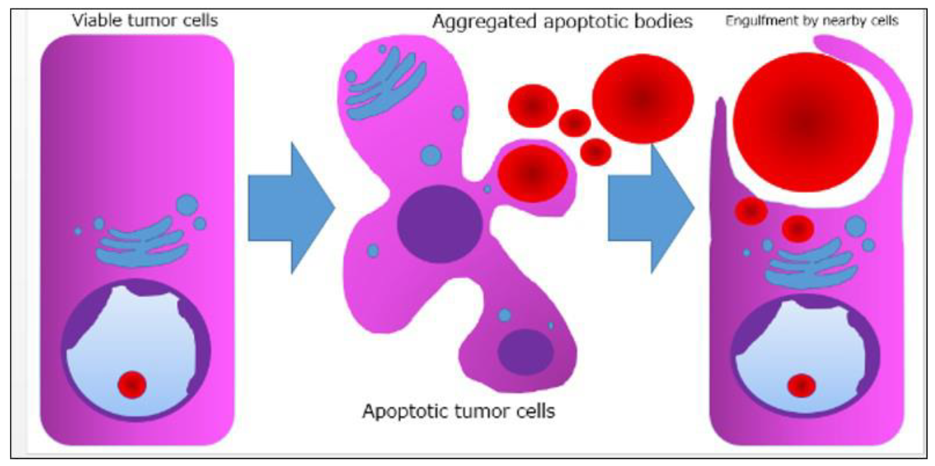
We present a rare case of apocrine breast cancer with thanatosomes (HGs). These globules are associated with apoptotic cell death, as indicated by their immunoreactivity to cleaved caspase-3 (Figure 5e and Figure 6) [8]. These nonspecific microscopic findings serve as histological markers of high cell turnover and apoptotic cell death; thus, in neoplastic conditions, they are typically linked to high-grade tumors [3]. This report is the first, to our knowledge, to show thanatosomes in apocrine breast carcinoma [3,4,5,6,7] and that these thanatosomes in breast cancer are immunoreactive for cleaved caspase-3. In this patient, the thanatosomes also exhibit immunoreactivity for ubiquitin (Figure 5f). Dikov et al. proposed that some HGs might arise from inhibition of the cytoplasmic alternative ubiquitin-proteasome system [2].
This report has one major limitation in that it is a single case report from one institution in Japan. Further clinicopathological analyses, including multicenter studies, will be necessary to definitively determine the cause and pathophysiology of this condition. Therefore, reports from other countries, cultures, and hospitals are eagerly anticipated.
In conclusion, thanatosomes are immunoreactive to cleaved caspase-3 and ubiquitin, indicating their connection to apoptosis. The study by D’Alfonso et al. demonstrated that thanatosomes were present in 16.3% of high-grade malignant breast tumors [3]. The present report is the first to show thanatosomes as a significant morphological feature in breast apocrine carcinoma.
Author Contributions
Conceptualization, M.T.; Validation, M.T.; Investigation, M.T.; Resources, M.N., T.I., and K.T.; Data Curation, M.T. and M.N.; Writing- Original Draft Preparation, M.T.; Writing- Review and Editing, M.N., K.T., T.I., and K.K.; Supervision, K.K.; Project Administration, M.T., K.T., T.I., and K.K. All authors have read and agreed to the published version of the manuscript.
Funding
This research received no external funding.
Institutional Review Board Statement
This study was conducted in accordance with the guidelines of the Declaration of Helsinki (1975) and was approved by the Ethics Committee of Shimada General Medical Center (protocol code: R7-1; date of approval: April 17, 2025).
Informed Consent Statement
Informed consent was obtained from the subject included in this study.
Data Availability Statement
Not applicable.
Acknowledgments
The authors thank Naoki Ooishi, Takayoshi Hirota, and Mizuki Naruse (Division of Pathology and Oral Pathology, Shimada General Medical Center, Shimada, Shizuoka, Japan) for helpful technical support with the special and immunohistochemical staining; and Hiroko Tina Tajima (St. Marianna University School of Medicine, Kawasaki, Kanagawa, Japan) for kindly reviewing and editing the English manuscript.
Abbreviations
The following abbreviations are used in this manuscript:
| HG | Hyaline globule |
| HER2 | Human epidermal growth factor receptor type 2 |
References
- Papadimitriou, J.C.; Drachenberg, C.B.; Brenner, D.S.; Newkirk, C.; Trump, B.F.; Silverberg, S.G. “Thanatosomes”: a unifying morphogenetic concept for tumor hyaline globules related to apoptosis. Hum. Pathol. 2000, 31, 1455–1465. [Google Scholar] [CrossRef] [PubMed]
- Dikov, D.I.; Auriault, M.L.; Boivin, J.F.; Sarafian, V.S.; Papadimitriou, J.C. Hyaline globules (thanatosomes) in gastrointestinal epithelium. Pathophysiologic correlations. Am. J. Clin. Pathol. 2007, 127, 792–799. [Google Scholar] [CrossRef] [PubMed]
- D’Alfonso, T.M.; Ginter, P.S.; Salvatore, S.P.; Antonio, L.B.; Hoda, S.A. Phylloides tumor with numerous thanatosomes (“death bodies”): a report of two cases and a study of thanatosomes in breast tumors. Int. J. Surg. Pathol. 2014, 22, 337–342. [Google Scholar] [CrossRef] [PubMed]
- Ozerdem, U.; Wells, J.; Hoda, S.A. Hyaline globules in mammary myofibroblastoma: a case report. Int. J. Surg. Pathol. 2015, 23, 89–91. [Google Scholar] [CrossRef] [PubMed]
- Panicker, N.K.; Buch, A.C.; Patel, A.R. Breast carcinoma with numerous large “thanatosomes. ” J. Cancer Res. Ther. 2015, 11, 980–982. [Google Scholar] [PubMed]
- Bezić, J. Invasive breast carcinoma with hyaline globules (“thanatosomes”). Breast Dis. 2020, 39, 43–45. [Google Scholar] [CrossRef] [PubMed]
- Datta, R.C.; Sandhya, B.N.; Swami, S.Y. Invasive lobular carcinoma of breast with hyaline globules (thanatosomes). Int. J. Clin. Diagn. Pathol. 2021, 4, 171–172. [Google Scholar]
- Tachibana, M.; Koreyasu, R.; Kamimura, K.; Tsutsumi, Y. Pancreatic intraductal papillary mucinous neoplasm with hyaline globules (Thanatosomes): report of two cases. Int. Med. Case Reports J. 2021, 14, 393–399. [Google Scholar] [CrossRef] [PubMed]
Figure 1.
Clinical imaging findings. (a) Contrast-enhanced computed tomography image shows an irregular mass in the right breast (arrow). (b) Breast ultrasound depicts an irregular, indistinct mass in the lateral region of the right breast. The internal echo is heterogeneous and displays numerous microcalcifications.
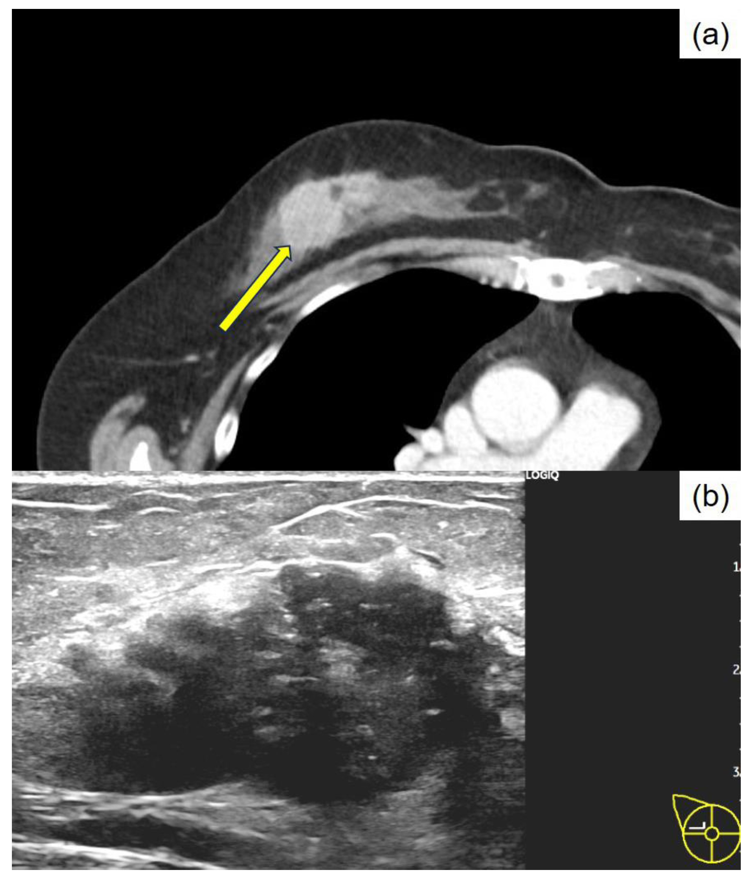
Figure 2.
Macroscopic findings of the resected specimen from the right total mastectomy. The tumor measures 39 × 25 × 24 mm in diameter and resembles a lobulated, solid mass with a milky white and focal yellowish appearance, accompanied by fat invasion.
Figure 2.
Macroscopic findings of the resected specimen from the right total mastectomy. The tumor measures 39 × 25 × 24 mm in diameter and resembles a lobulated, solid mass with a milky white and focal yellowish appearance, accompanied by fat invasion.
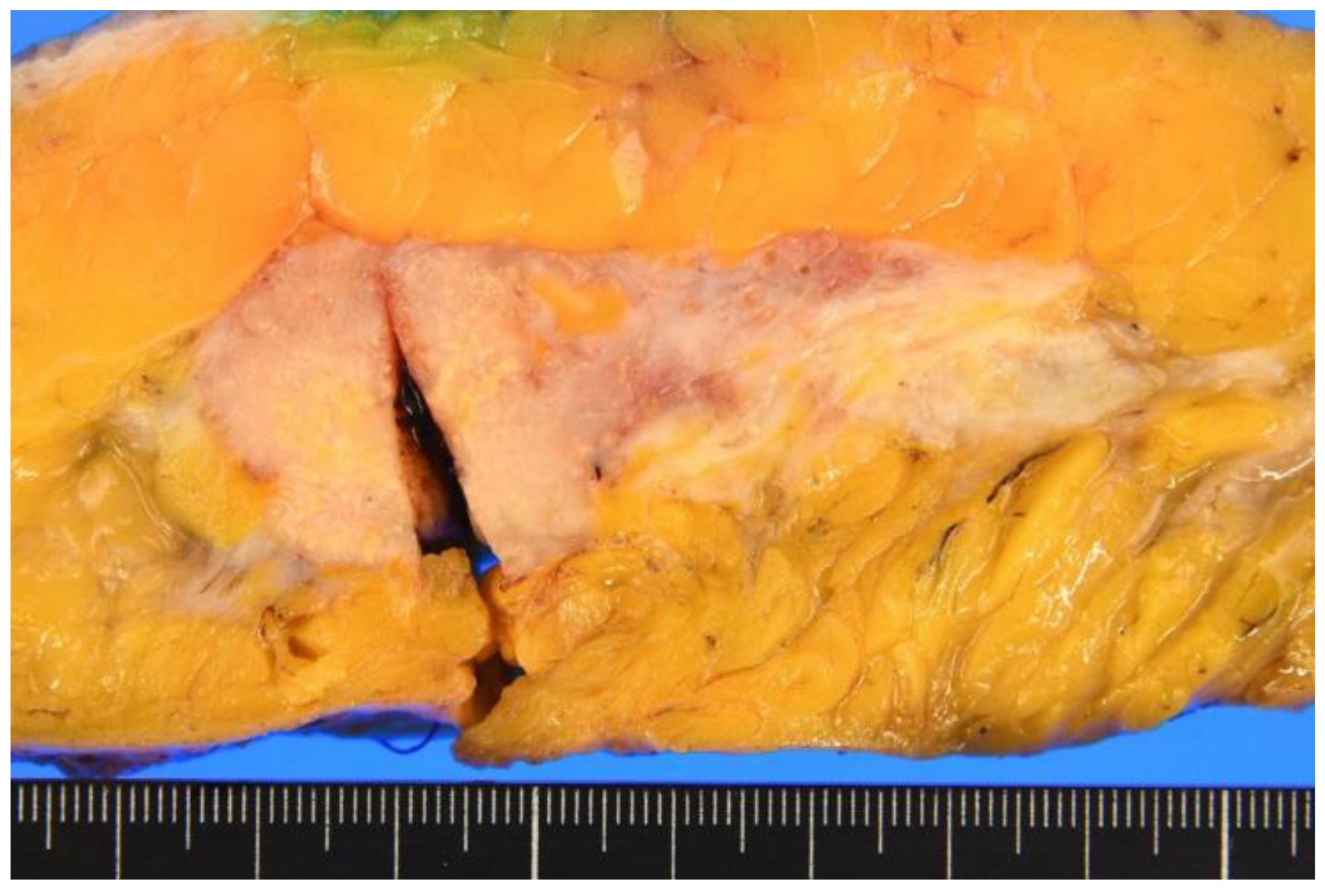
Figure 3.
Histopathology of the resected breast specimen stained with hematoxylin and eosin. (a) The tumor forms sheet-like solid nests, shows proliferation, and is accompanied by abundant capillaries. The cytoplasm appears eosinophilic and granular; the nuclei are round to oval and demonstrate notable nuclear pleomorphism. Occasional mitotic figures are observed (×200). (b) Various sizes of eosinophilic and uniform thanatosomes are intermittently noted within the breast cancer tissue (×1000).
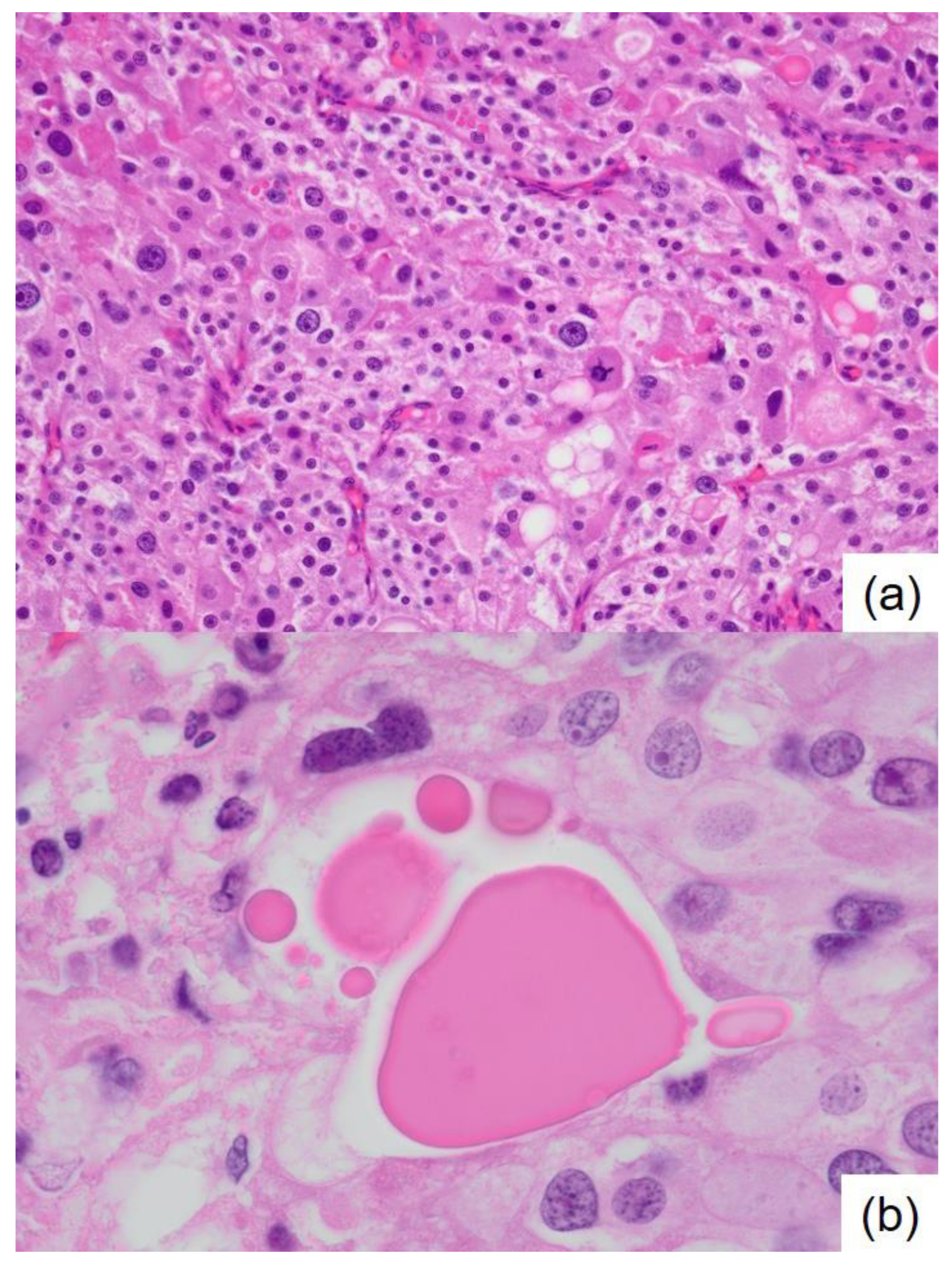
Figure 4.
Immunohistochemical staining of the breast cancer. (a) Androgen receptor, ×200; (b) FOXA1, ×200; (c) estrogen receptor, ×200; (d) progesterone receptor, ×200; (e) HER2/neu, ×200 (inset: enlarged image, ×400); and (f) GCDFP15, ×400 (inset: Ki-67, ×400).
Figure 4.
Immunohistochemical staining of the breast cancer. (a) Androgen receptor, ×200; (b) FOXA1, ×200; (c) estrogen receptor, ×200; (d) progesterone receptor, ×200; (e) HER2/neu, ×200 (inset: enlarged image, ×400); and (f) GCDFP15, ×400 (inset: Ki-67, ×400).
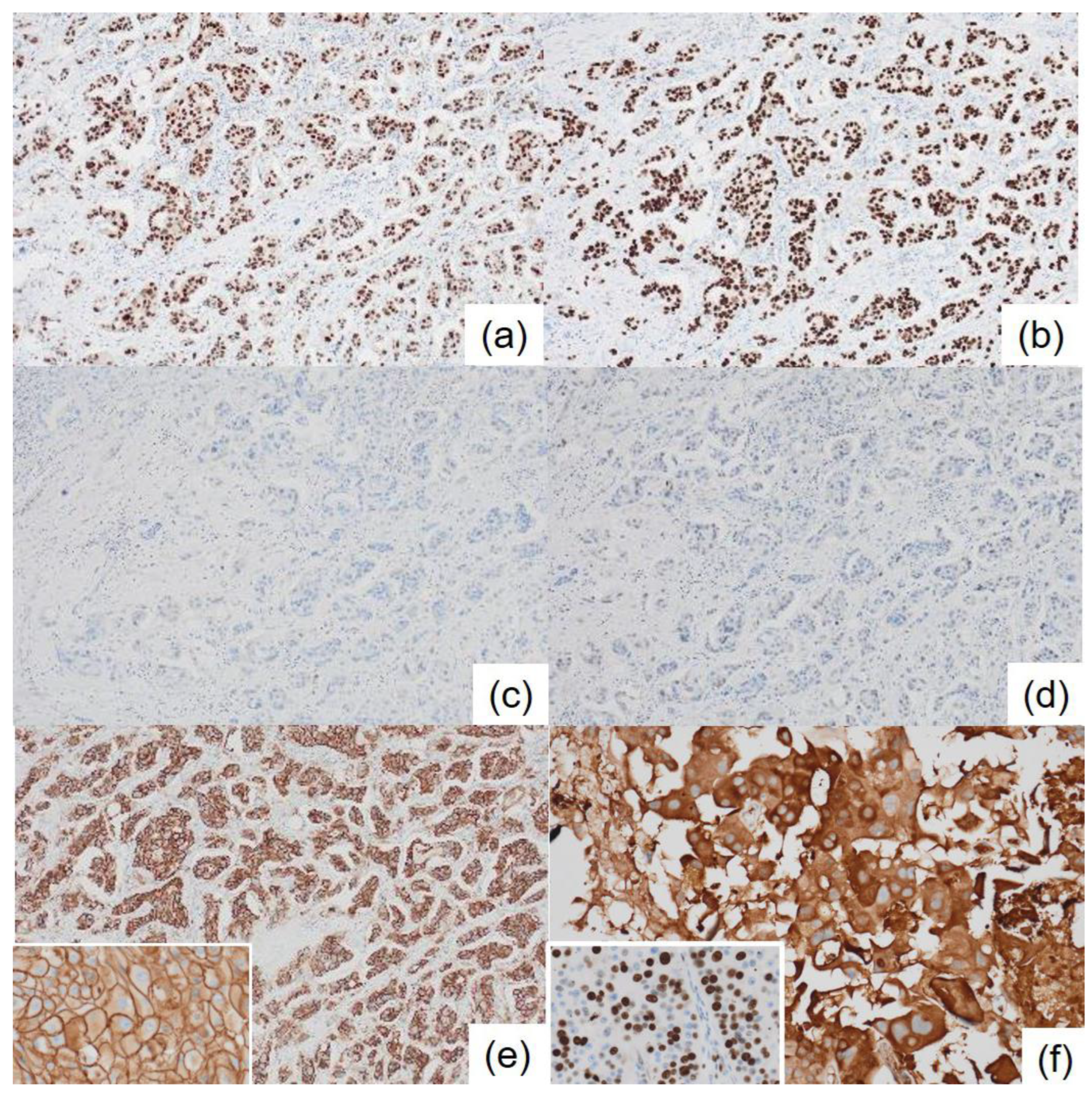
Figure 5.
Special staining and immunohistochemical staining of the thanatosomes. (a) Periodic acid-Schiff reaction, (b) Masson’s trichrome stain, (c) Mallory’s PTAH stain, and (d) Alcian blue. The thanatosomes are positive for cleaved caspase-3 (e) and ubiquitin (f) (a-f; ×1000).
Figure 5.
Special staining and immunohistochemical staining of the thanatosomes. (a) Periodic acid-Schiff reaction, (b) Masson’s trichrome stain, (c) Mallory’s PTAH stain, and (d) Alcian blue. The thanatosomes are positive for cleaved caspase-3 (e) and ubiquitin (f) (a-f; ×1000).
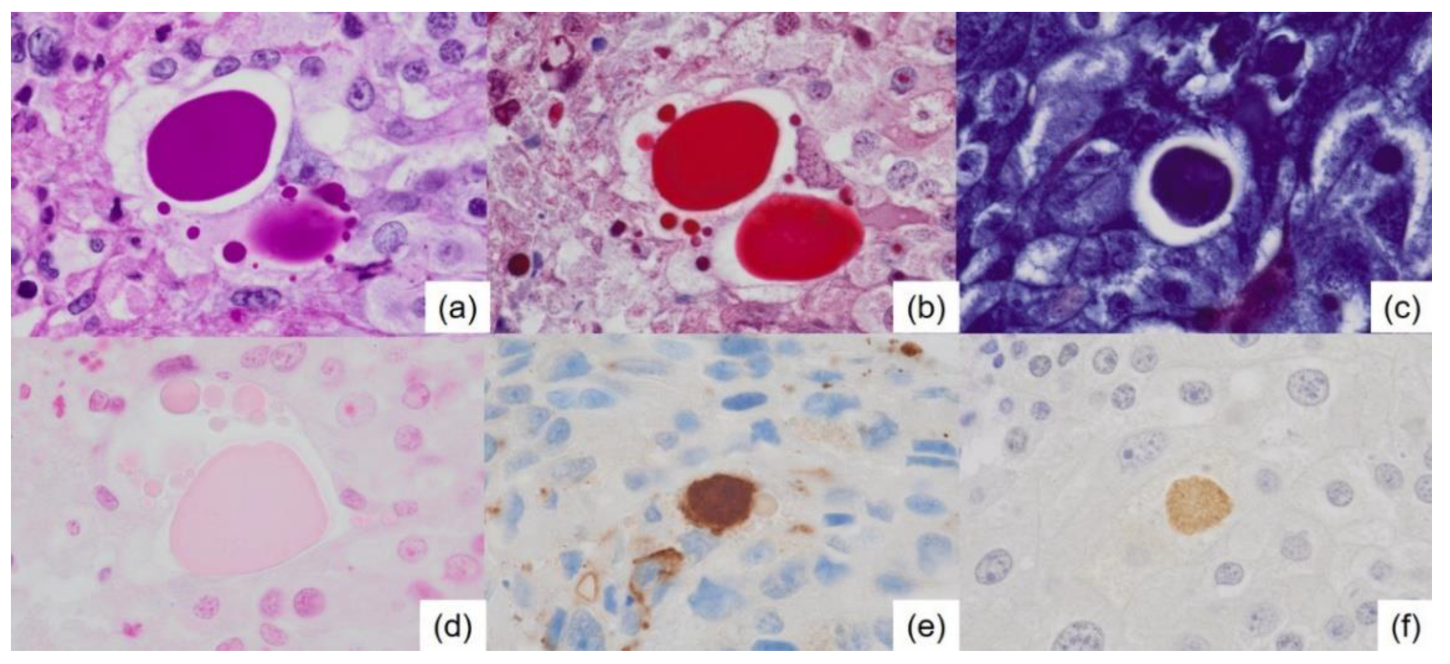
Figure 6.
A model for forming hyaline globules in the current neoplasm. An intact tumor cell responds to various injurious stimuli, ultimately leading to its demise through a process known as programmed cell death, also called apoptosis. The apoptotic bodies are engulfed by neighboring healthy cells.
Disclaimer/Publisher’s Note: The statements, opinions and data contained in all publications are solely those of the individual author(s) and contributor(s) and not of MDPI and/or the editor(s). MDPI and/or the editor(s) disclaim responsibility for any injury to people or property resulting from any ideas, methods, instructions or products referred to in the content. |
© 2025 by the authors. Licensee MDPI, Basel, Switzerland. This article is an open access article distributed under the terms and conditions of the Creative Commons Attribution (CC BY) license (http://creativecommons.org/licenses/by/4.0/).
Copyright: This open access article is published under a Creative Commons CC BY 4.0 license, which permit the free download, distribution, and reuse, provided that the author and preprint are cited in any reuse.


