
Submitted:
16 May 2025
Posted:
19 May 2025
You are already at the latest version
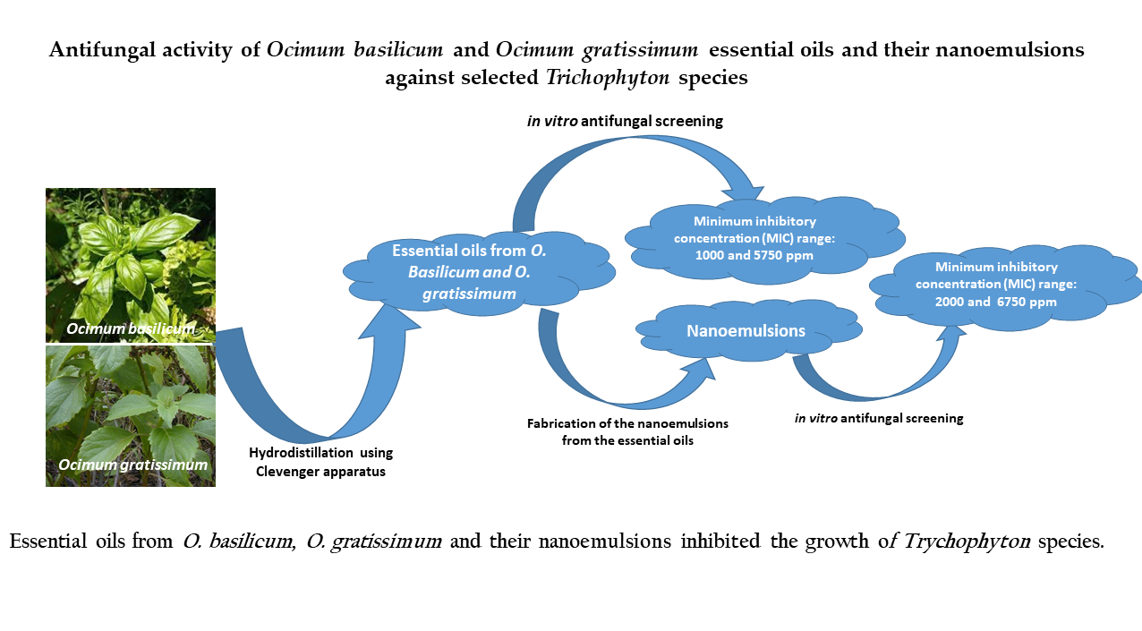
Abstract
The outcome of antifungal treatments is hindered by various conditions, such as resistance and tolerance of certain fungal pathogens. Mycoses of nails and skin, which are primarily caused by dermatophytes, are the most common fungal infections, with Trichophyton rubrum being the most common dermatophytic pathogen, followed by Trichophyton interdigitale. Thus, the search for effective treatments against dermatophyte infections is valuable. This study is sought to investigate the antidermatophytic and antioxidant activities of essential oils and nanoemulsions of the leaves and stems from Ocimum gratissimum and Ocimum basilicum. The plants’ essential oils, which were obtained by hydrodistillation using a Clevenger-type apparatus, were further analyzed by gas chromatography and gas chromatography coupled with spectrometry mass (GC/MS). The nanoemulsions were obtained by spontaneous emulsification using Tween 80 and their stabilities were evaluated using distilled water and methylene blue dye. The antidermatophytic effects of the essential oils were evaluated using an agar diffusion method and by determination of minimum inhibitory concentrations (MICs). The antioxidant activity was carried out by the 2,2-diphenyl-1-picrylhydrazyl (DPPH) colorimetric method. As a result, the GC/MS analysis of essential oil from O. gratissimum revealed the presence of γ-terpinene (33.73%), thymol (26.44%) and 1.8 cineol (16.65%), whereas the O. basilicum’s essential oil was dominated by linalool (55.32%), eucalyptol (16.78%) and eugenol (7.45%). Essential oils and nanoemulsions from O. gratissimum (MICs : 1000 and 2000 ppm, respectively) and O. basilicum (MICs : 5750 and 6750 ppm, respectively) revealed fungicidal activity against Trichophyton rubrum, whereas only O. gratissimum showed moderate activity against T. interdigitale. Moreover, essential oils and nanoemulsions from O. gratissimum and O. basilicum scavenged the free radicals of DPPH, thus revealing antioxidant activity. This novel contribution demonstrates the antidermatophytic activity of essential oils and nanoemulsions from O. gratissimum and O. basilicum, thus supporting the traditional use of these plants in ethnomedicine.
Keywords:
dermatophyte infections
; Trichophyton species
; Ocimum gratissimum
; Ocimum basilicum
; essential oils
; nanoemulsions
1. Introduction
Dermatophytoses are mycotic infections caused by a group of fungi that usually remain localized to the superficial layers of the skin, hair or nails [1,2]. Despite their propensity to infect the exterior aspects of the host, dermatophytic fungi prefer a warm, moist environment for growth, and as a consequence, infections are common in tropical regions [1]. These fungi are classified in the anamorphic genera Epidermophyton, Microsporum and Trichophyton [3]. Recent estimates proposed an annual incidence of 6·5 million invasive fungal infections and 3·8 million deaths, of which about 68% (2·5 million) were directly attributable [4]. Superficial mycoses are the most prevalent forms in humans, thus affecting about 20-25% of the world’s population [4]. In Africa, dermatophyte infection is widespread depending on geographic location, with a prevalence of 14-86% in children [1]. Mycoses of nails and skin, which are primarily caused by dermatophytes, are the most common fungal infections, with Trichophyton rubrum, being the most common dermatophytic pathogen, followed by Trichophyton interdigitale [5]. The reduced size of their genome with the expansion of particular gene families indicate that these fungi are highly specialized and have the ability to degrade hard keratin and escape the immune system during infection [6]. Dermatophytes establish infection following successful adherence of arthroconidia to the surface of keratinized tissues. The proteolytic enzymes released during adherence and invasion not only ascertain their survival but also allow the persistence of infection in the host [7]. The treatment of dermatophytosis involves the use of a number of drugs, such as griseofulvin, terbinafine, ketoconazole and itraconazole, among others. Although these treatments are effective against dermatophytes, their use is often limited due to the number of adverse effects they cause, their high cost, the length of treatment, and the development of resistance [8].
To address their healthcare needs, many people have turned to traditional healers, herbal medicines, ancient medicinal knowledge and home remedies [9]. In addition, there is a growing interest of researchers around the world in medicinal plants and their products, as a result of antifungal drug resistance [10,11]. This is due to their medicinal properties against infectious diseases. Interestingly, medicinal plant extracts and essential oils are factories of valuable natural bioactive compounds, which exhibit antibacterial and antifungal properties, thereby combating microbial resistance [12]. Examples of such plants include Ocimum basilicum and Ocimum gratissimum, which are widely used by local populations in Africa to cure various diseases, including fungal infections. For example, the infusion of Ocimum gratissimum is used to overcome headache, giddiness, cold and cough, whereas O. gratissimum-based formulations are recommended for the treatment of ear infections and dermatoses in Côte d'Ivoire [13]. In Nigeria and Togo, O. gratissimum is used to relieve fever, diarrhoea and dysentery [14]. The decoction of O. gratissimum stems is used to cure hepatitis, candidoses and wound infections [14].
On the other hand, O. basilicum or Sweet basil is an important essential oil crop, medicinal plant, and culinary herb that belongs to the Lamiaceae family [15,16]. In Africa, people have been using O. basilicum to treat malaria, typhoid, coughs and colds, yellow fever, candidiasis, influenza, genito-urinary infections, sore eyes and ear infections, among others [17]. O. basilicum and O. gratissimum are used in different places of Cameroon as spices and for the traditional treatment of of headaches, respiratory problems, influenza, stomach pain, inflammations of the throat, diarrhea, worms, and kidney malfunction [18].
Modern research has unveiled the antibacterial activity of O. basilicum [11,19,20,21] and O. gratissimum [22,23,24,25,26,27,28,29] against human and plant pathogenic bacteria. Antifungal activity of O. basilicum and O. gratissimum extracts was also revealed against fungal pathogens in plants (Botrytis cinerea, Colletotrichum gloeosporioides and Fusarium oxysporum ; [30,31]) ; and humans (Scopulariopsis breicaulis and Cryptococcus neoformans [32] ; Candida species [33,34,35]).
However, very little is known about the antifungal activity of botanicals from O. basilicum and O. gratissimum against the Trichophyton species, which are responsible for fungal infections of the scalp, groin (jock itch), body, feet (athlete's foot), fingernails, and toenails.
Thus, the present study aims to investigate the antifungal activity of essential oils and nanoemulsions from O. basilicum and O. gratissimum against Trichophyton rubrum and Trichophyton interdigitale. Moreover, the antioxidant activity of essential oils and their nanoemulsions is also evaluated.
2. Results and Discussion
2.1. Results
2.1.1. Yields of Extraction
The essential oils were obtained as bright and dark yellow substances for Ocimum gratissimum and Ocimum basilicum, respectively. The yields of extraction were found to be 1.12% and 0.62% for O. gratissimum and O. basilicum, respectively (Table 1).
To determine the phytochemical composition of the as-prepared essential oils from Ocimum gratissimum and Ocimum basilicum, the oil samples were subjected to gas chromatography and gas chromatography tandem mass spectrometry.
2.1.2. Chemical Composition of the Essential Oils
The chromatograms of the essential oils from Ocimum gratissimum and Ocimum basilicum are illustrated in Figure 1 and Figure 2. The Kovats indices were calculated following the analysis of the chromatograms to afford a tentative phytochemical analysis of the oils.
The GC/MS analysis aided in the complete characterization of the essential oils as shown in table 2. According to table 2, the essential oil from Ocimum gratissimum was dominated by hydrocarbonated monoterpenes, such as γ-terpinène (33.73%), α-thujène (6.05%), oxygenated monoterpenes (thymol, 26.44% ; and 1,8-cinéole, 16.65%), hydrocarbonated (β-caryophyllene, 2.64%), and oxygenated (β-6 elemene, 0.25%), sesquiterpenes. Moreover, the essential oil from Ocimum basilicum was found to be rich in oxygenated monoterpenes, such as eucalyptol (16.78%), linalol (55.32%), eugenol (7.45%), hydrocarbonated ((-)-endo-α-bergamotène, 2.75%), and oxygenated (tau-cadinol, 2.20%) (Table 2).
Overall, the essential oil from O. gratissimum was dominated by γ-terpinene (33.73%), thymol (26.44%), 1,8-cineole (16.65%) and thujene (6.05%), whereas O. basilicum essential oil comprised mostly eugenol (16.78%) and linalool (55.32%).
2.1.3. Characterization of the Nanoemulsions
a. Physical characteristics
The nanoemulsions were obtained by mixing separately the essential oils from O. basilicum and O. gratissimum with Tween 80 and distilled water in the respective proportions of 5/5/90 (w/w). Nanoemulsions from essential oils of O. basilicum and O. gratissimum were obtained as white, homogenous and milky substances.
b. Stability of emulsion
To evaluate the stability of the as-prepared nanoemulsions, a water soluble dye (methylene blue) was used for the centrifugation. After adding a few drops of the methylene blue solution in each nanoemulsion, the mixtures were centrifuged for 30 minutes at 1000, 2000 and 3000 rpm. Since the preparation was homogenous at different speeds of centrifugation, the nanoemulsions were considered to be stable.
c. Dilution test
This experiment was used to assess the type of emulsion, whether it was water/oil or oil/water emulsion for each nanoemulsion. The addition of water to each nanoemulsion did not modify the appearance of the emulsion, to conclude that the as-prepared nanoemulsions were water/oil emulsions.
d. Measure of pH
It is essential to measure the pH of a nanoformulated drug, which is intended to be used for potential application on the skin. In this study, the pH of the nanoemulsions from the essential oils of Ocimum basilicum and Ocimum gratissimum were obtained as 6.0 and 6.2, respectively. Accumulated evidence has shown that skin with pH values below 5.0 is in a better condition than skin with pH values above 5.0, as shown by measuring the biophysical parameters of barrier function, moisturization and scaling [36,37]. Other reports demonstrated that the mean pH value of the skin ranges between 5.4 and 7.0 [38,39].
2.1.4. In Vitro Antifungal Activity
The antifungal activity of essential oils and nanoformulations from O. basilicum and O. gratissimum were assayed against Trichophyton rubrum and Trichophyton interdigitale. Figure 3 and 4 illustrates, respectively, the inhibition zones of Trichophyton rubrum and Trichophyton interdigitale by O. gratissimum’s essential oil at various concentrations viz. 250, 350, 500, 750 and 1000 ppm. As the concentrations of the essential oils increase, there is a disappearance of the fungal growth. The highest antifungal activity was observed at 1000 ppm.
Figure 3.
: Inhibition of Trichophyton rubrum upon treatment with various concentrations of Ocimum gratissimum
Figure 3.
: Inhibition of Trichophyton rubrum upon treatment with various concentrations of Ocimum gratissimum
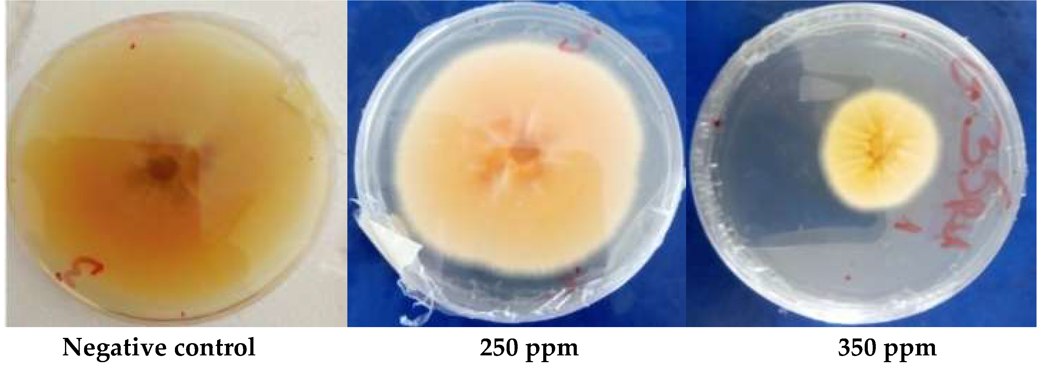
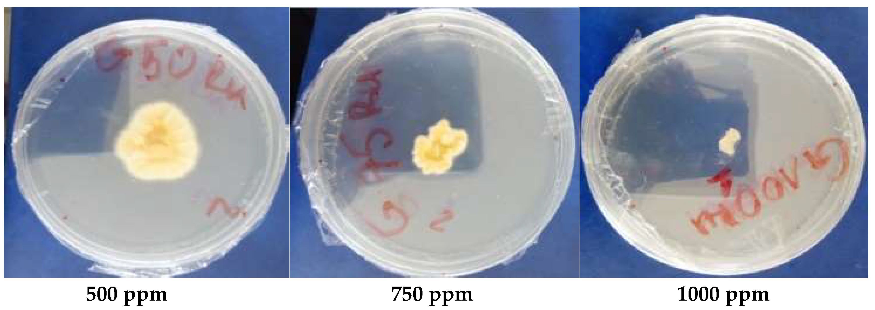
Figure 4.
Inhibition of Trichophyton interdigitale upon treatment with different concentrations of Ocimum gratissimum.
Figure 4.
Inhibition of Trichophyton interdigitale upon treatment with different concentrations of Ocimum gratissimum.
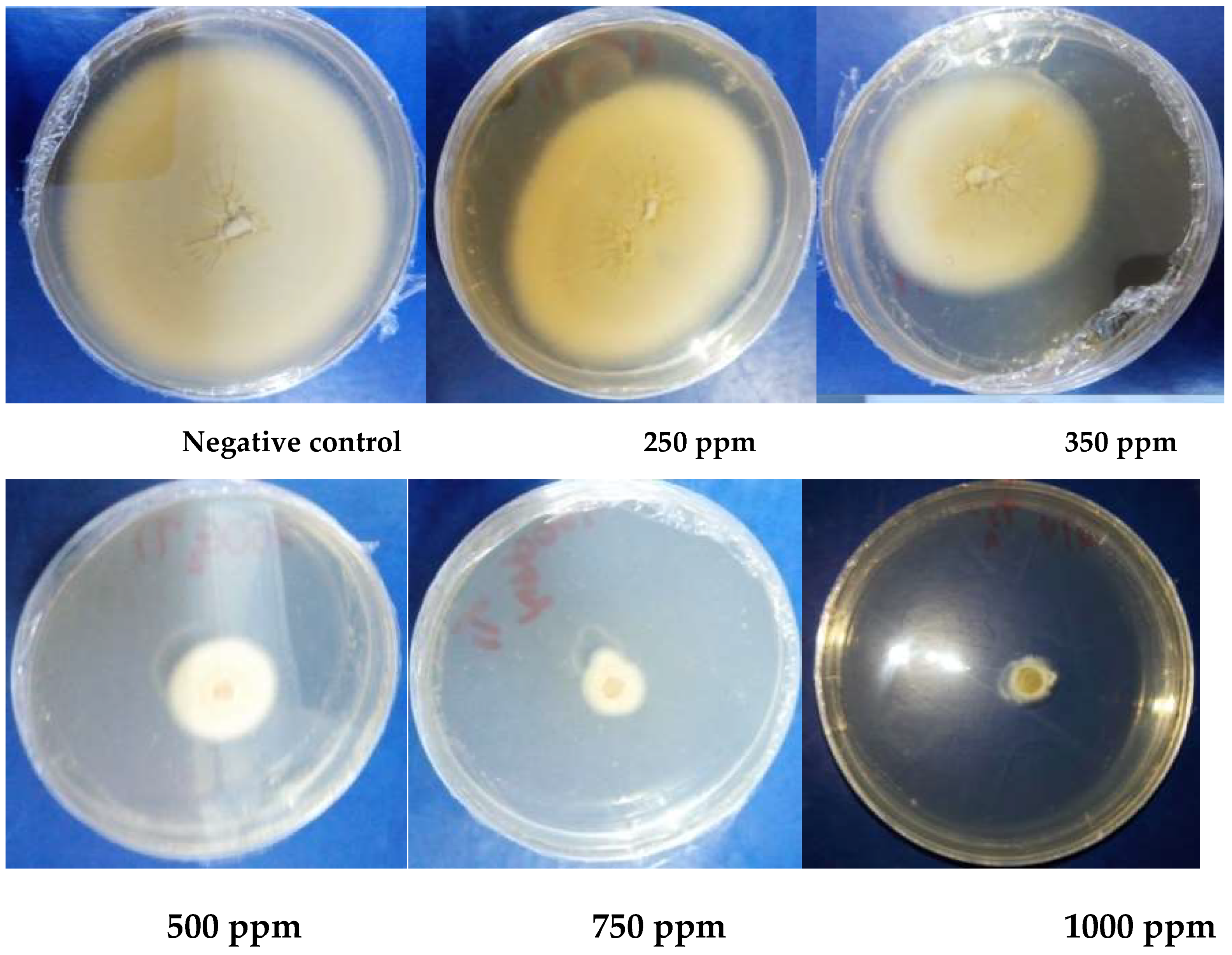
Figure 5 and 6 illustrates, respectively, the inhibition zones of Trichophyton rubrum and Trichophyton interdigitale by O. basilicum’s essential oil at various concentrations viz. 250, 350, 500, 750 and 1000 ppm. As the concentrations of the essential oils increase, there is a disappearance of the fungal growth. The highest antifungal activity was observed at 1000 ppm.
Figure 5.
Inhibition of Trichophyton rubrum by treatment with various concentrations of Ocimum basilicum.
Figure 5.
Inhibition of Trichophyton rubrum by treatment with various concentrations of Ocimum basilicum.
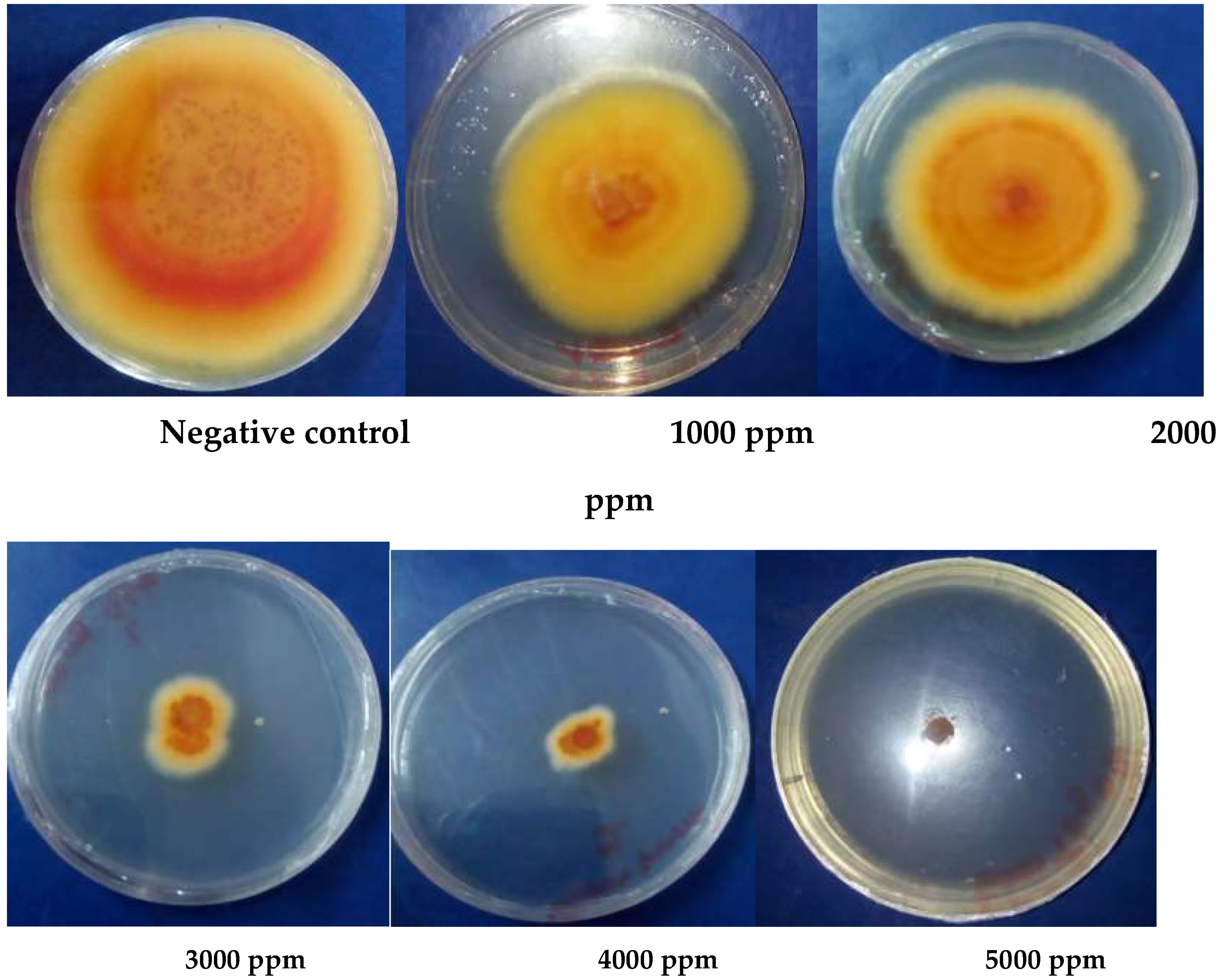
Figure 6.
Inhibitory effects of various concentrations of Ocimum basilicum on Trichophyton interdigitale.
Figure 6.
Inhibitory effects of various concentrations of Ocimum basilicum on Trichophyton interdigitale.
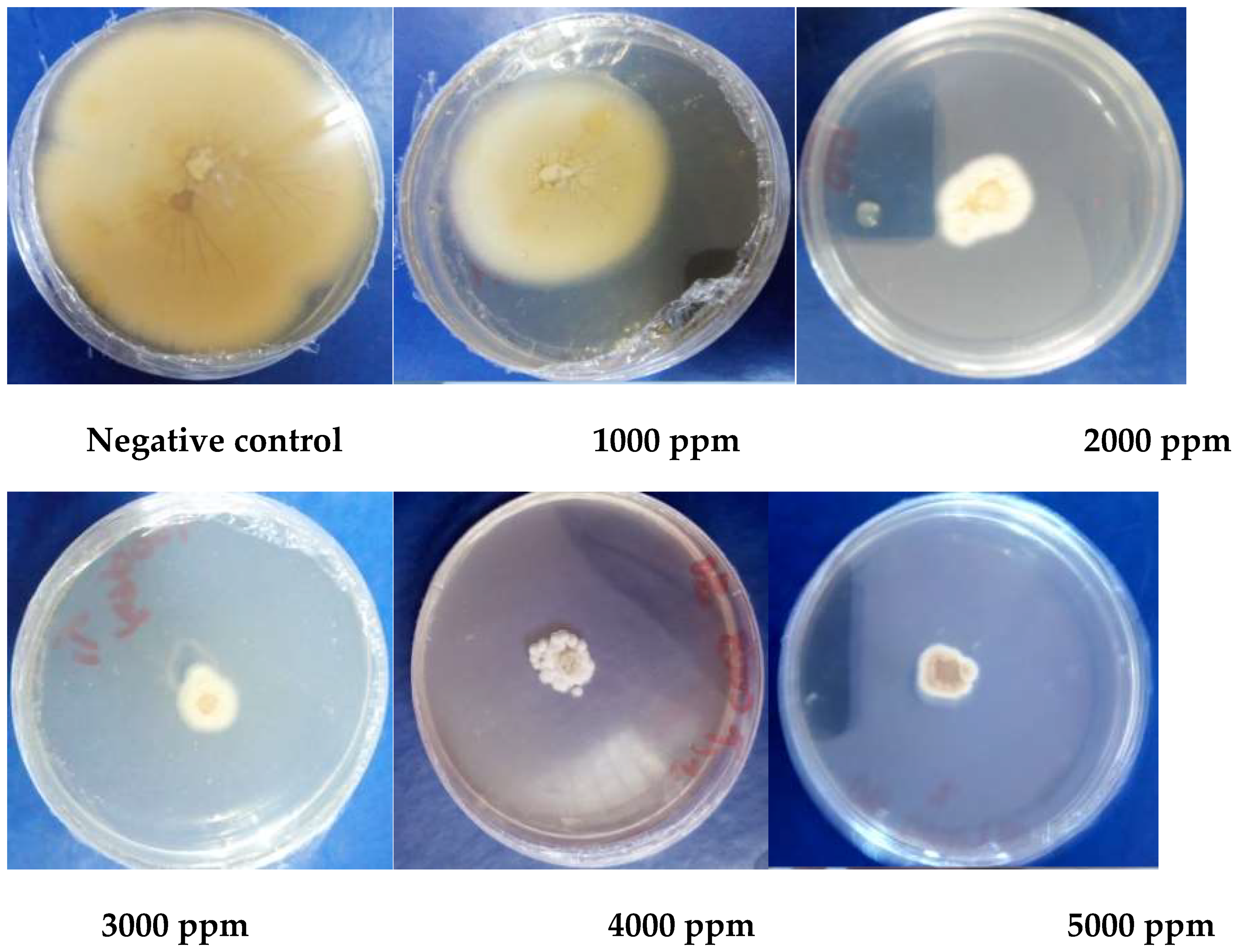
Figure 7 and 8 shows the inhibition of Trichophyton rubrum and Trichophyton interdigitale by the nanoemulsion prepared from O. gratissimum’s essential oil at various concentrations viz. 1000, 1250, 1500, 1750 and 2000 ppm. The reduction of the fungal growth is proportional to the increase in the concentration of the nanoemulsion. The highest antifungal activity was observed at 2000 ppm.
Figure 7.
Inhibitory effects of various concentrations of nanoemulsion from Ocimum gratissimum vis-à-vis Trichophyton rubrum.
Figure 7.
Inhibitory effects of various concentrations of nanoemulsion from Ocimum gratissimum vis-à-vis Trichophyton rubrum.
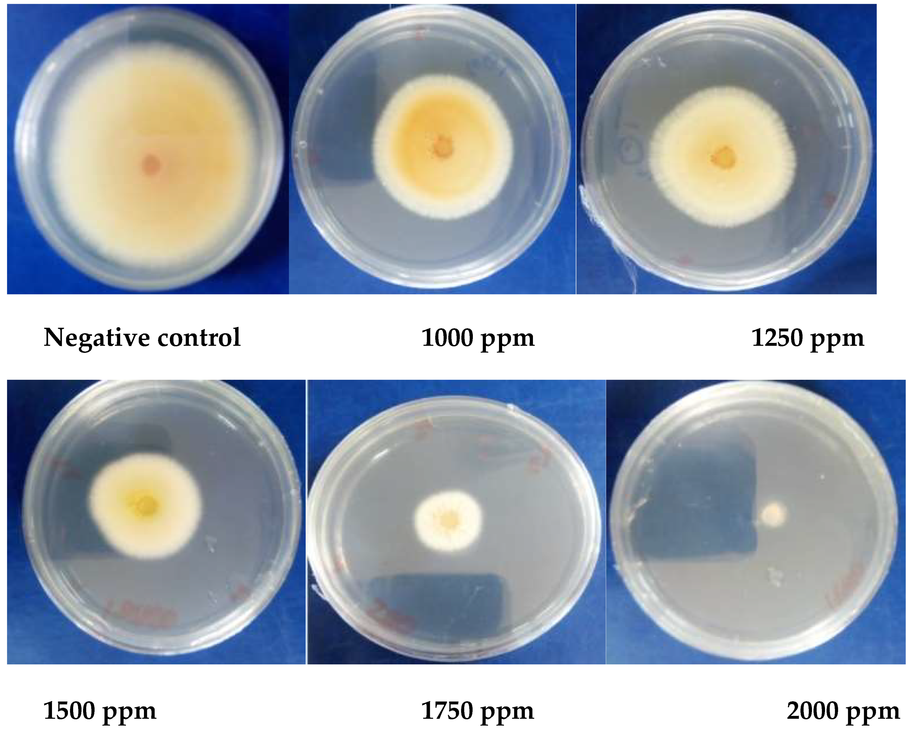
Figure 8.
Inhibitory activity of various concentrations of nanoemulsion from Ocimum gratissimum vis-à-vis Trichophyton interdigitale.
Figure 8.
Inhibitory activity of various concentrations of nanoemulsion from Ocimum gratissimum vis-à-vis Trichophyton interdigitale.
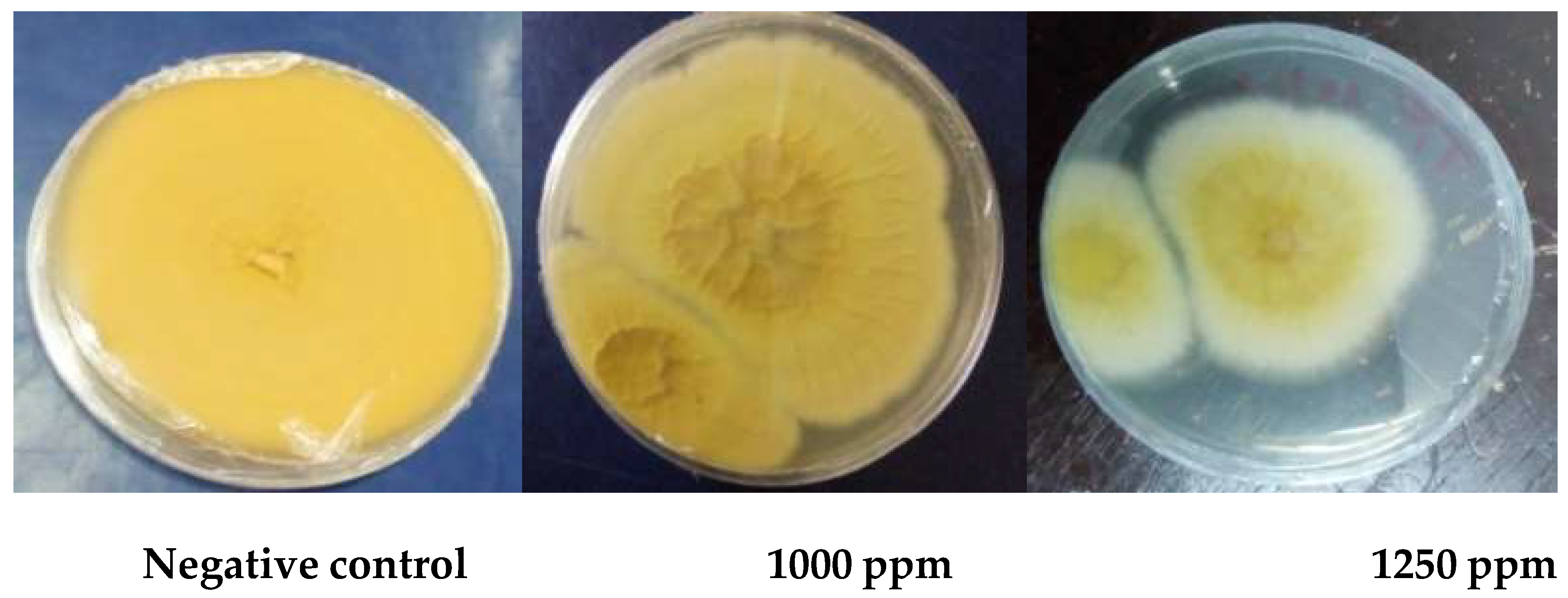
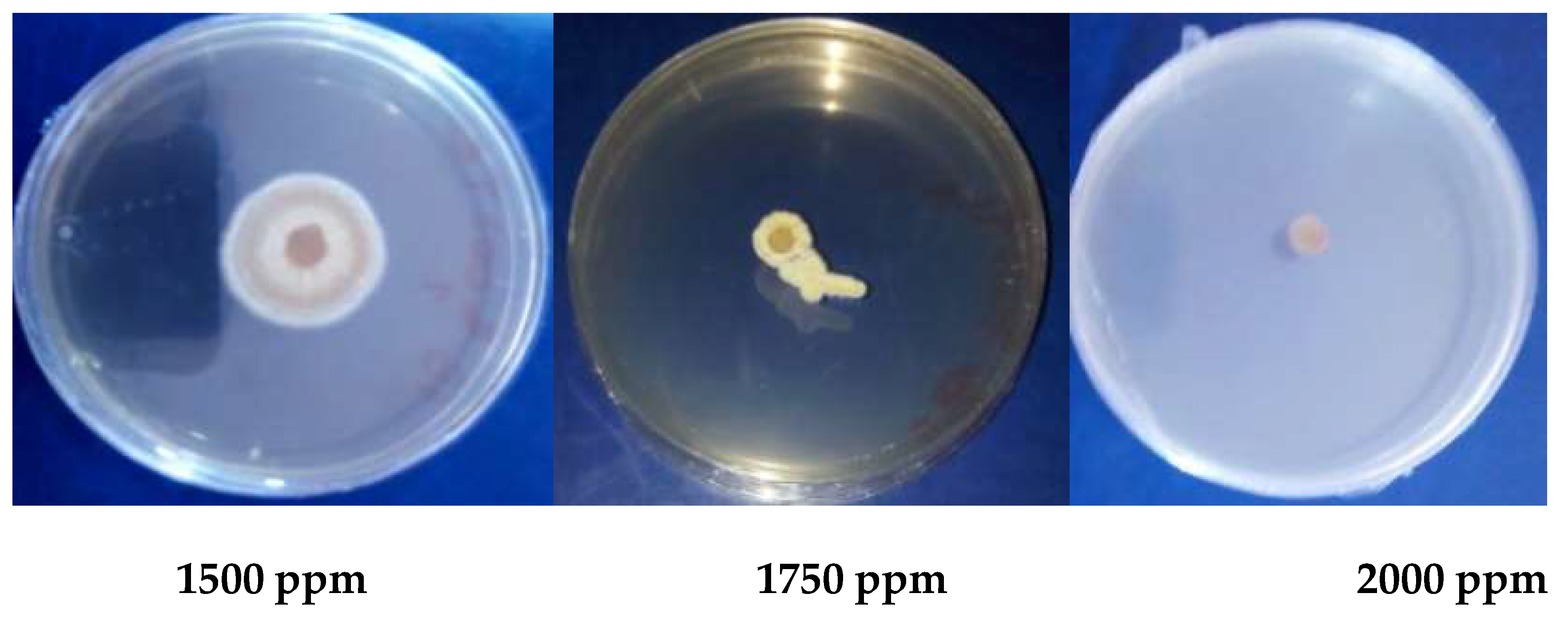
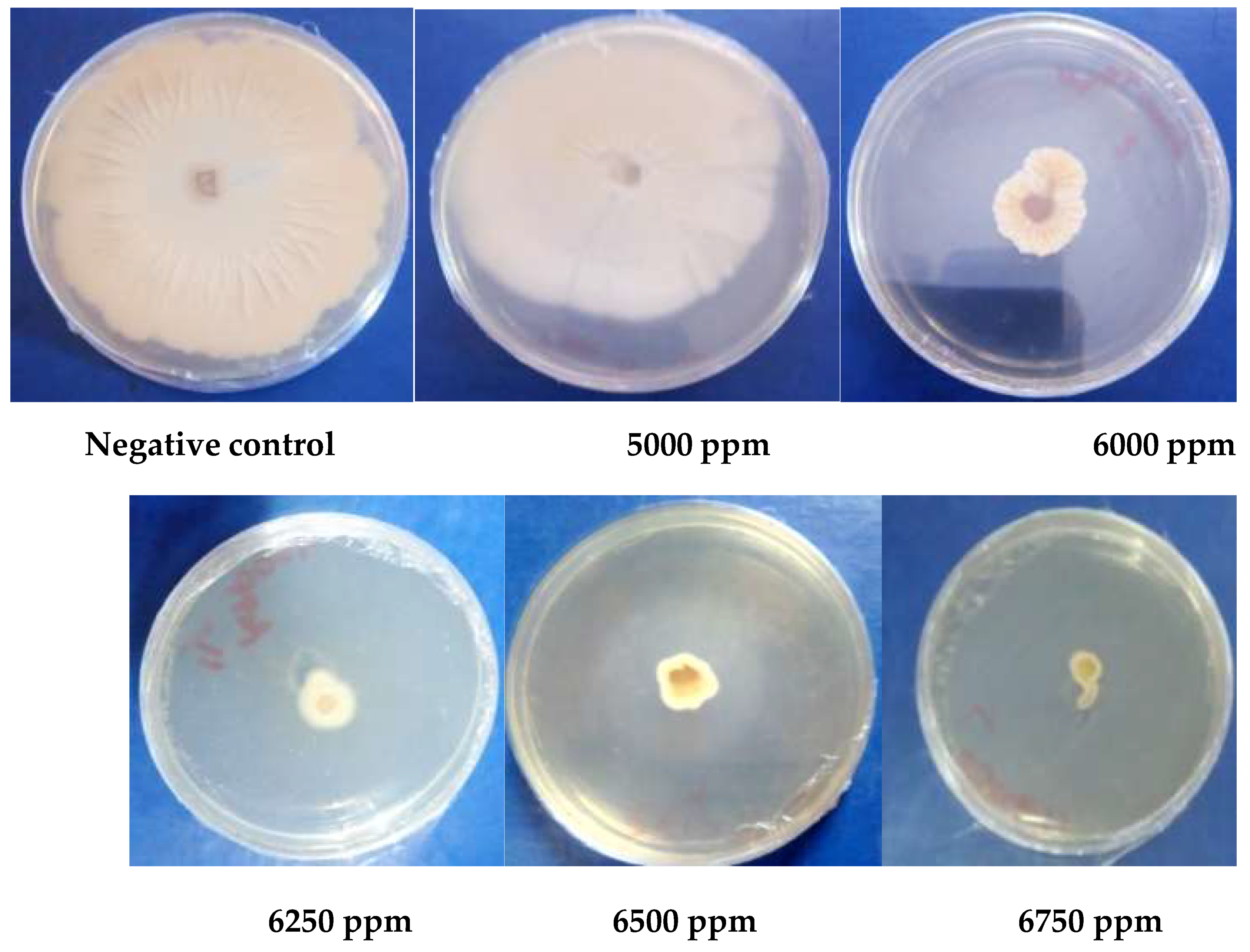
Figure 9 and 10 depicts the inhibitory effects of nanoemulsion from O. basilicum’s essential oil at various concentrations viz. 5000, 6000, 6250, 6500 and 6750 ppm against Trichophyton rubrum and Trichophyton interdigitale, respectively. The reduction of the dermatophyte’s growth is proportional to the increase in the concentration of the nanoemulsion. The highest antifungal activity was observed at 2000 ppm.
Figure 9.
Inhibitory effects of various concentrations of nanoemulsion from Ocimum basilicum vis-à-vis Trichophyton rubrum.
Figure 9.
Inhibitory effects of various concentrations of nanoemulsion from Ocimum basilicum vis-à-vis Trichophyton rubrum.
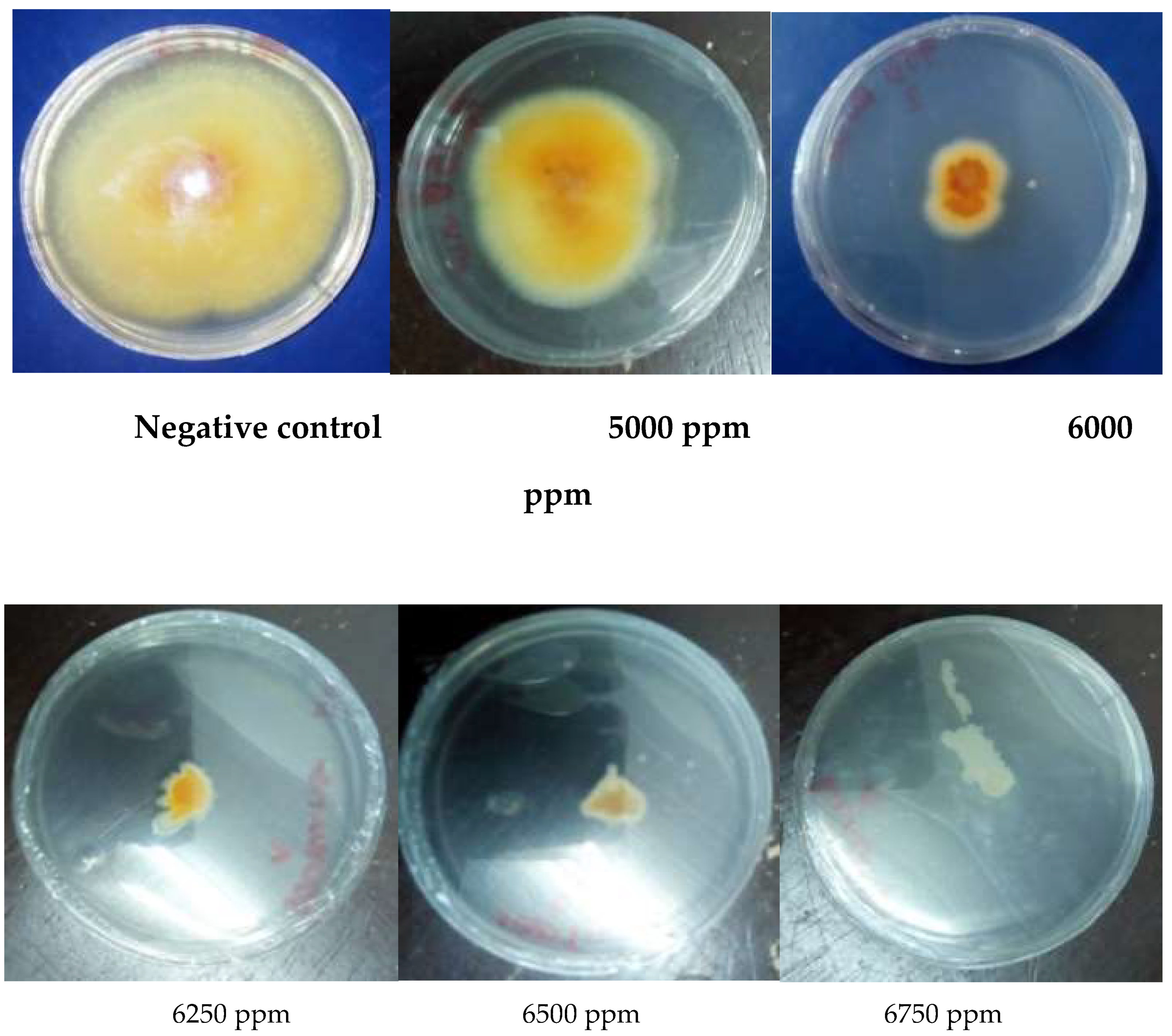
Figure 10.
Inhibitory activity of various concentrations of nanoemulsion from Ocimum basilicum vis-à-vis Trichophyton interdigitale.
Figure 10.
Inhibitory activity of various concentrations of nanoemulsion from Ocimum basilicum vis-à-vis Trichophyton interdigitale.
In antifungal assays, griseofulvin was used as a positive control. Figure 11 illustrates the inhibitory effects of griseofulvin on the growth of Trichophyton rubrum and Trichophyton interdigitale. As shown in Figure 11, as high as 5000 ppm of griseofulvin did not completely eradicate the growth of Trichophyton rubrum and Trichophyton interdigitale.
These results demonstrate that antifungal activity of the essential oils and nanoemulsion from Ocimum gratissimum is greater than that obtained with the reference antifungal compound griseofulvin. Indeed, 21 days’ incubation of essential oils or nanoemulsion from Ocimum gratissimum at lower concentrations (1000-5000 ppm) showed more pronounced inhibition of the mycelial growth compared with the activity of griseofulvin. However, the essential oils and nanoemulsion from Ocimum basilicum showed moderate antifungal activity at higher concentrations (5000-6750 ppm).
-Determination of the minimum inhibitory (MICs) and minimum fungicidal (MFCs) concentrations
The concentration where no radial growth of germs was visible after 21 days of incubation of fungi with essential oils and nanoformulations, corresponded to the minimum inhibitory concentrations (MICs) (Table 3). Against Trichophyton rubrum, the essential oil and nanoemulsion of Ocimum gratissimum showed minimum inhibitory (MICs) and minimum fungicidal (MFCs) concentrations of 1000 and 2000 ppm, respectively. Against Trichophyton interdigitale, Ocimum gratissimum afforded common minimum inhibitory and minimum fungicidal concentration (2000 ppm). The incubation of various concentrations of the essential oil and nanoemulsion of Ocimum basilicum with Trichophyton rubrum afforded a common MIC and MFC value of 5750 ppm. Against Trichophyton interdigitale, Ocimum basilicum’ essential oil and nanoemulsion yielded a common MIC and MFC value of 6750 ppm. Irrespective of the fungal strain tested, the standard antifungal agent griseofulvin yielded MIC value of more than 5000 ppm (Table 3).
3.1.5. Antioxidant Activity
In this study, the antioxidant activity of the essential oils and nanoemulsion from Ocimum gratissimum and Ocimum basilicum was evaluated using DPPH assay. As a result, the essential oils of Ocimum basilicum and Ocimum gratissimum scavenged the free radicals of DPPH with median scavenging concentrations (SC50S) of 100 and 500 ppm, respectively, vs butylated hydroxytoluene (SC50 : 200 ppm) (Figure 12).
The nanoemulsions, which were prepared from O. gratissimum and O. basilicum essential oils, scavenged the free radicals of DPPH with a common median radical scavenging concentration of 100 and 4800 ppm, respectively, vs butylated hydroxytoluene (SC50 : 200 ppm) (Figure 13).
The median efficient concentrations (CE50, g/mol) and the antiradical potential (AP, mol/g) of essential oils (5000 g/mol and 0.0002 mol/g) and nanoformulations (4800 g/mol and 0.00002 mol/g) were also calculated for O. gratissimum. Moreover, essential oils (1000 g/mol and 0.001 mol/g) and nanoformulations (1000 g/mol and 0.001 mol/g) obtained from O. basilicum showed more pronounced antioxidant activity, as evidenced by their median efficient concentrations and antiradical potential.
2.2. Discussion
Fungal pathogens have become a major threat to human health, infecting billions of people with approximately 1.5 million deaths annually [40,43].
There are several classes of azole antifungal drugs, including (i) imidazoles, such as clotrimazole, econazole, and miconazole, which are commonly used to treat skin and nail infections, (ii) triazoles, such as fluconazole, itraconazole, and voriconazole, which are used to treat systemic fungal infections and (iii) allylamines, such as terbinafine, which is used to treat dermatophyte infections [43,44]. However, most of these drugs have become resistant to the majority of fungal pathogens, thus reducing their effectiveness against re-emerging fungal strains. In addition to drug resistance, a number of undesirable side effects have been documented on these antifungals. Medicinal plants, which have been traditionally used for the treatment of superficial skin infections, can afford a number of botanicals with antifungal properties [45,46]. Such plants include Ocimum gratissimum and Ocimum basilicum, which are well known for their antimicrobial properties [47]. Ocimum gratissimum and Ocimum basilicum are two medicinal plants that have been extensively used to treat skin infections [48]. Thus, the scientific validation of Ocimum gratissimum and Ocimum basilicum in the treatment of certain dermatophytes, is valuable. The present study evaluated the antidermatophytic effects of essential oils from Ocimum gratissimum and Ocimum basilicum and their nanoformulations against Trichophyton rubrum and Trichophyton interdigitale. Essential oils from Ocimum basilicum (yield of extraction : 1.12%) and Ocimum gratissimum (yield of extraction : 0.62 %) were obtained as light yellowish and dark yellow colour, respectively. Accumulated evidence has shown that essential oils from Ocimum basilicum and Ocimum gratissimum collected in Benin and Nigeria, exhibited yields of extractions ranging from 0.63%-1.25%, and from 1.07%-1.25% [10,49,50]. The slight difference in the values of yields of extraction might be attributed to the difference in the plant harvesting period ; plants’ collection in dry season might afford higher yields of essential’s oil extraction than that from plants collected during wet season [51]. The GC-MS analysis of the essential oils from O. basilicum and O. gratissimum revealed that these oils were mainly dominated by volatile compounds, such as monoterpenes and sesquiterpenes. In fact, the essential oil from O. gratissimum was dominated by γ-terpinene (33.73%), thymol (26.44%), 1,8-cineole (16.65%) and thujene (6.05%), whereas O. basilicum essential oil comprised mostly eugenol (16.78%) and linalool (55.32%). Although Fokou et al. [34] (linalool (55.32%), eucalyptol (16.78%) and eugenol (7.45%)) reported almost similar composition of the essential oil from O. basilicum, another study by Kpodekon et al. [10] (estragole (31.3%), linalool (24.0%) and eugenol (21.7%)) revealed different composition for the essential oil of O. basilicum collected in a different country. This observation suggests that the phytochemical composition of a plant’s essential oil varies based on the place of plant’s collection. O. gratissimum’s essential oil, which was isolated by Tchoumbougnang et al. [52] from a plant collected in Douala (Cameroon), was dominated by monoterpenes, such as β-cymene (32.1%) and thymol (24.3%). The essentail oils prepared from two varieties of O. gratissimum, which were collected from Gabon by Agnaniet et al. [53] revealed 33.3% p-cymene and 31.5% thymol as thee main constituants for the first variety and 75.4% eugenol as the major compound of the second plant species. On the other hand, Kpodekon et al. [10] identified thymol (43.41%), as the main compound of O. gratissimum along with p-cymene (12.3%) and γ-terpinene (12.1%). The chemical variability of essential oils from each plant could be linked to the ecology and composition of the soil on which the plant grew up [54]. Since nanoemulsion technology has emerged as the most promising delivery channel for lipophilic components such as nutraceuticals, flavors, antioxidants and antimicrobial agents, nanoemulsions from essential oils of O. basilicum and O. gratissimum were prepared and evaluated for antifungal activity against selected fungi. The essential oil from O. gratissimum and its nanoformulation demonstrated antifungal activity against Trichophyton rubrum and Trichophyton interdigitale with minimum inhibitory (MICs) and minimum fungicidal (MFCs) concentrations varying from 1000 to 2000 ppm. Moreover, only Trichophyton rubrum was inhibited by the essential oil of O. basilicum and its nanoformulation, thus yielding common MIC and MFC values of 5750 and 6750 ppm, respectively. According to the MIC/MFC ratio values, which are found to be less than 2, it can be speculated that O. gratissimum and O. basilicum essential oils and their nanoformulation have a fungicidal orientation. Noteworthy, O. basilicum was found to be less active than O. gratissimum ; however, both the Trichophyton strains were found to be resistant to griseofulvin (MICs and MFCs ˃5000 ppm), the reference antifungal drug used. Growing evidence has shown the antifungal activity of 1,8-cineole [55,56], a compound that was identified among the major constituents of the essential oil of O. gratissimum. Major constituents of O. basilicum, such as eugenol (MIC value : 256 μg/ml [57]) and linalool (MIC value : ≤ 512μg/ml [58]) were reported to exhibit antifungal activity against Trichophyton rubrum. Thus, it is not unreasonable to speculate that 1,8-cineole might be the phytochemical responsible for the antifungal activity of O. gratissimum, whereas eugenol and linalool could have compounded the antifungal effects of O. basilicum. A recent paper published by Aliabasi et al. [59] revealed that eugenol anti-Trichophyton effects via inhibition of the ergosterol synthesis, or by affecting the keratinase activity, and SUB3 gene expression [59]. It has also been reported that linalool exert fungicidal effects by partially interacting with 1,3-β-glucan synthase [60]. On the other hand, 1,8-cineole induce ROS-dependent lethality and ROS-independent virulence inhibition in certain fungi [61]. Because of their nanosize, nanoemulsions are thought to interact with the cell wall biosynthesis in many fungi and induce several morphological disruption effects [62]. Donsi et al. [63] also demonstrated that nanosize particles below 200 nm activates passive transport mechanisms to easily cross the cell membrane of microorganisms.
Furthermore the antioxidant effects of essential oils of O. basilicum and O. gratissimum and their nanoformulations were also demonstrated through the DPPH assay. The major constituent of O. basilicum that is 1,8-cineole is well known for its antioxidant properties, which is mediated, in part, by activating the Nrf2/Keap1 system, a critical regulator of cellular protective responses to oxidative stress and inflammation [55]. The antioxidant activity of eugenol and linalool (major botanicals of O. gratissimum’s essential oil) is generally attributed to the inhibition of lipid peroxidation [64].
Overall, this study demonstrates the antifungal activity of the essential oils of O. basilicum and O. gratissimum, as well as their nanoformulations against Trichophyton rubrum and Trichophyton interdigitale, thus validating the traditional use of these plants in the treatment of certain dermatophytes. The antioxidant activity of O. basilicum and O. gratissimum is also unveiled. Nevertheless, additional studies, including toxicity and in vivo antidermatophytic tests, as well as antifungal mechanisms of action are desired to support the safe use of O. basilicum and O. gratissimum in ethnomedicine.
3. Conclusions
The present study sought to investigate anti-Trichophyton and antioxidant activities of essential oils from O. basilicum and O. gratissimum, and their nanoformulations. As a result, essential oils, which were obtained respectively as yellowish and dark yellow oily substances from O. basilicum and O. gratissimum by hydrodistillation using a clevenger apparatus, were further subjected to the fabrication of nanoemulsions. These nanoemulsions were obtained as whitist and milky substances. The GC-MS analysis of O. gratissimum’s essential oil yielded γ-terpinene (33.73%), thymol (26.44%), 1,8-cineole (16.65%) and thujene (6.05%) as its major constituents, whereas O. basilicum essential oil was dominated by eugenol (16.78%) and linalool (55.32%). The characterization of stability of the as-prepared nanoemulsions by centrifugation, dilution test and pH measurements revealed that the nanopreparations were stable under the experimental conditions. The incubation of Trichophyton rubrum and Trichophyton interdigitale with the essential oils of O. basilicum and O. gratissimum and their nanoformulations showed growth inhibition of these pathogenic fungi with MIC and MFC ranging from 1000 to 5750 ppm and 2000 to 6750 ppm, respectively. The antifungal activity was characterized as fungicidal since the MFC/MIC ratio was found to be less than 2. The antioxidant activity of essential oils and their nanoformulations was also unveiled with median scavenging concentrations varying respectively from 100 to 500 ppm and from 100 to 4800 ppm. The antiradical potential of O. basilicum and O. gratissimum were found to be greater than that of BHT, a standard antioxidant compound.
Overall, the present study demonstrates the antidermatophytic effects of essential oils of O. basilicum and O. gratissimum and their nanoformulations, thus substantiating the traditional use of these plants in the treatment of dermatophytic fungi. However, a detailed characterization of the fabricated nanoemulsions from essential oils of O. basilicum and O. gratissimum is warranted. Additional studies, including toxicity, in vivo antidermatophytic effects, and pharmacokinetics of O. basilicum and O. gratissimum botanicals are expected for the successful utilization of these plants in traditional healing practices.
3. Materials and Methods
3.1. Material
3.1.1. Plant Material
The leaves of Ocimum gratissimum and Ocimum basilicum were collected from Douala (Littoral Region of Cameroon). The identification of the plant was done at the National Herbarium of Cameroon (NHC) in Yaounde, where a voucher specimen was deposited under number 5817/SRF/Cam.
3.1.2. Fungal Strains
The fungal strains used in this study included Trichophyton rubrum and Trichophyton interdigitale, which were supplied by the Douala Obstetrics and Paediatrics Hospital, Douala, Cameroon. These fungal strains and isolates were maintained on Sabouraud Dextrose Agar.
3.1.3. Material for Fungal Cell Culture
In this study, Mueller Hinton Agar was used for the development of the bacterial strains, whereas Mueller Hinton Broth was employed for the determination of the minimum inhibitory concentration (MIC) and minimum fungicidal concentration (MFC). These media were obtained from Liofilchem® S.r.L (Scozia, Zona Industriale 64026, Roseto degli Abruzzi, Italy). Other reagents included McFarland standard 0.5, sterile distilled water, physiological water (normal saline), tween 80 and anhydrous sodium sulfate (Sigma-Aldrich, Darmstadt, Germany).
3.2. Methods
3.2.1. Extraction of the Essential Oils
The essential oils were extracted from fresh leaves of O. gratissimum and O. basilicum separately using a Clevenger-type apparatus. Briefly, the collected plant materials were washed and then chopped. Next, the plant material was introduced to a round bottom flask, with 1 kg for every 500 mL of water. Each mixture was then brought to a boil for a period of 4 to 5 h. During this process, the vapor underwent condensation and was divided into 2 phases, with the superior phase consisting of the EO of each plant, which was collected. The water contained in the essential oils was then dried using anhydrous sodium sulfate. The oils were further weighed and bottled in a tinted glass 60 mL bottle and refrigerated at 4 °C. Then, the yields of the EOs were expressed as percentages that were calculated using the following formula :
Y = (Me/Mp) × 100
Where
Y = yield of essential oil in percentage
Me = mass of essential oil in grams
Mp = mass of plant biomass in grams
3.2.2. GC-MS Analysis of Essential Oil of Ocimum Gratissimum and Ocimum basilicum
The essential oils were analyzed by gas chromatography (GC) on a Varian CP-3380 GC with a flame ionization detector fitted with a fused silica capillary column (30 m × 0.25 mm coated with DB5, film thickness 0.25 μm), with a temperature program of 50–200 °C at 5 °C/min, with common injection and and detection temperature of 200 °C, and N2 as the carrier gas (flow rate : 1 mL/min).
Afterwards, gas chromatography coupled with mass spectrometry (GC-MS) was conducted using a Hewlett-Packard apparatus (model 5970) equipped with an HP1 fused silica column (30 m × 0.25 mm, film thickness 0.25 μm), interfaced with a quadrupole detector (GC-quadrupole MS system). For GC-MS, the column temperature was programmed from 70° to 200 °C at 10 °C/min, whereas the injector temperature was set at 220 °C. Helium was used as the carrier gas at a flow rate of 0.6 mL/min, and the mass spectrometer was operated at 70 eV [65]. For each run, 0.1 µL of essential oil diluted in 10% hexane was injected. The linear retention indices of the compounds were relatively determined by the retention times of a series of n-alkanes, and the percentage compositions were obtained from electronic integration measurements, without taking into account the relative response factors [65,66]. After analysis by GC/GC-MS, the identification of different constituents of the oils was confirmed by comparison of retention times and mass spectra with known values reported in the literature [65,67]. For each compound identified, the retention index (Kovats retention index) was determined according to the following formula :
IK = Kovats retention index
Tr (Cn) = retention time of alkane at n atoms of carbons
Tr (Cn + 1) = retention time of alkane at (n + 1) atoms of carbons
Tr (x) = retention time for compound x
3.2.3. Preparation of the Nanoemulsions
The nanoemulsions were prepared according to a previously described protocol [68]. In this experiment, 5% tween 80 was used as a surfactant, corresponding to an hydrophile-lipophile balance (HLB) of 15. In fact, the nanoemulsion was prepared by mixing Tween 80 (5%), essential oil (5%) and distilled water (90%). Briefly, 0.5g of Tween 80 were mixed with 0.5g of each essential oil under mechanical agitation at 400 rotations per minute (rpm) for 30 min. Next, 9 mL of distilled water was added to the mixture at 2 mL/min while stirring at 400 rpm. After water’s addition, the preparation was kept under agitation for an additional two hours. Afterwards, the as-prepared nanoformulations were further characterized.
3.2.4. Characterization of the Nanoemulsions
a-Macroscopic examination
The characteristics of the as-prepared nanoemulsions were done by macroscopic examination using naked eyes.
b-Stability assessment by centrifugation
The stability was assessed by centrifugation of the nanoemulsion upon addition of a few drops of methylene blue at different velocities viz. 1000, 2000 and 3000 rpm for 10 minutes.
b-Direction of the emulsion
It is always necessary to determine the direction of the emulsion after its preparation. To this end, the as-prepared nanoemulsion was washed with water followed by a visual examination. On the other hand, the methylene blue dye was also used to assess the direction of the emulsion. In brief, a few drops of methylene blue were added to the nanoemulsion, followed by a visual examination.
c-Determination of pH
The pH was determined using a pH meter at room temperature.
3.2.5. Antidermatophytic Assay
a. Preparation of microbial inocula
The suspensions of selected fungi were prepared from 48 hours old fungal cultures, which were under incubation at 37°C on Sabouraud Dextrose Agar medium. Thus, two to three colonies of each microorganism were collected under sterile conditions (with a bec bunsen flamme) using a platinum loop and added to 10 mL of normal saline (NaCl 0.9%) and then homogenized to obtain a turbidity equivalent to 0.5 Mc Farland (1.5x10⁸ CFU/mL) as recommended by the « Comité de l’antibiogramme de la société française de microbiologie » [69]. The microbial suspension was further diluted 20 times using Sabouraud Dextrose Broth (SDB) to adjust the number of fungal colonies to approximately 1.5x10⁴ CFU/ml.
b. Preparation of solutions
b.1. Preparation of essential oils
The as-prepared essential oils were added to dimethylsulfoxide (DMSO) solution (1:9; v/v) to achieve a final concentration of 103000 ppm. This solution was further diluted using the SDB medium to yield test concentrations of 12800 ppm and 3200 ppm, the latter being considered as the concentration in the first well of the microplate [69].
b.2. Preparation of the sterility control
To verify whether the as-prepared essential oils are free of germs, the sterility control was assessed by inoculating a few microliters of stock solutions of essential oils onto the SDA (agar), followed by an incubation at 37°C for 24-48 hrs.
c. Inhibition test
The antidermatophytic activity of the essential oils was evaluated by incorporation in agar medium as previously described by Lahlou et al. [70]. Different concentrations of the SDB diluted essential oils were distributed into a 90 mm petri dish. After solidification of the culture medium, a 3 mm mycelial disc taken from a 4 days pre-culture was placed at the center of the Petri dishes. The preparation was incubated at room temperature and the mycelial growth was followed by measuring the diameter of the mycelial disc daily for 14 days. Each experiment was done in triplicate.
The percentages of inhibition were calculated using the following formula :
Dt = diameter of the fungal growth in the negative control Petri dish ; De = diameter of the fungal growth in the test Petri dish. Griseofulvin was used as positive control.
d. Determination of the minimal inhibitory concentration (MIC) and minimal fungicidal concentration (MFC)
The MIC of the essential oil were determined as the smallest concentration of essential oil which totally inhibited the visible growth of the dermatophyte until the end of the experiment. The MFC was determined by transferring the mycelial discs from the Petri dishes where the growth inhibition was complete to new dishes containing the SDA medium not supplemented with essential oil. The petri dishes were incubated for 14 days. Thus, the essential oil was considered to be fungicidal when there was no dermatophyte growth, or fungistatic when dermatophyte resume growth.
3.3. Statistical Analysis of Data
All tests were performed in triplicate. The results were expressed as a mean plus or minus standard deviation. Analysis of variance and testing of multiple ranges were performed to highlight significant differences between the means. Pearson's correlations were performed to test the significance link between percentages and concentration of essential oils and positive controls. The analyzes were all carried out using StatGraphics Centurium software 17.1.8 version, and the significance level was set at 0.05.
Author Contributions
Conceptualization, B.P.K., F.F.B. and P.M.J.D.; methodology, E.J.M., F.P.K.N., and S.N.P.; software, E.J.M., F.P.K.N., and S.N.P.; validation, B.P.K., F.F.B. and P.M.J.D.; formal analysis, E.J.M., F.P.K.N., and S.N.P.; investigation, E.J.M., F.P.K.N., and S.N.P; resources, B.P.K., and P.M.J.D.; data curation, E.J.M., F.P.K.N., and S.N.P.; writing—original draft preparation, E.J.M., F.P.K.N., and S.N.P.; writing—review and editing, B.P.K., F.F.B., and P.M.J.D.; visualization, E.J.M., B.P.K., and P.M.J.D.; supervision, B.P.K., F.F.B. and P.M.J.D.; project administration, B.P.K., F.F.B., and P.M.J.D.; funding acquisition, B.P.K., F.F.B. and P.M.J.D. All authors have read and agreed to the published version of the manuscript.
Funding
This research received no external funding.
Institutional Review Board Statement
“Not applicable”.
Informed Consent Statement
“Not applicable.”
Data Availability Statement
Data are available from the corresponding authors upon reasonable request. The data are not publicly available due to the sensitive nature of the research supporting the data.
Acknowledgments
Authors are thankful to the Cameroon National Herbarium (Yaounde, Cameroon) for the plants’ identification.
Conflicts of Interest
The authors declare no conflicts of interest.
References
- Smith MB, McGinnis MR. Tropical Infectious Diseases: Principles, Pathogens and Practice (Third Edition) 2011, Pages 559-564. [CrossRef]
- Jartarkar SR, Patil A, Goldust Y, Cockerell CJ, Schwartz RA, Grabbe S, Goldust M. Pathogenesis, immunology and management of dermatophytosis. J Fungi (Basel) 2021 ; 8(1): 39. [CrossRef]
- Belmokhtar Z, Djaroud S, Matmour D, Merad Y. Atypical and unpredictable superficial mycosis presentations: A narrative review. J Fungi (Basel) 2024 ; 10(4) 295. [CrossRef]
- Denning, DW. Global incidence and mortality of severe fungal disease. Lancet Infect Dis 2024 ; 24 (7) : e428-e438.
- Chanyachailert P, Leeyaphan C, Bunyaratavej S. Cutaneous fungal infections caused by dermatophytes and non-dermatophytes: An updated comprehensive review of epidemiology, clinical presentations, and diagnostic testing. J Fungi (Basel) 2023 ; 9(6) : 669. [CrossRef]
- Stajich, JE. Fungal genomes and insights into the evolution of the kingdom. Microbiol Spectr 2017 ; 5(4) : 10.1128/microbiolspec.FUNK-0055-2016.
- Gupta C, Das S, Gaurav V, Singh PK, Rai G, Datt S, Tigga RA, Pandhi D, Bhattacharya SN, Ansari MA, Dar SA. Review on host-pathogen interaction in dermatophyte infections. Med Mycol J 2023 ; 33 (1) : 101331. [CrossRef]
- Sonego B, Corio A, Mazzoletti V, Zerbato V, Benini A, di Meo N, Zalaudek I, Stinco G, Errichetti E, Zelin E. Trichophyton indotineae, an emerging drug-resistant dermatophyte : A review of the treatment options. J Clin Med 2024; 13 (12) 3558. [CrossRef]
- The World Health Organization (WHO, 2024). Traditional medicine has a long history of contributing to conventional medicine and continues to hold promise. https://www.who.int/news-room/feature-stories/detail/traditional-medicine-has-a-long-history-of-contributing-to-conventional-medicine-and-continues-to-hold-promise#:~:text=For%20centuries%20across%20countries%2C%20people,traditional%20medicine%20by%20their%20populations., Accessed on 31st October 2024.
- Kpodekon M, Boko C, Mainil J, Farougou S, Sessou P, Yehouenou B, Gbenou J, Duprez J-N ; Bardiau M. « Composition chimique et test d’efficacité in vitro des huiles essentielles extraites de feuilles fraîches du basilic commun (Ocimum basilicum) et du basilic tropical (Ocimum gratissimum) sur Salmonella enterica sérotype Oakland et Salmonella enterica sérotype Legon » J Soc Ouest-Afr Chim 2013 ; 35 : 41–48.
- Zhakipbekov K, Turgumbayeva A, Akhelova S, Bekmuratova K, Blinova O, Utegenova G, Shertaeva K, Sadykov N, Tastambek K, Saginbazarova A, Urazgaliyev K, Tulegenova G, Zhalimova Z, Karasova Z. Antimicrobial and other pharmacological properties of Ocimum basilicum, Lamiaceae. Molecules 2024 ; 29(2) : 388. [CrossRef]
- Thakur P, Chawla R, Chakotiya AS, Tanwar A, Goel R, Narula A, Arora R, Sharma RK. Camellia sinensis ameliorates the efficacy of last line antibiotics against carbapenem resistant Escherichia coli. Phytother Res 2016 ; 30 : 314-322.
- Kpadonou Kpoviessi BG, Kpoviessi SD, Yayi Ladekan E, Gbaguidi F, Frédérich M, Moudachirou M, Quetin-Leclercq J, Accrombessi GC, Bero J. 2014. In vitro antitrypanosomal and antiplasmodial activities of crude extracts and essential oils of Ocimum gratissimum Linn from Benin and influence of vegetative stage. J Ethnopharmacol 2014 ; 155 (3) 1417-23.
- Ugbogu OC, Emmanuel O, Agi GO, Ibe C, Ekweogu CN, Ude VC, Uche ME, Nnanna RO, Ugbogu EA. A review on the traditional uses, phytochemistry, and pharmacological activities of clove basil (Ocimum gratissimum L.). Heliyon 2021 ; 7(11) e08404. [CrossRef]
- Aminian AR, Mohebbati R, Boskabady MH. The effect of Ocimum basilicum L. and its main ingredients on respiratory disorders: An experimental, preclinical, and clinical review. Front Pharmacol 2022 ; 12 : 805391. [CrossRef]
- Azizah NS, Irawan B, Kusmoro J, Safriansyah W, Farabi K, Oktavia D, Doni F, Miranti, M. Sweet Basil (Ocimum basilicum L.)-A review of its botany, phytochemistry, pharmacological activities, and biotechnological development. Plants (Basel) 2023 ; 12(24) : 4148. [CrossRef]
- Chukwuma IF, Uchendu NO, Asomadu RO, Ezeorba WFC, Prince T, Ezeorba C. African and Holy Basil-a review of ethnobotany, phytochemistry, and toxicity of their essential oil: Current trends and prospects for antimicrobial/antiparasitic pharmacology. Arab J Chem 2023 ; 16 (7) : 104870. [CrossRef]
- Angu TC, Ngwasiri PN, Navti LK, Yimta Y, Angaba FFA. Preservation potentials of essential oils of Ocimum basilicum and Ocimum gratissimum from two agro-ecological zones on freshwater smoke-dried Oreochromis niloticus fish sold in some Local Markets in Cameroon. Adv Biol Chem 2023 ; 13 : 5. [CrossRef]
- Małgorzata N, Katarzyna G. 2016. Antibacterial activity of Ocimum basilicum L. essential oil against Gram-negative bacteria. Postępy Fitoterapii 2016 ; 17 (2) : 80-6.
- Araújo Silva V, Pereira da Sousa J, de Luna Freire Pessôa H, Fernanda Ramos de Freitas A, Douglas Melo Coutinho, H, Beuttenmuller Nogueira Alves L, Oliveira Lima E. 2016. Ocimum basilicum : Antibacterial activity and association study with antibiotics against bacteria of clinical importance. Pharm Biol 2016 ; 54(5) 863-7.
- Mahendran G, Vimolmangkang S. Chemical compositions, antioxidant, antimicrobial, and mosquito larvicidal activity of Ocimum americanum L. and Ocimum basilicum L. leaf essential oils. BMC Complement Med Ther 2023 ; 23 (1) 390. [CrossRef]
- Nakamura CV, Ueda-Nakamura T, Bando E, Melo AFN, Cortez DAG, Filho BPD. Antibacterial activity of Ocimum gratissimum L. essential oil. Mem Inst Oswaldo Cruz 1999 ; 94 (5) 675-8.
- Amengialue OO, Edobor O, Egharevba AP. 2013. Antibacterial activity of extracts of Ocimum gratissimum on bacteria associated with diarrhoea. BAJOPAS 2013 ; 6(2) : 143-5.
- Melo RS, Albuquerque Azevedo ÁM, Gomes Pereira AM, Rocha RR, Bastos Cavalcante RM, Carneiro Matos MN, Ribeiro Lopes PH, Gomes GA, Soares Rodrigues, TH, Santos HSD, Ponte IL, Costa RA, Brito GS, Catunda Júnior FEA, Carneiro VA. Chemical composition and antimicrobial effectiveness of Ocimum gratissimum L. essential oil against multidrug-resistant isolates of Staphylococcus aureus and Escherichia coli. Molecules 2019 ; 24(21) : 3864. [CrossRef]
- Onaebi C, Onyeke C, Osibe D, Ugwuja F, Okoro A, Onyegirim P. Antimicrobial activity of Ocimum gratissimum L. and Carica papaya L. against postharvest pathogens of avocado pear (Persea americana Mill.). Plant Pathol J 2020 ; 102 : 319–25.
- Xie Y, Zhang C, Mei J, Xie J. Antimicrobial effect of Ocimum gratissimum L. essential oil on Shewanella putrefaciens : Insights based on the cell membrane and external structure. Int J Mol Sci 2023 ; 24(13) 11066. [CrossRef]
- Silva JC, Pereira RLS, de Freitas TS, Rocha JE, Macedo NS, Nonato C de FA, Linhares ML, Tavares DSA, da Cunha FAB, Coutinho HDM, de Lima SG, Pereira-Junior FN, Maia FPA, Neto ICP, Rodrigues FFG, Santos GJG. Evaluation of antibacterial and toxicological activities of essential oil of Ocimum gratissimum L. and its major constituent eugenol. Food Biosci 2022 ; 50 : Part B, 102128. [CrossRef]
- Sharma S, Rolta R, Salaria D, Dev K. In vitro antibacterial and antifungal potentials of Ocimum tenuiflorum and Ocimum gratissimum essential oil. Pharmacological Research - Natural Products. 2024 ; Volume 4, 100065. [CrossRef]
- Hao PM, Quoc Le PT. Chemical profile and antimicrobial activity of Ocimum gratissimum L. essential oil from Dak Lak province, Vietnam. J Plant Biotechnol 2024 ; 51: 50-4. [CrossRef]
- Mohr FB, Lermen C, Gazim ZC, Gonçalves JE, Alberton O. Antifungal activity, yield, and composition of Ocimum gratissimum essential oil. Genet Mol Res 2017 ; 16 (1). [CrossRef]
- Akpo AF, Silué Y, Nindjin C, Tano K, Kouamé KA, Tetchi FA, Lopez-Laur F, In vitro antifungal activity of aqueous extract and essential oil of African basil (Ocimum gratissimum L.). NAJFNR, 2023 ; 7 (16) 136-45.
- Prabhu KS, Lobo R, Shirwaikar AA, Shirwaikar A. Ocimum gratissimum : A review of its chemical, pharmacological and ethnomedicinal properties. Open Complement Med J 2009 ; 1: 1-15.
- Nakamura CV, Ishida K, Faccin LC, Filho BPD, Diógenes AG, Cortez DAG, Rozental S, de Souza W, Ueda-Nakamura. In vitro activity of essential oil from Ocimum gratissimum L. against four Candida species. Research in Microbiology 2004 ; 155, 579-86.
- Fokou JBH, Dongmo PMJ, Boyom FF, Menkem EZ, Bakargna-Via I, Tsague IFK, Marguerite SK, Paul Henri AZ, Chantal M. Antioxidant and antifungal activities of the essential oils of Ocimum gratissimum from Yaoundé and Dschang (Cameroon). J Pharm Pharmacol 2014 ; 2 : 257-68.
- Pandey, S. Antibacterial and antifungal activities of Ocimum gratissimum L. Int J Pharm Pharm Sci 2017; 9 (12) : 26-31.
- Lambers H, Piessens S, Bloem A, Pronk H, Finkel P. Natural skin surface pH is on average below 5, which is beneficial for its resident flora. Int J Cosmet Sci 2006 ; 28 (5) : 359-70.
- Nguyen NNT, Nguyen TTD, Vo DL, Than DTM, Tien GP, Pham DT. Microemulsion-based topical hydrogels containing lemongrass leaf essential oil (Cymbopogon citratus (DC.) Stapf) and mango seed kernel extract (Mangifera indica Linn) for acne treatment : Preparation and in-vitro evaluations. PLoS One 2024 ; 19 (10) : e0312841. [CrossRef]
- Jeon JM, Park SJ, Choi TR, Park JH, Yoon JJ. Biodegradation of polyethylene and polypropylene by Lysinibacillus species JJY0216 isolated from soil grove. Polym Degrad Stab 2021 ; 191 (2021), Article 109662.
- Boothe WD, Tarbox JA, Tarbox MB. Atopic dermatitis : Pathophysiology. Adv Exp Med Biol. 2024 ; 1447, 21-35.
- Firacative, C. Invasive fungal disease in humans : are we aware of the real impact? Mem Inst Oswaldo Cruz 2020 ; 115 : e200430. [CrossRef]
- Fisher MC, Gurr SJ, Cuomo CA, Blehert DS, Jin H, Stukenbrock EH, Stajich JE, Kahmann R, Boone C, Denning DW, Gow NAR, Klein BS, Kronstad JW, Sheppard DC, Taylor JW, Wright GD, Heitman J, Casadevall A, Cowen LE. Threats posed by the fungal kingdom to humans, wildlife, and agriculture. mBio 2020 ;11(3): e00449-20. [CrossRef]
- Loh JT, Lam KP. Fungal infections : Immune defense, immunotherapies and vaccines. Adv Drug Deliv Rev 2023 ; 196: 114775. [CrossRef]
- Osset-Trénor P, Pascual-Ahuir A, Proft M. 2023. Fungal drug response and antimicrobial resistance. J Fungi (Basel) 2023 ; 9(5) : 565. [CrossRef]
- Kruithoff C, Gamal A, McCormick TS, Ghannoum MA. Dermatophyte infections worldwide : Increase in incidence and associated antifungal resistance. Life (Basel). 2023 ; 14(1): 1. [CrossRef]
- Aschale Y, Wubetu M, Abebaw A, Yirga T, Minwuyelet A, Toru M. A systematic review on traditional medicinal plants used for the treatment of viral and fungal infections in Ethiopia. J Exp Pharmacol 2021 ; 13 : 807-15.
- Mei A, Ricciardo B, Raby E, Kumarasinghe SP. Plant-based therapies for dermatophyte infections. Tasman Med Journal 2022 ; 3 : 21-37.
- Chanthaboury M, Choonharuangdej S, Shrestha B, Srithavaj T. Antimicrobial properties of Ocimum species : An in vitro study. J Int Soc Prev Community Dent 2022 ; 12 (6) : 596-602.
- Okoye FBC, Obonga WO, Onyegbule FA, Ndu OO, Ihekwereme CP. 2014. Chemical composition and anti-inflammatory activity of essential oils from The leaves of Ocimum basilicum L. and Ocimum gratissimum L. (Lamiaceae). Int J Pharm Sci Res 2014 ; 5 (6) : 2174-80.
- Dossoukpevi, R., Ahanhanzo, C., Gbaguidi, F., Agbangla, C., Agbidinoukoun, A., Cacai, G., 1997. « Incidence des plantes régénérées in vitro sur les huiles essentielles de deux espèces de Ocimum cultvées au Bénin. » Journal of Applied Biosciences 99, 9441-9449.
- Saliu BK, Usman LA, Sani A, Muhammad NO, Akolade JO. Chemical composition and antibacterial (oral isolates) activity of leaf essential oil of Ocimum gratissimum L. grown in north central Nigeria. Int J Curr Res 2011; 33: 22–28.
- Khalid AK, El-Gohary AE. Effect of seasonal variations on essential oil production and composition of Plectranthus amboinicus (Lour.) grow in Egypt. Int Food Res J 2014 ; 21(5) 1859-62.
- Tchoumbougnang F, Jazet DPM, Sameza ML, Mbanjo EGN, Fotso GBT, Amvam-Zollo PH, Menut C. « Activité larvicide sur Anopheles gambiae Giles et composition chimique des huiles essentielles extraites de quatre plantes cultivées au Cameroun ». Biotechnology, Agronomy, Society and Environment 2009 ; 13 (1) : 77-84.
- Agnaniet A, Mounzeo H, Menut C, Bessiere JM, Criton M. The essential oils of Rinorea subintegrifolia O ktze and Drypetes gosweileri S Moore occurring in Gabon. Flavour Fragr J 2003 ; 18 (3):207–10.
- Tursun, AO. 2022. Impact of soil types on chemical composition of essential oil of purple basil. Saudi J Biol Sci 2022 ; 29(7) : 103314. [CrossRef]
- Hoch CC, Petry J, Griesbaum L, Weiser T, Werner K, Ploch M, Verschoor A, Multhoff G, Bashiri Dezfouli A, Wollenberg B. 2023. 1,8-cineole (eucalyptol) : A versatile phytochemical with therapeutic applications across multiple diseases. Biomed Pharmacother 2023 ; 167 : 115467. [CrossRef]
- Ghazi Mirsaid, R., Falahati, M., Farahyar, S., Ghasemi, Z., Roudbary, M., Mahmoudi, S., 2024. In vitro antifungal activity of eucalyptol and its interaction with antifungal drugs against clinical dermatophyte isolates including Trichophyton indotineae. Discover Public Health 21, 73. [CrossRef]
- de Oliveira Pereira, F., Mendes, J.M., de Oliveira Lima, E., 2013. Investigation on mechanism of antifungal activity of eugenol against Trichophyton rubrum. Med Mycol J 2013 ; 51 (5) : 507-13.
- de Oliveira Lima MI, Araújo de Medeiros AC, Souza Silva KV, Cardoso GN, de Oliveira Lima E, de Oliveira Pereira F. Investigation of the antifungal potential of linalool against clinical isolates of fluconazole resistant Trichophyton rubrum. Med Mycol J 2017 ; 27 (2) : 195-202.
- Aliabasi S, Shams-Ghahfarokhi M, Razzaghi-Abyaneh M. 2023. Eugenol effectively inhibits Trichophyton rubrum growth via affecting ergosterol synthesis, keratinase activity, and SUB3 gene expression. J Herb Med 2023 ; 42 :100768. [CrossRef]
- Medeiros CIS, Sousa MNA, Filho GGA, Freitas FOR, Uchoa DPL, Nobre MSC, Bezerra ALD, Rolim LADMM, Morais AMB, Nogueira TBSS, Nogueira RBSS, Filho AAO, Lima EO. Antifungal activity of linalool against fluconazole-resistant clinical strains of vulvovaginal Candida albicans and its predictive mechanism of action. Braz J Med Biol Res 2022 ; 5 : e11831. [CrossRef]
- Shahina Z, Al Homsi R, Price JDW, Whiteway M, Sultana T, Dahms TES. Rosemary essential oil and its components 1,8-cineole and α-pinene induce ROS-dependent lethality and ROS-independent virulence inhibition in Candida albicans. PLoS ONE 2022 ; 17 : e0277097. [CrossRef]
- Krishnamoorthy R, Gassem MA, Athinarayanan J, Periyasamy VS, Prasad S, Alshatwi AA. Antifungal activity of nanoemulsion from Cleome viscosa essential oil against food-borne pathogenic Candida albicans. Saudi J Biol Sci 2021 ; 28 (1) : 286-93.
- Donsi F, Sessa M, Ferrari G. Nanoencapsulation of essential oils to enhance their antimicrobial activity in foods. LWT-Food Sci Technol 2011 ; 44 (9) : 1908-14.
- Gülçin, İ. Antioxidant activity of eugenol : a structure-activity relationship study. Journal of Medicinal Food 2011 ; 14 (9) : 975-85.
- Mahizan NA, Yang SK, Moo CL, Song AA, Chong CM, Chong CW, Abushelaibi A, Lim SE, Lai KS. Terpene derivatives as a potential agent against antimicrobial resistance (AMR) pathogens. Molecules 2019 ; 24 : 2631. [CrossRef]
- Arbab IA, Abdul AB, Aspollah M, Abdullah R, Abdelwahab SI, Ibrahim MY, Ali LZ. A review of traditional uses, phytochemical and pharmacological aspects of selected members of Clausena genus (Rutaceae). J Med Plants Res 2012 ; 6 : 5107–18.
- da Silva LYS, Paulo CLR, Moura TF, Alves DS, Pessoa RT, Araújo IM, de Morais Oliveira-Tintino CD, Tintino SR, Nonato CdFA, da Costa JGM, Ribeiro-Filho J, Coutinho HDM, Kowalska G, Mitura P, Bar M, Kowalski R, Menezes IRAd. Antibacterial activity of the essential oil of Piper tuberculatum Jacq. fruits against multidrug-resistant strains: Inhibition of efflux pumps and β-lactamase. Plants 2023 ; 12(12) : 2377. [CrossRef]
- Prinderre P, Piccerelle P, Cauture E, Kalantzis G, Reynier J, Joachim J. Formulation and evaluation of o/w emulsions using experimental design. Int J Pharm 1998; 163, 73-9.
- Soussy CJ, Carret G, Cavallo JD, Chardon H, Chidiac C, Choutet P, Courvalin P, Dabernat H, Drugeon H, Dubreuil L, Goldstein F, Jarlier V, Leclercq R, Nicolas-Chanoine MH, Philippon A, Quentin C, Rouveix B, Sirot J. « Comité de l'antibiogramme de la Société française de microbiologie. Communiqué 2000--2001 [Antibiogram Committee of the French Microbiology Society. Report 2000-2001] ». Pathologie Biologie (Paris) 2000 ; 48 (9) : 832-71.
- Lahlou, M. Methods to study the phytochemistry and bioactivity of essential oils. Phytother Res 2004 ; 18 (6) : 435-48.
Figure 1.
Chromatogram of the essential oil from Ocimum gratissimum.
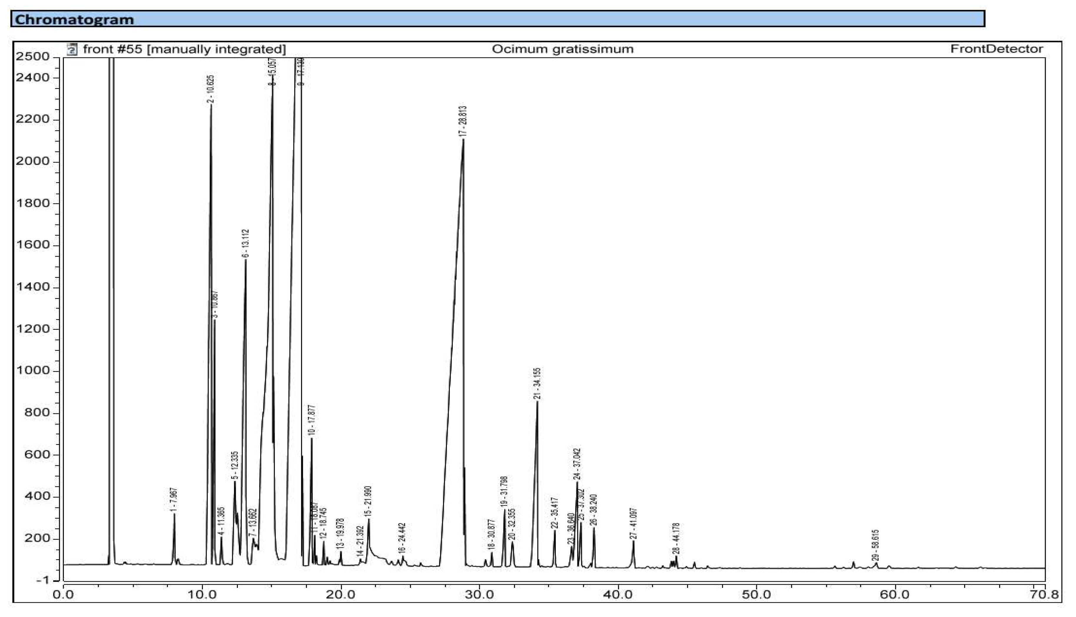
Figure 2.
: Chromatogram of the essential oil of Ocimum basilicum.

Figure 11.
Inhibitory effects of various concentrations of griseofulvin (positive control) against Trichophyton rubrum (A) and Trichophyton interdigitale (B).
Figure 11.
Inhibitory effects of various concentrations of griseofulvin (positive control) against Trichophyton rubrum (A) and Trichophyton interdigitale (B).

Figure 12.
: Percentages of DPPH radical scavenging as a function of concentrations of essential oils of O. gratissimum and O. basilicum and the butylated hydroxytoluene (BHT).
Figure 12.
: Percentages of DPPH radical scavenging as a function of concentrations of essential oils of O. gratissimum and O. basilicum and the butylated hydroxytoluene (BHT).
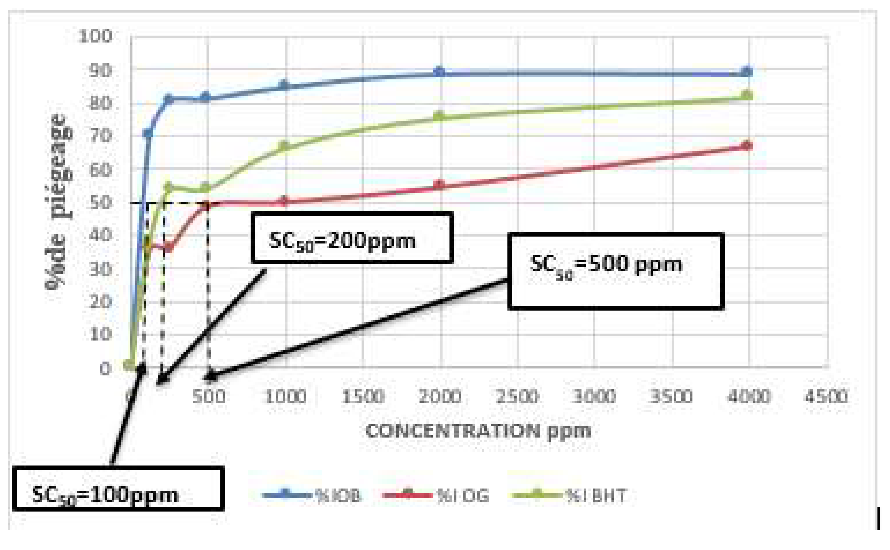
Figure 13.
: Percentages of DPPH radical scavenging as a function of concentrations of nanoformulations obtained from the essential oils of O. gratissimum and O. basilicum and the butylated hydroxytoluene (BHT).
Figure 13.
: Percentages of DPPH radical scavenging as a function of concentrations of nanoformulations obtained from the essential oils of O. gratissimum and O. basilicum and the butylated hydroxytoluene (BHT).
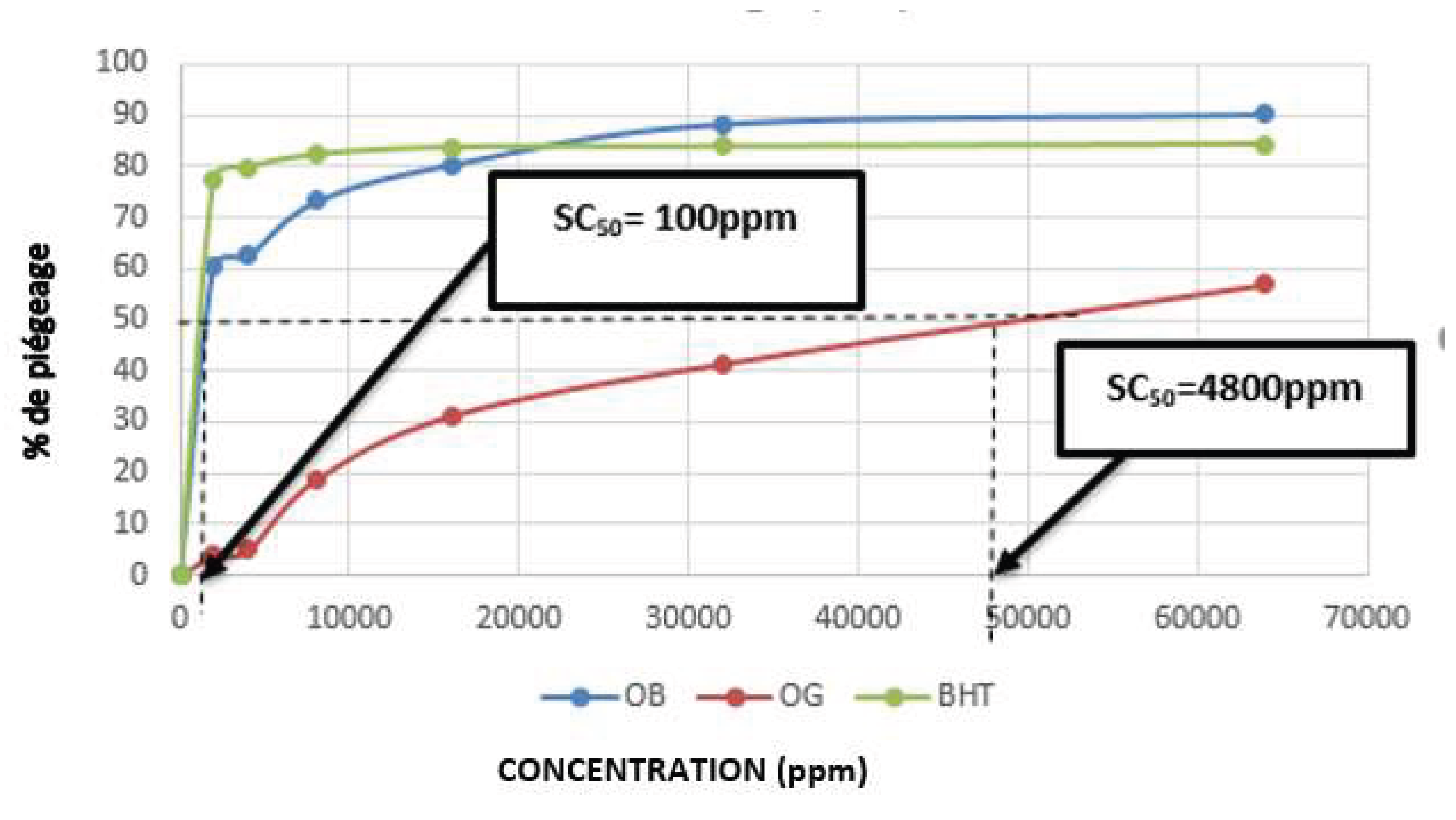

Table 1.
: Yield of extraction and physical characteristics of the essential oils from Ocimum gratissimum and Ocimum basilicum.
Table 1.
: Yield of extraction and physical characteristics of the essential oils from Ocimum gratissimum and Ocimum basilicum.
| Plant species | Organs | Yield of extraction | Color | Density |
| Ocimum gratissimum | Leaves and twigs | 1.12±0.02 | Bright yellow | 0.89 |
| Ocimum basilicum | Leaves and twigs | 0.62±0.01 | Dark yellow | 0.88 |
Table 2.
: Chemical composition of essential oils obtained from Ocimum gratissimum and Ocimum basilicum.
Table 2.
: Chemical composition of essential oils obtained from Ocimum gratissimum and Ocimum basilicum.
| Kovats’ indices | Name of the compound | Ocimum gratissimum | Ocimum basilicum | ||
| Monoterpenes | 93.87 | 92.51 | |||
| Hydrocarbonated monoterpenes | 48.22 | 7.93 | |||
| 1 | 812 | α-pinene | 1.55 | 1.20 | |
| 2 | 825 | β-pinene | - | 2.30 | |
| 3 | 828 | α-myrcene | 4.73 | 1.34 | |
| 4 | 848 | α-ocimene | - | 1.96 | |
| 5 | 926 | α-thujene | 6.05 | - | |
| 6 | 945 | Camphene | 0.18 | - | |
| 7 | 969 | Sabinene | 1.43 | - | |
| 8 | 1002 | α-terpinene | 0.42 | - | |
| 9 | 1076 | γ-terpinene | 33.73 | 1.13 | |
| 10 | 1097 | Terpinolene | 0.13 | - | |
| Oxygenated monoterpenes | 45.65 | 84.58 | |||
| 11 | 843 | Eucalyptol | - | 16.78 | |
| 12 | 865 | Fenchone | - | 1.09 | |
| 13 | 1032 | 1,8-cineole | 16.65 | - | |
| 14 | 1092 | Linalol | 0.97 | 55.32 | |
| 15 | 1110 | Trans-thujone | 0.11 | - | |
| 16 | 1135 | Isocitral exo | 0.09 | - | |
| 17 | 1164 | Borneol | 0.08 | - | |
| 18 | 1176 | Terpinen-4-ol | 1.08 | 3.94 | |
| 19 | 1226 | Thymol methyléther | 0.14 | - | |
| 20 | 1314 | Thymol | 26.44 | - | |
| 21 | 1360 | Eugenol | 0.09 | 7.45 | |
| Sesquiterpenes | 5.8 | 7.46 | |||
| Hydrocarbonated sesquiterpenes | 5.37 | 5.26 | |||
| 20 | 1015 | (-)-endo-α- bergamotene | - | 2.75 | |
| 21 | 1036 | Bicyclogermacrene | - | 0.67 | |
| 22 | 1042 | ϒ muurolene | - | 0.97 | |
| 23 | 1380 | β-cubebene | 0.47 | - | |
| 24 | 1393 | β-elemene | 0.27 | - | |
| 25 | 1432 | β-caryophyllene | 2.64 | - | |
| 26 | 1460 | α-humulene | 0.26 | - | |
| 27 | 1487 | Germacreme D | 0.13 | 0.87 | |
| 28 | 1496 | α-curcumene | 0.88 | - | |
| 29 | 1502 | α-selimene | 0.36 | - | |
| 30 | 1524 | d-cadinene | 0.36 | - | |
| Oxygenated sesquiterpenes | 0.43 | 2.20 | |||
| 31 | 1080 | Tau-cardinol | - | 2.20 | |
| 32 | 1591 | β-6-elemene | 0.25 | - | |
| 33 | 1665 | Hydroxycaryophyllene | 0.18 | - | |
| TOTAL | 99.67 | 99.97 | |||
Table 3.
Minimum inhibitory (MICs) and minimum fungicidal (MFCs) concentrations.
| Sample (OG) | Essential oil | Nanoemulsion | Griseofulvin | |||
| TR | TI | TR | TI | TR | TI | |
| MIC (ppm) | 1000 | 2000 | 2000 | 2000 | ˃5000 | |
| MFC (ppm) | 1000 | 2000 | 2000 | 2000 | ||
| Sample (OB) | Essential oil | Nanoemulsion | Griseofulvin | |||
| TR | TI | TR | TI | TR | TI | |
| MIC (ppm) | 5750 | / | 6750 | / | ˃5000 | |
| MFC (ppm) | 5750 | / | 6750 | / | ||
TR : Trichophyton rubrum and TI : Trichophyton interdigitale ; OB : Ocimum basilicum ; OG : Ocimum gratissimum.
Disclaimer/Publisher’s Note: The statements, opinions and data contained in all publications are solely those of the individual author(s) and contributor(s) and not of MDPI and/or the editor(s). MDPI and/or the editor(s) disclaim responsibility for any injury to people or property resulting from any ideas, methods, instructions or products referred to in the content. |
© 2025 by the authors. Licensee MDPI, Basel, Switzerland. This article is an open access article distributed under the terms and conditions of the Creative Commons Attribution (CC BY) license (http://creativecommons.org/licenses/by/4.0/).
Copyright: This open access article is published under a Creative Commons CC BY 4.0 license, which permit the free download, distribution, and reuse, provided that the author and preprint are cited in any reuse.


