Submitted:
10 May 2025
Posted:
12 May 2025
You are already at the latest version
Abstract
Echocardiography is vital in diagnosing and managing congenital heart disease (CHD), particularly in the pediatric population, necessitating detailed structural and functional assessments. Artificial intelligence (AI) has revolutionized echocardiographic analysis, particularly in functional assessments and the detection of valvular lesions. While convolutional neural networks (CNNs) dominate image-based tasks, emerging vision language models (VLMs) are transforming report generation by integrating multimodal data. This review explores the current state of AI in echocardiography, emphasizing the potential of VLMs to provide comprehensive reports and image-specific diagnoses. Despite significant advancements, several challenges hinder the development of holistic AI software for diagnosing complex congenital heart disease (CXCHD). These challenges include the heterogeneity of CHD, limited access to high-quality labeled datasets, variability in imaging techniques, and the need for expertise in image annotation. This review highlights the necessity for robust algorithms, standardized protocols, and diverse training datasets to fully realize the potential of AI in complex CHD diagnosis.
Keywords:
Artificial intelligence
; emerging visual language models
; complex congenital heart disease
Background:
Echocardiography remains a fundamental tool in diagnosing and managing congenital heart disease (CHD) owing to its accessibility, feasibility, and ability to provide real-time functional and structural assessment. Precise and reliable echocardiographic assessment is essential in guiding clinical decision-making, particularly in the pediatric population[1, 2].
Pediatric echocardiography differs significantly from adult echocardiography, particularly in focus and objectives. While adult echocardiography primarily emphasizes on functional assessment of the heart, such as ejection fraction and cardiac output, pediatric echocardiography prioritizes detailed structural assessment to identify congenital structural lesions that most commonly underlie cardiac conditions in children [3].
In pediatric patients, congenital heart disease (CHD) necessitates a comprehensive evaluation of cardiac anatomy; thereby, detailed assessment of the cardiac morphology, including chambers, valves, and great vessels, is required to identify structural anomalies. This level of diagnostic precision in anatomical assessment is essential in guiding clinical decisions, shaping potential interventions, and establishing long-term management interventions for young patients [4].
Echocardiography differs from other imaging modalities, such as cardiac MRI or CT angiography, in several key aspects, but most notably in the lack of standardization. While cardiac MRI and CT angiography are guided by standardized protocols to ensure consistent and reproducible results in imaging and interpretation across different operators and institutions, echocardiography varies widely in imaging technique, measurement criteria, and operator skillfulness. This variability in echocardiographic imaging introduces inconsistencies in interpretation and diagnostic accuracy, which may ultimately affect clinical decision-making [5].
In contrast, cardiac MRI and CT angiography benefit from established guidelines and protocols to promote reproducibility and consistency in imaging results. These standardized practices ensure reliable comparisons between studies and accurate evaluations of cardiac conditions, which is particularly essential in diagnosing complex cases, such as congenital heart diseases. Consequently, the relative lack of standardization in echocardiography emphasizes the imperative need for cultivating best practices to improve the consistency of image acquisition and interpretation within this modality [6,7,8].
This review aims to explore the current applications of artificial intelligence (Ai) in image analysis within the field of echocardiography while also addressing the challenges faced by Ai developers in the detailed assessment of complex congenital heart disease (CXCHD). Despite the advancements in AI technologies, significant hurdles hinder their practical application to CXHD's intricate and variable anatomical presentations.
Current state of developed AI software for Echocardiographic analysis:
Ai-assisted diagnosis in echocardiography is revolutionizing the assessment of cardiovascular conditions with significant applications in two key parameters: function assessment, wall thickness measurement, and detection of valvular lesions.
In function assessment, Ai algorithms have demonstrated remarkable accuracy in calculating left ventricular ejection fraction (LVEF), a crucial metric in diagnosing heart function, particularly in conditions such as heart failure. Additionally, Ai has introduced advanced strain measurement techniques through software like Echopac and Tomtec, which enable detailed evaluation of myocardial deformation. This advancement allows for early identification of subtle cardiac dysfunctions that may not be evident through traditional imaging methods. Furthermore, Ai can evaluate diastolic function by analyzing blood flow dynamics and tissue Doppler imaging, which can support the diagnosis of hypertensive heart disease. These advancements not only enhance diagnostic accuracy but can also allow for real-time analysis during echocardiographic examinations, supporting clinicians in making timely decisions regarding patient management.Automated speckle tracking echocardiography (STE) has evolved to enhance ease of use and diagnostic accuracy through varying levels of user interaction, often described in terms of "click" approaches. Single-click or one-step STE systems utilize machine learning algorithms to automatically identify and track myocardial speckles with minimal user intervention, enabling rapid and reproducible strain analysis. More interactive methods, such as two- or three-click techniques (Figure 1 and 2), incorporate additional user input to refine or validate the tracking process, particularly in challenging imaging conditions. The integration of machine learning into these systems significantly improves tracking robustness and accuracy by leveraging large datasets for model training, thereby reducing the reliance on manual adjustments. Overall, the progression from semi-automated to fully automated STE reflects ongoing advancements in artificial intelligence, aiming to provide reliable, operator-independent assessments of cardiac function in clinical settings.
Additionally, Ai-enhanced technologies can serve as vital tools in detecting valvular lesions and abnormal valvular function through analysis of Doppler flow patterns integrated with structural imaging. Thus, it facilitates detection of acquired valvular conditions such as mitral regurgitation and aortic stenosis during adulthood. When used in fetal echocardiography, Ai supports clinicians in the distinction between normal and abnormal cardiac anatomy by analyzing fetal heart structures and rhythms using feed forwards networks. Early identification is critical for timely intervention and optimizing neonatal outcomes. Integrating Ai into echocardiography enhances diagnostic precision, streamlines workflow, and ultimately improves patient care.
Table1 presents a landscape of software and Ai models used in echocardiographic analysis. A significant portion of these tools leverage Convolutional Neural Networks (CNNs) for various tasks, including assisting with image acquisition, functional assessment, and cardiac measurements. Several platforms like Vscan Air SL, GE Echo PAC/ Philips Intellispace, and Arterys Cardio AI incorporate CNNs to automate processes such as image capture, quality assessment, and ventricular segmentation. These platforms aim to streamline workflows, enhance accuracy, and provide comprehensive cardiac assessments.
Importantly, vision language models (V/LLM) are emerging as key players in the field, as seen in tools like EchoCLIP, EchoPrime and Ventripoint VMS. These advanced models are starting to be used in generating full reports, integrating image data with clinical notes and expert knowledge, offering potential for more holistic and image-specific diagnoses. Some tools, such as Bay Labs EchoMD, focus on simplifying the echocardiography process for varying expertise levels, while others, like Ultromics EchoGo and Ventripoint VMS, aim to offer advanced analysis such as global longitudinal strain and accurate 3D reconstructions. Finally, a couple of studies focused on fetal echocardiograms and incorporated either CNNs or feedforward networks for classification of normal and abnormal fetal heart conditions. A clear trend is that numerous companies and research groups are actively developing and deploying Ai-driven solutions to enhance the efficiency, accuracy, and accessibility of echocardiographic analysis across a variety of clinical needs, however none of them has yet achieved a holistic software for image specific diagnosis of complex congenital heart disease.
Top of Form
Challenges of Ai in echocardiography of complex congenital heart disease
The use of Ai algorithms in the evaluation of complex congenital heart disease presents significant challenges that impact their efficacy and reliability. A major limitation in the application of Ai in echocardiography is the heterogeneity of congenital heart conditions that present a range of anatomical and physiological variability[29]. Accordingly, diverse datasets that encompass a wide variety of presentations are needed to familiarize Ai models with differentiating different conditions, such as Tetralogy of Fallot or transposition of the great arteries, which differ vastly in structure and hemodynamics. However, the availability of such comprehensive datasets is often limited, especially for rare congenital defects that may be infrequently encountered in clinical practice. This scarcity is compounded by the requirement for high-quality labeled images, which are crucial for training Ai systems on recognizing and interpreting complex cases accurately. The process of annotating echocardiographic images is labor-intensive and necessitates the expertise of experienced cardiologists, which creates significant bottlenecks in the establishment of large, well-annotated datasets[30].
In addition to the challenges associated with data availability, variability in imaging techniques adds another layer of limitation to the application of Ai in echocardiography. Differences in operator skill, institutional protocols, and even the ultrasound machines used can lead to inconsistent image quality (Figure 3 and Figure 4 illustrate this challenge within a function GE software. In Figure 3, the software fails to detect the LV contour due to the transverse positioning of the LV in the 3-chamber view, conversely, Figure 4 demonstrates successful tracking when the LV is oriented differently). In other words, two echocardiographers may capture differing representations of the same cardiac structure based on their technique and experience. Furthermore, variations in imaging protocols—such as the specific views taken during an examination, the settings used (like frequency and gain), and the measurements emphasized—can impact the data introduced into Ai systems for analysis. Patient-specific factors, such as body habitus, age, and comorbid conditions, can influence echocardiographic image quality and impact the algorithms' generalizability across different populations[31].
The interplay of these challenges underlines the critical need for rigorous development and validation of Ai tools prior to their integration into clinical practice, to ensure effectiveness across different clinical settings and institutions. Training Ai algorithms on a narrow range of imaging data can severely compromise their performance when confronted with a more diverse array of cases; this risk of poor generalization can lead to decreased diagnostic accuracy and overlooked critical findings. Addressing these multifaceted challenges requires a concerted effort to curate diverse training datasets, design robust algorithms capable of accommodating various imaging techniques, and undertake ongoing testing and validation across multiple clinical environments. Consequently, the integration of Ai into echocardiography can be optimized, enhancing its role in the assessment of complex congenital heart disease and supporting clinicians in delivering better patient care.
Key solutions for achieving image-specific diagnosis in complex congenital heart disease.
Standardization of the image protocol and quality:
Ai has the potential for standardization of image acquisition protocols in echocardiography, thereby enhancing the quality and consistency of diagnostic outputs across different institutions and operators. One of the primary ways Ai contributes to standardization is through its capability to analyze vast amounts of data obtained from diverse echocardiographic practices. By identifying and learning from successful imaging techniques across various institutions, Ai can help define best practices for image acquisition. These insights can drive the development of standardized imaging protocols that operators can follow, regardless of the equipment or location[32, 33].
Moreover, Ai-driven systems can assist in automating aspects of image acquisition by providing real-time feedback to echocardiographers during the imaging process. For example, Ai algorithms can evaluate the quality of images as they are captured, alerting the operator when the images do not meet predefined quality standards or suggesting adjustments to improve image clarity and detail. This immediate feedback encourages adherence to standardized techniques, reducing operator variability and ensuring that each image meets the necessary diagnostic criteria. Additionally, Ai can facilitate the development of training modules and educational tools that promote standardized practices. By using machine learning to analyze successful methodologies, AI can generate training materials that highlight effective imaging techniques, common pitfalls, and practical guidelines. These resources can better equip echocardiographers with the knowledge and skills necessary to produce standardized images, thereby enhancing both individual performance and overall diagnostic accuracy[9].
In summary, Ai can play a pivotal role in standardizing image acquisition protocols in echocardiography through data analysis, real-time feedback, and educational resources. These efforts not only enhance image quality but also promote collaboration among healthcare providers, ultimately contributing to enhanced patient care in the diagnosis and management of cardiovascular diseases.
Segmental analysis
Another critical component in achieving effective Ai-driven diagnostic outcomes in complex echocardiography is segmental analysis, which is a detailed evaluation of various structural components of the heart, including the heart chambers’ position, the anatomy and functionality of the atrioventricular and semilunar valves, as well as the assessment of the cardiac septa. In addition, evaluating any discrepancies in ventricular size and analyzing the Doppler flow signals across the great vessels is essential. Segmental analysis is a cornerstone of effective echocardiographic evaluation in congenital heart disease. A comprehensive approach, incorporating detailed structural and functional assessments alongside the development of robust algorithms and datasets, is fundamental for accurately diagnosing and guiding appropriate management of these complex conditions[32,34].
The emergence of Visual large language models as a potential solution:
Three important subtypes of Ai used in image analysis include feedforward networks, convolutional neural networks (CNNs), and recently, visual language models. Feedforward neural networks, such as multilayer perceptrons, and recurrent neural networks (including bidirectional and reversed architectures) have been integral to the classification of fetal echocardiograms. These models are particularly suited for analyzing sequential or time-series data inherent in fetal heart imaging, with recurrent architecture effectively capturing temporal dynamics and subtle motion patterns across cardiac cycles. This capability enhances the accuracy of classification tasks, such as detecting congenital heart defects or assessing fetal cardiac function.
While CNNs are highly effective in extracting spatial features from static imaging data—enabling tasks such as image classification, segmentation, and detection—they excel at recognizing detailed patterns directly from pixel data, thus facilitating precise analysis of cardiac structures and motion. In contrast, visual language models aim to integrate multiple data modalities—such as combining imaging with clinical notes, reports, and contextual information—by understanding and generating meaningful multimodal representations. Applying these models to echocardiography offers the potential for richer insights, improving interpretability and diagnostic accuracy. Although CNNs currently dominate purely image-based tasks, visual language models are emerging as promising tools for more comprehensive, context-aware analysis. The future of echocardiographic AI likely involves hybrid approaches that leverage CNNs’ spatial robustness alongside the rich, multimodal understanding provided by visual language models—an advancement that could critically improve the diagnosis of complex congenital echocardiography. Table 2 summarizes the above-mentioned key strengths of each model[35].
Conclusion:
AI is revolutionizing echocardiography, and holds the promise to enhance diagnostic precision, streamline workflows, and ultimately improve patient care, as seen in emerging tools that implement vision language models and automate the generation of comprehensive clinical reports. VLMs have demonstrated potential in more holistic and image-specific diagnoses. The path towards creating a truly "AI-proof" echocardiography solution for complex CHD is not without obstacles. Overcoming limitations related to data availability, variability in imaging techniques, and the need for specialized expertise in image annotation is crucial. The field requires concerted efforts to curate diverse training datasets, develop robust algorithms capable of accommodating various imaging techniques, and establish standardized image acquisition protocols. By addressing these challenges, the field can unlock the full potential of AI in echocardiography and provide clinicians with the tools needed to deliver accurate and personalized care to patients with complex congenital heart disease.
List of Abbreviations:
| Abbreviation | Full Term |
| AI | Artificial Intelligence |
| CHD | Congenital Heart Disease |
| CXCHD | Complex Congenital Heart Disease |
| CNN | Convolutional Neural Network |
| VLM/VLLM | Vision Language Model/Large Language Model |
| LVEF | Left Ventricular Ejection Fraction |
| STE | Speckle Tracking Echocardiography |
Author Contributions
Conceptualization, AFA; Methodology, AFA, FM, FAM, MF, MA, NA, RG, RA, SA; software, AFA, FM, FAM, MF, MA, NA, RG, RA, SA; investigation, AFA, FM, FAM, MF, MA, NA, RG, RA, SA; resources, AFA, FM, FAM, MF, MA, NA, RG, RA, SA, data curation, AFA, FM, FAM, MF, MA, NA, RG, RA, SA; writing—original draft preparation, AFA, FM, FAM, MF, MA, NA, RG, RA, SA; writing—review and editing, AFA, FM, FAM, MF, MA, NA, RG, RA, SA; supervision, AFA; project administration, AFA; funding acquisition, (non-applicable). All authors have read and agreed to the published version of the manuscript.”
Institutional Review Board Statement
not applicable as this study is a hypothesis/Review article.
Informed Consent Statement
not applicable as this study is a viewpoint/editorial
Data Availability Statement
All data is made available within the manuscript and are owned by the authors.
Acknowledgement
To the peacekeepers in every part of the world, in every community, every family and every tiny relationship. Peace keeping might sometimes look like weakness, but it requires utmost strength,
Conflicts of Interest
The authors declare no conflict of interest. The manuscript is submitted under Creative Commons Licensing CC-BY-NC-ND. A large language model has been used to proof read the article.
References
- Tworetzky W, McElhinney DB, Brook MM, Mohan Reddy V, Hanley FL, Silverman NH (1999) Echocardiographic diagnosis alone for the complete repair of major congenital heart defects. J Am Coll Cardiol 33:228–233. [CrossRef]
- Mcleod G, Shum K, Gupta T, Chakravorty S, Kachur S, Bienvenu L, White M, Shah SB (2018) Echocardiography in Congenital Heart Disease. Prog Cardiovasc Dis 61:468–475. [CrossRef]
- Hurwitz RA, Caldwell RL (1998) Should Pediatric Echocardiography Be Performed in Adult Laboratories? Pediatrics 102:e15–e15. [CrossRef]
- Koestenberger M (2012) Transthoracic echocardiography in children and young adults with congenital heart disease. ISRN Pediatr 2012:753481. [CrossRef]
- White BR, Ho DY, Rogers LS, Natarajan SS (2019) A standardized imaging protocol improves quality and reduces practice variability in echocardiographic assessment of ventricular function by first-year pediatric cardiology fellows. Echocardiography 36:1515–1523. [CrossRef]
- Samyn MM, Yan K, Masterson C, Goot BH, Saudek D, Lavoie J, Kinney A, Krolikowski M, Hor K, Cohen S (2019) Echocardiography vs cardiac magnetic resonance imaging assessment of the systemic right ventricle for patients with d-transposition of the great arteries status post atrial switch. Congenit Heart Dis 14:1138–1148. [CrossRef]
- Baessato F, Fusini L, Muratori M, Tamborini G, Ghulam Ali S, Mantegazza V, Baggiano A, Mushtaq S, Pepi M, Patti G, Pontone G (2023) Echocardiography vs. CMR in the Quantification of Chronic Mitral Regurgitation: A Happy Marriage or Stormy Divorce? J Cardiovasc Dev Dis 10:150. [CrossRef]
- Kramer CM, Barkhausen J, Bucciarelli-Ducci C, Flamm SD, Kim RJ, Nagel E (2020) Standardized cardiovascular magnetic resonance imaging (CMR) protocols: 2020 update. J Cardiovasc Magn Reson 22:1–18. [CrossRef]
- Soemantoro R, Kardos A, Tang G, Zhao Y (2023) An AI-powered navigation framework to achieve an automated acquisition of cardiac ultrasound images. Sci Rep 13:1–13. [CrossRef]
- Yang PC, Jha A, Xu W, Song Z, Jamp P, Teuteberg JJ (2024) Cloud-Based Machine Learning Platform to Predict Clinical Outcomes at Home for Patients With Cardiovascular Conditions Discharged From Hospital: Clinical Trial. JMIR Cardio 8:. [CrossRef]
- Anisuzzaman DM, Malins JG, Jackson JI, Lee E, Naser JA, Rostami B, Greason G, Bird JG, Friedman PA, Oh JK, Pellikka PA, Thaden JJ, Lopez-Jimenez F, Attia ZI, Pislaru S V, Kane GC (2025) Leveraging Comprehensive Echo Data to Power Artificial Intelligence Models for Handheld Cardiac Ultrasound. Mayo Clin proceedings Digit Heal 3:100194. [CrossRef]
- Abdelmassih AF, Haidar W, Alabid S, Attallah T, Khraisat B, Khan A, Hafez B (2025) Understanding Why Postpericardiotomy Syndrome is More Common after Surgical Atrial Septal Defect Closure : A Sneak Peek at Atrial Functions by Speckle - tracking Echocardiography. 101–104. [CrossRef]
- Cioccari L, Baur HR, Berger D, Wiegand J, Takala J, Merz TM (2013) Hemodynamic assessment of critically ill patients using a miniaturized transesophageal echocardiography probe. Crit Care 17:. [CrossRef]
- Christensen M, Vukadinovic M, Yuan N, Ouyang D (2024) Vision–language foundation model for echocardiogram interpretation. Nat Med 30:1481–1488. [CrossRef]
- Holste G, Oikonomou EK, Tokodi M, Kovács A, Wang Z, Khera R (2024) PanEcho: Complete AI-enabled echocardiography interpretation with multi-task deep learning.
- Wessler BS, Huang Z, Long GM, Pacifici S, Prashar N, Karmiy S, Sandler RA, Sokol JZ, Sokol DB, Dehn MM, Maslon L, Mai E, Patel AR, Hughes MC (2023) Automated Detection of Aortic Stenosis Using Machine Learning. J Am Soc Echocardiogr 36:411–420. [CrossRef]
- Vukadinovic M, Tang X, Yuan N, Cheng P, Li D, Cheng S, He B, Ouyang D (2024) EchoPrime: A Multi-Video View-Informed Vision-Language Model for Comprehensive Echocardiography Interpretation.
- Amadou AA, Zhang Y, Piat S, Klein P, Schmuecking I, Passerini T, Sharma P (2024) EchoApex: A General-Purpose Vision Foundation Model for Echocardiography.
- Sadeghpour A, Jiang Z, Hummel YM, Frost M, Lam CSP, Shah SJ, Lund LH, Stone GW, Swaminathan M, Weissman NJ, Asch FM (2025) An Automated Machine Learning–Based Quantitative Multiparametric Approach for Mitral Regurgitation Severity Grading. JACC Cardiovasc Imaging 18:1–12. [CrossRef]
- O’Driscoll JM, Hawkes W, Beqiri A, Mumith A, Parker A, Upton R, McCourt A, Woodward W, Dockerill C, Sabharwal N, Kardos A, Augustine DX, Balkhausen K, Chandrasekaran B, Firoozan S, Marciniak A, Heitner S, Yadava M, Kaul S, Sarwar R, Sharma R, Woodward G, Leeson P (2022) Left ventricular assessment with artificial intelligence increases the diagnostic accuracy of stress echocardiography. Eur Hear J Open 2:1–10. [CrossRef]
- Ahmad A, Shigemitsu S, Termachi Y, Windram J, Khoo N, Colen T, Eckersley L (2022) Comparing a knowledge-based 3D reconstruction algorithm to TomTec 3D echocardiogram algorithm in measuring left cardiac chamber volumes in the pediatric population. Echocardiography 39:1180–1189. [CrossRef]
- Haji-Hassan M, Duțu B, Bolboacă SD (2023) Handheld Echocardiography Measurements Concordance and Findings Agreement: An Exploratory Study. Diagnostics 13:1–14. [CrossRef]
- Naghavi M, Reeves AP, Atlas K, Zhang C, Atlas T, Henschke CI, Yankelevitz DF, Budoff MJ, Li D, Roy SK, Nasir K, Molloi S, Fayad Z, McConnell M V., Kakadiaris I, Maron DJ, Narula J, Williams K, Shah PK, Levy D, Wong ND (2024) Artificial intelligence applied to coronary artery calcium scans (AI-CAC) significantly improves cardiovascular events prediction. npj Digit Med 7:309. [CrossRef]
- Asch FM, Poilvert N, Abraham T, Jankowski M, Cleve J, Adams M, Romano N, Hong H, Mor-Avi V, Martin RP, Lang RM (2019) Automated Echocardiographic Quantification of Left Ventricular Ejection Fraction Without Volume Measurements Using a Machine Learning Algorithm Mimicking a Human Expert. Circ Cardiovasc Imaging 12:1–9. [CrossRef]
- Muhtaseb R, Yaqub M (2022) EchoCoTr: Estimation of the Left Ventricular Ejection Fraction from Spatiotemporal Echocardiography. arXiv:arXiv:2209.04242v1.
- Strom JB, Playford D, Stewart S, Strange G (2024) An Artificial Intelligence Algorithm for Detection of Severe Aortic Stenosis: A Clinical Cohort Study. JACC Adv 3:1–12. [CrossRef]
- Edwards LA, Feng F, Iqbal M, Fu Y, Sanyahumbi A, Hao S, McElhinney DB, Ling XB, Sable C, Luo J (2023) Machine Learning for Pediatric Echocardiographic Mitral Regurgitation Detection. J Am Soc Echocardiogr 36:96-104.e4. [CrossRef]
- Karužas A, Balčiūnas J, Fukson M, Verikas D, Matuliauskas D, Platūkis T, Vaičiulienė D, Jurgaitytė J, Kupstytė-Krištaponė N, Dirsienė R, Jaruševičius G, Šakalytė G, Plisienė J, Lesauskaitė V (2022) Artificial intelligence for automated evaluation of aortic measurements in 2D echocardiography: Feasibility, accuracy, and reproducibility. Echocardiography 39:1439–1445. [CrossRef]
- Selmeryd J, Henriksen E, Leppert J, Hedberg P (2016) Interstudy heterogeneity of definitions of diastolic dysfunction severely affects reported prevalence. Eur Hear J – Cardiovasc Imaging 17:892–899. [CrossRef]
- Waisberg E, Ong J, Kamran SA, Paladugu P, Zaman N, Lee AG, Tavakkoli A (2023) Transfer learning as an AI-based solution to address limited datasets in space medicine. Life Sci Sp Res 36:36–38. [CrossRef]
- Ramlogan S, Aly D, France R, Schmidt S, Hinzman J, Sherman A, Goudar SP, Forsha D (2020) Reproducibility and Intervendor Agreement of Left Ventricular Global Systolic Strain in Children Using a Layer-Specific Analysis. J Am Soc Echocardiogr 33:110–119. [CrossRef]
- Hagiwara A, Fujita S, Ohno Y, Aoki S (2020) Variability and Standardization of Quantitative Imaging. Invest Radiol 55:601–616. [CrossRef]
- Li XT, Huang RY (2020) Standardization of imaging methods for machine learning in neuro-oncology. Neuro-oncology Adv 2:iv49–iv55. [CrossRef]
- Seetharam K, Thyagaturu H, Ferreira GL, Patel A, Patel C, Elahi A, Pachulski R, Shah J, Mir P, Thodimela A, Pala M, Thet Z, Hamirani Y (2024) Broadening Perspectives of Artificial Intelligence in Echocardiography. Cardiol Ther 13:267–279. [CrossRef]
- Li X, Li L, Jiang Y, Wang H, Qiao X, Feng T, Luo H, Zhao Y (2025) Vision-Language Models in medical image analysis: From simple fusion to general large models. Inf Fusion 118:102995. [CrossRef]
Figure 1.
2 click 3D speckle tracking echocardiography software by GE (requiring two clicks at the apex and base).
Figure 1.
2 click 3D speckle tracking echocardiography software by GE (requiring two clicks at the apex and base).
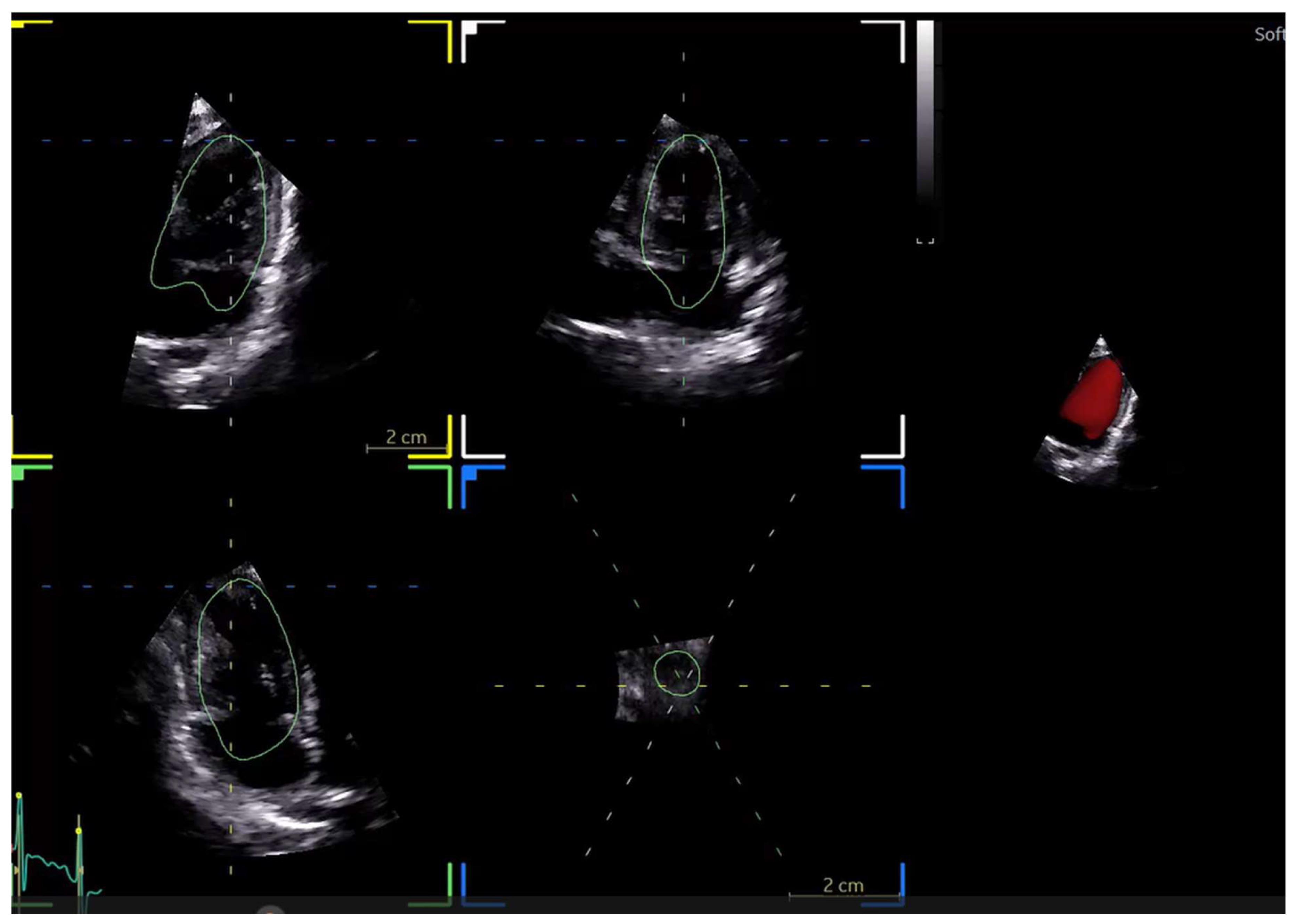
Figure 2.
No Click Auto Ejection fraction software by GE.
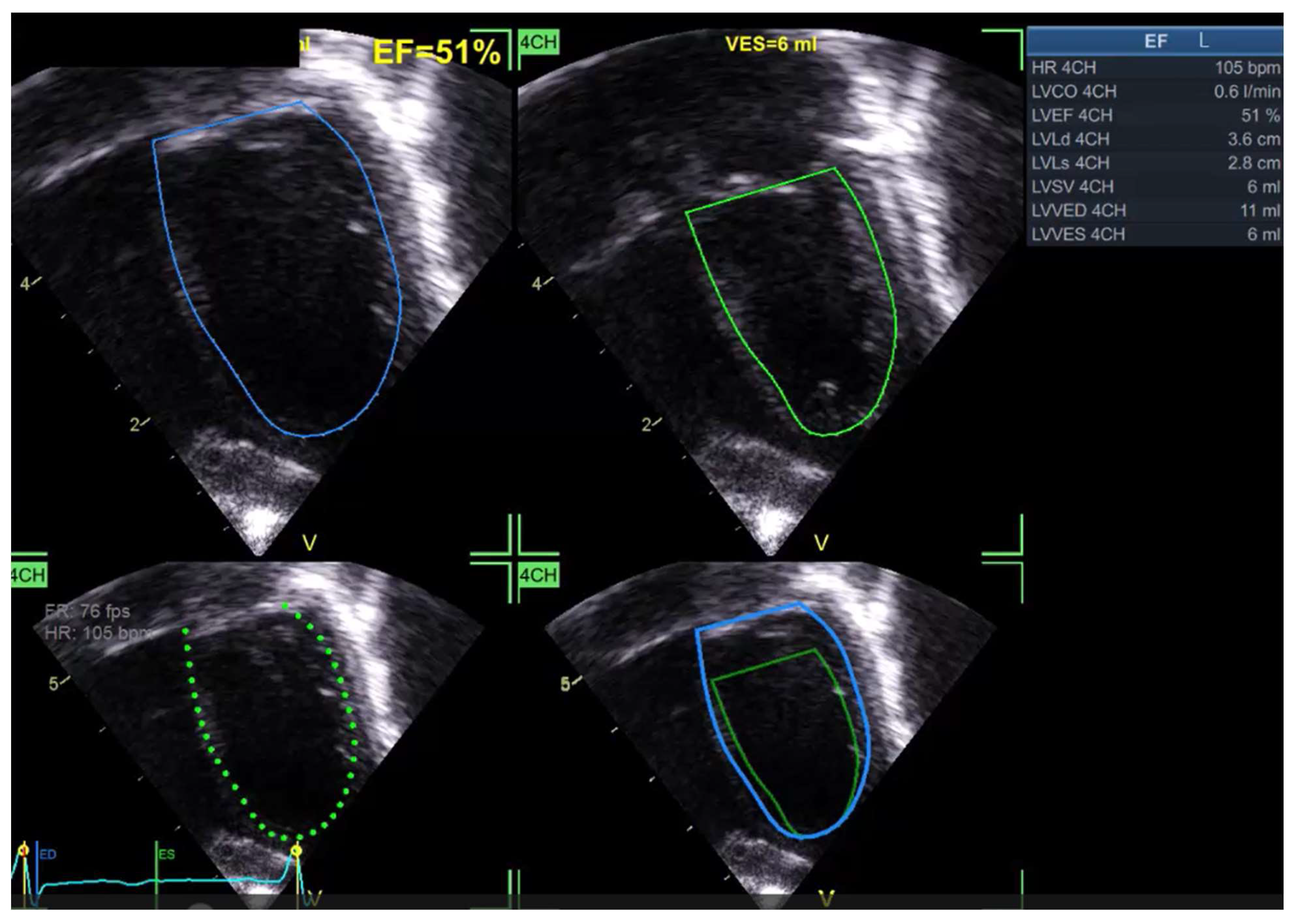
Figure 3.
Unsuccessful tracking of the LV due to transverse position of the LV in a 3 chambers view.
Figure 3.
Unsuccessful tracking of the LV due to transverse position of the LV in a 3 chambers view.
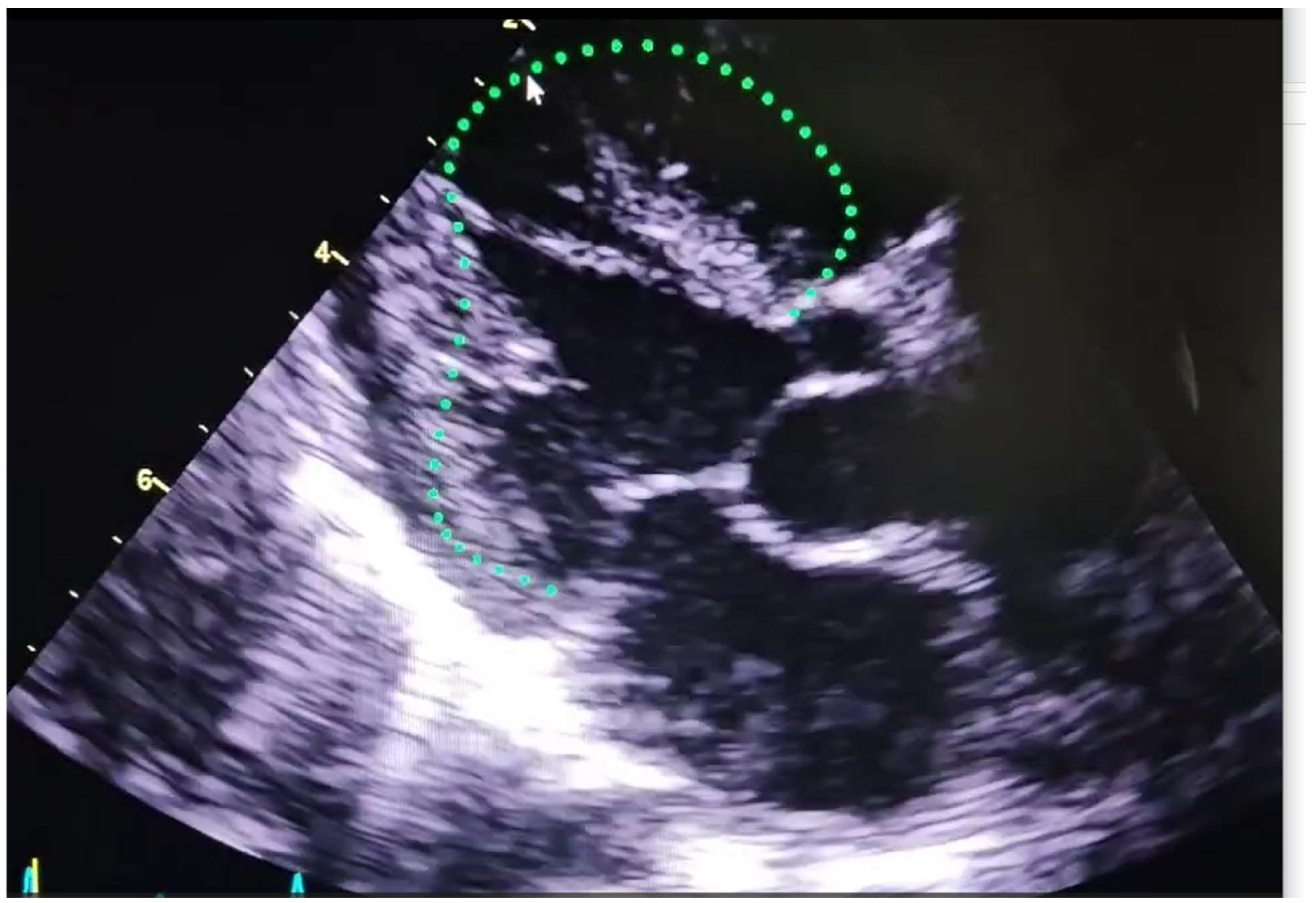
Figure 4.
Successful Tracking of the LV due to vertical position of the LV in a 3 chambers view.


Table 1.
Commercially available software employing Ai in the field of echocardiography.
| Software | Main Tools for Echocardiographic Analysis | Developing Company/Institute | Auto EF | Standardizing image acquisition | STE Analysis | Valve function | Image specific diagnosis | Ai model applied | ||
| Standardizing image acquisition | ||||||||||
| Vscan Air SL with Caption AI [9] | Step by step instructions, auto capture images, quality meter “real time feedback on image quality | GE | Yes | Yes | Yes | No | No | CNN | ||
| Kitware's AI-Powered Ultrasound(No available citations) | Assistance in probe placement, autonomous scanning, CHD identification in newborns. | Kitware | No | Yes | No | No | CNN | |||
| Cloud-based solutions | ||||||||||
| CardioCloud by Cardio-Care [10] | Remote access to studies, echocardiographic analysis | CardioCare | Yes | No | Yes | No | No | CNN | ||
| Main focus: Functional assessment | ||||||||||
| Philips Intellispace Cardiovascular and Tomtec [11] | Advanced echocardiography analysis, 3D reconstruction, integration with other imaging modalities | Philips Healthcare | Yes | No | Yes | Yes | No | CNN | ||
| GE Healthcare EchoPAC/Verisound [12] | Automated measurements, advanced reporting, comprehensive analysis | GE Healthcare | Yes | No | Yes | Yes | No | CNN | ||
| Imacor [13] | Myocardial strain assessment, 3D echocardiographic evaluation, hemodynamic ultrasound using TEE in critical care | Imacor | Yes | No | Yes | No | No | CNN | ||
| Automated decision-making regarding fluid therapy in ICU | ||||||||||
| EchoCLIP | Generates full echocardiographic reports, being trained not on individual data sets but on series of full exams and their corresponding expert reports | Researchers at Cedars Sinai Medical Center’s Smidt Heart Institute | Yes | No | No | Yes | Yes | Vision large V/LLM) | ||
| [14] | ||||||||||
| PanEcho [15] | Uses View-agnostic, multi-task deep learning architecture to process Echo videos. It can detect valvular diseases and chamber sizes. Can also accurately estimate LVEF and detect systolic dysfunction | Researchers at Yale School of Medicine and University of Texas at Austin | Yes | No | No | Yes | Yes | CNN | ||
| EchoMeasure [16] | Uses automated view classification and quality assessment. Provides comprehensive cardiac measurements including Left ventricular volumes, right ventricular diameter, posterior wall thickness, aortic annulus diameter. | iCardio.ai, a Los Angeles- based artificial intelligence company. | Yes | No | No | Yes (AS) | Yes | CNN | ||
| EchoPrime [17] | Consists of multiple modules integral for the interpretation of echocardiography, including video encoder, text encoder, view classifier, and anatomical attention module | Multi-disciplinary team at Cedars-Sinai Medical center | Yes | No | No | Yes | yes | V/LLM | ||
| EchoApex [18] | Uses task specific decoders for view classification, structure segmentation, left ventricular measurement and ejection fracture estimation. | Siemen Healthineers | Yes | No | No | Yes | Yes | CNN | ||
| Us2.ai[19] | Automated measures (45 echo parameters), disease detection like amyloidosis, generating real time report, POCUS AI guided HF screen | US2.ai Ltd. | Yes | No | Yes | No | Yes | CNN | ||
| Ultromics EchoGo [20] | Process ECHO data and provide detailed report. HF detection with preserve EF | Ultromics | Yes | No | Yes (including global longitudinal strain (GLS)) | Yes | Yes | CNN | ||
| Ventripoint VMS [21] | The Ventripoint point of care solution, KBR (Knowledge Based Reconstruction) artificial intelligence software algorithm and accurate sensor positioning allows to accurately reconstruct 2D and 3D echo images into accurate 3D models and precision metrics measurements including Heart structure sizes and ejection fraction. | Ventripoint Diagnostic Ltd. A Toronto based medical technology company | Yes | No | Yes | Yes | Yes | CNN/VLLM | ||
| First echocardiographic software for diagnosis of Single ventricle (study announced this year) | ||||||||||
| Full report | ||||||||||
| EchoConfidence by MyCardium Ai (no available citations) | Developed for highly accurate automated measurements. Utilizes fixed convolutional neural networks (CNN) trained on over 100,00 images. Comprehensive assessment of chambers, valves, vessles and Pericardium. | MyCardium Ai Ltd, a medical technology company in Liverpool | Yes | No | No (No information that confirms that it performs STE analysis on its website) | Yes | No | CNN | ||
| EchoNous Kosmos [22] | Real time anatomical labeling (fast exam), auto doppler images and auto present real time images quality optimization | EchoNous | Yes | No | No | No | CNN | |||
| Arterys Cardio Ai [23] | Automates the segmentation of cardiac ventricles, assess myocardial scarring and delay enhancement for conditions like hypertrophic myocardiopathy | Arterys | Yes | Yes (advanced tools for strain imaging similar to STE) | No | No | CNN | |||
| Bay Labs EchoMD [24] | Simplifies the process of ECHO analysis to make it accessible for varying expertise levels, integrates IT system for efficient data sharing and reporting | Bay Labs | Yes | No | No | No | No | CNN | ||
| EchoCoTr[25] | Deep learning-based LVEF estimation using spatiotemporal echocardiographic video analysis (CNN + Transformer) | Mohamed Bin Zayed University of Artificial Intelligence (MBZUAI) | Yes | No | No (not designed for speckle tracking) | No | No | CNN | ||
| Main Focus: Valvular Function | ||||||||||
| Echo iq [26] | A software developed to detect aortic stenosis | Boston, Massachusetts, USA | No | No | No | Yes | No | Yes | ||
| Stanford University research model [27] | Automated view recognition and mitral regurge classification | Stanford | No | No | No | Yes | No | CNN | ||
| Ligence Heart version 2[28] | Artificial intelligence for automated evaluation of aortic measurements in 2D echocardiography: Feasibility, accuracy, and reproducibility | Ligence | Yes | No | Yes | Yes | No | CNN | ||
| Main Focus: Fetal echocardiography classification | ||||||||||
| Komatsu et al | Classification of normal and abnormal fetal heart echocardiograms | University School of Medicine-Tokyo | No | No | No | No | No | CNN | ||
| Bridge et al | Classification of normal and abnormal fetal heart echocardiograms | Oxford University | No | No | No | No | No | Feed forward networks | ||
| Abbreviations:Ai: Artificial intelligence, CNN: Convoluted Neural Networks, EF: Ejection fraction, GE: General Electric, HF: Heart failure, LV: Left Ventricle, VLM: Vision Language models, IT: Information technology, MBZUAI: Mohamed Bin Zayed University for Artificial intelligence, USA: United states of America | ||||||||||
Table 2.
Subtypes of Ai and their current use and strengths in echocardiographic analysis.
| Aspect | Feedforward Networks | Convolutional Neural Networks | Visual Language Models |
| Strengths | Capturing temporal dynamics, motion patterns, fetal ECG analysis | Recognizing patterns from pixel data, structural analysis | Combining imaging with clinical notes, reports, enhancing interpretability |
| Application in Echocardiography | Classification of normal vs. abnormal fetal echocardiograms | Myocardial and Valvular functions | Providing full reports and thus allowing image specific diagnosis |
| Standardization barrier | Standard sequence of images required | Not necessarily required | |
Disclaimer/Publisher’s Note: The statements, opinions and data contained in all publications are solely those of the individual author(s) and contributor(s) and not of MDPI and/or the editor(s). MDPI and/or the editor(s) disclaim responsibility for any injury to people or property resulting from any ideas, methods, instructions or products referred to in the content. |
© 2025 by the authors. Licensee MDPI, Basel, Switzerland. This article is an open access article distributed under the terms and conditions of the Creative Commons Attribution (CC BY) license (http://creativecommons.org/licenses/by/4.0/).
Copyright: This open access article is published under a Creative Commons CC BY 4.0 license, which permit the free download, distribution, and reuse, provided that the author and preprint are cited in any reuse.


