Submitted:
06 May 2025
Posted:
06 May 2025
You are already at the latest version
Abstract
We performed an epidemiological and histological study on all cases of canine skin hemangiomas and hemangiosarcomas examined between 2018 and 2020. Of the 446 dogs with skin tumors examined, 25 were diagnosed with hemangiomas and 24 were diagnosed with hemangiosarcomas. Mixed-breed dogs were the most commonly affected by both tumors. The average ages of the dogs diagnosed with hemangioma and hemangiosarcoma were 8.70 and 8.36 years, respectively, and the mean age for hemangiosarcoma was slightly older than that for hemangioma; however, this difference was not statistically significant. Both types of tumors were more common in the trunk and limbs. Sex differences in tumor incidence were not detected in dogs with hemangiomas or hemangiosarcomas. Histologically, the mixed capillary and cavernous type was the most common in cases of hemangiomas, and the mixed capillary, cavernous, and solid types and mixed cavernous and solid types were more common in hemangiosarcoma cases compared to the other types. In addition, epithelioid hemangiosarcomas were frequently detected.
Keywords:
diagnostic pathology
; vascular tumors
; histopathology
; immunohistochemistry
; endothelial cell
; epithelioid hemangiosarcoma
; skin tumors
1. Introduction
Canine hemangiomas and hemangiosarcomas are tumors arising from vascular endothelial cells that occur on the skin of all body regions [1,2]. Hemangiomas can occur anywhere, at a single or multiple locations, in an animal [1,2,3]. Chronic solar damage has been suggested to be a risk factor for canine cutaneous hemangiomas [1,2,3,4]. In addition, hemangiomas present at birth or those developing during the first years of life are likely vascular malformations [3]. Similarly, some canine cutaneous hemangiosarcomas are associated with chronic solar irradiation, and dogs with short hair, light skin, or light pigmentation may be at increased risk of developing them [1,2,4]. The most frequent primary location of canine hemangiosarcoma is reported to be the spleen [1,2,5,6,7,8,9]; however, it has also been recorded in other visceral organs such as the heart, liver, lungs, tongue, intestinal tract, and in genitalia, such as, the penis [1,2,7,10,11,12,13,14,15,16,17,18]. Additionally, the skin is a common site of occurrence [1,2,3,15,16,18,19,20,21,22,23]. Morphological studies of cutaneous vascular tumors have classified hemangiomas as capillary or cavernous types based on the size of the vascular spaces and stromal quantity [3,16]. Hemangiosarcoma has two to four types (capillary, cavernous, solid, and epithelioid) based on the morphological characteristics and growth pattern of tumor cells [24,25,26,27,28,29].
We previously reported cases of primary splenic tumors in dogs in Uruguay [30,31]. Canine hemangiomas and hemangiosarcomas are common skin tumors in Uruguay. Therefore, we performed this study to clarify the epidemiology and morphological characteristics of these vascular tumors in Uruguay, using surgical materials obtained over 3 years.
2. Materials and Methods
2.1. Dogs
This retrospective study of all skin tumors was performed between 2018 and 2020. Of these, 25 hemangiomas and 24 hemangiosarcomas were identified. The clinical information (sex, age, breed, and location) of each dog was analysed.
2.2. Histopathology and Immunohistochemistry
Histological samples were submitted fixed in 10% formalin solution, processed, sectioned at 4 μm, stained with haematoxylin-eosin, and examined using light microscopy (Nikon Eclipse E200®, Nikon Corporation, China). Special staining (such as Masson's trichrome stain) was also performed to specifically stain collagen fibers in stromal connective tissue. Histopathological blinded examinations were performed using transmitted light microscopes by three different veterinary pathologists (B.V., K.Y. and J.M.V.).
In this study, we classified hemangiomas into three types as follows: capillary, cavernous, and mixed (capillary and cavernous), as previously described [3,16]. Hemangiosarcomas were classified as the following four types: capillary, cavernous, solid, epithelioid, and mixed (combined presence of at least two of these patterns in the same specimen), as previously reported [24,25,26,29]. For
immunohistochemical analysis, sections were stained with primary antibodies against: Cluster of Differentiation - 31 (CD31) (dilution 1:50, monoclonal, clone JC70A; Dako, Carpinteria, CA), Cluster of Differentiation - 34 (CD34) (dilution 1:25, monoclonal, clone ICO115; Santa Cruz, Santa Cruz, CA), and von Willebrand factor (factor VIII-related antigen) (dilution 1:100, polyclonal, A0082 Ig fraction; Dako, Carpinteria, CA) according to previously established protocols [32,33,34]. Human colonic adenocarcinoma specimens were initially used as positive controls to standardize the inmunohistochemical techniques. Endothelial cells from vessels in adjacent normal tissues were used as internal positive controls in all cases.
3. Results
3.1. Hemangiomas
3.1.1. Dogs
The results of sex, age, breed, tumor location, and histological type are presented in Table 1.
Of the 446 skin tumor samples, 26 were diagnosed with hemangiomas (5.8 %). Mixed-breed dogs were the most common, followed by German Shepherds, Boxers, Cocker Spaniels, and Sharpei. Their ages ranged from 5–15 years (mean ± standard deviation was 8.36 ± 2.08 years), 13 were females and 13 were male; none of the dogs examined in this study were neutered or under any contraceptive therapy. Tumors occurred most frequently in the skin of the trunk and limbs and less frequently in the skin of head and neck.
3.1.2. Histopathology
Histologically, hemangiomas were classified as cavernous, capillary, or mixed based on the size of the vascular spaces and stromal quantity (Figure 1). In the capillary type, the tumors were composed of vascular channels containing blood cells, and the blood vessel size was smaller than that in the other two types. The perivascular connective tissue was not prominent. In the cavernous type, neoplastic blood vessels are usually separated by fibrous connective tissue, and the stroma is accompanied by lymphocytes, plasmocytes, mast cells, and macrophages, some of which contain hemosiderin debris (hemosiderophages). The presence of connective tissue and size of blood vessels vary among individuals. One dog each of the cavernous and capillary types was observed, and the remaining 24 dogs had the mixed type. Most tumors (25/26; 96.2 %) were detected in the dermis, with the exception of one that originated in the subcutaneous tissue. Among those arising in the dermis, the superficial dermis was involved in eight of the 24 dogs (Fig. 1).
Tumor cells forming blood vessels were lined with a single layer of neoplastic cells with normochromic nuclei, and mitotic figures were rare. Thrombosis was observed in 12 cases, some of which were organized (Figure 2).
3.1.3. Immunohistochemistry
Immunohistochemically, almost all tumor cells were strongly positive for CD31 and factor VIII-related antigen, but negative for CD34 (Figure 3). The intensity and location of the immunohistochemical labelling did not differ between the histological types of hemangiomas and hemangiosarcomas.
3.2. Hemangiosarcomas
3.2.1. Dogs
Breed, sex, tumor location, and histological type data are shown in Table 2. Of the 30 hemangiosarcomas diagnosed during the study period, 24 were skin hemangiosarcomas that were analyzed in this study. Skin hemangiosarcomas diagnosed during the study period was 5.3% of all skin tumors. Mixed breeds were most commonly affected, with a small number of purebred Pitbull Terriers, Labrador Retrievers, and Boxers. Ages ranged from 5–13 years (mean ± standard deviation was 8.7 ± 2.5 years). Ten were female and 14 were male, and none of the animals were neutered or underwent contraceptive therapy. Tumors occurred most frequently in the trunk and limbs (50% and 34%, respectively). In addition, it occurred in the lip in one case and in the scrotum in two cases.
3.2.2. Histopathology
In all the studied cases, the information obtained from referring veterinarians or owners, confirmed that there were no other pathologies associated with the hemangiosarcoma. The first author macroscopically evaluated all the studied tumors, confirming that all of them were a single mass. Five of these tumors (20.8 %) were present in the dermis and 19 (79.2 %) extended into the subcutaneous tissue. Ulceration due to tumor growth was observed in 17 dogs, and no capsule formation was observed in these tumors. The degree of differentiation is extremely variable, ranging from well-differentiated tumors with well-defined vascular channels to poorly differentiated tumors with minimal vascular lumen formation. The tumors were classified into capillary, cavernous, solid, and epithelioid types based on neoplastic cell differentiation (Fig. 4).
Neoplastic cells often have prominent bulging nuclei that are pleomorphic and hyperchromatic, with numerous mitoses. Capillary-type tumors are tumor composed of capillary neoplastic endothelial cells that form vascular channels with variable numbers of erythrocytes. Connective tissue surrounding neoplastic vessels is not common. The cavernous type shows an atypical cavernous structure with well-differentiated spindle-shaped cells on a delicate connective tissue framework, forming blood-filled cavities and channels. In addition, many cases showed irregularly shaped and sized vessels with neoplastic endothelial cell lining and filling trabeculae between the vascular lumens. The amount of connective tissue in the stroma varies and lymphocytes and plasma cells, histiocytic, and hemosiderophages cells are frequently present in the stroma. In the solid type, neoplastic proliferation is composed of solid cords of immature neoplastic cells that have lost the ability to form vascular channels. Although visible vascular structures were rarely observed, some formed neoplastic blood vessels resembling an alveolar-type structure, in which the histologic features were a plump, epithelioid appearance of neoplastic endothelial cells, and occasional cytoplasmic vacuolation, which rarely contained erythrocytes. The patterns of epithelioid endothelial cell growth ranged from vasoformative structures to gland/acini-like anaplastic carcinomas, and short ducts to solid sheets. Regarding histological types, it was rare for each type described above to constitute a tumor alone. Therefore, capillary and epithelioid types were observed, whereas the other cases were mixed histological types. Nine capillary, cavernous, and solid mixed types and six cavernous and solid mixed types were observed. In addition, the epithelioid type was observed in seven mixed-type cases. Thrombi were commonly detected in all types; but were more frequent in hemangiomas.
3.2.3. Immunohistochemistry
In all types, immunohistochemistry revealed strong membranous staining for CD31 and moderately strong cytoplasmic staining for factor VIII-related antigen in neoplastic cells (Figure 4). Although the controls used to standardize the immunohistochemistry technique were positive, the tumor cells and internal controls tested negative for CD34.
4. Discussion
For hemangiomas, the mean age is > 8 years old [3,16]. In the diagnozed hemangioma, the average age of the dogs at the time of excision was 8.36 ± 2.08 years; therefore, there was a difference in the average age of dogs reported in previous studies [3]. In contrast, it has been reported that tumors had already developed at birth and during the first few years of life, suggesting a type of vascular malformation [3]. As the dogs in this study were over 5 years old, hemangiomas due to vascular malformations were unlikely. There are contradictory reports regarding sex; some argue that there are no differences between the two sexes, whereas others propose that hemangiomas are more frequent in females than in males [3], and vice versa [16]. In our study, no sex predilection was observed. Chronic solar damage has been suggested to be a cause of dermal hemangiomas [1,2,3]. In addition, dogs with short hair coats and lightly pigmented skin have been proposed to have more hemangiomas and hemangiosarcomas than those with variable-length hair coats or pigmentation [3,15]. In our study, hemangiomas and hemangiosarcomas were detected in long-haired dogs of multiple breeds. Therefore, we could not establish a clear relationship between the skin vascular tumors and ultraviolet light exposure. However, hemangiomas were found just below the epidermis in eight of the 25 dogs, indicating a relationship between skin hemangiomas and solar damage. Further research is required to determine the relationship between dog breed and solar damage. The present morphological changes in hemangiomas were similar to those previously reported [3,16,35]. In this study, the formation of multiple organized thrombi was considered one of characteristic changes. Further research is required to determine the pathogeny of thrombi formation in canine cutaneous hemangiomas. The large number of organized thrombi may be related to blood flow in the tumor and the time course from tumor formation. The present study also showed that the mixed type was more common than the capillary and cavernous types, as reported previously [3]. Depending on the time from the onset of the tumor and the condition of the dog, tumors may be complicated from capillary to cavernous types with proliferation of the connective tissue. Immunohistochemical studies have reported that factor VIII-related antigen staining is more sensitive in canine hemangiomas [36] and that CD31 markers have limited diagnostic relevance for vascular neoplasms in cats [32], but high diagnostic relevance in hemangiosarcomas and hemangiosarcomas in dogs [37]. In our cases, the tumor cells in dogs were immunoreactives against both CD31 and factor VIII-related antigen primary antibodies.
Hemangiosarcomas are malignant neoplasms of vascular endothelial origin that easily metastasize tumor to distant organs via the haematogenous routes [1,2,4,5,14,27]. In contrast, some studies suggested that the origin of these tumor cells in cats was derived from stem cells [38,47]. Although the primary site of canine hemangiosarcoma is the spleen, it has also been observed in other organs, including the heart and skin [1,2,7,10,11,12,13,14,15,16,17,18,39,41]. The canine cutaneous hemangiosarcomas were reported to be approximately 13% of all hemangiosarcomas in dogs [38]. The present incidence of the cutaneous hemangiosarcomas relative to all hemangiosarcomas was 89%, and this incidence was rather high compared to the previous reports. However, Brazilian epidemiological data demonstrated that the incidence of canine cutaneous hemangiosarcomas was 27–80 % of all hemangiosarcomas [42]. In addition, it has been reported that the incidence of hemangiomas is higher than that of hemangiosarcomas and that some dogs have a combination of both [3]. In our study, the incidence rate of hemangiomas was 5.8% and that of hemangiosarcomas was 5.3%; therefore, there was minimal difference in the incidence of both tumors. In addition, none of the dogs in this study had concurrent hemangiomas or hemangiosarcomas. Although it has been reported that this tumor affects breeds such as German Shepherds, Golden Retrievers, Labrador Retrievers, and Schnauzers [15], in the present study, hemangiosarcomas were not frequently observed in specific breeds. Some reports have indicated an increased prevalence in males [15,43]; however, no difference in the frequency of occurrence according to sex was observed in our study. Hemangiosarcoma occurs in older dogs, and the mean age of affected dogs at the time of diagnosis is 8–13 years [15,18,41,44,45]. The mean age of dogs with hemangiosarcomas in this study was 8.7 ± 2.5 years; however, seven out of 15 dogs were 8 years or younger, indicating that canine skin hemangiosarcomas in Uruguay may occur more frequently in younger dogs than in older dogs. For epidemiological studies, further investigations using more materials are needed.
In the future, we intend to clarify the relationship between tumor type and clinical prognosis after tumor surgery. Therefore, we classified hemangiosarcomas into four histological types based on previous reports [24-26,29]. As a result, the present hemangiosarcomas were classified into capillary, cavernous, solid, epithelioid, and mixed types, according to tumor cell differentiation. We considered the epithelioid type to be more malignant than the other types because of the lack of tumor cell differentiation. As a result of this classification, mixed capillary, cavernous, and solid, and mixed cavernous and solid types are frequently observed. To our knowledge, the incidence of each type of hemangiosarcoma has not been well-reported. Furthermore, we obtained interesting results in the present study: epithelioid tumor cells were detected in seven mixed types, and one case of hemangiosarcoma comprising epithelioid tumor cells alone was observed. Warren and Summers reported that key histologic features in the epithelioid hemangiosarcoma were the plump, epithelioid appearance of neoplastic endothelial cells and occasional cytoplasmic vacuolation [28]. Further, Shor et al. demonstrated that one of characteristic findings was discrete nests of anaplastic carcinoma-like tumor cells in epithelioid-type hemangiosarcomas [27]. The epithelioid-type hemangiosarcomas in this study showed similar histological changes as described above, and tumor cells were positive for endothelial markers such as factor VIII-related antigen and CD31, as described by Warren and Summers [28]. According to the report of Shor et al. [27], epithelioid hemanigiosarcoma had metastasized to various organs such as the prostate, lung, liver, kidney, testis, colon, stomach, and brain and was considered highly malignant. Immunohistochemistry results showed that the tumor cells of various types of hemangiomas reacted with factor VIII-related antigen and CD31. However, the tumor cells tested negative for CD34. Factor VIII-related antigen immunopositivity has been considered a prognostic marker for hemangiosarcoma, and CD31, alone or in combination with factor VIII-related antigen, has been shown to be more specific and necessary in cases of epithelioid hemangiosarcoma [1]. In addition, there have been many reports on the usefulness of factor VIII-related antigen and CD31 in the immunohistochemical diagnosis of hemangiosarcomas, including the epithelioid type [13,14,19,21,28,32,36,47]. In this study, tumor cells positive for both antibodies were also detected in the epithelioid hemangiosarcomas. Although CD34 has also been used as a marker for hemangiosarcomas, the lack of specificity limits its use of CD34 to confirm vascular tumors, and the broad expression of CD34 in cat neoplasms does not warrant its use as a marker for vascular neoplasms in non-human species [1,32]. In addition, CD31 immunohistochemistry for diagnosing canine epithelioid or poorly differentiated vascular neoplasms requires attention due to potential cross-reactivity [47].
5. Conclusions
The mean age of dogs with haemangioma at the time of excision was 8.36 ± 2.08 years, and for dogs with hemangiosarcoma was 8.7 ± 2.5 years.
In addition, no sex predilection was detected in dogs with hemangiomas or hemangiosarcomas.
Both types of tumors have been found in various dog breeds.
The hemangiomas in this study were classified into three types: capillary, cavernous, and mixed; the mixed type was more frequent than the capillary and cavernous types.
The hemangiosarcomas were classified into capillary, cavernous, solid, epithelioid, and mixed types, based on neoplastic cell differentiation.
As a result of this classification, nine mixed capillary, cavernous, and solid types and six mixed cavernous and solid types were observed, the epithelioid type was observed in seven mixed types and one case of hemangiosarcoma formed only by epithelioid tumor cells was also detected.
Author Contributions
Conceptualization, B.V.; KY; J.M.V.; methodology, B.V.; C.L.; V.Y.; K.Y.; software, B.V.;C.L; V.Y.; validation, B.V.; K.Y.; J.M.V.; formal analysis, B.V.; K.Y.; J.M.V.; investigation, B.V.; K.Y.; J.M.V.; resources, B.V.; K.J.; J.M.V.; data curation, B.V.; K.Y.; J.M.V.; writing—original draft preparation, B.V.; K.Y.; J.M.V.; writing—review and editing, B.V.; K.Y.; J.M.V.; visualization, B.V.; K.Y.; J.M.V.; supervision, K.Y.; J.M.V.; project administration, K.Y.; J.M.V.; funding acquisition, K.Y.; J.M.V.
Funding
This research was funded by Scientific Research and Development Commission Ov Faculty of Veterinary-Udelar (Comisión de Investigación y Desarrollo Científico, CIDEC, FVET-Udelar, Uruguay), B.V. was MSc fellowship recipient of Udelar’s Posgraduate Academic Commission (Comisión Académica de Posgrados,CAP-Udelar). J.M.V. is Research Career Member of the National Research System (SNI-ANII), Uruguay, and Program of Development of Basic Sciences (PEDECIBA), Uruguay.
Acknowledgments
The authors would like to thank all the owners and veterinary clinicians that referred the biopsy specimens to the Diagnostic Laboratory of the Pathology Unit, Department of Pathobiology, Faculty of Veterinary, Universidad de la República (Udelar) included in this study.
Conflicts of Interest
The authors declare no conflict of interest.
References
- Hendrick, M.J. Mesenchymal Tumors of the Skin and Soft Tissues. In Tumors in Domestic Animals, 5th ed.; Meuten, D.J., Ed. John Wiley & Sons Inc.: Ames, Iowa, USA, 2017; pp. 142–175. [Google Scholar] [CrossRef]
- Mauldin, E.A.; Peters-Kennedy, J. Integumentary System. In Jubb, Kennedy and Palmer’s Pathology of Domestic Animals, 6th ed.; Maxie, M.G., Ed.. Elsevier: Missouri, USA, 2016; Volume 1, pp. 509–736. [Google Scholar] [CrossRef]
- Hargis, A.M.; Ihrke, P.J.; Spangler, W.L.; Stannard, A.A. A retrospective clinicopathologic study of 212 dogs with cutaneous hemangiomas and hemangiosarcomas. Vet Pathol 1992, 29, 316–328. [Google Scholar] [CrossRef]
- Nóbrega, D.F.; Sehaber, V.F.; Madureira, R.; Bracarense, A.P.F.R.L. Canine cutaneous haemangiosarcoma: Biomarkers and survival. J Comp Pathol 2019, 166, 87–96. [Google Scholar] [CrossRef]
- Carloni, A.; Terragni, R.; Morselli-Labate, A.M.; Paninarova, M.; Graham, J.; Valenti, P.; Alberti, M.; Albarello, G.; Millanta, F.; Vignoli, M. Prevalence, distribution, and clinical characteristics of hemangiosarcoma-associated skeletal muscle metastases in 61 dogs: A whole body computed tomographic study. J Vet Intern Med 2019, 33, 812–819. [Google Scholar] [CrossRef] [PubMed]
- Fernandez, S.; Lang, J.M.; Maritato, K.C. Evaluation of nodular splenic lesions in 370 small-breed dogs (<15 kg). J Am Anim Hosp Assoc 2019, 55, 201–209. [Google Scholar] [CrossRef]
- Leyva, F.J.; Loughin, C.A.; Dewey, C.W.; Marino, D.J.; Akerman, M.; Lesser, M. L:. Histopathologic characteristics of biopsies from dogs undergoing surgery with concurrent gross splenic and hepatic masses: 125 cases (2012–2016). BMC Res Notes 2018, 11, 12. [Google Scholar] [CrossRef] [PubMed]
- Day, M.J.; Lucke, V.M.; Pearson, H. A review of pathological diagnoses made from 87 canine splenic biopsies. J Small Anim Pract 1995, 36, 426–433. [Google Scholar] [CrossRef] [PubMed]
- Johnson, K.A.; Powers, B.E.; Withrow, S.J.; Sheetz, M.J.; Curtis, C.R.; WrigleyR.H. Splenomegaly in dogs. Predictors of neoplasia and survival after splenectomy. J Vet Intern Med 1989, 3, 160-166. [CrossRef]
- Iwata, M.; Aikawa, T.; Miyazaki, Y.; Sadahiro, S. Primary colonic hemangiosarcoma in a dog. Can Vet J 2018, 59: 373–378. [PubMed Central]
- Osuga T, Nakamura K, Morita T, Sadahiro S. Diastolic heart failure associated with hemangiosarcoma infiltrating left ventricular walls in a dog. Can Vet J 2017, 58, 1167-1170. [PubMed Central]
- Burchell, R.K.; Kirberger, R.M.; van Rensberg, J.D.D. Haemangiosarcoma of the os penis in a dog: The most common neoplasm of the canine penis. J S Afr Vet Assoc 2014, 85, e1–e4. [Google Scholar] [CrossRef]
- Yamamoto, S.; Hoshi, K.; Hirakawa, A.; Chimura, S.; Kobayashi, M.; Machida, N. Epidemiological, clinical and pathological features of primary cardiac hemangiosarcoma in dogs: A review of 51 cases. J Vet Med Sci 2013, 75, 1433–1441. [Google Scholar] [CrossRef]
- Guinan, J.; Fischetti, A.; Garate, A.P.; Chalhoub, S. Primary peri-aortic hemangiosarcoma in a dog. Can Vet J 2012, 53, 1214–1218. [Google Scholar] [PubMed Central]
- Clifford, C.A.; Mackin, A.J.; Henry, C.J. Treatment of canine hemangiosarcoma: 2000 and beyond. J Vet Intern Med 2000, 14, 479–485. [Google Scholar] [CrossRef]
- van der Gaag, I.; Vos, J.H.; van der Linde-Sipman, J.S.; Koeman, J.P. Canine capillary and combined capillary-cavernous haemangioma. J Comp Pathol 1989, 101, 69–74. [Google Scholar] [CrossRef] [PubMed]
- Aronsohn, M. Cardic hemangiosarcoma in the dog: a review of 38 cases. J Am Vet Med Assoc 1985, 187, 922–926. [Google Scholar] [CrossRef]
- Brown, N,O.; Patnaik, A.K.; MacEwen, E,G. Canine hemangiosarcoma: retrospective analysis of 104 cases. J Am Vet Med Assoc 1985, 186, 56-58. [PubMed]
- Choi, E.W. Deep dermal and subcutaneous canine hemangiosarcoma in the perianal area: diagnosis of perianal mass in a dog. BMC Vet Res 2019, 15, 115. [Google Scholar] [CrossRef]
- Trappler, M.C.; Popovitch, C.A.; Goldschmidt, M.H.; Goldschmidt, K.H.; Risbon, R.E. Scrotal tumors in dogs: A retrospective study of 676 cases (1986–2010). Can Vet J 2014, 55, 1229–1233. [Google Scholar] [PubMed] [PubMed Central]
- Tsuji, N.; Furukawa, S.; Ozaki, K. Cutaneous hemangiosarcoma in a dog. J Toxicol Pathol 2013, 26, 193–195. [Google Scholar] [CrossRef] [PubMed]
- Schultheiss, P.C. A retrospective study of visceral and nonvisceral hemangiosarcoma and hemangiomas in domestic animals. J Vet Diagn Invest 2004, 16, 522–526. [Google Scholar] [CrossRef]
- Ward, H.; Fox, L.E.; Calderwood-Mays, M.B.; Hammer, A.S.; Couto, C.G. Cutaneous hemangiosarcoma in 25 dogs: a retrospective study. J Vet Intern Med 1994, 8, 345–348. [Google Scholar] [CrossRef]
- Valli, V.E.; Bienzle, D.; Meuten, D.J. Tumors of the hemolymphatic system. In Tumors in Domestic Animals, 5th ed.; Meuten, D.J., Ed. John Wiley & Sons Inc.: Ames, Iowa, USA, 2017; pp. 209–321. [Google Scholar] [CrossRef]
- Kim, J.H.; Graef, A.J.; Dickerson, E.B.; Modiano, J.F. . Pathobiology of hemangiosarcoma in dogs: research advances and future perspectives. Vet Sci 2015, 2, 388–405. [Google Scholar] [CrossRef]
- Gorden, B.H.; Kim, J.H.; Sarver, A.L; Frantz, A.M.; Breen, M.; Lindblad-Toh, K.; O'Brien, T.D.; Sharkey, L.C.; Modiano, J.F.; Dickerson, E.B. Identification of three molecular and functional subtypes in canine hemangiosarcoma through gene expression profiling and progenitor cell characterization. Am J Pathol 2014, 184, 985–995. [Google Scholar] [CrossRef]
- Shor, S.; Helefard, S.C.; Gorman, E.; Löhr, C.V. Diagnostic exercise: epithelioid hemangiosarcoma mimicking metastatic prostatic neoplasia in a dog. Vet Pathol 2009, 46, 548–552. [Google Scholar] [CrossRef]
- Warren, A.L.; Summers, B.A. Epithelioid variant of hemangioma and hemangiosarcoma in the dog, horse, and cow. Vet Pathol 2007, 44, 15–24. [Google Scholar] [CrossRef] [PubMed]
- Dickerson, E.B.; Thomas, R.; Fosmire, S.P.; Lamerato-Kozicki, A.R.; Bianco, S.R.; Wojcieszyn, J.W.; Breen, M.; Helfand, S.C.; Modiano, J.F. Mutations of phosphatase and tensin homolog deleted from chromosome 10 in canine hemangiosarcoma. Vet Pathol 2005, 42, 618–632. [Google Scholar] [CrossRef] [PubMed]
- Varela, B.; Larrañaga, C.; Yamasaki, K.; Verdes, J.M. Canine splenic tumors: histopathological study of 9 cases in Uruguay, 2019-2020. Braz J Vet Pathol 2022, 15, 127–132. [Google Scholar] [CrossRef]
- Varela, B.; Larrañaga, C.; Yamasaki, K.; Verdes, J.M. Histopathological case study of canine hemangiosarcoma with multiple organ metastases. Braz J Vet Pathol 2022, 15, 169–173. [Google Scholar] [CrossRef]
- Jennings, R.N.; Miller, M.A.; Ramos-Vara, J.A. Comparison of CD34, CD31, and factor VIII–related antigen immunohistochemical expression in feline vascular neoplasms and CD34 expression in feline nonvascular neoplasms. Vet Pathol 2012, 49, 532–537. [Google Scholar] [CrossRef]
- Ramos-Vara, J.A.; Miller, M.A.; Gilbreath, E.; Patterson, J.S. Immunohistochemical detection of CD34, E-cadherin, claudin-1, glucose transporter 1, laminin, and protein gene product 9.5 in 28 canine and 8 feline meningiomas. Vet Pathol 2010, 47, 725–737. [Google Scholar] [CrossRef]
- Ramos-Vara, J.A.; Beissenherz, M.E. Optimization of immunohistochemical methods using two different antigen retrieval methods on formalin-fixed, paraffin-embedded tissues: experience with 63 markers. J Vet Diagn Invest 2000, 12, 307–311. [Google Scholar] [CrossRef]
- Lather, D.; Nehra, V.; Gupta, R.P.; Jakhar, K.; Agnihotri, D.; Chaudhary, R. Cutaneous haemangioma in a dog - a case report. Haryana Vet 2014, 54, 89–90. [Google Scholar]
- Von Beust, B.R.; Suter, M.M.; Summers, B.A. Factor VIII-related antigen in canine endothelial neoplasms: An immunohistochemical study. Vet Pathol 1988, 25, 251–255. [Google Scholar] [CrossRef]
- Ferrer, L.; Fondevila, D.; Rabanal, R.M.; Vilafranca, M. Immunohistochemical detection of CD31 antigen in normal and neoplastic canine endothelial cells. J Comp Pathol 1995, 112, 319–326. [Google Scholar] [CrossRef]
- Tinsley, A. Canine hemangiosarcoma: a certainly less than ideal, very ugly cancer. Preprints 2020, 1, 1–14. [Google Scholar] [CrossRef]
- Robinson, K.L.; Bryan, M.E.; Atkinson, E.S.; Keeler, M.R.; Hahn, A.W.; Bryan, J.N. Neutering is associated with developing hemangiosarcoma in dogs in the Veterinary Medical Database: An age and time-period matched case-control study (1964–2003). Can Vet J 2022, 61, 499–505. [Google Scholar] [PubMed Central]
- Sprangler, W.L. ; Kass, P,H. Pathologic factors affecting postsplenectomy survival in dogs. J Vet Intern Med 1997, 11, 166–171. [Google Scholar] [CrossRef]
- Sprangler, W.L.; Culbertson, M.R. Prevalence, type, and importance of splenic diseases in dogs: 1,480 cases (1985–1989). J Am Vet Med Assoc 1992, 15, 829–834. [Google Scholar] [CrossRef] [PubMed]
- De Nardi, A.B.; Gomes, C.O.M.S.; Fonseca-Alves, C.E.; de Paiva F.N.; Linhares, S.C.M.; Carra, G.J.U.; Horta, R.D.S.; Sueiro, F.A.R.; Jark, P.C.; Nishiya, A.T.; Vasconcellos, C.H.C.; Ubukata, R.; Batschinski, K.; Sobral, R.A.; Fernandes, S.C.; Biondi, L.R.; Strefezzi, R.F.; Matera, J.M.; Rangel, M.M.M.; Dos Anjos, D.S.; Brunner, C.H.M.; Laufer-Amorim, R.; Cadrobbi, K.G.; Cirillo, J.V.; Martins, M.C.; Filho, N.P.R.; Lessa, D.F.S.; Portela, R.; Carneiro, C.S.; Lucas, S.R.R.; Fukumasu, H.; Feliciano, M.A.R.; 14, Quitzan, J.G.; Dagli, M.L.Z. Diagnosis, prognosis, and treatment of canine hemangiosarcoma: a review based on a consensus organized by the Brazilian Association of Veterinary Oncology, ABROVET. Cancers 2023, 15, 2025. [CrossRef]
- Gruntzig. K.; Graf, R.; Boo, G.; Guscetti, F.;Hässig, M.; Axhausen, K.W.; Fabrikant, S.; Welle, M.; Meier, D.; Folkers, G.; Pospischil, A. Swiss Canine Cancer Registry 1955-2008: occurrence of the most common tumour diagnoses and influence of age, breed, body size, sex and neutering status on tumour development. J Comp Pathol 2016, 155, 156-170. [CrossRef]
- Prymak, C.; McKee, L.J.; Goldschmidt, M.H.; Glickman, L.T. Epidemiologic, clinical, pathologic, and prognostic characteristics of splenic hemangiosarcoma and splenic hematoma in dogs: 217 cases. J Am Vet Med Assoc 1998, 193, 706–712. [Google Scholar] [CrossRef] [PubMed]
- Srebernik, N.; Appleby, E.C. Breed prevalence and sites of haemangioma and haemangiosarcoma in dogs. Vet Rec 1991, 129, 408–409. [Google Scholar] [CrossRef]
- Griffin, M.A.; Culp, W.T.N.; Rebhun, R.B. Canine and feline haemangiosarcoma. Vet Rec 2021, 585, e585. [Google Scholar] [CrossRef]
- Ramos-Vara, J.A.; Miller, M.A.; Dusold, D.M. Immunohistochemical expression of CD31 (PECAM-1) in nonendothelial tumors of dogs. Vet Pathol 2018, 55, 402–408. [Google Scholar] [CrossRef]
Figure 1.
Epidermal hemangioma, mixed type. Two areas are represented, a central one composed of small vessels and poor collagenous content and the peripherial one with large vessels and greater amount of connective tissue. H&E × 40.
Figure 1.
Epidermal hemangioma, mixed type. Two areas are represented, a central one composed of small vessels and poor collagenous content and the peripherial one with large vessels and greater amount of connective tissue. H&E × 40.
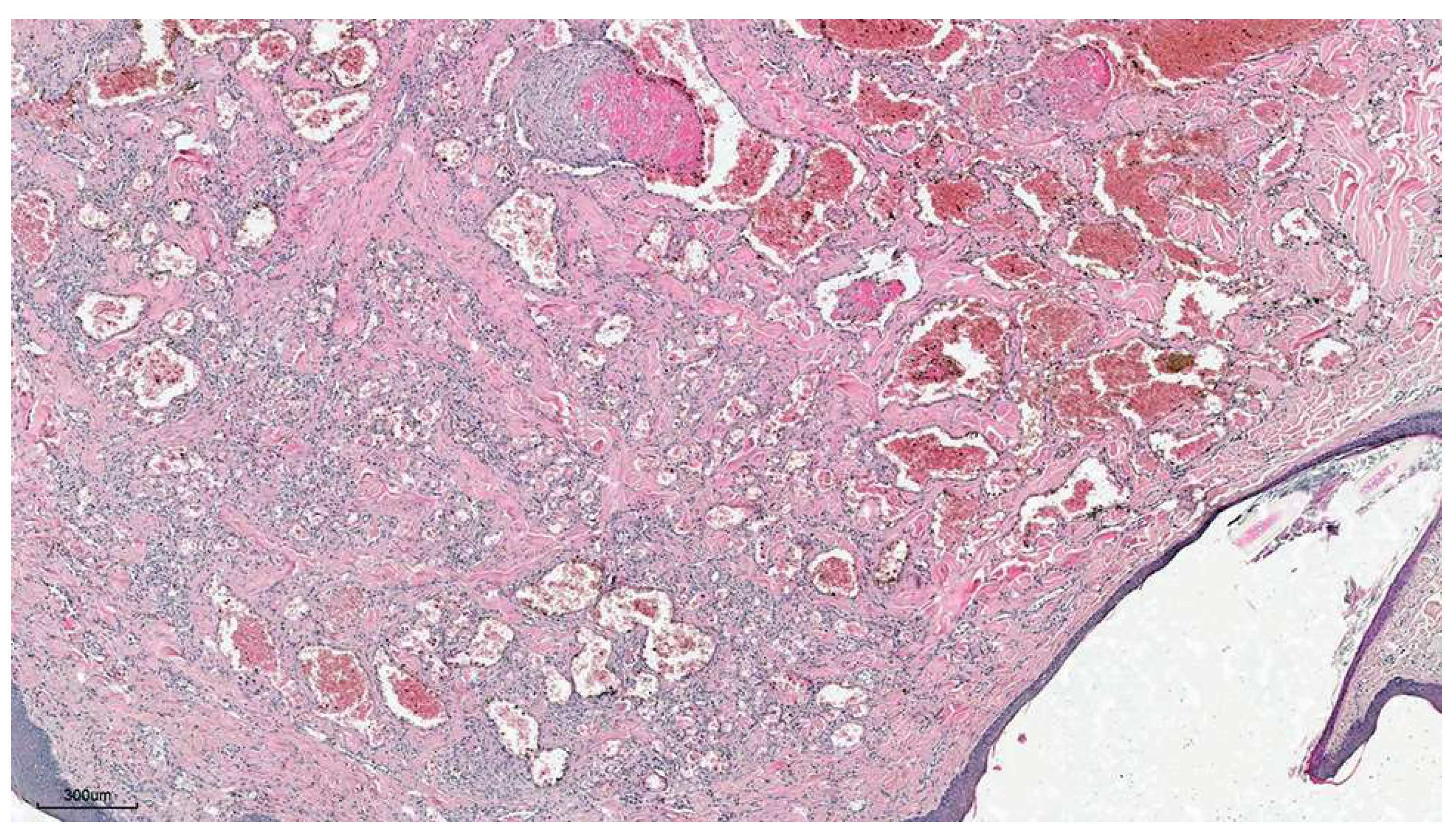
Figure 2.
Organized thrombi in mixed cutaneous hemangioma. H&E ×100.
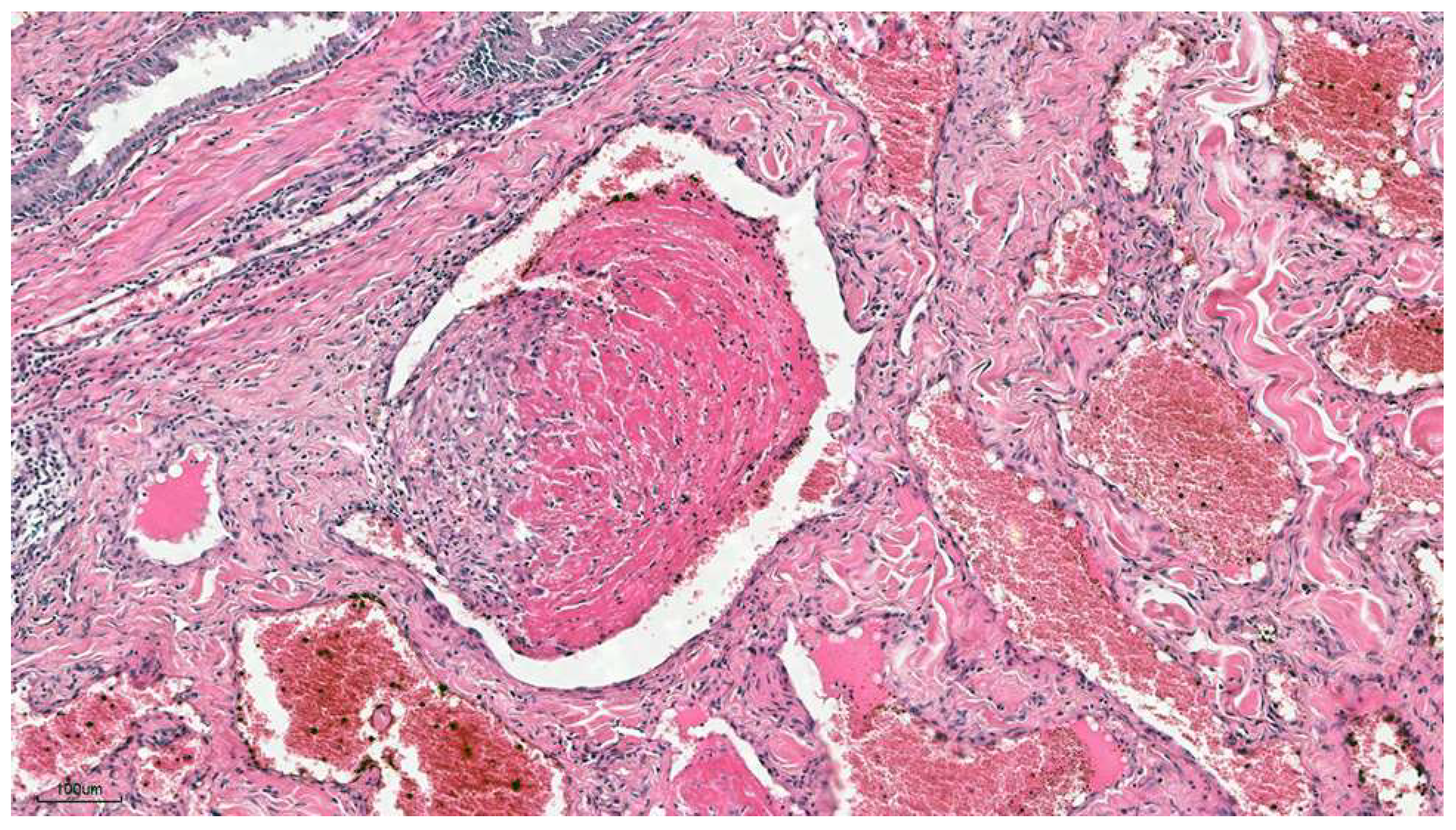
Figure 3.
Epidermal hemangioma, mixed type. H&E × 40. Upper inset: Strong and cytoplasmatic positive immunohistochemistry for factor VIII-related antigen × 400. Lower inset: Strong and membranous positive immunohistochemistry for CD31 × 400.
Figure 3.
Epidermal hemangioma, mixed type. H&E × 40. Upper inset: Strong and cytoplasmatic positive immunohistochemistry for factor VIII-related antigen × 400. Lower inset: Strong and membranous positive immunohistochemistry for CD31 × 400.
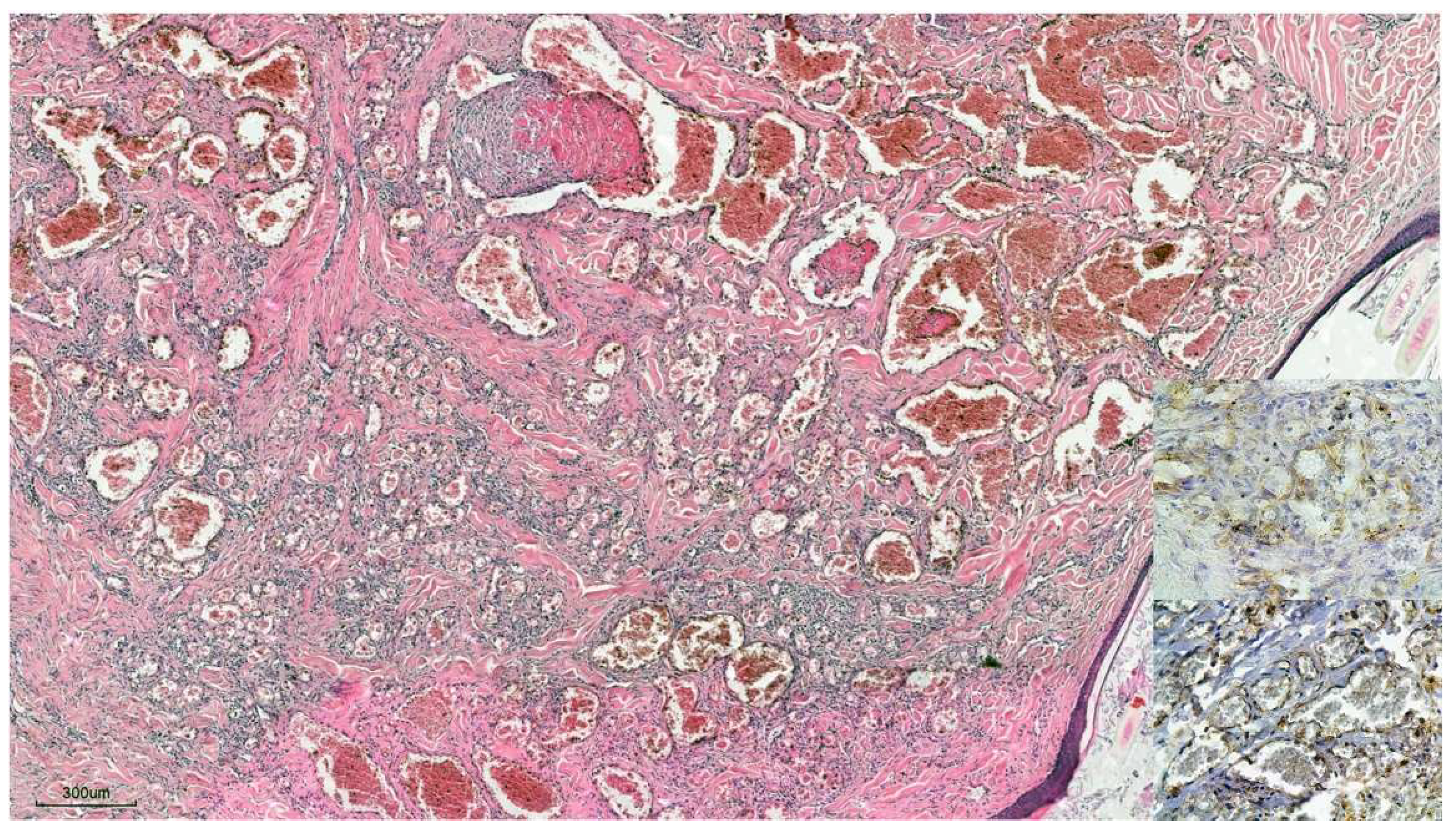
Figure 4.
Cutaneous hemangiosarcoma, mixed type composed of capillary at the left side and solid and epithelioid types at the right side of the same figure. H&E × 40. Upper inset: Strong and cytoplasmatic positive immunohistochemistry staining for factor VIII-related antigen × 400. Lower inset: Strong and membranous positive immunohistochemistry for CD31 × 400.
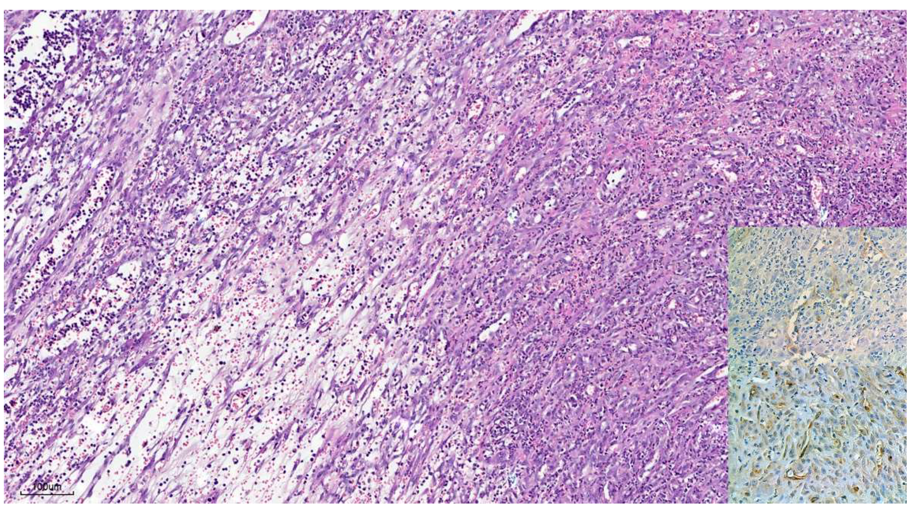

Table 1.
Clinical and histological data of hemangiomas.
| Items | Dogs (%) |
| Breeds (n = 25) | |
| Mixed | 11 (44) |
| German Shepherd | 3 (12) |
| Boxer | 2 (8) |
| Cocker Spaniel | 2 (8) |
| Sharpei | 2 (8) |
| Others | 5 (20) |
| Sex (n = 25) | |
| Male | 13 (52) |
| Female | 12 (48) |
| Localization (n = 25) | |
| Limbs | 10 (40) |
| Trunk | 9 (36) |
| Neck | 3 (12) |
| No information | 3 (12) |
| Histological occurrence site (n = 25) | |
| Dermis | 24 (86) |
| Subcutaneous tissue | 1 (4) |
| Histological grading (n = 25) | |
| Mixed capillary and cavernous types | 23 (92) |
| Capillary type | 1 (4) |
| Cavernous type | 1 (4) |
Table 2.
Clinical and histological data of hemangiosarcomas.
| Items | Dogs (%) |
| Breeds (n = 24) | |
| Mixed | 10 (42) |
| Pitbull Terrier | 3 (13) |
| Labrador Retriever | 2 (8) |
| Boxer | 2 (8) |
| Others | 7 (29) |
| Sex(n = 24) | |
| Male | 10 (42) |
| Female | 14 (58) |
| Localization (n = 24) | |
| Trunk | 12 (50) |
| Limbs | 8 (34) |
| Scrotum | 2 (8) |
| Lip | 1 (4) |
| No information | 1 (4) |
| Histological types (n = 24) | |
| Mixed capillary, cavernous, and solid types | 9 (39) |
| Mixed cavernous and solid types | 6 (25) |
| Mixed capillary, cavernous, solid, and epithelioid types | 2 (8) |
| Mixed solid and epithelioid types | 2 (8) |
| Mixed capillary, solid, and epithelioid types | 2 (8) |
| Mixed cavernous, solid, and epithelioid types | 1 (4) |
| Capillary type | 1 (4) |
| Epithelioid type | 1 (4) |
Disclaimer/Publisher’s Note: The statements, opinions and data contained in all publications are solely those of the individual author(s) and contributor(s) and not of MDPI and/or the editor(s). MDPI and/or the editor(s) disclaim responsibility for any injury to people or property resulting from any ideas, methods, instructions or products referred to in the content. |
© 2025 by the authors. Licensee MDPI, Basel, Switzerland. This article is an open access article distributed under the terms and conditions of the Creative Commons Attribution (CC BY) license (http://creativecommons.org/licenses/by/4.0/).
Copyright: This open access article is published under a Creative Commons CC BY 4.0 license, which permit the free download, distribution, and reuse, provided that the author and preprint are cited in any reuse.


