Submitted:
30 April 2025
Posted:
30 April 2025
You are already at the latest version
Abstract
The development of efficient scintillators emitting in the red and/or infrared spectral range represents an important scientific challenge, as such materials could find numerous practical applications. This work studies newly grown CsI:Yb,Sm and CsI:Eu,Sm single crystals demonstrating red and infrared luminescence. We measured luminescence spectra in the visible and near-IR range, excitation spectra across visible, UV, VUV, and X-ray ranges, Raman absorption spectra, and thermoluminescence spectra. The results show that divalent europium and ytterbium ions can efficiently transfer excitation to samarium ions. The measured X-ray light yield reached 37,000 photons/MeV for CsI:Yb,Sm and 40,000 photons/MeV for CsI:Eu,Sm.
Keywords:
cesium iodide
; scintillator
; europium
; ytterbium
; samarium
; luminescence
; single crystal
; crystal growth
1. Introduction
Red-emitting scintillators represent a crucial class of materials capable of converting ionizing radiation energy into red and near-infrared luminescence. These materials are of significant interest for radiation monitoring, medical imaging, and high-energy physics, as their spectral characteristics closely match the sensitivity range of semiconductor photodetectors. Ultra-compact yet highly sensitive scintillator-photodetector assemblies could be particularly valuable for real-time dosimetry monitoring during radiation therapy[1,2,3,4]. Traditional scintillator materials often rely on trivalent rare-earth ions as activators; however, divalent samarium () has emerged as a promising alternative for NIR emission[5,6]. exhibits characteristic 5d→4f transitions that result in strong emission in the red and NIR regions, making it suitable for applications requiring efficient light conversion at these wavelengths. Also, divalent samarium exhibits low self-absorption, which contributes to high light yield even at high activator concentrations[7]. However, the practical application of -activated scintillators can be limited by the need for efficient energy transfer mechanisms[8,9,10]. To address these limitation, sensitization strategies involving co-doping with other rare-earth ions such as ytterbium ()[6,11] or europium () have been explored[9,12]. and can act as efficient sensitizers, absorbing energy from the host lattice and transferring it to via energy transfer processes, thereby enhancing the overall scintillation efficiency. Our previous studies[13,14] of activated CsI single crystals have revealed that while samarium ions exhibit efficient red emission through 4f55d1→4f6 transitions, their practical application is limited by poor spectral overlap between the host exciton emission band and absorption. Therefore, the luminescence of exhibits low intensity under X-ray excitation. However, co-doping with suitable sensitizers can enhance energy transfer and increase X-ray-excited luminescence. The optimal impurities for this co-doping strategy are and , as the energy transfer processes involving these activators are relatively efficient[13,15]. This article focuses on the development and characterization of novel scintillator materials based on CsI crystals activated with divalent samarium () and sensitized with ytterbium () or europium (). We explore the luminescence and scintillation properties of these materials under visible, near ultraviolet, synchrotron and X-ray excitation.
2. Materials and Methods
Granulated CsI with a purity of 99.998% (with respect to metal impurities), produced by Lanhit, was used as the starting material for crystal growth. The activators consisted of and powders, as well as granulated , all with a purity of 99.99% (Lanhit). The initial mixture contained 150 g of CsI and 2.6 g of each activator (approximately 1 mol%). The materials were loaded into a vitreous carbon crucible inside the furnace and heated under vacuum at 500°C for six hours to remove adsorbed oxygen and moisture. By the end of the drying process, the residual vapor pressure in the furnace was below 0.01 Pa. The growth setup comprises a spherical water-cooled stainless steel chamber. A crystal pulling rod, thermostatically water-cooled and driven by two stepper motors, provides simultaneous rotation and vertical displacement of the growing crystal. A graphite adapter fitted with a quartz capillary is mounted at the rod ’s extremity (Figure 1). The furnace is equipped with a cylindrical resistive heater surrounded by pyrolytic graphite thermal insulation. Temperature regulation is achieved using a K-type thermocouple fixed to the crucible base. To suppress melt evaporation, both melting and crystal growth were conducted in an argon atmosphere maintained at 110 kPa.
Crystal growth was initiated by seeding onto a quartz capillary. To grow the colored crystal, the Kyropoulos method was employed, which enables higher incorporation of dopant ions into the crystal lattice compared to the Czochralski technique. The crystal was grown at a cooling rate of 0.3°C/h. Although the Kyropoulos method typically involves crystal pulling and rotation from the melt, neither the pulling motor nor rotation were utilized in this case to maximize the dopant ion concentration in the as-grown crystal. As a result, a single crystal approximately 35 mm in diameter and 8 mm thick was obtained. It grew as a layer on the remaining part of the boule, where the melt began to solidify non-uniformly, exhibiting off-stoichiometry and inclusions of dendritic structures with unclear composition. This approach enables higher dopant concentrations but limits the growth of large, homogeneous crystals. Nevertheless, samples of sufficient quality for spectroscopic studies can still be cut from the resulting crystals. For comparative analysis, the spectra of CsI:Eu, CsI:Sm, and CsI:Yb crystals will be presented. These crystals were grown under identical conditions with similar activator concentrations. The luminescence and excitation spectra were registered by a spectrometer based on SDL-1 and MDR-2 grating monochromators (LOMO, Saint-Petersburg, Russia) equipped with a grating of 1200 and 600 lines per mm. A Hamamatsu H6780-04 photomultiplier module was used as a photodetector. Excitation was carried out using a high-pressure 150W xenon arc lamp DKSH-150 through an MDR-2 monochromator with a diffraction grating of 1200 lines/mm. Additionally, 405 and 450 nm lasers were used for excitation. A closed-cycle helium cryostat was used for studies in the temperature range of 7-300 K. The X-ray luminescence spectra were measured using an MDR-2 monochromator coupled with a cooled FEU-83 photomultiplier tube (S-1 photocathode type), under excitation by a 10L-01 X-ray tube operated at 50 kV voltage and 1 mA current. The luminescence experiments under VUV excitations were carried out using synchrotron radiation from 1.5 GeV storage ring of MAX IV synchrotron facility (Lund, Sweden). The luminescence experiments under synchrotron radiation excitations are a powerful tool for the study of scintillators[16,17]. The experiments have been performed at the photoluminescence endstation of FinEstBeAMS beamline. The parameters of the beamline and the experimental setup are given in[18]. The luminescence spectra were processed using an ArDI web-application (https://ardi.fmm.ru).
Raman spectra were measured using a WITec alpha300R confocal Raman spectrometer coupled with a 532 nm Nd:YAG laser at room temperature, calibrated on crystalline silicon. The spectra were recorded with a diffraction grating of 1800 lines per millimeter and a spectral resolution of 3 . The laser beam had an output power of 10 mW, and the focal spot diameter sample was between 5 and 10m. The backscattered Raman signal was collected by using a Zeiss 50x/NA 0.55 objective in a UHTS300 spectrometer equipped with a Peltier-cooled, front-illuminated CCD camera. Spectral scan durations were 20 s, with signals averaging over 5 scans.
3. Results
The undoped crystal does not exhibit Raman scattering due to its NaCl-type crystal lattice. However, Raman signals were detected in CsI crystals doped with 1 mol.% of and , as well as in those doped with 1 mol.% of Sm2+ and 1 mol.% of Eu2+. The Raman spectra of these doped crystals are presented in Figure 2. The bands observed at 80, 94, 112, 136, and 164 cm−1 in the doped crystals can be attributed to (Ln2+I8)6-vc clusters[19].
The luminescence spectra of CsI:Yb,Sm shown in Figure 3 were measured at temperatures of 298 K and 7 K. At room temperature, a band corresponding to the 4f55d1→4f6 luminescence of divalent samarium[20] is observed in the red and infrared spectral regions, with a maximum intensity in the range of 1.4-1.5 eV (≈827-886 nm) under 405 nm laser excitation. In the long-wavelength region, the edge of the 4f55d1→4f6 emission band is truncated due to the declining sensitivity of the detector used. In the blue spectral region at room temperature, two bands (high-spin and low-spin) of 4f135d1→4f14 luminescence from divalent ytterbium ions are visible[21]. However, the emission intensity in these bands is two orders of magnitude lower than that of the divalent samarium band.
Upon cooling, the characteristic Yb2+ emission band nearly completely loses its intensity, while the 4f55d1→4f6 emission band narrows, revealing a shoulder at 1.55 eV (≈800 nm). Additionally, 4f6→4f6 luminescence bands characteristic of divalent samarium ions appear[22,23], with observed transitions 5D0→7F0, 5D0→7F1 and 5D0→7F2 showing maxima at 1.81 eV (≈685 nm), 1.77 eV (≈700 nm), and 1.71 eV (≈725 nm), respectively. When excited at 450 nm within the ytterbium emission band, the intensity of the 4f55d1→4f6 emission increases significantly relative to the 4f6→4f6 transitions.
The excitation spectrum (Figure 4) consists of 4fn5d1→4fn electronic transitions of Yb and Sm ions. In the region of 4f6→4f6 transitions of samarium, the main excitation bands show good agreement with the excitation bands of the reference CsI:Yb sample[13]. In the 4f55d1→4f6 emission band of samarium ions, the excitation spectrum exhibits some anticorrelation compared to the excitation spectrum of 4f6→4f6 luminescence, suggesting different types of samarium emission centers responsible for 4f6→4f6 and 4f55d1→4f6 luminescence.
The luminescence of the CsI:Eu,Sm sample (Figure 5) at room temperature consists of a broadened 4f55d1→4f6 Sm2+ emission band, compared to CsI:Yb,Sm. Both CsI:Yb,Sm and CsI:Eu,Sm exhibit nearly identical 4f6→4f6 luminescence band profiles, indicating that the Sm2+ centers responsible for this emission experience similar crystal field environments. The 4f65d1→4f7 Eu2+ emission band, peaking at 2.67 eV (≈464 nm)[24], also exhibits low intensity. Under 2.75 eV (450 nm laser) excitation, the samarium emission band (4f55d1→4f6) broadens and develops a vibrational structure, the energy spacing between these vibrational maxima falls within the 210-280 cm−1 range, that corresponds to doubled Raman frequency of divalent rare earth-iodine complexes (Figure 2). Upon cooling, four bands emerge in this spectral region, with maxima at 2.58 eV (≈481 nm), 2.66 eV (≈466 nm), 2.76 eV (≈449 nm), and 2.85 eV (≈435 nm).
The excitation spectrum (Figure 6) of CsI:Eu,Sm in the 4f6→4f6 luminescence band is completely identical to that of CsI:Yb,Sm in the 4f6→4f6 band. When exciting within the 4f55d1→4f6 luminescence band of Sm ions, a certain correlation with the excitation spectrum of Eu2+ ions is observed. From the luminescence spectra, it is evident that both Eu and Yb act as sensitizers for Sm2+ ions.
The excitation spectrum of CsI:Eu,Sm in the vacuum ultraviolet (VUV) region was measured in the 4f6→4f6and 4f55d1→4f6 emission bands at 7 K (Figure 7, curves (1) and (2)). A correlation is observed between the high-energy excitation spectra. In the 5-5.9 eV (≈248-210 nm) range, the 4f6→4f6 excitation spectrum exhibits two bands with maxima at 5.3 eV (≈234 nm) and 5.6 eV (≈221 nm), showing good correlation with the excitation spectrum of europium emission. In contrast, the 4f55d1→4f6 band displays a broad continuum without characteristic dip. Notably, these excitation bands are absent in the 4f55d1→4f6 excitation spectrum of CsI:Sm (Figure 7, curve 5). In the STE excitation region, the Eu2+ and Sm2+ crystals are not found intensive bands. However the similar to STE excitation bands structure could be observed (Figure 7, curves 2 and 3). In contrast, the only Sm2+ doped CsI crystal is not excited in the STE region (Figure 7, curve 5). The excitation spectrum in the 4f65d1→4f7 band of CsI:Eu (Figure 8) correlates with the STE excitation spectrum, indicating an efficient energy transfer mechanism from self-trapped excitons to Eu2+ ions.
For a more detailed study of the divalent europium luminescence band, we measured the luminescence spectra of CsI:Eu crystals at 7 K under excitation at 5.6 eV (≈221 nm), 5.7 eV (≈218 nm), 5.3 eV (≈234 nm), and 3.7 eV (≈335 nm) (Figure 9). The Gaussian fitting analysis of the emission band demonstrates that the spectrum consists of five overlapping components with maxima at 2.69 eV (≈461 nm), 2.73 eV (≈454 nm), 2.78 eV (≈446 nm), 3.03 eV (≈409 nm), and 3.17 eV (≈391 nm). The relative intensities of these bands show significant variation with increasing excitation energy. Furthermore, the spectrum contains an additional luminescence peak at 3.66 eV (≈339 nm) corresponding to self-trapped exciton emission.
To determine the light yield, we measured the X-ray luminescence spectra (Figure 10) of the synthesized samples and a commercial CsI:Tl scintillator crystal (light yield: 54,000 photons/MeV) used as a reference. All samples were prepared with identical geometric dimensions. Given the broad spectral range of the emission bands, the measurements employed a cooled photomultiplier tube with a silver-oxygen-cesium (S-1 type) photocathode operating in pulse-counting mode. The recorded spectra were corrected for the instrument’s spectral sensitivity. To verify that Eu and Yb ions act as effective sensitizers in scintillation process, we additionally measured the luminescence spectrum of a CsI:Sm crystal grown under the same conditions as the studied samples. The light yield and integrated emission intensity (expressed as a percentage relative to the reference) are summarized in Table 1. The measurement uncertainty of this method was 10%.
The thermoluminescence glow curves are given in Figure 11. The weak glow peaks at 125 and 140 K and intense glow peaks at 200, 255 and 275 K were observed in CsI:Yb, Sm crystals. In the crystals CsI: Eu, Sm the peaks at 205 and 290 K were observed.
4. Discussion
The most effective scintillators are materials in which energy transfer from intrinsic excitations to the activator ions occurs via excitons excitons[26,27,28,29]. In crystals activated by Yb2+, this process also takes place, making them quite promising as scintillators[13].
In CsI-Eu crystals, the exciton mechanism is similarly observed[30,31], with the formation of near-impurity excitons also noted in these crystals. In the emission spectrum of Eu2+ in CsI crystals, excitation near the edge of the forbidden band reveals several bands. The broad band around 2.75 eV corresponds to the emission of a near-impurity exciton localized near sodium[32]. This band is also observed in unactivated, nominally impurity-free crystals. In europium-activated crystals, four additional bands appear at 2.7 eV, 2.77 eV, 3.00 eV, and 3.17 eV. Their excitation spectra differ from one another. The bands at 2.7 eV and 2.77 eV exhibit excitation bands in the 3 to 5 eV range, associated with intracenter excitation of 4f-5d transitions of Eu2+[15,33]. Higher-energy bands are excited only within the energy range close to interband transitions of 5-5.6 eV. In the energy region where electron multiplications are observed, the luminescence bands at 2.7 eV and 2.77 eV are excited much more efficiently. Thus, these bands are associated with intracenter 5d-4f transitions in europium centers of different types is confirmed[30]. Such centers are most likely Eu2+ compensated by cation vacancies in nearest-neighbor (nn) and next-nearest-neighbor (nnn) positions. The nature of the higher-energy bands is likely associated with the decay of a near-impurity exciton near two types of Eu2+ centers. In this case, excitation transfer from these excitons to europium ions occurs, as indicated by an intense band in the 5-5.7 eV region observed in the excitation spectrum of 5d-4f europium luminescence (bands at 2.7 eV and 2.77 eV).
From the excitation spectra of crystals with double activation (Figs. Figure 6 and Figure 7), it is evident that excitation transfer from the divalent ions of the sensitizers Eu2+ or Yb2+ to Sm2+ occurs. In particular, in crystals activated by europium, bands appear in the 5d-4f excitation spectrum in the 5.3 and 5.6 eV regions, associated with the excitation of a near-impurity exciton near europium[15] and weak bands in the region associated with self-trapped excitons at 5.9-6.0 eV. A similar pattern is observed in CsI-Yb, Sm crystals.
The energy transfer process is associated with the overlap of the emission bands of Eu2+ (Figure 9) and Yb2+ [13] and the absorption characteristics of Sm2+ [14]. When excited in the 4f-5d bands of Yb2+ or Eu2+, intense luminescence related to Sm2+ is observed, causing the entire volume of the crystal to glow. The intensity of 5d-4f luminescence from Sm2+ increases with rising concentrations of divalent lanthanide ions, as decreasing distances between Sm2+ ions and sensitizer ions (Yb2+ or Sm2+) facilitate energy transfer. Thus, dipole-dipole nonradiative energy transfer from sensitizer ions Yb2+ or Eu2+ to Sm2+ ions presumably occurs.
In the excitation spectrum of 5d-4f luminescence, particularly within the region of interband transitions, coactivation of CsI-Sm2+ crystals with Yb2+ or Sm2+ ions reveals the occurrence of electron multiplications. This phenomenon suggests the establishment of an excitation transfer channel from the crystal lattice to Sm2+ ions, facilitated by the presence of Eu2+ and Yb2+ ions. Notably, a distinct band emerges in the 5d-4f excitation spectrum of Sm2+ luminescence, corresponding to a near-impurity exciton localized on either Eu2+ or Yb2+. The precise origin of these excitons remains uncertain; however, it may be attributed to the deep energy states of Eu2+ and Yb2+ within the bandgap of CsI crystals, which could provide stability to the impurity exciton. Additionally, it is plausible that the decay of cationic excitons occurs at these impurity sites, as the core levels of Eu2+ and Yb2+ are situated within the energy range conducive to the formation of cationic excitons[34,35,36].
The thermoluminescence (TSL) bands in CsI-Yb, Sm and CsI-Eu, Sm crystals are associated with VkA hole centers near the divalent lanthanide ions. Since these bands differ between crystals with various sensitizers, they likely result from the nonradiative decay of a near-impurity exciton close to Yb2+ and Eu2+ ions. The TSL peaks in CsI-Eu, Sm crystals occur below room temperature, which means these crystals do not exhibit significant afterglow, unlike CsI-Tl crystals. In contrast, CsI-Yb, Sm crystals show a slight afterglow; however, this is several orders of magnitude less than that observed in CsI-Tl. Similarly, in CsI-Tl crystals co-activated with divalent lanthanides ions (Eu2+, Yb2+ or Tm2+), the afterglow intensity diminishes [37,38,39,40]. The light yield of the double-activated crystals was assessed using X-ray luminescence spectra, revealing a notable enhancement compared to crystals activated solely by Sm2+. Specifically, CsI-Yb,Sm and particularly CsI-Eu,Sm crystals demonstrate great potential as scintillators emitting in the near-infrared region. Their light yield surpasses that of previously studied crystals[41] and may approach that of CsI-Tl.
5. Conclusions
For the first time, CsI crystals doubly activated with rare-earth ion pairs (Eu,Sm and Yb,Sm) have been successfully grown. Our studies demonstrate that divalent europium and ytterbium ions serve as efficient sensitizers for samarium ions (Sm2+) in CsI crystals, where Sm2+ exhibits a broad 5d-4f emission band in the red and near-infrared spectral region. The light yield of the obtained crystals under X-ray excitation was evaluated to be approximately 37,000 photons/MeV for CsI:Yb,Sm and 40,000 photons/MeV for CsI:Eu,Sm. Further optimization of crystal growth techniques could significantly enhance the light output, potentially leading to the development of cost-effective infrared scintillators. These findings open new possibilities for creating efficient scintillation materials with emission in the near-infrared range.
Author Contributions
Conceptualization, D.S. and R.S.; methodology, D.S., R.S., V.P. (Vladimir Pankratov), V.P. and A.M.; validation, D.S., E.K., and V.P. (Vladimir Pankratov); formal analysis, V.P. (Viktorija Pankratova); investigation, D.S., R.S., V.G., V.P. (Vladimir Pankratov), V.P. (Viktorija Pankratova), and E.K.; resources, D.S.; writing—original draft preparation, D.S. and R.S.; writing—review and editing, V.P. (Vladimir Pankratov); visualization, D.S. and R.S.; supervision, D.S.; project administration, D.S. and V.P. (Vladimir Pankratov); funding acquisition, D.S. All authors have read and agreed to the published version of the manuscript.
Funding
This work (crystal growth and spectroscopy) was funded by the Russian Science Foundation (project No. 23-72-01097).
Data Availability Statement
The data presented in this study are available on request from the corresponding author. The Raman absorption spectra are available in the ArDI (Advanced spectRa Deconvolution Instrument) database – a web application designed for searching, processing, and analyzing vibrational spectra (https://ardi.fmm.ru/).
Acknowledgments
The synchrotron studies of CsI:Eu were conducted within the framework of State Assignment No. 0350-2016-0024 "Crystalline and Amorphous Functional Materials with Predictable Properties". The crystal growth facility utilizes equipment from the Isotope-Geochemical Research Shared Equipment Center at the Vinogradov Institute of Geochemistry (IGC SB RAS). Raman spectra were measured in Center for Geodynamics and Geochronology of Institute of Earth Crust SB RAS. V.P. acknowledges LZP grant lzp-2023/1-0063.
Conflicts of Interest
The authors declare no conflicts of interest. The funders had no role in the design of the study; in the collection, analyses, or interpretation of data; in the writing of the manuscript; or in the decision to publish the results.
Abbreviations
The following abbreviations are used in this manuscript:
| NIR | Near-Infrared Light |
| VUV | Vacuum ultraviolet |
| UV | Ultraviolet |
| STE | Self-trapped exciton |
References
- Kim, J.; Park, J.; Park, B.; Kim, Y.; Park, B.; Park, S.H. Compact and Real-Time Radiation Dosimeter Using Silicon Photomultipliers for In Vivo Dosimetry in Radiation Therapy. Sensors 2025, 25, 857. [CrossRef]
- Strigari, L.; Marconi, R.; Solfaroli-Camillocci, E. Evolution of Portable Sensors for In-Vivo Dose and Time-Activity Curve Monitoring as Tools for Personalized Dosimetry in Molecular Radiotherapy. Sensors 2023, 23, 2599. [CrossRef]
- Kodama, S.; Kurosawa, S.; Morishita, Y.; Usami, H.; Torii, T.; Hayashi, M.; Sasano, M.; Azuma, T.; Tanaka, H.; Kochurikhin, V.; et al. Growth and Scintillation Properties of a New Red-Emitting Scintillator Rb2HfI6 for the Fiber-Reading Radiation Monitor. IEEE Transactions on Nuclear Science 2020, 67, 1055–1062. [CrossRef]
- Bartram, R.H.; Kappers, L.A.; Hamilton, D.S.; Brecher, C.; Ovechkina, E.E.; Miller, S.R.; Nagarkar, V.V. Multiple Thermoluminescence Glow Peaks and Afterglow Suppression in CsI:Tl Co-Doped with Eu2+ or Yb2+. IOP Conference Series: Materials Science and Engineering 2015, 80, 012003. [CrossRef]
- van Aarle, C.; Krämer, K.W.; Dorenbos, P. Lengthening of the Sm2+ 4f55d → 4f6 Decay Time through Interplay with the 4f6[5D0] Level and Its Analogy to Eu2+ and Pr3+. Journal of Luminescence 2024, 266, 120329. [CrossRef]
- Wen, X.; Kucerkova, R.; Babin, V.; Prusa, P.; Kotykova, M.; Nikl, M.; Li, W.; Wang, Q.; Kurosawa, S.; Wu, Y. Scintillator-Oriented near-Infrared Emitting Cs4SrI6:Yb2+, Sm2+ Single Crystals via Sensitization Strategy. Journal of the American Ceramic Society 2023, 106, 6762–6768. [CrossRef]
- Alekhin, M.S.; Awater, R.H.P.; Biner, D.A.; Krämer, K.W.; de Haas, J.T.M.; Dorenbos, P. Luminescence and Spectroscopic Properties of Sm2+ and Er3+ Doped SrI2. Journal of Luminescence 2015, 167, 347–351. [CrossRef]
- Dixie, L.C.; Edgar, A.; Bartle, M.C. Spectroscopic and Radioluminescence Properties of Two Bright X-ray Phosphors: Strontium Barium Chloride Doped with Eu2+ or Sm2+ Ions. Journal of Luminescence 2014, 149, 91–98. [CrossRef]
- Wolszczak, W.; Krämer, K.W.; Dorenbos, P. CsBa2I5:Eu2+,Sm2+—The First High-Energy Resolution Black Scintillator for γ-Ray Spectroscopy. physica status solidi (RRL) – Rapid Research Letters 2019, 13, 1900158. [CrossRef]
- Wolszczak, W.; Krämer, K.W.; Dorenbos, P. Engineering Near-Infrared Emitting Scintillators with Efficient Eu2+ → Sm2+ Energy Transfer. Journal of Luminescence 2020, 222, 117101. [CrossRef]
- van Aarle, C.; Krämer, K.W.; Dorenbos, P. The Role of Yb2+ as a Scintillation Sensitiser in the Near-Infrared Scintillator CsBa2I5:Sm2+. Journal of Luminescence 2021, 238, 118257. [CrossRef]
- Awater, R.H.P.; Alekhin, M.S.; Biner, D.A.; Krämer, K.W.; Dorenbos, P. Converting SrI2:Eu2+ into a near Infrared Scintillator by Sm2+ Co-Doping. Journal of Luminescence 2019, 212, 1–4. [CrossRef]
- Sofich, D.; Myasnikova, A.; Bogdanov, A.; Pankratova, V.; Pankratov, V.; Kaneva, E.; Shendrik, R. Crystal Growth and Spectroscopy of Yb2+-Doped CsI Single Crystal. Crystals 2024, 14, 500. [CrossRef]
- Sofich, D.O.; Bogdanov, A.I.; Shendrik, R.Y. Spectroscopic and Vibrational Properties of CsI Single Crystals Doped with Divalent Samarium. Optical Materials 2025, 162, 116958. [CrossRef]
- Gektin, A.; Shiran, N.; Belsky, A.; Vasyukov, S. Luminescence Properties of CsI:Eu Crystals. Optical Materials 2012, 34, 2017–2020. [CrossRef]
- Pankratova, V.; Kozlova, A.P.; Buzanov, O.A.; Chernenko, K.; Shendrik, R.; Šarakovskis, A.; Pankratov, V. Time-Resolved Luminescence and Excitation Spectroscopy of Co-Doped Gd3Ga3Al2O12 Scintillating Crystals. Scientific Reports 2020, 10, 20388. [CrossRef]
- Pankratov, V.; Kotlov, A. Luminescence Spectroscopy under Synchrotron Radiation: From SUPERLUMI to FINESTLUMI. Nuclear Instruments and Methods in Physics Research Section B: Beam Interactions with Materials and Atoms 2020, 474, 35–40. [CrossRef]
- Chernenko, K.; Kivimäki, A.; Pärna, R.; Wang, W.; Sankari, R.; Leandersson, M.; Tarawneh, H.; Pankratov, V.; Kook, M.; Kukk, E.; et al. Performance and Characterization of the FinEstBeAMS Beamline at the MAX IV Laboratory. Journal of Synchrotron Radiation 2021, 28, 1620–1630. [CrossRef]
- Rodriguez-Betancourtt, V.M.; Nattland, D. Raman Spectroscopic Study of Mixed Valence Neodymium and Cerium Chloride Solutions in Eutectic LiCl–KCl Melts. Physical Chemistry Chemical Physics 2005, 7, 173–179. [CrossRef]
- Radzhabov, E.A. Spectroscopy of Divalent Samarium in Alkaline-Earth Fluorides. Optical Materials 2018, 85, 127–132. [CrossRef]
- Suta, M.; Urland, W.; Daul, C.; Wickleder, C. Photoluminescence Properties of Yb2+ Ions Doped in the Perovskites CsCaX3 and CsSrX3 (X = Cl, Br, and I) – a Comparative Study. Physical Chemistry Chemical Physics 2016, 18, 13196–13208. [CrossRef]
- Zeng, P.; Cao, Z.; Chen, Y.; Yin, M. Investigation of the Temperature Characteristic in SrB4O7:Sm2+ Phosphor-in-Glass by Analyzing the Lifetime of 684 Nm. Journal of Rare Earths 2017, 35, 783–786. [CrossRef]
- Guzzi, M.; Baldini, G. Luminescence and Energy Levels of Sm2+ in Alkali Halides. Journal of Luminescence 1973, 6, 270–284. [CrossRef]
- Shiran, N.; Gektin, A.; Boyarintseva, Y.; Vasyukov, S.; Boyarintsev, A.; Pedash, V.; Tkachenko, S.; Zelenskaya, O.; Zosim, D. Modification of NaI Crystal Scintillation Properties by Eu-doping. Optical Materials 2010, 32, 1345–1348. [CrossRef]
- Shendrik, R.; Radzhabov, E. Absolute Light Yield Measurements on SrF2 and BaF2 Doped With Rare Earth Ions. IEEE Transactions on Nuclear Science 2014, 61, 406–410. [CrossRef]
- Ucer, K.B.; Bizarri, G.; Burger, A.; Gektin, A.; Trefilova, L.; Williams, R.T. Electron Thermalization and Trapping Rates in Pure and Doped Alkali and Alkaline-Earth Iodide Crystals Studied by Picosecond Optical Absorption. Physical Review B 2014, 89, 165112. [CrossRef]
- Shendrik, R.; Radzhabov, E. Energy Transfer Mechanism in Pr-Doped SrF2 Crystals. IEEE Transactions on Nuclear Science 2012, 59, 2089–2094. [CrossRef]
- Khanin, V.; Venevtsev, I.; Chernenko, K.; Pankratov, V.; Klementiev, K.; van Swieten, T.; van Bunningen, A.J.; Vrubel, I.; Shendrik, R.; Ronda, C.; et al. Exciton Interaction with Ce3+ and Ce4+ Ions in (LuGd)3(Ga,Al)5O12 Ceramics. Journal of Luminescence 2021, 237, 118150. [CrossRef]
- Li, P.; Gridin, S.; Ucer, K.B.; Williams, R.T.; Del Ben, M.; Canning, A.; Moretti, F.; Bourret, E. Picosecond Absorption Spectroscopy of Excited States in BaBrCl with and without Eu Dopant and Au Codopant. Physical Review Applied 2019, 12, 014035. [CrossRef]
- Gektin, A.; Shiran, N.; Vasyukov, S.; Belsky, A.; Sofronov, D. Europium Emission Centers in CsI:Eu Crystal. Optical Materials 2013, 35, 2613–2617. [CrossRef]
- Yakovlev, V.; Trefilova, L.; Karnaukhova, A.; Ovcharenko, N. Energy Transfer Mechanism in CsI:Eu Crystal. Journal of Luminescence 2014, 148, 274–276. [CrossRef]
- Hsu, O.L.; Bates, C.W. Excitonic Emission from CsI(Na). Physical Review B 1977, 15, 5821–5833. [CrossRef]
- Yakovlev, V.; Trefilova, L.; Meleshko, A.; Ovcharenko, N. Luminescence of Eu2+–Vc- Dipoles and Their Associates in CsI:Eu Crystals. Journal of Luminescence 2012, 132, 2476–2478. [CrossRef]
- Lushchik, Ch.; Lushchik, A. Evolution of Anion and Cation Excitons in Alkali Halide Crystals. Physics of the Solid State 2018, 60, 1487–1505. [CrossRef]
- Shalaev, A.A.; Shendrik, R.; Myasnikova, A.S.; Bogdanov, A.; Rusakov, A.; Vasilkovskyi, A. Luminescence of BaBrI and SrBrI Single Crystals Doped with Eu2+. Optical Materials 2018, 79, 84–89. [CrossRef]
- Lushchik, A.; Feldbach, E.; Kink, R.; Lushchik, Ch.; Kirm, M.; Martinson, I. Secondary Excitons in Alkali Halide Crystals. Physical Review B 1996, 53, 5379–5387. [CrossRef]
- Sisodiya, D.S.; Singh, S.G.; Chandrakumar, K.R.S.; Patra, G.D.; Ghosh, M.; Pitale, S.; Sen, S. Optimizing the Scintillation Kinetics of CsI Scintillator Single Crystals by Divalent Cation Doping: Insights from Electronic Structure Analysis and Luminescence Studies. The Journal of Physical Chemistry C 2024, 128, 197–209. [CrossRef]
- Brecher, C.; Lempicki, A.; Miller, S.R.; Glodo, J.; Ovechkina, E.E.; Gaysinskiy, V.; Nagarkar, V.V.; Bartram, R.H. Suppression of Afterglow in CsI:Tl by Codoping with Eu2+—I: Experimental. Nuclear Instruments and Methods in Physics Research Section A: Accelerators, Spectrometers, Detectors and Associated Equipment 2006, 558, 450–457. [CrossRef]
- Bartram, R.H.; Kappers, L.A.; Hamilton, D.S.; Lempicki, A.; Brecher, C.; Glodo, J.; Gaysinskiy, V.; Ovechkina, E.E. Suppression of Afterglow in CsI:Tl by Codoping with Eu2+—II: Theoretical Model. Nuclear Instruments and Methods in Physics Research Section A: Accelerators, Spectrometers, Detectors and Associated Equipment 2006, 558, 458–467. [CrossRef]
- Shahmaleki, S.; Rahmani, F. Scintillation Properties of CsI(Tl) Co-Doped with Tm2+. Radiation Physics and Engineering 2021, 2, 13–19. [CrossRef]
- Takase, S.; Miyazaki, K.; Nakauchi, D.; Kato, T.; Kawaguchi, N.; Yanagida, T. Development of Nd-doped CsI Single Crystal Scintillators Emitting near-Infrared Light. Journal of Luminescence 2024, 267, 120400. [CrossRef]
Figure 1.
Schematic of the crystal growth setup and photographs of samples prepared for spectroscopic studies.
Figure 1.
Schematic of the crystal growth setup and photographs of samples prepared for spectroscopic studies.
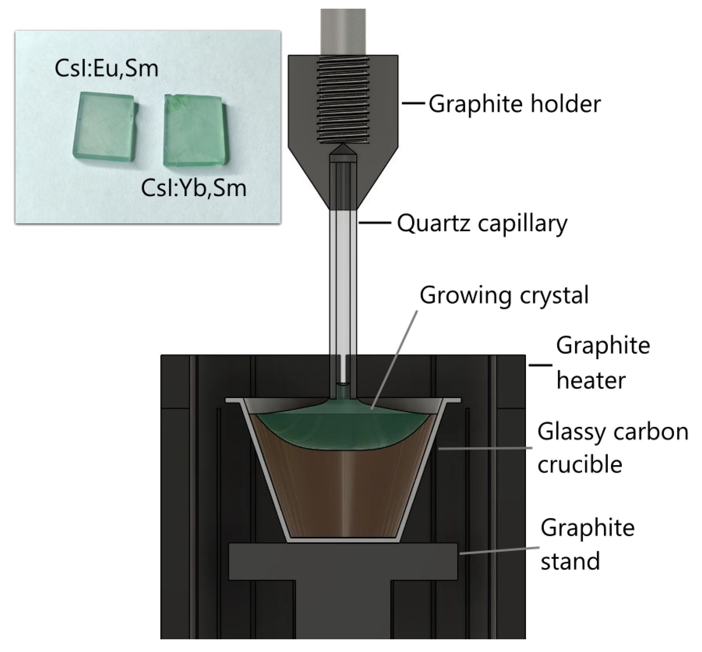
Figure 2.
Raman absorption spectra of CsI:Sm (1); CsI:Yb,Sm (2) and CsI:Eu,Sm (3).
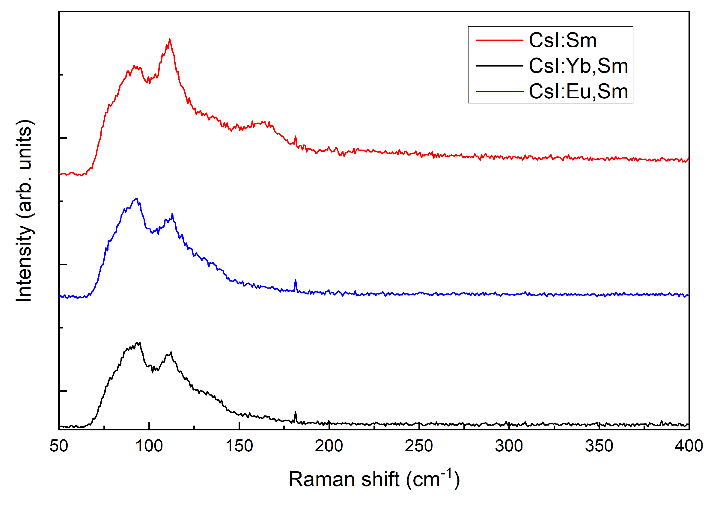
Figure 3.
Luminescence spectra of CsI:Yb,Sm measured at 298 K and 7 K.
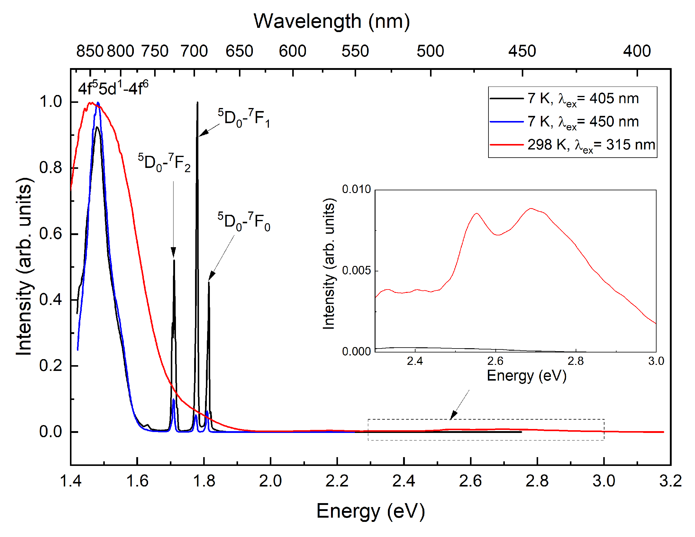
Figure 4.
Excitation spectra of CsI:Yb,Sm measured at 7 K. The dashed line represents the excitation spectrum of CsI:Yb crystal measured at 298 K.
Figure 4.
Excitation spectra of CsI:Yb,Sm measured at 7 K. The dashed line represents the excitation spectrum of CsI:Yb crystal measured at 298 K.
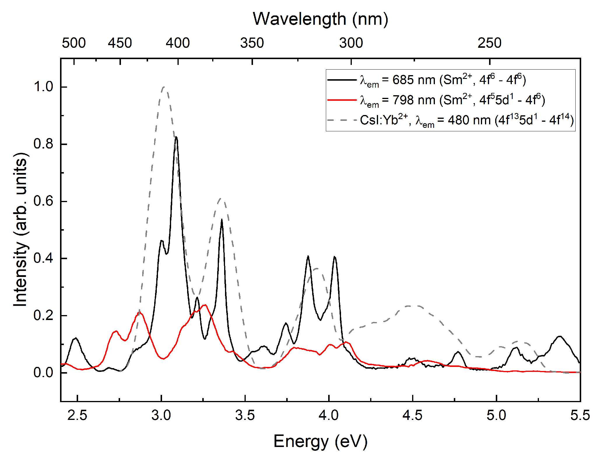
Figure 5.
Luminescence spectra of CsI:Eu,Sm measured at 298 K and 7 K.
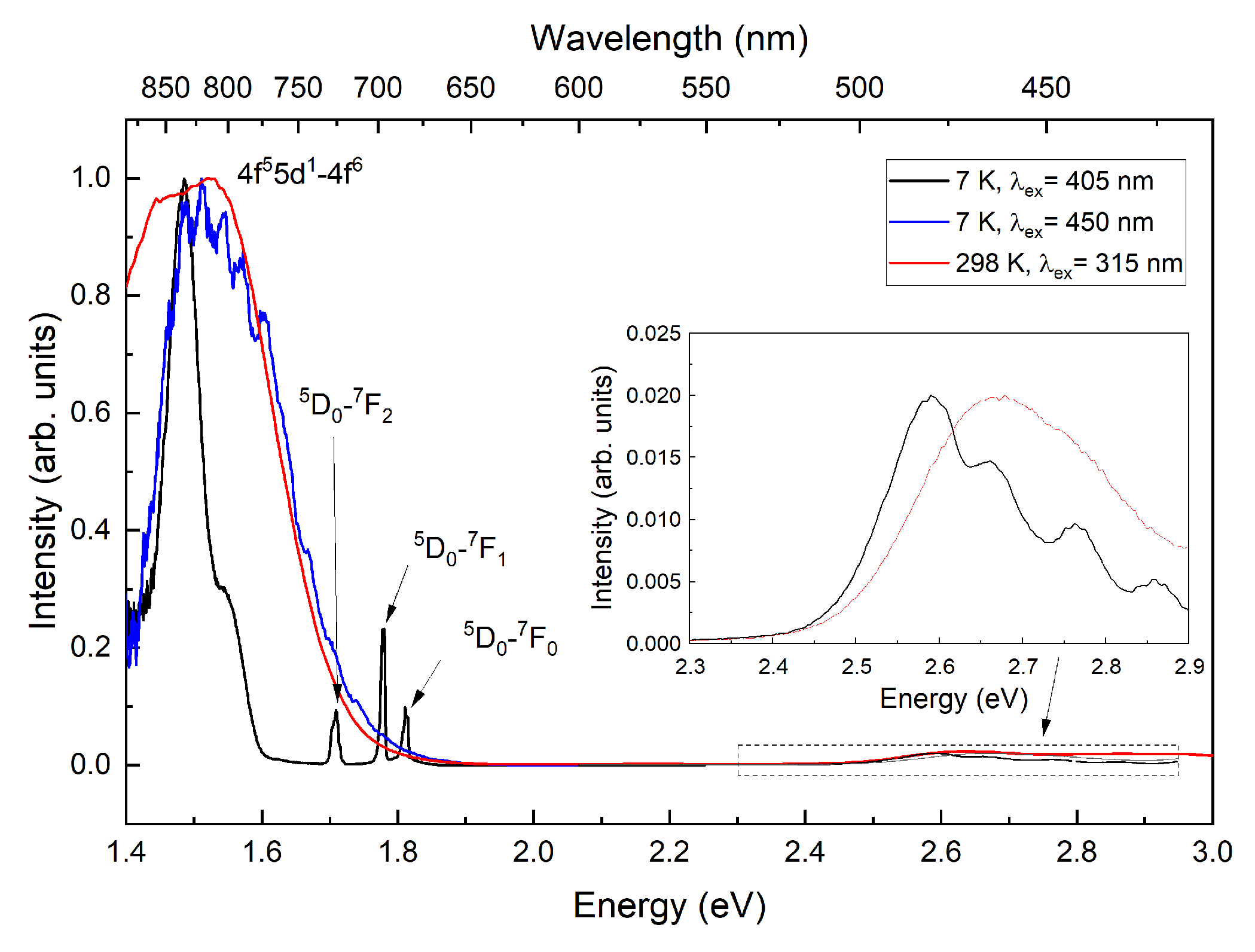
Figure 6.
Excitation spectra in the 4f6→4f6 luminescence bands at 7 K.
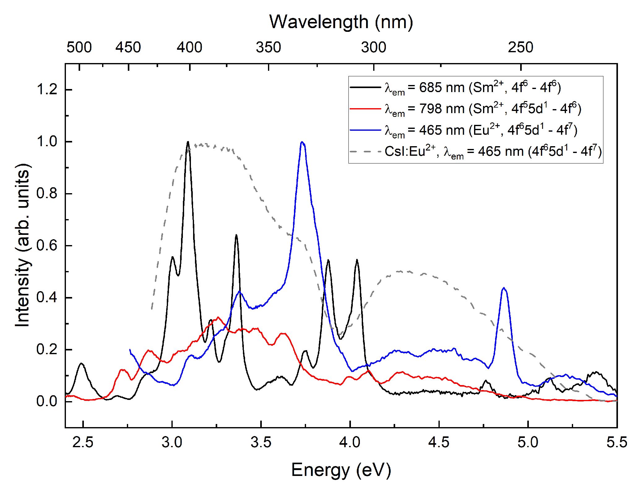
Figure 7.
Excitation spectra of CsI:Eu,Sm and CsI:Sm (dot line) samples in VUV range.
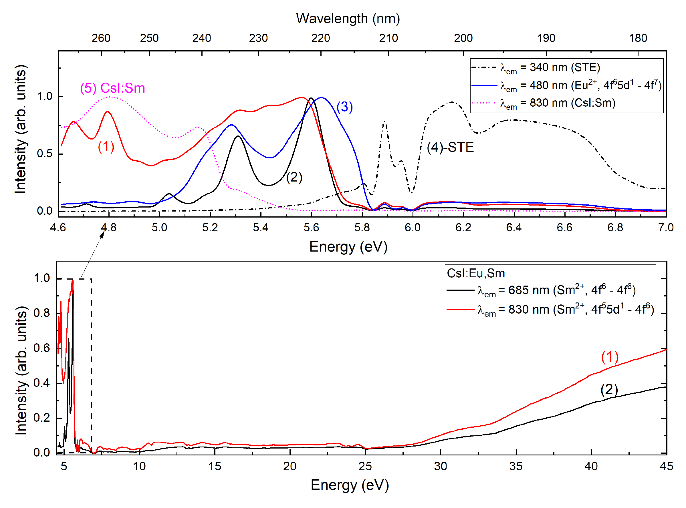
Figure 8.
Excitation spectra of CsI:Eu in VUV range.

Figure 9.
Luminescence spectra of CsI:Eu under VUV excitation at 7 K.
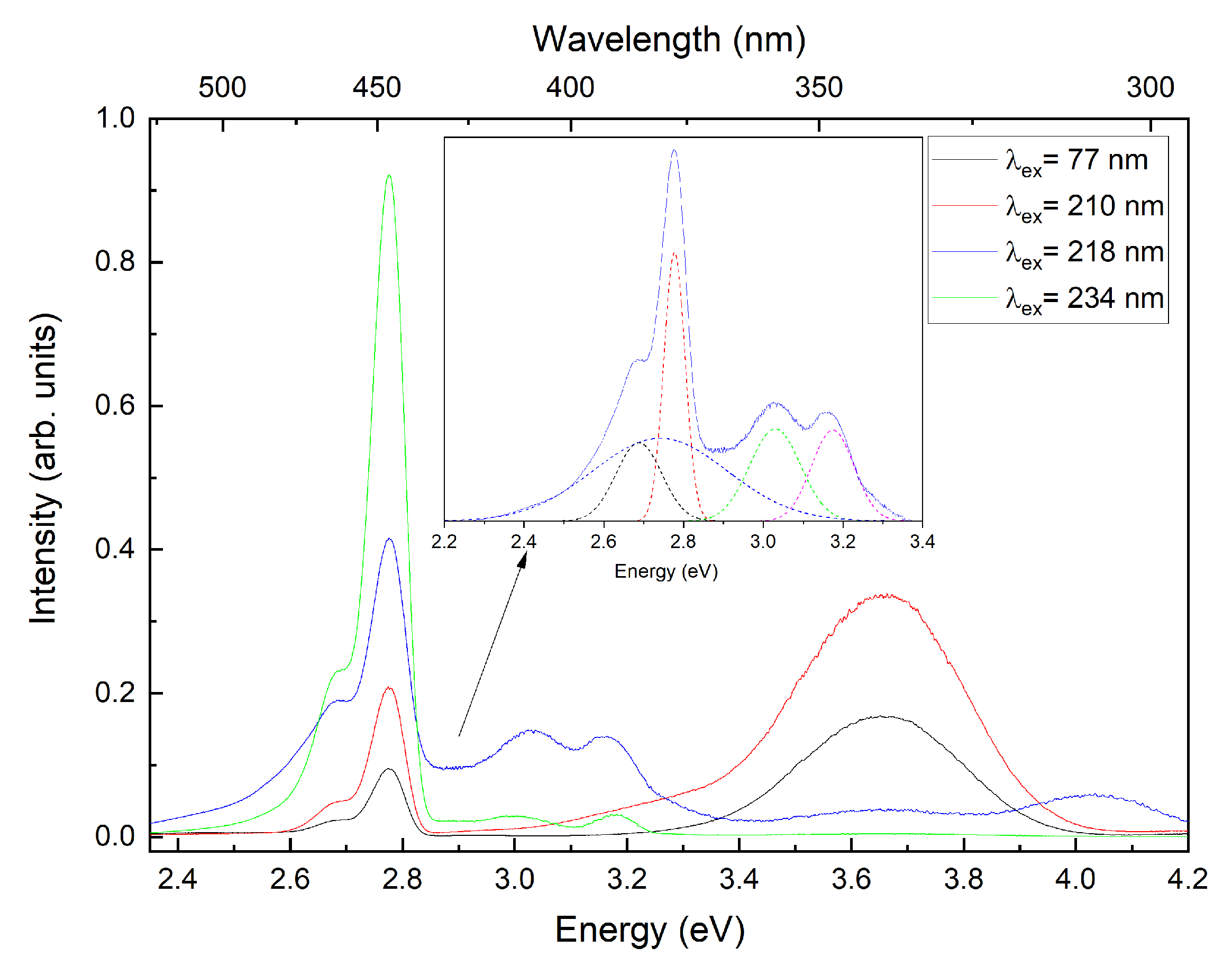
Figure 10.
Luminescence spectra of CsI:Tl, CsI:Yb,Sm; CsI:Eu,Sm; and CsI:Yb,Sm crystals under X-ray excitation.
Figure 10.
Luminescence spectra of CsI:Tl, CsI:Yb,Sm; CsI:Eu,Sm; and CsI:Yb,Sm crystals under X-ray excitation.
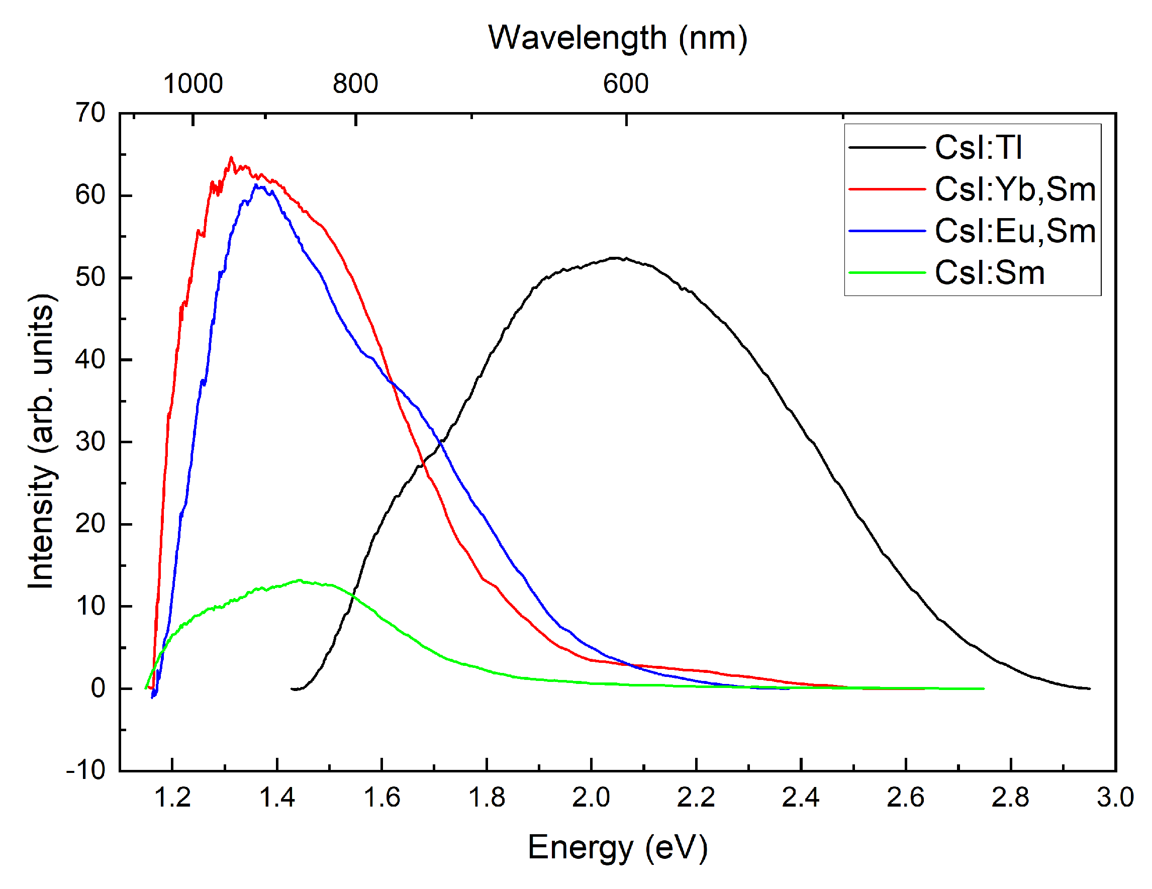
Figure 11.
Thermoluminescence spectra of CsI:Yb,Sm and CsI:Eu,Sm.
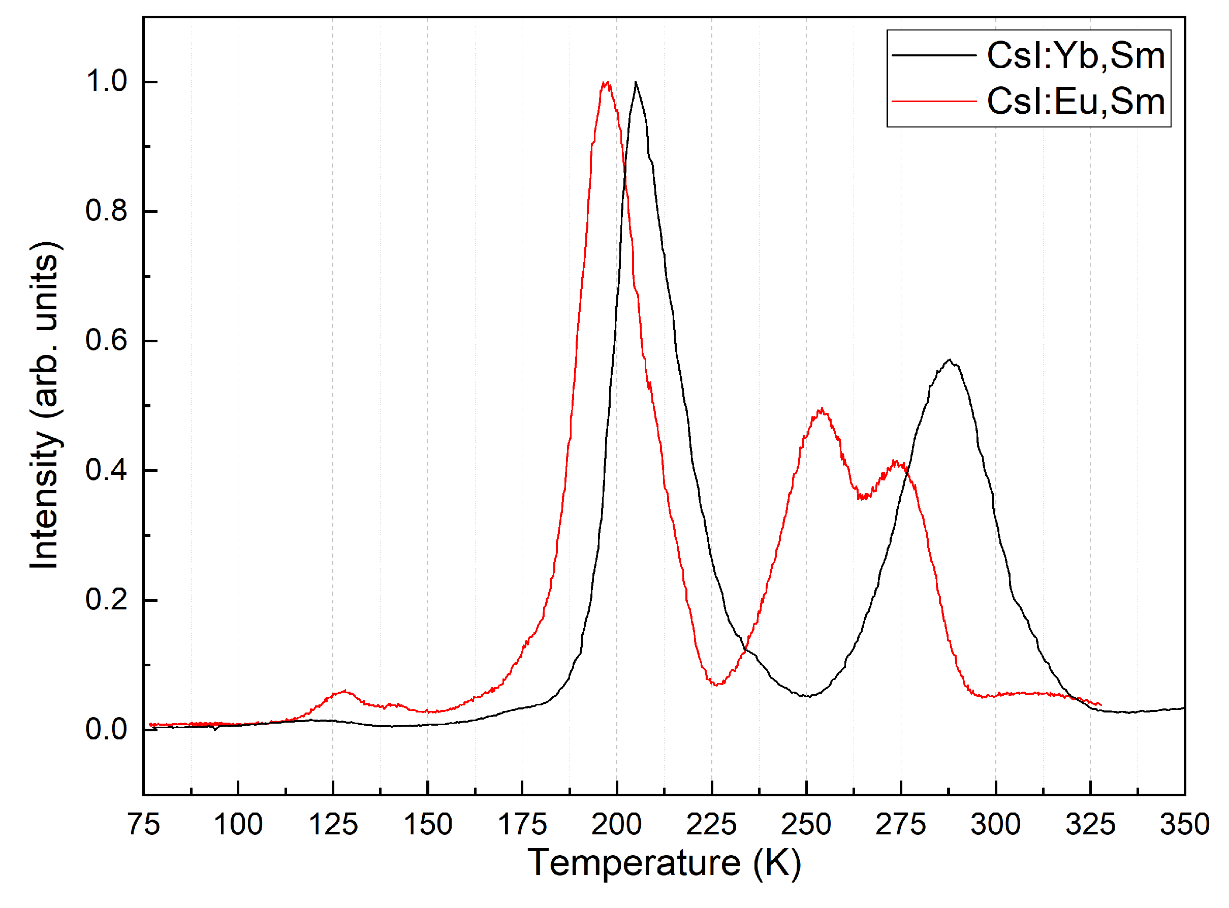

Table 1.
Light yield of samples under X-ray excitation.
| Sample | Light yield relative to standard (%) | Light yield (photons/MeV) |
|---|---|---|
| CsI:Tl | 100 | 54000[25] |
| CsI:Yb,Sm | 68 | 36720 |
| CsI:Eu,Sm | 74 | 39960 |
| CsI:Sm | 14 | 7560 |
Disclaimer/Publisher’s Note: The statements, opinions and data contained in all publications are solely those of the individual author(s) and contributor(s) and not of MDPI and/or the editor(s). MDPI and/or the editor(s) disclaim responsibility for any injury to people or property resulting from any ideas, methods, instructions or products referred to in the content. |
© 2025 by the authors. Licensee MDPI, Basel, Switzerland. This article is an open access article distributed under the terms and conditions of the Creative Commons Attribution (CC BY) license (http://creativecommons.org/licenses/by/4.0/).
Copyright: This open access article is published under a Creative Commons CC BY 4.0 license, which permit the free download, distribution, and reuse, provided that the author and preprint are cited in any reuse.


