
Submitted:
28 April 2025
Posted:
28 April 2025
You are already at the latest version
Abstract
Single-cell RNA sequencing (scRNA-seq) has revolutionized neuroscience by enabling the analysis of cellular heterogeneity and dynamic molecular processes at single-cell resolution. In spinal cord research, scRNA-seq provides critical insights into cell type diversity, developmental trajectories, and pathological mechanisms. This review summarizes recent progress in applying scRNA-seq to spinal cord development, injury, and neurodegenerative diseases, and discusses current challenges and future directions. Relevant studies were retrieved from PubMed and Web of Science, focusing on key applications of scRNA-seq, including advances in spatial transcriptomics and mul-ti-omics integration. scRNA-seq has enabled the identification of distinct spinal cord cell populations and revealed gene regulatory networks driving development. In injury models, it has uncovered the temporal dynamics of immune and glial responses, alongside potential regenerative processes. In neurodegenerative conditions, scRNA-seq has highlighted cell-specific vulnerabilities and molecular changes. Inte-gration with spatial transcriptomics and computational tools, such as machine learning, has further improved the resolution of spinal cord biology. However, challenges remain in data complexity, sample acquisition, and clinical translation. Single-cell tran-scriptomics is a powerful approach to understanding spinal cord biology. Its integration with emerging technologies will advance both basic research and clinical applications, supporting personalized and regenerative therapies. Addressing technical and analyt-ical barriers is essential to fully realize the potential of scRNA-seq in spinal cord science.
Keywords:
single-cell RNA sequencing
; spinal cord
; transcriptomics
; spinal cord injury
; neurodegeneration
; developmental biology
; spatial transcriptomics
; multi-omics integration
; cell atlas
; regenerative medicine
1. Introduction
The spinal cord is a fundamental component of the central nervous system, facilitating the transmission of sensory and motor signals between the brain and peripheral tissues. Additionally, the spinal cord coordinates reflex activities, enabling rapid responses independently of the brain. Its structure is intricately connected to its function: the outer white matter comprises myelinated ascending and descending nerve tracts, while the central gray matter contains neuronal cell bodies and local neural circuits [1].
The complexity of the spinal cord is evident not only from its diverse cellular composition—including neurons, glial cells, and other specialized cell types—but also from the precise functional specialization and intricate interactions among these cells [2]. Recent advances in single-cell transcriptomics have further unveiled the heterogeneity of neuronal and non-neuronal cell populations within the spinal cord, providing novel insights into their functions and underlying mechanisms in disease [3]. For example, motor neurons in the spinal cord have been found to consist of multiple subtypes, each characterized by distinct gene expression profiles and specialized functional roles [4].
Moreover, the response of glial cells following spinal cord injury has attracted considerable attention. Research has shown significant alterations in astrocytes, microglia, and oligodendrocytes post-injury, critically influencing neuronal survival and regeneration [5,6,7]. These discoveries underscore the importance of thoroughly exploring spinal cord cellular composition and function, contributing to our understanding of its role in both health and disease and opening new avenues for therapeutic intervention.
In recent years, the emergence of single-cell transcriptomics and spatial transcriptomics has marked a significant breakthrough in biological research, providing unprecedented insights into cellular functions and tissue architecture. The groundbreaking development of single-cell transcriptomics dates back to 2009 when Tang et al. first successfully sequenced the transcriptome of individual mouse embryonic cells, overcoming the challenge posed by low RNA content in single cells [8]. Subsequently, advancements in microfluidics and laboratory automation have dramatically scaled up the capacity of single-cell transcriptomic analyses, facilitating the commercialization of platforms capable of processing thousands to tens of thousands of cells per experiment [9]. The rapid evolution of single-cell RNA sequencing (scRNA-seq) has thus emerged as a powerful tool for unraveling cellular heterogeneity within spinal cord tissues.
Spatial transcriptomics has further advanced this field by integrating gene expression profiles with spatial tissue contexts. Its origins are traced back to the application of in situ hybridization by Walter Gehring's group in 1986, which localized antennapedia mRNA in Drosophila embryos and imaginal discs [10]. The term "spatial transcriptomics" was creatively coined by Ståhl et al. in 2016, describing a novel method that captures mRNA molecules directly from tissue sections on glass slides, enabling high-resolution mapping of gene expression integrated with tissue morphology [11]. While single-cell transcriptomics provides genome-wide expression profiles at single-cell resolution, it lacks contextual tissue information—a gap effectively addressed by spatial transcriptomics, which elucidates intercellular interactions and tissue heterogeneity [9,12]. Nonetheless, spatial transcriptomics still faces challenges, particularly regarding resolution and the detection of low-abundance transcripts [12].
The integration of single-cell and spatial transcriptomics is driving advances in precision medicine, offering robust tools for disease research, drug development, and regenerative medicine [13]. By sequencing transcripts at the single-cell level, researchers can identify distinct cellular subpopulations and clarify their specific roles in spinal cord development, homeostasis, and injury repair. For instance, Delile et al. (2019) employed scRNA-seq to profile the developing mouse spinal cord, uncovering spatial and temporal gene-expression dynamics essential for neural progenitor differentiation and neuronal maturation, significantly enriching our understanding of spinal cord developmental processes [14]. Moreover, Blum et al. (2021) utilized single-nucleus transcriptomics to reveal substantial heterogeneity among motor neurons in adult mouse spinal cords, identifying subtype-specific transcriptional signatures closely associated with motor functions and disease mechanisms [4]. Recently, Zhang et al. (2024) combined spatial transcriptomics with single-nucleus RNA sequencing to create a detailed spatial atlas of adult human spinal cord cells, identifying and localizing 21 neuronal subclusters. This comprehensive mapping linked molecular identities directly to anatomical structures, greatly expanding knowledge regarding cellular composition in the human spinal cord [15]. Collectively, advances in single-cell and spatial transcriptomics offer unparalleled opportunities to elucidate the complex cellular architecture of the spinal cord and open innovative pathways for precise therapeutic interventions in spinal cord injuries and associated diseases.
With the widespread application of single-cell and spatial transcriptomics in spinal cord research, a growing body of studies has uncovered the cellular heterogeneity and dynamic changes that occur in the spinal cord across development, physiological function, and pathological conditions [16,17]. Despite these advances, a comprehensive review that systematically integrates these findings and explores their implications for neuroscience and clinical translation is still lacking. This review aims to fill this gap by providing a thorough overview of recent progress in the application of single-cell and spatial transcriptomic technologies in spinal cord research. By synthesizing current findings, we seek to offer a conceptual framework that deepens the understanding of complex cellular behaviors within the spinal cord and guides future developments in related fields.
The review is structured to cover several key aspects, including technological advancements, the construction of comprehensive cellular atlases, the analysis of microenvironmental dynamics, and the challenges associated with translational medicine. By tracing the evolution of these technologies and highlighting representative clinical applications, we aim to bridge basic and translational research, offering insights into both the fundamental biology of the spinal cord and its relevance in regenerative medicine and therapeutic development for neurological disorders. Ultimately, this review aspires to serve as a valuable resource for neuroscientists and clinicians alike, providing a systematic reference that promotes a deeper understanding of spinal cord complexity and informs future research directions and treatment strategies.
2. Single-Cell Transcriptomics: Technologies and Methodologies
2.1. Principles and Evolution of Single-Cell RNA Sequencing Technologies
Single-cell RNA sequencing (scRNA-seq) is a powerful technique that enables transcriptomic analysis at the resolution of individual cells, allowing for the exploration of cellular heterogeneity and complex gene expression patterns. The core workflow of scRNA-seq typically involves the isolation of single cells, cell lysis, reverse transcription of mRNA into cDNA, library construction, and subsequent high-throughput sequencing. These steps allow researchers to capture a comprehensive transcriptional profile from individual cells [18].
Since its initial introduction by Tang et al. in 2009 [8], scRNA-seq has undergone rapid technological advancement. In 2012, the development of SMART-seq significantly improved full-length cDNA amplification strategies, resulting in enhanced transcript coverage [19]. One of the most notable breakthroughs in scRNA-seq has been the exponential increase in throughput. While early methods such as STRT-seq could only process hundreds of cells [20], the advent of droplet-based microfluidics in 2015—exemplified by Drop-seq and the 10x Genomics Chromium system—enabled the profiling of tens of thousands to millions of cells in a single experiment [21,22,23]. This high-throughput capacity has made it feasible to investigate cellular heterogeneity in complex tissues such as the spinal cord.
Moreover, these technical advancements have not only increased scalability but also reduced the cost per cell, facilitating the broad application of scRNA-seq across various disciplines, including developmental biology, oncology, and neuroscience. In summary, the development of scRNA-seq has provided a transformative tool for dissecting cellular functions and tissue architecture, especially in elucidating the diverse cellular landscape of complex systems such as the spinal cord.
2.2. Comparison of Major scRNA-seq Platforms and Their Features
The selection of single-cell RNA sequencing (scRNA-seq) technologies should be guided by specific research objectives and experimental conditions, as different platforms vary significantly in throughput, sensitivity, and cost. Among the currently available scRNA-seq platforms, the 10x Genomics Chromium system is one of the most widely adopted high-throughput technologies. Based on droplet microfluidics, it encapsulates thousands of individual cells into nanoliter droplets containing unique barcodes, enabling efficient and parallel transcriptomic profiling [22]. Its high throughput allows the detection of rare cell populations, and its relatively low cost and scalability make it particularly suitable for complex experimental designs involving multiple conditions or time points [24].
In contrast, SMART-seq2 and SMART-seq3 are well-established plate-based scRNA-seq methods that enable full-length mRNA amplification and sequencing. These technologies are especially advantageous for detecting low-abundance transcripts and transcript isoforms [25,26]. SMART-seq3 further improves sensitivity over its predecessor and supports quantitative transcript detection at the single-molecule level [26]. However, these methods are generally limited by lower throughput, longer processing times, and higher per-cell costs, making them more suitable for studies focusing on rare cell populations or functional subtypes.
Drop-seq and inDrops, both droplet-based microfluidic approaches, offer cost-effective and scalable solutions for high-throughput scRNA-seq. Drop-seq, developed by Macosko et al., co-encapsulates single cells with barcoded beads in droplets, providing a robust method with high efficiency and relatively low cost [21]. inDrops introduces improvements in barcode design and reaction chemistry, enhancing cell capture rates and data consistency, while also exhibiting remarkably low background noise [27]. Furthermore, its flexible architecture facilitates adaptation to other sequencing-based assays [27]. While both methods maintain high throughput, their sensitivity is generally lower compared to SMART-seq-based techniques [28].
SPLiT-seq represents a unique combinatorial barcoding approach that eliminates the need for physical cell isolation. By sequentially tagging fixed cells through multiple rounds of barcode delivery, SPLiT-seq significantly reduces reliance on specialized instrumentation and greatly increases throughput [16]. Using this method, Rosenberg and colleagues mapped the spatiotemporal dynamics of 30 neuronal subtypes in the developing mouse spinal cord, demonstrating the utility of SPLiT-seq for comprehensive transcriptomic profiling in complex multicellular systems [16].
In recent years, spatial transcriptomics technologies such as 10x Genomics Visium and BGI's Stereo-seq have enabled in situ mapping of gene expression across complex tissues like the spinal cord. For instance, Zhang et al. utilized single-nucleus RNA sequencing in combination with Visium spatial transcriptomics to delineate the spatial distribution of spinal cord neurons across nine human donors [15]. Stereo-seq further advances the field by improving spatial resolution and expanding the capture area, offering the potential to generate spatial gene expression maps at subcellular resolution [29].
2.3. Technology Selection Strategies and Application Cases in Spinal Cord Research
In spinal cord research, the selection of an appropriate single-cell RNA sequencing (scRNA-seq) technology must be carefully tailored to the specific research objectives and the nature of the biological samples. For studies aiming to identify novel transcript isoforms or rare cell populations, SMART-seq is often preferred due to its high sensitivity and ability to capture full-length transcripts. In contrast, high-throughput platforms such as 10x Genomics Chromium or Drop-seq are more suitable for constructing large-scale single-cell atlases of the spinal cord.
In the context of spinal cord development, Delile et al. employed the 10x Genomics Chromium platform to perform scRNA-seq on cervical and thoracic regions of the embryonic mouse spinal cord, covering multiple developmental stages from embryonic day 9.5 to 13.5. This work generated a high-resolution transcriptional atlas, revealing both spatial and temporal dynamics of neural progenitors and neuronal populations. Moreover, the study identified novel marker genes and regulatory sequences involved in neuronal differentiation, providing a valuable resource for elucidating the cellular diversity and molecular mechanisms underlying spinal cord development [14].
To construct a comprehensive cellular map of the human spinal cord, Andersen et al. applied the 10x Genomics Chromium system to systematically profile human fetal spinal cords. Their study uncovered substantial cellular heterogeneity and regional specialization during development. By integrating data across multiple developmental time points, the authors tracked temporal changes in cell types and mapped disease-associated genes to specific cellular populations, offering novel insights into the cellular architecture of motor control and the pathogenesis of neurological disorders [30]. This work represents a critical step toward building a detailed single-cell atlas of human spinal cord development.
In studies of spinal cord injury (SCI), researchers have utilized single-nucleus RNA sequencing (snRNA-seq) to profile transcriptional changes in the injured spinal cord at high resolution. For example, Matson et al. used the 10x Genomics Chromium platform to generate a cellular map of the lumbar spinal cord following injury in a mouse model. Their findings revealed injury-induced alterations in distinct neuronal subpopulations and highlighted specific neuronal subsets with regenerative potential [31]. These results provide a theoretical framework for future therapeutic strategies targeting defined neuronal groups to promote spinal cord repair.
In summary, the 10x Genomics Chromium system has emerged as the predominant platform used in most spinal cord studies due to its balance of throughput and resolution. Thoughtful selection and application of scRNA-seq technologies, aligned with the specific aims and sample characteristics of a study, are essential for advancing our understanding of spinal cord development, function, and regeneration.
3. Applications of Single-Cell Transcriptomics in Spinal Cord Research
Single-cell transcriptomics has become a powerful tool for investigating spinal cord biology, with applications spanning developmental processes, injury responses, and neurodegenerative diseases. Additionally, the integration of spatial transcriptomics and multi-omics approaches offers deeper insights into the complex molecular landscape of the spinal cord. Figure 1 summarizes the major research areas where scRNA-seq technologies have been applied in spinal cord studies.
3.1. Decoding Spinal Cord Developmental Biology
Single-cell transcriptomic technologies have emerged as a pivotal approach for deciphering cellular diversity and lineage specification during spinal cord development. By generating cell type-specific gene expression maps, researchers can precisely track processes such as neural tube formation, fate specification, and circuit assembly. This section highlights key discoveries enabled by single-cell approaches, with a focus on temporal regulation of neurogenesis, mechanisms underlying cell type diversification, and the dissection of critical signaling pathways at single-cell resolution (Table 1).
Delile et al. employed the 10x Genomics platform to perform scRNA-seq on cervical and thoracic spinal cord regions from embryonic day (E) 9.5 to 13.5 in mice, generating a high-resolution developmental atlas that revealed spatial and temporal dynamics of neural progenitor and neuronal populations [14]. The study identified novel marker genes expressed in distinct dorsoventral domains of progenitors and neurons, providing insights into transcriptional programs guiding cell fate decisions during spinal cord development [14].
In human spinal cord development, Andersen et al. conducted scRNA-seq analysis on mid-gestation (~22 weeks) fetal samples and uncovered pronounced heterogeneity both between and within major cell types [30]. For example, glial cells showed spatially dependent transcriptional programs along the dorsoventral and rostrocaudal axes, and astrocytes displayed differential gene expression based on their localization in white versus gray matter. Moreover, the study integrated multiple existing datasets covering the same developmental window to trace temporal changes in cellular composition, and mapped disease-related genes to specific cell types, offering novel insights into the cellular basis of motor control and opportunities for stem cell-based disease modeling [30].
Significant progress has also been made in resolving motor neuron diversity at the single-cell level. Sathyamurthy et al. utilized massively parallel single-nucleus RNA sequencing (snRNA-seq) to profile spinal cord neurons in adult mice and identified 43 transcriptionally distinct neuronal subtypes with region-specific distributions [17]. Their work established a comprehensive molecular map of spinal neurons and linked transcriptional identities to anatomical and functional domains.
Building on these findings, Blum et al. focused specifically on adult spinal motor neurons and used scRNA-seq to investigate transcriptional heterogeneity within this population [4]. Despite similar morphological and positional features, individual motor neurons exhibited distinct transcriptional programs related to electrophysiological properties, spatial organization, and axonal targeting [4]. This study provided a detailed molecular atlas of motor neurons, laying the groundwork for future investigations into motor system function and dysfunction.
In a broader developmental context, Cao et al. performed large-scale scRNA-seq during mouse organogenesis and constructed a multi-organ cell atlas, identifying developmental trajectories across multiple germ layers [32]. Their analysis showed that spinal cord progenitors undergo clear transcriptional transitions over time, driven by key regulatory factors such as Hox genes and the Sonic Hedgehog (Shh) signaling pathway [32]. This work offered a valuable resource for studying neural development and the emergence of cellular heterogeneity in the spinal cord.
Advances in computational methods have further enhanced our ability to analyze developmental dynamics. The reversed graph embedding (RGE) algorithm proposed by Qiu et al. enables the reconstruction of pseudotemporal trajectories from scRNA-seq data without time labels [33]. Implemented in Monocle 2, this method has been widely used to model nonlinear developmental progressions in complex systems, including the differentiation of spinal cord progenitors into diverse neuronal lineages.
Recently, Zhang et al. combined snRNA-seq with spatial transcriptomics to profile spinal cord neurons from nine human donors, identifying 21 neuronal subtypes and defining their spatial distributions [15]. Comparative analysis between human and mouse spinal cords revealed broad similarities in neuronal composition, along with species-specific transcriptional signatures. Notably, sex-specific expression differences were also observed in motor neurons, including genes such as SCN10A and HCN1 [15].
Collectively, these studies illustrate the power of single-cell and spatial transcriptomics to resolve the cellular diversity and complex regulatory networks underpinning spinal cord development. They provide foundational insights into neurodevelopmental biology and inform our understanding of the molecular basis of spinal disorders.
3.2. Investigating Spinal Cord Injury and Neurodegeneration
scRNA-seq has provided unprecedented resolution for dissecting cellular heterogeneity and molecular mechanisms underlying spinal cord injury (SCI) and neurodegenerative diseases. By enabling high-throughput profiling of cell populations at various time points and pathological states, researchers can identify critical cellular subtypes, signaling pathways, and transcription factors, thereby advancing the understanding of disease mechanisms and uncovering potential therapeutic targets.
SCI is a severe central nervous system disorder characterized by disruption of spinal cord structure and function, leading to impairments in motor, sensory, and autonomic functions [34]. The application of single-cell transcriptomic technologies offers unique opportunities to explore cellular responses following SCI at single-cell resolution, allowing for the characterization of cellular composition, state transitions, and gene expression dynamics in injured spinal tissue [35]. Following SCI, the spinal cord undergoes complex cellular and molecular alterations, with glial cell responses particularly well-delineated by scRNA-seq studies.
Research has demonstrated that microglia and astrocytes exhibit pronounced dynamic changes post-injury, forming multiple functional subtypes. Notably, microglia undergo a second wave of activation around 14 days post-injury, coinciding with secondary neuronal damage [36]. While other cell types tend to revert toward pre-injury states, microglia remain persistently activated, potentially altering the immune microenvironment long-term [36]. For instance, certain microglial subtypes with high expression of Cd68 may suppress regenerative capacities [36]. Zhang et al. (2025) conducted a cross-species study utilizing single-nucleus RNA sequencing and lineage tracing to comprehensively characterize neural cells in the spinal cord from development to injury [37]. This study revealed, for the first time, that astrocytes in the adult spinal cord possess significant multipotency, capable of transdifferentiating into mature oligodendrocytes following injury [37].
Oligodendrocytes and their progenitors are crucial for maintaining myelin integrity and promoting axonal regeneration [38]. scRNA-seq has enabled in-depth analysis of the cellular heterogeneity within the oligodendrocyte lineage, elucidating their functional changes and intercellular interactions at various post-injury stages [39]. Notably, the Psap (Prosaposin)/Gpr37l1 and Psap/Gpr37 ligand-receptor pairs have been implicated as key regulators within the oligodendrocyte lineage, and several transcription factors with potential regulatory roles have been predicted [39].
Moreover, single-nucleus RNA sequencing (snRNA-seq) has revealed significant alterations in cells distal to the injury site. In a mouse model, researchers analyzed lumbar spinal cord cells following thoracic SCI and observed dynamic responses across multiple cell types during acute, subacute, and chronic phases [31]. Particularly, a rare neuronal population, including major spinocerebellar projection neurons, was found to express regeneration-associated genes post-injury [31]. These neurons demonstrated axonal preservation, extension, and remodeling, suggesting a potential role in functional recovery [31].
snRNA-seq has also shown that neurons exhibit distinct gene expression changes and structural and functional plasticity following SCI. Sarathwath et al. (2024) investigated regenerative processes in adult zebrafish six weeks post-severe SCI and discovered that adult neurogenesis and neural plasticity synergistically promote spinal repair [40]. They identified a population of injury-responsive neurons (iNeurons), which displayed significant plasticity and adopted neuroblast-like transcriptional features one-week post-injury, thereby establishing zebrafish as a model for plasticity-driven neural repair [40].
Single-cell transcriptomic analyses have also elucidated the complex immune responses following SCI. Gillespie et al. (2024), using humanized NSG-SGM3 mice, systematically analyzed human immune responses post-SCI via scRNA-seq [41]. The study identified diverse immune cell subpopulations and revealed downregulation of T cell receptor signaling and antigen presentation, confirming the presence of spinal cord injury-induced immune deficiency syndrome (SCI-IDS) [41]. Concurrently, immune cells in the local lesion microenvironment were activated, with upregulation of genes associated with proliferation and oxidative phosphorylation [41]. This model provides a valuable platform for translational studies on immune responses in SCI.
Disruption of the spinal vascular system is a critical pathological factor post-SCI, contributing to a deteriorating microenvironment, exacerbated inflammation, and nutrient deprivation at the injury site. Yao et al. (2022) conducted a comprehensive scRNA-seq analysis of vascular endothelial cells (ECs) in a rat SCI model and discovered that microglia and macrophages regulate specific EC subtypes via SPP1 and IGF signaling pathways, promoting endogenous angiogenesis [42]. This study highlights the importance of immune-endothelial interactions in post-SCI vascular remodeling, offering new avenues for therapies aimed at enhancing angiogenesis after SCI.
Spinal muscular atrophy (SMA) is a neurodegenerative disorder caused by the loss of function of the SMN protein [43]. While SMA primarily affects anterior horn motor neurons, non-neuronal cells also play significant roles in its pathogenesis [44]. scRNA-seq of spinal cords from severe SMA mouse models identified ten distinct cell types and their differentially expressed genes. CellChat analysis revealed markedly reduced intercellular communication in SMA spinal cords [45]. Furthermore, a specific vascular fibroblast subpopulation was significantly diminished, potentially leading to vascular defects and widespread impairments in protein synthesis and energy metabolism [45].
During the secondary phase of SCI, the immune microenvironment at the injury site plays a critical role in spinal regeneration [46]. Previous studies have shown that macrophages/microglia exhibit both pro-inflammatory and anti-inflammatory functions during the subacute phase of SCI [47]. Zhang et al. (2023) integrated scRNA-seq and bulk RNA-seq data to delineate the immune infiltration landscape and potential therapeutic agents in SCI [48]. During the subacute phase, B2m, Itgb5, and Vav1 were identified as key molecular targets in macrophages/microglia, potentially promoting neural regeneration [48]. Additionally, low-dose decitabine was shown to modulate immune cell polarization and enhance regenerative capacity in spinal tissues [48]. Identifying hub genes and targeted therapies for macrophage/microglial responses post-SCI is thus crucial for advancing neural regeneration.
In conclusion, single-cell transcriptomic technologies have been instrumental in unveiling cellular heterogeneity, disrupted cell-cell communication, and immune microenvironmental changes associated with SCI and neurodegenerative diseases. These tools have enabled researchers to dissect complex cellular responses and molecular mechanisms at an unprecedented resolution. Through these studies, critical processes such as neuronal plasticity, glial trans-differentiation, immune modulation, and vascular remodeling have been elucidated, providing a robust theoretical foundation for the development of novel therapeutic strategies.
3.3. Spatial Transcriptomics and Multi-Omics Integration
scRNA-seq has significantly advanced our understanding of cellular heterogeneity within the spinal cord. Proteomic insights, such as UBL3-mediated EV protein secretion in cancer cells [49], highlight the value of integrating transcriptomics with extracellular vesicle studies in spinal cord research. However, its lack of spatial context limits comprehensive insights into the localization and interactions of cells within their tissue microenvironment. The emergence of spatial transcriptomics addresses this limitation by enabling transcriptome-wide gene expression profiling while preserving the spatial organization of tissues. This allows researchers to capture the spatial distribution of gene expression at high resolution within tissue sections, facilitating a deeper understanding of spinal cord structure and function. Compared to conventional scRNA-seq, spatial transcriptomics can precisely map the spatial distribution patterns of distinct cell types within the spinal cord, retaining structural information and thereby offering a more holistic view of the spatiotemporal characteristics of neurons and glial cells.
Delile et al. (2019) utilized single-cell transcriptomic analysis to construct a spatiotemporal atlas of gene expression during mouse spinal cord development, identifying various neuronal subtypes and their spatial localization, thereby enhancing our understanding of spinal cord developmental mechanisms [14]. Russ et al. (2021) integrated six independent scRNA-seq datasets to generate a comprehensive cell-type atlas of the mouse spinal cord [50]. This study identified 84 distinct cell types and validated their spatial distribution using high-throughput in situ hybridization, revealing cell-type-specific distributions along the dorsoventral and rostrocaudal axes [50]. Additionally, they developed an open-source cell type classification tool, SeqSeek, to facilitate standardized cell type identification, providing a valuable resource for exploring spinal cord cellular diversity and spatial organization [50].
Zhang et al. (2024) applied the 10x Genomics Visium platform to generate spatial transcriptomes of lumbar spinal cord sections from six adult human donors (three males and three females, aged 47–59) [15]. They identified 17 cell types with distinct spatial distribution patterns and further integrated spatial transcriptomics with single-nucleus RNA sequencing to classify neuronal subtypes systematically and map their localization [15]. Notably, they uncovered sex-specific differences in gene expression within motor neurons, including differential expression of SCN10A and HCN1, providing a molecular basis for sex-specific spinal cord functions [15].
Han et al. (2024) employed spatial transcriptomics and scRNA-seq to analyze the molecular characteristics of ischemic hemispheres in a mouse stroke model [51]. They found that the interaction between galectin-9 (LGALS9) and CD44 plays a crucial role in post-stroke neuroinflammation and repair [51]. Lgals9, delivered via extracellular vesicles, promoted functional recovery, highlighting its potential as a therapeutic target and revealing cell-type-specific responses in injured regions [51].
Spatial multi-omics has demonstrated unique value in spinal cord injury (SCI) research. By integrating immunohistochemistry with multiparametric analyses, spatial multi-omics can delineate microenvironmental changes during secondary injury phases post-SCI [52]. Researchers have systematically reviewed the advancements in spatial multi-omics for studying the post-injury spinal microenvironment, including alterations in the immune milieu, and discussed potential future therapeutic strategies [52].
Spatial multi-omics technologies offer novel perspectives for understanding spinal cord-related diseases. In amyotrophic lateral sclerosis (ALS) research, spatial transcriptomic analyses have shown that ALS risk genes are enriched in motor neurons and microglia [53]. This pioneering study introduced TF-seqFISH, an image-based single-cell transcription factor spatial decoding method, to investigate the spatial expression and regulation of transcription factors during human spinal cord development [53]. By integrating TF-seqFISH data with scRNA-seq, the researchers elucidated the spatial distribution of neural progenitors along the dorsoventral axis and identified molecular and spatial characteristics governing neuronal generation, migration, and differentiation along the mediolateral axis [53]. These findings provide a valuable spatiotemporal transcriptomic resource for the developing human spinal cord and offer potential intervention strategies for SCI repair and ALS treatment.
The integration of single-cell/nucleus transcriptomics with spatial transcriptomics offers complementary insights: while scRNA-seq provides high-resolution cell type definitions, spatial transcriptomics preserves the spatial context within tissues [54]. Effective multi-omics data integration requires robust computational methods to handle the heterogeneity and high dimensionality of different data types. Gao et al. (2023) proposed a graph convolutional network-based framework (GCN-SC) for single-cell multi-omics integration, effectively fusing diverse modalities and enhancing cell type classification accuracy [55]. Wang et al. developed contrastive cycle adversarial autoencoders to align and integrate single-cell multi-omics data, maintaining structural integrity while minimizing cross-modality discrepancies, thereby improving integration performance [56].
Cao et al. (2022) introduced GLUE (graph-linked unified embedding), a modular framework for integrating unpaired single-cell multi-omics data and inferring regulatory interactions [57]. By explicitly modeling cross-omics regulatory interactions, GLUE bridges the gap between data types, outperforming existing tools in accuracy, robustness, and scalability [57]. Recently, Yang et al. (2025) proposed SIMO, a probabilistic alignment-based computational method for multi-omics spatial integration, capable of efficiently addressing modality differences and uncovering multi-level cellular topologies and regulatory patterns [58]. Applications in both simulated and real biological datasets demonstrated its high accuracy and robustness in capturing multimodal spatial heterogeneity, providing a powerful computational tool for dissecting molecular spatial organization [58].
Deep learning approaches have also shown tremendous potential for integrating spatial transcriptomics with other modalities. Researchers have systematically reviewed deep learning applications for integrating spatial transcriptomics with histological images, chromatin imaging, and scRNA-seq data [59]. These methods can enhance spatial transcriptomic data interpretation, while spatial data, in turn, provides essential contextual information for single-cell analyses.
In summary, spatial transcriptomics and multi-omics integration offer unprecedented opportunities in spinal cord research, enabling comprehensive molecular and functional characterization of spinal cells within their native spatial contexts. With continuous advancements in technologies and computational methodologies, this field is poised to make significant breakthroughs in spinal cord development, disease, and injury research.
4. Current Challenges and Limitations
Single-cell transcriptomics in spinal cord research is confronted with several key limitations that hinder both experimental and clinical progress. These challenges can be broadly categorized into technical, computational, and biological aspects, as illustrated in Figure 2.
4.1. Technical Challenges
Single-cell RNA sequencing (scRNA-seq) has demonstrated tremendous potential in spinal cord research; however, it still faces numerous technical challenges and limitations. This section systematically discusses these obstacles to provide insights for future studies. Key technical issues include sample preparation and cell dissociation, data sparsity and technical noise, the difficulty in detecting rare cell types, the loss of spatial information, batch effects, and standardization concerns.
Sample preparation for spinal cord tissue presents unique challenges. The spinal cord is characterized by high structural complexity and cellular heterogeneity, comprising tightly packed neurons, glial cells, and vascular components [60]. Moreover, distal neuronal structures such as axons and dendrites are rich in RNA [61]. Conventional tissue dissociation methods used in scRNA-seq can disrupt these structures, leading to the loss of specific RNA species, thereby compromising data integrity and completeness. Ament et al. reported that scRNA-seq often fails to capture transcripts localized in distal compartments of neurons, including dendrites, axons, growth cones, synapses, and endfeet—collectively referred to as the "dark transcriptome"—which play crucial roles in neural development and function [62]. In injured spinal cord tissue, additional challenges arise: many cell types do not survive the harsh dissociation protocols while surviving cells often exhibit stress-induced transcriptional artifacts [63].
To address this, researchers have adopted single-nucleus RNA sequencing (snRNA-seq), which does not require intact cells and better preserves cellular diversity, especially in tissues that are difficult to dissociate. For instance, Sathyamurthy et al. utilized snRNA-seq to construct a cellular atlas of the adult mouse spinal cord, identifying 43 neuronal populations, thus demonstrating the utility of this method in complex tissues [17].
Detecting rare cell types remains another critical limitation. The spinal cord contains rare but functionally important cell types, such as specific neuronal or glial subpopulations [31]. Due to their low abundance, conventional scRNA-seq often fails to efficiently capture these cells, limiting our understanding of their biological roles. Although high-throughput platforms like 10x Genomics have improved detection sensitivity, enrichment strategies or specific markers are still required to reliably identify rare populations [31]. For example, Milich et al. investigated the origin and fate of macrophages after spinal cord injury and highlighted their critical role in injury pathology, though their heterogeneity and dynamic changes remain insufficiently characterized [64].
Data sparsity and technical noise pose significant analytical challenges. scRNA-seq datasets are inherently sparse, with many genes exhibiting zero expression in individual cells. This zero inflation results from low mRNA content, inefficient reverse transcription, and amplification biases [65]. High technical noise and biological variability further complicate data interpretation. Standard RNA-seq analytical approaches may be biased by cell-to-cell variability. Kharchenko et al. modeled single-cell measurements as a mixture of success and failure in transcript detection, proposing a Bayesian framework to mitigate expression distortions and enhance differential expression analysis accuracy [66]. Additionally, Liu et al. noted that in small-scale organisms, single-cell transcriptomics is severely impacted by high variability, affecting data reliability and interpretation [67]. The heterogeneity of spinal cord cell populations exacerbates these issues. Studies on spinal cord injury and neurodegenerative diseases indicate that different cell types exhibit distinct responses to SMN protein deficiency, necessitating highly sensitive techniques to discern subtle transcriptional differences [68].
The growing volume of scRNA-seq datasets enables researchers to systematically map cellular transcriptomes in diverse biological and clinical contexts. However, integrating multiple datasets remains challenging due to biological and technical heterogeneity across platforms [69].
Another critical limitation is the loss of spatial context. Traditional scRNA-seq dissociates cells from their native tissue architecture, thus losing spatial information essential for understanding cell-cell interactions and microenvironments within the spinal cord. To overcome this, spatial transcriptomics techniques such as Visium and LCM-seq have been developed to capture gene expression data while preserving tissue structure [11,70]. Despite advancements in spatial methods like Slide-seq and MERFISH, challenges remain in resolution, throughput, and cost, preventing them from fully replacing scRNA-seq [71,72]. As Marx (2021) noted, while spatial transcriptomics allows transcriptome profiling within tissue context, further technological improvements are needed for broader application [73].
Batch effects also threaten the reliability of scRNA-seq data. Technical variability between experimental batches can introduce significant batch effects, compromising data comparability and integration. Although several algorithms (e.g., MNN, Harmony, Seurat) exist to correct batch effects, challenges persist, especially when integrating data across conditions, platforms, or species [74]. Furthermore, the lack of standardized workflows limits the comparability of datasets across studies. Luecken and Theis outlined current best practices in scRNA-seq analysis, including preprocessing, normalization, and batch correction strategies [75].
Despite these challenges, ongoing methodological advancements and computational innovations are gradually overcoming these limitations, offering new opportunities for mechanistic insights and therapeutic development in spinal cord diseases.
4.2. Computational and Interpretive Bottlenecks
The application of single-cell transcriptomics (scRNA-seq) in spinal cord research has expanded significantly, yet it faces numerous computational and interpretive challenges. Compared to traditional bulk RNA sequencing, scRNA-seq data is inherently noisier and more complex, presenting substantial analytical difficulties [76]. Due to the limited starting material, scRNA-seq data confronts various computational challenges, including normalization, differential gene expression analysis, and dimensionality reduction [77]. These bottlenecks not only compromise the accuracy and reproducibility of data but have also become critical limiting factors for the broader application of single-cell transcriptomics in spinal cord studies.
A major issue in scRNA-seq data is its high sparsity and technical noise, complicating data preprocessing and quality control [78]. For instance, the dropout phenomenon of lowly expressed genes and batch effects significantly impact the accuracy of downstream analyses [78,79]. Therefore, quality control (QC) is essential for identifying and removing low-quality cells to ensure the reliability and reproducibility of results. Although various normalization and batch correction methods such as Seurat and Harmony have been developed, their efficacy remains limited in complex tissues like the spinal cord [74].
The high sparsity of scRNA-seq data represents one of the principal barriers to analysis. Imputation of missing values is a crucial strategy to address this issue. Several imputation methods tailored for scRNA-seq data, including ScImpute, SAVER, and MAGIC, have been proposed [80,81,82]. These methods vary in their assumptions and scalability, influencing their performance. However, some researchers argue that dropout patterns might carry biologically meaningful information useful for cell type identification, offering new directions for computational method development [83].
In the context of spinal cord research, these computational and interpretive bottlenecks are particularly pronounced. A study on the single-cell transcriptomics of the developing human spinal cord revealed a significantly higher number of cells detected in the CS17 sample compared to others, potentially due to environmental RNA accumulation [84]. This phenomenon was closely linked to a 24-hour delay in sample preparation, underscoring the critical impact of sample preservation conditions and processing timeliness on data quality [84]. Moreover, the complexity and high heterogeneity of spinal cord tissues impose greater demands on the interpretation of single-cell sequencing data.
Integrating scRNA-seq with other omics data, such as ATAC-seq and proteomics, can provide a comprehensive understanding of cellular functions and regulatory mechanisms. However, differences in scale, noise, and missing data across these data types pose significant challenges for multi-omics integration. While methods such as MOFA and Seurat v3 have attempted to address these issues, limitations persist in practical applications [85,86].
Biological interpretation and validation of scRNA-seq results are crucial yet challenging steps. The potential for false positives, technical artifacts, and dependency on extensive biological knowledge complicates the interpretive process. Furthermore, the lack of standardized validation protocols and reference databases limits the reproducibility and credibility of findings.
In summary, the computational and interpretive bottlenecks in spinal cord scRNA-seq research include complex data preprocessing, difficulties in cell type identification, limitations in trajectory inference, challenges in multi-omics integration, computational resource constraints, and obstacles in result interpretation. Addressing these issues will require the development of more efficient, accurate, and interpretable analytical methods to advance the application of scRNA-seq in spinal cord research.
4.3. Biological Complexity and Clinical Translation
Single-cell RNA sequencing (scRNA-seq) has significantly advanced our understanding of cellular heterogeneity in the spinal cord. However, the clinical translation of these findings is impeded by several factors, including the intricacies of biological complexity, interspecies differences, and barriers in transitioning from experimental research to clinical applications.
The cellular heterogeneity and dynamic changes within the spinal cord represent a major biological challenge. The spinal cord consists of various cell types, such as neurons, astrocytes, oligodendrocytes, microglia, and vascular-associated cells [87,88]. These cells exhibit substantial heterogeneity and undergo dynamic shifts under different physiological and pathological conditions. For example, in models of spinal cord injury (SCI), microglia and astrocytes have been shown to display distinct subtypes and functional states at various stages post-injury, indicating the presence of complex regulatory mechanisms in response to injury and repair [36]. Moreover, Fan et al. (2023) applied single-cell molecular profiling to rhesus macaque SCI models and identified unique molecular heterogeneity, spatiotemporal cellular dynamics, and intricate intercellular interactions, further highlighting the dynamic complexity of spinal cord tissue during injury processes [89].
Interspecies differences and their impact on clinical translation constitute another critical challenge. Although animal models, particularly mice, are widely employed in spinal cord studies, there are substantial discrepancies in cell composition and gene expression profiles between species. Zhang et al. (2024) reported that certain genes highly expressed in the mouse spinal cord are minimally expressed or entirely absent in humans, and vice versa [15]. A comprehensive transcriptomic comparison of canonical neuronal markers between human and mouse spinal cords revealed notable divergences in genes such as CALCA and GRP, underscoring the need for caution regarding species-specific gene expression in clinical translation [15]. These interspecies variations may result in therapeutic strategies validated in animal models failing during human clinical trials. Additionally, sex differences further complicate translational efforts, as Zhang et al. (2024) demonstrated sex-specific gene expression patterns in the human spinal cord that could influence disease susceptibility and treatment efficacy [15].
Furthermore, differences in glial cell development timing between humans and mice add another layer of complexity. Zhang et al. (2021) found that human astrocytes and oligodendrocytes begin differentiating as early as the 8th gestational week, while in mice, GFAP expression—an astrocytic marker—appears only at embryonic day 16 (E16) [90]. Such species-specific developmental timelines complicate the translation of findings from animal models to human contexts, as also emphasized by Kushnarev et al. [91].
Barriers to translating experimental research to clinical applications further impede progress. One major limitation is the difficulty in acquiring human spinal cord samples, which restricts large-scale single-cell studies in clinical contexts. While Zhang et al. (2024) successfully performed sequencing on lumbar spinal cord samples from nine human donors, such studies are rare [15]. The acquisition of paired pre- and post-treatment clinical samples is particularly challenging, limiting the exploration of therapeutic mechanisms at the molecular level.
Although scRNA-seq offers new avenues for precision medicine in spinal cord disorders, the translation from bench to bedside remains a formidable task. Kumar et al. (2023) highlighted the potential of scRNA-seq to revolutionize clinical practice in neurological diseases by providing personalized insights and improving prognoses [92]. However, most current research remains descriptive, and substantial work is needed to translate these data into diagnostic tools, prognostic biomarkers, or therapeutic targets.
Finally, technical and operational hurdles in clinical settings remain significant. There is no universally accepted standard for sample collection and processing, resulting in difficulties when comparing or integrating data across studies [93]. Additionally, the analysis and interpretation of scRNA-seq data require sophisticated bioinformatics expertise, which currently limits widespread clinical implementation [93]. The high cost and technical complexity of scRNA-seq also pose barriers, although technological advancements have somewhat reduced these issues. Further efforts are required to decrease costs and simplify workflows for large-scale clinical research.
In summary, despite these challenges, continued technological innovation and interdisciplinary collaboration are expected to enable scRNA-seq to drive transformative progress in spinal cord research, ultimately benefiting patients with spinal cord disorders.
5. Future Directions and Perspectives
5.1. Toward a Comprehensive Spinal Cord Cell Atlas
The construction of a comprehensive spinal cord cell atlas is one of the primary goals of current single-cell transcriptomics research. Although several studies have classified the cell types of the mouse spinal cord, these datasets have yet to be integrated into a unified reference framework. For instance, Russ et al. compiled existing single-cell transcriptomic data to construct a mouse spinal cord cell atlas, revealing the hierarchical organization and spatial distribution of diverse cell types [50].
In human spinal cord studies, Yadav et al. employed single-nucleus RNA sequencing combined with spatial transcriptomics to classify adult spinal cord cell types, thus enriching our understanding of human spinal cord cellular diversity [60]. Furthermore, Zhang et al. also utilized single-nucleus RNA sequencing and spatial transcriptomics to create a detailed cell atlas of the adult human spinal cord, identifying 21 neuronal subtypes and elucidating their spatial distributions and sex-specific differences [15].
To achieve a fully comprehensive spinal cord cell atlas, future research must integrate single-cell data from diverse studies and establish standardized criteria for cell type classification. It is also essential to consider inter-individual variability, including factors such as sex, age, and disease states, which may influence cellular composition. For example, the MOp census and atlas provided the neuroscience community with a foundational platform to accumulate and integrate cross-species cell type information [94].
Moreover, future spinal cord atlases should integrate multi-omics data, extending beyond transcriptomics alone. A recent example, Tabulae Paralytica, exemplifies this trend by incorporating four spinal cord injury datasets, including a single-nucleus transcriptomic atlas of 500,000 cells, multi-omics data combining transcriptomic and epigenomic measurements, and two spatial transcriptomic maps covering four spatial and temporal dimensions [63]. This platform facilitates the integration of four-dimensional, multimodal, genome-scale datasets for biological and medical research [63].
With the growing availability of spinal cord single-cell datasets, the establishment of a unified reference framework and standardized cell-type identification methodologies has become increasingly critical. For instance, prior studies have generated spinal cord cell-type atlases based on single-cell transcriptomic data, integrating existing datasets into a shared reference framework [50].
Additionally, future research should focus on the development of more advanced computational tools, such as SeqSeek—an open-source cell type classifier—to promote standardized cell type annotation [50].
These efforts will collectively contribute to a deeper understanding of the cellular composition and functional organization of the spinal cord, laying the groundwork for advancing research and therapies targeting neurological diseases.
5.2. AI and Machine Learning in Single-Cell Analysis
With the exponential growth of single-cell transcriptomic data, artificial intelligence (AI) and machine learning (ML) techniques have emerged as critical tools for managing and analyzing such complex datasets, offering vast potential for discovery. Deep learning, in particular, has demonstrated strong promise in single-cell data analysis and is expected to unlock further opportunities in the future [95]. In spinal cord research, these computational methods have revolutionized research paradigms by optimizing data preprocessing, enhancing cell type identification and marker gene prediction, improving annotation accuracy, elucidating dynamic trajectories, and integrating multimodal data.
Recent advances in deep learning, especially graph neural networks (GNNs) and autoencoders have shown great efficacy in preprocessing and denoising scRNA-seq data. For example, Wang et al. introduced the scGNN framework, which constructs graph-based representations of cellular relationships and applies multimodal autoencoders to mitigate data sparsity, improving both cell clustering and gene expression imputation, with broad applicability in neural single-cell studies [96]. Further, Xi et al. systematically evaluated various autoencoder architectures for single-cell data imputation, and their optimized designs significantly enhanced downstream analysis accuracy, providing a solid foundation for high-quality data mining [97].
AI/ML methods have also excelled in cell type identification and marker gene prediction. Cao et al. systematically assessed 13 supervised learning algorithms for phenotypic classification in scRNA-seq data and found that ElasticNet and XGBoost consistently outperformed others across diverse dataset scales, demonstrating their potential for robust cell-type classification [98].
Moreover, scDeepSort, a weighted GNN-based pre-trained annotation tool, enables high-accuracy cell-type labeling without reliance on predefined marker genes or RNA-seq references, making it broadly applicable across tissues and platforms [99]. Similarly, ACTINN (Automated Cell Type Identification using Neural Networks) offers rapid and accurate identification of cell types, validated across murine leukocytes, human PBMCs, and human T-cell subtypes, suggesting its utility as an adjunct in scRNA-seq pipelines [100]. Yang et al. developed scBERT, inspired by BERT architectures in natural language processing, which pre-trains on large-scale unlabeled scRNA-seq data to automatically learn intricate gene expression patterns [101]. This model achieves high annotation accuracy across datasets and can identify novel cell types [101]. Additionally, Wang and Du proposed WCSGNet, a GNN-based model that constructs weighted cell-specific gene association networks, effectively capturing cell-specific expression features for high-precision annotations, especially under heterogeneous cellular contexts [102].
With established cell atlases, the full potential of gene expression analysis can be leveraged to define and decode spinal cord cell types [103]. The ScnML model, specifically designed for spinal cord neuron subpopulation recognition, enhances classification accuracy in high-dimensional feature spaces and addresses challenges of computational efficiency and overfitting seen in conventional models [104].
Developmental trajectory inference and dynamic process modeling are crucial for uncovering cell state transitions, fate decisions, and tissue developmental mechanisms. Fang et al. introduced Chronocell, a trajectory inference approach based on "process time," improving the biological interpretability of developmental pathways, particularly suited for complex dynamic reconstructions [105]. In spinal cord development, Shi et al. integrated scRNA-seq, spatial transcriptomics, and TF-seqFISH to delineate the migration and differentiation trajectories of neural progenitors along dorsoventral and mediolateral axes, highlighting finely tuned spatiotemporal regulation by transcription factors [53]. This study deepened the understanding of spinal cord developmental dynamics and proposed novel time-space-integrated modeling approaches [53].
Deep learning also shows great potential in multimodal integration and spatial transcriptomics. Biancalani et al. developed Tangram, a deep learning-based framework for precise alignment of scRNA-seq data with various spatial transcriptomics technologies (e.g., MERFISH, Visium), enabling genome-wide spatial expression reconstruction at single-cell resolution [106]. More recently, researchers introduced scNET, which combines scRNA-seq data with protein-protein interaction networks, leveraging GNNs to enhance gene function representation and pathway identification, providing a novel tool for multi-omics integration [107].
In the context of spinal cord injury (SCI), researchers have pursued multi-dimensional explorations from molecular characterization to clinical trajectory analysis. Liu et al. applied the ScnML model to analyze scRNA-seq data, achieving high-precision prediction of neuronal subpopulations and identifying functional state-related marker genes, thus offering molecular insights into post-injury cellular dynamics [104]. Concurrently, Jaja et al. developed a classification system for recovery trajectories in cervical SCI patients based on longitudinal clinical data, revealing dynamic heterogeneity in functional recovery and underscoring the prognostic significance of recovery patterns over initial injury severity [108]. These studies jointly establish a comprehensive framework linking single-cell molecular features with clinical outcomes, advancing precision diagnostics and therapy for SCI.
The combined application of ML, statistical methods, and AI in scRNA-seq analysis significantly enhances cell type identification, understanding of cellular heterogeneity, and insights into biological processes [109]. These approaches are anticipated to improve disease stratification, foster innovative therapeutic strategies, and propel precision medicine forward [110].
Despite these advancements, AI/ML techniques still face challenges in data quality control, model interpretability, and cross-platform data integration. Future research should focus on developing more robust algorithms, enhancing interpretability, and standardizing data processing protocols to facilitate the widespread application of AI/ML in spinal cord single-cell research.
In conclusion, AI and machine learning hold vast promise for single-cell transcriptomic analysis, particularly in spinal cord studies. Through continuous algorithm refinement and multi-source data integration, these technologies are poised to drive spinal cord disease diagnosis and treatment into a new era of precision medicine.
5.3. Personalized Medicine and Regenerative Therapies
With the rapid advancement of single-cell RNA sequencing (scRNA-seq) technologies, spinal cord research is entering a new era characterized by personalized medicine and regenerative therapies. By enabling a detailed analysis of cellular heterogeneity and molecular signatures, scRNA-seq provides a robust foundation for precise diagnosis and individualized therapeutic strategies.
The application of scRNA-seq in personalized medicine lies in its capacity to delineate the transcriptional profiles of diverse cell types within spinal cord tissues at high resolution. Particularly following spinal cord injury (SCI), scRNA-seq facilitates the identification of specific cellular subpopulations and their dynamic responses. For instance, during the subacute phase of SCI, immune-related genes such as B2m, Itgb5, and Vav1 are upregulated in microglia and macrophages, suggesting their potential as key targets for modulating immune responses and promoting neural regeneration [48]. Furthermore, machine learning models, such as ScnML, integrated with scRNA-seq data, have demonstrated high efficiency in predicting spinal neural cell subtypes and identifying novel marker genes, thereby offering powerful tools for personalized interventions [104]. Moreover, scRNA-seq has revealed significant heterogeneity and dynamic changes among various cell types in the post-injury microenvironment, underscoring its pivotal role in elucidating injury mechanisms and guiding regenerative strategies [111].
In the realm of regenerative medicine, scRNA-seq serves as a powerful tool for understanding cellular diversity and developing novel therapeutic approaches. In neural stem cell studies, scRNA-seq has shown that Nestin-positive neural stem cells are activated post-SCI, exhibiting regenerative potential, which implies that these endogenous cells may contribute to neural repair [112]. Additionally, astrocytes have been found to transdifferentiate into oligodendrocytes following injury, participating in remyelination processes, thereby offering new strategies for regeneration [90]. Notably, adult zebrafish exhibit an intrinsic capacity for spinal cord regeneration, with the neurogenesis of glutamatergic and GABAergic neurons helping to restore excitatory/inhibitory balance, presenting an ideal model for plasticity-driven repair [40]. Furthermore, Li et al. utilized human pluripotent stem cell-derived spinal cord organoid (SSCO) models in conjunction with scRNA-seq to map the developmental trajectory of the spine and spinal cord, identifying HMMR-positive bipotent neuro-mesodermal progenitors, thereby offering novel tools for regenerative therapies and disease modeling [113].
Looking ahead, the integration of scRNA-seq with spatial transcriptomics, epigenomics, and proteomics promises a more comprehensive understanding of the molecular mechanisms underlying spinal cord injury, thereby accelerating the development of personalized medicine and regenerative treatments. For example, integrating single-cell and spatial transcriptomics has enabled the construction of a cellular atlas of the adult human spinal cord, providing new insights into its complex architecture [60]. Moreover, the application of artificial intelligence and machine learning will further expedite the extraction of biologically meaningful information from large-scale single-cell datasets, driving the realization of precision medicine.
In summary, single-cell transcriptomics has brought about transformative changes in spinal cord research, deepening our understanding of spinal development and pathology, while laying a solid foundation for the advancement of personalized medicine and regenerative therapies. As technologies continue to evolve and interdisciplinary collaborations intensify, scRNA-seq will undoubtedly drive further progress in both basic research and clinical applications related to the spinal cord.
6. Conclusions
In this review, we systematically summarize the recent advancements in single-cell transcriptomics (scRNA-seq) within the field of spinal cord research. By integrating findings from recent studies, it is evident that scRNA-seq has led to groundbreaking progress in various areas, including spinal cord developmental biology [14,53], mechanisms of injury repair [111,114], and neurodegenerative disease research [68]. Through high-throughput sequencing of transcripts at the single-cell level, scRNA-seq has revealed the cellular heterogeneity, developmental trajectories, and intercellular interactions within spinal cord tissues, offering unprecedented resolution for deciphering the complexity of spinal cord biology. Clinically, single-cell technologies are reshaping the paradigms of diagnosis and treatment for spinal cord disorders.
Despite these notable advantages, the application of single-cell transcriptomics in spinal cord research still faces several challenges. These include difficulties in sample acquisition, the computational complexity of data analysis, and the necessity for biological validation of the findings. Moreover, high technical costs and a lack of standardized protocols limit its broader implementation in clinical studies.
Looking forward, the integration of scRNA-seq with emerging spatial transcriptomics and multi-omics technologies is expected to facilitate the construction of a more comprehensive cellular atlas of the spinal cord, elucidating the spatiotemporal dynamics of cellular processes. The adoption of artificial intelligence and machine learning approaches will further enhance the efficiency and precision of data analysis, thereby advancing personalized medicine and regenerative therapies. The convergence of single-cell transcriptomics with these cutting-edge technologies is poised to propel spinal cord research into a new era.
In conclusion, single-cell transcriptomics offers a powerful tool for spinal cord research, deepening our understanding of spinal cord development, injury, and disease mechanisms. With continuous technological innovations and interdisciplinary collaborations, scRNA-seq is anticipated to play an increasingly vital role in both fundamental research and clinical applications, fostering sustained progress in the field of neuroscience.
Conflicts of Interest
The authors declare no conflicts of interest.
Abbreviations
The following abbreviations are used in this manuscript:
| scRNA-seq | Single-cell RNA sequencing |
| CNS | Central Nervous System |
| SCI | Spinal Cord Injury |
| SMA | Spinal Muscular Atrophy |
| QC | Quality Control |
| AI | Artificial Intelligence |
| ML | Machine Learning |
| PBMCs | Peripheral Blood Mononuclear Cells |
| SMN | Survival Motor Neuron |
| ALS | Amyotrophic Lateral Sclerosis |
| GNNs | Graph Neural Networks |
References
- Harrow-Mortelliti M, Reddy V, Jimsheleishvili G. Physiology, Spinal Cord. [Updated 2023 Mar 17]. In: StatPearls [Internet]. Treasure Island (FL): StatPearls Publishing; 2025 Jan-. Available from: https://www.ncbi.nlm.nih.gov/books/NBK544267/.
- Bahney, J., & von Bartheld, C. S. (2018). The Cellular Composition and Glia-Neuron Ratio in the Spinal Cord of a Human and a Nonhuman Primate: Comparison With Other Species and Brain Regions. Anatomical record (Hoboken, N.J. : 2007), 301(4), 697–710. [CrossRef]
- Liau, E. S., Jin, S., Chen, Y. C., Liu, W. S., Calon, M., Nedelec, S., Nie, Q., & Chen, J. A. (2023). Single-cell transcriptomic analysis reveals diversity within mammalian spinal motor neurons. Nature communications, 14(1), 46. [CrossRef]
- Blum, J. A., Klemm, S., Shadrach, J. L., Guttenplan, K. A., Nakayama, L., Kathiria, A., Hoang, P. T., Gautier, O., Kaltschmidt, J. A., Greenleaf, W. J., & Gitler, A. D. (2021). Single-cell transcriptomic analysis of the adult mouse spinal cord reveals molecular diversity of autonomic and skeletal motor neurons. Nature neuroscience, 24(4), 572–583. [CrossRef]
- Wang, R., Zhou, R., Chen, Z., Gao, S., & Zhou, F. (2022). The Glial Cells Respond to Spinal Cord Injury. Frontiers in neurology, 13, 844497. [CrossRef]
- Jäkel, S., & Dimou, L. (2017). Glial Cells and Their Function in the Adult Brain: A Journey through the History of Their Ablation. Frontiers in cellular neuroscience, 11, 24. [CrossRef]
- Gaudet, A. D., & Fonken, L. K. (2018). Glial Cells Shape Pathology and Repair After Spinal Cord Injury. Neurotherapeutics : the journal of the American Society for Experimental NeuroTherapeutics, 15(3), 554–577. [CrossRef]
- Tang, F., Barbacioru, C., Wang, Y., Nordman, E., Lee, C., Xu, N., Wang, X., Bodeau, J., Tuch, B. B., Siddiqui, A., Lao, K., & Surani, M. A. (2009). mRNA-Seq whole-transcriptome analysis of a single cell. Nature methods, 6(5), 377–382. [CrossRef]
- Shen, X., Zhao, Y., Wang, Z., & Shi, Q. (2022). Recent advances in high-throughput single-cell transcriptomics and spatial transcriptomics. Lab on a chip, 22(24), 4774–4791. [CrossRef]
- Wirz, J., Fessler, L. I., & Gehring, W. J. (1986). Localization of the Antennapedia protein in Drosophila embryos and imaginal discs. The EMBO journal, 5(12), 3327–3334. [CrossRef]
- Ståhl, P. L., Salmén, F., Vickovic, S., Lundmark, A., Navarro, J. F., Magnusson, J., Giacomello, S., Asp, M., Westholm, J. O., Huss, M., Mollbrink, A., Linnarsson, S., Codeluppi, S., Borg, Å., Pontén, F., Costea, P. I., Sahlén, P., Mulder, J., Bergmann, O., Lundeberg, J., … Frisén, J. (2016). Visualization and analysis of gene expression in tissue sections by spatial transcriptomics. Science (New York, N.Y.), 353(6294), 78–82. [CrossRef]
- Molla Desta, G., & Birhanu, A. G. (2025). Advancements in single-cell RNA sequencing and spatial transcriptomics: transforming biomedical research. Acta biochimica Polonica, 72, 13922. [CrossRef]
- Aldridge, S., & Teichmann, S. A. (2020). Single cell transcriptomics comes of age. Nature communications, 11(1), 4307. [CrossRef]
- Delile, J., Rayon, T., Melchionda, M., Edwards, A., Briscoe, J., & Sagner, A. (2019). Single cell transcriptomics reveals spatial and temporal dynamics of gene expression in the developing mouse spinal cord. Development (Cambridge, England), 146(12), dev173807. [CrossRef]
- Zhang, D., Chen, Y., Wei, Y., Chen, H., Wu, Y., Wu, L., Li, J., Ren, Q., Miao, C., Zhu, T., Liu, J., Ke, B., & Zhou, C. (2024). Spatial transcriptomics and single-nucleus RNA sequencing reveal a transcriptomic atlas of adult human spinal cord. eLife, 12, RP92046. [CrossRef]
- Rosenberg, A. B., Roco, C. M., Muscat, R. A., Kuchina, A., Sample, P., Yao, Z., Graybuck, L. T., Peeler, D. J., Mukherjee, S., Chen, W., Pun, S. H., Sellers, D. L., Tasic, B., & Seelig, G. (2018). Single-cell profiling of the developing mouse brain and spinal cord with split-pool barcoding. Science (New York, N.Y.), 360(6385), 176–182. [CrossRef]
- Sathyamurthy, A., Johnson, K. R., Matson, K. J. E., Dobrott, C. I., Li, L., Ryba, A. R., Bergman, T. B., Kelly, M. C., Kelley, M. W., & Levine, A. J. (2018). Massively Parallel Single Nucleus Transcriptional Profiling Defines Spinal Cord Neurons and Their Activity during Behavior. Cell reports, 22(8), 2216–2225. [CrossRef]
- Jovic, D., Liang, X., Zeng, H., Lin, L., Xu, F., & Luo, Y. (2022). Single-cell RNA sequencing technologies and applications: A brief overview. Clinical and translational medicine, 12(3), e694. [CrossRef]
- Ramsköld, D., Luo, S., Wang, Y. C., Li, R., Deng, Q., Faridani, O. R., Daniels, G. A., Khrebtukova, I., Loring, J. F., Laurent, L. C., Schroth, G. P., & Sandberg, R. (2012). Full-length mRNA-Seq from single-cell levels of RNA and individual circulating tumor cells. Nature biotechnology, 30(8), 777–782. [CrossRef]
- Wu, X., Yang, B., Udo-Inyang, I., Ji, S., Ozog, D., Zhou, L., & Mi, Q. S. (2018). Research Techniques Made Simple: Single-Cell RNA Sequencing and its Applications in Dermatology. The Journal of investigative dermatology, 138(5), 1004–1009. [CrossRef]
- Macosko, E. Z., Basu, A., Satija, R., Nemesh, J., Shekhar, K., Goldman, M., Tirosh, I., Bialas, A. R., Kamitaki, N., Martersteck, E. M., Trombetta, J. J., Weitz, D. A., Sanes, J. R., Shalek, A. K., Regev, A., & McCarroll, S. A. (2015). Highly Parallel Genome-wide Expression Profiling of Individual Cells Using Nanoliter Droplets. Cell, 161(5), 1202–1214. [CrossRef]
- Zheng, G. X., Terry, J. M., Belgrader, P., Ryvkin, P., Bent, Z. W., Wilson, R., Ziraldo, S. B., Wheeler, T. D., McDermott, G. P., Zhu, J., Gregory, M. T., Shuga, J., Montesclaros, L., Underwood, J. G., Masquelier, D. A., Nishimura, S. Y., Schnall-Levin, M., Wyatt, P. W., Hindson, C. M., Bharadwaj, R., … Bielas, J. H. (2017). Massively parallel digital transcriptional profiling of single cells. Nature communications, 8, 14049. [CrossRef]
- Anaparthy, N., Ho, Y. J., Martelotto, L., Hammell, M., & Hicks, J. (2019). Single-Cell Applications of Next-Generation Sequencing. Cold Spring Harbor perspectives in medicine, 9(10), a026898. [CrossRef]
- Wang, X., He, Y., Zhang, Q., Ren, X., & Zhang, Z. (2021). Direct Comparative Analyses of 10X Genomics Chromium and Smart-seq2. Genomics, proteomics & bioinformatics, 19(2), 253–266. [CrossRef]
- Picelli, S., Björklund, Å. K., Faridani, O. R., Sagasser, S., Winberg, G., & Sandberg, R. (2013). Smart-seq2 for sensitive full-length transcriptome profiling in single cells. Nature methods, 10(11), 1096–1098. [CrossRef]
- Hagemann-Jensen, M., Ziegenhain, C., Chen, P., Ramsköld, D., Hendriks, G. J., Larsson, A. J. M., Faridani, O. R., & Sandberg, R. (2020). Single-cell RNA counting at allele and isoform resolution using Smart-seq3. Nature biotechnology, 38(6), 708–714. [CrossRef]
- Klein, A. M., Mazutis, L., Akartuna, I., Tallapragada, N., Veres, A., Li, V., Peshkin, L., Weitz, D. A., & Kirschner, M. W. (2015). Droplet barcoding for single-cell transcriptomics applied to embryonic stem cells. Cell, 161(5), 1187–1201. [CrossRef]
- Ziegenhain, C., Vieth, B., Parekh, S., Reinius, B., Guillaumet-Adkins, A., Smets, M., Leonhardt, H., Heyn, H., Hellmann, I., & Enard, W. (2017). Comparative Analysis of Single-Cell RNA Sequencing Methods. Molecular cell, 65(4), 631–643.e4. [CrossRef]
- Chen, A., Liao, S., Cheng, M., Ma, K., Wu, L., Lai, Y., Qiu, X., Yang, J., Xu, J., Hao, S., Wang, X., Lu, H., Chen, X., Liu, X., Huang, X., Li, Z., Hong, Y., Jiang, Y., Peng, J., Liu, S., … Wang, J. (2022). Spatiotemporal transcriptomic atlas of mouse organogenesis using DNA nanoball-patterned arrays. Cell, 185(10), 1777–1792.e21. [CrossRef]
- Andersen, J., Thom, N., Shadrach, J. L., Chen, X., Onesto, M. M., Amin, N. D., Yoon, S. J., Li, L., Greenleaf, W. J., Müller, F., Pașca, A. M., Kaltschmidt, J. A., & Pașca, S. P. (2023). Single-cell transcriptomic landscape of the developing human spinal cord. Nature neuroscience, 26(5), 902–914. [CrossRef]
- Matson, K. J. E., Russ, D. E., Kathe, C., Hua, I., Maric, D., Ding, Y., Krynitsky, J., Pursley, R., Sathyamurthy, A., Squair, J. W., Levi, B. P., Courtine, G., & Levine, A. J. (2022). Single cell atlas of spinal cord injury in mice reveals a pro-regenerative signature in spinocerebellar neurons. Nature communications, 13(1), 5628. [CrossRef]
- Cao, J., Spielmann, M., Qiu, X., Huang, X., Ibrahim, D. M., Hill, A. J., Zhang, F., Mundlos, S., Christiansen, L., Steemers, F. J., Trapnell, C., & Shendure, J. (2019). The single-cell transcriptional landscape of mammalian organogenesis. Nature, 566(7745), 496–502. [CrossRef]
- Qiu, X., Mao, Q., Tang, Y., Wang, L., Chawla, R., Pliner, H. A., & Trapnell, C. (2017). Reversed graph embedding resolves complex single-cell trajectories. Nature methods, 14(10), 979–982. [CrossRef]
- Ahuja, C. S., Wilson, J. R., Nori, S., Kotter, M. R. N., Druschel, C., Curt, A., & Fehlings, M. G. (2017). Traumatic spinal cord injury. Nature reviews. Disease primers, 3, 17018. [CrossRef]
- Wahane, S., Zhou, X., Zhou, X., Guo, L., Friedl, M. S., Kluge, M., Ramakrishnan, A., Shen, L., Friedel, C. C., Zhang, B., Friedel, R. H., & Zou, H. (2021). Diversified transcriptional responses of myeloid and glial cells in spinal cord injury shaped by HDAC3 activity. Science advances, 7(9), eabd8811. [CrossRef]
- Li, C., Wu, Z., Zhou, L., Shao, J., Hu, X., Xu, W., Ren, Y., Zhu, X., Ge, W., Zhang, K., Liu, J., Huang, R., Yu, J., Luo, D., Yang, X., Zhu, W., Zhu, R., Zheng, C., Sun, Y. E., & Cheng, L. (2022). Temporal and spatial cellular and molecular pathological alterations with single-cell resolution in the adult spinal cord after injury. Signal transduction and targeted therapy, 7(1), 65. [CrossRef]
- Zhang, Q., Wu, X., Fan, Y., Zhang, H., Yin, M., Xue, X., Yin, Y., Jin, C., Quan, R., Jiang, P., Liu, Y., Yu, C., Kuang, W., Chen, B., Li, J., Chen, Z., Hu, Y., Xiao, Z., Zhao, Y., & Dai, J. (2025). Characterizing progenitor cells in developing and injured spinal cord: Insights from single-nucleus transcriptomics and lineage tracing. Proceedings of the National Academy of Sciences of the United States of America, 122(2), e2413140122. [CrossRef]
- Hildebrand, C., Remahl, S., Persson, H., & Bjartmar, C. (1993). Myelinated nerve fibres in the CNS. Progress in neurobiology, 40(3), 319–384. [CrossRef]
- Wu, Z., Li, G., Wang, S., Zhang, N., Li, X., Zhang, F., Niu, J., Wang, N., Zu, J., & Wang, Y. (2023). Single-cell analysis of spinal cord injury reveals functional heterogeneity of oligodendrocyte lineage cells. Gene, 886, 147713. [CrossRef]
- Saraswathy, V. M., Zhou, L., & Mokalled, M. H. (2024). Single-cell analysis of innate spinal cord regeneration identifies intersecting modes of neuronal repair. Nature communications, 15(1), 6808. [CrossRef]
- Gillespie, E. R., Grice, L. F., Courtney, I. G., Lao, H. W., Jung, W., Ramkomuth, S., Xie, J., Brown, D. A., Walsham, J., Radford, K. J., Nguyen, Q. H., & Ruitenberg, M. J. (2024). Single-cell RNA sequencing reveals peripheral blood leukocyte responses to spinal cord injury in mice with humanised immune systems. Journal of neuroinflammation, 21(1), 63. [CrossRef]
- Yao, C., Cao, Y., Wang, D., Lv, Y., Liu, Y., Gu, X., Wang, Y., Wang, X., & Yu, B. (2022). Single-cell sequencing reveals microglia induced angiogenesis by specific subsets of endothelial cells following spinal cord injury. FASEB journal : official publication of the Federation of American Societies for Experimental Biology, 36(7), e22393. [CrossRef]
- Lefebvre, S., Bürglen, L., Reboullet, S., Clermont, O., Burlet, P., Viollet, L., Benichou, B., Cruaud, C., Millasseau, P., & Zeviani, M. (1995). Identification and characterization of a spinal muscular atrophy-determining gene. Cell, 80(1), 155–165. [CrossRef]
- Lobsiger, C. S., & Cleveland, D. W. (2007). Glial cells as intrinsic components of non-cell-autonomous neurodegenerative disease. Nature neuroscience, 10(11), 1355–1360. [CrossRef]
- Sun, J., Qiu, J., Yang, Q., Ju, Q., Qu, R., Wang, X., Wu, L., & Xing, L. (2022). Single-cell RNA sequencing reveals dysregulation of spinal cord cell types in a severe spinal muscular atrophy mouse model. PLoS genetics, 18(9), e1010392. [CrossRef]
- Feng, Y., Peng, Y., Jie, J., Yang, Y., & Yang, P. (2022). The immune microenvironment and tissue engineering strategies for spinal cord regeneration. Frontiers in cellular neuroscience, 16, 969002. [CrossRef]
- Fu, S. P., Chen, S. Y., Pang, Q. M., Zhang, M., Wu, X. C., Wan, X., Wan, W. H., Ao, J., & Zhang, T. (2022). Advances in the research of the role of macrophage/microglia polarization-mediated inflammatory response in spinal cord injury. Frontiers in immunology, 13, 1014013. [CrossRef]
- Zhang, Q., Yu, B., Zhang, Y., Tian, Y., Yang, S., Chen, Y., & Wu, H. (2023). Combination of single-cell and bulk RNA seq reveals the immune infiltration landscape and targeted therapeutic drugs in spinal cord injury. Frontiers in immunology, 14, 1068359. [CrossRef]
- Mimi, M. A., Hasan, M. M., Takanashi, Y., Waliullah, A. S. M., Mamun, M. A., Chi, Z., Kahyo, T., Aramaki, S., Takatsuka, D., Koizumi, K., & Setou, M. (2024). UBL3 overexpression enhances EV-mediated Achilles protein secretion in conditioned media of MDA-MB-231 cells. Biochemical and biophysical research communications, 738, 150559. [CrossRef]
- Russ, D. E., Cross, R. B. P., Li, L., Koch, S. C., Matson, K. J. E., Yadav, A., Alkaslasi, M. R., Lee, D. I., Le Pichon, C. E., Menon, V., & Levine, A. J. (2021). A harmonized atlas of mouse spinal cord cell types and their spatial organization. Nature communications, 12(1), 5722. [CrossRef]
- Han, B., Zhou, S., Zhang, Y., Chen, S., Xi, W., Liu, C., Zhou, X., Yuan, M., Yu, X., Li, L., Wang, Y., Ren, H., Xie, J., Li, B., Ju, M., Zhou, Y., Liu, Z., Xiong, Z., Shen, L., Zhang, Y., … Yao, H. (2024). Integrating spatial and single-cell transcriptomics to characterize the molecular and cellular architecture of the ischemic mouse brain. Science translational medicine, 16(733), eadg1323. [CrossRef]
- Peng, R., Zhang, L., Xie, Y., Guo, S., Cao, X., & Yang, M. (2024). Spatial multi-omics analysis of the microenvironment in traumatic spinal cord injury: a narrative review. Frontiers in immunology, 15, 1432841. [CrossRef]
- Shi, Y., Huang, L., Dong, H., Yang, M., Ding, W., Zhou, X., Lu, T., Liu, Z., Zhou, X., Wang, M., Zeng, B., Sun, Y., Zhong, S., Wang, B., Wang, W., Yin, C., Wang, X., & Wu, Q. (2024). Decoding the spatiotemporal regulation of transcription factors during human spinal cord development. Cell research, 34(3), 193–213. [CrossRef]
- Miao, Z., Humphreys, B. D., McMahon, A. P., & Kim, J. (2021). Multi-omics integration in the age of million single-cell data. Nature reviews. Nephrology, 17(11), 710–724. [CrossRef]
- Gao, H., Zhang, B., Liu, L., Li, S., Gao, X., & Yu, B. (2023). A universal framework for single-cell multi-omics data integration with graph convolutional networks. Briefings in bioinformatics, 24(3), bbad081. [CrossRef]
- Wang, X., Hu, Z., Yu, T., Wang, Y., Wang, R., Wei, Y., Shu, J., Ma, J., & Li, Y. (2023). Con-AAE: contrastive cycle adversarial autoencoders for single-cell multi-omics alignment and integration. Bioinformatics (Oxford, England), 39(4), btad162. [CrossRef]
- Cao, Z. J., & Gao, G. (2022). Multi-omics single-cell data integration and regulatory inference with graph-linked embedding. Nature biotechnology, 40(10), 1458–1466. [CrossRef]
- Yang, P., Jin, K., Yao, Y., Jin, L., Shao, X., Li, C., Lu, X., & Fan, X. (2025). Spatial integration of multi-omics single-cell data with SIMO. Nature communications, 16(1), 1265. [CrossRef]
- Luo, J., Fu, J., Lu, Z., & Tu, J. (2024). Deep learning in integrating spatial transcriptomics with other modalities. Briefings in bioinformatics, 26(1), bbae719. [CrossRef]
- Yadav, A., Matson, K. J. E., Li, L., Hua, I., Petrescu, J., Kang, K., Alkaslasi, M. R., Lee, D. I., Hasan, S., Galuta, A., Dedek, A., Ameri, S., Parnell, J., Alshardan, M. M., Qumqumji, F. A., Alhamad, S. M., Wang, A. P., Poulen, G., Lonjon, N., Vachiery-Lahaye, F., … Levine, A. J. (2023). A cellular taxonomy of the adult human spinal cord. Neuron, 111(3), 328–344.e7. [CrossRef]
- Taylor, R., & Nikolaou, N. (2024). RNA in axons, dendrites, synapses and beyond. Frontiers in molecular neuroscience, 17, 1397378. [CrossRef]
- Ament, S. A., & Poulopoulos, A. (2023). The brain's dark transcriptome: Sequencing RNA in distal compartments of neurons and glia. Current opinion in neurobiology, 81, 102725. [CrossRef]
- Skinnider, M. A., Gautier, M., Teo, A. Y. Y., Kathe, C., Hutson, T. H., Laskaratos, A., de Coucy, A., Regazzi, N., Aureli, V., James, N. D., Schneider, B., Sofroniew, M. V., Barraud, Q., Bloch, J., Anderson, M. A., Squair, J. W., & Courtine, G. (2024). Single-cell and spatial atlases of spinal cord injury in the Tabulae Paralytica. Nature, 631(8019), 150–163. [CrossRef]
- Milich, L. M., Ryan, C. B., & Lee, J. K. (2019). The origin, fate, and contribution of macrophages to spinal cord injury pathology. Acta neuropathologica, 137(5), 785–797. [CrossRef]
- Jiang, R., Sun, T., Song, D., & Li, J. J. (2022). Statistics or biology: the zero-inflation controversy about scRNA-seq data. Genome biology, 23(1), 31. [CrossRef]
- Kharchenko, P. V., Silberstein, L., & Scadden, D. T. (2014). Bayesian approach to single-cell differential expression analysis. Nature methods, 11(7), 740–742. [CrossRef]
- Liu, Z., Hu, S. K., Campbell, V., Tatters, A. O., Heidelberg, K. B., & Caron, D. A. (2017). Single-cell transcriptomics of small microbial eukaryotes: limitations and potential. The ISME journal, 11(5), 1282–1285. [CrossRef]
- Sun, J., Qiu, J., Yang, Q., Ju, Q., Qu, R., Wang, X., Wu, L., & Xing, L. (2022). Single-cell RNA sequencing reveals dysregulation of spinal cord cell types in a severe spinal muscular atrophy mouse model. PLoS genetics, 18(9), e1010392. [CrossRef]
- Korsunsky, I., Millard, N., Fan, J., Slowikowski, K., Zhang, F., Wei, K., Baglaenko, Y., Brenner, M., Loh, P. R., & Raychaudhuri, S. (2019). Fast, sensitive and accurate integration of single-cell data with Harmony. Nature methods, 16(12), 1289–1296. [CrossRef]
- Nichterwitz, S., Chen, G., Aguila Benitez, J., Yilmaz, M., Storvall, H., Cao, M., Sandberg, R., Deng, Q., & Hedlund, E. (2016). Laser capture microscopy coupled with Smart-seq2 for precise spatial transcriptomic profiling. Nature communications, 7, 12139. [CrossRef]
- Chen, K. H., Boettiger, A. N., Moffitt, J. R., Wang, S., & Zhuang, X. (2015). RNA imaging. Spatially resolved, highly multiplexed RNA profiling in single cells. Science (New York, N.Y.), 348(6233), aaa6090. [CrossRef]
- Rodriques, S. G., Stickels, R. R., Goeva, A., Martin, C. A., Murray, E., Vanderburg, C. R., Welch, J., Chen, L. M., Chen, F., & Macosko, E. Z. (2019). Slide-seq: A scalable technology for measuring genome-wide expression at high spatial resolution. Science (New York, N.Y.), 363(6434), 1463–1467. [CrossRef]
- Marx V. (2021). Method of the Year: spatially resolved transcriptomics. Nature methods, 18(1), 9–14. [CrossRef]
- Tran, H. T. N., Ang, K. S., Chevrier, M., Zhang, X., Lee, N. Y. S., Goh, M., & Chen, J. (2020). A benchmark of batch-effect correction methods for single-cell RNA sequencing data. Genome biology, 21(1), 12. [CrossRef]
- Luecken, M. D., & Theis, F. J. (2019). Current best practices in single-cell RNA-seq analysis: a tutorial. Molecular systems biology, 15(6), e8746. [CrossRef]
- Chen, G., Ning, B., & Shi, T. (2019). Single-Cell RNA-Seq Technologies and Related Computational Data Analysis. Frontiers in genetics, 10, 317. [CrossRef]
- Adil, A., Kumar, V., Jan, A. T., & Asger, M. (2021). Single-Cell Transcriptomics: Current Methods and Challenges in Data Acquisition and Analysis. Frontiers in neuroscience, 15, 591122. [CrossRef]
- Haque, A., Engel, J., Teichmann, S. A., & Lönnberg, T. (2017). A practical guide to single-cell RNA-sequencing for biomedical research and clinical applications. Genome medicine, 9(1), 75. [CrossRef]
- Haghverdi, L., Lun, A. T. L., Morgan, M. D., & Marioni, J. C. (2018). Batch effects in single-cell RNA-sequencing data are corrected by matching mutual nearest neighbors. Nature biotechnology, 36(5), 421–427. [CrossRef]
- Li, W. V., & Li, J. J. (2018). An accurate and robust imputation method scImpute for single-cell RNA-seq data. Nature communications, 9(1), 997. [CrossRef]
- Huang, M., Wang, J., Torre, E., Dueck, H., Shaffer, S., Bonasio, R., Murray, J. I., Raj, A., Li, M., & Zhang, N. R. (2018). SAVER: gene expression recovery for single-cell RNA sequencing. Nature methods, 15(7), 539–542. [CrossRef]
- van Dijk, D., Sharma, R., Nainys, J., Yim, K., Kathail, P., Carr, A. J., Burdziak, C., Moon, K. R., Chaffer, C. L., Pattabiraman, D., Bierie, B., Mazutis, L., Wolf, G., Krishnaswamy, S., & Pe'er, D. (2018). Recovering Gene Interactions from Single-Cell Data Using Data Diffusion. Cell, 174(3), 716–729.e27. [CrossRef]
- Qiu P. (2020). Embracing the dropouts in single-cell RNA-seq analysis. Nature communications, 11(1), 1169. [CrossRef]
- Rayon, T., Maizels, R. J., Barrington, C., & Briscoe, J. (2021). Single-cell transcriptome profiling of the human developing spinal cord reveals a conserved genetic programme with human-specific features. Development (Cambridge, England), 148(15), dev199711. [CrossRef]
- Argelaguet R, Velten B, Arnol D, Dietrich S, Zenz T, Marioni JC, Buettner F, Huber W, Stegle O. Multi-Omics Factor Analysis-a framework for unsupervised integration of multi-omics data sets. Mol Syst Biol. 2018 Jun 20;14(6):e8124. doi: 10.15252/msb.20178124. PMID: 29925568; PMCID: PMC6010767.
- Stuart, T., Butler, A., Hoffman, P., Hafemeister, C., Papalexi, E., Mauck, W. M., 3rd, Hao, Y., Stoeckius, M., Smibert, P., & Satija, R. (2019). Comprehensive Integration of Single-Cell Data. Cell, 177(7), 1888–1902.e21. [CrossRef]
- Sofroniew, M. V., & Vinters, H. V. (2010). Astrocytes: biology and pathology. Acta neuropathologica, 119(1), 7–35. [CrossRef]
- Peirs, C., & Seal, R. P. (2016). Neural circuits for pain: Recent advances and current views. Science (New York, N.Y.), 354(6312), 578–584. [CrossRef]
- Fan, Y., Wu, X., Han, S., Zhang, Q., Sun, Z., Chen, B., Xue, X., Zhang, H., Chen, Z., Yin, M., Xiao, Z., Zhao, Y., & Dai, J. (2023). Single-cell analysis reveals region-heterogeneous responses in rhesus monkey spinal cord with complete injury. Nature communications, 14(1), 4796. [CrossRef]
- Zhang, Q., Wu, X., Fan, Y., Jiang, P., Zhao, Y., Yang, Y., Han, S., Xu, B., Chen, B., Han, J., Sun, M., Zhao, G., Xiao, Z., Hu, Y., & Dai, J. (2021). Single-cell analysis reveals dynamic changes of neural cells in developing human spinal cord. EMBO reports, 22(11), e52728. [CrossRef]
- Kushnarev, M., Pirvulescu, I. P., Candido, K. D., & Knezevic, N. N. (2020). Neuropathic pain: preclinical and early clinical progress with voltage-gated sodium channel blockers. Expert opinion on investigational drugs, 29(3), 259–271. [CrossRef]
- Awuah, W. A., Ahluwalia, A., Ghosh, S., Roy, S., Tan, J. K., Adebusoye, F. T., Ferreira, T., Bharadwaj, H. R., Shet, V., Kundu, M., Yee, A. L. W., Abdul-Rahman, T., & Atallah, O. (2023). The molecular landscape of neurological disorders: insights from single-cell RNA sequencing in neurology and neurosurgery. European journal of medical research, 28(1), 529. [CrossRef]
- Lähnemann, D., Köster, J., Szczurek, E., McCarthy, D. J., Hicks, S. C., Robinson, M. D., Vallejos, C. A., Campbell, K. R., Beerenwinkel, N., Mahfouz, A., Pinello, L., Skums, P., Stamatakis, A., Attolini, C. S., Aparicio, S., Baaijens, J., Balvert, M., Barbanson, B., Cappuccio, A., Corleone, G., … Schönhuth, A. (2020). Eleven grand challenges in single-cell data science. Genome biology, 21(1), 31. [CrossRef]
- BRAIN Initiative Cell Census Network (BICCN) (2021). A multimodal cell census and atlas of the mammalian primary motor cortex. Nature, 598(7879), 86–102. [CrossRef]
- Ma, Q., & Xu, D. (2022). Deep learning shapes single-cell data analysis. Nature reviews. Molecular cell biology, 23(5), 303–304. [CrossRef]
- Wang, J., Ma, A., Chang, Y., Gong, J., Jiang, Y., Qi, R., Wang, C., Fu, H., Ma, Q., & Xu, D. (2022). Author Correction: scGNN is a novel graph neural network framework for single-cell RNA-Seq analyses. Nature communications, 13(1), 2554. [CrossRef]
- Xi, N. M., & Li, J. J. (2023). Exploring the optimization of autoencoder design for imputing single-cell RNA sequencing data. Computational and structural biotechnology journal, 21, 4079–4095. [CrossRef]
- Cao, X., Xing, L., Majd, E., He, H., Gu, J., & Zhang, X. (2022). A Systematic Evaluation of Supervised Machine Learning Algorithms for Cell Phenotype Classification Using Single-Cell RNA Sequencing Data. Frontiers in genetics, 13, 836798. [CrossRef]
- Shao, X., Yang, H., Zhuang, X., Liao, J., Yang, P., Cheng, J., Lu, X., Chen, H., & Fan, X. (2021). scDeepSort: a pre-trained cell-type annotation method for single-cell transcriptomics using deep learning with a weighted graph neural network. Nucleic acids research, 49(21), e122. [CrossRef]
- Ma, F., & Pellegrini, M. (2020). ACTINN: automated identification of cell types in single cell RNA sequencing. Bioinformatics (Oxford, England), 36(2), 533–538. [CrossRef]
- Yang, F., Wang, W., Wang, F. et al. scBERT as a large-scale pretrained deep language model for cell type annotation of single-cell RNA-seq data. Nat Mach Intell 4, 852–866 (2022). [CrossRef]
- Wang, Y. R., & Du, P. F. (2025). WCSGNet: a graph neural network approach using weighted cell-specific networks for cell-type annotation in scRNA-seq. Frontiers in genetics, 16, 1553352. [CrossRef]
- Dobrott, C. I., Sathyamurthy, A., & Levine, A. J. (2019). Decoding Cell Type Diversity Within the Spinal Cord. Current opinion in physiology, 8, 1–6. [CrossRef]
- Liu, L., Huang, Y., Zheng, Y., Liao, Y., Ma, S., & Wang, Q. (2024). ScnML models single-cell transcriptome to predict spinal cord neuronal cell status. Frontiers in genetics, 15, 1413484. [CrossRef]
- Fang, M., Gorin, G., & Pachter, L. (2025). Trajectory inference from single-cell genomics data with a process time model. PLoS computational biology, 21(1), e1012752. [CrossRef]
- Biancalani, T., Scalia, G., Buffoni, L., Avasthi, R., Lu, Z., Sanger, A., Tokcan, N., Vanderburg, C. R., Segerstolpe, Å., Zhang, M., Avraham-Davidi, I., Vickovic, S., Nitzan, M., Ma, S., Subramanian, A., Lipinski, M., Buenrostro, J., Brown, N. B., Fanelli, D., Zhuang, X., … Regev, A. (2021). Deep learning and alignment of spatially resolved single-cell transcriptomes with Tangram. Nature methods, 18(11), 1352–1362. [CrossRef]
- A graph neural network that combines scRNA-seq and protein-protein interaction data. (2025). Nature methods, 22(4), 660–661. [CrossRef]
- Jaja, B. N. R., Badhiwala, J., Guest, J., Harrop, J., Shaffrey, C., Boakye, M., Kurpad, S., Grossman, R., Toups, E., Geisler, F., Kwon, B., Aarabi, B., Kotter, M., Fehlings, M. G., & Wilson, J. R. (2021). Trajectory-Based Classification of Recovery in Sensorimotor Complete Traumatic Cervical Spinal Cord Injury. Neurology, 96(22), e2736–e2748. [CrossRef]
- Patel, M., Magre, N., Motwani, H., & Brown, N. B. (2024). Advances in machine learning, statistical methods, and AI for single-cell RNA annotation using raw count matrices in scRNA-seq data (No. arXiv:2406.05258; 版 1). arXiv. [CrossRef]
- Shen, X., & Li, X. (2024). Deep-learning methods for unveiling large-scale single-cell transcriptomes. Cancer biology & medicine, 20(12), 972–980. [CrossRef]
- Cao, Y., Zhu, S., Yu, B., & Yao, C. (2022). Single-cell RNA sequencing for traumatic spinal cord injury. FASEB journal : official publication of the Federation of American Societies for Experimental Biology, 36(12), e22656. [CrossRef]
- Shu, M., Xue, X., Nie, H., Wu, X., Sun, M., Qiao, L., Li, X., Xu, B., Xiao, Z., Zhao, Y., Fan, Y., Chen, B., Zhang, J., Shi, Y., Yang, Y., Lu, F., & Dai, J. (2022). Single-cell RNA sequencing reveals Nestin+ active neural stem cells outside the central canal after spinal cord injury. Science China. Life sciences, 65(2), 295–308. [CrossRef]
- Li, D., Ma, Y., Huang, W., Li, X., Liu, H., Xiong, C., Zhao, Q., Wang, B., Lai, X., Huang, S., Wei, Y., Chen, J., Zhang, X., Wei, L., Ye, W., Chen, Q., Rong, L., Xiang, A. P., & Li, W. (2023). Modeling human spine-spinal cord organogenesis by hPSC-derived neuromesodermal progenitors (页 2023.07.20.549829). bioRxiv. [CrossRef]
- Chelyshev, Y. A., & Ermolin, I. L. (2023). RNA Sequencing and Spatial Transcriptomics in Traumatic Spinal Cord Injury (Review). Sovremennye tekhnologii v meditsine, 15(6), 75–86. [CrossRef]
Figure 1.
Applications of Single-Cell Transcriptomics in Spinal Cord Research.
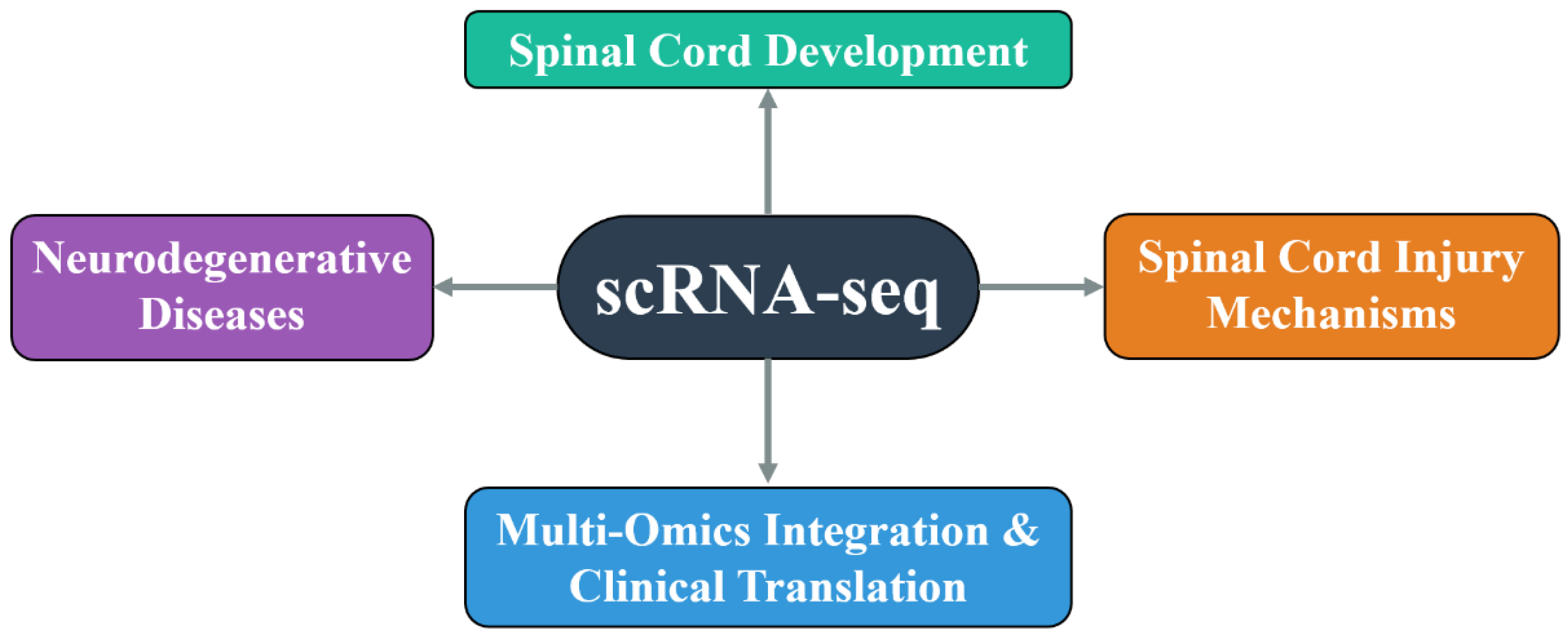
Figure 2.
Current Challenges and Limitations in Applying Single-Cell Transcriptomics to Spinal Cord Research.
Figure 2.
Current Challenges and Limitations in Applying Single-Cell Transcriptomics to Spinal Cord Research.
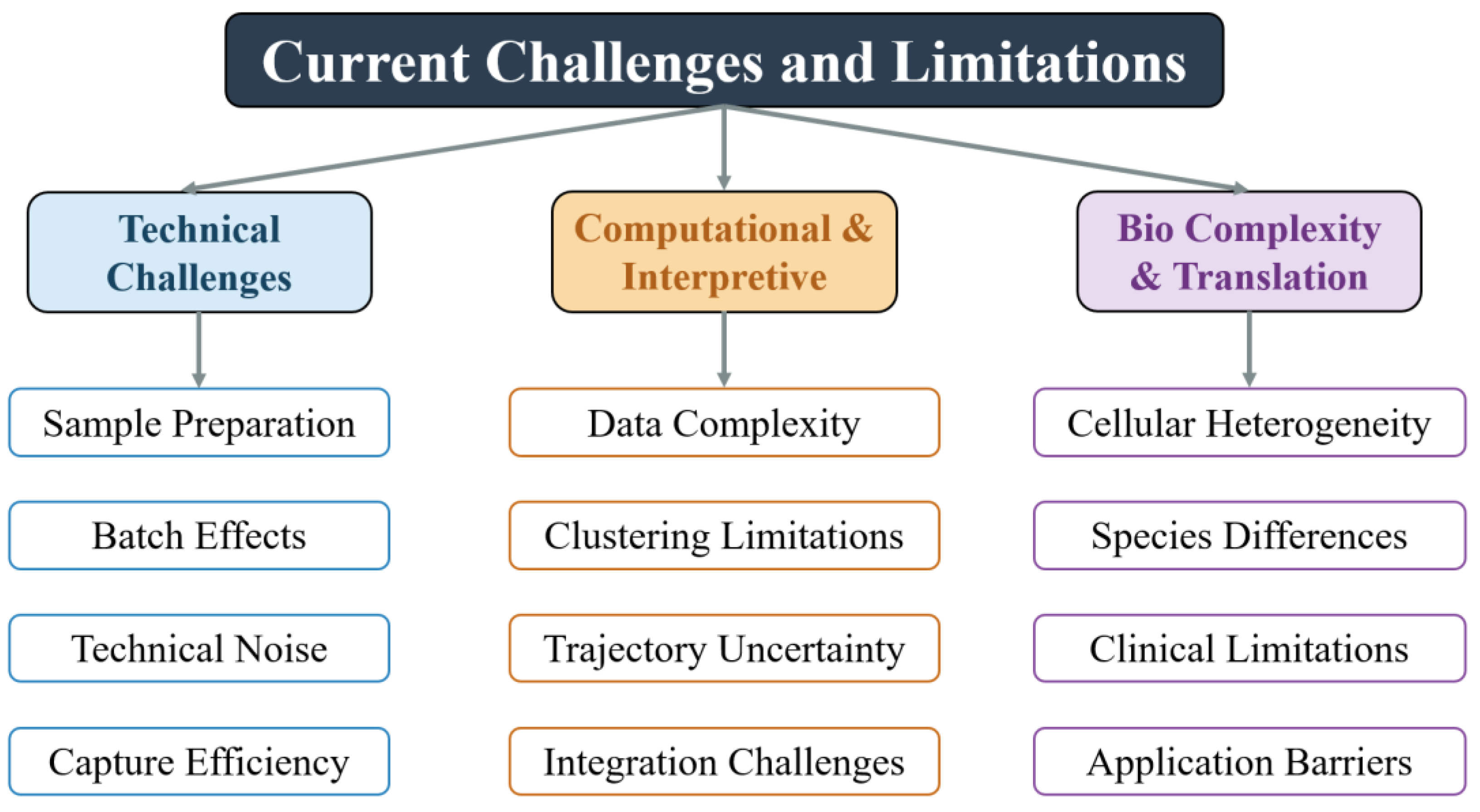

Table 1.
Representative Single-Cell Transcriptomic Studies on Spinal Cord Development.
| Study | Species | Developmental Stage | Key Findings |
|---|---|---|---|
| Delile et al. | Mouse | E9.5–E13.5 | Spatial/temporal dynamics of neural progenitors; novel markers in dorsoventral domains |
| Andersen et al. | Human | ~22 weeks gestation | Glial heterogeneity, astrocyte regionalization, disease gene mapping to specific cell types |
| Sathyamurthy et al. | Mouse | Adult | 43 neuronal subtypes, region-specific distribution, spinal neuron molecular map |
| Blum et al. | Mouse | Adult | Motor neuron heterogeneity, transcriptional profiles linked to axonal targeting and function |
| Cao et al. | Mouse | Organogenesis (multi-stage) | Spinal progenitor transcriptional transitions, Hox genes, Shh pathway in developmental regulation |
| Zhang et al. | Human | Adult | 21 neuronal subtypes, spatial distribution, human-mouse comparison, sex-specific transcription |
Disclaimer/Publisher’s Note: The statements, opinions and data contained in all publications are solely those of the individual author(s) and contributor(s) and not of MDPI and/or the editor(s). MDPI and/or the editor(s) disclaim responsibility for any injury to people or property resulting from any ideas, methods, instructions or products referred to in the content. |
© 2025 by the authors. Licensee MDPI, Basel, Switzerland. This article is an open access article distributed under the terms and conditions of the Creative Commons Attribution (CC BY) license (http://creativecommons.org/licenses/by/4.0/).
Copyright: This open access article is published under a Creative Commons CC BY 4.0 license, which permit the free download, distribution, and reuse, provided that the author and preprint are cited in any reuse.


