Submitted:
24 April 2025
Posted:
24 April 2025
You are already at the latest version
Abstract
Background and Objectives
Upper tract urothelial carcinoma (UTUC) is one of the most underdiagnosed but at the same time one of the most lethal cancers. In this review article we investigated the application of artificial intelligence and novel technologies in the prompt identification of high grade UTUC to prevent metastases and facilitate timely treatment.
Materials and Methods
We conducted an extensive search of the literature from the Pubmed and Google scholar databases for studies investigating the application of artificial intelligence for the diagnosis of UTUC. After the exclusion of non-associated and non-English studies, we included 12 articles in our review.
Results
Artificial intelligence systems have been shown to enhance post radical nephroureterectomy urine cytology reporting, in order to facilitate early diagnosis of bladder recurrence, as well as improve diagnostic accuracy in atypical cells, by being trained in annotated cytology images. Apart from that, data from computed tomography urograms by extracting textural radiomics features, can develop machine learning models to predict UTUC tumors grade and stage in small size and especially high grade tumors. Random forest models have been shown to have the best performance in predicting high grade UTUC, while hydronephrosis is the most significant independent factor for high grade tumors. ChatGPT, although not mature to provide information on diagnosis and treatment, can assist patients' understanding of the disease’s epidemiology and risk factors. Computer vision models in real-time can augment visualisation during endoscopic ureteral tumor diagnosis and ablation. Deep learning workflow can also be applied in histopathological slides to predict UTUC protein- based subtypes.
Conclusion
Artificial intelligence has been shown to greatly facilitate the timely diagnosis of high-grade UTUC by improving the diagnostic accuracy of urine cytology, CT Urogram and ureteroscopy visualisation. Deep learning systems can become a useful and easily accessible tool in physicians' armamentarium to deal with urothelial cancer diagnostic uncertainties.
Keywords:
Urothelial carcinoma
; Artificial intelligence
; Upper tract urothelial carcinoma
; urothelial carcinoma diagnosis
1. Introduction
Upper tract urothelial cancer (UTUC) is one of the most aggressive but understudied tumors in both sexes and includes neoplasms of the ureters and the pelvicalyceal systems. It is associated with very high mortality in advanced stages and thus there is an increased need to improve imaging modalities which will facilitate early diagnosis. [1]
Nowadays, initial diagnosis of UTUC and upper urinary tract recurrence after conservative or radical management are achieved with urine cytology reporting, computed tomography urogram (CTU) findings and with semirigid and flexible diagnostic ureteroscopy. Nevertheless, none of these techniques have managed to provide high diagnostic accuracy especially in identifying high grade and stage tumors. [1]
During the last few years there have been significant advancements in the application of artificial intelligence (AI) in the medical field and especially in improving the diagnosis of aggressive cancers, such as ovarian, prostate and bladder cancer. [2,3,4,5,6]
However, effective imaging modalities to identify advanced stages of upper tract urothelial carcinoma are still lacking. In this review article we have researched the available literature and present, for the first time to the best of our knowledge, a narrative review about the novel technologies available in urologists’ armamentarium, which can provide timely diagnosis of muscle invasive cancers of the upper urothelial tract.
Furthermore, we have investigated and discussed the potential role of artificial intelligence in facilitating selective and effective identification of aggressive disease, in order to decrease delayed diagnosis of metastatic UTUC as well as overtreatment.
2. Materials and Methods
An extensive research was conducted including all the available studies from Pubmed, Google scholar and Cochrane library databases, using associated keywords such as: “upper tract urothelial carcinoma” AND “artificial intelligence” AND “diagnosis” OR “diagnosis of upper tract urothelial carcinoma”, without restrictions concerning the date of publication, according to the PRISMA guidelines. [Figure 1]
Inclusion criteria for articles were that they involved artificial intelligence application for the diagnosis of UTUC or diagnosis of recurrence after conservative or radical treatment of UTUC. Our initial search retrieved 54 studies. After the initial title and abstract screening, we excluded duplicates, non-English studies and articles not associated with our criteria.
Studies involving non-AI methods such as statistical analysis methods and existing nomograms were excluded from the full manuscript screening process. Apart from that, studies focusing on other cancers or bladder cancer as the initial diagnosis or generally urothelial cancer without clarifying the inclusion of UTUC cases were also excluded, as well as studies not concerning exclusively the diagnosis of UTUC. After the final full manuscript screening, 12 studies were selected to be included in our review article.
Title and abstract screening was conducted by two independent reviewers (NK and VA) and disagreements were resolved by a third reviewer (AK). The selected studies were assessed by three different reviewers (NK, TB, SK) and the results and discussion section was conducted by them. The final review and corrections of the manuscript was independently done by two reviewers (VA, AK).
3. Results
After the screening process 12 studies were included in our review article. [Table 1] 11 of the selected studies were retrospective in design, with a relatively small number of patients (6-483 patients) with a mean of 136,73 patients with UTUC [7,8,9,10,11,12,13,14,15,16,17]. Only 1 selected study was a review article, which presented the available technologies to improve the performance of urine cytology. [18] From the selected studies, 2 retrospective studies and 1 review article were associated with urine cytology [7,8,18], 3 retrospective studies presented deep learning models for computed urograms findings [9,10,11], 2 retrospective studies were found about diagnostic ureteroscopy [12,13] and 4 retrospective studies were included that concerned ChatGPT artificial intelligence [14], histopathological slides interpretation [15], inflammatory markers pre-Radical Nephroureterectomy (RNU) [16] and N-glycans scoring systems [17].
The available literature presents promising retrospective data concerning artificial intelligence systems application for the successful and timely diagnosis of UTUC. The most crucial applications have been shown to be associated with post radical nephroureterectomy urine cytology reporting [7,8] and with deep learning systems that can be trained to improve ureteroscopic vision [12,13], as well as extract textural radiomics features from computed tomography urograms, to predict UTUC tumors grade and stage in <2cm tumors. [9,10,11]
Artificial intelligence such as ChatGPT, have been proven helpful to inform patients about the general information about the risk factors and epidemiology of UTUC [14], while deep learning workflow applied in histopathological slides can predict utuc protein- based subtypes. [15]
The use of machine learning has been shown promising results from a novel pre-RNU systemic immune-inflammation score (SIIS) nomogram, as an independent predictor of prognosis in patients with UTUC [16], as well as in creating urological disease-specific scoring systems with the use of biomarkers such as N-glycans. [17]
4. Discussion
Although UTUC is a relatively rare type of cancer, consisting of only 5-10% of urothelial cancers, it is commonly diagnosed in advanced stages with 60% being aggressive upon diagnosis and 30% metastatic. [1,9] Intravesical recurrence after radical nephroureterectomy (RNU) for the treatment of UTUC is very high with rates of 22-47% [1]. Thus, strict follow-up with cystoscopy and urine cytology is necessary. Using artificial intelligence to increase urine cytology sensitivity has been suggested recently [7]. The AIxURO system is a deep learning model that performs segmentation and is aligned with the guidelines in the Paris System 2.0 for urine cytology, in order to particularly distinguish between high-risk “suspicious cells” and low-risk “atypical cells” in urine cytology samples of patients that had undergone RNU.
The AIxURO system increased postoperative urine cytology reporting confidence and managed to diagnose intravesical recurrence in misdiagnosed patients from their urine cytology samples. [7] In a retrospective study by Chen CC. Et al. [7], the results of postoperative digitized cytology slides, assessed by the AIxURO system, were compared to cytopathologists reports.
With the assistance of the artificial intelligence system AIxURO, bladder tumour recurrence post RNU, was detected in 37% (10/27) of patients, by combining advanced urine analysis with artificial intelligence techniques, whereas traditional urine cytology examination identified only 29,6% (8/27) of recurrences. Hence, AIxURO showed greater sensitivity and accuracy in comparison to traditional urine cytology, although the authors admit many limitations, such as the small sample size and retrospective study design. [7]
These findings were confirmed by a larger study of post-RNU cytologies evaluated by AIxURO. [8] The AIxURO system provided more accurate and consistent results especially in atypical cell samples, in comparison to the assessments by the cytopathologist and cytotechnologist. The cooperation of artificial intelligence with clinicians experience has been proposed to reduce diagnostic discrepancies and significantly improve the reliability of the urine cytology diagnosis of UTUC bladder recurrence after RNU. [8]
Significant correlation has been proven between pathological grade of UTUC and prognosis of the disease, with higher grade associated with poorer prognosis. Hence, precise preoperative identification of the UTUC pathological stage could significantly improve survival of patients. [9]
Radiomics technology by using the random forest machine learning (MC) algorithm, based on Computed tomography urogram (CTU), could greatly assist in the prediction of high grade UTUC and potentially reduce unnecessary invasive procedures to some extent.
Many studies have proposed the construction of nomogram models to predict high grade UTUC, by extracting images from CTU. [9,10,11] In one study maximal tumour diameter was recorded in 3 phases from CTU images (unenhanced, medullary, excretory), by using the Radiomics module of the 3D Slicer software (version 5.0.3). [9]
The segmentation was performed manually for each tumour by a specialised radiologist. The radiomics scores obtained from the mixed-phase characteristics of 167 patients, showed statistically significant differences between low grade and high grade UTUC (p<0,05). The nomogram showed a probability of predicting high grade UTUC of 96,8% and compared to urine cytology it achieved higher accuracy and sensitivity. However, it also showed lower specificity, which limits the nomograms ability to replace postoperative tissue pathological examination. [9]
In another study by Alqahtani A. et al. [10], using the same radiomics technology, the authors managed to successfully differentiate high grade from low grade tumours, as well as early stage from advanced UTUC. The application of machine learning and enhanced CT Urograms is suggested as the beginning of “virtual biopsy”, which they showed performed significantly better than ureteroscopic histopathology in small (<2cm) UTUC stage and grade prediction. Future prospective studies with larger populations are certainly needed to standardise these radiomics-based models. [10]
The random forest (RF) machine learning model has shown the best performance in predicting high-grade UTUC, in comparison to other machine learning models, by extracting features from three periods of CTU images. It also had the highest area under the curve (AUC) values with 0.914 (95% Confidence Interval [95%CI] 0.852–0.977) and 0.903 (95%CI 0.809–0.997) in the training set and validation set, and accuracy of 0.878 and 0.857, respectively. Hydronephrosis has been proven the best independent influence factor for high-grade disease. [11]
Apart from that, a well recognised difficulty during conservative treatment of UTUC, via ureteroscopic laser ablation, is poor endoscopic visibility, due to bleeding from the tumour and narrow ureteral lumen [12]. U-net computer vision models have been proven to augment visualisation during treatment. By comparing 20 different videos of patients that received endoscopic treatment, the U-net system managed to successfully segment both the UTUC tumours and the areas of ablation. These deep learning computer vision models have demonstrated excellent real-time performance for automated UTUC segmentation during ureteroscopy, facilitating more accurate conservative treatment. [12]
Computer assisted interventions (CAI) can potentially provide information to urologists during diagnostic ureteroscopies for UTUC, or during treatment of UTUC with ureteroscopic laser ablation. Several navigation systems have been proposed and could be considered precursors of robotic ureteroscopy. CAI has been shown to provide spatial temporal information to improve hollow lumen segmentation in ureteroscopic images, especially in challenging situations such as: cases with poor visibility, occasional bleeding and specular reflections [13].
Large language models (LLM) such as ChatGPT, which are powered by artificial intelligence have been suggested to potentially assist in medical diagnosis of several diseases in the near future. [14]
However, their use is still limited because of reproducibility of authors bias and potential spread of misinformation.
A study evaluated the responses of ChatGPT in regard to patients questions associated with UTUC, such as the risk factors of the disease, it’s definition, most common symptoms and treatment methods. [14]
It was shown that ChatGPT had a suboptimal performance especially in the questions associated with the treatment of UTUC. It achieved it’s highest scores concerning the risk factors of UTUC.
Thus ChatGPT can be considered a very useful tool in assisting the public to understand basic aspects of the disease and gain general knowledge of prophylaxis against UTUC. It is certain that ChatGPT can not replace proper medical care and assessment by urologists, but nevertheless it can simplify information concerning the disease for the patients. [14]
The use of artificial intelligence has also been proposed to predict UTUC protein-based subtypes, by developing a deep-learning workflow (DL) in order to identify them directly from histopathological H&E slides. [15]
In a retrospective study by Angeloni M. Et al. [8], these subtypes were identified using the immunohistochemical expression of three luminal (FOXA1, GATA3, and CK20) and three basal (CD44, CK5, and CK14) markers with the assistance of a DL model based on a transfer-learning approach by fine-tuning a pre-trained ResNet50. The DL model successfully predicted protein- based UTUC subtypes, with basal predictions mainly containing PD-L1 positive samples, while luminal predictions containing a higher proportion of FGFR-3-mutated samples. [15]
Other novel technologies involving artificial intelligence have also shown promising results in recent years. [16] A novel pre-RNU systemic immune-inflammation score (SIIS) nomogram with the assistance of machine learning, has been found to be an independent predictor of prognosis in patients with UTUC. [16]
An SIIS-related nomogram was created to predict 1-, 3- and 5- year overall survival in UTUC patients, using Cox regression model and machine learning with random survival forest model. Combined preoperative higher serum levels of neutrophil-to-lymphocyte ratio, monocyte-to-lymphocyte ratio, platelet-to-lymphocyte ratio, systemic immune-inflammation index and systemic inflammation response index were associated with significantly shorter overall survival. [16]
Machine learning urological disease-specific scoring systems with the use of biomarkers such as N-glycans, and specifically monogalactosyl biantennary N-glycan (G1) and agalactosyl bisecting GlcNAc core fucosyl N-glycan (G0FB), have been shown to achieve a high impact on the early detection of UTUC. [17]
Furthermore these biomarkers are able to discriminate between bladder cancer and UTUC, in order to select a disease-specific treatment, using only a one-time serum collection. [17]
To summarize the available data from the literature, it is well established that the diagnosis of UTUC is challenging even in recent years, because of the limited accuracy of the available diagnostic tools. [18]
The Paris system for Reporting Urine Cytology (TPS) performance in UTUC is hindered by difficult- to- access location of tumours, instrumentation artifacts and ureteroscopic trauma. This is resulting in poor overall performance especially for low-grade UTUC, while for high-grade disease, urine cytology demonstrates unreliable sensitivity and high specificity. Based on these findings, artificial intelligence and machine learning may facilitate objective application of TPS criteria on urine samples, for early UTUC diagnosis, as well as for its distinction from bladder cancer. [18]
This is the first literature review to the best of our knowledge, to summarize all the recent updates in the application of artificial intelligence technologies and machine learning models, for the successful diagnosis of UTUC. Other reviews have already addressed the application of AI for urothelial carcinoma in general [19] or for other aspects of UTUC such as prediction of the most crucial prognostic factors of the disease. [20]
5. Conclusions
The application of artificial intelligence and machine learning models have been shown to be successful in facilitating the timely identification of high-grade UTUC by improving the diagnostic accuracy of urine cytology, CT Urogram and ureteroscopy visualisation. Deep learning systems can become a useful and easily accessible tool in urologists' everyday clinical practice to deal with upper tract urothelial cancer diagnostic uncertainties
6. Future Directions
Randomized prospective studies with larger populations are needed to verify the above findings and standardize the use of these novel technologies in clinical practice guidelines, in the same way as it has already been initiated in general for urothelial carcinomas with prospective validation of study results concerning the use of artificial intelligence . [21]
Author Contributions
Conceived the study and design: Nikolaos Kostakopoulos, Vasileios Argyropoulos, Themistoklis Bellos. Data collection and analysis: Nikolaos Kostakopoulos, Vasileios Argyropoulos, Stamatios Katsimperis, Athanasios Kostakopoulos. Manuscript writing and statistical analysis: Nikolaos Kostakopoulos, Themistoklis Bellos, Stamatios Katsimperis. Manuscript editing and review: Vasileios Argyropoulos, Athanasios Kostakopoulos .
Funding
None.
Data Availability Statement
n/a.
Conflicts of Interest
None.
References
- Masson-Lecomte A, Birtle A, Pradere B, Capoun O, Compérat E, Domínguez-Escrig JL, Liedberg F, Makaroff L, Mariappan P, Moschini M, Rai BP, van Rhijn BWG, Shariat SF, Smith EJ, Teoh JYC, Soukup V, Wood R, Xylinas EN, Soria F, Seisen T, Gontero P. European Association of Urology Guidelines on Upper Urinary Tract Urothelial Carcinoma: Summary of the 2025 Update. Eur Urol. 2025 Mar 7:S0302-2838(25)00145-9. [CrossRef]
- Wang Y, Lin W, Zhuang X, Wang X, He Y, Li L, Lyu G. Advances in artificial intelligence for the diagnosis and treatment of ovarian cancer (Review). Oncol Rep. 2024 Mar;51(3):46. [CrossRef]
- Cai G, Huang F, Gao Y, Li X, Chi J, Xie J, Zhou L, Feng Y, Huang H, Deng T, Zhou Y, Zhang C, Luo X, Xie X, Gao Q, Zhen X, Liu J. Artificial intelligence-based models enabling accurate diagnosis of ovarian cancer using laboratory tests in China: a multicentre, retrospective cohort study. Lancet Digit Health. 2024 Mar;6(3):e176-e186. [CrossRef]
- Kartasalo K, Bulten W, Delahunt B, Chen PC, Pinckaers H, Olsson H, Ji X, Mulliqi N, Samaratunga H, Tsuzuki T, Lindberg J, Rantalainen M, Wählby C, Litjens G, Ruusuvuori P, Egevad L, Eklund M. Artificial Intelligence for Diagnosis and Gleason Grading of Prostate Cancer in Biopsies-Current Status and Next Steps. Eur Urol Focus. 2021 Jul;7(4):687-691. [CrossRef]
- Marletta S, Eccher A, Martelli FM, Santonicco N, Girolami I, Scarpa A, Pagni F, L'Imperio V, Pantanowitz L, Gobbo S, Seminati D, Dei Tos AP, Parwani A. Artificial intelligence-based algorithms for the diagnosis of prostate cancer: A systematic review. Am J Clin Pathol. 2024 Jun 3;161(6):526-534. [CrossRef]
- Chan EO, Pradere B, Teoh JY; European Association of Urology – Young Academic Urologists (EAU-YAU) Urothelial Carcinoma Working Group. The use of artificial intelligence for the diagnosis of bladder cancer: a review and perspectives. Curr Opin Urol. 2021 Jul 1;31(4):397-403.
- Chen CC, Yen TH, Li JR, Chen CJ, Yang CS, Lai JY, Lin SJ, Yeh CH, Hsu SW, Lin MY, Liu TJ, Chen CS. Artificial intelligence algorithms enhance urine cytology reporting confidence in postoperative follow-up for upper urinary tract urothelial carcinoma. Int Urol Nephrol. 2024 Nov 7.
- Chang KY, Yang CS, Lai JY, Lin SJ, Li JR, Liu TJ, Yang WL, Lin MY, Yeh CH, Hsu SW, Chen CJ. An Artificial Intelligence-assisted Diagnostic System Improves Upper Urine Tract Cytology Diagnosis. In Vivo. 2024 Nov-Dec;38(6):3016-3021.
- Zheng Y, Shi H, Fu S, Wang H, Li X, Li Z, Hai B, Zhang J. Development and validation of a radiomics-based nomogram for predicting pathological grade of upper urinary tract urothelial carcinoma. BMC Cancer. 2024 Dec 18;24(1):1546. [CrossRef]
- Alqahtani A, Bhattacharjee S, Almopti A, Li C, Nabi G. Radiomics-based machine learning approach for the prediction of grade and stage in upper urinary tract urothelial carcinoma: a step towards virtual biopsy. Int J Surg. 2024 Jun 1;110(6):3258-3268. [CrossRef]
- Zheng Y, Shi H, Fu S, Wang H, Wang J, Li X, Li Z, Hai B, Zhang J. A computed tomography urography-based machine learning model for predicting preoperative pathological grade of upper urinary tract urothelial carcinoma. Cancer Med. 2024 Jan;13(1):e6901. [CrossRef]
- Lu D, Reed A, Pace N, Luckenbaugh AN, Pallauf M, Singla N, Oguz I, Kavoussi N. Automated Upper Tract Urothelial Carcinoma Tumor Segmentation During Ureteroscopy Using Computer Vision Techniques. J Endourol. 2024 Aug;38(8):836-842. [CrossRef]
- Lazo JF, Marzullo A, Moccia S, Catellani M, Rosa B, de Mathelin M, De Momi E. Using spatial-temporal ensembles of convolutional neural networks for lumen segmentation in ureteroscopy. Int J Comput Assist Radiol Surg. 2021 Jun;16(6):915-922. [CrossRef]
- Łaszkiewicz J, Krajewski W, Tomczak W, Chorbińska J, Nowak Ł, Chełmoński A, Krajewski P, Sójka A, Małkiewicz B, Szydełko T. Performance of ChatGPT in providing patient information about upper tract urothelial carcinoma. Contemp Oncol (Pozn). 2024;28(2):172-181. [CrossRef]
- Angeloni M, van Doeveren T, Lindner S, Volland P, Schmelmer J, Foersch S, Matek C, Stoehr R, Geppert CI, Heers H, Wach S, Taubert H, Sikic D, Wullich B, van Leenders GJ, Zaburdaev V, Eckstein M, Hartmann A, Boormans JL, Ferrazzi F, Bahlinger V. A deep-learning workflow to predict upper tract urothelial carcinoma protein-based subtypes from H&E slides supporting the prioritization of patients for molecular testing. J Pathol Clin Res. 2024 Mar;10(2):e12369. [CrossRef]
- Liu J, Wu P, Lai S, Wang J, Hou H, Zhang Y. Prognostic models for upper urinary tract urothelial carcinoma patients after radical nephroureterectomy based on a novel systemic immune-inflammation score with machine learning. BMC Cancer. 2023 Jun 22;23(1):574. [CrossRef]
- Iwamura H, Mizuno K, Akamatsu S, Hatakeyama S, Tobisawa Y, Narita S, Narita T, Yamashita S, Kawamura S, Sakurai T, Fujita N, Kodama H, Noro D, Kakizaki I, Nakaji S, Itoh K, Tsuchiya N, Ito A, Habuchi T, Ohyama C, Yoneyama T. Machine learning diagnosis by immunoglobulin N-glycan signatures for precision diagnosis of urological diseases. Cancer Sci. 2022 Jul;113(7):2434-2445. [CrossRef]
- Chukwudebe O, Lynch E, Vira M, Vaickus L, Khan A, Shaheen Cocker R. A review of the performance of urinary cytology with a focus on atypia, upper tract and updates on novel ancillary testing. J Am Soc Cytopathol. 2025 Jan-Feb;14(1):23-35. [CrossRef]
- Malik S, Wu J, Bodnariuc N, Narayana K, Gupta N, Malik M, Kwong JCC, Khondker A, Johnson AEW, Kulkarni GS. Existing trends and applications of artificial intelligence in urothelial cancer A scoping review. Can Urol Assoc J. 2023 Nov;17(11):E395-E401.
- Michael Staehler et al. Artificial intelligence and identifying prognostic patterns in upper tract urothelial carcinoma (UTUC). JCO 42, e16616-e16616(2024). [CrossRef]
- Wu S, Shen R, Hong G, Luo Y, Wan H, Feng J, Chen Z, Jiang F, Wang Y, Liao C, Li X, Liu B, Huang X, Liu K, Qin P, Wang Y, Xie Y, Ouyang N, Huang J, Lin T. Development and validation of an artificial intelligence-based model for detecting urothelial carcinoma using urine cytology images: a multicentre, diagnostic study with prospective validation. EClinicalMedicine. 2024 Mar 27;71:102566. [CrossRef]
Figure 1.
PRISMA guidelines flowchart.
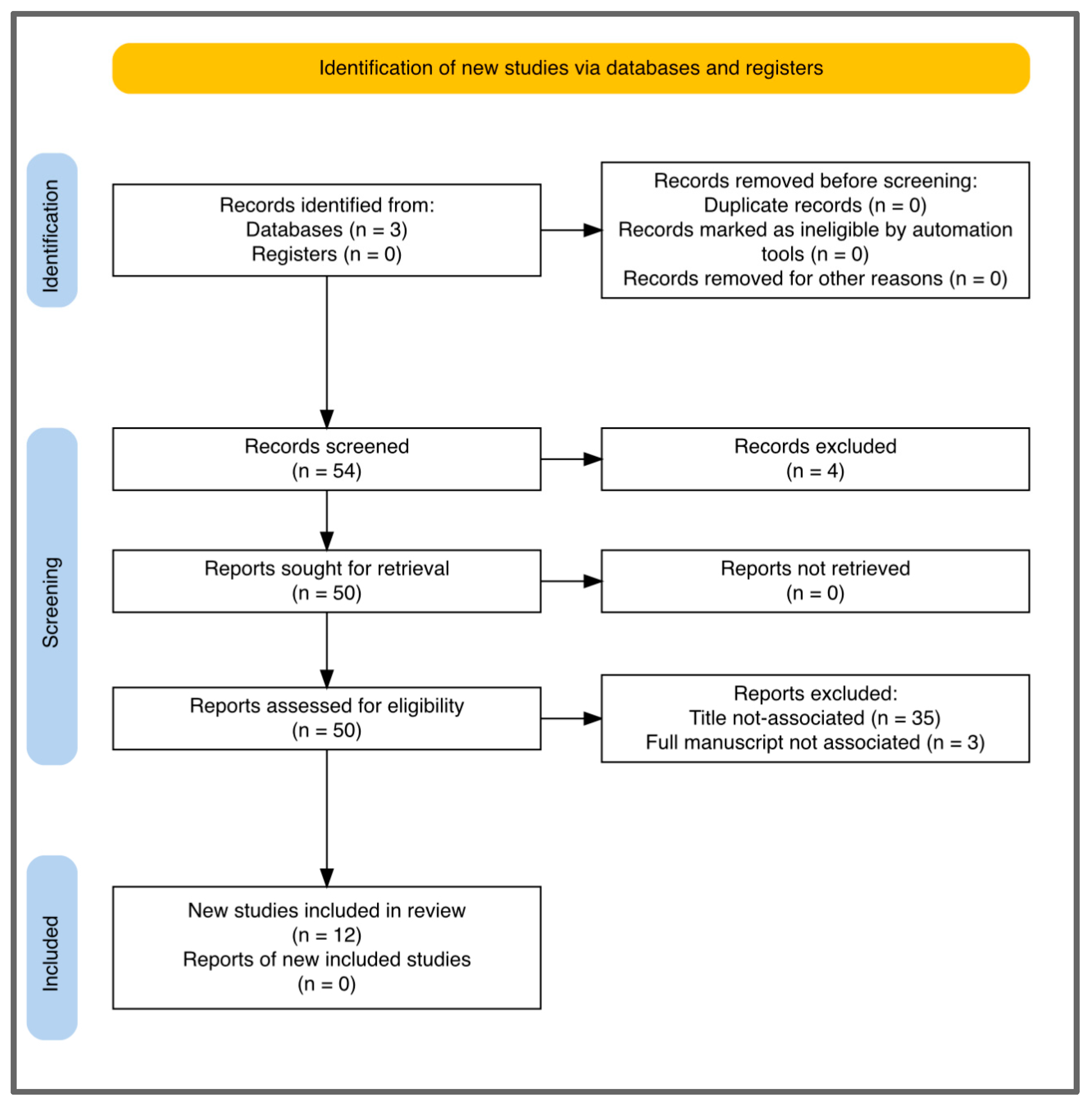

Table 1.
Artificial intelligence applications for the diagnosis of UTUC.
| Study | Number of patients (mean 136,73) | AI Application type for UTUC diagnosis | Type of study |
|---|---|---|---|
|
113 | urine cytology diagnostic accuracy | Retrospective study |
|
185 | urine cytology diagnostic accuracy | Retrospective study |
|
140 | radiomics CTU nomogram | Retrospective study |
|
106 | radiomics CTU nomogram | Retrospective study |
|
167 | machine learning CTU model | Retrospective study |
|
20 | ureteroscopic vision enhancement | Retrospective study |
|
6 | ureteroscopic vision enhancement | Retrospective study |
|
16 | ChatGPT performance | Retrospective study |
|
163 | histopathology slides deep learning system | Retrospective study |
|
483 | systemic immune-inflammation score machine learning | Retrospective study |
|
105 | immunoglobulin N-glycan machine learning | Retrospective study |
|
n/a | urine cytology diagnostic accuracy | Narrative review |
Disclaimer/Publisher’s Note: The statements, opinions and data contained in all publications are solely those of the individual author(s) and contributor(s) and not of MDPI and/or the editor(s). MDPI and/or the editor(s) disclaim responsibility for any injury to people or property resulting from any ideas, methods, instructions or products referred to in the content. |
© 2025 by the authors. Licensee MDPI, Basel, Switzerland. This article is an open access article distributed under the terms and conditions of the Creative Commons Attribution (CC BY) license (http://creativecommons.org/licenses/by/4.0/).
Copyright: This open access article is published under a Creative Commons CC BY 4.0 license, which permit the free download, distribution, and reuse, provided that the author and preprint are cited in any reuse.


