Submitted:
15 April 2025
Posted:
16 April 2025
You are already at the latest version
Abstract
Oxidative stress and its relation on the onset of several chronic diseases has been in-creasingly studied and highlighted in recent years. This fact has been concurrent with increased reports on the antioxidant properties of various phytochemicals derived from fruits, vegetables, herbs, or seaweed, which can be accessible by intake or obtained following chemical extraction from these sources. These phytochemicals are majorly the result of each plant’s secondary metabolism, out of which the main chemical groups include structural polysaccharides, unsaturated fatty acids (PUFAs), pigments (chlo-rophylls, carotenoids, anthocyanins) and chief among them, phenolic compounds. The structural features of each group allow them to act at different sites, levels, by different molecular mechanisms and display diverse effectiveness as chemo-preventive agents for various diseases. Beyond their antioxidant properties, these phytochemicals have been described to exert a plethora of chemo-preventive and therapeutic effects, in-cluding anti-inflammatory, antidiabetic, anti-obesity or neuroprotective, acting through various mechanisms and paths involved. This knowledge has led to the development of various nutraceuticals enriched in antioxidant phytochemicals to be used as functional ingredients in foods, or for their periodic separated intake, whether as enriched extracts, or isolated compounds of high efficacy. Overall, phytochemical antioxidants are attrac-tive biomolecules to be used as nutraceuticals of relevant chemo-preventive and ther-apeutic properties on the onset of various diseases related to antioxidant stress.
Keywords:
Phytochemicals
; Natural antioxidants
; Chemo-preventive
; Nutraceuticals
1. Introduction
Phytochemicals are secondary metabolites synthesized by plants which comprise a diverse array of chemical groups. These include phenolic compounds, fatty acids, pigments, various polysaccharides, terpenoids, alkaloids, phytosterols or saponins, among others [1]. Phytochemicals have always been a matter of interest, as they serve as extremely relevant agents in medicine, agriculture, food, and various other fields, such as the textile and cosmetic industries. Thus, our world has a strong dependance on phytochemicals with various uses and purposes. However, in recent decades the interest in dietary phytochemicals has risen, as are thought to be of many health benefits such as cardio-protection, neuroprotection, anti-inflammatory effects and improved gut health, among others [2]. These effects are mainly attributed to the antioxidant properties of various groups of phytochemicals, with phenolic compounds, unsaturated fatty acids (UFAs) or polysaccharides being chief among them [3]. Thus, diets which include more vegetables and fruits that may act as source of phytochemicals have attracted more interest from researchers, industry, and society, with focus on a more conscious and healthy diet. This has also prompted the surge of a nutraceutical industry based on producing purified or selected phytochemicals that have the potential to confer specific health benefits (e.g., phenolic compounds) [4]. These phytochemicals can thus be obtained from different sources. UFAs for example, can be feasibly obtained from oily crops, such as olive, sunflower, rapeseed, sesame, palm or peanut, among others [5]. Therefore, depending on the source, the composition and quantity of fatty acids will vary. These may be obtained from the mentioned fruits and seeds, but also from some less conventional sources like fruits seeds, such as pepper, grape or avocado [6,7]. Microalgae have also been increasingly highlighted as excellent sources of UFAs of interest in recent years [8]. Given their nature and metabolic function as reducing agents, plant pigments are used as colorants and antioxidants. Carotenoids can be distinguished between carotenes, which include carotene and lycopene, and xanthophylls, with some main examples being lutein, zeaxanthin or lycopene [9]. Moreover, they can be further distinguished between provitamin A carotenoids, which include carotenes and β-cryptoxanthin, and non-provitamin A carotenoids, including mainly xanthophylls [10]. Intuitively, carotenoids can be obtained from yellow, orange, and red-colored fruits and vegetables, with higher concentrations in the peels of these. Moreover, in recent years, both seaweeds and microalgae have become feasible and cost-effective sources of carotenoids, with some major examples being fucoxanthin from brown seaweeds and astaxanthin from red microalgae [11] In this sense, carotenoids have been highlighted as relevant phytochemicals with various potential health benefits, ranging from their nutritional value as precursors of vitamin A to health-related properties such as antioxidant, metabolic modulation, anti-inflammatory or neuroprotective [12]. Polysaccharides with health-related properties tend to be structural polysaccharides. This means that polysaccharides serve as structural support to the plant organism. Among these, cellulose, hemicellulose or pectin are the major polysaccharides present in plants that constitute dietary fiber [13]. These polysaccharides may be found more abundantly in the “harder” parts of plants, such as seeds, leaves or peels, and are of high interest as promoters of healthy gut microbiota [14]. However, storage polysaccharides (that act as stored energy) include starches and other polysaccharides, such as fructans. These are more abundant in bulbs, tubers and pulps [15]. Beyond these, phenolic compounds are antioxidant phytochemicals of paramount importance that have been exhaustively studied in the last two decades and have a ubiquitous distribution in the plant kingdom. Phenolic compounds are phytochemicals with great antioxidant activity and are synthesized in response to biotic (predators, infections) or abiotic (drought, salinity) factors [16]. The diverse structural complexity and degree of polymerization of phenolic compounds also determine their bioavailability, activities, and occurrence. Some well-known examples of phenolic compounds and sources include tannins and catechins in grapes or tea, flavonoids in citrus or anthocyanins in berries [16]. Moreover, given their metabolic role in the plant, peels and leaves are usually the fractions of fruits and vegetables that contain the greatest concentration of them [17]. Altogether, there a great interest in these phytochemicals. phytochemicals, either by chemical or/and microbial synthesis, as these methods are more economically feasible and consistent [18]. Although these methods may be cost-effective, chemical extraction of phytochemicals from their natural sources is becoming increasingly extended, especially using fruit and vegetable agri-food waste as source of phytochemicals [19].
In this review, we discuss the role of antioxidants in plants, their significance in the mitigation of oxidative damage, and their implications for plant resilience and human overall health.
2. Chemical Classes of Antioxidant Phytochemicals: Structural Significance
2.1. Unsaturated Fatty Acids
Fatty acids are hydrophobic aliphatic carbon chains of variable length that contain a carboxyl (-COOH) group at the end of the chain [20]. The fatty acids with no double bonds present are called saturated fatty acids (MFAs), whereas those that present at some point of the chain a double bond are called UFAs. In addition, fatty acids can also be distinguished depending on their stereochemistry, presenting cis or trans isomers, with cis being more prevalent in plants, and trans in animal fats [21]. In the case of plant organisms, fatty acids may usually present a length in the range of 12 to 22 carbons, with 16 and 18 being generally the most abundant [22]. Among MFAs, plant organisms generally accumulate palmitic (C16:0) and stearic (C18:0) acids. Moreover, plants are the main source in nature and thus in the human diet of UFAs, with some major representatives being oleic acid (C18:1) and linoleic acid (C18:2) [5].
2.2. Carotenoids
Carotenoids represent a class of pigments naturally produced by plants, fungi, algae, and specific bacteria [23]. These compounds are responsible for the vibrant red, yellow, and orange colors seen in various fruits and vegetables [24]. Structurally, carotenoids are classified as tetraterpenoids, composed of a 40-carbon polyene chain, providing their antioxidant properties [25]. This antioxidant capacity arises from their ability to neutralize reactive oxygen species (ROS) and neutralize free radicals, providing protection against oxidative stress, which is linked to diseases such as cancer and cardiovascular conditions [26]. The conjugated double bond system present in the structure of carotenoids facilitates electron delocalization, thereby increasing their ability to neutralize free radicals and singlet oxygen [27]. Additionally, their antioxidant efficiency is modulated by the degree of unsaturation and the presence of functional groups, like hydroxyl (-OH) or keto (C=O) groups [28].
Based on their structure, carotenoids can be divided into two primary categories based on their chemical structure: carotenes, which are purely hydrocarbons, and xanthophylls, which contain oxygenated groups [29]. The functional groups in xanthophylls generally improve their solubility and interaction with cellular membranes, potentially influencing their antioxidant effectiveness [30]. In fact, the presence of additional oxygen-containing groups often makes xanthophylls more potent antioxidants compared to carotenes [31].
Plants, algae, and fungi can produce different isomers of carotenes, with α-carotene and β-carotene being the most prevalent in plants (Table 1). Other isomeric forms include γ-carotene, δ-carotene, ε-carotene, and ζ-carotene. Both α-carotene and β-carotene serve as precursors to vitamin A [32], although β-carotene exhibits greater antioxidant activity compared to α-carotene [33]. Among xanthophylls, key molecules include fucoxanthin, astaxanthin, lutein, zeaxanthin, and β-cryptoxanthin [29]. These compounds are recognized for their strong antioxidant properties, which play a role in their anticancer, anti-inflammatory, and neuroprotective effects, as well as their ability to enhance immune responses [29,34]. Owing to these attributes, carotenoids hold considerable potential for therapeutic use in managing OS-related diseases [35]. The variation in their efficacy stems from differences in their structural characteristics, such as terminal functional groups and chain length [29]. For instance, lutein and zeaxanthin, commonly found in leafy greens, are known to accumulate in the retina’s macular region, where they filter harmful blue light and serve as powerful antioxidants [34].
Additionally, several studies support the inclusion of carotenoids in functional foods and dietary supplements, linking their intake to a reduced risk of chronic diseases, including cardiovascular disorders, specific cancers, and age-related macular degeneration [24]. Their role as precursors to vitamin A further enhances their significance in promoting visual health and supporting the immune system [36]. The numerous benefits of these compounds have favored the inclusion of these molecules as ingredients in the development of nutraceuticals aimed at improving health and preventing diseases.
2.3. Polysaccharides
Polysaccharides are complex polymers composed of monosaccharide units connected by glycosidic bonds, widely distributed in plants, fungi, algae, and certain bacteria [37]. These natural polymers have attracted significant attention due to their potential as nutraceuticals, particularly for their antioxidant, immunomodulatory, and anti-inflammatory activities [38,39]. As antioxidants (Table 1), polysaccharides protect biological systems by neutralizing ROS, limiting free radical damage, and shielding cellular components from OS [40]. Their protective effects are mediated through the regulation of antioxidant signaling pathways, enhancement of the body’s antioxidant enzyme defenses, and reduction in ROS production. Additionally, polysaccharides prevent ROS-induced tissue damage through both free radical scavenging and modulation of the immune response [41,42]. However, under specific conditions, certain polysaccharides may also exhibit prooxidant behavior, adding complexity to their antioxidant profile [40,43].
Overall, the antioxidant capacity of polysaccharides is influenced by multiple factors rather than a single property [44]. Structural features such as monosaccharide composition, glycosidic linkages, molecular weight, and branching degree contribute to their antioxidant activity [45]. Moreover, polysaccharides with higher molecular weights, increased branching, or specific functional groups, such as sulfate or carboxyl groups, often demonstrate stronger antioxidant potential [46,47]. Moreover, the method of extraction plays a crucial role in determining their functional properties [48,49]. The ability of polysaccharides ability to donate electrons or protons, chelate metal ions, and scavenge free radicals forms the foundation of their antioxidant mechanism [44]. However, despite the established antioxidant potential of polysaccharides, there is limited research on highly purified forms, making it important to consider the possible influence of other bioactive components, such as proteins, peptides, or polyphenols, which may be present in combination with polysaccharides [44].
Polysaccharides are classified into different groups based on their structure, origin, and function. Structurally, they can be divided into heteropolysaccharides (e.g., hyaluronic acid, chondroitin sulfate, heparin) and homopolysaccharides (e.g., fructans, galactan, glycogen) [50]. Based on their source, they are categorized into several groups: plant-based (e.g., pectin, cellulose, starch), microbial-based (e.g., curdlan, dextran, bacterial cellulose), fungi-derived (e.g., β-glucans, chitin derivatives), animal-derived (e.g., chitosan, chitin, heparin), and marine-derived (e.g., agar, alginate, fucoidans) [51]. Polysaccharides like pectin, rich in galacturonic acid, exhibit significant antioxidant properties. This bioactivity is influenced by the uronic acid content and degree of polymerization [52]. Moreover, sulfated polysaccharides from marine algae are particularly effective in neutralizing free radicals and chelating metal ions, enhancing their antioxidant performance [53].
The growing recognition of polysaccharides’ antioxidant properties underscores their potential in nutraceutical development, as they not only combat OS but also offer additional health benefits [49]. Therefore, the ability of polysaccharides to address OS, as well as other bioactivities, make these compounds valuable substances in the nutraceutical market for dietary supplements and functional foods. [54]. Ongoing research continues to shed light on the mechanisms by which these polysaccharides exert their health-promoting effects, opening new avenues for their use in preventive and therapeutic nutrition [55]. Moreover, beyond their role as prebiotics, polysaccharides are also widely utilized in industry for various functions, including as thickeners, emulsifiers, stabilizers, gelling agents, and controlled-release agents, broadening their applications [50].
2.4. Phenolic Compounds
Phenolic compounds, a diverse group of secondary metabolites, are produced by plants primarily in response to environmental stress factors such as drought, extreme temperatures, UV exposure, pollution, and pathogen attacks [56]. Their biosynthesis follows pathways like the pentose phosphate, shikimate, and phenylpropanoid routes [57]. Chemically, phenolics are defined by the presence of one or more hydroxyl groups attached to an aromatic ring, though their complexity varies across different subclasses. These compounds encompass a broad range of molecules, including phenolic acids, flavonoids, stilbenes, lignans, and related structures like chalcones, humulones, and alcohols [56,58].
These compounds are classified according to their structure, with more than 8,000 variations currently identified. The classification is typically based on the number of phenolic rings and the nature of the linkages between them. Major groups include phenolic acids, flavonoids, stilbenes, and lignans [59].
Phenolic acids are divided into two main categories: hydroxybenzoic acids (such as gallic acid) and hydroxycinnamic acids (including caffeic, ferulic, and coumaric acids) [59,60]. These acids are widely recognized for their ability to neutralize free radicals, reducing oxidative damage in cells. Flavonoids, another key category, have a C6–C3–C6 backbone and are further categorized into subgroups like flavonols, flavones, isoflavones, anthocyanins, flavanones, flavanols, and chalcones [60]. In plant cells, flavonoids are typically stored as glycosides, and their antioxidant potential is influenced by the degree of glycosylation. Aglycones like quercetin and myricetin are generally more potent antioxidants than their glycoside forms [61].
Tannins, another class of phenolics, are categorized into hydrolyzable tannins (e.g., gallotannins, ellagitannins) and condensed tannins (e.g., proanthocyanidins). These compounds are known for their protein-binding capabilities, contributing to the astringent taste of certain fruits and their antimicrobial activities [59,62].
Additionally, some phenolics like curcumin (diferuloylmethane) display a broad spectrum of biological functions, including antioxidant, anti-inflammatory, antimicrobial, antitumor, and liver-protective effects [63]. The structure of phenolic compounds is closely linked to their bioactivity, with different chemical arrangements providing specific biological functions [64,65].
Given their broad spectrum of bioactivities, phenolic compounds are increasingly being incorporated into nutraceuticals and functional foods aimed at improving human health and preventing chronic diseases [66]. Furthermore, these compounds contribute to the sensory qualities of many plant-based foods and beverages, particularly in terms of flavor and color [67]. In the context of human nutrition, phenolic compounds are known for their strong antioxidant properties (Figure 1) and represent a significant portion of the antioxidant content in plant-based foods [68]. In terms of dietary applications, phenolic acids are commonly found in functional foods due to their beneficial health effects, such as reducing inflammation, preventing allergic reactions, and offering cardiovascular protection [69]. As research continues to elucidate the specific mechanisms underlying their health benefits, phenolic compounds hold promise as key ingredients in the development of novel therapeutic agents and functional food products [69,70]. Additionally, phenolic compounds have extensive applications in the food, cosmetic, and pharmaceutical industries, where they are used for their antioxidant and preservative properties, as well as their ability to improve the stability and shelf life of products [2].

Table 1.
Concentration and antioxidant activity of antioxidant phytochemicals in principal sources: data from chemical assays.
Table 1.
Concentration and antioxidant activity of antioxidant phytochemicals in principal sources: data from chemical assays.
| Class | Compound | Main source | Concentration | Assay | AA | Ref. |
|---|---|---|---|---|---|---|
| UFAs* | ||||||
| ω-3 | ALA | Olive, sunflower, linseed, rapeseed, fruit and vegetable seeds, other oily crops | 5.5–61.5% | ROS | 16.86 mM | [71,72,73,74] |
| EPA | Seaweed, microalgae, fish oil | 6.6–22.5% | ROS | 150 µM | [75,76] | |
| DHA | Seaweed, microalgae, fish oil | 1–6.6% | ROS | 100 µM | [75,77,78] | |
| ω-6 | LA | Olive, sunflower, linseed, rapeseed, nuts, fruit and vegetable seeds, other oily crops | 16.5–62.5% | ROS | 39.5 mM | [71,74,79,80,81] |
| ω-7 | PA | Olive, nuts, macadamia nuts, microalgae | 0.6–50.1 | – | – | [82,83,84,85,86] |
| ω-9 | OA | Microalgae, linseed, rapeseed, nuts, fruit and vegetable seeds, other oily crops | 1.4–79.6% | SOD | 53.1 mM | [74,75,83,87,88,89] |
| Carotenoids | ||||||
| Carotenes | α-carotene | Carrots, pumpkins | 13.44–30.11 mg/kg fw | ROS | 40.6 µmol TE/g dw | [90,91] |
| β-carotene | Carrots, red peppers, oranges, potatoes, green vegetables | 41.60–71.2 mg/kg fw | ROS | 7.2 µmol TE/g dw | [90,91] | |
| Xanthophylls | Fucoxanthin | Brown algae | 0.02-18.60 mg/g dw | ROS | 201 μg/mL | [29,92] |
| Astaxanthin | Haematococcus pluvialis | 3.8 % | ROS | 1.33 mM | [93] | |
| Lutein | Microalgae, algae, vegetables (i.e., kale, spinach) | 0.7-5% dw | ROS | 1.8-22 μg/mL | [94] | |
| Zeaxanthin | Red and brown seaweed, red/orange vegetables/fruits | 0.49-1230 µg/g dw | ROS | 2.2 μg/mL | [29,95] | |
| β-cryptoxanthin | Algae, red/orange vegetables/fruits | 409-1103 µg/g dw | ROS | 38.30 μg/mL | [96,97] | |
| Polysaccharides | ||||||
| HE | Hyaluronic acid | Streptococcus spp., Tremella fuciformis | 1300 µg/mL | ROS | 69.2-78.4% | [98,99] |
| Chondroitin sulfate | Bacteria and cartilage | - | Metal cations chelation | 3.33 mg/mL | [100,101] | |
| Heparin | Marine organism, Asteraceae | - | Enzymatic antioxidants | 2.20 mg/mL | [102,103] | |
| HO | Fructan | Prokaryotes, lower and higher plants | 0.9–1.8 g/100 g in different wheat cultivars | Enzymatic antioxidants, metal cations chelation | 0.12 mg/mL | [15,104] |
| Galactan | Seaweeds, seeds of some plants | - | SOD and GSH-Px | 9 μM | [105] | |
| Plant | Pectin | Cell walls of terrestrial plants | Citrus peels 30% fw, oranges 0.5-3.5 % fw, carrots 1.4 % fw | ROS | 161.94 ppm | [106,107] |
| Cellulose | Cell walls of terrestrial plants | 40-50% fw | ROS | 80.9 ppm | [108,109] | |
| Starch | Cereals, pseudocereals,umes | 60-75 % fw | ROS | 97 µg/mL | [110,111] | |
| Microbial | Curdlan | Agrobacterium sp., Rhizobium sp. | 34.04 mg/g | ROS | 82 % DPPH, 72% ABTS | [112,113] |
| Dextran | Lactic acid bacteria | 580 mg/100 mL dw | ROS | 97 μg/mL | [114,115] | |
| Cellulose | Acetobacter spp., Sarcina spp., Agrobacterium spp. | 60.7 % dw | ROS | 80.9 ppm | [108,116] | |
| Fungi | β-glucans | Mycetes’ cell walls | 31% dw | ROS, antioxidant enzyme | 161-4019 μg/mL | [117,118] |
| Chitosan | Cell wall of filamentous fungi | 20-45% dw | ROS | 0.022 mg/mL | [119,120] | |
| Marine | Fucoidan | Brown seaweed | 20% dw | ROS | 0.058 mg/mL | [121,122] |
| Alginate | Brown seaweed | 20-60 % dw | ROS | 121.4-346.3 mol/g | [123,124] | |
| Cellulose | Green algae | 1.5-34 %dw | ROS | 0.15–0.39 mg/mL | [116,125,126] | |
| Phenolic compounds | ||||||
| Total PC | - | Phoenix dactylifera var Bunarinja | 34.90 mg/ 100g fw | ROS | 0.875 μg/mL | [127] |
| Phenolic acids | Caffeic acid | Green coffee | 6.56 μg/mL | ROS | 6.31 μg/mL | [128] |
| Cichoric acid | Echinacea purpurea | 56.03 mg/g dw | ROS | 15 μg/mL | [129] | |
| Ferulic acid | Rice bran | 8.71 mg/g | ROS | 9.9 μg/mL | [130,131] | |
| Flavonoids | Quercetin | Onion skin | 2, 122 mg/g | ROS | 62.27 μg/mL | [132] |
| Myricetin | Green tea | 0.40-0.79 mg/g | ROS | 4.68 µg/mL | [133] | |
| Apigenin | Gentiana veitchiorum | 37.50 mg/L | ROS | 8.26 mg/mL | [134] | |
| Total tannin | - | Ginger | 35.08 mg/g | ROS | 1 mg/mL | [135] |
| - | Garlic | 7.44 mg/g | ROS | 3.7 mg/mL | [135] | |
| - | Myristica fragrans | 14.03 % w/w | ROS | 89.98 μg/mL | [136] | |
Abbreviatures: UFAs: unsaturated fatty acids, EFAs: essential fatty acids, OA: oleic acid, DHA: docosohexanoic acid, EPA: eicosapentaenoic acid, HE: heteropolysaccharides, HO: homopolysaccharides, PC: phenolic compounds; dw: dry weight; fw: fresh weight; ROS: reactive oxygen species; AA: antioxidant activity (IC50). *UFAs concentrations are presented as % of total fatty acids. ns: not specified.
3. Chemopreventive and Therapeutic Properties of Antioxidant Phytochemicals
Many studies have explored the link between dietary intake and cancer mortality, predominantly focusing on pre-diagnosis intake and overall cancer mortality. However, evidence remains inconclusive, with mixed results on the impact of fruits, vegetables, nuts, legumes, and olive oil on cancer outcomes (Figure 1) [137]. Despite this, diets rich in plant-based foods are generally associated with reduced cancer risk. This suggests that dietary phytochemicals from various plant species hold potential for cancer chemoprevention [138]. This section will explore in vivo and in vitro studies on the biological properties of phytochemicals, highlighting their effects and mechanisms (Table 2). Their activities, e.g., antioxidant, neuroprotective and anti-inflammatory, will be classified and related to specific compounds.
Figure 1.
Overall view of the therapeutic properties of antioxidant phytochemicals.
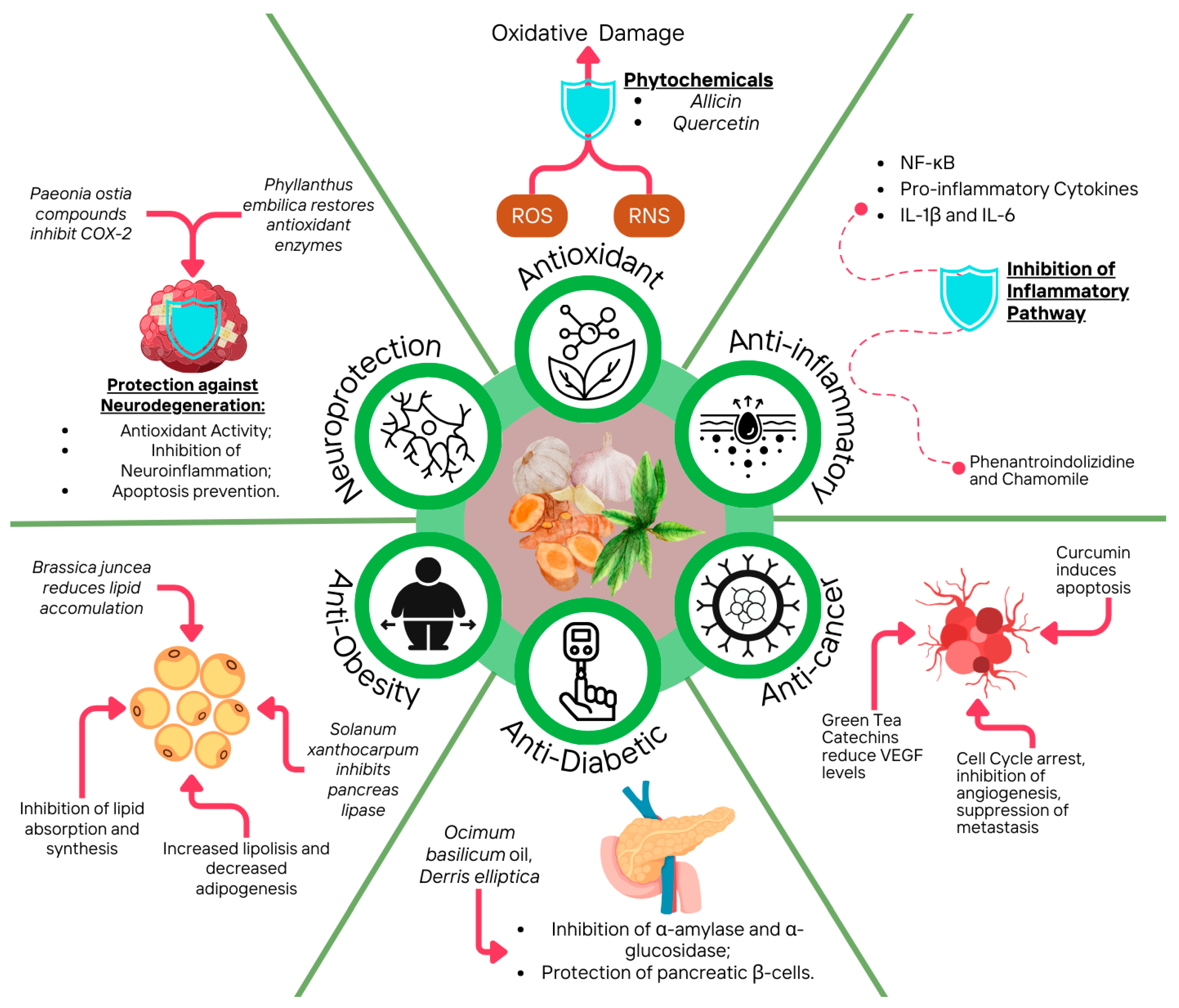
3.1. Antioxidant
Antioxidants and phytochemicals act through two main mechanisms: chain breaking by donating electrons to free radicals, and removal of ROS/reactive nitrogen species (RNS) by quenching their catalysts. These processes are critical in the prevention and treatment of cancer [139]. In a recent study, authors examined the antioxidant properties of allicin, a phytochemical from garlic, in in vitro and in vivo models of cholangiocarcinoma (CCA). Allicin was found to reduce OS and inhibit the signal transducer and activator of transcription 3 (STAT3) signaling pathway, which promotes tumor growth. The study demonstrated that allicin suppressed CCA cell proliferation, migration, and invasion, while inducing apoptosis. Its dual action as an antioxidant and inhibitor of tumor-promoting signals suggests allicin could be a promising therapeutic option for CCA [140].
In a study of HS-1793, a plant-derived resveratrol analog demonstrated its ability to enhance the efficacy of radiation therapy. HS-1793 improved lymphocyte proliferation, reduced DNA damage in irradiated mice, and decreased interleukin-10 (IL-10) and transforming growth factor-beta (TGF-β) levels. It also decreased regulatory T cells (Tregs) and inhibited CD206+ tumor-associated macrophages (TAMs), highlighting its potential as an antioxidant and immunomodulatory agent in cancer treatment [141]. An in vivo study evaluated the phytochemicals green tea (Camellia sinensis) catechins and curcumin (Curcuma longa) for chemopreventive effects in a hamster buccal pouch carcinoma model. Green tea polyphenols reduced vascular endothelial growth factor (VEGF) levels and inhibited angiogenesis, while curcumin induced apoptosis through cytochrome c release and caspase activation. Both agents reduced cell proliferative and angiogenic activity, and when combined, they were more effective at inducing apoptosis and cell cycle arrest [138]. In one study, dicoumarol (DC), a coumarin-like compound, exhibited anti-proliferative potential by disrupting key cellular processes, including cell proliferation and survival. It effectively reduces cell viability and induces apoptosis in several cell tumor lines. Additionally, DC increases OS by disrupting antioxidant defenses. Importantly, it does not harm mouse ovarian tissue or developing oocytes, suggesting its promise as a chemopreventive agent without adverse effects on fertility [142]. The aqueous extract of Fagonia cretica was tested for its effects on breast cancer cell lines. The results proved that the extract induced cell cycle arrest and apoptosis through both p53-dependent and independent mechanisms. Specifically, it led to a significant increase in Tumor Protein p53 and Forkhead Box O3 (FOXO3a) expression [143]. On the other hand, plant-derived quercetin exhibited chemopreventive effects in HepG2 (human liver cancer) cells by reducing ROS, downregulating phosphatidylinositol 3-kinase (PI3K), protein kinase C (PKC), and cyclooxygenase-2 (COX-2), while increasing pro-apoptotic p53 and BAX expression, highlighting its antioxidant and anticarcinogenic action [144]. One in silic and in vitro study attributed Markhamia lutea’s strong antioxidant properties primarily to its rich phytochemicals, including flavonoids and phenolic acids, which effectively scavenge free radicals and reduce OS by neutralizing ROS and chelating metal ions[145].
A study on Elephantopus mollis Kunth. highlighted 3,4-di-O-caffeoyl quinic acid for its significant antioxidant activity and chemopreventive properties, including apoptosis induction in NCI-H23 (human lung adenocarcinoma) cells and β-glucosidase inhibition [146]. Finally, the leaf extract of Onobrychis argyrea has demonstrated significant antioxidant, anti-diabetic, anti-Alzheimer’s disease and anti-cancer in vitro properties. It induced apoptosis in human colorectal adenocarcinoma (HT-29) cells by disrupting mitochondrial membranes and activating caspases, and showed high antioxidant activity, enzyme inhibitory activities and strong anti-cancer capacity [147].
3.2. Anti-Inflammatory
Anti-inflammatory phytochemicals may help prevent cancer by targeting inflammation-related mechanisms. They induce cell cycle arrest by inhibiting cell cycle regulators, leading to halted cancer cell proliferation [148]. Moreover, they promote apoptosis by upregulating pro-apoptotic proteins and caspases while downregulating anti-apoptotic factors, thus facilitating cancer cell death [149]. They also promote autophagy by upregulating autophagy-related proteins and inhibit mTOR signaling, leading to the elimination of damaged cells. These combined effects decrease inflammation and suppress tumor progression [150]. For instance, phytochemical phenanthroindolizidine alkaloids from Tylophora ovata exhibit anti-inflammatory and chemopreventive properties against triple-negative breast cancer (TNBC). O-methyltylophorinidine (1) and its synthetic form (1s) inhibit nuclear factor kappa B (NFκB), arrest the cell cycle, reduce spheroid growth, and block invasion [151].
In one study, real-time PCR analysis showed that Mangifera indica polyphenols (mainly gallic acid, hydroxybenzoic acid hexoside, and hydro- lysable tannins such as monogalloyl-glucoside) reduced Nuclear Factor kappa B (NF-κB) expression and phosphorylation in tumor necrosis factor-alpha (TNF-α)-treated MCF-12A cells, indicating anti-inflammatory effects. Mango indentified phytochemicals also upregulated microRNA-126 and downregulated microRNA-21 and modulated the phosphoinositide 3-kinase/protein kinase B/mechanistic target of rapamycin (PI3K/AKT/mTOR) signaling pathway [152]. Commiphora leptophloeos extract, rich in phenolic acids and flavonoids, showed anti-inflammatory effects through downregulation of NF-κB/COX-2 and reduction of pro-inflammatory cytokines (TNF-α, IL-1β, IL-6). This suggests its potential in the treatment of inflammatory bowel disease (IBD) [153]. The anti-inflammatory activity of Helicteres isora fruit extracts has been focused on its effects on pro-inflammatory mediators such as Prostaglandin E2 (PGE-2), COX-2, and Tumor Necrosis Factor-alpha (TNF-α). In the investigation, Enzyme-Linked Immunosorbent Assay (ELISA) assays were used to measure the inhibition of the production of PGE-2 in LPS-stimulated Human Monocyte Cell Line THP-1 (THP-1 cells). Compared to celecoxib, a known COX-2 inhibitor, the hexane extract showed the strongest inhibition of PGE-2 (69.68%), followed by ethanol extract (57.17%). All extracts were less effective than dexamethasone, a standard anti-inflammatory drug, but the hexane extract showed 51.61% inhibition of TNF-α [154]. The extract of Euphorbia hirta has been shown to have anti-inflammatory activity in vitro, which is relevant to its potential chemoprotective effects. By reducing nitric oxide (NO) production through inhibition of inducible NO synthase (iNOS), E. hirta can help to lower inflammation, a known contributor to cancer advancement. Chronic inflammation can facilitate tumor growth and spread, so the extract’s ability to suppress NO and inflammatory cytokines may play a role in its anticancer properties. Besides, compounds such as 5-hydroxymethylfurfural (5-HMF) in the ethanol extract are known for their anti-inflammatory effects, supporting the extract’s potential to moderate cancer risk by managing inflammation [155]. Chamomile phytochemicals, including β-amyrin, quercetin, and lupeol, act as NF-κB inhibitors and induce G2/M cell cycle arrest. These compounds lower pro-inflammatory cytokines IL-1β and IL-6 and also interact with microtubules, which supports their potential as anti-cancer modulators of inflammatory pathways [156].
3.3. Antidiabetic
Diabetes mellitus is a metabolic syndrome condition which increases the levels of glucose in the blood [158,159]. The main chronic complications of this disease are linked to the level of exposure of patients with hyperglycemia [158]. Despite the availability of chemical drugs acting as therapeutical agents, there is a will to find natural alternatives to control the complications derived from the disease and improve glycemic control and glucose intolerance [159]. In this sense, traditional medicine revealed several molecules present in plants with antidiabetic activity, as well as their mechanisms of action [158].
For instance, leaves extracts have been widely studied to determine their antidiabetic potential. An in vivo and in vitro study investigated the volatile and phenolic compounds present in Ocimun baslicum L. essential oil to determine its potential antidiabetic activity. Chemical agent streptozotocin (STZ) (50 mg/Kg of body weight) was used as a model to induce diabetes mellitus (DM) in Wistar Albino rats. After 28 days of feeding with basil essential oil (70 mg/Kg of body weight), rats showed a protection effect in pancreatic β-cells and a decrease in blood glucose levels. Regarding in vitro assays, both α-amylase and α-glucosidase inhibitory activity were studied, showing a strong inhibition potential. Authors linked the antidiabetic effect of basil essential oil to the phenolic and flavonoid compounds content. However, the mechanism of action was not dilucidated [160]. In another in vivo study, Derris elliptica methanolic leaf extract phytochemical profile and antidiabetic potential was determined. STZ was used to induce DM in Sprague Dawley rats and after 14 days feeding with the plant’s extract at 2 different concentrations (200 and 400 mg/Kg body weight), results showed hypoglycemic effect, and reduction of cholesterol levels in a dose dependent manner. Moreover, the highest dose increased serum insulin levels, and antihyperglycemic effect was shown. Although authors did not explained the mechanism of action, quercetin was the compounds linked to these antidiabetic properties [159]. Eugenia sonderiana leaves extracts were also studied to determine the antidiabetic potential and to develop a structure-activity correlation. Both in vivo and in vitro studies were assessed, showing great α-amylase and α-glucosidase inhibitory activity, reduction of glucose and pancreatic enzymes and triglycerides, while HDL cholesterol levels were maintained within normal ranges. To explain the antidiabetic ability of Eugenia sonderiana leaves extracts, authors suggested a synergistic effect between polyphenols by increasing glycogen synthesis and α-amylase, and saponins by a complex formation with the cholesterol [158].
3.4. Antiobesity
Obesity is becoming a significant health problem worldwide. This condition contributes to the increase of prevalence of chronic diseases, including T2D, hypertension, dyslipidemia, cardiovascular diseases and some types of cancer [161,162]. Common strategies to decrease the incidence of obesity are physical activity, surgery, drugs, and diet modifications [162]. In this regard, natural compounds present in plants have been suggested as potential chemicals to be used for both prevention and treatment of obesity [162]. Several studies have assessed the anti-obesity potential of phytochemicals. For instance, green tea has been traditionally used as a medical plant, and scientific studies have revealed its potential used for obesity prevention. In vitro results regarding green tea leaves suggest the anti-obesity potential of this plant by inhibiting adipocyte differentiation and proliferation [163].
In a study, C57BL/6J mice fed a high-fat diet supplemented with 0.25% Rosa centifolia polyphenols for 35 days showed decreased body weight and adipose tissue, as well as reduced serum cholesterol and hepatic triglyceride levels. The treatment increased fecal triglycerides and increased lipolysis-related proteins while suppressing lipid synthesis enzymes. Ellagic acid, a key component of R.centifolia, is likely responsible for these anti-obesity effects, emphasizing its capability for treating obesity [161]. Solanum xanthocarpum exhibits anti-obesity properties mainly by inhibiting pancreatic lipase, an enzyme essential for digesting lipids. The active phytochemicals in the plant, including solasodine, carpesterol, β-sitosterol, and diosgenin, are responsible for this effect. Although pancreatic lipase inhibition shows some potential, the efficacy is modest compared to orlistat. In addition, the hypoglycemic and hepatoprotective properties of the plant, largely attributed to solasodine, support its overall metabolic benefits [164].
Brassica juncea attenuated lipid accumulation in 3T3-L1 adipocytes and lowered epididymal white adipose tissue (eWAT) mass in obese mice fed a high-fat diet [165]. One study tested five rhubarb hydroxyanthraquinones (HAQ’s) for their obesity-related activity. Rhein and emodin showed the most success, significantly reducing triglycerides and body fat in 3T3-L1 cells and high-fat diet-induced obese rats. Rhein was particularly effective in reducing plasma cholesterol and attenuating obesity-associated changes, making it a potential obesity-reducing agent [166]. Anthophycus longifolius compounds delayed lipid and carbohydrate digestion by inhibiting α-amylase, α-glucosidase, and pancreatic lipase [167]. Finally, acetone fractions from Rumex rothschildianus, rich in flavonoids and phenolics, strongly inhibited lipase activity, comparable to orlistat, and also affected α-amylase and α-glucosidase [168].
3.5. Neuroprotective
In the past five decades, there has been a rise in the incidence of neurodegenerative disorders (NDs), which has been linked to the increase of longevity that has occurred. NDs encompass a wide range of conditions that affect the central nervous system, having alterations in the neuronal structure and cellular dysfunction that leads to progressive degeneration [169]. The most prevalent NDs are Alzheimer disease (AD), Parkinson disease (PD) and multiple sclerosis MS). Despite the drugs available to ameliorate these conditions, their efficacy is limited. In the past years, it has been suggested natural compounds as potential alternatives with the ability to delay the disease onset, reduce its progression and regenerate the damage via their anti-amyloid, antioxidant, and ant-inflammatory properties [170].
Paeonia ostia has traditionally been used as medical and ornamental plant in China. The neuroprotective effect of the stamens of this plant has been studied by NO inhibition assay in lipopolysaccharides-induced BV-2 cells (LPS-induced BV-2) and molecular docking. LPS-induced BV-2 results showed (+)-3′′-methoxy-oxylactiflorin excellent inhibitory property in inhibiting NO production. Moreover, molecular docking results showed that the anti-inflammatory mechanism of this molecule was through a binding action with protein cyclooxygenase-2 (COX-2) [171]. In another study, Phyllantys emblica fruits extract were tested in rats with sodium valproate-induced postnatal autism. After treatment with the fruit’s extracts in 100mg/Kg of body weight, amelioration of social interaction, social affiliation, anxiety, and motor coordination was achieved. Moreover, results showed the restoring effect of the extract in glutathione-S-transferase (GST) and glutathione reductase (GR), which are oxidative enzymes, and reduction of malondialdehyde (MDA) and NO levels [172]. Another in vivo study determined the neuroprotective potential of Tabebuia impetginosa leaves by using a chemo brain model. Authors fed rats with 200 mg/Kg of body weight. Cyclophosphamide inducing cognitive impairment and treated the animals with 30 mg/kg of body weight of the extract for 14 days. Behavioral tests including locomotor, Y-maze, and passive avoidance tests were conducted and results showed pronounced antioxidant and anti-inflammatory potentials. Moreover, the restoration of normal hippocampal histological features and attenuated apoptotic caspase-3 expression was achieved, demonstrating the neuroprotective activity of the leaves’ extracts [173].
Table 2.
Chemopreventive activities of plant extracts and their phytochemicals: in silico, in vivo and in vitro examples.
Table 2.
Chemopreventive activities of plant extracts and their phytochemicals: in silico, in vivo and in vitro examples.
| Plant species | Extract/ Fraction | Phytochemical compound | Chemopreventive activity | Analysis Method | Trial type | Mechanism of action | Results | Ref. |
|---|---|---|---|---|---|---|---|---|
| Antioxidant activity | ||||||||
| Garlic (Allium sativum L.) | ns. | Allicin (purity >90%) | Anti-tumor (Cholangiocarcinoma) | CCK-8, colony formation, FC, WB | In vitro/In vivo | STAT3 inhibition via SHP-1 upregulation | Suppressed proliferation, invasion, EMT, and tumor growth | [140] |
| ns. | HS-1793 | Resveratrol analogue | Anti-tumor (Murine breast cancer) | LYM proliferation, DNA damage assessment, Treg and TAM analysis | In vivo | Inhibition of LYM damage and immune suppression by Tregs and TAMs | Enhanced LYM proliferation, reduced Tregs, and decreased IL-10/TGF-β | [141] |
| Green tea‒Curcuma longa L. | ns. | Catechins and curcumin | Anti-tumor (OSCC) | Histology, Immunofluorescence, FC | In vivo | AP induction and anti-angiogenesis | Increase in AP and reduction in tumor growth | [138] |
| Melilotus officinalis L. | ns. | DC (coumarin derivative) | Anti-proliferative, gonad-safe | DC injection in BALB/c mice ovarian apoptosis, meiotic spindle | In vitro/In vivo | Alteration of cell cycle dynamics without effect on microtubule stability | DC suppressed cell proliferation and increased AP in Vero and MCF-7 cells | [142,174] |
| Polyalthia longifolia L. | ME | Tetranorditerpene | Anti-cancer (prostate, leukemia cells) | Proteomic analysis | In vitro | Activates ER stress, induces apoptosis | Inhibits prostate cancer, leukemia cell growth | [175] |
| Fagonia cretica L. | AqE | ns. | Cytotoxic, induces cell cycle arrest | siRNA knockdown, MTT and FC, comet assay, WB | In vitro | Induction of DNA damage, and activation of p53 and FOXO3a | Induce cell cycle arrest and AP in two phenotypic breast cancer cell lines | [143] |
| Onobrychis argyrea L. | ME (leaves) | Quinic Acid, Isoquercitrin, Epicatechin, Routine | Antioxidant, antidiabetic, anticancer | LC-MS/MS, DPPH, Iron Reduction, Enzyme Inhibition, XTT, FC | In vitro | ME induces apoptosis in HT-29 cells by disrupting mitochondrial membranes and activating caspases | High antioxidant, enzyme inhibition, strong anti-cancer capacity | [147] |
| Elephantopus mollis Kunth. | ME | 3,4-di-O-caffeoyl quinic acid | Cytotoxicity and -glucosidase inhibitory effects | DPPH, FRAP, Metal Chelating, β-Carotene, Cytotoxicity | In vitro | Induces cell death in NCI-H23 cells by triggering apoptosis | High antioxidant, induces apoptosis | [146] |
| Thymus vulgaris L. | MetE, EAE, ChE, BolE, AqE, PEE | Polyphenols, tannins, flavonoids and sterols/triterpènes | Antioxidant | DPPH, ABTS, Ferrous Ion Chelation, CVT | In vitro | Radical scavenging, metal chelation, and electrochemical reduction | Strong antioxidant capacity correlated with phenol and flavonoid content | [176] |
| Markhamia lutea L. | Leaves extract | Flavonoids (O-glycosides) | Antioxidant, Anti-AChE, Anti-BChE, Aβ-amyloid-42 inhibition | DPPH, ORAC, Iron Reduction, FRAP | In silico/ in vitro | Induces antioxidant effects and inhibits AChE, BChE, and Aβ-amyloid 42 | DPPH: 35.69 µg/mL, ORAC: 16,694.4 μM TE/mg, and iron chelation: 70.7 μM EDTA eq/mg | [145] |
| Hertia cheirifolia L. | Organic and ethyl acetate fraction | Total phenolics (100–250 mg GAE/g) | Antioxidant | DPPH, ABTS, FRAP, β-carotene | In vitro | Synergistic mechanisms between different biomolecules | DPPH: 38.83 µg/ml,ABTS: 23.76 µg/ml; FRAP: 2628.87 µmol Fe²⁺ Eq/mL; β-carotene: 58.91% | [177] |
| ns. | AqE | QUE | Anticarcinogenic (hepatocellular carcinoma) | WB, RT-PCR | In vitro | Downregulation of ROS, PKC, PI3K, COX-2; Upregulation of p53, BAX | QUE modulated OS and apoptotic pathways in HepG2 cells | [144] |
| Anti-inflammatory activity | ||||||||
| Tylophora ovata L. | Natural and synthetic PAs | O-methyltylophorinidine (1, 1s) | Anti-tumor against TNBC | NFκB inhibition, 3D co-culture | In vitro | Stabilizes IκBα, blocks NFκB | Inhibits spheroid growth, surpasses paclitaxel | [151] |
| Mangifera indica L. | ns. | Polyphenols | Anti-inflammatory, anticancer | Real-time PCR analysis and protein expression | In vitro | Modulated PI3K/AKT/mTOR, , NF-κB, PARP-1, Bcl-2 | Reduced cancer cell growth by 90% | [152] |
| Helicteres isora L. | DCM-E/HeE | Rosmarinic Acid | Anti-inflammatory, Antioxidant | ELISA assays | In vitro | Differentiation in cancer cells and showed no cytotoxic effect at high levels | Reduced TNF-α, PGE-2, and NO levels; highest COX-2 inhibition | [154] |
| Waltheria indica L. | Roots and aerial parts/ CH2Cl2 extract | Flavonoids | Anti-inflammatory, Cancer chemoprevention | NF-κB inhibition, luciferase reporter assay, QR inducing assay | In vitro | Induce Phase 2 enzyme activity via QR induction assay | Of 29 compounds in the study, 7 showed inhibitory activity on the NF-κB pathway | [178] |
| Commiphora leptophloeos L. | Hydroalcoholic leaf extract | Phenolic acids and flavonoids | Anti-inflammatory | NO radical inhibition analysis, qPCR, physicochemical tests | In vitro/ In vivo | Downregulates NF-κB, COX-2, reduces cytokines | Reduces inflammatory markers, promising for inflammatory bowel disease | [153] |
| Matricaria chamomilla L. | ns. | β-Amyrin, β-Eudesmol, β-Sitosterol, Apigenin, Lupeol, Quercetin, Myricetin | Anti-inflammatory, anticancer | Proteome analysis, WB, Quantitative Real-Time RT-PCR, Thermophoresis | In silico/ In vitro | Inhibition of NF-κB pathway, reduction of IL-1β, IL6 mRNA expression, and G2/M cell cycle arrest | NF-κB inhibition, potential cancer prevention, reduced proinflammatory cytokine expression | [156] |
| Asparagusdensiflorus meyeri L. | Root and aerial parts/ DCM-E | Saponins, glycosides, sterols, triterpenes | Cytotoxic, anti-inflammatory | MTT assay, MCF-7 cell stimulation using TNF—α, RT-PRC | In vitro | Reduces NO release and NF-κB gene expression | Significant cytotoxicity (IC50 26.13 μg/ml) | [179] |
| Capparis cartilaginea L. | Ethanolic leaf extract | Alkaloids, flavonoids, phenols, fatty acids, carotenes | Antioxidant, cytotoxic, anti-inflammatory | FBRC, FRAP, MTT assay, COX-1 inhibition | In vitro | Dose-dependent inhibition of thermally induced protein denaturation | Anti-inflammatory (IC50 60.23 and 17.67 µg/mL) better than standards | [180] |
| Corchorus olitorius L. | Hydroethanolic leaf extract | Tannins, flavonoids, phenolics, terpenoids, cardiac glycosides | Pro-estrogenic, anti-inflammatory | Phytochemical and ELISA analyses | In vivo | Lowers IL-6 and inhibits proliferation by binding phytoestrogens to ER-β | Strong antioxidative potential due to its high tannin content. | [181] |
| Amaranthus hybridus L. | Tannins, flavonoids, phenolics, cardiac glycosides, coumarins | Reduction in tumor size and incidence | ||||||
| Euphorbia hirta L. | Whole extract | Phytol, fatty acids and 5-HMF | Anti-inflammatory | NO Production | In vitro | Suppression of PG generation | Inhibition of iNOS directly involved in inflammation | [157] |
| Antidiabetic activity | ||||||||
| Tradescantia pallida L. | Leave extract | Syringic acid, p-coumaric acid, morin, and catechin | Glycosylation and hemoglobin activity | α-amylase assay | In vitro | Glycosylation inhibition non-enzymatically | Boosts insulin production, revitalizes β-cells, inhibits AGEs, stimulates glucose transporters and AMPK | [182] |
| Cissampelos capensis L. | Leaf, stem, and rhizome | Glaziovine, Pronuciferine and, cissamanine | Antihyperglycemic | α-amylase assay | In vitro | Enzyme inhibition pathway | Reduction of glucose levels | [183] |
| Phyllanthus emblica L. | ns. | Flavonoids | Antihyperglycemic | Molecular docking assay | In silico | Hypoglycemic action, reduction of relative risk of T2D, and PPAR inhibition of T2D | High binding affinity and selectivity for T2D therapeutic targets | [184] |
| Ocinum sanctum L. | Leaves | Eugenol | Antihyperglycemic | ELISA, RIA, and Neutral Red assay | In vitro | Physiological pathway | Reductions in plasma glucose levels in T2D are associated with enhanced insulin secretion from pancreatic islets, perfused pancreas | [185] |
| Ocinum basilicum L. | Leaves | TPC and FC | Antihyperglycemic | Enzyme inhibitory activity assay | In vitro | Enzymes inhibition pathway (α-glucosidase, α-amylase, DPP-IV, PTP1B, and SGLT2) | Inhibition of intestinal sucrase, maltase, and porcine pancreatic α-amylase | [186] |
| Derris elliptica L. | Leaves | QUE and ceramide | Antihyperglycemic | Biochemical analysis and histopathology study | In vivo | Enzymes inhibition pathway | Increase insulin secretion, protect pancreatic β–cells from oxidative stress | [159] |
| Carica papaya L. | Seeds | Hexadecanoic acid Methyl ester, 11-ODA Oleic acid | Antihyperglycemic | α-amylase and α-glucosidase inhibition assay | In vitro | Enzymes inhibition pathway | Reduction of glucose levels | [187] |
| Rhazya stricta L. | Root | Hexadecanoic acid, Methyl ester | Antihyperlipidemic activity and hepatoprotective effect | DPP-IV, α-amylase, α-secretase inhibition assay, GLP-1 measurement | In vitro/ in vivo | Enzymes inhibition pathway | Reduce blood glucose and HbA1c, reduce cholesterol and triglyceride levels, reduce liver enzyme activity | [188] |
| Halooxylon stocksii L. | Root and aerial parts | 8-ODA Methyl ester | Antidiabetic | α-amylase and α-glucosidase assay | In vitro | Enzymes inhibition pathway | Reduction of glucose levels | [189] |
| Antiobesity activity | ||||||||
| Rosa centifolia L. | Petals | Ellagic acid (polyphenols) | Lipid metabolism improvement | PCR | In vivo | Suppression of lipid synthesis, Inhibition of intestinal absorption, Downregulation of Scd1 and Hmcgr mRNAs in the liver | Reduced body weight and adipose tissue, increased fecal triglycerides, improved lipid, and cholesterol metabolism | [161] |
| Rheum rhabarbarum L. | ns. | Emodin, rhein (anthraquinones) | Lipid lowering | ELISA assay and histological evaluation | In vitro/ in vivo | FAS and ACC production was prevented through decreased PPARγ and C/EBPα expression, leading to a reduction in lipid accumulation | Body weight and adipose tissue reduction | [166] |
| Brassica juncea L. | ns. | Sinigrin (glucosinolate) | Anti-obesity | Cell Culture and XTT Assay, WB, Histological Analysis | In vitro/ in vivo | Reduce expression of adipogenic and lipid synthesis proteins | Inhibition of lipid accumulation in 3T3-L1 and decrease eWAT mass in obese mice fed a high-fat diet | [165] |
| Anthophycus longifolius L. | ns. | Rhodomycinone, salsolinol, 5-HCO, 2-COS, demethylalangiside | Anti-obesity and Anti-hyperglycemia | α-amylase, α-glucosidase, and pancreatic lipase assay | In vitro | Enzymes inhibition pathway | Delay lipid and CH digestion and absorption | [167] |
| Solanum xanthocarpum L. | Fresh and dry leaves | Solasodine, Carpesterol, β-Sitosterol, Diosgenin | Hypoglycemic, hepatoprotective hypotensive | Pancreatic Lipase Inhibition Assay and MTT | In vitro | ns. | At 62.5 µg/mL, the fresh leaf extract reduced cancer cell viability by 50% | [164] |
| Rumex rothschildianus L. | Acetone fraction | Flavonoids, phenolics | Anti-α-amylase, anti-α-glucosidase, anti-lipase | Lipase inhibition activity | In vitro | Inhibits OS, α-amylase, α-glucosidase, and lipase | Strong lipase inhibition (acetone fraction IC50 26.3 μg/ml), close to orlistat (IC50 12.3 μg/ml) | [168] |
| Neuroprotective activity | ||||||||
| Paeonia ostii | Stamen | (+)-3′′-methoxy-oxylactiflorin | Anti-inflammatory | Molecular docking and NO inhibition assay | In vitro/ in silico | Inhibition of NO production by binding with protein COX-2 | NO production reduced to values of EC50 3.02 μM | [171] |
| Phyllanthus emblica | Fruit extract | ns. | Anti-inflammatory | NO inhibition assay | In vivo | Reduction of pro-inflammatory markers IL-1β and TNF-α and up-regulation of expression of up-regulated 5-HT1D, 5-HT2A, and D2 receptor proteins | Amelioration of social interaction, social affiliation, anxiety, and motor coordination | [172] |
| Tabebuia impetiginosa | Leaves | Iridoids and organic acids | Anti-inflammatory | AChE inhibitory activity, LA detection, Y-maze test, PA assay | In vitro/ in vivo | Attenuation of cognitive impairment induced by CP supported by its effect on rats’ performance in Y-maze and PA tests | Reduction of CP-induced chemo brain and restoration of normal hippocampal function | [173] |
Abbreviations: CCK-8: Cell Counting Kit-8; STAT3: Signal Transducer and Activator of Transcription 3; WB: Western blotting ; FC: Flow cytometry ; SHP-1: Src Homology 2 Domain-Containing Phosphatase-1; EMT: Epithelial-Mesenchymal Transition; Treg: Regulatory T cell; TAM: Tumor-associated macrophage; LYM: lymphocyte; IL-10: Interleukin-10; TGF-β: Transforming growth factor-beta; ME: Methanol Extract; OSCC: oral squamous cell carcinoma; AP: Apoptosis; DC: Dicoumarol; BALB/c; Bagg Albino Laboratory strain/c; Vero: African green monkey kidney epithelial cells; MCF-7: Human breast cancer cells; siRNA: Small interfering RNA; MTT assay: 3-(4,5-Dimethylthiazol-2-yl)-2,5-diphenyltetrazolium bromide assay; p53: Tumor Protein p53; FOXO3a: Forkhead Box O3; QUE: Quercetin; ROS: Reactive Oxidative Species; OS: Oxidative Stress; PKC: Protein Kinase C; PI3K: Phosphatidylinositol 3-Kinase; COX-2: Cyclooxygenase-2; RT-PCR: Reverse Transcription Polymerase Chain Reaction; HepG2 cells: Human liver cancer cell line; AMPK: adenosine monophosphate-activated protein; AGES: advanced glycation end products; T2D: type 2 diabetes; PPAR: peroxisome proliferator activated receptor; NFκB: Nuclear Factor kappa B; IκBα: Inhibitor of Nuclear Factor kappa B alpha; HT-29 cells: Human colorectal Adenocarcinoma cell line; DPPH: 2,2-Diphenyl-1-picrylhydrazyl, FRAP: Ferric Reducing Antioxidant Power; NCI-H23 cells: Human Lung Adenocarcinoma cell line; ORAC: Oxygen Radical Absorbance Capacity; ER: Endoplasmic Reticulum; HeE: Hexane Extract; DCM-E: Dichloromethane Extract; MetE: Methanolic Extract; EAE: Ethyl Acetate Extract; ChE: Chloroform Extract; BolE : Butanol Extract; AqE: Aqueous Extract ; PEE: Petroleum Ether Extract; CVT: Cyclic Voltammetry Techniques; PI3K/AKT/mTOR: Phosphoinositide 3-Kinase/Protein Kinase B/Mechanistic Target of Rapamycin; -κB: Nuclear Factor kappa B; PARP-1: Poly (ADP-ribose) Polymerase 1; Bcl-2: B-cell Lymphoma 2; TNF-α: Tumor Necrosis Factor-alpha; THP-1 cells: Human Monocyte Cell Line THP-1: ELISA: Enzyme-Linked Immunosorbent Assay; QR: Quinone reductase ; NO: Nitric Oxide; 5-HMF: hydroxymethyl-2-furancarboxaldehyde; iNOS: Inducible NO synthase; PG: Prostaglandins; T2D: type 2 diabetes; RIA: radioimmune assay; TPC: total polyphenol compounds; FC: flavonoids content; 11-ODA: 11-octadecanoic acid; HbA1c: glycosylated hemoglobin; PCR: polymerase chain reaction; FAS: fatty acid synthase; ACC: acetyl-CoA carboxylase; 5-HCA: 5-hydroxyconiferyl; 2-COS: 2-caffeoylisocitrate; DPP-IV:p ipeptidyl peptidase-IV; PTP1B: protein tyrosine phosphatase 1B; SGLT2: sodium-glucose co-transporter 2; qPCR: polymerase chain reaction; RT-PCR: Reverse Transcription-Polymerase Chain Reaction Assay; FBRC: Ferric-bipyridine reducing capacity of total antioxidant; ER-β; Strogen Receptor Beta; eWAT: Epididymal white adipose tissue; IL-1β: interleucina 1 β; TNF-α: tumor necrosis factor alpha; 5-HT1D:serotonin 1D receptor ; 5-HT2A: serotonin 2A receptor; D2: dopamine 2 receptor; AChE: acetylcholinesterase; LA: locomotor activity; PA: passive avoidance. ns: not specified.
4. Development of Phytochemicals as Nutraceuticals
4.1. Extraction, Purification and Encapsulation
Based on the above information, phytochemicals, which include a diverse range of compounds, have been recognized for their potential influence on health promotion and disease prevention. Due to their therapeutic effect, phytochemicals have become prime candidates in the development of nutraceuticals, which are food-derived products that offer additional health benefits beyond basic nutrition [190]. However, direct ingestion of phytochemicals within their native plant matrices presents several challenges [191]. The bioavailability of these compounds is often restricted by factors such as low concentrations, poor solubility, instability under gastrointestinal conditions, and rapid metabolic degradation and excretion [192]. These obstacles highlight the need for developing methods to concentrate, purify, and encapsulate phytochemicals, thereby enhancing their effectiveness as nutraceuticals (Figure 2) [193].
Figure 2.
Process of development of phytochemicals as nutraceuticals.
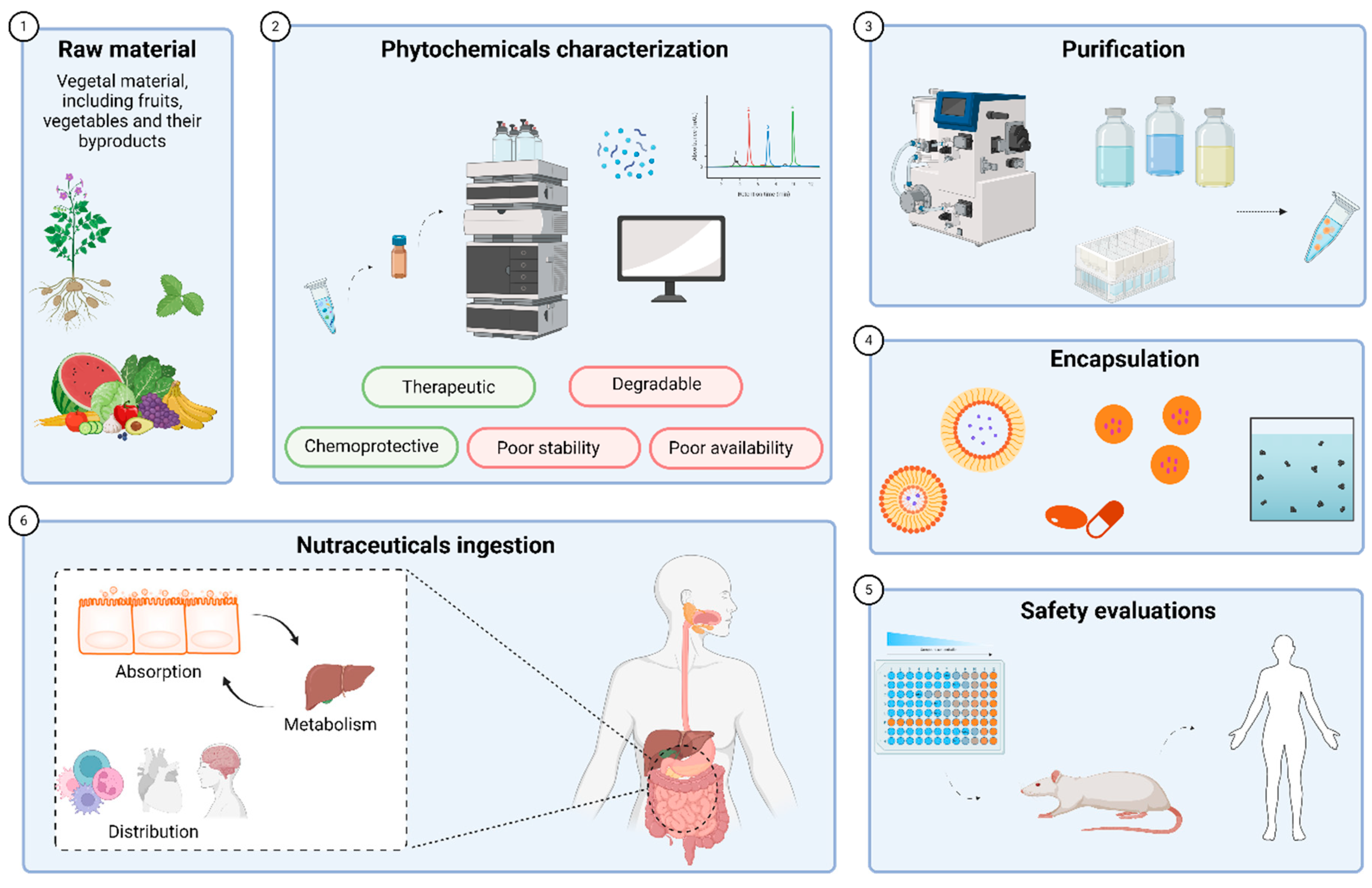
The initial phase in the utilization of phytochemicals for nutraceutical purposes involves their extraction from plant sources [194]. This extraction process is complex and must be carefully optimized to preserve the structure and bioactivity of the target compounds [195]. Traditional extraction techniques, such as solvent extraction, have been improved by emerging methods like microwave-assisted extraction and ultrasound-assisted extraction, since they offer increased efficiency and selectivity [196]. A major challenge in phytochemical extraction and recovery is to incorporate environmental sustainability into the design and optimization of production processes, with the aim of developing more eco-friendly methodologies [197] and enhancing economic outcomes within the food industry [198]. The choice of appropriate extraction techniques is crucial, as it directly impacts the quality and characteristics of the final product [199]. Key factors influencing the efficacy of both conventional and emerging extraction techniques include the solid-to-solvent ratio, solvent concentration, particle size, and the mode of extraction (flow or batch) [200]. These parameters critically affect the efficiency of extraction and the phytochemical composition of the resultant extract [201].
Following extraction, the subsequent step involves the purification of the bioactive compounds from the crude extract. This is necessary because the initial phytochemical mixture typically contains a combination of active and inactive substances, along with impurities that could affect the safety and efficacy of the final product [202].
Recent advancements in purification and isolation techniques for bioactive compounds from plant sources have addressed some of the challenges posed by the complexity and diversity of phytochemicals [203,204]. These advancements focus on optimizing methods to concentrate the desired compounds, thereby enhancing bioactivity—such as antioxidant, antibacterial, or cytotoxic effects—while maintaining simplicity, specificity, and efficiency in processing [205,206]. Techniques such as high-performance liquid chromatography (HPLC), solid-phase extraction (SPE), and preparative thin-layer chromatography (TLC) are commonly used for the isolation and purification of bioactive molecules due to their practicality, cost-effectiveness, and the availability of various stationary phases [199,205]. Notably, silica, alumina, cellulose, and polyamide have proven effective in separating phytochemicals [205]. To achieve the separation of complex mixtures, the use of multiple mobile phases with varying polarity is often required [199,205]. The degree of purification achieved is directly linked to the pharmacokinetic properties of the compounds, influencing their bioavailability, distribution, metabolism, and excretion [207].
Despite the successful purification of phytochemicals, the formulation of these compounds into nutraceuticals remains challenged by issues of stability, solubility, and bioavailability. Encapsulation technologies have emerged as vital strategies to overcome these challenges [208,209]. Additionally, encapsulation can improve the quality of food products by masking undesirable odors and flavors and extending shelf life [198]. This process involves enclosing phytochemicals within a protective matrix or carrier system, which shields them from environmental degradation, enhances their solubility in biological fluids, and facilitates controlled release in the gastrointestinal tract [210,211]. Various encapsulation methods have been developed, each offering unique advantages depending on the physicochemical properties of the phytochemicals and their intended application [212]. Microencapsulation, for instance, is widely used to protect sensitive compounds and improve their stability during processing and storage [213,214]. Conversely, nanoencapsulation provides enhanced bioavailability and targeted delivery due to the small particle size and large surface area [215,216], which improve interactions with biological membranes and allow for more precise control over release kinetics [217,218].
In conclusion, the effective application of phytochemicals as nutraceuticals is closely linked to advances in extraction, purification, and encapsulation techniques. These processes not only enhance the pharmacological potential of phytochemicals but also ensure their stability, bioavailability, and functional integration into various nutraceutical products.
4.2. Considerations of Bioavailability
Phytochemicals offer a wide range of health benefits, yet their effectiveness is often compromised by challenges related to bioavailability, including issues with water dispersibility, chemical stability, and gastrointestinal absorption [219]. For phytochemicals to be effective, they must be released from their food matrices, solubilized in gastrointestinal fluids, and absorbed by enterocytes in the gastrointestinal tract (GIT). However, these compounds may precipitate, interact with other dietary molecules, or be chemically altered by digestive enzymes, metabolic pathways, or gut microbiota, which can significantly reduce their bioactive forms by the time they enter systemic circulation [220].
Therefore, the bioavailability of phytochemicals is affected by various factors, including the complexity of the food matrix, the chemical form of the compound, and individual physiological differences such as gut microbiota composition, mucosal health, and metabolic activity. These factors contribute to considerable inter- and intra-individual variability in bioavailability, complicating the prediction of their efficacy [221]. Additionally, the impact of food processing on phytochemical stability and bioavailability is critical, as processing techniques can either enhance or diminish their bioactive potential [222].
To address these issues, in vitro digestion models and cellular uptake assays are employed to assess factors affecting both bioaccessibility and bioavailability. These models are essential for screening and optimization of formulations prior to more expensive in vivo studies. Despite the availability of various assessment methods, a universally accepted standard for bioaccessibility remains elusive, and current methods must account for the diverse structural characteristics of phytochemicals [207]. For lipophilic compounds such as carotenoids and fat-soluble vitamins, formulation techniques like encapsulation and emulsification have shown potential in enhancing bioaccessibility and protecting these compounds from degradation [207].
In silico modeling, commonly used in pharmaceutical research, offers an interesting approach as it allows to predict phytochemical bioavailability based on their physicochemical properties. However, these models are still developing for dietary phytochemicals, indicating a need for further research to establish effective predictive tools [223]. Moreover, technological processes such as thermal processing and biotechnological interventions, including enzymatic hydrolysis and fermentation, have demonstrated their influence in improving the bioavailability of phenolic compounds. However, while these strategies have shown promise in animal studies, further human trials are necessary to validate their effectiveness [224]. Thus, both research and innovation in processing and formulation techniques are essential for advancing nutraceuticals and optimizing the bioavailability of phytochemicals.
5. Current Applications of Nutraceuticals
Nutraceuticals, which combine nutritional and medicinal properties, are increasingly being incorporated into food products to promote health and prevent disease. These bioactive compounds, derived from a variety of matrices, offer a wide range of benefits, such as improving cardiovascular and immune system function, or managing chronic diseases. In addition, advances in encapsulation and nanotechnology have further improved their efficacy in food applications, expanding the potential of health-promoting foods [199].
5.1. Ingredients of Functional Foods
The incorporation of antioxidants in functional foods has been widely researched due to their health benefits and potential enhancement food quality and shelf life. This incorporation can be done in several ways, including through the addition of crude extracts, fibers, color additives or other food enrichment (Figure 3) [225].
Crude extracts are raw, unrefined substances obtained from plants, animals, or other natural sources that contain bioactive compounds. The most widely utilized are plant polyphenol extract, such as green tea extract, grape seed extract, rosemary extract, but also mushroom extracts, spice and herbs extracts (garlic extract, ginger extract, turmeric extract), vanilla extract and more recently, algae extract (spirulina, chlorella extracts). Plant polyphenols are often used to delay lipid oxidation, thereby enhancing the shelf-life of lipid-bearing foods [226]. These extracts also exhibit antimicrobial activity with green tea extract having major antimicrobial activity against major foodborne pathogens such as Listeria monocytogenes, Salmonella Typhimurium, Escherichia coli O157:H7, and Campylobacter jejuni, contributing to food safety [227,228]. Some crude extracts also contribute to flavoring such as mushroom extracts, garlic extracts, rosemary extracts and spices and herbs extracts in general. These can also provide distinctive aroma and color enhancing their sensory attributes. For example, spirulina extracts that are naturally greenish blue can modify the color of a food [229].
Fiber can be added to foods due to their functional and health benefits. They can be roughly divided into soluble and insoluble fibers. Soluble fibers include pectins and inulin while insoluble fibers include cellulose, psyllium and wheat bran. Functionally, adding fiber to foods can impact their sensory properties, such as appearance, texture, taste, and flavor [230]. Pectins can be found on fruit jams and jellies, acting as a gelling agent, giving jams and jellies their thick, spreadable consistency. Additionally, it allows for a lower sugar amount to be used while improving the product’s shelf life and stability [231]. Inulin, usually extracted from chicory root is used as a fat replacer and provides a creamy texture in low-fat or reduced-calorie foods [232]. It also serves as a prebiotic, promoting the growth of beneficial gut bacteria [233]. Cellulose and its derivatives, such as microcrystalline cellulose (MCC) and bacterial cellulose, are commonly used as stabilizers and thickeners in food products, including meat products, emulsions, beverages, dairy products, and bakery items [234]. Furthermore, MCC and other cellulose derivatives are used in functional and nutraceutical foods for their positive effects on gastrointestinal health and lipid metabolism [234]. Cellulose gels are also widely used in the food industry for their high-water absorption capacity and biocompatibility. They are applied in food packaging, functional foods, and food safety due to their structural flexibility and stimuli-responsive properties [235]. Other more unrefined fibers such as psyllium husk and wheat bran are also often used in the food industry. Unmodified wheat bran and psyllium husk are used as nutritional enhancements as they are a source of dietary fiber, minerals, vitamins, and bioactive compounds such as phenolic acids, which contribute to improved bowel health, prevention of diseases like colon cancer, and cardiovascular health [236]. These fibers, when modified through processes such as mechanical milling, enzymatic hydrolysis, and thermal treatments can be utilized as functional ingredients. For example, modified wheat bran can be incorporated into cereal foods, baked products, and fried snacks to reduce oil content and increase fiber content [236].
To specifically modify the color of a food, color additives can be used. The main natural color additives used in the food industry are anthocyanins, beta-carotene, betanin and curcumin. Unlike synthetic dyes, these are usually less resistant to degradation via sunlight or heat exposure, but do not carry risks like causing hyperactivity in children [237]. Anthocyanins are widely used as natural food colorants due to their vibrant colors, which range from red to blue depending on pH levels [238,239]. Often, these anthocyanins are chemically modified, such as acylation and glycosylation, which offers enhanced color stability, making them more suitable for industrial applications as natural colorants [240]. These also provide additional health benefits, improving carbohydrate metabolism and decreasing the risk factors of metabolic disorders [240]. Anthocyanins are used in beverages such as fruit juices, smoothies, teas, and energy drinks, dairy products such as flavored milk and ice creams, candies, jams and jellies, bakery products and many others [241]. Beta-carotene is also known as pro-vitamin A, contributing to the recommended intake of this essential nutrient on foods where its added [36]. Beta-carotene is used to provide an orange hue to various food products such as margarine and butter substitutes, fruit juices and smoothies, dairy products such as cheese and yogurt, snack, baked goods, infant formula among many others [242]. Betanin, a red-violet pigment found in beetroots, is also widely used in the food industry. Betanin exhibits high antioxidant activity, which helps in scavenging reactive oxygen species and preventing lipid oxidation in foods, thereby preserving food quality [243]. It retains its antioxidant ability even after simulated digestion, making it effective in protecting against oxidative stress [244]. Betanin shows significant stability at low temperatures, which is beneficial for its use in frozen or refrigerated foods [244]. Encapsulation techniques, such as liposomal nanocarriers and microencapsulation, improve the stability and bioavailability of betanin [245,246]. Furthermore betanin can delay the retrogradation of starches, which is beneficial for maintaining the quality and texture of starchy foods like bread and pastries [247].
Figure 3.
Ingredient types, uses and benefits in food applications.
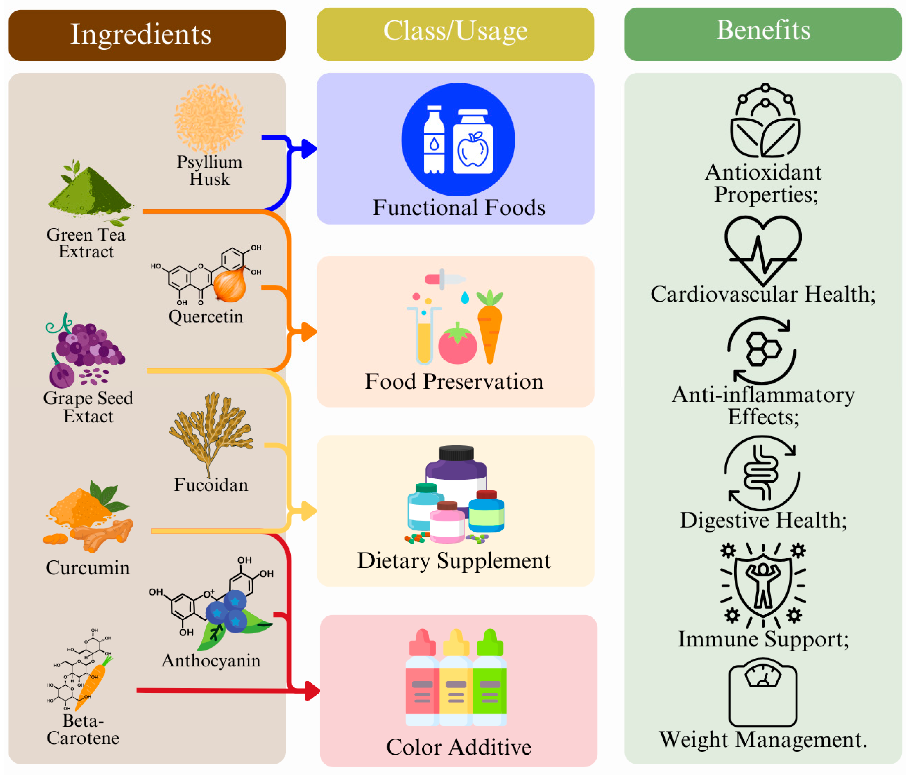
Curcumin, whether as a turmeric extract or in its unprocessed form, is commonly used to impart a yellowish-orange hue to foods [248]. It has also demonstrated potential in the prevention and management of various health conditions, including cardiovascular diseases, diabetes, metabolic syndrome, arthritis, and mental disorders [249,250].
In conclusion, the incorporation of antioxidants, fibers, crude extracts, and natural color additives into functional foods has been shown to enhance not only the nutritional value but also the sensory qualities, shelf life, and safety of various products. Both soluble and insoluble fibers, such as pectin, inulin, cellulose, psyllium husk, and wheat bran, play crucial roles in improving texture and stability, while providing health benefits like enhanced digestive health and disease prevention. Crude extracts, including plant polyphenols, as well as mushroom and spice extracts, contribute to preserving food quality by delaying lipid oxidation, offering antimicrobial properties, and improving both flavor and color.
5.2. Isolated Phytochemicals as Nutraceuticals
Isolated phytochemicals, bioactive compounds derived from plants, have gained significant attention as nutraceuticals due to their potent health-promoting properties. These compounds, which include flavonoids, alkaloids, terpenoids, and polyphenols, are increasingly being isolated and studied for their potential to prevent and manage various chronic diseases, such as cancer, cardiovascular disorders, and neurodegenerative conditions (Table 3). Phytochemicals are often extracted from agricultural and food waste streams, promoting a circular economy by converting waste into value-added functional ingredients [251]. By concentrating the active ingredients, isolated phytochemicals offer more targeted therapeutic effects compared to whole plant extracts, making them valuable in the development of functional foods and dietary supplements [195].
Table 3.
Nutraceutical Applications and Functionality.
| Nutraceutical | Source | Applications | Functionality | Health Benefits | Ref. |
|---|---|---|---|---|---|
| Pectin | Fruits (Apple, citrus…) | Jams, jellies, dairy products | Gelling agent, thickener | Anti-cancer, immunomodulatory, anti-inflammatory, cholesterol-lowering (…) | [252] |
| Inulin | Chicory root | Low-fat foods, fiber supplements | Prebiotic, fat replacer | Gut-microbiota regulating, lipid metabolism regulating, mineral absorption enhancing, anti-inflammatory (…) | [253] |
| Cellulose | Several plants | Low-fat foods, plant-based meats, bakery products | Stabilizer, thickener | Gut-microbiota regulating, cholesterol reducing, blood glucose levels regulating, anti-inflammatory (…) | [234] |
| Wheat Bran | Wheat | Cereals, bread and bakery products | Texture enhancer, fiber source | Gut-microbiota regulating, cancer-risk reducing, cardioprotective (…) | [254] |
| Psyllium husk | Plantago ovata seeds | Fiber supplement, cereals | Fiber source, thickener | Anti-diabetic, reduces cholesterol levels, and aids in gastrointestinal health (…) | [255,256] |
| Green tea extract and catechins | Green tea leaves | Beverages, supplements, snacks | Antioxidant, antimicrobial | Antioxidant, anti-inflammatory, antiviral, antiobesity (…) | [257,258] |
| Grapeseed extract | Grape seeds | Beverages, supplements | Antioxidant, antimicrobial | Anti-inflammatory, antioxidant, cardioprotective, antimicrobial, anti-cancer (…) | [259] |
| β-carotene | Carrots, sweet potatoes | Supplements, snacks, beverages, candies | Natural colorant, antioxidant | Antioxidant, supports immune function (…) | [260] |
| Anthocyanins | Berries, red cabbage | Supplements, snacks, beverages, candies | Natural colorant, antioxidant | Antioxidant, anti-inflammatory, antidiabetic, anti-obesity (…) | [261] |
| Betanins | Beetroot | Supplements, snacks, beverages, candies | Natural colorant, antioxidant | Antioxidative, anti-inflammatory, antidiabetic, potential anticancer benefits (…) | [243] |
| Curcumin | Turmeric root | Supplements, snacks, beverages, candies | Natural colorant, antioxidant | Antioxidant, anti-inflammatory, anticancer, and immune-regulatory properties (…) | [262] |
| Resveratrol | Grapes, red wine | Beverages, supplements | Antioxidant, antimicrobial | Antioxidant, anti-inflammatory, anti-cancer, cardioprotective (…) | [263] |
| Fucoidans | Blown algae | Supplements, fortified foods | Gelling agents, thickeners | Antioxidant, anti-inflammatory, anticoagulant, antitumor, antiviral (…) | [264] |
| Agar | Red algae | Jellies, jams, candy, plant-based gelatin | Gelling agent, texture enhancer | Antioxidant, antiviral, antibacterial, prebiotic, anti-tumor (…) | [265] |
| Carrageenan | Red algae | Jellies, jams, candy, plant-based gelatin | Thickener, gelling agent | Cardioprotective, anticancer, antiviral, anticoagulant, antioxidant (…) | [266] |
| Quercetin | Onions, apple peels | Supplements, fortified foods | Antioxidant, preservative | Anti-inflammatory, antimicrobial, anticancer, cardioprotective (…) | [267,268] |
Polyphenol extracts, concentrated from fruits, vegetables, herbs, and seeds, are well-known for their antioxidant properties. Green tea catechins, particularly epigallocatechin-3-gallate (EGCG), are effective in delaying lipid oxidation and extending the shelf life of lipid-containing foods [226,249]. These polyphenols are also incorporated into convenient products such as chewing gum and gelatin gummies, enhancing consumer accessibility and palatability [269]. Furthermore, green tea extracts demonstrate antimicrobial properties against common foodborne pathogens, enhancing both food safety and quality [227]. Similarly, resveratrol, often sourced from grape industry waste, exhibits strong antioxidant and antibacterial activity, making it suitable for use in oil-in-water emulsions, bulk oils, ground meat, and as a potential natural preservative in food packaging and processing [270,271].
Omega-3 oils, particularly eicosatetraenoic acid (EPA) and docosahexaenoic acid (DHA), are widely recognized for their roles in supporting brain development and cardiovascular health [272]. Sourced from marine species such some microalgae like Schizochytrium and Nannochloropsis, these oils are extracted through solvent methods [273]. Due to growing consumer demand for plant-based omega-3s, microalgae supplements have become popular as they contain lower levels of heavy metals compared to traditional fish oils [274]. However, the high cost and instability of algae oils limit their use in food products, though nano-emulsification techniques have shown promise in stabilizing omega-3s for functional food applications [275]. Additionally, algae-derived omega-3s are used in livestock feed, enriching meat, milk, and eggs with bioactive compounds that benefit human health [276], including increasing omega-3 blood indices and reducing diastolic blood pressure [277].
Fucoidans, polysaccharides extracted from brown algae, have gained recognition for their multifunctional bioactive properties. These include anticoagulant, antiviral, antitumor, antibacterial, and immunomodulatory effects [278]. Fucoidans are also valuable as gelling and thickening agents in the food industry, offering both functional and health benefits [279]. heir solubility and ease of incorporation into food formulations make them suitable for various applications, including the development of UVB-protective cosmetics and functional foods [280,281].
Agar and carrageenans, two polysaccharides derived from red algae, are widely used as gelling, thickening, and stabilizing agents in food products. Agar’s ability to form gels at low concentrations makes it ideal for candies, soups, snacks, and plant-based formulations [265,282]. Agar is also often used on plant-based and halal formulations of products that otherwise require animal gelatin [283]. Additionally, agar and its derivatives exhibit multiple bioactive functions such as antioxidant, antiviral, antibacterial, prebiotic, anti-tumor, anticoagulant, and antidiabetic activities. These bioactive properties make agar a valuable ingredient in functional foods and nutraceuticals [265,282]. Carrageenans are similarly utilized as thickening, gelling, and protein-suspending agents, enhancing texture and water retention in food products while also providing various health benefits [284]. In low-fat meat formulations, the incorporation of carrageenans promotes gel formation and water retention, thereby improving textural properties [284]. In parallel with agar, carrageenans exhibit anti-inflammatory, anticoagulant, antitumor, and antithrombotic effects [285].
Quercetin, a flavonoid present in a variety of plants, is predominantly extracted from onions and apple peels due to their commercial availability [286]. In food products like bread, quercetin demonstrates antioxidant and antiglycation effects in a dose-dependent manner. Furthermore, it inhibits the formation of advanced glycation end-products (AGEs), thereby enhancing the shelf life of these products [287]. Quercetin has also been shown to offer protective benefits against cardiovascular and neurodegenerative diseases, underscoring its potential as a valuable bioactive compound for the development of functional foods aimed at promoting health [288].
In summary, phytochemicals such as polyphenols, omega-3 oils, fucoidans, agar, carrageenan, and quercetin are increasingly recognized for their dual roles in improving both the quality and health benefits of food products. These compounds, sourced from plants and algae, contribute to enhanced shelf life, nutritional value, and functional properties, while offering a wide range of health benefits including antioxidant, antimicrobial, and cardiovascular protective effects.
6. Conclusions
This review uncovers the versatile functions of antioxidants in plant organisms and their involvement in protection to OS. Several antioxidant compounds, such as flavonoids, carotenoids, and phenolic acids, promote plant resistance and are also beneficial for human health. A better understanding of these mechanisms may allow the optimization of agricultural practices and food supply. Further research should be directed towards more efficient use of plant antioxidants to improve their productive capacity and nutraceutical value.
Author Contributions
Conceptualization: A.G.P, J.E., A.O.S.J., R.N.M., E.N.Y., P.B., A.P.V., F.C., M.B.P.P.O., M.C. and M.A.P.; methodology: A.G.P, J.E., A.O.S.J., R.N.M., E.N.Y., P.B., A.P.V., F.C., M.C.; validation, A.G.P, M.B.P.P.O., M.C. and M.A.P.; formal analysis: A.G.P, J.E., A.O.S.J., R.N.M., E.N.Y., P.B., A.P.V., F.C., M.C.; investigation: A.G.P, J.E., A.O.S.J., R.N.M., E.N.Y., P.B., A.P.V., F.C., M.C.; resources: A.G.P, J.E., A.O.S.J., R.N.M., E.N.Y., P.B., A.P.V., F.C., M.C.; data curation: A.G.P, A.O.S.J., P.B., A.P.V. and M.C.; writing—original draft preparation: A.G.P, J.E., A.O.S.J., R.N.M., E.N.Y., P.B., A.P.V., F.C., M.C.; writing—review and editing: A.G.P, A.O.S.J., M.C., M.B.P.P.O., and M.A.P.; visualization: A.G.P, A.O.S.J., P.B., A.P.V. and F.C.; supervision: A.G.P, M.C., M.B.P.P.O., and M.A.P.; project administration: M.B.P.P.O. and M.A.P.; funding acquisition: M.B.P.P.O. and M.A.P.. All authors have read and agreed to the published version of the manuscript.
Funding
This research received no external funding.
Informed Consent Statement
Not applicable.
Data Availability Statement
Not applicable.
Acknowledgments
The research leading to these results was supported by MICIU/AEI/10.13039/501100011033 supporting the predoctoral industrial grant for A. Perez-Vazquez (DIN2024-013416) in collaboration with Mercantia Desarrollos Alimentarios S.L; by Xunta de Galicia for supporting the post-doctoral grant of A.G. Pereira (IN606B-2024/011), and the pre-doctoral grant of P. Barciela (ED481A-2024-230). The authors thank the EU-FORA Fellowship Program (EUBA-EFSA-2023-ENREL-01) that supports the work of F. Chamorro (INNOV2SAFETY-GA13). The authors are grateful to the National funding by FCT, Foundation for Science and Technology, through the individual research grants of J. Echave (2023.04987.BD) and A.O.S. Jorge (2023.00981.BD).
Conflicts of Interest
The authors declare no conflicts of interest.
References
- Carpena, M.; Garcia-Oliveira, P.; Pereira, A.G.; Soria-Lopez, A.; Chamorro, F.; Collazo, N.; Jarboui, A.; Simal-Gandara, J.; Prieto, M.A. Plant Antioxidants from Agricultural Waste: Synergistic Potential with Other Biological Properties and Possible Applications. In Plant Antioxidants and Health; Ekiert, H.M., Ramawat, K.G., Arora, J., Eds.; Springer, Cham, 2021; pp. 1–38 ISBN 9783030452995.
- Shahidi, F.; Ambigaipalan, P. Phenolics and Polyphenolics in Foods, Beverages and Spices: Antioxidant Activity and Health Effects - A Review. J. Funct. Foods 2015, 18, 820–897. [Google Scholar] [CrossRef]
- Zhang, Y.-J.; Gan, R.-Y.; Li, S.; Zhou, Y.; Li, A.-N.; Xu, D.-P.; Li, H.-B. Antioxidant Phytochemicals for the Prevention and Treatment of Chronic Diseases. Molecules 2015, 20, 21138–21156. [Google Scholar] [CrossRef] [PubMed]
- Allaqaband, S.; Dar, A.H.; Patel, U.; Kumar, N.; Nayik, G.A.; Khan, S.A.; Ansari, M.J.; Alabdallah, N.M.; Kumar, P.; Pandey, V.K.; et al. Utilization of Fruit Seed-Based Bioactive Compounds for Formulating the Nutraceuticals and Functional Food: A Review. Front. Nutr. 2022, 9, 1–13. [Google Scholar] [CrossRef]
- Knothe, G. Vegetable Oils. In Handbook of Bioenergy Crop Plants; Wiley, 2012; pp. 793–810 ISBN 9781439816851.
- Chouaibi, M.; Rezig, L.; Hamdi, S.; Ferrari, G. Chemical Characteristics and Compositions of Red Pepper Seed Oils Extracted by Different Methods. Ind. Crops Prod. 2019, 128, 363–370. [Google Scholar] [CrossRef]
- García-Lomillo, J.; González-SanJosé, M.L. Applications of Wine Pomace in the Food Industry: Approaches and Functions. Compr. Rev. Food Sci. Food Saf. 2017, 16, 3–22. [Google Scholar] [CrossRef]
- Lu, Q.; Li, H.; Xiao, Y.; Liu, H. A State-of-the-Art Review on the Synthetic Mechanisms, Production Technologies, and Practical Application of Polyunsaturated Fatty Acids from Microalgae. Algal Res. 2021, 55, 102281. [Google Scholar] [CrossRef]
- Russo, G.L.; Moccia, S.; Russo, M.; Spagnuolo, C. Redox Regulation by Carotenoids: Evidence and Conflicts for Their Application in Cancer. Biochem. Pharmacol. 2021, 194, 114838. [Google Scholar] [CrossRef]
- Bohn, T. Provitamin a Carotenoids: Occurrence, Intake and Bioavailability. Food Nutr. Components Focus 2012, 1, 142–161. [Google Scholar]
- Poojary, M.M.; Barba, F.J.; Aliakbarian, B.; Donsì, F.; Pataro, G.; Dias, D.A.; Juliano, P. Innovative Alternative Technologies to Extract Carotenoids from Microalgae and Seaweeds. Mar. Drugs 2016, 14. [Google Scholar] [CrossRef]
- Rodriguez-Concepcion, M.; Avalos, J.; Bonet, M.L.; Boronat, A.; Gomez-Gomez, L.; Hornero-Mendez, D.; Limon, M.C.; Meléndez-Martínez, A.J.; Olmedilla-Alonso, B.; Palou, A.; et al. A Global Perspective on Carotenoids: Metabolism, Biotechnology, and Benefits for Nutrition and Health. Prog. Lipid Res. 2018, 70, 62–93. [Google Scholar] [CrossRef]
- Nie, Q.; Hu, J.; Gao, H.; Li, M.; Sun, Y.; Chen, H.; Zuo, S.; Fang, Q.; Huang, X.; Yin, J.; et al. Bioactive Dietary Fibers Selectively Promote Gut Microbiota to Exert Antidiabetic Effects. J. Agric. Food Chem. 2021, 69, 7000–7015. [Google Scholar] [CrossRef] [PubMed]
- Baiano, A. Recovery of Biomolecules from Food Wastes - A Review. Molecules 2014, 19, 14821–14842. [Google Scholar] [CrossRef] [PubMed]
- Saeed, F.; Pasha, I.; Arshad, M.U.; Muhammad Anjum, F.; Hussain, S.; Rasheed, R.; Nasir, M.A.; Shafique, B. Physiological and Nutraceutical Perspectives of Fructan. Int. J. Food Prop. 2015, 18, 1895–1904. [Google Scholar] [CrossRef]
- Jimenez-Lopez, C.; Fraga-Corral, M.; Carpena, M.; García-Oliveira, P.; Echave, J.; Pereira, A.G.; Lourenço-Lopes, C.; Prieto, M.A.; Simal-Gandara, J. Agriculture Waste Valorisation as a Source of Antioxidant Phenolic Compounds within a Circular and Sustainable Bioeconomy. Food Funct. 2020, 11, 4853–4877. [Google Scholar] [CrossRef]
- Barjoveanu, G.; Pătrăuțanu, O.-A.; Teodosiu, C.; Volf, I. Life Cycle Assessment of Polyphenols Extraction Processes from Waste Biomass. Sci. Rep. 2020, 10, 13632. [Google Scholar] [CrossRef]
- Li, S.; Li, Y.; Smolke, C.D. Strategies for Microbial Synthesis of High-Value Phytochemicals. Nat. Chem. 2018, 10, 395–404. [Google Scholar] [CrossRef]
- Reguengo, L.M.; Salgaço, M.K.; Sivieri, K.; Maróstica Júnior, M.R. Agro-Industrial by-Products: Valuable Sources of Bioactive Compounds. Food Res. Int. 2022, 152. [Google Scholar] [CrossRef]
- Watkins, P.A. Fatty Acids: Metabolism. In Encyclopedia of Human Nutrition; Elsevier, 2013; Vol. 2–4, pp. 220–230 ISBN 9780123848857.
- Moghadasian, M.H.; Shahidi, F. Fatty Acids. In International Encyclopedia of Public Health; Elsevier, 2016; Vol. 3, pp. 114–122 ISBN 9780128037089.
- He, M.; Ding, N.Z. Plant Unsaturated Fatty Acids: Multiple Roles in Stress Response. Front. Plant Sci. 2020, 11, 1–15. [Google Scholar] [CrossRef]
- Maoka, T. Carotenoids as Natural Functional Pigments. J. Nat. Med. 2020, 74, 1–16. [Google Scholar] [CrossRef]
- Crupi, P.; Faienza, M.F.; Naeem, M.Y.; Corbo, F.; Clodoveo, M.L.; Muraglia, M. Overview of the Potential Beneficial Effects of Carotenoids on Consumer Health and Well-Being. Antioxidants 2023, 12. [Google Scholar] [CrossRef]
- Lakshminarayana, R.; Paul, B. Free Radical Chemistry of Carotenoids and Oxidative Stress Physiology of Cancer. In Handbook of Oxidative Stress in Cancer: Therapeutic Aspects: Volume 1; Chakraborti, S., Ed.; Springer Nature Singapore: Singapore, 2022; Vol. 1, pp. 3077–3097. ISBN 9789811654220. [Google Scholar]
- Genç, Y.; Bardakci, H.; Yücel, Ç.; Karatoprak, G.Ş.; Akkol, E.K.; Barak, T.H.; Sobarzo-Sánchez, E. Oxidative Stress and Marine Carotenoids: Application by Using Nanoformulations. Mar. Drugs 2020, 18, 423. [Google Scholar] [CrossRef]
- Srivastava, R. Physicochemical, Antioxidant Properties of Carotenoids and Its Optoelectronic and Interaction Studies with Chlorophyll Pigments. Sci. Rep. 2021, 11, 18365. [Google Scholar] [CrossRef]
- Saini, R.K.; Prasad, P.; Lokesh, V.; Shang, X.; Shin, J.; Keum, Y.S.; Lee, J.H. Carotenoids: Dietary Sources, Extraction, Encapsulation, Bioavailability, and Health Benefits—A Review of Recent Advancements. Antioxidants 2022, 11. [Google Scholar] [CrossRef]
- Pereira, A.G.; Otero, P.; Echave, J.; Carreira-Casais, A.; Chamorro, F.; Collazo, N.; Jaboui, A.; Lourenço-Lopes, C.; Simal-Gandara, J.; Prieto, M.A. Xanthophylls from the Sea: Algae as Source of Bioactive Carotenoids. Mar. Drugs 2021, 19, 188. [Google Scholar] [CrossRef]
- Widomska, J.; Zareba, M.; Subczynski, W.K. Can Xanthophyll-Membrane Interactions Explain Their Selective Presence in the Retina and Brain? Foods 2016, 5, 1–19. [Google Scholar] [CrossRef]
- Ojulari, O.V.; Gi Lee, S.; Nam, J.O. Therapeutic Effect of Seaweed Derived Xanthophyl Carotenoid on Obesity Management; Overview of the Last Decade. Int. J. Mol. Sci. 2020, 21, 2502. [Google Scholar] [CrossRef]
- Gürbüz, M.; Aktaç, Ş. Understanding the Role of Vitamin A and Its Precursors in the Immune System. Nutr. Clin. Metab. 2022, 36, 89–98. [Google Scholar] [CrossRef]
- Ebadi, M.; Mohammadi, M.; Pezeshki, A.; Jafari, S.M. Health Benefits of Beta-Carotene. In Handbook of Food Bioactive Ingredients; Jafari, S.M., Rashidinejad, A., Simal-Gandara, J., Eds.; Springer International Publishing: Cham, 2023; ISBN 978-3-030-81404-5. [Google Scholar]
- Aziz, E.; Batool, R.; Akhtar, W.; Rehman, S.; Shahzad, T.; Malik, A.; Shariati, M.A.; Laishevtcev, A.; Plygun, S.; Heydari, M.; et al. Xanthophyll: Health Benefits and Therapeutic Insights. Life Sci. 2020, 240, 117104. [Google Scholar] [CrossRef]
- Singh, A.; Omer, K. Recent Advancement in Therapeutic Activity of Carotenoids. In Dietary Carotenoids; Rao, A.V., Rao, L., Eds.; IntechOpen: Rijeka, 2024. [Google Scholar]
- Grune, T.; Lietz, G.; Palou, A.; Ross, A.C.; Stahl, W.; Tang, G.; Thurnham, D.; Yin, S.A.; Biesalski, H.K. β-Carotene Is an Important Vitamin A Source for Humans. J. Nutr. 2010, 140, 2268S–2285S. [Google Scholar] [CrossRef]
- Murphy, E.J.; Fehrenbach, G.W.; Abidin, I.Z.; Buckley, C.; Montgomery, T.; Pogue, R.; Murray, P.; Major, I.; Rezoagli, E. Polysaccharides—Naturally Occurring Immune Modulators. Polymers (Basel). 2023, 15. [Google Scholar] [CrossRef]
- Pedrosa, L. de F.; de Vos, P.; Fabi, J.P. Nature’s Soothing Solution: Harnessing the Potential of Food-Derived Polysaccharides to Control Inflammation. Curr. Res. Struct. Biol. 2023, 6, 100112. [Google Scholar] [CrossRef] [PubMed]
- Baghel, R.S.; Choudhary, B.; Pandey, S.; Pathak, P.K.; Patel, M.K.; Mishra, A. Rehashing Our Insight of Seaweeds as a Potential Source of Foods, Nutraceuticals, and Pharmaceuticals. Foods 2023, 12. [Google Scholar] [CrossRef]
- Zeng, F. Sen; Yao, Y.F.; Wang, L.F.; Li, W.J. Polysaccharides as Antioxidants and Prooxidants in Managing the Double-Edged Sword of Reactive Oxygen Species. Biomed. Pharmacother. 2023, 159, 114221. [Google Scholar] [CrossRef]
- Lin, G. peng; Wu, D. sheng; Xiao, X. wei; Huang, Q. yun; Chen, H. bin; Liu, D.; Fu, H. qing; Chen, X. hua; Zhao, C. Structural Characterization and Antioxidant Effect of Green Alga Enteromorpha Prolifera Polysaccharide in Caenorhabditis Elegans via Modulation of MicroRNAs. Int. J. Biol. Macromol. 2020, 150, 1084–1092. [Google Scholar] [CrossRef]
- Zhang, Y.; Xu, M.; Hu, C.; Liu, A.; Chen, J.; Gu, C.; Zhang, X.; You, C.; Tong, H.; Wu, M.; et al. Sargassum Fusiforme Fucoidan SP2 Extends the Lifespan of Drosophila Melanogaster by Upregulating the Nrf2-Mediated Antioxidant Signaling Pathway. Oxid. Med. Cell. Longev. 2019. [Google Scholar] [CrossRef]
- Cui, H.; Wu, S.; Shang, Y.; Li, Z.; Chen, M.; Li, F.; Wang, C. Pleurotus Nebrodensis Polysaccharide(PN50G) Evokes A549 Cell Apoptosis by the ROS/AMPK/PI3K/AKT/MTOR Pathway to Suppress Tumor Growth. Food Funct. 2016, 7, 1616–1627. [Google Scholar] [CrossRef]
- Wang, J.; Hu, S.; Nie, S.; Yu, Q.; Xie, M. Reviews on Mechanisms of in Vitro Antioxidant Activity of Polysaccharides. Oxid. Med. Cell. Longev. 2016, 2016, 5692852. [Google Scholar] [CrossRef]
- Devi, N.; Sarmah, M.; Khatun, B.; Maji, T.K. Encapsulation of Active Ingredients in Polysaccharide–Protein Complex Coacervates. Adv. Colloid Interface Sci. 2017, 239, 136–145. [Google Scholar] [CrossRef]
- Liu, Z.; Sun, X. A Critical Review of the Abilities, Determinants, and Possible Molecular Mechanisms of Seaweed Polysaccharides Antioxidants. Int. J. Mol. Sci. 2020, 21, 1–20. [Google Scholar] [CrossRef]
- Chen, N.; Jiang, T.; Xu, J.; Xi, W.; Shang, E.; Xiao, P.; Duan, J. ao The Relationship between Polysaccharide Structure and Its Antioxidant Activity Needs to Be Systematically Elucidated. Int. J. Biol. Macromol. 2024, 270, 132391. [Google Scholar] [CrossRef]
- Fernandes, P.A.R.; Coimbra, M.A. The Antioxidant Activity of Polysaccharides: A Structure-Function Relationship Overview. Carbohydr. Polym. 2023, 314, 120965. [Google Scholar] [CrossRef] [PubMed]
- Bai, L.; Xu, D.; Zhou, Y.M.; Zhang, Y.B.; Zhang, H.; Chen, Y.B.; Cui, Y.L. Antioxidant Activities of Natural Polysaccharides and Their Derivatives for Biomedical and Medicinal Applications. Antioxidants 2022, 11. [Google Scholar] [CrossRef] [PubMed]
- Niyigaba, T.; Liu, D.; Habimana, J. de D. The Extraction, Functionalities and Applications of Plant Polysaccharides in Fermented Foods: A Review. Foods 2021, 10. [Google Scholar] [CrossRef] [PubMed]
- Sheibani, E.; Hosseini, A.; Sobhani Nasab, A.; Adib, K.; Ganjali, M.R.; Pourmortazavi, S.M.; Ahmadi, F.; Marzi Khosrowshahi, E.; Mirsadeghi, S.; Rahimi-Nasrabadi, M.; et al. Application of Polysaccharide Biopolymers as Natural Adsorbent in Sample Preparation. Crit. Rev. Food Sci. Nutr. 2023, 63, 2626–2653. [Google Scholar] [CrossRef]
- Teng, C.; Qin, P.; Shi, Z.; Zhang, W.; Yang, X.; Yao, Y.; Ren, G. Structural Characterization and Antioxidant Activity of Alkali-Extracted Polysaccharides from Quinoa. Food Hydrocoll. 2021, 113, 106392. [Google Scholar] [CrossRef]
- Bhuyan, P.P.; Nayak, R.; Patra, S.; Abdulabbas, H.S.; Jena, M.; Pradhan, B. Seaweed-Derived Sulfated Polysaccharides; The New Age Chemopreventives: A Comprehensive Review. Cancers (Basel). 2023, 15. [Google Scholar] [CrossRef]
- Anand, S.; Bharadvaja, N. Potential Benefits of Nutraceuticals for Oxidative Stress Management. Rev. Bras. Farmacogn. 2022, 32, 211–220. [Google Scholar] [CrossRef]
- Livingstone, K.M.; Ramos-Lopez, O.; Pérusse, L.; Kato, H.; Ordovas, J.M.; Martínez, J.A. Precision Nutrition: A Review of Current Approaches and Future Endeavors. Trends Food Sci. Technol. 2022, 128, 253–264. [Google Scholar] [CrossRef]
- Tsao, R. Chemistry and Biochemistry of Dietary Polyphenols. Nutrients 2010, 2, 1231–1246. [Google Scholar] [CrossRef]
- Jimenez-Lopez, C.; Pereira, A.G.; Lourenço-Lopes, C.; Garcia-Oliveira, P.; Cassani, L.; Fraga-Corral, M.; Prieto, M.A.; Simal-Gandara, J. Main Bioactive Phenolic Compounds in Marine Algae and Their Mechanisms of Action Supporting Potential Health Benefits. Food Chem. 2021, 341, 128262. [Google Scholar] [CrossRef]
- Di Lorenzo, C.; Colombo, F.; Biella, S.; Stockley, C.; Restani, P. Polyphenols and Human Health: The Role of Bioavailability. Nutrients 2021, 13, 1–30. [Google Scholar] [CrossRef] [PubMed]
- Han, X.; Shen, T.; Lou, H. Dietary Polyphenols and Their Biological Significance. Int. J. Mol. Sci. 2007, 8, 950–988. [Google Scholar] [CrossRef]
- Zhang, Z.; Li, X.; Sang, S.; McClements, D.J.; Chen, L.; Long, J.; Jiao, A.; Jin, Z.; Qiu, C. Polyphenols as Plant-Based Nutraceuticals: Health Effects, Encapsulation, Nano-Delivery, and Application. Foods 2022, 11. [Google Scholar] [CrossRef]
- Haminiuk, C.W.I.; Maciel, G.M.; Plata-Oviedo, M.S.V.; Peralta, R.M. Phenolic Compounds in Fruits - an Overview. Int. J. Food Sci. Technol. 2012, 47, 2023–2044. [Google Scholar] [CrossRef]
- Smeriglio, A.; Barreca, D.; Bellocco, E.; Trombetta, D. Proanthocyanidins and Hydrolysable Tannins: Occurrence, Dietary Intake and Pharmacological Effects. Br. J. Pharmacol. 2017, 174, 1244–1262. [Google Scholar] [CrossRef]
- Rashmi, H.B.; Negi, P.S. Phenolic Acids from Vegetables: A Review on Processing Stability and Health Benefits. Food Res. Int. 2020, 136, 109298. [Google Scholar] [CrossRef]
- Caleja, C.; Ribeiro, A.; Barreiro, M.F.; Ferreira, I.C.F.R. Phenolic Compounds as Nutraceuticals or Functional Food Ingredients. Curr. Pharm. Des. 2017, 23, 2787–2806. [Google Scholar] [CrossRef]
- Vuolo, M.M.; Lima, V.S.; Maróstica Junior, M.R. Phenolic Compounds. In Bioactive Compounds: Health Benefits and Potential Applications; Campos, M.R.S., Ed.; Elsevier: Amsterdam, The Netherlands, 2019; ISBN 9780128147740. [Google Scholar]
- Sorrenti, V.; Burò, I.; Consoli, V.; Vanella, L. Recent Advances in Health Benefits of Bioactive Compounds from Food Wastes and By-Products: Biochemical Aspects. Int. J. Mol. Sci. 2023, 24. [Google Scholar] [CrossRef]
- Lin, J.K.; Weng, M.S. Flavonoids as Nutraceuticals. Sci. Flavonoids 2006, 7, 213–238. [Google Scholar] [CrossRef]
- Dias, M.; Romaní-Pérez, M.; Romaní, A.; de la Cruz, A.; Pastrana, L.; Fuciños, P.; Amado, I.R. Recent Technological Advances in Phenolic Compounds Recovery and Applications: Source of Nutraceuticals for the Management of Diabetes. Appl. Sci. 2022, 12. [Google Scholar] [CrossRef]
- Rahman, M.M.; Rahaman, M.S.; Islam, M.R.; Rahman, F.; Mithi, F.M.; Alqahtani, T.; Almikhlafi, M.A.; Alghamdi, S.Q.; Alruwaili, A.S.; Hossain, M.S.; et al. Role of Phenolic Compounds in Human Disease: Current Knowledge and Future Prospects. Molecules 2022, 27. [Google Scholar] [CrossRef] [PubMed]
- Vignesh, A.; Amal, T.C.; Sarvalingam, A.; Vasanth, K. A Review on the Influence of Nutraceuticals and Functional Foods on Health. Food Chem. Adv. 2024, 5, 100749. [Google Scholar] [CrossRef]
- Rhazi, L.; Depeint, F.; Gotor, A.A. Loss in the Intrinsic Quality and the Antioxidant Activity of Sunflower (Helianthus Annuus L.) Oil during an Industrial Refining Process. Molecules 2022, 27, 916. [Google Scholar] [CrossRef] [PubMed]
- Ambra, R.; Natella, F.; Lucchetti, S.; Forte, V.; Pastore, G. α-Tocopherol, β-Carotene, Lutein, Squalene and Secoiridoids in Seven Monocultivar Italian Extra-Virgin Olive Oils. Int. J. Food Sci. Nutr. 2017, 68, 538–545. [Google Scholar] [CrossRef]
- Fagali, N.; Catalá, A. Antioxidant Activity of Conjugated Linoleic Acid Isomers, Linoleic Acid and Its Methyl Ester Determined by Photoemission and DPPH Techniques. Biophys. Chem. 2008, 137, 56–62. [Google Scholar] [CrossRef]
- Lewinska, A.; Zebrowski, J.; Duda, M.; Gorka, A.; Wnuk, M. Fatty Acid Profile and Biological Activities of Linseed and Rapeseed Oils. Molecules 2015, 20, 22872–22880. [Google Scholar] [CrossRef]
- Hulatt, C.J.; Wijffels, R.H.; Bolla, S.; Kiron, V. Production of Fatty Acids and Protein by Nannochloropsis in Flat-Plate Photobioreactors. PLoS One 2017, 12, e0170440. [Google Scholar] [CrossRef]
- Xiao, B.; Li, Y.; Lin, Y.; Lin, J.; Zhang, L.; Wu, D.; Zeng, J.; Li, J.; Liu, J. wen; Li, G. Eicosapentaenoic Acid (EPA) Exhibits Antioxidant Activity via Mitochondrial Modulation. Food Chem. 2022, 373, 131389. [Google Scholar] [CrossRef]
- Li, G.; Li, Y.; Xiao, B.; Cui, D.; Lin, Y.; Zeng, J.; Li, J.; Cao, M.-J.; Liu, J. Antioxidant Activity of Docosahexaenoic Acid (DHA) and Its Regulatory Roles in Mitochondria. J. Agric. Food Chem. 2021, 69, 1647–1655. [Google Scholar] [CrossRef]
- Li, Z.; Chen, X.; Li, J.; Meng, T.; Wang, L.; Chen, Z.; Shi, Y.; Ling, X.; Luo, W.; Liang, D.; et al. Functions of PKS Genes in Lipid Synthesis of Schizochytrium Sp. by Gene Disruption and Metabolomics Analysis. Mar. Biotechnol. 2018, 20, 792–802. [Google Scholar] [CrossRef]
- Rodrigues, N.; Casal, S.; Pinho, T.; Cruz, R.; Peres, A.M.; Baptista, P.; Pereira, J.A. Fatty Acid Composition from Olive Oils of Portuguese Centenarian Trees Is Highly Dependent on Olive Cultivar and Crop Year. Foods 2021, 10, 496. [Google Scholar] [CrossRef] [PubMed]
- Petraru, A.; Ursachi, F.; Amariei, S. Nutritional Characteristics Assessment of Sunflower Seeds, Oil and Cake. Perspective of Using Sunflower Oilcakes as a Functional Ingredient. Plants 2021, 10. [Google Scholar] [CrossRef]
- Ali, Y.M.; Kadir, A.A.; Ahmad, Z.; Yaakub, H.; Zakaria, Z.A.; Hakim Abdullah, M.N. Free Radical Scavenging Activity of Conjugated Linoleic Acid as Single or Mixed Isomers. Pharm. Biol. 2012, 50, 712–719. [Google Scholar] [CrossRef]
- Zhou, W.; Wang, H.; Chen, L.; Cheng, W.; Liu, T. Heterotrophy of Filamentous Oleaginous Microalgae Tribonema Minus for Potential Production of Lipid and Palmitoleic Acid. Bioresour. Technol. 2017, 239, 250–257. [Google Scholar] [CrossRef]
- Borges, T.H.; Pereira, J.A.; Cabrera-Vique, C.; Lara, L.; Oliveira, A.F.; Seiquer, I. Characterization of Arbequina Virgin Olive Oils Produced in Different Regions of Brazil and Spain: Physicochemical Properties, Oxidative Stability and Fatty Acid Profile. Food Chem. 2017, 215, 454–462. [Google Scholar] [CrossRef]
- Hu, W.; Fitzgerald, M.; Topp, B.; Alam, M.; O’Hare, T.J. A Review of Biological Functions, Health Benefits, and Possible de Novo Biosynthetic Pathway of Palmitoleic Acid in Macadamia Nuts. J. Funct. Foods 2019, 62. [Google Scholar] [CrossRef]
- Mohammed, D.; Freije, A.; Abdulhussain, H.; Khonji, A.; Hasan, M.; Ferraris, C.; Gasparri, C.; Aziz Aljar, M.A.; Ali Redha, A.; Giacosa, A.; et al. Analysis of the Antioxidant Activity, Lipid Profile, and Minerals of the Skin and Seed of Hazelnuts (Corylus Avellana L.), Pistachios (Pistacia Vera) and Almonds (Prunus Dulcis)—A Comparative Analysis. AppliedChem 2023, 3, 110–118. [Google Scholar] [CrossRef]
- Shuai, X.; Dai, T.; Chen, M.; Liang, R.; Du, L.; Chen, J.; Liu, C. Comparative Study of Chemical Compositions and Antioxidant Capacities of Oils Obtained from 15 Macadamia (Macadamia Integrifolia) Cultivars in China. Foods 2021, 10. [Google Scholar] [CrossRef]
- Górnaś, P.; Rudzińska, M.; Grygier, A.; Lācis, G. Diversity of Oil Yield, Fatty Acids, Tocopherols, Tocotrienols, and Sterols in the Seeds of 19 Interspecific Grapes Crosses. J. Sci. Food Agric. 2019, 99, 2078–2087. [Google Scholar] [CrossRef]
- Henry, G.E.; Momin, R.A.; Nair, M.G.; Dewitt, D.L. Antioxidant and Cyclooxygenase Activities of Fatty Acids Found in Food. J. Agric. Food Chem. 2002, 50, 2231–2234. [Google Scholar] [CrossRef]
- Wang, J.; Zhang, Y.; Fang, Z.; Sun, L.; Wang, Y.; Liu, Y.; Xu, D.; Nie, F.; Gooneratne, R. Oleic Acid Alleviates Cadmium-Induced Oxidative Damage in Rat by Its Radicals Scavenging Activity. Biol. Trace Elem. Res. 2019, 190, 95–100. [Google Scholar] [CrossRef] [PubMed]
- Xavier, A.A.O.; Pérez-Gálvez, A. Carotenoids as a Source of Antioxidants in the Diet. In Sub-Cellular Biochemistry; Stange, C., Ed.; Springer International Publishing: Cham, 2016; Vol. 79, pp. 359–375. ISBN 978-3-319-39126-7. [Google Scholar]
- Koca Bozalan, N.; Karadeniz, F. Carotenoid Profile, Total Phenolic Content, and Antioxidant Activity of Carrots. Int. J. Food Prop. 2011, 14, 1060–1068. [Google Scholar] [CrossRef]
- Lourenço-Lopes, C.; Fraga-Corral, M.; Jimenez-Lopez, C.; Carpena, M.; Pereira, A.G.; Garcia-Oliveira, P.; 57,92, M. A.; Simal-Gandara, J. Biological Action Mechanisms of Fucoxanthin Extracted from Algae for Application in Food and Cosmetic Industries. Trends Food Sci. Technol. 2021, 117, 163–181. [Google Scholar] [CrossRef]
- Ambati, R.R.; Moi, P.S.; Ravi, S.; Aswathanarayana, R.G. Astaxanthin: Sources, Extraction, Stability, Biological Activities and Its Commercial Applications - A Review. Mar. Drugs 2014, 12, 128–152. [Google Scholar] [CrossRef]
- Iwamoto, H.; Soccol, C.R.; Molina-Aulestia, D.T.; Cardoso, J.; de Melo Pereira, G.V.; de Souza Vandenberghe, L.P.; Manzoki, M.C.; Ambati, R.R.; Ravishankar, G.A.; de Carvalho, J.C. Lutein from Microalgae: An Industrial Perspective of Its Production, Downstream Processing, and Market. Fermentation 2024, 10. [Google Scholar] [CrossRef]
- Tudor, C.; Pintea, A. A Brief Overview of Dietary Zeaxanthin Occurrence and Bioaccessibility. Molecules 2020, 25. [Google Scholar] [CrossRef]
- Burri, B.J.; La Frano, M.R.; Zhu, C. Absorption, Metabolism, and Functions of β-Cryptoxanthin. Nutr. Rev. 2016, 74, 69–82. [Google Scholar] [CrossRef]
- Brahma, D.; Dutta, D. Evaluating β-Cryptoxanthin Antioxidant Properties against ROS-Induced Macromolecular Damages and Determining Its Photo-Stability and in-Vitro SPF. World J. Microbiol. Biotechnol. 2023, 39, 310. [Google Scholar] [CrossRef]
- Galla, R.; Ruga, S.; Aprile, S.; Ferrari, S.; Brovero, A.; Grosa, G.; Molinari, C.; Uberti, F. New Hyaluronic Acid from Plant Origin to Improve Joint Protection—An In Vitro Study. Int. J. Mol. Sci. 2022, 23. [Google Scholar] [CrossRef]
- Mohammed, A.A.; Niamah, A.K. Identification and Antioxidant Activity of Hyaluronic Acid Extracted from Local Isolates of Streptococcus Thermophilus. Mater. Today Proc. 2022, 60, 1523–1529. [Google Scholar] [CrossRef]
- Schiraldi, C.; Cimini, D.; De Rosa, M. Production of Chondroitin Sulfate and Chondroitin. Appl. Microbiol. Biotechnol. 2010, 87, 1209–1220. [Google Scholar] [CrossRef]
- Campo, G.M.; Avenoso, A.; Campo, S.; Ferlazzo, A.M.; Calatroni, A. Antioxidant Activity of Chondroitin Sulfate. In Advances in Pharmacology; Advances in Pharmacology; Academic Press, 2006; Vol. 53, pp. 417–431 ISBN 0120329557.
- Pavão, M.S.G.; Mourão, P.A.S. Challenges for Heparin Production: Artificial Synthesis or Alternative Natural Sources? Glycobiol. Insights 2012, 3, 1–6. [Google Scholar] [CrossRef]
- Bijak, M.; Saluk, J.; Szelenberger, R.; Nowak, P. Popular Naturally Occurring Antioxidants as Potential Anticoagulant Drugs. Chem. Biol. Interact. 2016, 257, 35–45. [Google Scholar] [CrossRef]
- Medlej, M.K.; Batoul, C.; Olleik, H.; Li, S.; Hijazi, A.; Nasser, G.; Maresca, M.; Pochat-Bohatier, C. Antioxidant Activity and Biocompatibility of Fructo-Polysaccharides Extracted from a Wild Species of Ornithogalum from Lebanon. Antioxidants 2021, 10, 1–20. [Google Scholar] [CrossRef]
- Delattre, C.; Fenoradosoa, T.A.; Michaud, P. Galactans: An Overview of Their Most Important Sourcing and Applications as Natural Polysaccharides. Brazilian Arch. Biol. Technol. 2011, 54, 1075–1092. [Google Scholar] [CrossRef]
- Srivastava, P.; Malviya, R. Sources of Pectin, Extraction and Its Applications in Pharmaceutical Industry - an Overview. Indian J. Nat. Prod. Resour. 2011, 2, 10–18. [Google Scholar]
- Wathoni, N.; Yuan Shan, C.; Yi Shan, W.; Rostinawati, T.; Indradi, R.B.; Pratiwi, R.; Muchtaridi, M. Characterization and Antioxidant Activity of Pectin from Indonesian Mangosteen (Garcinia Mangostana L.) Rind. Heliyon 2019, 5. [Google Scholar] [CrossRef] [PubMed]
- Indrianingsih, A.W.; Rosyida, V.T.; Apriyana, W.; Hayati, S.N.; Darsih, C.; Nisa, K.; Ratih, D. Antioxidant and Antibacterial Properties of Bacterial Cellulose-Indonesian Plant Extract Composites for Mask Sheet. J. Appl. Pharm. Sci. 2020, 10, 37–42. [Google Scholar] [CrossRef]
- Cabañas-Romero, L.V.; Valls, C.; Valenzuela, S. V.; Roncero, M.B.; Pastor, F.I.J.; Diaz, P.; Martínez, J. Bacterial Cellulose-Chitosan Paper with Antimicrobial and Antioxidant Activities. Biomacromolecules 2020, 21, 1568–1577. [Google Scholar] [CrossRef]
- Hashem, A.H.; Al Abboud, M.A.; Alawlaqi, M.M.; Abdelghany, T.M.; Hasanin, M. Synthesis of Nanocapsules Based on Biosynthesized Nickel Nanoparticles and Potato Starch: Antimicrobial, Antioxidant, and Anticancer Activity. Starch/Staerke 2022, 74, 2100165. [Google Scholar] [CrossRef]
- Mikulíková, D.; Kraic, J.Á.N. Natural Sources of Health-Promoting Starch Natural Sources of Health-Promoting Starch. J. Food Nutr. Res. 2018, 45, 69–76. [Google Scholar]
- Zhang, R.; Edgar, K.J. Properties, Chemistry, and Applications of the Bioactive Polysaccharide Curdlan. Biomacromolecules 2014, 15, 1079–1096. [Google Scholar] [CrossRef]
- Li, H.; Xu, S.; Xie, Y.; Zhang, Q.; Ding, S.; Wang, R.; Fu, F.; Zhan, X. Curdlan-Polyphenol Complexes Prepared by PH-Driven Effectively Enhanced Their Physicochemical Stability, Antioxidant and Prebiotic Activities. Int. J. Biol. Macromol. 2024, 267, 131579. [Google Scholar] [CrossRef]
- Kareem, A.J.; Salman, J.A.S. Production of Dextran from Locally Lactobacillus Spp. Isolates. Reports Biochem. Mol. Biol. 2019, 8, 278–286. [Google Scholar]
- Rosca, I.; Turin-Moleavin, I.A.; Sarghi, A.; Lungoci, A.L.; Varganici, C.D.; Petrovici, A.R.; Fifere, A.; Pinteala, M. Dextran Coated Iron Oxide Nanoparticles Loaded with Protocatechuic Acid as Multifunctional Therapeutic Agents. Int. J. Biol. Macromol. 2024, 256, 128314. [Google Scholar] [CrossRef]
- Jonas, R.; Farah, L.F. Production and Application of Microbial Cellulose. Polym. Degrad. Stab. 1998, 59, 101–106. [Google Scholar] [CrossRef]
- Rop, O.; Mlcek, J.; Jurikova, T. Beta-Glucans in Higher Fungi and Their Health Effects. Nutr. Rev. 2009, 67, 624–631. [Google Scholar] [CrossRef]
- Mebrek, S.; Djeghim, H.; Mehdi, Y.; Meghezzi, A.; Anwar, S.; Ali Awadh, N.A.; Benali, M. Antioxidant, Anti-Cholinesterase, Anti-α-Glucosidase and Prebiotic Properties of Beta-Glucan Extracted from Algerian Barley. Int. J. Phytomedicine 2018, 10, 58. [Google Scholar] [CrossRef]
- Huq, T.; Khan, A.; Brown, D.; Dhayagude, N.; He, Z.; Ni, Y. Sources, Production and Commercial Applications of Fungal Chitosan: A Review. J. Bioresour. Bioprod. 2022, 7, 85–98. [Google Scholar] [CrossRef]
- Xing, R.; Yu, H.; Liu, S.; Zhang, W.; Zhang, Q.; Li, Z.; Li, P. Antioxidant Activity of Differently Regioselective Chitosan Sulfates in Vitro. Bioorganic Med. Chem. 2005, 13, 1387–1392. [Google Scholar] [CrossRef]
- Rocha De Souza, M.C.; Marques, C.T.; Guerra Dore, C.M.; Ferreira Da Silva, F.R.; Oliveira Rocha, H.A.; Leite, E.L. Antioxidant Activities of Sulfated Polysaccharides from Brown and Red Seaweeds. J. Appl. Phycol. 2007, 19, 153–160. [Google Scholar] [CrossRef] [PubMed]
- Rhein-Knudsen, N.; Reyes-Weiss, D.; Horn, S.J. Extraction of High Purity Fucoidans from Brown Seaweeds Using Cellulases and Alginate Lyases. Int. J. Biol. Macromol. 2023, 229, 199–209. [Google Scholar] [CrossRef]
- Pereira, L.; COTAS, J. Alginates - Recent Uses of This Natural Polymer; IntechOpen: Rijeka, 2019; ISBN 978-1-78985-642-2. [Google Scholar]
- El-Sheekh, M.; Kassem, W.M.A.; Alwaleed, E.A.; Saber, H. Optimization and Characterization of Brown Seaweed Alginate for Antioxidant, Anticancer, Antimicrobial, and Antiviral Properties. Int. J. Biol. Macromol. 2024, 278, 134715. [Google Scholar] [CrossRef]
- Machado, B.; Costa, S.M.; Costa, I.; Fangueiro, R.; Ferreira, D.P. The Potential of Algae as a Source of Cellulose and Its Derivatives for Biomedical Applications. Cellulose 2024, 31, 3353–3376. [Google Scholar] [CrossRef]
- Petchsomrit, A.; Chanthathamrongsiri, N.; Jiangseubchatveera, N.; Manmuan, S.; Leelakanok, N.; Plianwong, S.; Siranonthana, N.; Sirirak, T. Extraction, Antioxidant Activity, and Hydrogel Formulation of Marine Cladophora Glomerata. Algal Res. 2023, 71. [Google Scholar] [CrossRef]
- Al Harthi, S.S.; Mavazhe, A.; Al Mahroqi, H.; Khan, S.A. Quantification of Phenolic Compounds, Evaluation of Physicochemical Properties and Antioxidant Activity of Four Date (Phoenix Dactylifera L.) Varieties of Oman. J. Taibah Univ. Med. Sci. 2015, 10, 346–352. [Google Scholar] [CrossRef]
- Palmieri, M.G.S.; Cruz, L.T.; Bertges, F.S.; Húngaro, H.M.; Batista, L.R.; da Silva, S.S.; Fonseca, M.J.V.; Rodarte, M.P.; Vilela, F.M.P.; Amaral, M. da P.H. do Enhancement of Antioxidant Properties from Green Coffee as Promising Ingredient for Food and Cosmetic Industries. Biocatal. Agric. Biotechnol. 2018, 16, 43–48. [Google Scholar] [CrossRef]
- Chiou, S.Y.; Sung, J.M.; Huang, P.W.; Lin, S.D. Antioxidant, Antidiabetic, and Antihypertensive Properties of Echinacea Purpurea Flower Extract and Caffeic Acid Derivatives Using in Vitro Models. J. Med. Food 2017, 20, 171–179. [Google Scholar] [CrossRef]
- Gharat, N.N.; Rathod, V.K. Extraction of Ferulic Acid from Rice Bran Using NADES-Ultrasound-Assisted Extraction: Kinetics and Optimization. J. Food Process Eng. 2023, 46, e14158. [Google Scholar] [CrossRef]
- Rivero-Cruz, J.F.; Granados-Pineda, J.; Pedraza-Chaverri, J.; Pérez-Rojas, J.M.; Kumar-Passari, A.; Diaz-Ruiz, G.; Rivero-Cruz, B.E. Phytochemical Constituents, Antioxidant, Cytotoxic, and Antimicrobial Activities of the Ethanolic Extract of Mexican Brown Propolis. Antioxidants 2020, 9. [Google Scholar] [CrossRef]
- M. Al-Ansari, M.; Al-Humaid, L.; Aldawsari, M.; Abid, I.F.; Jhanani, G.K.; Shanmuganathan, R. Quercetin Extraction from Small Onion Skin (Allium Cepa L. Var. Aggregatum Don.) and Its Antioxidant Activity. Environ. Res. 2023, 224. [Google Scholar] [CrossRef]
- Priyandoko, D.; Widowati, W.; Kusuma, H.S.W.; Afifah, E.; Wijayanti, C.R.; Wahyuni, C.D.; Idris, A.M.; Putdayani, R.A.; Rizal, R. Antioxidant Activity of Green Tea Extract and Myricetin. In Proceedings of the InHeNce 2021 - 2021 IEEE International Conference on Health, Instrumentation and Measurement, and Natural Sciences; 2021; pp. 1–5. [Google Scholar]
- Dou, X.; Zhou, Z.; Ren, R.; Xu, M. Apigenin, Flavonoid Component Isolated from Gentiana Veitchiorum Flower Suppresses the Oxidative Stress through LDLR-LCAT Signaling Pathway. Biomed. Pharmacother. 2020, 128. [Google Scholar] [CrossRef] [PubMed]
- Akullo, J.O.; Kiage-Mokua, B.N.; Nakimbugwe, D.; Ng’ang’a, J.; Kinyuru, J. Phytochemical Profile and Antioxidant Activity of Various Solvent Extracts of Two Varieties of Ginger and Garlic. Heliyon 2023, 9. [Google Scholar] [CrossRef]
- Antasionasti, I.; Datu, O.S.; Lestari, U.S.; Abdullah, S.S.; Jayanto, I. Correlation Analysis of Antioxidant Activities with Tannin, Total Flavonoid, and Total Phenolic Contents of Nutmeg (Myristica Fragrans Houtt) Fruit Precipitated by Egg White. Borneo J. Pharm. 2021, 4, 301–310. [Google Scholar] [CrossRef]
- Molina-Montes, E.; Salamanca-Fernández, E.; Garcia-Villanova, B.; Sánchez, M.J. The Impact of Plant-Based Dietary Patterns on Cancer-Related Outcomes: A Rapid Review and Meta-Analysis. Nutrients 2020, 12, 1–29. [Google Scholar] [CrossRef]
- Saleh, M.M. , Darwish, Z.E., El Nouaem, M.I. et al. The Potential Preventive Effect of Dietary Phytochemicals In Vivo. Nature 2023, 9, 1–10. [Google Scholar] [CrossRef]
- Samuel, H.S.; Undie, D.A.; Okibe, G.; Ochepo, O.E.; Mahmud, F.; Ilumunter, M.M. Antioxidant and Phytochemical Classification of Medicinal Plants Used in the Treatment of Cancer Disease. J. Chem. Lett. 2024, 5, 108–119. [Google Scholar]
- Chen, H.; Zhu, B.; Zhao, L.; Liu, Y.; Zhao, F.; Feng, J.; Jin, Y.; Sun, J.; Geng, R.; Wei, Y. Allicin Inhibits Proliferation and Invasion in Vitro and in Vivo via SHP-1-Mediated STAT3 Signaling in Cholangiocarcinoma. Cell. Physiol. Biochem. 2018, 47, 641–653. [Google Scholar] [CrossRef]
- Kim, J.S.; Jeong, S.K.; Oh, S.J.; Lee, C.G.; Kang, Y.R.; Jo, W.S.; Jeong, M.H. The Resveratrol Analogue, HS-1793, Enhances the Effects of Radiation Therapy through the Induction of Anti-Tumor Immunity in Mammary Tumor Growth. Int. J. Oncol. 2020, 56, 1405–1416. [Google Scholar] [CrossRef]
- Aras, D.; Cinar, O.; Cakar, Z.; Ozkavukcu, S.; Can, A. Can Dicoumarol Be Used as a Gonad-Safe Anticancer Agent : An in Vitro and in Vivo Experimental Study. Mol. Hum. Reprod. 2016, 22, 57–67. [Google Scholar] [CrossRef]
- Lam, M.; Carmichael, A.R.; Griffiths, H.R. An Aqueous Extract of Fagonia Cretica Induces DNA Damage, Cell Cycle Arrest and Apoptosis in Breast Cancer Cells via FOXO3a and P53 Expression. PLoS One 2012, 7, 1–11. [Google Scholar] [CrossRef]
- Kumar, A.; Manjula, M. Anticarcinogenic Action of Quercetin by Downregulation of Phosphatidylinositol 3-Kinase ( PI3K ) and Protein Kinase C ( PKC ) via Induction of P53 in Hepatocellular Carcinoma ( HepG2 ) Cell Line. Mol. Biol. Rep. 2015, 42, 1407–1417. [Google Scholar] [CrossRef]
- Magdy, M.; Elosaily, A.H.; Mohsen, E.; EL Hefnawy, H.M. Chemical Profile, Antioxidant and Anti-Alzheimer Activity of Leaves and Flowers of Markhamia Lutea Cultivated in Egypt: In Vitro and in Silico Studies. Futur. J. Pharm. Sci. 2024, 10, 1–17. [Google Scholar] [CrossRef]
- Ooi, K.L.; Muhammad, T.S.T.; Tan, M.L.; Sulaiman, S.F. Cytotoxic, Apoptotic and Anti-α-Glucosidase Activities of 3,4-Di-O-Caffeoyl Quinic Acid, an Antioxidant Isolated from the Polyphenolic-Rich Extract of Elephantopus Mollis Kunth. J. Ethnopharmacol. 2011, 135, 685–695. [Google Scholar] [CrossRef]
- Yeniçeri, E.; Altay, A.; Koksal, E.; Altın, S.; Taslimi, P.; Yılmaz, M.A.; Cakir, O.; Tarhan, A.; Kandemir, A. Phytochemical Profile by LC-MS/MS Analysis and Evaluation of Antioxidant, Antidiabetic, Anti-Alzheimer, and Anticancer Activity of Onobrychis Argyrea Leaf Extracts. Eur. J. Integr. Med. 2024, 66. [Google Scholar] [CrossRef]
- Almilaibary, A. Phyto-Therapeutics as Anti-Cancer Agents in Breast Cancer: Pathway Targeting and Mechanistic Elucidation. Saudi J. Biol. Sci. 2024, 31, 103935. [Google Scholar] [CrossRef]
- Yun, C.W.; Jeon, J.; Go, G.; Lee, J.H.; Lee, S.H. The Dual Role of Autophagy in Cancer Development and a Therapeutic Strategy for Cancer by Targeting Autophagy. Int. J. Mol. Sci. 2021, 22, 1–22. [Google Scholar] [CrossRef]
- Koh, Y.-C.; Ho, C.-T.; Pan, M.-H. Recent Advances in Cancer Chemoprevention with Phytochemicals. J. Food Drug Anal. 2020, 28, 14–37. [Google Scholar] [CrossRef]
- Reimche, I.; Yu, H.; Ariantari, N.P.; Liu, Z.; Merkens, K.; Rotfuß, S.; Peter, K.; Jungwirth, U.; Bauer, N.; Kiefer, F.; et al. Phenanthroindolizidine Alkaloids Isolated from Tylophora Ovata as Potent Inhibitors of Inflammation, Spheroid Growth, and Invasion of Triple-Negative Breast Cancer. Int. J. Mol. Sci. 2022, 23. [Google Scholar] [CrossRef]
- Arbizu-Berrocal, S.H.; Kim, H.; Fang, C.; Krenek, K.A.; Talcott, S.T.; Mertens-Talcott, S.U. Polyphenols from Mango (Mangifera Indica L.) Modulate PI3K/AKT/MTOR-Associated Micro-RNAs and Reduce Inflammation in Non-Cancer and Induce Cell Death in Breast Cancer Cells. J. Funct. Foods 2019, 55, 9–16. [Google Scholar] [CrossRef]
- Da Silva, V.C.; Guerra, G.C.B.; Araújo, D.F.D.S.; De Araújo, E.R.; De Araújo, A.A.; Dantas-Medeiros, R.; Zanatta, A.C.; Da Silva, I.L.G.; De Araújo Júnior, R.F.; Esposito, D.; et al. Chemopreventive and Immunomodulatory Effects of Phenolic-Rich Extract of Commiphora Leptophloeos against Inflammatory Bowel Disease: Preclinical Evidence. J. Ethnopharmacol. 2024, 328. [Google Scholar] [CrossRef]
- Rattanamaneerusmee, A.; Thirapanmethee, K.; Nakamura, Y.; Bongcheewin, B.; Chomnawang, M.T. Chemopreventive and Biological Activities of Helicteres Isora L. Fruit Extracts. Res. Pharm. Sci. 2018, 13, 476–483. [Google Scholar] [CrossRef]
- Sharma, N.; Samarakoon, K.W.; Gyawali, R.; Park, Y.H.; Lee, S.J.; Oh, S.J.; Lee, T.H.; Jeong, D.K. Evaluation of the Antioxidant, Anti-Inflammatory, and Anticancer Activities of Euphorbia Hirta Ethanolic Extract. Molecules 2014, 19, 14567–14581. [Google Scholar] [CrossRef]
- Drif, A.I.; Yücer, R.; Damiescu, R.; Ali, N.T.; Abu Hagar, T.H.; Avula, B.; Khan, I.A.; Efferth, T. Anti-Inflammatory and Cancer-Preventive Potential of Chamomile (Matricaria Chamomilla L.): A Comprehensive In Silico and In Vitro Study. Biomedicines 2024, 12, 1484. [Google Scholar] [CrossRef]
- Sharma, N.; Samarakoon, K.W.; Gyawali, R.; Park, Y.H.; Lee, S.J.; Oh, S.J.; Lee, T.H.; Jeong, D.K. Evaluation of the Antioxidant, Anti-Inflammatory, and Anticancer Activities of Euphorbia Hirta Ethanolic Extract. Molecules 2014, 19, 14567–14581. [Google Scholar] [CrossRef]
- Bastos, R.G.; Rodrigues, S. de O.; Marques, L.A.; Oliveira, C.M. d.; Salles, B.C.C.; Zanatta, A.C.; Rocha, F.D.; Vilegas, W.; Pagnossa, J.P.; Fernanda, F.B.; et al. Eugenia Sonderiana O. Berg Leaves: Phytochemical Characterization, Evaluation of in Vitro and in Vivo Antidiabetic Effects, and Structure-Activity Correlation. Biomed. Pharmacother. 2023, 165, 115126. [Google Scholar] [CrossRef]
- Abd Rahman, R.; Jayasingh Chellammal, H.S.; Ali Shah, S.A.; Mohd Zohdi, R.; Ramachandran, D.; Mohsin, H.F. Exploring the Therapeutic Potential of Derris Elliptica (Wall.) Benth in Streptozotocin-Induced Diabetic Rats: Phytochemical Characterization and Antidiabetic Evaluation. Saudi Pharm. J. 2024, 32, 102016. [Google Scholar] [CrossRef]
- Kanmaz, H.; Gokce, Y.; Hayaloglu, A.A. Volatiles, Phenolic Compounds and Bioactive Properties of Essential Oil and Aqueous Extract of Purple Basil (Ocimum Basilicum L.) and Antidiabetic Activity in Streptozotocin-Induced Diabetic Wistar Rats. Food Chem. Adv. 2023, 3. [Google Scholar] [CrossRef]
- Ye, Y.; Kawaguchi, Y.; Takeuchi, A.; Zhang, N.; Mori, R.; Mijiti, M.; Banno, A.; Okada, T.; Hiramatsu, N.; Nagaoka, S. Rose Polyphenols Exert Antiobesity Effect in High-Fat–Induced Obese Mice by Regulating Lipogenic Gene Expression. Nutr. Res. 2023, 119, 76–89. [Google Scholar] [CrossRef]
- Tung, Y.C.; Hsieh, P.H.; Pan, M.H.; Ho, C.T. Cellular Models for the Evaluation of the Antiobesity Effect of Selected Phytochemicals from Food and Herbs. J. Food Drug Anal. 2017, 25, 100–110. [Google Scholar] [CrossRef]
- Wolfram, S.; Wang, Y.; Thielecke, F. Anti-Obesity Effects of Green Tea: From Bedside to Bench. Mol. Nutr. Food Res. 2006, 50, 176–187. [Google Scholar] [CrossRef] [PubMed]
- Brattiya, K.; Jagdish, R.; Ahamed, A.; R, A.; J, J. Anti-Obesity and Anticancer Activity of Solanum Xanthocarpum Leaf Extract: An in Vitro Study. Natl. J. Physiol. Pharm. Pharmacol. 2023, 13, 1. [Google Scholar] [CrossRef]
- Im, J.H.; Lim, J. seok; Han, X.; Men, X.; Oh, G.; Fu, X.; Cho, G. hee; Hwang, W. sang; Choi, S. Il; Lee, O.H. Anti-Obesogenic Effect of Standardized Brassica Juncea Extract on Bisphenol A-Induced 3T3-L1 Preadipocytes and C57BL/6J Obese Mice. Food Sci. Biotechnol. 2024, 16, 13. [Google Scholar] [CrossRef]
- Fang, J.Y.; Huang, T.H.; Chen, W.J.; Aljuffali, I.A.; Hsu, C.Y. Rhubarb Hydroxyanthraquinones Act as Antiobesity Agents to Inhibit Adipogenesis and Enhance Lipolysis. Biomed. Pharmacother. 2022, 146, 112497. [Google Scholar] [CrossRef]
- Magwaza, S.N.; Erukainure, O.L.; Olofinsan, K.; Meriga, B.; Islam, M.S. Evaluation of the Antidiabetic, Antiobesity and Antioxidant Potential of Anthophycus Longifolius ((Turner) Kützing). Sci. African 2024, 23. [Google Scholar] [CrossRef]
- Jaradat, N.; Hawash, M. Anti-Obesity Activities of Rumex Rothschildianus Aarons. Extracts. BMC Complement. Med. Ther. 2021, 21, 1–11. [Google Scholar] [CrossRef]
- Suescun, J.; Chandra, S.; Schiess, M.C. The Role of Neuroinflammation in Neurodegenerative Disorders. Transl. Inflamm. 2019, 241–267. [Google Scholar] [CrossRef]
- Ayaz, M.; Mosa, O.F.; Nawaz, A.; Hamdoon, A.A.E.; Elkhalifa, M.E.M.; Sadiq, A.; Ullah, F.; Ahmed, A.; Kabra, A.; Khan, H.; et al. Neuroprotective Potentials of Lead Phytochemicals against Alzheimer’s Disease with Focus on Oxidative Stress-Mediated Signaling Pathways: Pharmacokinetic Challenges, Target Specificity, Clinical Trials and Future Perspectives. Phytomedicine 2024, 124. [Google Scholar] [CrossRef]
- Peng, X.N.; Zhou, Y.; Liu, Y.X.; Huo, Y.L.; Ren, J.Y.; Bai, Z.Z.; Zhang, Y.L.; Tang, J.J. Neuroprotective Potential of Phytochemicals Isolated from Paeonia Ostii ‘Feng Dan’ Stamen. Ind. Crops Prod. 2023, 200, 116808. [Google Scholar] [CrossRef]
- Gouda, B.; Sinha, S.N.; Sangaraju, R.; Huynh, T.; Patangay, S.; Venkata Mullapudi, S.; Mungamuri, S.K.; Patil, P.B.; Periketi, M.C. Extraction, Phytochemical Profile, and Neuroprotective Activity of Phyllanthus Emblica Fruit Extract against Sodium Valproate-Induced Postnatal Autism in BALB/c Mice. Heliyon 2024, 10, e34992. [Google Scholar] [CrossRef]
- Khaled, N.; Ibrahim, N.; Ali, A.E.; Youssef, F.S.; El-Ahmady, S.H. LC-QTOF-MS/MS Phytochemical Profiling of Tabebuia Impetiginosa (Mart. Ex DC.) Standl. Leaf and Assessment of Its Neuroprotective Potential in Rats. J. Ethnopharmacol. 2024, 331. [Google Scholar] [CrossRef]
- Kerry Bone, S.M (Ed.) Principles of Herbal Pharmacology, 2nd ed. Elsevier, 2013; ISBN 9780443069925.
- Chen, Y.C.; Chia, Y.C.; Huang, B.M. Phytochemicals from Polyalthia Species: Potential and Implication on Anti-Oxidant, Anti-Inflammatory, Anti-Cancer, and Chemoprevention Activities. Molecules 2021, 26. [Google Scholar] [CrossRef]
- Amamra, S.; Cartea, M.E.; Belhaddad, O.E.; Soengas, P.; Baghiani, A.; Kaabi, I.; Arrar, L. Determination of Total Phenolics Contents, Antioxidant Capacity of Thymus Vulgaris Extracts Using Electrochemical and Spectrophotometric Methods. Int. J. Electrochem. Sci. 2018, 13, 7882–7893. [Google Scholar] [CrossRef]
- Abdelouhab, K.; Guemmaz, T.; Karamać, M.; Kati, D.E.; Amarowicz, R.; Arrar, L. Phenolic Composition and Correlation with Antioxidant Properties of Various Organic Fractions from Hertia Cheirifolia Extracts. J. Pharm. Biomed. Anal. 2023, 235. [Google Scholar] [CrossRef]
- Monteillier, A.; Cretton, S.; Ciclet, O.; Marcourt, L.; Ebrahimi, S.N.; Christen, P.; Cuendet, M. Cancer Chemopreventive Activity of Compounds Isolated from Waltheria Indica. J. Ethnopharmacol. 2017, 203, 214–225. [Google Scholar] [CrossRef]
- Sobhy, Y.S.; Abo-Zeid, Y.S.; Mahgoub, S.S.; Mina, S.A.; Mady, M.S. In-Vitro Cytotoxic and Anti-Inflammatory Potential of Asparagus Densiflorus Meyeri and Its Phytochemical Investigation. Chem. Biodivers. 2024, 1–12. [Google Scholar] [CrossRef]
- Thamer, F.H.; Al-opari, A.M.; Al-Gani, A.M.S.; Al-jaberi, E.A.; Allugam, F.A.; Almahboub, H.H.; Mosik, H.M.; Khalil, H.H.; Abduljalil, M.M.; Alpogosh, M.Y.; et al. Capparis Cartilaginea Decne. as a Natural Source of Antioxidant, Anti-Inflammatory, and Anti-Cancer Herbal Drug. Phytomedicine Plus 2024, 4, 100502. [Google Scholar] [CrossRef]
- Dania, O.E.; Dokunmu, T.M.; Adegboye, B.E.; Adeyemi, A.O.; Chibuzor, F.C.; Iweala, E.E.J. Pro-Estrogenic and Anti-Inflammatory Effects of Corchorus Olitorius and Amaranthus Hybridus Leaves in DMBA-Induced Breast Cancer. Phytomedicine Plus 2024, 4, 100567. [Google Scholar] [CrossRef]
- Imtiaz, F.; Islam, M.; Saeed, H.; Ishaq, M.; Shareef, U.; Qaisar, M.N.; Ullah, K.; Mansoor Rana, S.; Yasmeen, A.; Saleem, A. HPLC Profiling for the Simultaneous Estimation of Antidiabetic Compounds from Tradescantia Pallida. Arab. J. Chem. 2024, 17, 105703. [Google Scholar] [CrossRef]
- Latolla, N.; Reddy, S.; van de Venter, M.; Hlangothi, B. Phytochemical Composition and Antidiabetic Potential of the Leaf, Stem, and Rhizome Extracts of Cissampelos Capensis L.F. South African J. Bot. 2023, 163, 468–477. [Google Scholar] [CrossRef]
- Sharma, P.; Joshi, T.; Joshi, T.; Chandra, S.; Tamta, S. In Silico Screening of Potential Antidiabetic Phytochemicals from Phyllanthus Emblica against Therapeutic Targets of Type 2 Diabetes. J. Ethnopharmacol. 2020, 248. [Google Scholar] [CrossRef] [PubMed]
- Hannan, J.M.A.; Marenah, L.; Ali, L.; Rokeya, B.; Flatt, P.R.; Abdel-Wahab, Y.H.A. Ocimum Sanctum Leaf Extracts Stimulate Insulin Secretion from Perfused Pancreas, Isolated Islets and Clonal Pancreatic β-Cells. J. Endocrinol. 2006, 189, 127–136. [Google Scholar] [CrossRef] [PubMed]
- El-Beshbishy, H.A.; Bahashwan, S.A. Hypoglycemic Effect of Basil (Ocimum Basilicum) Aqueous Extract Is Mediated through Inhibition of α-Glucosidase and α-Amylase Activities: An in Vitro Study. Toxicol. Ind. Health 2012, 28, 42–50. [Google Scholar] [CrossRef]
- Agada, R.; Thagriki, D.; Esther Lydia, D.; Khusro, A.; Alkahtani, J.; Al Shaqha, M.M.; Alwahibi, M.S.; Soliman Elshikh, M. Antioxidant and Anti-Diabetic Activities of Bioactive Fractions of Carica Papaya Seeds Extract. J. King Saud Univ. - Sci. 2021, 33. [Google Scholar] [CrossRef]
- Mahmood, R.; Kayani, W.K.; Ahmed, T.; Malik, F.; Hussain, S.; Ashfaq, M.; Ali, H.; Rubnawaz, S.; Green, B.D.; Calderwood, D.; et al. Assessment of Antidiabetic Potential and Phytochemical Profiling of Rhazya Stricta Root Extracts. BMC Complement. Med. Ther. 2020, 20, 1–17. [Google Scholar] [CrossRef]
- Rizvi, S.N.R.; Afzal, S.; Khan, K.U.R.; Aati, H.Y.; Rao, H.; Ghalloo, B.A.; Shahzad, M.N.; Khan, D.A.; Esatbeyoglu, T.; Korma, S.A. Chemical Characterisation, Antidiabetic, Antibacterial, and In Silico Studies for Different Extracts of Haloxylon Stocksii (Boiss.) Benth: A Promising Halophyte. Molecules 2023, 28, 3847. [Google Scholar] [CrossRef]
- Puri, V.; Nagpal, M.; Singh, I.; Singh, M.; Dhingra, G.A.; Huanbutta, K.; Dheer, D.; Sharma, A.; Sangnim, T. A Comprehensive Review on Nutraceuticals: Therapy Support and Formulation Challenges. Nutrients 2022, 14, 4637. [Google Scholar] [CrossRef]
- Gil-Martín, E.; Forbes-Hernández, T.; Romero, A.; Cianciosi, D.; Giampieri, F.; Battino, M. Influence of the Extraction Method on the Recovery of Bioactive Phenolic Compounds from Food Industry By-Products. Food Chem. 2022, 378. [Google Scholar] [CrossRef]
- Aqil, F.; Munagala, R.; Jeyabalan, J.; Vadhanam, M. V. Bioavailability of Phytochemicals and Its Enhancement by Drug Delivery Systems. Cancer Lett. 2013, 334, 133–141. [Google Scholar] [CrossRef]
- Siddiqui, R.A.; Moghadasian, M.H. Nutraceuticals and Nutrition Supplements: Challenges and Opportunities. Nutrients 2020, 12, 10–13. [Google Scholar] [CrossRef]
- Xu, J. Harnessing the Power of Plants: A Green Factory for Bioactive Compounds. Life 2023, 13. [Google Scholar] [CrossRef] [PubMed]
- Kumar, A.; Nirmal, P.; Kumar, M.; Jose, A.; Tomer, V.; Oz, E.; Proestos, C.; Zeng, M.; Elobeid, T.; Sneha, V.; et al. Major Phytochemicals: Recent Advances in Health Benefits and Extraction Method. Molecules 2023, 28, 1–41. [Google Scholar] [CrossRef]
- El Maaiden, E.; Bouzroud, S.; Nasser, B.; Moustaid, K.; El Mouttaqi, A.; Ibourki, M.; Boukcim, H.; Hirich, A.; Kouisni, L.; El Kharrassi, Y. A Comparative Study between Conventional and Advanced Extraction Techniques: Pharmaceutical and Cosmetic Properties of Plant Extracts. Molecules 2022, 27. [Google Scholar] [CrossRef]
- Cannavacciuolo, C.; Pagliari, S.; Celano, R.; Campone, L.; Rastrelli, L. Critical Analysis of Green Extraction Techniques Used for Botanicals: Trends, Priorities, and Optimization Strategies-A Review. TrAC - Trends Anal. Chem. 2024, 173, 117627. [Google Scholar] [CrossRef]
- Domínguez, R.; Pateiro, M.; Munekata, P.E.S.; McClements, D.J.; Lorenzo, J.M. Encapsulation of Bioactive Phytochemicals in Plant-Based Matrices and Application as Additives in Meat and Meat Products. Molecules 2021, 26. [Google Scholar] [CrossRef]
- Bommakanti, V.; Puthenparambil Ajikumar, A.; Sivi, C.M.; Prakash, G.; Mundanat, A.S.; Ahmad, F.; Haque, S.; Prieto, M.A.; Rana, S.S. An Overview of Herbal Nutraceuticals, Their Extraction, Formulation, Therapeutic Effects and Potential Toxicity. Separations 2023, 10. [Google Scholar] [CrossRef]
- Belwal, T.; Chemat, F.; Venskutonis, P.R.; Cravotto, G.; Jaiswal, D.K.; Bhatt, I.D.; Devkota, H.P.; Luo, Z. Recent Advances in Scaling-up of Non-Conventional Extraction Techniques: Learning from Successes and Failures. TrAC Trends Anal. Chem. 2020, 127, 115895. [Google Scholar] [CrossRef]
- Pateiro, M.; Barba, F.J.; Domínguez, R.; Sant’Ana, A.S.; Mousavi Khaneghah, A.; Gavahian, M.; Gómez, B.; Lorenzo, J.M. Essential Oils as Natural Additives to Prevent Oxidation Reactions in Meat and Meat Products: A Review. Food Res. Int. 2018, 113, 156–166. [Google Scholar] [CrossRef]
- Bitwell, C.; Indra, S. Sen; Luke, C.; Kakoma, M.K. A Review of Modern and Conventional Extraction Techniques and Their Applications for Extracting Phytochemicals from Plants. Sci. African 2023, 19. [Google Scholar] [CrossRef]
- Altemimi, A.; Lightfoot, D.A.; Kinsel, M.; Watson, D.G. Employing Response Surface Methodology for the Optimization of Ultrasound Assisted Extraction of Lutein and β-Carotene from Spinach. Molecules 2015, 20, 6611–6625. [Google Scholar] [CrossRef]
- Altemimi, A.; Lakhssassi, N.; Baharlouei, A.; Watson, D.G.; Lightfoot, D.A. Phytochemicals: Extraction, Isolation, and Identification of Bioactive Compounds from Plant Extracts. Plants 2017, 6. [Google Scholar] [CrossRef] [PubMed]
- Zhang, Z.; Pang, X.; Xuewu, D.; Ji, Z.; Jiang, Y. Role of Peroxidase in Anthocyanin Degradation in Litchi Fruit Pericarp. Food Chem. 2005, 90, 47–52. [Google Scholar] [CrossRef]
- Mulinacci, N.; Prucher, D.; Peruzzi, M.; Romani, A.; Pinelli, P.; Giaccherini, C.; Vincieri, F.F. Commercial and Laboratory Extracts from Artichoke Leaves: Estimation of Caffeoyl Esters and Flavonoidic Compounds Content. J. Pharm. Biomed. Anal. 2004, 34, 349–357. [Google Scholar] [CrossRef]
- Nicolescu, A.; Babotă, M.; Barros, L.; Rocchetti, G.; Lucini, L.; Tanase, C.; Mocan, A.; Bunea, C.I.; Crișan, G. Bioaccessibility and Bioactive Potential of Different Phytochemical Classes from Nutraceuticals and Functional Foods. Front. Nutr. 2023, 10. [Google Scholar] [CrossRef]
- Manocha, S.; Dhiman, S.; Grewal, A.S.; Guarve, K. Nanotechnology: An Approach to Overcome Bioavailability Challenges of Nutraceuticals. J. Drug Deliv. Sci. Technol. 2022, 72. [Google Scholar] [CrossRef]
- Zheng, B.; McClements, D.J. Formulation of More Efficacious Curcumin Delivery Systems Using Colloid Science: Enhanced Solubility, Stability, and Bioavailability. Molecules 2020, 25. [Google Scholar] [CrossRef]
- Agriopoulou, S.; Tarapoulouzi, M.; Varzakas, T.; Jafari, S.M. Microorganisms Switzerland November. 2023.
- Kato, L.S.; Lelis, C.A.; da Silva, B.D.; Galvan, D.; Conte-Junior, C.A. Micro- and Nanoencapsulation of Natural Phytochemicals: Challenges and Recent Perspectives for the Food and Nutraceuticals Industry Applications. In Advances in Food and Nutrition Research; Toldrá, F., Ed.; Advances in Food and Nutrition Research; Academic Press, 2023; Vol. 104, pp. 77–137 ISBN 9780443193026.
- Alu’datt, M.H.; Alrosan, M.; Gammoh, S.; Tranchant, C.C.; Alhamad, M.N.; Rababah, T.; Zghoul, R.; Alzoubi, H.; Ghatasheh, S.; Ghozlan, K.; et al. Encapsulation-Based Technologies for Bioactive Compounds and Their Application in the Food Industry: A Roadmap for Food-Derived Functional and Health-Promoting Ingredients. Food Biosci. 2022, 50. [Google Scholar] [CrossRef]
- Choudhury, N.; Meghwal, M.; Das, K. Microencapsulation: An Overview on Concepts, Methods, Properties and Applications in Foods. Food Front. 2021, 2, 426–442. [Google Scholar] [CrossRef]
- Ozkan, G.; Franco, P.; De Marco, I.; Xiao, J.; Capanoglu, E. A Review of Microencapsulation Methods for Food Antioxidants: Principles, Advantages, Drawbacks and Applications. Food Chem. 2019, 272, 494–506. [Google Scholar] [CrossRef]
- Pateiro, M.; Gómez, B.; Munekata, P.E.S.; Barba, F.J.; Putnik, P.; Kovačević, D.B.; Lorenzo, J.M. Nanoencapsulation of Promising Bioactive Compounds to Improve Their Absorption, Stability, Functionality and the Appearance of the Final Food Products. Molecules 2021, 26, 26. [Google Scholar] [CrossRef] [PubMed]
- Altemimi, A.B.; Farag, H.A.M.; Salih, T.H.; Awlqadr, F.H.; Al-Manhel, A.J.A.; Vieira, I.R.S.; Conte-Junior, C.A. Application of Nanoparticles in Human Nutrition: A Review. Nutrients 2024, 16. [Google Scholar] [CrossRef]
- Singh, A.K.; Pal, P.; Pandey, B.; Goksen, G.; Sahoo, U.K.; Lorenzo, J.M.; Sarangi, P.K. Development of “Smart Foods” for Health by Nanoencapsulation: Novel Technologies and Challenges. Food Chem. X 2023, 20. [Google Scholar] [CrossRef]
- Puttasiddaiah, R.; Lakshminarayana, R.; Somashekar, N.L.; Gupta, V.K.; Inbaraj, B.S.; Usmani, Z.; Raghavendra, V.B.; Sridhar, K.; Sharma, M.; Rennes-angers, L.I.A.; et al. Advances in Nanofabrication Technology for Nutraceuticals : New Insights and Future Trends. Bioengineered 2022, 9, 1–22. [Google Scholar] [CrossRef]
- Hu, Y.; Lin, Q.; Zhao, H.; Li, X.; Sang, S.; McClements, D.J.; Long, J.; Jin, Z.; Wang, J.; Qiu, C. Bioaccessibility and Bioavailability of Phytochemicals: Influencing Factors, Improvements, and Evaluations. Food Hydrocoll. 2023, 135, 108165. [Google Scholar] [CrossRef]
- Holst, B.; Williamson, G. Nutrients and Phytochemicals: From Bioavailability to Bioefficacy beyond Antioxidants. Curr. Opin. Biotechnol. 2008, 19, 73–82. [Google Scholar] [CrossRef]
- Epriliati, I. Phytochemicals. In Phytochemicals - A Global Perspective of Their Role in Nutrition and Health; Rao, V., Ed.; IntechOpen: Rijeka, 2012. [Google Scholar]
- Shahidi, F.; Pan, Y. Influence of Food Matrix and Food Processing on the Chemical Interaction and Bioaccessibility of Dietary Phytochemicals: A Review. Crit. Rev. Food Sci. Nutr. 2022, 62, 6421–6445. [Google Scholar] [CrossRef]
- Selby-Pham, S.N.B.; Miller, R.B.; Howell, K.; Dunshea, F.; Bennett, L.E. Physicochemical Properties of Dietary Phytochemicals Can Predict Their Passive Absorption in the Human Small Intestine. Sci. Rep. 2017, 7, 1931. [Google Scholar] [CrossRef]
- Polia, F.; Pastor-Belda, M.; Martínez-Blázquez, A.; Horcajada, M.N.; Tomás-Barberán, F.A.; García-Villalba, R. Technological and Biotechnological Processes To Enhance the Bioavailability of Dietary (Poly)Phenols in Humans. J. Agric. Food Chem. 2022, 70, 2092–2107. [Google Scholar] [CrossRef]
- Helal, N.A.; Eassa, H.A.; Amer, A.M.; Eltokhy, M.A.; Edafiogho, I.; Nounou, M.I. Nutraceuticals’ Novel Formulations: The Good, the Bad, the Unknown and Patents Involved. Recent Pat. Drug Deliv. Formul. 2019, 13, 105–156. [Google Scholar] [CrossRef]
- Namal Senanayake, S.P.J. Green Tea Extract: Chemistry, Antioxidant Properties and Food Applications - A Review. J. Funct. Foods 2013, 5, 1529–1541. [Google Scholar] [CrossRef]
- Perumalla, A.V.S.; Hettiarachchy, N.S. Green Tea and Grape Seed Extracts - Potential Applications in Food Safety and Quality. Food Res. Int. 2011, 44, 827–839. [Google Scholar] [CrossRef]
- Massounga Bora, A.F.; Ma, S.; Li, X.; Liu, L. Application of Microencapsulation for the Safe Delivery of Green Tea Polyphenols in Food Systems: Review and Recent Advances. Food Res. Int. 2018, 105, 241–249. [Google Scholar] [CrossRef] [PubMed]
- Nieto, G.; Martínez-Zamora, L.; Peñalver, R.; Marín-Iniesta, F.; Taboada-Rodríguez, A.; López-Gómez, A.; Martínez-Hernández, G.B. Applications of Plant Bioactive Compounds as Replacers of Synthetic Additives in the Food Industry. Foods 2023, 13, 47. [Google Scholar] [CrossRef]
- Duizer, L.M.; West, R.; Campanella, O.H. Fiber Addition to Cereal Based Foods: Effects on Sensory Properties. In Food Engineering Series; 2020; pp. 419–435.
- Muñoz-Almagro, N.; Garrido-Galand, S.; Taladrid, D.; Moreno-Arribas, M.V.; Villamiel, M.; Montilla, A. Use of Natural Low-Methoxyl Pectin from Sunflower by-Products for the Formulation of Low-Sucrose Strawberry Jams. J. Sci. Food Agric. 2022, 102, 5957–5964. [Google Scholar] [CrossRef]
- Tiwari, A.; Sharma, H.K.; Kumar, N.; Kaur, M. The Effect of Inulin as a Fat Replacer on the Quality of Low-Fat Ice Cream. Int. J. Dairy Technol. 2015, 68, 374–380. [Google Scholar] [CrossRef]
- Le Bastard, Q.; Chapelet, G.; Javaudin, F.; Lepelletier, D.; Batard, E.; Montassier, E. The Effects of Inulin on Gut Microbial Composition: A Systematic Review of Evidence from Human Studies. Eur. J. Clin. Microbiol. Infect. Dis. 2020, 39, 403–413. [Google Scholar] [CrossRef]
- Nsor-Atindana, J.; Chen, M.; Goff, H.D.; Zhong, F.; Sharif, H.R.; Li, Y. Functionality and Nutritional Aspects of Microcrystalline Cellulose in Food. Carbohydr. Polym. 2017, 172, 159–174. [Google Scholar] [CrossRef]
- Nath, P.C.; Debnath, S.; Sharma, M.; Sridhar, K.; Nayak, P.K.; Inbaraj, B.S. Recent Advances in Cellulose-Based Hydrogels: Food Applications. Foods 2023, 12, 350. [Google Scholar] [CrossRef]
- Onipe, O.O.; Ramashia, S.E.; Jideani, A.I.O. Wheat Bran Modifications for Enhanced Nutrition and Functionality in Selected Food Products. Molecules 2021, 26, 3918. [Google Scholar] [CrossRef]
- Maiani, G.; Castón, M.J.P.; Catasta, G.; Toti, E.; Cambrodón, I.G.; Bysted, A.; Granado-Lorencio, F.; Olmedilla-Alonso, B.; Knuthsen, P.; Valoti, M.; et al. Carotenoids: Actual Knowledge on Food Sources, Intakes, Stability and Bioavailability and Their Protective Role in Humans. Mol. Nutr. Food Res. 2009, 53, 194–218. [Google Scholar] [CrossRef] [PubMed]
- Wang, Y.; Julian McClements, D.; Chen, L.; Peng, X.; Xu, Z.; Meng, M.; Ji, H.; Zhi, C.; Ye, L.; Zhao, J.; et al. Progress on Molecular Modification and Functional Applications of Anthocyanins. Crit. Rev. Food Sci. Nutr. 2023, 1–19. [Google Scholar] [CrossRef]
- Pereira, A.G.; Fraga, M.; Oliveira, P.G.; Jimenez-Lopez, C.; Lourenço-Lopes, C.; Barros, L.; Ferreira, I.C.F.R.; Prieto, M.A.; Simal-Gandara, J. Identification, Quantification, and Method Validation of Anthocyanins. Chem. Proc. 2020, 5, 51–83. [Google Scholar] [CrossRef]
- Jokioja, J.; Yang, B.; Linderborg, K.M. Acylated Anthocyanins: A Review on Their Bioavailability and Effects on Postprandial Carbohydrate Metabolism and Inflammation. Compr. Rev. Food Sci. Food Saf. 2021, 20, 5570–5615. [Google Scholar] [CrossRef]
- Mattioli, R.; Francioso, A.; Mosca, L.; Silva, P. Anthocyanins: A Comprehensive Review of Their Chemical Properties and Health Effects on Cardiovascular and Neurodegenerative Diseases. Molecules 2020, 25. [Google Scholar] [CrossRef]
- Niu, B.; Shao, P.; Feng, S.; Qiu, D.; Sun, P. Rheological Aspects in Fabricating Pullulan-Whey Protein Isolate Emulsion Suitable for Electrospraying: Application in Improving β-Carotene Stability. Lwt 2020, 129, 109581. [Google Scholar] [CrossRef]
- Esatbeyoglu, T.; Wagner, A.E.; Schini-Kerth, V.B.; Rimbach, G. Betanin-A Food Colorant with Biological Activity. Mol. Nutr. Food Res. 2015, 59, 36–47. [Google Scholar] [CrossRef]
- da Silva, D.V.T.; dos Santos Baião, D.; de Oliveira Silva, F.; Alves, G.; Perrone, D.; Del Aguila, E.M.; Flosi Paschoalin, V.M. Betanin, a Natural Food Additive: Stability, Bioavailability, Antioxidant and Preservative Ability Assessments. Molecules 2019, 24, 458. [Google Scholar] [CrossRef]
- Pagano, A.P.E.; Khalid, N.; Kobayashi, I.; Nakajima, M.; Neves, M.A.; Bastos, E.L. Microencapsulation of Betanin in Monodisperse W/O/W Emulsions. Food Res. Int. 2018, 109, 489–496. [Google Scholar] [CrossRef]
- Amjadi, S.; Ghorbani, M.; Hamishehkar, H.; Roufegarinejad, L. Improvement in the Stability of Betanin by Liposomal Nanocarriers: Its Application in Gummy Candy as a Food Model. Food Chem. 2018, 256, 156–162. [Google Scholar] [CrossRef]
- Dai, T.; He, X.; Xu, J.; Geng, Q.; Li, C.; Sun, J.; Liu, C.; Chen, J.; He, X. Effects of Betanin on Pasting, Rheology and Retrogradation Properties of Different Starches. Foods 2022, 11, 1600. [Google Scholar] [CrossRef] [PubMed]
- Tsuda, T. Curcumin as a Functional Food-Derived Factor: Degradation Products, Metabolites, Bioactivity, and Future Perspectives. Food Funct. 2018, 9, 705–714. [Google Scholar] [CrossRef] [PubMed]
- Munekata, P.E.S.; Pateiro, M.; Zhang, W.; Dominguez, R.; Xing, L.; Fierro, E.M.; Lorenzo, J.M. Health Benefits, Extraction and Development of Functional Foods with Curcuminoids. J. Funct. Foods 2021, 79, 104392. [Google Scholar] [CrossRef]
- Xu, X.Y.; Meng, X.; Li, S.; Gan, R.Y.; Li, Y.; Li, H. Bin Bioactivity, Health Benefits, and Related Molecular Mechanisms of Curcumin: Current Progress, Challenges, and Perspectives. Nutrients 2018, 10, 1553. [Google Scholar] [CrossRef]
- McClements, D.J.; Öztürk, B. Utilization of Nanotechnology to Improve the Application and Bioavailability of Phytochemicals Derived from Waste Streams. J. Agric. Food Chem. 2022, 70, 6884–6900. [Google Scholar] [CrossRef]
- Zhang, S.; Waterhouse, G.I.N.; Xu, F.; He, Z.; Du, Y.; Lian, Y.; Wu, P.; Sun-Waterhouse, D. Recent Advances in Utilization of Pectins in Biomedical Applications: A Review Focusing on Molecular Structure-Directing Health-Promoting Properties. Crit. Rev. Food Sci. Nutr. 2023, 63, 3386–3419. [Google Scholar] [CrossRef]
- Qin, Y.Q.; Wang, L.Y.; Yang, X.Y.; Xu, Y.J.; Fan, G.; Fan, Y.G.; Ren, J.N.; An, Q.; Li, X. Inulin: Properties and Health Benefits. Food Funct. 2023, 14, 2948–2968. [Google Scholar] [CrossRef]
- Deroover, L.; Tie, Y.; Verspreet, J.; Courtin, C.M.; Verbeke, K. Modifying Wheat Bran to Improve Its Health Benefits. Crit. Rev. Food Sci. Nutr. 2020, 60, 1104–1122. [Google Scholar] [CrossRef]
- Ziai, S.A.; Larijani, B.; Akhoondzadeh, S.; Fakhrzadeh, H.; Dastpak, A.; Bandarian, F.; Rezai, A.; Badi, H.N.; Emami, T. Psyllium Decreased Serum Glucose and Glycosylated Hemoglobin Significantly in Diabetic Outpatients. J. Ethnopharmacol. 2005, 102, 202–207. [Google Scholar] [CrossRef]
- Chen, C.; Shang, C.; Xin, L.; Xiang, M.; Wang, Y.; Shen, Z.; Jiao, L.; Ding, F.; Cui, X. Beneficial Effects of Psyllium on the Prevention and Treatment of Cardiometabolic Diseases. Food Funct. 2022, 13, 7473–7486. [Google Scholar] [CrossRef]
- Saeed, M.; Naveed, M.; Arif, M.; Kakar, M.U.; Manzoor, R.; Abd El-Hack, M.E.; Alagawany, M.; Tiwari, R.; Khandia, R.; Munjal, A.; et al. Green Tea (<i>Camellia Sinensis</IZ) and L-Theanine: Medicinal Values and Beneficial Applications in Humans—A Comprehensive Review. Biomed. Pharmacother. 2017, 95, 1260–1275. [Google Scholar] [CrossRef] [PubMed]
- Bogdanski, P.; Suliburska, J.; Szulinska, M.; Stepien, M.; Pupek-Musialik, D.; Jablecka, A. Green Tea Extract Reduces Blood Pressure, Inflammatory Biomarkers, and Oxidative Stress and Improves Parameters Associated with Insulin Resistance in Obese, Hypertensive Patients. Nutr. Res. 2012, 32, 421–427. [Google Scholar] [CrossRef]
- Gupta, M.; Dey, S.; Marbaniang, D.; Pal, P.; Ray, S.; Mazumder, B. Grape Seed Extract: Having a Potential Health Benefits. J. Food Sci. Technol. 2020, 57, 1205–1215. [Google Scholar] [CrossRef]
- Bendich, A. From 1989 to 2001: What Have We Learned about the “Biological Actions of Beta-Carotene”? J. Nutr. 2004, 134, 225S–230S. [Google Scholar] [CrossRef]
- Li, D.; Wang, P.; Luo, Y.; Zhao, M.; Chen, F. Health Benefits of Anthocyanins and Molecular Mechanisms: Update from Recent Decade. Crit. Rev. Food Sci. Nutr. 2017, 57, 1729–1741. [Google Scholar] [CrossRef]
- Hewlings, S.J.; Kalman, D.S. Curcumin: A Review of Its Effects on Human Health. Foods 2017, 6, 92. [Google Scholar] [CrossRef]
- Barber, T.M.; Kabisch, S.; Randeva, H.S.; Pfeiffer, A.F.H.; Weickert, M.O. Implications of Resveratrol in Obesity and Insulin Resistance: A State-of-the-Art Review. Nutrients 2022, 14, 2870. [Google Scholar] [CrossRef]
- Wang, Y.; Xing, M.; Cao, Q.; Ji, A.; Liang, H.; Song, S. Biological Activities of Fucoidan and the Factors Mediating Its Therapeutic Effects: A Review of Recent Studies. Mar. Drugs 2019, 17. [Google Scholar] [CrossRef]
- Li, F.; Liu, K. Research Progress in the Preparation, Structural Characterization, Bioactivities, and Potential Applications of Sulfated Agarans from the Genus Gracilaria. J. Food Biochem. 2022, 46. [Google Scholar] [CrossRef]
- du Preez, R.; Paul, N.; Mouatt, P.; Majzoub, M.E.; Thomas, T.; Panchal, S.K.; Brown, L. Carrageenans from the Red Seaweed Sarconema Filiforme Attenuate Symptoms of Diet-Induced Metabolic Syndrome in Rats. Mar. Drugs 2020, 18, 97. [Google Scholar] [CrossRef]
- Deepika; Maurya, P. K. Health Benefits of Quercetin in Age-Related Diseases. Molecules 2022, 27, 2498. [Google Scholar] [CrossRef]
- Aghababaei, F.; Hadidi, M. Recent Advances in Potential Health Benefits of Quercetin. Pharmaceuticals 2023, 16, 1020. [Google Scholar] [CrossRef]
- Naware, N.; Ambatkar, S.; Kamble, T.; Bangar, S.; Uppar, K.; Shirke, K.; Patil, M.; Jain, A. A Review Focusing on the Benefits of Green Tea Catechins as Nutraceuticals. Sci. Phytochem. 2023, 2, 1–12. [Google Scholar] [CrossRef]
- Ma, D.S.L.; Tan, L.T.H.; Chan, K.G.; Yap, W.H.; Pusparajah, P.; Chuah, L.H.; Ming, L.C.; Khan, T.M.; Lee, L.H.; Goh, B.H. Resveratrol-Potential Antibacterial Agent against Foodborne Pathogens. Front. Pharmacol. 2018, 9. [Google Scholar] [CrossRef]
- Ko, S.K.; Chung, S.J.; Park, Y.H.; Park, A.S.; Kim, H.J.; Park, G.Y.; Oh, Y.H. Determination of Hazardous Metals in Nail Enamel Containing Glitter. Korean J. Environ. Heal. Sci. 2017, 43, 103–110. [Google Scholar] [CrossRef]
- Cohen, S.; Emery, P.; Greenwald, M.; Yin, D.; Becker, J.C.; Melia, L.A.; Li, R.; Gumbiner, B.; Thomas, D.; Spencer-Green, G.; et al. A Phase I Pharmacokinetics Trial Comparing PF-05280586 (a Potential Biosimilar) and Rituximab in Patients with Active Rheumatoid Arthritis. Br. J. Clin. Pharmacol. 2016, 82, 129–138. [Google Scholar] [CrossRef]
- Khan, M.I.; Shin, J.H.; Kim, J.D. The Promising Future of Microalgae: Current Status, Challenges, and Optimization of a Sustainable and Renewable Industry for Biofuels, Feed, and Other Products. Microb. Cell Fact. 2018, 17, 1–21. [Google Scholar] [CrossRef]
- Hajipour, H.; Ghorbani, M.; Kahroba, H.; Mahmoodzadeh, F.; Emameh, R.Z.; Taheri, R.A. Arginyl-Glycyl-Aspartic Acid (RGD) Containing Nanostructured Lipid Carrier Co-Loaded with Doxorubicin and Sildenafil Citrate Enhanced Anti-Cancer Effects and Overcomes Drug Resistance. Process Biochem. 2019, 84, 172–179. [Google Scholar] [CrossRef]
- Bush, L.; Stevenson, L.; Lane, K.E. The Oxidative Stability of Omega-3 Oil-in-Water Nanoemulsion Systems Suitable for Functional Food Enrichment: A Systematic Review of the Literature. Crit. Rev. Food Sci. Nutr. 2019, 59, 1154–1168. [Google Scholar] [CrossRef]
- Kusmayadi, A.; Leong, Y.K.; Yen, H.W.; Huang, C.Y.; Chang, J.S. Microalgae as Sustainable Food and Feed Sources for Animals and Humans – Biotechnological and Environmental Aspects. Chemosphere 2021, 271. [Google Scholar] [CrossRef]
- Stanton, A. V.; James, K.; Brennan, M.M.; O’Donovan, F.; Buskandar, F.; Shortall, K.; El-Sayed, T.; Kennedy, J.; Hayes, H.; Fahey, A.G.; et al. Omega-3 Index and Blood Pressure Responses to Eating Foods Naturally Enriched with Omega-3 Polyunsaturated Fatty Acids: A Randomized Controlled Trial. Sci. Rep. 2020, 10, 15444. [Google Scholar] [CrossRef]
- Vo, T.S.; Kim, S.K. Fucoidans as a Natural Bioactive Ingredient for Functional Foods. 2013.
- Vickers, C.; Liu, F.; Abe, K.; Salama-Alber, O.; Jenkins, M.; Springate, C.M.K.; Burke, J.E.; Withers, S.G.; Boraston, A.B. Endo-Fucoidan Hydrolases from Glycoside Hydrolase Family 107 (GH107) Display Structural and Mechanistic Similarities to -L-Fucosidases from GH29. J. Biol. Chem. 2018, 293, 18296–18308. [Google Scholar] [CrossRef]
- Wang, M.; Veeraperumal, S.; Zhong, S.; Cheong, K.L. Fucoidan-Derived Functional Oligosaccharides: Recent Developments, Preparation, and Potential Applications. Foods 2023, 12, 878. [Google Scholar] [CrossRef]
- Fernando, I.P.S.; Dias, M.K.H.M.; Madusanka, D.M.D.; Han, E.J.; Kim, M.J.; Heo, S.J.; Ahn, G. Fucoidan Fractionated from Sargassum Coreanum via Step-Gradient Ethanol Precipitation Indicate Promising Uvb-Protective Effects in Human Keratinocytes. Antioxidants 2021, 10, 1–17. [Google Scholar] [CrossRef]
- Chen, X.; Fu, X.; Huang, L.; Xu, J.; Gao, X. Agar Oligosaccharides: A Review of Preparation, Structures, Bioactivities and Application. Carbohydr. Polym. 2021, 265, 118076. [Google Scholar] [CrossRef]
- Karim, A.A.; Bhat, R. Gelatin Alternatives for the Food Industry: Recent Developments, Challenges and Prospects. Trends Food Sci. Technol. 2008, 19, 644–656. [Google Scholar] [CrossRef]
- Trius, A.; Sebranek, J.G. Carrageenans and Their Use in Meat Products. Crit. Rev. Food Sci. Nutr. 1996, 36, 69–85. [Google Scholar] [CrossRef]
- Zhu, B.; Ni, F.; Sun, Y.; Zhu, X.; Yin, H.; Yao, Z.; Du, Y. Insight into Carrageenases: Major Review of Sources, Category, Property, Purification Method, Structure, and Applications. Crit. Rev. Biotechnol. 2018, 38, 1261–1276. [Google Scholar] [CrossRef]
- Dai, Y.; Row, K.H. Application of Natural Deep Eutectic Solvents in the Extraction of Quercetin from Vegetables. Molecules 2019, 24, 2300. [Google Scholar] [CrossRef]
- Lin, J.; Zhou, W. Role of Quercetin in the Physicochemical Properties, Antioxidant and Antiglycation Activities of Bread. J. Funct. Foods 2018, 40, 299–306. [Google Scholar] [CrossRef]
- Bhat, I.U.H.; Bhat, R. Quercetin: A Bioactive Compound Imparting Cardiovascular and Neuroprotective Benefits: Scope for Exploring Fresh Produce, Their Wastes, and by-Products. Biology (Basel). 2021, 10, 586. [Google Scholar] [CrossRef] [PubMed]
Disclaimer/Publisher’s Note: The statements, opinions and data contained in all publications are solely those of the individual author(s) and contributor(s) and not of MDPI and/or the editor(s). MDPI and/or the editor(s) disclaim responsibility for any injury to people or property resulting from any ideas, methods, instructions or products referred to in the content. |
© 2025 by the authors. Licensee MDPI, Basel, Switzerland. This article is an open access article distributed under the terms and conditions of the Creative Commons Attribution (CC BY) license (https://creativecommons.org/licenses/by/4.0/).
Copyright: This open access article is published under a Creative Commons CC BY 4.0 license, which permit the free download, distribution, and reuse, provided that the author and preprint are cited in any reuse.


