
Submitted:
14 April 2025
Posted:
14 April 2025
You are already at the latest version
Abstract
Background/objectives: Propolis (bee glue) is a complex biological substance produced by honeybee (Apis mellifera L.) and used as a natural remedy due to its therapeutic properties, including anti-inflammatory potential. The present research aimed to investigate the phenolic content, antioxidant, acetylcholinesterase inhibitory, antihemolytic activity and ex vivo anti-inflammatory activities of dimethylsulphoxide (DMSO) extracts of six propolis samples (P1-P6) from three regions of Bulgaria. Methods: the total phenolic content (TPC) and the total flavonoid content (TFC) were evaluated according to the standard methods; the antioxidant activity was assessed by the DPPH and FRAP assays; the anti-acetylcholinesterase activity was determined by the acetylcholinesterase inhibition assay; the antihemolytic activity was assessed by the degree of red blood cell (RBC) membrane stabilization; ex vivo anti-inflammatory activity was assessed by the expression of interleukin-1β (IL-1β) and nitric oxide synthase (NOS3) in smooth muscle preparations (SMPs) of a rat’s stomach. Results: The DMSO propolis extracts exhibited TPC from 189.30 to 290.80 mg GAE/g, TFC 64.41 to 151.60 mg QE/g and significant antioxidant activity (from 863.73 to 1799.22 mM TE/g determined by the DPPH assay, and from 796.86 to 1168.02 mM TE/g by the FRAP assay). The values of acetylcholinesterase inhibitory activity at concentration of 1 mg/mL ranged between 40.12 % and 46.44 %, with IC50 values between 1.07 and 1.39 mg/mL. All propolis extracts demonstrated high antihemolytic activity, which at concentration of 0.5 mg/mL showed protection of RBC between 76.28 and 89.32 %, with IC50 values varying from 0.06 to 0.14 mg/mL. The results of the immunohistochemical analysis showed that propolis extracts P1, P2, P3, P4 and P6 suppressed the expressions of IL-1β and NOS3 in varying degrees. The samples P1 and P2 completely suppressed NOS3 expression and strongly reduced the intensity of IL-1β expression; consequently, they had the highest ex vivo anti-inflammatory activity. Conclusions: Based on the results obtained, we can conclude that the investigated Bulgarian propolis samples possessed promising antioxidant capacity and anti-inflammatory potential. Therefore, they can find application as prospective therapeutic agents.
Keywords:
propolis
; anti-inflammatory activity
; anti-acetylcholinesterase activity
; antihemolytic activity
1. Introduction
Propolis, popular also as bee glue, is a complex mixture of biological substances produced by honeybees (Apis mellifera L.) and used mainly as a building material (to repair damaged cells of honeycomb, fill cracks, smooth out the inner beehive walls, reduce the size of the entrance, thus regulating the temperature during cold seasons), but also to protect the bee colony from infections by mummifying the invaders that have penetrated and died inside the hive, thus avoiding their decomposition and maintaining aseptic conditions [1,2]. Production of propolis is a complex physiological and biochemical process that begins with the collection of various plant exudates by worker bees, which subsequently enrich the collected material with secretions and beeswax, conferring its specific physical properties [3].
Similar to other bee products, the chemical composition of propolis is highly variable and can be influenced by multiple factors, such as the botanical source, food availability, bee race, bee diseases, harvesting season and environmental conditions in the certain geographical area [4]. Generally, propolis is a biological product consisting of lipids (50 %), beeswax (30 %), essential oils (10 %), pollen (5 %) and organic compounds (5 %). The organic components include carboxylic acids (20 %), terpenoids (15 %), steroids (12 %), hydrocarbons (10 %), sugars (6 %), alkaloids (6 %), flavonoids (4 %), phenols (3 %), vitamins (2 %), amino acids (2 %), ketones (2 %), proteins (1 %), and other compounds (14 %) The major biologically active compounds of propolis are polyphenols presented by flavonoids (flavanones, flavones, flavonols and dihydroflavonols), hydroquinones, phenolic acids and esters and phenolic aldehydes. It has been found that propolis contains numerous nonphenolic substances belonging to different classes such as aliphatic acids and aliphatic esters, coumarins, aliphatic and aromatic hydrocarbons, terpenoids, steroids and isoprenylated benzophenones. Propolis has also proved to be a rich source of amino acids, macro- and microelements (magnesium, nickel, calcium, iron, zinc, cesium, manganese, silver, copper, aluminum, vanadium), and vitamins B, C and E that also contribute to its biological activities [5,6]. It should be noted here that the amounts of chemical constituents in the propolis extracts can vary greatly depending on the type of extractant and extraction method used [7].
The most common biologically active compounds that determine the therapeutic potential of propolis belong to the group of phenolic compounds, namely flavonoids (apigenin, chrysin, galangin, luteolin, kaempferol, pinobanksin, pinocembrin, quercetin) and phenolic acids (cinnamic acid, ferulic acid, p-coumaric acid and its derivative artepillin C, caffeic acid and its caffeic acid phenethyl ester - CAPE) [6]. They contribute to the significant anticancer [8,9,10,11], anti-inflammatory [12,13,14], antioxidant, antimicrobial [15,16,17], antiviral, immunomodulatory [18,19,20], acetylcholinesterase inhibitory [21,22], anti-allergic [23], anti-diabetic [21,24], anti-atherogenic and anti-angiogenic [25] activities of propolis.
The search for biologically active compounds as an alternative to conventional anti-inflammatory agents, which are known to possess negative effects on the organism, especially with prolonged use, is of special interest to the pharmaceutical industry. Inflammation is a defensive response of the organism to external and internal stimuli resulting from mechanical injury or pathogen infections. In the early phases of the inflammatory process macrophages trigger the release of pro-inflammatory cytokines, such as interleukin 1β (IL-1β), interleukin 6 (IL-6), and tumor necrosis factor alpha (TNF-α). Besides being involved in the expression of pro-inflammatory and inflammatory cytokines, macrophages also stimulate the activation of nuclear factor kappa B (NF-kB) that mediates the expression of genes implicated in the apoptosis. Nitric oxide (NO) is another mediator of inflammation that can damage tissues when produced in excess by the inflamed endothelial cells. In some cases, untreated or uncontrolled inflammation process may result in chronic inflammation diseases, including diabetes, rheumatoid arthritis, cardiovascular and respiratory disorders, and even cancer [12]. Different mechanisms of the anti-inflammatory activity of propolis and its components (phenolic compounds, terpenoids, steroids and amino acids) have been reported, the main one being the inhibition of mediators of inflammation [26].
Presently, acetylcholinesterase inhibitory activity is not among the most studied therapeutic activities of propolis. Acetylcholinesterase (AChE) is a key enzyme, involved in the hydrolysis of the neurotransmitter acetylcholine (ACh) in the synaptic gaps of the central and peripheral nervous system. By reducing the ACh level in the synapses, AChE terminates the nerve impulses. The abnormal hydrolysis of ACh, leading to its progressive decline, is associated with the development of some neurodegenerative conditions with loss of cognitive functions, such as Alzheimer's disease (AD). Therefore, the application of AChE inhibitors in the treatment of AD is crucial for slowing and stabilizing ACh hydrolysis [27,28]. Myasthenia gravis (MG) is an autoimmune disorder of neuromuscular transmission characterized by the production of antibodies against the acetylcholine receptors and clinically manifested with pronounced weakness and fatigue of the voluntary muscles [29]. The application of AChE inhibitors in the treatment of MG does not modify the pathological immune process but enhances the neuromuscular transmission by slowing the hydrolysis of ACh in the synapses, thus prolonging the interaction of ACh with its muscle membrane receptor. Therefore, AChE inhibitors are recommended in addition to the basic immunosuppressive therapy in patients with MG [30]. As a source of bioactive compounds with significant AChE inhibitory potential, propolis can be successfully used as a natural substitute for synthetic AChE inhibitors, thereby overcoming their adverse health effects (toxicity).
As a continuation of our previous research on six selected Bulgarian propolis samples, this study aimed to investigate polyphenolic content and antioxidant activity of their dimethylsulphoxide (DMSO) extracts and to compare the results with those for the ethanolic extracts the same propolis [31] as well as to extend the knowledge about their potential therapeutic effects, such as anti-acetylcholinesterase, antihemolytic and ex vivo anti-inflammatory activities.
2. Materials and Methods
2.1. Raw Propolis Material
The propolis samples (Table 1) were investigated in our previous research [31]. The raw propolis material was collected by beekeepers from six different locations in Northern Bulgaria in 2022, thereafter the samples were delivered to the laboratory by a courier. The samples were labeled and stored in plastic containers at room temperature in darkness until analysis.
2.2. Animals
Male Wistar rats (age of 10-12 weeks) with body weight in the range of 250-280 g were used in the experiments. The animals were housed under standard conditions: temperature 22 ± 2 °С, free access to food, and 12 h light/dark cycle. The animals were provided by the Animal House of Medical University of Plovdiv, Bulgaria.
2.3. Preparation of Propolis Extracts
In order to prepare the extracts (20 mg/mL), the raw propolis samples were finely chopped and macerated with dimethylsulphoxide—DMSO (Carlo Erba Reagents SAS, Val de Reuil, France) as previously described [31]. To conduct the extraction, the samples were left for 3 days at room temperature in darkness. Afterwards, the extracts were filtered through a filter paper and stored at identical conditions prior to analyses.
2.4. Total Phenolic Content
The total phenolic content (TPC) of DMSO propolis extracts was assessed according to the method of Ivanov et al. [32] using the Folin–Ciocalteu reagent (Sigma-Aldrich, St. Louis, USA). The results were expressed as mg equivalents of gallic acid (GAE)/g of extract.
2.5. Total Flavonoid Content
The total flavonoid content (TFC) of DMSO propolis extracts was evaluated following the method described by Ivanov et al. [32]. The results were expressed as mg of quercetin equivalents (QE)/g of extract.
2.6. Total Caffeic Acid Derivatives
The total caffeic acid derivatives (TCADs) of DMSO propolis extracts were determined by the method of Ivanov et al. [33] using Arnow’s reagent. The total caffeic acid derivatives were expressed as mg of caffeic acid equivalents (CAE)/g of extract.
2.7. High-Performance Liquid Chromatography (HPLC) Analysis of Phenolic Compounds
The phenolic compounds in DMSO propolis extracts were determined using an HPLC unit Elite LaChrome (VWR™ Hitachi, Tokyo, Japan) equipped with a diode array detector (DAD) as previously described [31]. The results were expressed as mg/g of extract.
2.8. Antioxidant Activity
2.8.1. DPPH Radical Scavenging Assay
The DPPH assay was performed by the method of Ivanov et al. [32] using DPPH (2,2-diphenyl-1-picrylhydrazyl) reagent (Sigma-Aldrich, St. Louis, USA). The antioxidant activity was expressed as mM Trolox equivalents (TE)/g of extract. The half-maximal inhibitory concentration values (IC50) were expressed as mg/mL of extract.
2.8.2. Ferric-Reducing Antioxidant Power (FRAP) Assay
The FRAP assay was performed according to the method of Ivanov et al. [32] using 2,4,6-Tris(2-pyridyl)-s-triazine (TPTZ) (Sigma-Aldrich, St. Louis, USA). The antioxidant activity was expressed as mM Trolox equivalents (TE)/g of extract.
2.9. Acetylcholinesterase Inhibitory Activity
Acetylcholinesterase (AChE) inhibitory assay was performed using a colorimetric method according to López et al. [34] with slight modifications by Ivanov et al. [35]. A quantity of 0.86 U AChE (type VI-S; Sigma-Aldrich, St. Louis, MO, USA) was dissolved in 1.0 mL of 50 mmol phosphate buffer (pH 8.0), supplied with 0.15 mol NaCl and 0.05% (v/v) Tween 80 (Duchefa Biochemie, Haarlem, The Netherlands). The prepared enzyme solution (20 μL) was added into 2.0 mL of 50 mmol phosphate buffer (pH 8.0) and mixed with 20 μL of analyzed propolis extract. The samples were incubated at 4 °C for 20 min in darkness, and then the reaction was started by adding 20 μL 6.0 mmol (in 50 mmol phosphate buffer with pH 7.0) acetylthiocholine iodide (Sigma) and 20 μL 5.0 mmol (50 mmol phosphate buffer with pH 7.0) 5,5′-dithiobis-(2-nitrobenzoic acid) (DTNB, Sigma). Samples were vortexed and incubated at 37 °C for 20 min in darkness. After the reaction time, the samples were cooled down in ice and 20 μL 1.8 mmol (50 mmol phosphate buffer pH 7.0) was added. Eserine salicylate (Sigma) was added to inactivate the enzyme. A blank sample with pure methanol instead of propolis extract was prepared. Positive controls were developed for the blank and experimental samples, following the same procedure, but the enzyme was fully inhibited by adding 20 μL of 1.8 mmol eserine salicylate solution before starting the enzyme reaction. Changes in the absorption of samples against their positive controls were measured spectrophotometrically at 405 nm. The results were expressed as % inhibition of acetylcholinesterase.
2.10. Red Blood Cell Membrane Stabilization (Antihemolytic Activity)
The red blood cell (RBC) membrane stabilization assay or antihemolytic activity test was performed according to Alamgeer [36] with minor modifications.
2.10.1. Preparation of Solutions
Alsever’s solution was prepared by dissolving 0.8 g sodium citrate, 2 g dextrose, 0.05 g citric acid and 0.42 g sodium chloride in 100 mL of distilled water. Hypotonic saline (0.36 %) was prepared by dissolving 0.36 g of sodium chloride in 100 mL of distilled water. Isotonic saline (0.85 %) was prepared by dissolving 0.85 g of sodium chloride in 100 mL of distilled water. Phosphate buffer saline (PBS) was prepared by dissolving 8 g sodium chloride, 1.44 g disodium hydrogen phosphate, 0.24 g of potassium dihydrogen phosphate and 0.2 g potassium chloride in 1 L of distilled water. The pH of PBS was adjusted to 7.2 - 7.4 using 0.1 N HCl. All solutions were sterilized by autoclaving at 121 °C for 20 min (liquid cycle).
2.10.2. Preparation of RBC Suspension
Bovine blood sample was taken from a healthy animal and gently mixed during the taking with an equal volume of sterilized Alsever’s solution. Thereafter, the blood sample was centrifuged at 3000 rpm for 10 min. The packed blood cells were collected, washed with isotonic saline solution, centrifuged at identical conditions, and then 10 % RBC suspension with isosaline was prepared for further analysis.
2.10.3. Experimental Procedure
To perform the test, 1 mL of PBS, 2 mL of hypotonic saline, 0.5 mL of DMSO propolis extract at various concentrations (0.5, 0.25, 0.1 and 0.05 mg/mL) and 0.5 mL of 10 % RBC suspension were mixed. 1mL of PBS, 2 mL of hypotonic saline, 0.5 mL of DMSO and 0.5 mL of 10 % RBC suspension in isosaline was used as a control. 1mL of PBS, 2 mL of hypotonic saline, 0.5 mL of standard non-steroid and steroid drug solutions (Aspirin and Prednisolone Cortico) in DMSO at the same concentrations and 0.5 mL of 10 % RBC suspension served as additional controls. Next, the assay mixtures were incubated at 37 °C for 30 min, centrifuged at 3000 rpm for 10 min, the supernatant was poured in a cuvette and hemoglobin content was estimated spectrophotometrically at 560 nm. Percentage protection of RBC against hemolysis was estimated using the following equation:
2.11. Ex Vivo Anti-Inflammatory Activity
Ex vivo anti-inflammatory activity assay was performed according to Milusheva et al. [37].
2.11.1. Immunohistochemical Analysis
In order to obtain smooth muscle preparations (SMPs), the animals were sacrificed, and their stomachs were washed with a physiological serum and cut immediately into stripes (12-13 mm long and 1.0-1.1 mm wide). The SMPs were incubated in 20 mL of Krebs solution containing 50 µL of the tested DMSO propolis extract (20 mg/mL) for 1 hour. Next, the SMPs were fixed with 10 % neutral formalin. After the conventional paraffin wax embedding, serial sections (4 µm thick) were cut to observe the circular and longitudinal layer of the smooth muscle (SM) cells as well as the myenteric plexus of the stomach. They were used for the immunohistochemistry tests.
The sections described above were deparaffinized, and then subjected to the following procedures: detection of antigenic epitopes with citrate buffer, blocking endogenous peroxidase with 3 % hydrogen peroxide, blocking endogenous biotin using a kit (ref. No BBK 120, Scy Tek, Lab. Inc., Logan, UT, USA), blocking non-specific binding using a reagent (Superblock, Scy Tek, Lab. Inc., Logan, UT, USA), followed by incubation with interleukin-1β (IL-1β) (E-AB-52153) and nitric oxide synthase (NOS3) (E-AB-70065) (Elabscience Biotechnology Inc., Houston, TX, USA), after which a second 10 min incubation followed, with a biotinylated secondary antibody (ref. No AGL015, Scy Tek Lab. Inc., Logan, UT, USA). The reaction was visualized by 3,3′-diaminobenzidine tetrachloride (DAB, Scy Tek Lab. Inc., Logan, UT, USA), and the slices were counterstained with Mayer’s hematoxylin. All microphotographs were taken using a Leica DM1000 LED microscope (Leica Microsystems GmbH, Oxford, UK), combined with Leica ICC50 W digital camera (Leica Microsystems GmbH, Oxford, UK).
2.11.2. Morphometric Analysis
The intensity of the immune reaction in the stomach was measured in arbitrary units (AU) on the slices immunostained for IL-1β and NOS3. Using software, the average intensity of pixels was recorded in arbitrary units in the range 0–256 on microphotographs of the stomach, 0 being black, and 256 being white. A minimum of 50 points were measured in the stomach at magnification ×400. All measurements involved five slices per animal and an examination of all cross-sections of the stomach. The measurements were performed using the LAS X software (Leica Microsystems GmbH, Oxford, UK).
2.12. Ethics Statement
Animals used in experiments were male Wistar rats. The experiments were approved by the Ethical Committee of the Bulgarian Food Agency with # 391/09.05.2024 and were carried out following the guidelines of the European Directive 2010/63/EU.
2.13. Statistical Analysis
Using statistical methods of MS Office Excel 2010 software, data from triplicates were analyzed to determine the standard deviation (± SD) and the maximum estimation error at significant level p < 0.05. One-sample t-test and Wilcoxon test were used for immunohistochemical analysis. Quantitative data were analyzed using the GraphPad Prism software (GraphPad Software 8.0.1 version, Inc., La Jolla, CA, USA). Asterisk indicates significant differences between groups—*** p < 0.001.
3. Results and Discussion
3.1. Total Phenolic Content (TPC), Total Flavonoid Content (TFC), Total Caffeic Acid Derivatives Content (TCADC), Antioxidant Activity and Phenolic Profile of DMSO Propolis Extracts
As seen from the results in Table 2, propolis extract P4 exhibited the highest TPC, TFC and TCADC values. Propolis extract P3 showed the lowest TPC and TCADC, while extract P1 had the lowest TFC values. TPC values of DMSO extracts were similar compared to the ethanolic extracts of the same propolis samples at the same concentration (190.4 – 317.0 mg GAE/g) tested in our previous research [31]. It is noteworthy that the TFC and TCADC values of DMSO extracts were significantly higher than those obtained by ethanolic extraction (53.4 - 79.3 mg QE/g and 5.9 - 12.1 mg CAE/g, respectively) at the same concentration as we reported earlier [31]. Consequently, the type of solvent used in propolis extraction is an important factor in determining the amount of biologically active compounds in its extracts.
Antioxidant activity is a biological characteristic that primarily relies on the polyphenolic content. The results presented in Table 3 show that DMSO propolis extracts P4 and P2 had the highest values of the antioxidant capacity determined by the DPPH and FRAP assays, which correlated with the highest TPC and TFC values. Propolis extract P3 exhibited the lowest antioxidant activity by the two methods, which corresponded to the lowest TPC and TCADC values. It should be noted here that the antioxidant activity values of DMSO extracts by the DPPH and FRAP methods were identical to those of ethanolic ones of the same propolis samples at the same concentration (DPPH values from 1000.3 to 1606.0 mM TE/g and FRAP values from 634.1 to 1134.5 mM TE/g) as we previously reported [31].
The results from HPLC analysis of DMSO propolis extracts showed the presence of five phenolic acids and four flavonoids in various concentrations (Table 4), which were consistent with those obtained for the ethanolic extracts of the same propolis samples. In addition, DMSO extracts exhibited higher flavonoid concentrations (isorhamnetin, pinocembrin, chrysin and pinobanksin-3-O-propionate) and higher caffeic acid and caffeic acid benzyl ester contents compared to the ethanolic extracts of the same propolis samples examined in our previous research [31]. Consequently, as a polar solvent DMSO led to a better extraction of certain bioactive compounds.
Propolis is known as a rich source of polyphenolic compounds, which determine its antioxidant potential and other biological activities. It is important to note that the chemical composition of propolis, in particular the polyphenolic content, depends on the plant source, collection site and environmental conditions of the certain geographical region. It has been established that propolis from Central and Eastern Europe, which is collected from the buds of black poplar (Populus nigra L.), birch (Betula pendula Roth), alder (Alnus glutinosa L.), pine (Pinus sylvestris L.) and willow species (Salix sp. L.) contains mainly phenolic compounds (flavonoids, phenolic acids, and their esters) [38], whereas in propolis from the Mediterranean region, where the predominant flora is represented by coniferous species, diterpenes are considered to be the main bioactive constituents [39].
Presently, the literature is deficient in information regarding the polyphenolic content and antioxidant activity of DMSO propolis extracts. Consistent with our results were obtained by Bozkuş and Değer [40] who determined that DMSO extracts showed the highest TPC values (141.2 mg GAE/g), TFC values (55.3 mg QE/g) and antioxidant activity (AOA) by the FRAP method (273.8 mg TE/g) compared to the ethanolic (TPC 122.7 mg GAE/g, TFC 47.8 mg QE/g, AOA 236.9 mg TE/g), acetone (TPC 100.0 mg GAE/g, TFC 47.3 mg QE/g, AOA 221.3 mg TE/g), glycerol (TPC 88.0 mg GAE/g, TFC 23.3 mg QE/g, AOA 141.8 mg TE/g) and aqueous (TPC 19.7 mg GAE/g, TFC 1.3 mg QE/g, AOA 26.2 mg TE/g) extracts of the same propolis sample. High values of TPC and TFC of a mixed Turkish propolis sample were reported by Ozdal et al. [41]. The authors found that the TPC of the ethanolic extract was 314.36 mg GAE/g, which agreed with our results from the present and the previous study [31]; however, the TFC value was 522.71 mg QE/g, which was significantly higher than our results. The values of antioxidant activity measured by the DPPH and FRAP assays were 391.73 mg TE/g and 156.59 mg TE/g, respectively.
In contrast, lower than our results for the total phenolic and flavonoid contents were obtained by Belmehdi et al. [42]. The authors investigated four Moroccan ethanolic propolis extracts and established that TPC values varied from 27.80 to 91.46 mg GAE/g, whereas the TFC ranged between 6.14 and 29.79 mg QE/g. Significantly lower values compared to our results for DMSO and ethanolic propolis extracts were also reported by Shehata et al. [43] who examined propolis from Egypt, Saudi Arabia, Oman, China, Bulgaria and Brazil, and determined that TPC values of ethanolic extracts varied between 210.17 and 313.67 mg GAE/100 g, while TFC values were from 96.30 and 162.03 mg catechol equivalents (CAT)/100 g. Lower TPC, but higher TFC values compared to our results were also detected by Altuntaş et al. [17], who evaluated 24 propolis samples from different regions of Turkey. The TPC values of 70 % ethanolic propolis extracts ranged between 16.73 and 125.83 mg GAE/g, while the TFC varied from 57.98 to 327.38 mg QE/g. In terms of antioxidant activity, the authors stated that values varied from 46.72 to 228.23 mg TE/g according to the DPPH assay and between 61.55 and 378.93 mg TE/g according to the CUPRAC method. A study on 12 ethanolic propolis extracts from Iran conducted by Fathi Hafshejani et al. [44] revealed that the TPC values varied between 26.59 and 221.38 mg GAE/g, which correlated with the lowest (4.8 mg QE/g) and the highest (100.03 mg QE/g) TFC values, respectively, as well as with the lowest (IC50 = 1031.57 μg/mL) and the highest (IC50 = 4.62 μg/mL) ability of the same samples to inhibit the DPPH-free radicals.
3.2. Acetylcholinesterase Inhibitory Activity
The results in Table 5 indicate that all DMSO propolis extracts possessed the ability to inhibit acetylcholinesterase (AChE) activity in a dose-dependent manner. As seen from the results, at the highest concentration of propolis extracts evaluated (1 mg/mL), the AChE inhibitory activity values ranged between 40.12 % (P3) and 46.44 % (P5). With regard to the individual IC50 values, they varied between 1.07 mg/mL (P5) and 1.39 mg/mL (P3).
According to the literature, phenolic compounds, especially flavonoids, can act as inhibitors of acetylcholinesterase (AChE). For example, the flavonoid derivative quercetin at a concentration of 1 mg/mL was found to be active against AChE with inhibition rate of 76.2 % [45]. The existing data from the studies on anti-acetylcholinesterase activity of propolis have shown different inhibitory activity depending on the origin of the samples and their polyphenolic content. Variable IC50 (0.081 - 1.353 mg/mL) of ethanolic propolis extracts from Turkey, Azerbaijan and Brazil were reported by Baltas et al. [46] as three of them originating from Turkey showed values close to our results. El-Hady et al. [47] studied the AChE inhibitory activity of three propolis samples from Sudan and determined that the inhibition values varied from 25.5 to 91.7 %, compared to the conventional drug Distigmine bromide (72.4 %). In another research, the same authors evaluated the AChE inhibitory activity of two propolis samples from Egypt and determined IC50 values of 360 µg/mL and 600 µg/mL [48]. AChE inhibitory activity ranging between 78 % and 95 % of 13 propolis samples (10 mg/mL) originating from different parts of Europe and America was reported by Osés et al. [49]. Likewise, the authors team stated that anti-acetylcholinesterase activity correlated with the catechin and p-coumaric acid content of the extracts. The analysis of 12 propolis samples from different regions of Iran showed that they were capable to inhibit AChE in varying degree with IC50 values from 14.37 to 239 μg/mL compared to the conventional cholinesterase inhibitor Neostigmine (IC50 = 0.023 μg/mL) [44].
Some researchers have investigated the anti-acetylcholinesterase potential of the individual phenolic compounds of propolis. In this regard, Shahinozzaman et al. [28] purified and characterized five prenylated flavonoids from Okinawa propolis, and stated that isonymphaeol-B and nymphaeol-A exhibited higher AChE inhibitory activity (IC50 = 7.23 and 7.77 μM, respectively) compared to the conventional drug Donepezil (IC50 = 8.13 μM), while 3’-geranyl- naringenin, nymphaeol-B and nymphaeol-C also inhibited AChE, but in lower degree than the standard drug used as a control, with IC50 values of 12.34, 15.09, and 15.70 μM, respectively.
Based on the results obtained in the present research as well as the literature review on the topic, we can conclude that propolis is a natural product with promising AChE inhibitory potential, which can be administered to relieve symptoms and improve cognitive abilities in patients with various cholinergic dysfunctions such as Alzheimer's disease, Myasthenia gravis and others.
3.3. Red Blood Cell Membrane Stabilization (Antihemolytic Activity)
As seen from the results in Table 6, all propolis extracts demonstrated remarkable ability to protect bovine erythrocytes from hypotonicity-induced hemolysis in a dose-dependent manner. At the highest concentration of propolis extracts evaluated (0.5 mg/mL), the antihemolytic activity ranged between 76.28 % (P3) and 89.32 % (P5). These values (except P3) were found to be very close to that of the steroid anti-inflammatory drug Prednisolone Cortico (94.97 %) and significantly higher as compared to that of the non-steroid anti-inflammatory drug Aspirin (60.12 %) used as controls at the same concentrations. With respect to the individual IC50 values, they varied between 0.06 mg/mL (P4 and P6) and 0.14 mg/mL (P2). IC50 values of the controls were 0.06 mg/mL (Prednisolone Cortico) and 0.19 mg/mL (Aspirin).
The erythrocyte membrane is analogous to the lysosomal membrane and its stabilization hypothesizes that the propolis extracts may also stabilize the lysosomal membrane. Stabilization of the lysosomal membrane is of great importance in limiting inflammatory processes by preventing the release of lysosomal constituents (enzymes) from the activated neutrophils, leading to further inflammation and tissue damage. Most of the anti-inflammatory drugs act by membrane stabilization or by inhibiting of hydrolytic enzymes. Due to the structural similarities between the erythrocyte and lysosomal membranes, prevention of erythrocyte hemolysis is considered a measure of anti-inflammatory activity [50].
Currently, the literature is lacking of sufficient information on the antihemolytic activity of propolis. A research conducted by Humaira et al. [51] showed that propolis produced by Trigona sp. bees at concentrations from 10 % to 30 % protected human erythrocytes from hypotonicity-induced hemolysis with values ranging between 57.92 % and 84.45 %. The results obtained by Mendez-Encinas et al. [12] revealed that Sonoran propolis extracts were capable to stabilize erythrocyte membrane against hypotonicity -induced hemolysis. The highest concentration of extracts evaluated (1111 µg/mL) showed protection values from 54 % to 97 %, while the control drug Diclofenac sodium at the same concentration showed protection of only 28 %. Some studies emphasize the key role of flavonoids as natural antihemolytic agents. In this regard, Veiko et al. [52] demonstrated the efficacy of quercetin in preventing hemolysis of sheep erythrocytes by α-hemolysin from Staphylococcus aureus.
3.4. Ex Vivo Anti-Inflammatory Activity
The SMPs obtained from the stomach wall of rats were incubated in 20 mL of Krebs solution containing 50 µL of the tested DMSO propolis extract (20 mg/mL) for 1 hour. The activity of pro-inflammatory cytokine interleukin-1β (IL-1β) and nitric oxide synthase (NOS3) were evaluated using immunohistochemical methods. The results obtained are illustrated in Figure 1 and Figure 2A,B.
The data of the measurement of IL-1β expression intensity and density in SMPs incubated with propolis sample P1 (125.7 ± 9.3 AU) were comparable to those incubated with samples P2 (145 ± 8.8 AU) and P3 (138.5 ± 6.6 AU). In these SMPs the intensity of the immune response was lower than the control sample (166.9 ± 6.03 AU, p<0.001) (Figure 1C,E,G and Figure 2A). Propolis samples P4 (152.2 ± 10.8 AU) and P6 (149 ± 4.9 AU) also exhibited lower values compared to the control, but without statistical significance (Figure 1I,M and Figure 2A). Only propolis sample P5 (175.5 ± 7.3 AU) was found to have a higher intensity of IL-1β expression in comparison with the control (Figure 1K and Figure 2A).
Regarding NOS3, we found weak to absent expression in cells and neurons of the myenteric plexus in SMPs incubated with propolis samples P1 (95.9 ± 6.6 AU) and P2 (107.6 ± 6.9 AU) compared to the control (152.5 ± 11 AU, p<0.001) (Figure 1D,F and Figure 2B). In SMPs incubated with propolis samples P3 (116.3 ± 4.07 AU), P4 (129.3 ± 4.5 AU) and P6 (142.1 ± 4.9 AU), we also found weaker expression of NOS3 Figure 1H,J,N and Figure 2B) in comparison with the control. In SMPs incubated with propolis sample P5 (160.5 ± 5.5 AU) we found higher intensity of NOS3 expression compared to the control, but without statistical significance (Figure 1L and Figure 2B). Consequently, the analysis of the experimental data showed that propolis samples P1, P2, P3, P4 and P6 suppressed IL-1β and NOS3 expressions in varying degrees, as the samples P1 and P2 completely suppressed NOS3 expression and strongly reduced the intensity of IL-1β expression, indicating higher anti-inflammatory effect. In our previous research, the six propolis samples exhibited remarkable in vitro anti-inflammatory potential by inhibition of albumin denaturation ranging from 73.59 % to 78.44 %, which was higher than that of the commercially available anti-inflammatory drugs Aspirin (58.44 %) and Prednisolone Cortico (57.34 %) [31]. The immunohistochemical analysis revealed that DMSO propolis extracts (except P5) significantly reduced the expression of inflammatory marker IL-1β. The results also indicated a significant reduction in NOS3 expression in the samples treated with propolis extracts (except P5). These findings are aligned with some recent studies demonstrating that propolis reduced the levels of hydroxyarginine and citrulline—products of NO formation [39].
The anti-inflammatory activity of propolis is attributed to its polyphenolic content, especially the presence of caffeic acid phenethyl ester (CAPE) and flavonoids, such as quercetin, chrysin, galangin, kaempferol and others [53]. Among these, CAPE, its analogues and chrysin were reported as potent inhibitors of IL-1β expression and NO production [54,55]. It was established that flavonoids pinobanksin and its derivatives [56] and pinocembrin [57] also contribute to in vitro and in vivo anti-inflammatory activity of propolis. In line with our results, the anti-inflammatory potential may be related to the high flavonoid concentrations of the six propolis samples, particularly chrysin (31.42 - 82.21 mg/g), pinocembrin (11.48 - 36.32 mg/g) and pinobanksin-3-O-propionate (34.27 - 75.28 mg/g) (DMSO extracts). Other authors as Demestre et al. [58] found that flavonoids in propolis can inhibit inducible cyclooxygenase and iNOS by binding to the PPAR-γ receptor on macrophages, thus contributing to their anti-inflammatory properties. As primary bioactive components of propolis, phenolic acids and their esters are reported to inhibit the expression of inflammatory cytokines and increase the production of anti-inflammatory cytokines, including IL-10 and IL-4 [59,60].
Based on the results obtained in the present and previous study, the propolis extracts investigated can be utilized as effective anti-inflammatory agents and substitutes for the conventional ones. Further research is needed to identify the key bioactive components responsible for the anti-inflammatory potential and their mechanism of action.
4. Conclusions
The studied DMSO extracts of six propolis samples from three different regions of Bulgaria exhibited high total phenolic, total flavonoid and total caffeic acid derivatives contents, resulting in significant antioxidant activity, which coincided with the values of ethanolic extracts of the same samples – a subject of our previous research. For the first time Bulgarian propolis samples were evaluated for anti-acetylcholinesterase activity, antihemolytic activity and ex vivo anti-inflammatory activity on rat’s gastric smooth muscle preparations. The propolis samples exhibited acetylcholinesterase inhibitory activity, remarkable antihemolytic potential, expressed as protection of bovine erythrocytes from hypotonicity-induced hemolysis and promising ex vivo anti-inflammatory activity, expressed by suppression of IL-1β and NOS3 expression. Based on the results obtained, we can conclude that investigated Bulgarian propolis is a high-quality beekeeping product, which can find successful therapeutic application as an anti-acetylcholinesterase, antihemolytic and anti-inflammatory agent to be used as an alternative to conventional drugs, thus reducing their adverse health effects.
Author Contributions
Conceptualization, Y.T. and I.I.; methodology, Y.T., I.I. and M.P.; software, M.P.; validation, Y.T., I.I. and M.P.; formal analysis, Y.T., I.I. and M.P.; investigation, Y.T., I.I. and M.P.; resources, Y.T., I.I. and M.P.;; data curation, Y.T., I.I. and M.P.; writing—original draft preparation, Y.T.; writing—review and editing, Y.T.; visualization, M.P.; supervision, Y.T.; project administration, Y.T.; funding acquisition, Y.T. All authors have read and agreed to the published version of the manuscript.
Funding
This research received no external funding.
Institutional Review Board Statement
The procedures used in this study agreed with the European Communities Council Directive 2010/63/EU for animal experiments. The experimental procedures were conducted following national rules on animal experiments and were approved by the Bulgarian Food Safety Agency (No. 229/No. 145/09 April 2019).
Informed Consent Statement
Not applicable.
Data Availability Statement
Datasets from the time of this study are available from the respective authors upon reasonable request.
Conflicts of Interest
The authors declare no conflicts of interest.
References
- Przybyłek, I.; Karpiński, T.M. Antibacterial Properties of Propolis. Molecules 2019, 24, 2047. [Google Scholar] [CrossRef]
- Kasote, D.; Bankova, V.; Viljoen, A.M. Propolis: chemical diversity and challenges in quality control. Phytochem. Rev. 2022, 21, 1887–1911. [Google Scholar] [CrossRef] [PubMed]
- Almuhayawi, M.S. Propolis as a novel antibacterial agent. Saudi J. Biol. Sci. 2020, 27, 3079–3086. [Google Scholar] [CrossRef] [PubMed]
- Mountford-McAuley, R.; Prior, J.; McCormick, A.C. Factors affecting propolis production. J. Apic. Res. 2021, 1–9. [Google Scholar] [CrossRef]
- Hossain, R.; Quispe, C.; Khan, R.A.; Saikat, A.S.M.; Ray, P.; Ongalbek, D.; Yeskaliyeva, B.; Jain, D.; Smeriglio, A.; Trombetta, D.; Kiani, R.; Kobarfard, F.; Mojgani, N.; Saffarian, P.; Ayatollahi, S.A.; Sarkar, C.; Islam, M.T.; Keriman, D.; Uçar, A.; Martorell, M.; Sureda, A.; Pintus, G.; Butnariu, M.; Sharifi-Rad, J.; Cho, W.C. Propolis: An update on its chemistry and pharmacological applications. Chin. Med. 2022, 17, 100. [Google Scholar] [CrossRef]
- Zullkiflee, N.; Taha, H.; Usman, A. Propolis: Its Role and Efficacy in Human Health and Diseases. Molecules 2022, 27, 6120. [Google Scholar] [CrossRef] [PubMed]
- Bankova, V.; Trusheva, B.; Popova, M. Propolis extraction methods: a review. J. Apic. Res. 2021, 1–10. [Google Scholar] [CrossRef]
- Forma, E.; Bryś, M. Anticancer Activity of Propolis and Its Compounds. Nutrients 2021, 13, 2594. [Google Scholar] [CrossRef]
- Oršolić, N.; Šaranović, A.B.; Bašić, I. Direct and Indirect Mechanism(s) of Antitumour Activity of Propolis and its Polyphenolic Compounds. Planta Med. 2006, 72, 20–27. [Google Scholar] [CrossRef]
- Oršolić, N.; Jembrek, M.J. Molecular and Cellular Mechanisms of Propolis and Its Polyphenolic Compounds against Cancer. Int. J. Mol. Sci. 2022, 23, 10479. [Google Scholar] [CrossRef]
- Catchpole, O.; Mitchell, K.; Bloor, S.; Davis, P.; Suddes, A. Antiproliferative activity of New Zealand propolis and phenolic compounds vs human colorectal adenocarcinoma cells. Fitoterapia 2015, 106, 167–174. [Google Scholar] [CrossRef] [PubMed]
- Mendez-Encinas, M.A.; Valencia, D.; Ortega-García, J.; Carvajal-Millan, E.; Díaz-Ríos, J.C.; Mendez-Pfeiffer, P.; Soto-Bracamontes, C.M.; Garibay-Escobar, A.; Alday, E.; Velazquez, C. Anti-Inflammatory Potential of Seasonal Sonoran Propolis Extracts and Some of Their Main Constituents. Molecules 2023, 28, 4496. [Google Scholar] [CrossRef] [PubMed]
- Zulhendri, F.; Lesmana, R.; Tandean, S.; Christoper, A.; Chandrasekaran, K.; Irsyam, I.; Suwantika, A.A.; Abdulah, R.; Wathoni, N. Recent Update on the Anti-Inflammatory Activities of Propolis. Molecules 2022, 27, 8473. [Google Scholar] [CrossRef] [PubMed]
- Funakoshi-Tago, M.; Okamoto, K.; Izumi, R.; Tago, K.; Yanagisawa, K.; Narukawa, Y.; Kiuchi, F.; Kasahara, T.; Tamura, H. Anti-inflammatory activity of flavonoids in Nepalese propolis is attributed to inhibition of the IL-33 signaling pathway. Int. Immunopharmacol. 2015, 25, 189–198. [Google Scholar] [CrossRef]
- Nichitoi, M.M.; Josceanu, A.M.; Isopescu, R.D.; Isopencu, G.O.; Geana, E.-I.; Ciucure, C.T.; Lavric, V. Polyphenolics profile effects upon the antioxidant and antimicrobial activity of propolis extracts. Sci Rep. 2021, 11, 20113. [Google Scholar] [CrossRef]
- Peixoto, M.; Freitas, A.S.; Cunha, A.; Oliveira, R.; Almeida-Aguiar, C. Antioxidant and antimicrobial activity of blends of propolis samples collected in different years. LWT-Food Sci. Technol. 2021, 145, 111311. [Google Scholar] [CrossRef]
- Altuntaş, Ü.; Güzel, İ.; Özçelik, B. Phenolic Constituents, Antioxidant and Antimicrobial Activity and Clustering Analysis of Propolis Samples Based on PCA from Different Regions of Anatolia. Molecules 2023, 28, 1121. [Google Scholar] [CrossRef]
- Wolska, K.; Górska, A.; Antosik, K.; Ługowska, K. Immunomodulatory Effects of Propolis and its Components on Basic Immune Cell Functions. Indian J. Pharm. Sci. 2019, 81, 575–588. [Google Scholar] [CrossRef]
- Ripari, N.; Sartori, A.A.; da Silva Honorio, M.; Conte, F.L.; Tasca, K.I.; Santiago, K.B.; Sforcin, J.M. Propolis antiviral and immunomodulatory activity: a review and perspectives for COVID-19 treatment. J. Pharm. Pharmacol. 2021, 73, 281–299. [Google Scholar] [CrossRef]
- Magnavacca, A.; Sangiovanni, E.; Racagni, G.; Dell'Agli, M. The antiviral and immunomodulatory activities of propolis: An update and future perspectives for respiratory diseases. Med. Res. Rev. 2022, 42, 897–945. [Google Scholar] [CrossRef]
- El-Guendouz, S.; Aazza, S.; Lyoussi, B.; Antunes, M.D.; Faleiro, M.L.; Miguel, M.G. Anti-acetylcholinesterase, antidiabetic, anti-inflammatory, antityrosinase and antixanthine oxidase activities of Moroccan propolis. Int. J. Food Sci. Technol. 2016, 51, 1762–1773. [Google Scholar] [CrossRef]
- Miguel, M.G.; Doughmi, O.; Aazza, S.; Antunes, D.; Lyoussi, B. Antioxidant, anti-inflammatory and acetylcholinesterase inhibitory activities of propolis from different regions of Morocco. Food Sci. Biotechnol. 2014, 23, 313–322. [Google Scholar] [CrossRef]
- Liew, K.Y.; Kamise, N.I.; Ong, H.M.; Aw Yong, P.Y.; Islam, F.; Tan, J.W.; Tham, C.L. Anti-Allergic Properties of Propolis: Evidence From Preclinical and Clinical Studies. Front. Pharmacol. 2022, 12, 785371. [Google Scholar] [CrossRef]
- Shahinozzaman, M.; Taira, N.; Ishii, T.; Halim, M.A.; Hossain, M.A.; Tawata, S. Anti-Inflammatory, Anti-Diabetic, and Anti-Alzheimer’s Effects of Prenylated Flavonoids from Okinawa Propolis: An Investigation by Experimental and Computational Studies. Molecules 2018, 23, 2479. [Google Scholar] [CrossRef] [PubMed]
- Daleprane, J.B.; da Silva Freitas, V.; Pacheco, A.; Rudnicki, M.; Faine, L.A.; Dörr, F.A.; Ikegaki, M.; Salazar, L.A.; Ong, T.P.; Abdalla, D.S.P. Anti-atherogenic and anti-angiogenic activities of polyphenols from propolis. J. Nutr. Biochem. 2012, 23, 557–566. [Google Scholar] [CrossRef]
- Araujo, M.A.R.; Libério, S.A.; Guerra, R.N.M.; Ribeiro, M.N.S.; Nascimento, F.R.F. Mechanisms of action underlying the antiinfl ammatory and immunomodulatory effects of propolis: a brief review. Braz. J. Pharmacogn. 2012, 22, 208–219. [Google Scholar] [CrossRef]
- Necip, A.; Demirtas, I.; Tayhan, S.E.; Işık, M.; Bilgin, S.; Turan, İ.F.; İpek, Y.; Beydemir, Ş. Isolation of phenolic compounds from eco-friendly white bee propolis: Antioxidant, wound-healing, and anti-Alzheimer effects. Food Sci. Nutr. 2024, 12, 1928–1939. [Google Scholar] [CrossRef]
- Shahinozzaman, M.; Taira, N.; Ishii, T.; Halim, M.A.; Hossain, M.A.; Tawata, S. Anti-Inflammatory, Anti-Diabetic, and Anti-Alzheimer’s Effects of Prenylated Flavonoids from Okinawa Propolis: An Investigation by Experimental and Computational Studies. Molecules 2018, 23, 2479. [Google Scholar] [CrossRef] [PubMed]
- Sieb, J.P. Myasthenia gravis: an update for the clinician. Clin. Exp. Immunol. 2014, 175, 408–418. [Google Scholar] [CrossRef]
- Maggi, L.; Mantegazza, R. Treatment of Myasthenia Gravis. Clin. Drug Investig. 2011, 31, 691–701. [Google Scholar] [CrossRef]
- Tumbarski, Y.; Ivanov, I.; Todorova, M.; Apostolova, S.; Tzoneva, R.; Nikolova, K. Phenolic Content, Antioxidant Activity and In Vitro Anti-Inflammatory and Antitumor Potential of Selected Bulgarian Propolis Samples. Biomedicines 2025, 13, 334. [Google Scholar] [CrossRef] [PubMed]
- Ivanov, I.; Vrancheva, R.; Marchev, A.; Petkova, N.; Aneva, I.; Denev, P.; Georgiev, V.; Pavlov, A. Antioxidant Activities and Phenolic Compounds in Bulgarian Fumaria Species. Int. J. Curr. Microbiol. Appl. Sci. 2014, 3, 296–306. [Google Scholar]
- Ivanov, I. Polyphenols Content and Antioxidant Activities of Taraxacum officinale F.H.Wigg (Dandelion) Leaves. Int. J. Pharmacogn. Phytochem. Res. 2014, 6, 889–893. [Google Scholar]
- López, S.; Bastida, J.; Viladomat, F.; Codina, C. Acetylcholinesterase inhibitory activity of some Amaryllidaceae alkaloids and Narcissus extracts. Life Sci. 2002, 71, 2521–2529. [Google Scholar] [CrossRef]
- Ivanov, I.G.; Vrancheva, R.Z.; Petkova, N.T.; Tumbarski, Y.; Dincheva, I.N.; Badjakov, I.K. Phytochemical compounds of anise hyssop (Agastache foeniculum) and antibacterial, antioxidant, and acetylcholinesterase inhibitory properties of its essential oil. J. Appl. Pharm. Sci. 2019, 9, 72–78. [Google Scholar] [CrossRef]
- Alamgeer, H.U.H. Evaluation of in vitro and in vivo therapeutic efficacy of Ribes alpestre Decne in Rheumatoid arthritis. Braz. J. Pharm. Sci. 2019, 55, e17832. [Google Scholar] [CrossRef]
- Milusheva, M.; Todorova, M.; Gledacheva, V.; Stefanova, I.; Feizi-Dehnayebi, M.; Pencheva, M.; Nedialkov, P.; Tumbarski, Y.; Yanakieva, V.; Tsoneva, S.; Nikolova, S. Novel Anthranilic Acid Hybrids—An Alternative Weapon against Inflammatory Diseases. Pharmaceuticals 2023, 16, 1660. [Google Scholar] [CrossRef]
- Kurek-Górecka, A.; Rzepecka-Stojko, A.; Górecki, M.; Stojko, J.; Sosada, M.; Świerczek-Zięba, G. Structure and Antioxidant Activity of Polyphenols Derived from Propolis. Molecules 2014, 19, 78–101. [Google Scholar] [CrossRef]
- El-Guendouz, S.; Lyoussi, B.; Miguel, M.G. Insight on propolis from Mediterranean countries chemical composition, biological activities and application fields. Chem. Biodiversity 2019, 16, e1900094. [Google Scholar] [CrossRef]
- Bozkuş, T.N.; Değer, O. Comparison of total phenolic contents and antioxidant activities of propolis in different solvents. Food Health, 2022, 8, 111–117. [Google Scholar] [CrossRef]
- Ozdal, T.; Sari-Kaplan, G.; Mutlu-Altundag, E.; Boyacioglu, D.; Capanoglu, E. Evaluation of Turkish propolis for its chemical composition, antioxidant capacity, anti-proliferative effect on several human breast cancer cell lines and proliferative effect on fibroblasts and mouse mesenchymal stem cell line. J. Apic. Res. 2018, 1–12. [Google Scholar] [CrossRef]
- Belmehdi, O.; Bouyahya, A.; Jekő, J.; Cziáky, Z.; Zengin, G.; Sotkó, G.; El baaboua, A.; Senhaji, N.S.; Abrini, J. Chemical analysis, antibacterial, and antioxidant activities of flavonoid-rich extracts from four Moroccan propolis. J. Food Process. Preserv. 2021, e15816. [Google Scholar] [CrossRef]
- Shehata, M.G.; Ahmad, F.T.; Badr, A.N.; Masry, S.H.; El-Sohaimy, S.A. Chemical analysis, antioxidant, cytotoxic and antimicrobial properties of propolis from different geographic regions. Ann. Agric. Sci. 2020, 65, 209–217. [Google Scholar] [CrossRef]
- Fathi Hafshejani, S.; Lotfi, S.; Rezvannejad, E.; Mortazavi, M.; Riahi-Madvar, A. Correlation between total phenolic and flavonoid contents and biological activities of 12 ethanolic extracts of Iranian propolis. Food Sci. Nutr. 2023, 11, 4308–4325. [Google Scholar] [CrossRef] [PubMed]
- Orhan, I.; Kartal, M.; Tosun, F.; Şener, B. Screening of various phenolic acids and flavonoid derivatives for their anticholinesterase potential. Z. Naturforsch. C 2007, 62, 829–832. [Google Scholar] [CrossRef] [PubMed]
- Baltas, N.; Yildiz, O.; Kolayli, S. Inhibition properties of propolis extracts to some clinically important enzymes. J. Enzyme Inhib. Med. Chem. 2016, 31, 52–55. [Google Scholar] [CrossRef]
- El-Hady, F.K.A.; Souleman, A.M.A.; Ibrahim, I.G.; Abdel-Aziz, M.S.; El-Shahid, Z.A.; Ali, E.A.; Elsarrag, M.S.A. Cytotoxic, Anti-acetylcholinesterase, Antioxidant and Antimicrobial Activities of Sudanese Propolis with Correlation to its GC/MS and HPLC Analysis. Pharm. Lett. 2016, 8, 339–350. [Google Scholar]
- El-Hady, F.K.A.; Souleman, A.M.A.; El-Shahid, Z.A. Antiacetylcholinesterase and Cytotoxic Activities of Egyptian Propolis with Correlation to its GC/M S and HPLC Analysis. Int. J. Pharm. Sci. Rev. Res. 2015, 34, 32–42. [Google Scholar]
- Osés, S.M.; Marcos, P.; Azofra, P.; de Pablo, A.; Fernández-Muíño, M.Á.; Sancho, M.T. Phenolic Profile, Antioxidant Capacities and Enzymatic Inhibitory Activities of Propolis from Different Geographical Areas: Needs for Analytical Harmonization. Antioxidants 2020, 9, 75. [Google Scholar] [CrossRef]
- De, P.; Sarkar, S.; Mukhophadhyay, M.J. Study the antioxidant and In vitro Anti-inflammatory activity by membrane stabilization method of Amaranthus gangeticus leaf extract. J. Pharmacogn. Phytochem. 2017, 6, 103–105. [Google Scholar]
- Humaira, A.F.; Aini, S.R.; Hasina, R. Anti –Inflammatory Activity of Propolis Trigona sp. Water Extract from North Lombok with Red Blood Cell Membrane Stability Method. Biol. Med. Natural Prod. Chem. 2024, 13, 555–558. [Google Scholar] [CrossRef]
- Veiko, A.G.; Olchowik-Grabarek, E.; Sekowski, S.; Roszkowska, A.; Lapshina, E.A.; Dobrzynska, I.; Zamaraeva, M.; Zavodnik, I.B. Antimicrobial Activity of Quercetin, Naringenin and Catechin: Flavonoids Inhibit Staphylococcus aureus-Induced Hemolysis and Modify Membranes of Bacteria and Erythrocytes. Molecules 2023, 28, 1252. [Google Scholar] [CrossRef] [PubMed]
- Valenzuela-Barra, G.; Castro, C.; Figueroa, C.; Barriga, A.; Silva, X.; de las Heras, B.; Hortelano, S.; Delporte, C. Anti-inflammatory activity and phenolic profile of propolis from two locations in Región Metropolitana de Santiago, Chile. J. Ethnopharmacol. 2015, 168, 37–44. [Google Scholar] [CrossRef] [PubMed]
- Nagaoka, T.; Banksota, A.H.; Tezuka, Y.; Midorikawa, K.; Matsushige, K.; Kadota, S. Caffeic acid phenethyl ester (CAPE) analogues: potent nitric oxide inhibitors from The Netherlands propolis. Biol. Pharm. Bull. 2003, 26, 487–491. [Google Scholar] [CrossRef]
- Blonska, M.; Bronikowska, J.; Pietsz, G.; Czuba, Z.P.; Scheller, S.; Krol, W. Effect of ethanol extract of propolis (EEP) and its flavones on inducible gene expression in J774A.1 macrophages. J. Ethnopharmacol. 2004, 91, 25–30. [Google Scholar] [CrossRef]
- Elangovan, B. A review on pharmacological studies of natural flavanone: pinobanksin. 3 Biotech 2024, 14, 111. [Google Scholar] [CrossRef]
- Xool-Tamayo, J.; Chan-Zapata, I.; Arana-Argaez, V.E.; Villa-de la Torre, F.; Torres-Romero, J.C.; Araujo-Leon, J.A.; Aguilar-Ayala, F.J.; Rejón-Peraza, M.E.; Castro-Linares, N.C.; Vargas-Coronado, R.F.; Cauich-Rodríguez, J.V. In vitro and in vivo anti-inflammatory properties of Mayan propolis. Eur. J. Inflamm. 2020, 18. [Google Scholar] [CrossRef]
- Demestre, M.; Messerli, S.M. , Celli. N.; Shahhossini, M.; Kluwe, L.; Mautner, V.; Maruta, H. CAPE (caffeic acid phenethyl ester)-based propolis extract (Bio 30) suppresses the growth of human neurofibromatosis (NF) tumor xenografts in mice. Phytother. Res. 2009, 23, 226–230. [Google Scholar] [CrossRef]
- Shinde, U.A.; Phadke, A.S.; Nair, A.M.; Mungantiwar, A.A.; Dikshit, V.J.; Saraf, M.N. Membrane stabilizing activity—A possible mechanism of action for the anti-inflammatory activity of Cedrus deodara wood oil. Fitoterapia 1999, 70, 251–257. [Google Scholar] [CrossRef]
- Bueno-Silva, B.; Kawamoto, D.; Ando-Suguimoto, E.S.; Alencar, S.M.; Rosalen, P.L.; Mayer, M.P.A. Brazilian Red Propolis Attenuates Inflammatory Signaling Cascade in LPS-Activated Macrophages. PLoS ONE 2015, 10, e0144954. [Google Scholar] [CrossRef]
Figure 1.
Representative photomicrographs of SM cells and myenteric ganglia (MG) from the rat’s stomach corpus incubated with propolis samples P1, P2, P3, P4, P5, and P6 for 1 hour. (A) Control, stained for IL-1β, ×400; (B) Control, stained for NOS3, ×400; (C,E,G,I,M) - samples incubated with propolis P1, P2, P3, P4, and P6, showing the presence of IL-1β expression in the myenteric plexus (black arrow), ×200; (K) - samples incubated with propolis P5, showing strong IL-1β expression in the myenteric plexus (black arrow), ×200; (D,F,H) - samples incubated with propolis P1, P2, and P3, showing weakly stained cells for NOS3 in the myenteric plexus (orange arrow), ×200; (J,L,N) - samples incubated with propolis P4, P5, and P6, showing NOS3 expression in the myenteric plexus (orange arrow), ×200.
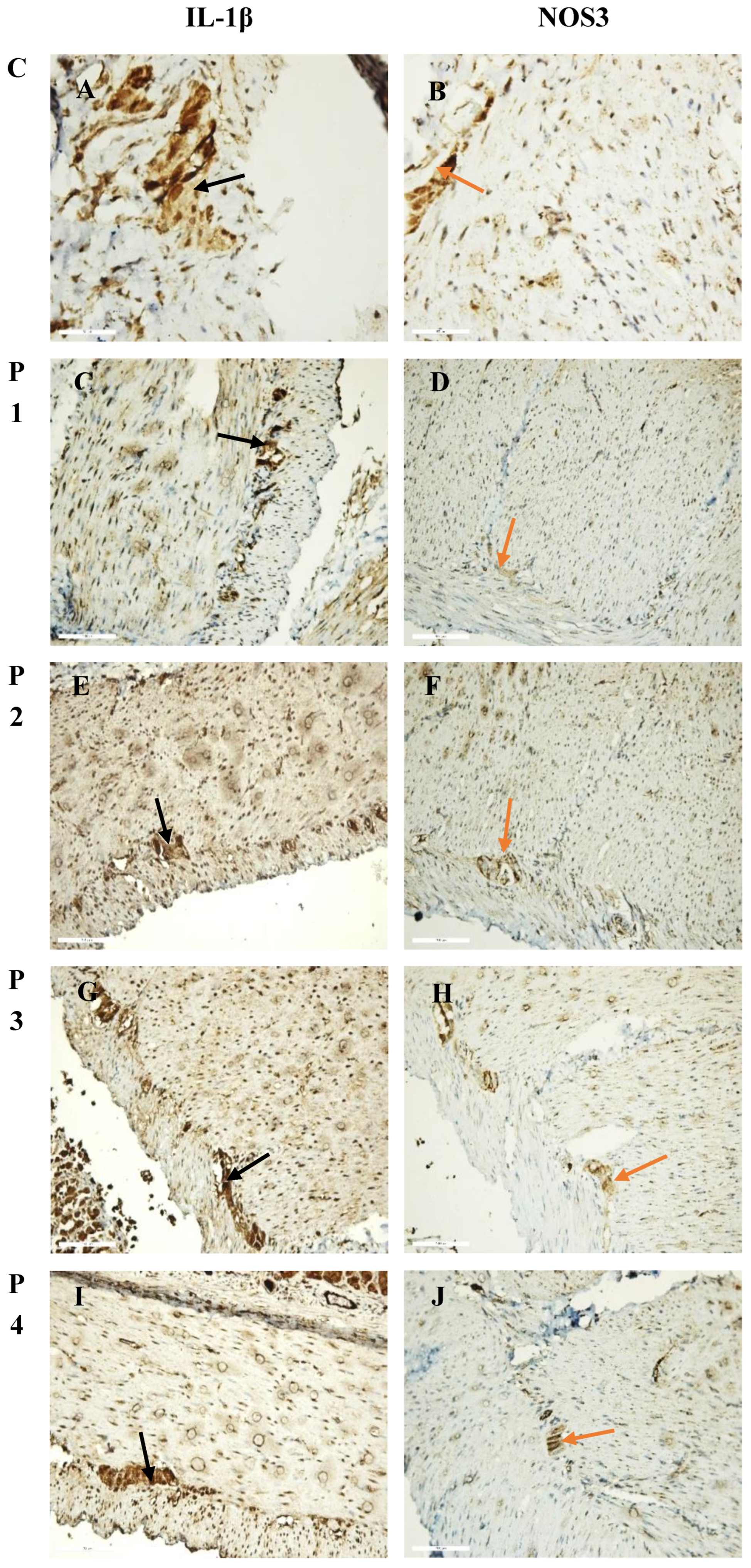
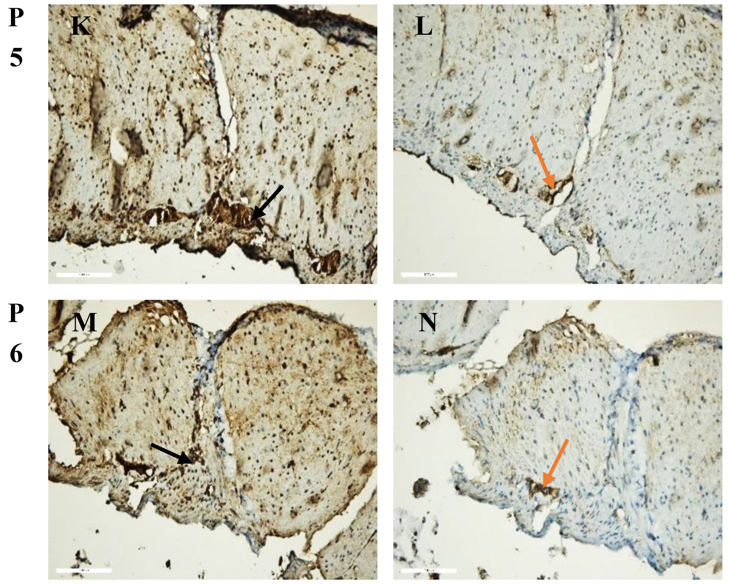
Figure 2.
Intensity of IL-1β (A) and NOS3 (B) immunoreaction, expressed in arbitrary units (AU) in smooth muscle preparations (SMPs) of rat’s stomach. ***, p < 0.001.
Figure 2.
Intensity of IL-1β (A) and NOS3 (B) immunoreaction, expressed in arbitrary units (AU) in smooth muscle preparations (SMPs) of rat’s stomach. ***, p < 0.001.
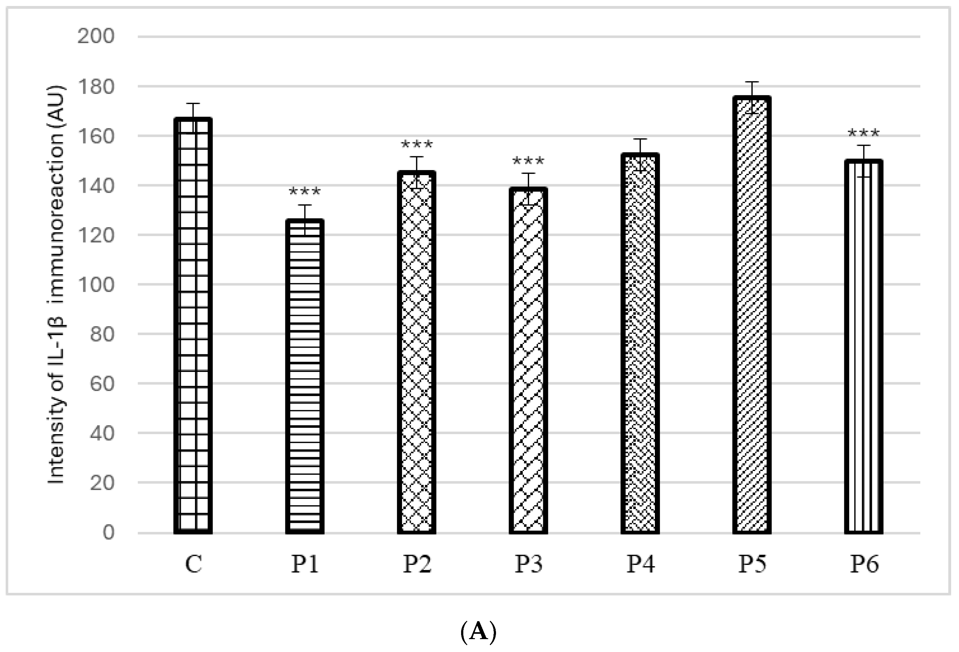
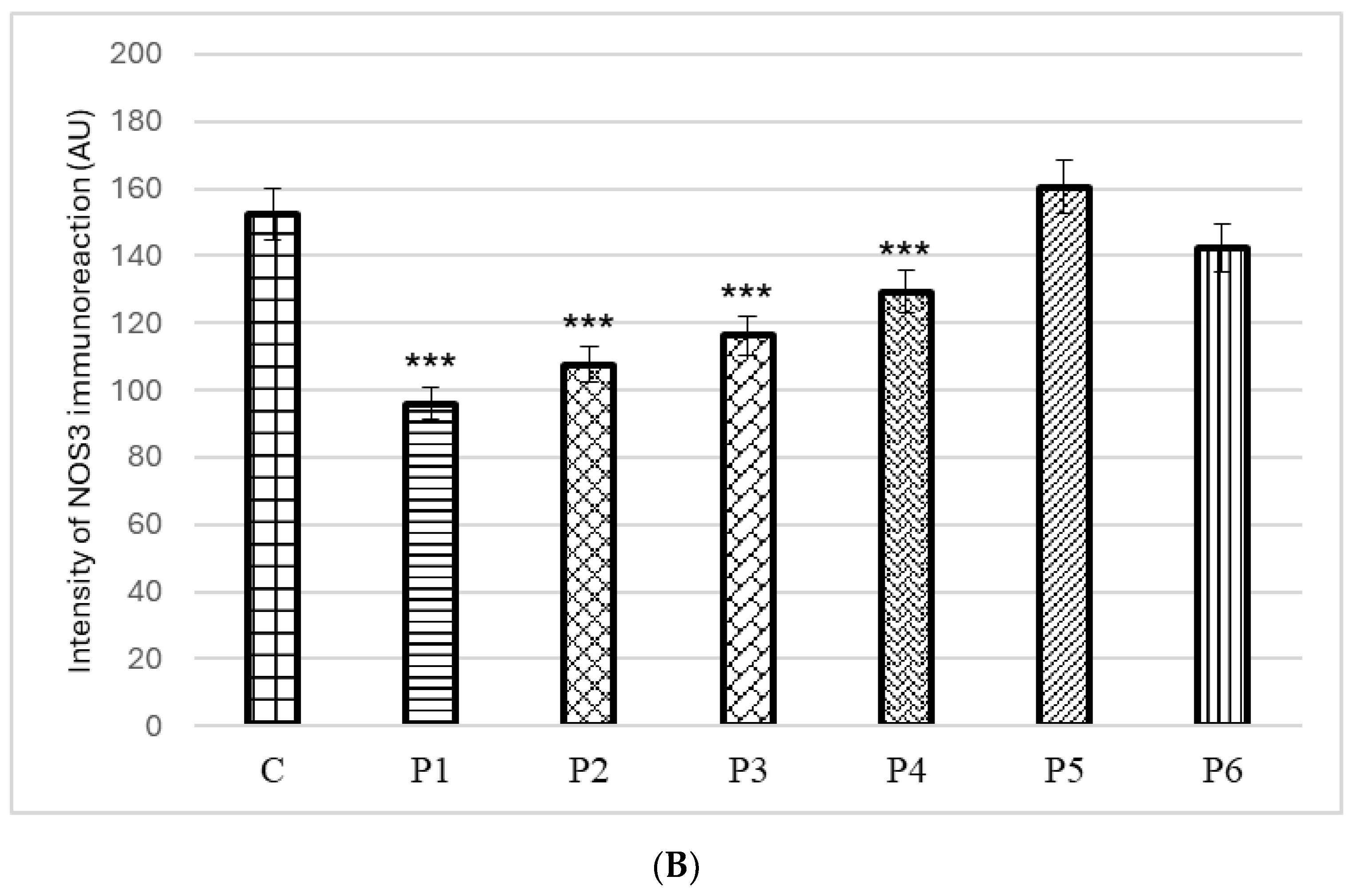

Table 1.
Origin of the propolis samples.
| Propolis Sample* | Village | District/Region | GPS Coordinates |
|---|---|---|---|
| P1 | Gamzovo | Vidin | 44°05′ N 22°45′ E |
| P2 | Parsha | Gabrovo | 42°57′ N 25°29′ E |
| P3 | Ritya | Gabrovo | 42°59′ N 25°25′ E |
| P4 | Kozi rog | Gabrovo | 42°57′ N 25°16′ E |
| P5 | Burya | Gabrovo | 43°02′ N 25°19′ E |
| P6 | Malinovo | Lovech | 42°90′ N 24°90′ E |
* the coding of the samples corresponds to that in our previous study [31].
Table 2.
Total phenolic content (TPC), total flavonoid content (TFC) and total caffeic acid derivatives content (TCADC) of DMSO propolis extracts (20 mg/mL).
Table 2.
Total phenolic content (TPC), total flavonoid content (TFC) and total caffeic acid derivatives content (TCADC) of DMSO propolis extracts (20 mg/mL).
| Propolis sample | TPC, mg GAE/g of extract |
TFC, mg QE/g of extract |
TCADC, mg CAE/g of extract |
|---|---|---|---|
| P1 | 213.75 ± 0.30 | 64.41 ± 0.26 | 15.85 ± 0.69 |
| P2 | 275.09 ± 0.60 | 136.70 ± 0.13 | 18.77 ± 0.30 |
| P3 | 189.30 ± 0.10 | 95.75 ± 0.55 | 11.75 ± 0.85 |
| P4 | 290.80 ± 0.23 | 151.60 ± 0.66 | 21.79 ± 0.31 |
| P5 | 256.03 ± 0.50 | 125.24 ± 0.19 | 19.06 ± 0.27 |
| P6 | 226.01 ± 0.95 | 114.94 ± 0.52 | 15.84 ± 0.21 |
Table 3.
Antioxidant activity of DMSO propolis extracts (20 mg/mL).
| Propolis sample | Antioxidant activity | ||||
|---|---|---|---|---|---|
| DPPH, mM TE/g of extract |
IC50, mg/mL of extract |
FRAP, mM TE/g of extract |
|||
| P1 | 1257.50 ± 2.12 | 0.34 ± 0.02 | 976.77 ± 2.06 | ||
| P2 | 1522.55 ± 2.24 | 0.29 ± 0.01 | 1168.02 ± 2.43 | ||
| P3 | 863.73 ± 1.56 | 0.48 ± 0.03 | 796.86 ± 0.59 | ||
| P4 | 1799.22 ± 3.02 | 0.24 ± 0.01 | 1145.24 ± 0.99 | ||
| P5 | 1479.69 ± 1.58 | 0.28 ± 0.01 | 1080.10 ± 0.67 | ||
| P6 | 1260.21 ± 1.42 | 0.32 ± 0.02 | 968.07 ± 1.52 | ||
Table 4.
Phenolic compounds identified in DMSO propolis extracts by HPLC analysis.
| Phenolic Compounds, mg/g of Extract |
Propolis Samples | |||||
|---|---|---|---|---|---|---|
| P1 | P2 | P3 | P4 | P5 | P6 | |
| Phenolic acids | ||||||
| Caffeic acid | 5.18 ± 0.12 | 6.60 ± 0.13 | 4.21 ± 0.04 | 6.85 ± 0.14 | 5.17 ± 0.09 | 4.98 ± 0.03 |
| p-coumaric acid | 3.00 ± 0.09 | 5.42 ± 0.11 | 2.27 ± 0.02 | 2.99 ± 0.03 | 3.54 ± 0.03 | 3.20 ± 0.04 |
| Sinapic acid | 1.47 ± 0.02 | 1.27 ± 0.05 | 3.12 ± 0.07 | 3.43 ± 0.01 | 1.76 ± 0.01 | 2.76 ± 0.07 |
| Caffeic acid benzyl ester | 4.25 ± 0.05 | 5.06 ± 0.08 | 4.71 ± 0.09 | 9.43 ± 0.15 | 7.83 ± 0.05 | 5.19 ± 0.11 |
| Cinnamic acid | 0.26 ± 0.02 | 1.96 ± 0.01 | 0.18 ± 0.00 | 0.48 ± 0.00 | 0.59 ± 0.01 | 1.25 ± 0.02 |
| Flavonoids | ||||||
| Isorhamnetin | 1.91 ± 0.07 | 3.51 ± 0.06 | 1.84 ± 0.01 | 2.85 ± 0.02 | 1.98 ± 0.04 | 2.21 ± 0.08 |
| Pinocembrin | 11.48 ± 0.15 | 36.32 ± 0.17 | 11.90 ± 0.10 | 18.61 ± 0.13 | 25.75 ± 0.26 | 22.11 ± 0.23 |
| Chrysin | 31.42 ± 0.55 | 53.98 ± 0.36 | 59.62 ± 0.44 | 81.50 ± 0.62 | 82.21 ± 0.57 | 81.90 ± 0.39 |
| Pinobanksin-3-O-propionate | 35.26 ± 0.28 | 56.85 ± 0.51 | 34.27 ± 0.18 | 75.28 ± 0.46 | 63.74 ± 0.42 | 54.28 ± 0.21 |
Table 5.
Acetylcholinesterase inhibitory activity of DMSO propolis extracts.
| Sample | Inhibition, % | IC50, mg/mL | |||
|---|---|---|---|---|---|
| 1 mg/mL | 0.5 mg/mL | 0.25 mg/mL | 0.1 mg/mL | ||
| P1 | 40.99 ± 0.28 | 39.76 ± 0.52 | 37.64 ± 0.61 | 23.13 ± 0.83 | 1.23 ± 0.03 |
| P2 | 43.58 ± 0.44 | 41.40 ± 0.08 | 35.00 ± 0.96 | 26.73 ± 0.57 | 1.25 ± 0.02 |
| P3 | 40.12 ± 0.09 | 31.85 ± 0.82 | 24.74 ± 0.04 | 18.82 ± 0.29 | 1.39 ± 0.03 |
| P4 | 43.58 ± 0.44 | 34.57 ± 0.56 | 27.14 ± 0.18 | 18.14 ± 0.11 | 1.18 ± 0.02 |
| P5 | 46.44 ± 0.17 | 38.84 ± 0.29 | 23.93 ± 0.28 | 17.33 ± 0.04 | 1.07 ± 0.03 |
| P6 | 41.00 ± 0.07 | 35.06 ± 0.88 | 29.12 ± 0.25 | 12.75 ± 0.89 | 1.36 ± 0.24 |
Table 6.
Red blood cell membrane stabilization activity of DMSO propolis extracts.
| Sample | Protection, % | IC50, mg/mL | |||
|---|---|---|---|---|---|
| 0.5 mg/mL | 0.25 mg/mL | 0.1 mg/mL | 0.05 mg/mL | ||
| P1 | 86.59 ± 0.39 | 72.97 ± 0.44 | 53.78 ± 0.23 | 34.03 ± 0.63 | 0.11 ± 0.01 |
| P2 | 85.29 ± 0.06 | 73.32 ± 0.23 | 48.56 ± 0.74 | 21.30 ± 0.59 | 0.14 ± 0.02 |
| P3 | 76.28 ± 0.54 | 68.53 ± 0.87 | 45.47 ± 0.55 | 39.61 ± 0.68 | 0.13 ± 0.02 |
| P4 | 86.40 ± 0.17 | 77.09 ± 0.08 | 66.69 ± 0.67 | 41.65 ± 0.89 | 0.06 ± 0.01 |
| P5 | 89.32 ± 0.20 | 78.94 ± 0.49 | 55.93 ± 0.69 | 41.71 ± 0.59 | 0.08 ± 0.01 |
| P6 | 84.28 ± 0.45 | 72.74 ± 0.84 | 64.74 ± 0.40 | 42.31 ± 0.25 | 0.06 ± 0.01 |
| Aspirin | 60.12 ± 0.36 | 55.22 ± 0.26 | 52.18 ± 0.10 | 22.68 ± 0.38 | 0.19 ± 0.02 |
| Prednisolone | 94.97 ± 0.86 | 88.26 ± 0.48 | 74.74 ± 0.69 | 35.71 ± 0.63 | 0.06 ± 0.01 |
Disclaimer/Publisher’s Note: The statements, opinions and data contained in all publications are solely those of the individual author(s) and contributor(s) and not of MDPI and/or the editor(s). MDPI and/or the editor(s) disclaim responsibility for any injury to people or property resulting from any ideas, methods, instructions or products referred to in the content. |
© 2025 by the authors. Licensee MDPI, Basel, Switzerland. This article is an open access article distributed under the terms and conditions of the Creative Commons Attribution (CC BY) license (http://creativecommons.org/licenses/by/4.0/).
Copyright: This open access article is published under a Creative Commons CC BY 4.0 license, which permit the free download, distribution, and reuse, provided that the author and preprint are cited in any reuse.


