Submitted:
07 April 2025
Posted:
08 April 2025
You are already at the latest version
Abstract
Three-dimensional (3D) spheroid models have revolutionized in vitro cancer research by offering more physiologically relevant alternatives to traditional two-dimensional (2D) cultures. This systematic review evaluates methods for spheroid formation and cellular sources used, highlighting diverse applications and preferences in the field. The five most investigated cancer origins for spheroid seeding are breast, colon, lung, ovary, and brain cancers, reflecting their clinical importance and research focus. Among seeding methodologies, forced-floating and scaffold-based methods predominate, demonstrating reliability and versatility in spheroid generation. Other techniques, including microfluidics, bioprinting, hanging drop and suspension culture also play significant roles, each with distinct advantages and limitations. This review underscores the growing adoption of spheroid models and the need for standardization in methodologies to enhance reproducibility and translational potential in cancer research.
Keywords:
spheroids
; personalized medicine
; drug screening
; organoids
; 3D models
1. Introduction
In recent years, there has been a paradigm shift in biomedical research towards developing more clinically relevant models to study human physiology and diseases. Personalized medicine, aiming to tailor medical interventions to individual patient characteristics, has gained prominence in the quest for more effective and targeted treatments. Within this context, three-dimensional cell culture models, particularly spheroids, have emerged as valuable tools for bridging the gap between traditional two-dimensional cell cultures and in vivo studies. Spheroids, characterized by their spherically shaped cellular aggregates, exhibit enhanced physiological relevance by recreating cell-cell and cell-matrix interactions found in native tissues. This article aims to explore the developing field of spheroid research, focusing on their potential applications in personalized medicine and elucidating methodologies employed for spheroid seeding.
Traditional cell culture systems are often inadequate in reproducing the complexity of in vivo tissue structures and functions. Spheroids, formed through the self-assembly of cells, provide a closer representation of the native tissue microenvironment. This three-dimensional architecture facilitates cell-cell interactions, nutrient gradients, and spatial organization, closely mimicking the in vivo conditions. As a result, spheroids have gained prominence as an advanced in vitro model that bridges the gap between conventional cell cultures and in vivo studies.
1.1. Summary of Models for In Vitro Studies
The concept of growing cells outside of the human body began in the early 20th century. In 1907, Ross Harrison obtained the first successful tissue culture by growing nerve fibers from frog embryos. This work paved the way for cell culture techniques that have since become foundational in biomedical research [1].
Furthermore, the establishment of the first continuous human cell line, HeLa cells, by George Otto Gey in 1951 marked a revolutionary step in cancer research. Derived from cervical cancer cells, HeLa provided researchers with an immortal cell line capable of indefinite growth under proper conditions. This breakthrough enabled consistent and reproducible experiments, fostering a deeper understanding of cancer progression and therapeutic responses [2].
The 2D monolayer culture technique dominated in vitro cancer research for much of the 20th century. 2D monolayer cultures have been the cornerstone of in vitro cancer research for decades due to their simplicity, cost-effectiveness, and adaptability to high-throughput screening. In these systems, cancer cells are grown on flat, rigid substrates, providing uniform exposure to nutrients, oxygen, and drugs. The standardized conditions in 2D cultures enable reproducible experiments and straightforward readouts, making them ideal for initial mechanistic studies and large-scale drug screening. Cells are grown on flat plastic or glass surfaces in a single layer, making it possible to study their morphology, growth, and responses to drugs in a controlled environment. These models were integral to early breakthroughs in cancer biology, such as identifying signaling pathways and testing chemotherapeutics. However, their limitations became increasingly apparent. The two-dimensional format failed to replicate the complexity of the tumor microenvironment, cell-cell interactions, and gradients of nutrients and oxygen present, and cellular architecture found in vivo. Cells grown in 2D often exhibit altered morphology, gene expression, and behavior compared to their in vivo counterparts [3].
The limitations of 2D models drifted the development of 3D models. In recent years, three-dimensional (3D) culture systems, including spheroids, organoids, and other, have emerged as advanced in vitro models that better mimic the in vivo tumor microenvironment. Spheroids, first described in the 1970s, allowed researchers to grow cells in a structure that mimics the spatial architecture of tumors. Spheroids, for instance, are self-organizing aggregates of cancer cells that develop gradients of oxygen, nutrients, and waste products, closely resembling the structural and functional characteristics of solid tumors. These models better simulate cell-cell and cell-matrix interactions, as well as the nutrient and oxygen gradients found in actual tumors. This advancement has significantly improved the study of tumor biology and drug resistance [4].
Today, 2D and 3D models are commonly used in tandem, each complementing the other’s strengths. 2D cultures remain essential for high-throughput screening due to their simplicity and reproducibility. In contrast, 3D models provide a more physiologically relevant context, making them indispensable for studying tumor progression, metastasis, and resistance to therapy. Hybrid approaches combining 2D and 3D cultures, as well as emerging technologies like organ-on-a-chip and patient-derived organoids, are bridging the gap between in vitro and in vivo systems. These models aim to capture the advantages of both 2D simplicity and 3D physiological relevance, providing a more comprehensive understanding of tumor biology and therapeutic responses. While 2D cultures remain essential for preliminary studies due to their simplicity and efficiency, incorporating 3D models enhances the translational potential of in vitro research by mimicking critical aspects of tumor biology. The complementary use of both systems, along with advances in hybrid models, offers a robust framework for cancer research, drug discovery, and personalized medicine [5,6,7].
1.2. Comparison of 2D and 3D Models
In vitro models are indispensable tools for studying cancer biology, screening therapeutic agents, and elucidating disease mechanisms. Among these, traditional two-dimensional (2D) cancer cell cultures and advanced three-dimensional (3D) models are widely utilized. Each model possesses distinct advantages and limitations. The most appropriate model selection should be made based on the specific context of the application. Both models also represent defined potentials. Table 1 highlights how each model is best suited to specific research objectives, emphasizing the importance of selecting the appropriate system based on the experimental needs [5,6,7,8,10,11,12,13].
1.3. Spheroids in Personalized Medicine
Personalized medicine aims to customize healthcare interventions based on individual patient characteristics, and spheroids emerge as a powerful tool in this endeavor. The inherent heterogeneity within patient populations can be more accurately represented using spheroids, enabling the development of personalized therapeutic strategies. By incorporating patient-specific cells into spheroid models, researchers can assess drug responses and tailor treatment regimens for improved clinical outcomes [14].
Spheroids have emerged as a pivotal three-dimensional (3D) in vitro model in cancer research, offering a significant leap forward in replicating the complexity of in vivo tumor biology. Unlike traditional two-dimensional (2D) monolayer cultures, spheroids self-assemble into multicellular aggregates, creating a structurally and functionally relevant microenvironment that mimics many key aspects of solid tumors. This enhanced physiological relevance makes spheroids an invaluable tool for advancing our understanding of cancer biology and improving preclinical evaluations of therapeutic agents [15].
The defining characteristic of spheroids is their ability to form gradients of oxygen, carbon dioxide, nutrients, and metabolites. These gradients lead to the development of distinct cellular zones within the spheroid [15].
Cellular zones of spheroids are visualized and presented in Figure 1. Three cellular zones of spheroids are:
- Proliferative outer layer: Consisting of actively dividing cells, with high accessibility to oxygen and nutrients.
- Quiescent intermediate layer: Consisting of quiescent and senescent cells with reduced metabolic activity due to limited nutrient and oxygen availability.
- Hypoxic apoptotic core: Consisting of cells in apoptotic state due to severe nutrient and oxygen deprivation. Core environment mimics what is observed in poorly vascularized tumor regions in vivo.
This zonal architecture replicates the heterogeneous microenvironment of solid tumors, which is critical for studying tumor progression, metastasis, and resistance to therapies.
1.4. Applications of Spheroids Models
Spheroids are widely utilized in cancer research as robust models for examining critical biological processes in a three-dimensional context. They provide significant insights into tumor progression by enabling detailed investigation of invasion, metastasis, and interactions between the tumor and surrounding stroma. In the realm of therapeutic screening, spheroids facilitate the evaluation of anti-cancer drug efficacy, penetration dynamics within the tumor microenvironment, and mechanisms of drug resistance. They are also essential for modeling tumor responses to hypoxia and radiation therapy, offering a physiologically relevant system that closely mimics in vivo conditions. Additionally, spheroids play a pivotal role in immunotherapy research by supporting the study of immune cell-tumor interactions, thereby providing a more representative alternative to conventional two-dimensional culture models [15,16]
2. Materials and Methods
A comprehensive search was conducted in the PubMed database to identify relevant articles available until December 2024. The search utilized specific free words and Medical Subject Headings (MeSH) terms, including key terms such as 'spheroid,' 'cancer,' 'drug,' 'patient-derived,' and 'tumor.' Exclusion terms 'co-culture' and 'xenograft' were applied. Only articles published in English were considered for inclusion.
Inclusion criteria encompassed studies involving spheroids formed from patient-derived cancer cells, while exclusion criteria covered articles that did not meet these inclusion criteria, reviews, case reports, and works focusing on the application or use of spheroids in areas other than drug screening.
Data extraction from included articles comprised information on authors, publication year, title, cancer type, cell aggregation protocol, culture conditions, spheroid measurements, in vitro functional properties, therapeutic effects in vivo, and study model employed.
The inclusion criterion for English-language publications may introduce potential bias, excluding relevant literature in other languages. Consequently, the findings should be interpreted cautiously, acknowledging the potential limitations associated with the language-based selection criteria.
3. Results
3.1. Database Screening
Following a screening of 190 articles retrieved through searches in PubMed pertaining to seeding of spheroids from human cancer cells for drug sensitivity screening, only 143 articles were evaluated for eligibility based on the search strategy outlined in the materials and methods section. The selection process is illustrated in Figure 2.
3.2. Systematic Review
Table 2 displays the selected 143 articles along with their fundamental details, including: first author and publication year, cancer type by type of tissue in which cancer originates (histological type) and by primary site, spheroid seeding method.
3.2.1. Source of Spheroids
Spheroids derived from various cancer types are extensively utilized in research to mimic in vivo tumor characteristics, providing insights into diverse cancer-specific processes. The ability to generate spheroids from a variety of cell sources, including patient-derived tumor cells, further enhances their value in studying personalized therapeutic responses. These models allow researchers to recreate the complexity of individual tumors and test specific treatment regimens in vitro, paving the way for advances in precision and personalized medicine. In the present systematic review, the most common cell sources of spheroids were identified. Five most investigated cancer origins associated with spheroids were breast (n=46, accounting for 25% of all investigated cancers), followed by colon (n=23, 12%), lung (n=21, 11%), ovary (n=19, 10%), and brain (n=18, 10%). A complete summary of systematic review results is presented in Figure 3 and Table 3.
Breast Cancer
Spheroids derived from breast cancer cells represent one of the most extensively studied in vitro models, reflecting the prominence of breast cancer as both a clinical challenge and leading research focus. Systematic review of the literature revealed that breast cancer cell lines and patient-derived cells were the most frequently used sources for spheroid generation.
Breast cancer spheroid models facilitate the exploration of unique tumor characteristics (e.g. variations in growth dynamics, gene expression profiles, and interactions with the tumor microenvironment) and aspects of cancer biology (e.g. immortality, telomerase activation, antiapoptotic strategy) [20,28,48,156].
Moreover, this approach facilitates the exploration of therapies targeting estrogen-metabolizing enzymes and receptors, enabling the discovery of novel treatments that may prevent tumor initiation or inhibit cancer growth [26].
Additionally, this methodology supports the development of patient-specific drugs, thereby aligning with the principles of precision medicine to optimize therapeutic outcomes for individual breast cancer patients [59].
Colon Cancer
Colon cancer is the second most common source of cells used for spheroid generation, reflecting its critical role in cancer research.
To enhance the utility of human 3D colorectal cancer spheroid models in preclinical drug assessment, there is the need for standardized and validated methodologies. While monoculture spheroids are useful for high-throughput drug screening due to their simplicity, spheroids provide deeper insights into tumor biology and chemoresistance mechanisms, offering a more accurate preclinical tool for evaluating therapeutic efficacy and developing new drug candidates [33,157].
3D cultures still face challenges in clinical implementation, and advancements in co-culture techniques, addressing tumor heterogeneity, and improving laboratory protocols are essential for enhancing reproducibility and drug testing reliability in colorectal cancer research [158]. Moreover, collecting cells during biopsy from different tumor sites might provide more comprehensive representation of tumor subclones, offering greater insight into the tumor's diverse properties and improving the accuracy of preclinical models for drug testing and personalized medicine [158].
Lung Cancer
Lung cancer represents the third most common source of cells for generating spheroids. Lung cancer-derived spheroids are utilized to investigate key aspects of tumor biology.
Lung cancer spheroids are particularly valuable for studying the progression of both non-small cell lung cancer (NSCLC) and small cell lung cancer (SCLC), which differ significantly in their biological behavior and response to therapy [159,160].
Therapeutically, lung cancer spheroids serve as platforms for evaluating the efficacy of novel anti-cancer agents, including small molecules, biologics, and combination therapies. Their three-dimensional structure facilitates studies of drug delivery systems aimed at overcoming barriers such as limited penetration into solid tumors [40,47,49,67,77,119].
Ovarian Cancer
Ovarian cancer is the fourth most common source of cells used for spheroid generation. Spheroids derived from ovarian cancer cells are utilized in research to study processes central to ovarian cancer pathology, such as peritoneal metastasis, chemoresistance, and interactions with the tumor microenvironment. Given the propensity of ovarian cancer to spread via the peritoneal cavity through multicellular aggregates, spheroids serve as a physiologically relevant model to replicate these metastatic behaviors in vitro [31,52,76,106,107].
Brain Cancer
Brain cancers, including glioblastoma and other gliomas, rank as the fifth most common source of cells used for spheroid generation.
These three-dimensional models are pivotal for studying the unique microenvironment and invasive properties of brain tumors, which are characterized by their aggressive behavior and resistance to standard therapies. Brain cancer-derived spheroids closely mimic the in vivo conditions of brain tumors, providing insights into key processes such as tumor invasion, therapeutic resistance, and interactions with the extracellular matrix (ECM). Glioblastoma-derived spheroids are among the most studied in this category. They are particularly valuable for investigating the highly invasive nature of glioblastoma cells, which infiltrate surrounding healthy brain tissue, making complete surgical resection nearly impossible [24,63,74,93,104].
Recent advancements include patient-derived brain cancer spheroids, which preserve the genetic and phenotypic heterogeneity of primary tumors. These models are increasingly used for personalized medicine, enabling the testing of individualized therapeutic regimens in a controlled in vitro setting [30,64,147].
3.2.2. Spheroid Seeding Methods
The successful implementation of spheroids in personalized medicine relies on robust and reproducible methodologies for their generation. The generation of three-dimensional (3D) spheroids as in vitro models requires careful consideration of seeding methods to ensure reproducibility, scalability, and physiological relevance. A variety of techniques have been developed to create spheroids, ranging from traditional methods to advanced approaches incorporating cutting-edge technologies. An overview of the most common and recent seeding methods used for the seeding of spheroids from human cancer cells is presented below.
In conclusion, this article aims to provide a thorough understanding of the methodologies employed in spheroid seeding and highlights the manifold applications of spheroids in advancing personalized medicine.
Our systematic review revealed that, among spheroid seeding methodologies, the most common were forced floating (n=70), scaffold-based methods (n=41), recent advances (n = 20), hanging drop (n=14) and suspension culture (n=4). A summary of systematic review results is presented in Figure 4 and Table 4.
Based on the systematic review, a summary of the most common spheroid seeding methods is illustrated in Figure 5. The figure visualizes the most utilized spheroid seeding methods such as hanging drop (1), forced-floating (2), magnetic levitation (3), scaffold-based (4), suspension culture (5), and recent scientific advances (6) methods: microencapsulation (6a), bioprinting (6b), nanoparticles (6c), chips and microfluidics (6d).
Forced Floating
The forced floating method is the most employed technique for spheroid formation due to its simplicity and scalability. This approach uses non-adhesive surfaces to prevent cell attachment, encouraging cells to aggregate and form three-dimensional (3D) spheroids. The technique involves seeding cells in multi-well plates that have been treated to inhibit surface adhesion, either through coating with low-attachment materials like poly-HEMA (Poly(2-hydroxyethyl methacrylate)) or using ultra-low attachment (ULA) culture plates designed specifically for this purpose. In the absence of adhesion sites, cells naturally aggregate in the medium, forming spheroids under the influence of gravity and intercellular interactions. The process begins with the preparation of a single-cell suspension of the desired cell density, typically ranging from 103 to 105 cells per well, depending on the type of cells and the intended spheroid size. The cell suspension is then distributed into the wells of the plate. Over the course of 24–96 hours, depending on the cell type and experimental conditions, the cells self-assemble into compact spheroids [22,100,138,163].
The forced floating method is particularly advantageous for producing uniform spheroids with consistent size and morphology, making it suitable for high-throughput applications such as drug screening and toxicity assays. Moreover, one of the significant benefits of the forced floating method is its compatibility with automated systems, allowing for large-scale spheroid generation and analysis. Additionally, this method does not require specialized equipment beyond the plates or coatings, making it accessible for most laboratories. However, the method has certain limitations. The reliance on non-adhesive surfaces can lead to variability in spheroid integrity and size if the cell density or culture conditions are not carefully optimized. Additionally, long-term culture may be constrained by limited nutrient and oxygen diffusion, necessitating periodic medium exchange or supplementation with perfusion systems [7,31 55,57,92,139,113].
Scaffold-Based
The scaffold-based method for spheroid seeding is the second most utilized method for generating three-dimensional (3D) cellular aggregates. This method uses biomaterials, known as scaffolds, that mimic the extracellular matrix (ECM) to provide the structural support and environment conducive to cell adhesion, proliferation, and aggregation into spheroids. Scaffolds can be fabricated from a wide range of materials, including natural polymers such as collagen, gelatin, and alginate, as well as synthetic polymers like PLGA (poly(lactic-co-glycolic acid)) and PEG (polyethylene glycol). The process typically begins with the preparation of the scaffold material, which may be in the form of hydrogels, porous matrices, or microcarriers. Cells are then seeded onto or encapsulated within the scaffold. Once seeded, the cells interact with the scaffold material and with one another, eventually forming spheroid structures. The scaffold not only facilitates cell aggregation, but it also supports nutrient and oxygen diffusion, which is crucial for maintaining cell viability and function in 3D cultures [140,165,166].
Scaffold-based methods offer significant advantages, including ability to recreate a more physiologically relevant microenvironment compared to non-adhesive-based techniques. The structural and biochemical properties of the scaffold can be engineered to closely mimic the in vivo ECM, supporting the growth and differentiation of specific cell types. This makes the method particularly suitable for modeling complex tissues and for co-culture systems involving multiple cell types. Additionally, scaffold-based systems are compatible with long-term culture, as the scaffold provides a sustained environment for nutrient and waste exchange. However, there are limitations to this approach. The use of scaffolds introduces variability in spheroid size and shape, depending on the uniformity of the material and the seeding protocol. The composition and mechanical properties of the scaffold can also influence cellular behavior, which may complicate the interpretation of results. Furthermore, the cost and complexity of scaffold fabrication, particularly for advanced synthetic materials, may be a barrier for some applications [7,165].
Hanging Drop
The hanging drop method is a robust and third most utilized technique for generating three-dimensional (3D) spheroids in vitro, particularly valued for its ability to produce uniform and physiologically relevant cellular aggregates. This method capitalizes on gravity-driven cellular self-assembly within droplets of culture medium, facilitating interactions that mimic those found in vivo. To implement this approach, a cell suspension of the desired density is prepared, often ranging from 10² to 10⁴ cells per droplet. Droplets, typically 20–50 μL in volume, are then dispensed onto the inner surface of an inverted petri dish lid. The droplets are retained by surface tension, allowing them to remain suspended. This setup is placed over a dish containing a hydrating agent, such as phosphate-buffered saline (PBS) or water, to maintain humidity and prevent evaporation during incubation. Over 24 to 72 hours under standard culture conditions, the suspended cells settle at the bottom of the droplets and aggregate into spheroids through intercellular adhesion and natural cell-cell interactions [17,34,53,113,115,143].
The hanging drop method is particularly advantageous due to its simplicity, low cost, and minimal equipment requirements. This method offers significant benefits, including enhanced cellular aggregation and adhesion driven by gravitational forces, which minimize mechanical damage to spheroids. It also allows for precise control over the spheroid size and cell composition by adjusting the initial cell density and droplet volume. However, this technique has limitations, such as the potential disruption of spheroids during transfer to conventional culture plates due to mechanical stress. Moreover, it is labor-intensive and not easily scalable for high-throughput applications, as each droplet must be individually prepared and managed. Additionally, nutrient and waste exchange are limited by the small volume of medium, necessitating careful monitoring to maintain spheroid viability. It has also been adapted for co-culture systems to explore interactions between different cell types, such as tumor and stromal cells [7,53,58,113,132,167].
Suspension Culture
The suspension culture method is the fourth most utilized approach for generating spheroids in in vitro studies, particularly in cancer research, developmental biology, and drug discovery. This technique relies on culturing cells in a liquid medium without a solid substrate, allowing them to aggregate and form spheroids due to intercellular adhesion and natural aggregation tendencies. Typically, the suspension culture is conducted in low-attachment culture vessels, such as non-adherent plates or spinner flasks, to prevent cell adhesion to the container surface and promote spheroid formation [140,168].
The process begins with the preparation of a single-cell suspension at a defined density, which is a critical parameter for achieving uniform spheroid size and morphology. These vessels prevent cells from adhering to the surface and maintain them in suspension. Spinner flasks or bioreactors are dynamic systems, where gentle agitation or rotation keeps the cells suspended and evenly distributed, which can enhance the uniformity of spheroid formation and improve mass transport of nutrients and oxygen [23,140,168].
The suspension culture method offers several advantages. It is relatively simple and cost-effective, requiring minimal specialized equipment beyond vessels. The method is highly versatile, accommodating various cell types and allowing for easy incorporation of co-culture systems to model complex cell-cell interactions, such as those between tumor and stromal cells. Furthermore, the suspension culture can be adapted for high-throughput applications, making it suitable for large-scale drug screening and toxicity testing. Despite its strengths, the suspension culture method has limitations. Nutrient and oxygen diffusion can be inadequate in larger spheroids, leading to hypoxic or necrotic cores. This limitation necessitates careful control of spheroid size and medium composition to maintain viability. Additionally, while the method is relatively straightforward, achieving consistent spheroid size and morphology can be challenging without precise control over cell seeding density and culture conditions. Moreover, prolonged culture durations may require frequent medium changes or the use of perfusion systems to sustain spheroid health [7,168].
Recent Advances in Spheroid Seeding Methods
Recent advances in spheroid seeding methods are represented by microencapsulation, bioprinting, nanoparticle-assisted techniques, microfluidics and lab-on-a-chip methods.
Microencapsulation encapsulates cells in biocompatible hydrogels like alginate, mimicking the extracellular matrix (ECM) and facilitating spheroid formation. It promotes intercellular interactions within a controlled microenvironment, protecting cells from shear stress and supporting co-culture systems. However, challenges include nutrient diffusion limitations and difficulties in spheroid retrieval [157,169].
Bioprinting is a method utilizing 3D printing technologies, allowing for the precise placement of cells and biomaterials to replicate native tissue architecture. It offers exceptional control over spheroid organization and is particularly effective for high-throughput applications and heterotypic co-cultures. Limitations include the need for bioink optimization and high costs of equipment [170,171].
Nanoparticle-assisted methods precisely guide cell aggregation into spheroids by leveraging functionalized nanoparticles. Magnetic nanoparticles enable external manipulation, while adhesive ligands support aggregation. It offers reproducibility and integration with imaging or therapeutic applications but raises concerns about nanoparticle cytotoxicity and scalability [39,66,130,172].
Microfluidics and lab-on-a-chip are technologies that provide highly controlled environments for spheroid formation, mimicking in vivo conditions through precise nutrient gradients and mechanical control. Microfluidics supports real-time monitoring and complex co-cultures, although challenges include high costs, technical expertise, and scalability [28,34,50,83,87,101,173].
Magnetic Levitation
The magnetic levitation method is a less frequently used approach to spheroid formation. This method utilizes leveraging magnetic fields to promote cellular aggregation and 3D structure development. This technique involves the use of magnetic nanoparticles that are internalized by cells through incubation. The nanoparticles are typically composed of biocompatible materials such as iron oxide and may be functionalized with extracellular matrix (ECM) proteins or other molecules to enhance cellular uptake and minimize toxicity. After the cells have internalized the nanoparticles, they are exposed to a magnetic field, which forces them to aggregate and suspend in the culture medium. The magnetic field enables the controlled formation of compact spheroids by facilitating cell-cell and cell-ECM interactions [168,174].
Magnetic levitation offers several advantages. It allows for rapid and reproducible spheroid generation and provides a high degree of control over spheroid size and structure. Additionally, this method supports the formation of co-culture spheroids by enabling the simultaneous aggregation of different cell types, which is particularly useful for modeling tumor-stroma or tumor-immune cell interactions. The technique also facilitates the incorporation of ECM components, improving the physiological relevance of the spheroid microenvironment. Furthermore, magnetic levitation is amenable to high-throughput applications and can be easily scaled up for drug screening or other large-scale studies. Despite its advantages, the method has limitations. The requirement for magnetic nanoparticles introduces potential concerns regarding biocompatibility and cellular toxicity, particularly for long-term studies. Additionally, the uniformity of nanoparticle uptake among cells can vary, potentially leading to inconsistencies in spheroid formation. The cost of magnetic nanoparticles and specialized magnetic devices may also be a barrier for some laboratories [7,168,174].
Summary of Spheroid Seeding Method
A summary of advantages and limitations of selected spheroid seeding methods is presented in Table 5.
4. Conclusions
The findings of this systematic review underscore the growing interest in 3D cancer cell models, particularly spheroids, for cancer research, therapeutic testing, and personalized medicine.
Breast cancer emerged as the most investigated cancer source for spheroid seeding, accounting for 25% of the studies included in this review, followed by colon, lung, ovary, and brain cancers. This distribution reflects the cancers' high clinical relevance, particularly in research into tumor progression, drug resistance, and the need for better preclinical models to predict patient responses to therapies.
Among the various methods for spheroid formation, forced floating and scaffold-based methods were the most employed, each used in 71 (32% of all methods) and 41 studies (19% of all methods), respectively. These approaches provide reproducible, easy-to-implement techniques for spheroid generation. Forced floating involves the aggregation of cells in suspension, which then self-assemble into spheroids. This technique has been adopted due to its simplicity and ability to consistently produce spheroids across different cancer types. The use of this method across a wide variety of cancer types underscores its applicability in generating tumor models for drug discovery and testing. Scaffold-based methods involve embedding cells in a 3D matrix, are well-established and widely adopted in cancer research. Both methods facilitate the development of spheroids that closely resemble in vivo tumor environments, with the ability to evaluate drug efficacy and tumor microenvironment interactions. In recent years, more advanced techniques have emerged, contributing to the versatility of spheroid culture. Notably, the hanging drop method is becoming increasingly popular, offering distinct advantages. Hanging drop, which involves placing small drops of cell suspension on an inverted culture dish lid, results in the spontaneous aggregation of cells into 3D spheroids due to gravity and surface tension. While this method is more labor-intensive than forced floating or scaffold-based methods, it offers more physiologically relevant 3D structures and is gaining traction for use in drug screening and tumor modeling.
Whereas these established methods have significantly advanced the field of cancer research, there are still several challenges and limitations in spheroid culture. One major issue is the inherent difficulty in achieving the necessary level of reproducibility and standardization for large-scale therapeutic testing. While methods like forced floating and scaffold-based systems provide relatively consistent spheroid formation, techniques such as hanging drop are more prone to variability depending on experimental conditions. Standardizing spheroid protocols, particularly for high-throughput drug screening, remains an ongoing challenge in the field.
Interestingly, the review also highlighted the increasing application of patient-derived spheroids, which offer a more clinically relevant model by capturing the genetic and phenotypic variability of individual tumors. The ability to generate spheroids from different regions of the same tumor has the potential to capture tumor heterogeneity, which could be critical for personalized medicine approaches. This is especially important for cancers like breast and colon cancer, where the tumor microenvironment and its interactions with various cell types play a key role in metastasis, drug resistance, and immune evasion.
In conclusion, spheroid culture techniques, particularly those employing forced floating, scaffold-based methods, have proven valuable in the study of cancer biology and the testing of novel therapies. However, to move towards more clinically translatable models, additional research is needed to refine the methods, particularly in terms of reproducibility, standardization, and incorporation of tumor heterogeneity. The adoption of patient-derived and co-culture spheroid models may significantly improve the predictive power of in vitro drug testing and lead to better-targeted therapies for cancer patients. This section is not mandatory but can be added to the manuscript if the discussion is unusually long or complex.
Author Contributions
All authors have read and agreed to the published version of the manuscript.
Funding
This research received no external funding.
Institutional Review Board Statement
Not applicable.
Informed Consent Statement
Not applicable.
Data Availability Statement
The original contributions presented in this study are included in the article. Further inquiries can be directed to the corresponding author.
Acknowledgments
Not applicable.
Conflicts of Interest
The authors declare no conflicts of interest.
Abbreviations
The following abbreviations are used in this manuscript:
| 2D | Two-dimensional |
| 3D | Three-dimensional |
| ECM | Extracellular Matrix |
| NSCLC | Non-small cell lung lymphoma |
| PEG | Polyethylene glycol |
| Poly-HEMA | Poly(2-hydroxyethyl methacrylate |
| PLGA | poly(lactic-co-glycolic acid |
| SCLC | Small cell lung cancer |
| ULA | Ultra-low attachment |
References
- Harrison, R.G. The Outgrowth of the Nerve Fiber as a Mode of Protoplasmic Movement. J Exp Zool 1910, 9, 787–846. [Google Scholar] [CrossRef]
- Scherer, W.F.; Syverton, J.T.; Gey, G.O. Studies on the Propagation in Vitro of Poliomyelitis Viruses. IV. Viral Multiplication in a Stable Strain of Human Malignant Epithelial Cells (Strain HeLa) Derived from an Epidermoid Carcinoma of the Cervix. J Exp Med 1953, 97, 695–710. [Google Scholar] [CrossRef]
- Liu, X.; Raju, P. 5.42 - In Vitro Cancer Model for Drug Testing. In Comprehensive Biotechnology (Second Edition); Moo-Young, M., Ed.; Academic Press: Burlington, 2011; pp. 543–549. [Google Scholar] [CrossRef]
- Sutherland, R.M.; McCredie, J.A.; Inch, W.R. Growth of Multicell Spheroids in Tissue Culture as a Model of Nodular Carcinomas2. JNCI: Journal of the National Cancer Institute 1971, 46, 113–120. [Google Scholar] [CrossRef]
- Breslin, S.; O’Driscoll, L. The Relevance of Using 3D Cell Cultures, in Addition to 2D Monolayer Cultures, When Evaluating Breast Cancer Drug Sensitivity and Resistance. Oncotarget 2016, 7. [Google Scholar] [CrossRef] [PubMed]
- Hoarau-Véchot, J.; Rafii, A.; Touboul, C.; Pasquier, J. Halfway between 2D and Animal Models: Are 3D Cultures the Ideal Tool to Study Cancer-Microenvironment Interactions? International Journal of Molecular Sciences 2018, 19. [Google Scholar] [CrossRef]
- Breslin, S.; O’Driscoll, L. Three-Dimensional Cell Culture: The Missing Link in Drug Discovery. Drug Discovery Today 2013, 18, 240–249. [Google Scholar] [CrossRef]
- Jensen, C.; Teng, Y. Is It Time to Start Transitioning From 2D to 3D Cell Culture? Front Mol Biosci. 2020, 7. [Google Scholar] [CrossRef]
- Goodbye, Flat Biology? Nature 2003, 424, 861–861. [CrossRef] [PubMed]
- Fontoura, J.C.; Viezzer, C.; dos Santos, F.G.; Ligabue, R.A.; Weinlich, R.; Puga, R.D.; Antonow, D.; Severino, P.; Bonorino, C. Comparison of 2D and 3D Cell Culture Models for Cell Growth, Gene Expression and Drug Resistance. Materials Science and Engineering: C 2020, 107, 110264. [Google Scholar] [CrossRef]
- Franchi-Mendes, T.; Eduardo, R.; Domenici, G.; Brito, C. 3D Cancer Models: Depicting Cellular Crosstalk within the Tumour Microenvironment. Cancers 2021, 13. [Google Scholar] [CrossRef]
- Habanjar, O.; Diab-Assaf, M.; Caldefie-Chezet, F.; Delort, L. 3D Cell Culture Systems: Tumor Application, Advantages, and Disadvantages. International Journal of Molecular Sciences 2021, 22. [Google Scholar] [CrossRef]
- Kamb, A. What’s Wrong with Our Cancer Models? Nature Reviews Drug Discovery 2005, 4, 161–165. [Google Scholar] [CrossRef] [PubMed]
- Gilazieva, Z.; Ponomarev, A.; Rutland, C.; Rizvanov, A.; Solovyeva, V. Promising Applications of Tumor Spheroids and Organoids for Personalized Medicine. Cancers 2020, 12. [Google Scholar] [CrossRef] [PubMed]
- Nayak, P.; Bentivoglio, V.; Varani, M.; Signore, A. Three-Dimensional In Vitro Tumor Spheroid Models for Evaluation of Anticancer Therapy: Recent Updates. Cancers 2023, 15. [Google Scholar] [CrossRef] [PubMed]
- El Harane, S.; Zidi, B.; El Harane, N.; Krause, K.-H.; Matthes, T.; Preynat-Seauve, O. Cancer Spheroids and Organoids as Novel Tools for Research and Therapy: State of the Art and Challenges to Guide Precision Medicine. Cells 2023, 12. [Google Scholar] [CrossRef]
- Agastin, S.; Giang, U.-B.T.; Geng, Y.; DeLouise, L.A.; King, M.R. Continuously Perfused Microbubble Array for 3D Tumor Spheroid Model. Biomicrofluidics 2011, 5, 024110. [Google Scholar] [CrossRef]
- Alhasan, L.; Qi, A.; Al-Abboodi, A.; Rezk, A.; Chan, P.P.Y.; Iliescu, C.; Yeo, L.Y. Rapid Enhancement of Cellular Spheroid Assembly by Acoustically Driven Microcentrifugation. ACS Biomater. Sci. Eng 2016, 2, 1013–1022. [Google Scholar] [CrossRef]
- An, H.J.; Kim, H.S.; Kwon, J.A.; Song, J.; Choi, I. Adjustable and Versatile 3D Tumor Spheroid Culture Platform with Interfacial Elastomeric Wells. ACS Appl. Mater. Interfaces 2020, 12, 6924–6932. [Google Scholar] [CrossRef]
- Árnadóttir, S.S.; Jeppesen, M.; Lamy, P.; Bramsen, J.B.; Nordentoft, I.; Knudsen, M.; Vang, S.; Madsen, M.R.; Thastrup, O.; Thastrup, J.; L Andersen, C. Characterization of Genetic Intratumor Heterogeneity in Colorectal Cancer and Matching Patient-Derived Spheroid Cultures. Molecular Oncology 2018, 12, 132–147. [Google Scholar] [CrossRef]
- Baek, N.; Seo, O.W.; Lee, J.; Hulme, J.; An, S.S.A. Real-Time Monitoring of Cisplatin Cytotoxicity on Three-Dimensional Spheroid Tumor Cells. Drug Des Devel Ther 2016, 10, 2155–2165. [Google Scholar] [CrossRef]
- Barone, R.M.; Calabro-Jones, P.; Thomas, T.N.; Sharp, T.R.; Byfield, J.E. Surgical Adjuvant Therapy in Colon Carcinoma: A Human Tumor Spheroid Model for Evaluating Radiation Sensitizing Agents. Cancer 1981, 47, 2349–2357. [Google Scholar] [CrossRef]
- Bartholomä, P.; Impidjati; Reininger-Mack, A.; Zhang, Z.; Thielecke, H.; Robitzki, A. A More Aggressive Breast Cancer Spheroid Model Coupled to an Electronic Capillary Sensor System for a High-Content Screening of Cytotoxic Agents in Cancer Therapy: 3-Dimensional in Vitro Tumor Spheroids as a Screening Model. J Biomol Screen 2005, 10, 705–714. [Google Scholar] [CrossRef]
- Boo, L.; Yeap, S.K.; Ali, N.M.; Ho, W.Y.; Ky, H.; Satharasinghe, D.A.; Liew, W.C.; Tan, S.W.; Wang, M.-L.; Cheong, S.K.; Ong, H.K. Phenotypic and microRNA Characterization of the Neglected CD24+ Cell Population in MCF-7 Breast Cancer 3-Dimensional Spheroid Culture. Journal of the Chinese Medical Association 2020, 83. [Google Scholar] [CrossRef]
- Brooks, E.A.; Gencoglu, M.F.; Corbett, D.C.; Stevens, K.R.; Peyton, S.R. An Omentum-Inspired 3D PEG Hydrogel for Identifying ECM-Drivers of Drug Resistant Ovarian Cancer. APL Bioengineering 2019, 3, 026106. [Google Scholar] [CrossRef]
- Bruns, J.; Egan, T.; Mercier, P.; Zustiak, S.P. Glioblastoma Spheroid Growth and Chemotherapeutic Responses in Single and Dual-Stiffness Hydrogels. Acta Biomaterialia 2023, 163, 400–414. [Google Scholar] [CrossRef]
- Calori, I.R.; Alves, S.R.; Bi, H.; Tedesco, A.C. Type-I Collagen/Collagenase Modulates the 3D Structure and Behavior of Glioblastoma Spheroid Models. ACS Appl. Bio Mater. 2022, 5, 723–733. [Google Scholar] [CrossRef]
- Chang, S.; Wen, J.; Su, Y.; Ma, H. Microfluidic Platform for Studying the Anti-Cancer Effect of Ursolic Acid on Tumor Spheroid. ELECTROPHORESIS 2022, 43, 1466–1475. [Google Scholar] [CrossRef]
- Chen, G.; Liu, W.; Yan, B. Breast Cancer MCF-7 Cell Spheroid Culture for Drug Discovery and Development. J Cancer Ther. 2022, 13, 117–130. [Google Scholar] [CrossRef]
- Chen, M.C.W.; Gupta, M.; Cheung, K.C. Alginate-Based Microfluidic System for Tumor Spheroid Formation and Anticancer Agent Screening. Biomedical Microdevices 2010, 12, 647–654. [Google Scholar] [CrossRef]
- Chen, Z.; Ma, N.; Sun, X.; Li, Q.; Zeng, Y.; Chen, F.; Sun, S.; Xu, J.; Zhang, J.; Ye, H.; Ge, J.; Zhang, Z.; Cui, X.; Leong, K.; Chen, Y.; Gu, Z. Automated Evaluation of Tumor Spheroid Behavior in 3D Culture Using Deep Learning-Based Recognition. Biomaterials 2021, 272, 120770. [Google Scholar] [CrossRef]
- Cheng, V.; Esteves, F.; Chakrabarty, A.; Cockle, J.; Short, S.; Brüning-Richardson, A. High-Content Analysis of Tumour Cell Invasion in Three-Dimensional Spheroid Assays. Oncoscience 2015, 2, 596–606. [Google Scholar] [CrossRef]
- Close, D.A.; Johnston, P.A. Detection and Impact of Hypoxic Regions in Multicellular Tumor Spheroid Cultures Formed by Head and Neck Squamous Cell Carcinoma Cells Lines. SLAS Discovery 2022, 27, 39–54. [Google Scholar] [CrossRef]
- Das, T.; Meunier, L.; Barbe, L.; Provencher, D.; Guenat, O.; Gervais, T.; Mes-Masson, A.-M. Empirical Chemosensitivity Testing in a Spheroid Model of Ovarian Cancer Using a Microfluidics-Based Multiplex Platform. Biomicrofluidics 2013, 7, 011805. [Google Scholar] [CrossRef]
- Das, V.; Fürst, T.; Gurská, S.; Džubák, P.; Hajdúch, M. Evaporation-Reducing Culture Condition Increases the Reproducibility of Multicellular Spheroid Formation in Microtiter Plates. JoVE 2017, e55403. [Google Scholar] [CrossRef]
- Das, V.; Fürst, T.; Gurská, S.; Džubák, P.; Hajdúch, M. Reproducibility of Uniform Spheroid Formation in 384-Well Plates: The Effect of Medium Evaporation. SLAS Discovery 2016, 21, 923–930. [Google Scholar] [CrossRef]
- De Angelis, M.L.; Bruselles, A.; Francescangeli, F.; Pucilli, F.; Vitale, S.; Zeuner, A.; Tartaglia, M.; Baiocchi, M. Colorectal Cancer Spheroid Biobanks: Multi-Level Approaches to Drug Sensitivity Studies. Cell Biology and Toxicology 2018, 34, 459–469. [Google Scholar] [CrossRef]
- Dhamecha, D.; Le, D.; Chakravarty, T.; Perera, K.; Dutta, A.; Menon, J.U. Fabrication of PNIPAm-Based Thermoresponsive Hydrogel Microwell Arrays for Tumor Spheroid Formation. Materials Science and Engineering: C 2021, 125, 112100. [Google Scholar] [CrossRef]
- Dias, D.R.; Moreira, A.F.; Correia, I.J. The Effect of the Shape of Gold Core–Mesoporous Silica Shell Nanoparticles on the Cellular Behavior and Tumor Spheroid Penetration. J. Mater. Chem. B 2016, 4, 7630–7640. [Google Scholar] [CrossRef]
- Domenici, G.; Eduardo, R.; Castillo-Ecija, H.; Orive, G.; Montero Carcaboso, Á.; Brito, C. PDX-Derived Ewing’s Sarcoma Cells Retain High Viability and Disease Phenotype in Alginate Encapsulated Spheroid Cultures. Cancers 2021, 13. [Google Scholar] [CrossRef]
- Dufau, I.; Frongia, C.; Sicard, F.; Dedieu, L.; Cordelier, P.; Ausseil, F.; Ducommun, B.; Valette, A. Multicellular Tumor Spheroid Model to Evaluate Spatio-Temporal Dynamics Effect of Chemotherapeutics: Application to the Gemcitabine/CHK1 Inhibitor Combination in Pancreatic Cancer. BMC Cancer 2012, 12, 15. [Google Scholar] [CrossRef]
- Eetezadi, S.; Evans, J.C.; Shen, Y.-T.; de Souza, R.; Piquette-Miller, M.; Allen, C. Ratio-Dependent Synergism of a Doxorubicin and Olaparib Combination in 2D and Spheroid Models of Ovarian Cancer. Mol. Pharmaceutics 2018, 15, 472–485. [Google Scholar] [CrossRef]
- Eguchi, H.; Kimura, R.; Onuma, S.; Ito, A.; Yu, Y.; Yoshino, Y.; Matsunaga, T.; Endo, S.; Ikari, A. Elevation of Anticancer Drug Toxicity by Caffeine in Spheroid Model of Human Lung Adenocarcinoma A549 Cells Mediated by Reduction in Claudin-2 and Nrf2 Expression. International Journal of Molecular Sciences 2022, 23. [Google Scholar] [CrossRef]
- Eimer, S.; Dugay, F.; Airiau, K.; Avril, T.; Quillien, V.; Belaud-Rotureau, M.-A.; Belloc, F. Cyclopamine Cooperates with EGFR Inhibition to Deplete Stem-like Cancer Cells in Glioblastoma-Derived Spheroid Cultures. Neuro-Oncology 2012, 14, 1441–1451. [Google Scholar] [CrossRef]
- El-Sadek, I.A.; Miyazawa, A.; Shen, L.T.-W.; Makita, S.; Mukherjee, P.; Lichtenegger, A.; Matsusaka, S.; Yasuno, Y. Three-Dimensional Dynamics Optical Coherence Tomography for Tumor Spheroid Evaluation. Biomed. Opt. Express 2021, 12, 6844–6863. [Google Scholar] [CrossRef]
- Enmon, R.M.; O’Connor, K.C.; Lacks, D.J.; Schwartz, D.K.; Dotson, R.S. Dynamics of Spheroid Self-Assembly in Liquid-Overlay Culture of DU 145 Human Prostate Cancer Cells. Biotechnol Bioeng 2001, 72, 579–591. [Google Scholar] [CrossRef]
- Flørenes, V.A.; Flem-Karlsen, K.; McFadden, E.; Bergheim, I.R.; Nygaard, V.; Nygård, V.; Farstad, I.N.; Øy, G.F.; Emilsen, E.; Giller-Fleten, K.; Ree, A.H.; Flatmark, K.; Gullestad, H.P.; Hermann, R.; Ryder, T.; Wernhoff, P.; Mælandsmo, G.M. A Three-Dimensional Ex Vivo Viability Assay Reveals a Strong Correlation Between Response to Targeted Inhibitors and Mutation Status in Melanoma Lymph Node Metastases. Translational Oncology 2019, 12, 951–958. [Google Scholar] [CrossRef]
- Fu, J.; Li, X.B.; Wang, L.X.; Lv, X.H.; Lu, Z.; Wang, F.; Xia, Q.; Yu, L.; Li, C.M. One-Step Dip-Coating-Fabricated Core–Shell Silk Fibroin Rice Paper Fibrous Scaffolds for 3D Tumor Spheroid Formation. ACS Appl. Bio Mater. 2020, 3, 7462–7471. [Google Scholar] [CrossRef]
- Fu, J.J.; Zhou, Y.; Shi, X.X.; Kang, Y.J.; Lu, Z.S.; Li, Y.; Li, C.M.; Yu, L. Spontaneous Formation of Tumor Spheroid on a Hydrophilic Filter Paper for Cancer Stem Cell Enrichment. Colloids and Surfaces B: Biointerfaces 2019, 174, 426–434. [Google Scholar] [CrossRef]
- Gao, Y.; Wu, M.; Luan, Q.; Papautsky, I.; Xu, J. Acoustic Bubble for Spheroid Trapping, Rotation, and Culture: A Tumor-on-a-Chip Platform (ABSTRACT Platform). Lab Chip 2022, 22, 805–813. [Google Scholar] [CrossRef]
- Gencoglu, M.F.; Barney, L.E.; Hall, C.L.; Brooks, E.A.; Peyton, S.R. Comparative Study of Multicellular Tumor Spheroid Formation Methods and Implications for Drug Screening. ACS Biomater Sci Eng. 2018, 4, 410–420. [Google Scholar] [CrossRef]
- Gendre, D.A.J.; Ameti, E.; Karenovics, W.; Perriraz-Mayer, N.; Triponez, F.; Serre-Beinier, V. Optimization of Tumor Spheroid Model in Mesothelioma and Lung Cancers and Anti-Cancer Drug Testing in H2052/484 Spheroids. Oncotarget 2021, 12, No–24. [Google Scholar] [CrossRef] [PubMed]
- Gheytanchi, E.; Naseri, M.; Karimi-Busheri, F.; Atyabi, F.; Mirsharif, E.S.; Bozorgmehr, M.; Ghods, R.; Madjd, Z. Morphological and Molecular Characteristics of Spheroid Formation in HT-29 and Caco-2 Colorectal Cancer Cell Lines. Cancer Cell International 2021, 21, 204. [Google Scholar] [CrossRef]
- Goisnard, A.; Daumar, P.; Dubois, C.; Aubel, C.; Roux, M.; Depresle, M.; Gauthier, J.; Vidalinc, B.; Penault-Llorca, F.; Mounetou, E.; Bamdad, M. LightSpot®-FL-1 Fluorescent Probe: An Innovative Tool for Cancer Drug Resistance Analysis by Direct Detection and Quantification of the P-Glycoprotein (P-Gp) on Monolayer Culture and Spheroid Triple Negative Breast Cancer Models. Cancers 2021, 13. [Google Scholar] [CrossRef]
- Guo, X.; Chen, Y.; Ji, W.; Chen, X.; Li, C.; Ge, R. Enrichment of Cancer Stem Cells by Agarose Multi-Well Dishes and 3D Spheroid Culture. Cell and Tissue Research 2019, 375, 397–408. [Google Scholar] [CrossRef]
- Hagemann, J.; Jacobi, C.; Hahn, M.; Schmid, V.; Welz, C.; Schwenk-Zieger, S.; Stauber, R.; Baumeister, P.; Becker, S. Spheroid-Based 3D Cell Cultures Enable Personalized Therapy Testing and Drug Discovery in Head and Neck Cancer. Anticancer Res 2017, 37, 2201. [Google Scholar] [CrossRef]
- Han, S.; Lim, J.Y.; Cho, K.; Lee, H.W.; Park, J.Y.; Ro, S.W.; Kim, K.S.; Seo, H.R.; Kim, D.Y. Anti-Cancer Effects of YAP Inhibitor (CA3) in Combination with Sorafenib against Hepatocellular Carcinoma (HCC) in Patient-Derived Multicellular Tumor Spheroid Models (MCTS). Cancers 2022, 14. [Google Scholar] [CrossRef]
- Harmer, J.; Struve, N.; Brüning-Richardson, A. Characterization of the Effects of Migrastatic Inhibitors on 3D Tumor Spheroid Invasion by High-Resolution Confocal Microscopy. JoVE 2019, No. 151, e60273. [Google Scholar] [CrossRef]
- Herter, S.; Morra, L.; Schlenker, R.; Sulcova, J.; Fahrni, L.; Waldhauer, I.; Lehmann, S.; Reisländer, T.; Agarkova, I.; Kelm, J.M.; Klein, C.; Umana, P.; Bacac, M. A Novel Three-Dimensional Heterotypic Spheroid Model for the Assessment of the Activity of Cancer Immunotherapy Agents. Cancer Immunology, Immunotherapy 2017, 66, 129–140. [Google Scholar] [CrossRef]
- Ho, W.Y.; Yeap, S.K.; Ho, C.L.; Rahim, R.A.; Alitheen, N.B. Development of Multicellular Tumor Spheroid (MCTS) Culture from Breast Cancer Cell and a High Throughput Screening Method Using the MTT Assay. PLOS ONE 2012, 7, e44640. [Google Scholar] [CrossRef]
- Ho, W.Y.; Liew, S.S.; Yeap, S.K.; Alitheen, N.B. Synergistic Cytotoxicity between Elephantopus Scaber and Tamoxifen on MCF-7-Derived Multicellular Tumor Spheroid. Evidence-Based Complementary and Alternative Medicine 2021, 2021, 6355236. [Google Scholar] [CrossRef]
- Hofmann, S.; Cohen-Harazi, R.; Maizels, Y.; Koman, I. Patient-Derived Tumor Spheroid Cultures as a Promising Tool to Assist Personalized Therapeutic Decisions in Breast Cancer. Translational Cancer Research; Vol 11, No 1 (January 26, 2022): Translational Cancer Research 2021.
- Hornung, A.; Poettler, M.; Friedrich, R.P.; Weigel, B.; Duerr, S.; Zaloga, J.; Cicha, I.; Alexiou, C.; Janko, C. Toxicity of Mitoxantrone-Loaded Superparamagnetic Iron Oxide Nanoparticles in a HT-29 Tumour Spheroid Model. Anticancer Res 2016, 36, 3093. [Google Scholar] [PubMed]
- Huang, Z.; Yu, P.; Tang, J. Characterization of Triple-Negative Breast Cancer MDA-MB-231 Cell Spheroid Model. Onco Targets Ther. 2020, 13, 5395–5405. [Google Scholar] [CrossRef] [PubMed]
- Jove, M.; Spencer, J.A.; Hubbard, M.E.; Holden, E.C.; O’Dea, R.D.; Brook, B.S.; Phillips, R.M.; Smye, S.W.; Loadman, P.M.; Twelves, C.J. Cellular Uptake and Efflux of Palbociclib In Vitro in Single Cell and Spheroid Models. J Pharmacol Exp Ther 2019, 370, 242. [Google Scholar] [CrossRef]
- Ju, F.N.; Kim, C.-H.; Lee, K.-H.; Kim, C.-D.; Lim, J.; Lee, T.; Park, C.G.; Kim, T.-H. Gold Nanostructure-Integrated Conductive Microwell Arrays for Uniform Cancer Spheroid Formation and Electrochemical Drug Screening. Biosensors and Bioelectronics 2023, 222, 115003. [Google Scholar] [CrossRef]
- Karamikamkar, S.; Behzadfar, E.; Cheung, K.C. A Novel Approach to Producing Uniform 3-D Tumor Spheroid Constructs Using Ultrasound Treatment. Biomedical Microdevices 2018, 20, 27. [Google Scholar] [CrossRef]
- Karlsson, H.; Fryknäs, M.; Larsson, R.; Nygren, P. Loss of Cancer Drug Activity in Colon Cancer HCT-116 Cells during Spheroid Formation in a New 3-D Spheroid Cell Culture System. Experimental Cell Research 2012, 318, 1577–1585. [Google Scholar] [CrossRef] [PubMed]
- Karshieva, S.S.; Glinskaya, E.G.; Dalina, A.A.; Akhlyustina, E.V.; Makarova, E.A.; Khesuani, Y.D.; Chmelyuk, N.S.; Abakumov, M.A.; Khochenkov, D.A.; Mironov, V.A.; Meerovich, G.A.; Kogan, E.A.; Koudan, E.V. Antitumor Activity of Photodynamic Therapy with Tetracationic Derivative of Synthetic Bacteriochlorin in Spheroid Culture of Liver and Colon Cancer Cells. Photodiagnosis and Photodynamic Therapy 2022, 40, 103202. [Google Scholar] [CrossRef]
- Kato, E.E.; Sampaio, S.C. Crotoxin Modulates Events Involved in Epithelial–Mesenchymal Transition in 3D Spheroid Model. Toxins 2021, 13. [Google Scholar] [CrossRef]
- Kim, C.-H.; Suhito, I.R.; Angeline, N.; Han, Y.; Son, H.; Luo, Z.; Kim, T.-H. Vertically Coated Graphene Oxide Micro-Well Arrays for Highly Efficient Cancer Spheroid Formation and Drug Screening. Advanced Healthcare Materials 2020, 9, 1901751. [Google Scholar] [CrossRef]
- Ko, J.; Ahn, J.; Kim, S.; Lee, Y.; Lee, J.; Park, D.; Jeon, N.L. Tumor Spheroid-on-a-Chip: A Standardized Microfluidic Culture Platform for Investigating Tumor Angiogenesis. Lab Chip 2019, 19, 2822–2833. [Google Scholar] [CrossRef]
- Kochanek, S.J.; Close, D.A.; Johnston, P.A. High Content Screening Characterization of Head and Neck Squamous Cell Carcinoma Multicellular Tumor Spheroid Cultures Generated in 384-Well Ultra-Low Attachment Plates to Screen for Better Cancer Drug Leads. ASSAY and Drug Development Technologies 2019, 17, 17–36. [Google Scholar] [CrossRef] [PubMed]
- Kochanek, S.J.; Close, D.A.; Camarco, D.P.; Johnston, P.A. Maximizing the Value of Cancer Drug Screening in Multicellular Tumor Spheroid Cultures: A Case Study in Five Head and Neck Squamous Cell Carcinoma Cell Lines. SLAS Discovery 2020, 25, 329–349. [Google Scholar] [CrossRef] [PubMed]
- Koshkin, V.; Ailles, L.E.; Liu, G.; Krylov, S.N. Metabolic Suppression of a Drug-Resistant Subpopulation in Cancer Spheroid Cells. Journal of Cellular Biochemistry 2016, 117, 59–65. [Google Scholar] [CrossRef] [PubMed]
- Kroupová, J.; Hanuš, J.; Štěpánek, F. Surprising Efficacy Twist of Two Established Cytostatics Revealed by A-La-Carte 3D Cell Spheroid Preparation Protocol. European Journal of Pharmaceutics and Biopharmaceutics 2022, 180, 224–237. [Google Scholar] [CrossRef]
- Kudláčová, J.; Kotrchová, L.; Kostka, L.; Randárová, E.; Filipová, M.; Janoušková, O.; Fang, J.; Etrych, T. Structure-to-Efficacy Relationship of HPMA-Based Nanomedicines: The Tumor Spheroid Penetration Study. Pharmaceutics 2020, 12. [Google Scholar] [CrossRef]
- Kumari, P.; Jain, S.; Ghosh, B.; Zorin, V.; Biswas, S. Polylactide-Based Block Copolymeric Micelles Loaded with Chlorin E6 for Photodynamic Therapy: In Vitro Evaluation in Monolayer and 3D Spheroid Models. Mol. Pharmaceutics 2017, 14, 3789–3800. [Google Scholar] [CrossRef]
- Lal-Nag, M.; McGee, L.; Titus, S.A.; Brimacombe, K.; Michael, S.; Sittampalam, G.; Ferrer, M. Exploring Drug Dosing Regimens In Vitro Using Real-Time 3D Spheroid Tumor Growth Assays. SLAS Discovery 2017, 22, 537–546. [Google Scholar] [CrossRef]
- Lama, R.; Zhang, L.; Naim, J.M.; Williams, J.; Zhou, A.; Su, B. Development, Validation and Pilot Screening of an in Vitro Multi-Cellular Three-Dimensional Cancer Spheroid Assay for Anti-Cancer Drug Testing. Bioorganic & Medicinal Chemistry 2013, 21, 922–931. [Google Scholar] [CrossRef]
- Landgraf, L.; Kozlowski, A.; Zhang, X.; Fournelle, M.; Becker, F.-J.; Tretbar, S.; Melzer, A. Focused Ultrasound Treatment of a Spheroid In Vitro Tumour Model. Cells 2022, 11. [Google Scholar] [CrossRef]
- Le, V.-M.; Lang, M.-D.; Shi, W.-B.; Liu, J.-W. A Collagen-Based Multicellular Tumor Spheroid Model for Evaluation of the Efficiency of Nanoparticle Drug Delivery. Artificial Cells, Nanomedicine, and Biotechnology 2016, 44, 540–544. [Google Scholar] [CrossRef]
- Lee, S.W.; Hong, S.; Jung, B.; Jeong, S.Y.; Byeon, J.H.; Jeong, G.S.; Choi, J.; Hwang, C. In Vitro Lung Cancer Multicellular Tumor Spheroid Formation Using a Microfluidic Device. Biotechnology and Bioengineering 2019, 116, 3041–3052. [Google Scholar] [CrossRef] [PubMed]
- Lee, Y.; Chen, Z.; Lim, W.; Cho, H.; Park, S. High-Throughput Screening of Anti-Cancer Drugs Using a Microfluidic Spheroid Culture Device with a Concentration Gradient Generator. Current Protocols 2022, 2, e529. [Google Scholar] [CrossRef] [PubMed]
- Lemmo, S.; Atefi, E.; Luker, G.D.; Tavana, H. Optimization of Aqueous Biphasic Tumor Spheroid Microtechnology for Anti-Cancer Drug Testing in 3D Culture. Cellular and Molecular Bioengineering 2014, 7, 344–354. [Google Scholar] [CrossRef]
- Li, M.; Lu, B.; Dong, X.; Zhou, Y.; He, Y.; Zhang, T.; Bao, L. Enhancement of Cisplatin-Induced Cytotoxicity against Cervical Cancer Spheroid Cells by Targeting Long Non-Coding RNAs. Pathology - Research and Practice 2019, 215, 152653. [Google Scholar] [CrossRef]
- Lim, W.; Park, S. A Microfluidic Spheroid Culture Device with a Concentration Gradient Generator for High-Throughput Screening of Drug Efficacy. Molecules 2018, 23. [Google Scholar] [CrossRef]
- Lin, Z.-T.; Gu, J.; Wang, H.; Wu, A.; Sun, J.; Chen, S.; Li, Y.; Kong, Y.; Wu, M.X.; Wu, T. Thermosensitive and Conductive Hybrid Polymer for Real-Time Monitoring of Spheroid Growth and Drug Responses. ACS Sens. 2021, 6, 2147–2157. [Google Scholar] [CrossRef]
- Liu, X.; Lin, H.; Song, J.; Zhang, T.; Wang, X.; Huang, X.; Zheng, C. A Novel SimpleDrop Chip for 3D Spheroid Formation and Anti-Cancer Drug Assay. Micromachines 2021, 12. [Google Scholar] [CrossRef]
- Lorenzo, C.; Frongia, C.; Jorand, R.; Fehrenbach, J.; Weiss, P.; Maandhui, A.; Gay, G.; Ducommun, B.; Lobjois, V. Live Cell Division Dynamics Monitoring in 3D Large Spheroid Tumor Models Using Light Sheet Microscopy. Cell Division 2011, 6, 22. [Google Scholar] [CrossRef] [PubMed]
- Luan, Q.; Becker, J.H.; Macaraniag, C.; Massad, M.G.; Zhou, J.; Shimamura, T.; Papautsky, I. Non-Small Cell Lung Carcinoma Spheroid Models in Agarose Microwells for Drug Response Studies. Lab Chip 2022, 22, 2364–2375. [Google Scholar] [CrossRef]
- Madsen, N.H.; Nielsen, B.S.; Nhat, S.L.; Skov, S.; Gad, M.; Larsen, J. Monocyte Infiltration and Differentiation in 3D Multicellular Spheroid Cancer Models. Pathogens 2021, 10. [Google Scholar] [CrossRef]
- Marshall, S.K.; Saelim, B.; Taweesap, M.; Pachana, V.; Panrak, Y.; Makchuchit, N.; Jaroenpakdee, P. Anti-EGFR Targeted Multifunctional I-131 Radio-Nanotherapeutic for Treating Osteosarcoma: In Vitro 3D Tumor Spheroid Model. Nanomaterials 2022, 12. [Google Scholar] [CrossRef] [PubMed]
- Maruhashi, R.; Akizuki, R.; Sato, T.; Matsunaga, T.; Endo, S.; Yamaguchi, M.; Yamazaki, Y.; Sakai, H.; Ikari, A. Elevation of Sensitivity to Anticancer Agents of Human Lung Adenocarcinoma A549 Cells by Knockdown of Claudin-2 Expression in Monolayer and Spheroid Culture Models. Biochimica et Biophysica Acta (BBA) - Molecular Cell Research 2018, 1865, 470–479. [Google Scholar] [CrossRef] [PubMed]
- Melnik, D.; Sahana, J.; Corydon, T.J.; Kopp, S.; Nassef, M.Z.; Wehland, M.; Infanger, M.; Grimm, D.; Krüger, M. Dexamethasone Inhibits Spheroid Formation of Thyroid Cancer Cells Exposed to Simulated Microgravity. Cells 2020, 9. [Google Scholar] [CrossRef]
- Molyneaux, K.; Wnek, M.D.; Craig, S.E.L.; Vincent, J.; Rucker, I.; Wnek, G.E.; Brady-Kalnay, S.M. Physically-Cross-Linked Poly(Vinyl Alcohol) Cell Culture Plate Coatings Facilitate Preservation of Cell–Cell Interactions, Spheroid Formation, and Stemness. Journal of Biomedical Materials Research Part B: Applied Biomaterials 2021, 109, 1744–1753. [Google Scholar] [CrossRef]
- Monazzam, A.; Josephsson, R.; Blomqvist, C.; Carlsson, J.; Långström, B.; Bergström, M. Application of the Multicellular Tumour Spheroid Model to Screen PET Tracers for Analysis of Early Response of Chemotherapy in Breast Cancer. Breast Cancer Research 2007, 9, R45. [Google Scholar] [CrossRef] [PubMed]
- Morimoto, T.; Takemura, Y.; Miura, T.; Yamamoto, T.; Kakizaki, F.; An, H.; Maekawa, H.; Yamaura, T.; Kawada, K.; Sakai, Y.; Yuba, Y.; Terajima, H.; Obama, K.; Taketo, M.M.; Miyoshi, H. Novel and Efficient Method for Culturing Patient-Derived Gastric Cancer Stem Cells. Cancer Science 2023, 114, 3259–3269. [Google Scholar] [CrossRef]
- Mosaad, E.O.; Chambers, K.F.; Futrega, K.; Clements, J.A.; Doran, M.R. The Microwell-Mesh: A High-Throughput 3D Prostate Cancer Spheroid and Drug-Testing Platform. Scientific Reports 2018, 8, 253. [Google Scholar] [CrossRef]
- Mueggler, A.; Pilotto, E.; Perriraz-Mayer, N.; Jiang, S.; Addeo, A.; Bédat, B.; Karenovics, W.; Triponez, F.; Serre-Beinier, V. An Optimized Method to Culture Human Primary Lung Tumor Cell Spheroids. Cancers 2023, 15. [Google Scholar] [CrossRef]
- Nashimoto, Y.; Okada, R.; Hanada, S.; Arima, Y.; Nishiyama, K.; Miura, T.; Yokokawa, R. Vascularized Cancer on a Chip: The Effect of Perfusion on Growth and Drug Delivery of Tumor Spheroid. Biomaterials 2020, 229, 119547. [Google Scholar] [CrossRef]
- Nigjeh, S.E.; Yeap, S.K.; Nordin, N.; Kamalideghan, B.; Ky, H.; Rosli, R. Citral Induced Apoptosis in MDA-MB-231 Spheroid Cells. BMC Complementary and Alternative Medicine 2018, 18, 56. [Google Scholar] [CrossRef]
- Nittayaboon, K.; Leetanaporn, K.; Sangkhathat, S.; Roytrakul, S.; Navakanitworakul, R. Cytotoxic Effect of Metformin on Butyrate-Resistant PMF-K014 Colorectal Cancer Spheroid Cells. Biomedicine & Pharmacotherapy 2022, 151, 113214. [Google Scholar] [CrossRef]
- Ohya, S.; Kajikuri, J.; Endo, K.; Kito, H.; Matsui, M. KCa1.1 K+ Channel Inhibition Overcomes Resistance to Antiandrogens and Doxorubicin in a Human Prostate Cancer LNCaP Spheroid Model. International Journal of Molecular Sciences 2021, 22. [Google Scholar] [CrossRef]
- Oliveira, M.S.; Aryasomayajula, B.; Pattni, B.; Mussi, S.V.; Ferreira, L.A.M.; Torchilin, V.P. Solid Lipid Nanoparticles Co-Loaded with Doxorubicin and α-Tocopherol Succinate Are Effective against Drug-Resistant Cancer Cells in Monolayer and 3-D Spheroid Cancer Cell Models. International Journal of Pharmaceutics 2016, 512, 292–300. [Google Scholar] [CrossRef]
- Ono, K.; Sato, K.; Nakamura, T.; Yoshida, Y.; Murata, S.; Yoshida, K.; Kanemoto, H.; Umemori, K.; Kawai, H.; Obata, K.; Ryumon, S.; Hasegawa, K.; Kunisada, Y.; Okui, T.; Ibaragi, S.; Nagatsuka, H.; Sasaki, A. Reproduction of the Antitumor Effect of Cisplatin and Cetuximab Using a Three-Dimensional Spheroid Model in Oral Cancer. Int J Med Sci 2022, 19, 1320–1333. [Google Scholar] [CrossRef] [PubMed]
- Pampaloni, F.; Mayer, B.; Kabat Vel-Job, K.; Ansari, N.; Hötte, K.; Kögel, D.; Stelzer, E.H.K. A Novel Cellular Spheroid-Based Autophagy Screen Applying Live Fluorescence Microscopy Identifies Nonactin as a Strong Inducer of Autophagosomal Turnover. SLAS Discovery 2017, 22, 558–570. [Google Scholar] [CrossRef] [PubMed]
- Park, M.C.; Jeong, H.; Son, S.H.; Kim, Y.; Han, D.; Goughnour, P.C.; Kang, T.; Kwon, N.H.; Moon, H.E.; Paek, S.H.; Hwang, D.; Seol, H.J.; Nam, D.-H.; Kim, S. Novel Morphologic and Genetic Analysis of Cancer Cells in a 3D Microenvironment Identifies STAT3 as a Regulator of Tumor Permeability Barrier Function. Cancer Research 2016, 76, 1044–1054. [Google Scholar] [CrossRef]
- Pattni, B.S.; Nagelli, S.G.; Aryasomayajula, B.; Deshpande, P.P.; Kulkarni, A.; Hartner, W.C.; Thakur, G.; Degterev, A.; Torchilin, V.P. Targeting of Micelles and Liposomes Loaded with the Pro-Apoptotic Drug, NCL-240, into NCI/ADR-RES Cells in a 3D Spheroid Model. Pharmaceutical Research 2016, 33, 2540–2551. [Google Scholar] [CrossRef]
- Perche, F.; Patel, N.R.; Torchilin, V.P. Accumulation and Toxicity of Antibody-Targeted Doxorubicin-Loaded PEG–PE Micelles in Ovarian Cancer Cell Spheroid Model. Journal of Controlled Release 2012, 164, 95–102. [Google Scholar] [CrossRef]
- Preda, P.; Enciu, A.-M.; Tanase, C.; Dudau, M.; Albulescu, L.; Maxim, M.-E.; Darie-Niță, R.N.; Brincoveanu, O.; Avram, M. Assessing Polysaccharides/Aloe Vera–Based Hydrogels for Tumor Spheroid Formation. Gels 2023, 9. [Google Scholar] [CrossRef]
- Pulze, L.; Congiu, T.; Brevini, T.A.L.; Grimaldi, A.; Tettamanti, G.; D’Antona, P.; Baranzini, N.; Acquati, F.; Ferraro, F.; de Eguileor, M. MCF7 Spheroid Development: New Insight about Spatio/Temporal Arrangements of TNTs, Amyloid Fibrils, Cell Connections, and Cellular Bridges. International Journal of Molecular Sciences 2020, 21. [Google Scholar] [CrossRef]
- Raghavan, S.; Mehta, P.; Horst, E.N.; Ward, M.R.; Rowley, K.R.; Mehta, G. Comparative Analysis of Tumor Spheroid Generation Techniques for Differential in Vitro Drug Toxicity. Oncotarget 2016, 7, 13. [Google Scholar] [CrossRef] [PubMed]
- Raghavan, S.; Mehta, P.; Xie, Y.; Lei, Y.L.; Mehta, G. Ovarian Cancer Stem Cells and Macrophages Reciprocally Interact through the WNT Pathway to Promote Pro-Tumoral and Malignant Phenotypes in 3D Engineered Microenvironments. J Immunother Cancer 2019, 7, 190. [Google Scholar] [CrossRef]
- Ralph, A.C.L.; Valadão, I.C.; Cardoso, E.C.; Martins, V.R.; Oliveira, L.M.S.; Bevilacqua, E.M.A.F.; Geraldo, M.V.; Jaeger, R.G.; Goldberg, G.S.; Freitas, V.M. Environmental Control of Mammary Carcinoma Cell Expansion by Acidification and Spheroid Formation in Vitro. Scientific Reports 2020, 10, 21959. [Google Scholar] [CrossRef] [PubMed]
- Roering, P.; Siddiqui, A.; Heuser, V.D.; Potdar, S.; Mikkonen, P.; Oikkonen, J.; Li, Y.; Pikkusaari, S.; Wennerberg, K.; Hynninen, J.; Grenman, S.; Huhtinen, K.; Auranen, A.; Carpén, O.; Kaipio, K. Effects of Wee1 Inhibitor Adavosertib on Patient-Derived High-Grade Serous Ovarian Cancer Cells Are Multiple and Independent of Homologous Recombination Status. Frontiers in Oncology 2022, 12. [Google Scholar] [CrossRef]
- Roudi, R.; Madjd, Z.; Ebrahimi, M.; Najafi, A.; Korourian, A.; Shariftabrizi, A.; Samadikuchaksaraei, A. Evidence for Embryonic Stem-like Signature and Epithelial-Mesenchymal Transition Features in the Spheroid Cells Derived from Lung Adenocarcinoma. Tumor Biology 2016, 37, 11843–11859. [Google Scholar] [CrossRef] [PubMed]
- Rouhani, M.; Goliaei, B.; Khodagholi, F.; Nikoofar, A. Lithium Increases Radiosensitivity by Abrogating DNA Repair in Breast Cancer Spheroid Culture. Arch Iran Med. 2014, 17, 352–360. [Google Scholar]
- Sakumoto, M.; Oyama, R.; Takahashi, M.; Takai, Y.; Kito, F.; Shiozawa, K.; Qiao, Z.; Endo, M.; Yoshida, A.; Kawai, A.; Kondo, T. Establishment and Proteomic Characterization of Patient-Derived Clear Cell Sarcoma Xenografts and Cell Lines. In Vitro Cellular & Developmental Biology - Animal 2018, 54, 163–176. [Google Scholar] [CrossRef]
- Salehi, F.; Jamali, T.; Kavoosi, G.; Ardestani, S.K.; Vahdati, S.N. Stabilization of Zataria Essential Oil with Pectin-Based Nanoemulsion for Enhanced Cytotoxicity in Monolayer and Spheroid Drug-Resistant Breast Cancer Cell Cultures and Deciphering Its Binding Mode with gDNA. International Journal of Biological Macromolecules 2020, 164, 3645–3655. [Google Scholar] [CrossRef]
- Sambi, M.; Samuel, V.; Qorri, B.; Haq, S.; Burov, S.V.; Markvicheva, E.; Harless, W.; Szewczuk, M.R. A Triple Combination of Metformin, Acetylsalicylic Acid, and Oseltamivir Phosphate Impacts Tumour Spheroid Viability and Upends Chemoresistance in Triple-Negative Breast Cancer. Drug Des Devel Ther 2020, 14, 1995–2019. [Google Scholar] [CrossRef]
- Sankar, S.; Mehta, V.; Ravi, S.; Sharma, C.S.; Rath, S.N. A Novel Design of Microfluidic Platform for Metronomic Combinatorial Chemotherapy Drug Screening Based on 3D Tumor Spheroid Model. Biomedical Microdevices 2021, 23, 50. [Google Scholar] [CrossRef]
- Särchen, V.; Shanmugalingam, S.; Kehr, S.; Reindl, L.M.; Greze, V.; Wiedemann, S.; Boedicker, C.; Jacob, M.; Bankov, K.; Becker, N.; Wehner, S.; Theilen, T.M.; Gretser, S.; Gradhand, E.; Kummerow, C.; Ullrich, E.; Vogler, M. Pediatric Multicellular Tumor Spheroid Models Illustrate a Therapeutic Potential by Combining BH3 Mimetics with Natural Killer (NK) Cell-Based Immunotherapy. Cell Death Discovery 2022, 8, 11. [Google Scholar] [CrossRef]
- Sarıyar, E.; Karpat, O.; Sezan, S.; Baylan, S.M.; Kıpçak, A.; Guven, K.; Erdal, E.; Fırtına Karagonlar, Z. EGFR and Lyn Inhibition Augments Regorafenib Induced Cell Death in Sorafenib Resistant 3D Tumor Spheroid Model. Cellular Signalling 2023, 105, 110608. [Google Scholar] [CrossRef] [PubMed]
- Sauer, S.J.; Tarpley, M.; Shah, I.; Save, A.V.; Lyerly, H.K.; Patierno, S.R.; Williams, K.P.; Devi, G.R. Bisphenol A Activates EGFR and ERK Promoting Proliferation, Tumor Spheroid Formation and Resistance to EGFR Pathway Inhibition in Estrogen Receptor-Negative Inflammatory Breast Cancer Cells. Carcinogenesis 2017, 38, bgx003. [Google Scholar] [CrossRef]
- Shaheen, S.; Ahmed, M.; Lorenzi, F.; Nateri, A.S. Spheroid-Formation (Colonosphere) Assay for in Vitro Assessment and Expansion of Stem Cells in Colon Cancer. Stem Cell Reviews and Reports 2016, 12, 492–499. [Google Scholar] [CrossRef] [PubMed]
- Shen, K.; Lee, J.; Yarmush, M.L.; Parekkadan, B. Microcavity Substrates Casted from Self-Assembled Microsphere Monolayers for Spheroid Cell Culture. Biomedical Microdevices 2014, 16, 609–615. [Google Scholar] [CrossRef] [PubMed]
- Sheth, D.B.; Gratzl, M. Electrochemical Mapping of Oxygenation in the Three-Dimensional Multicellular Tumour Hemi-Spheroid. Proceedings of the Royal Society A: Mathematical, Physical and Engineering Sciences 2019, 475, 20180647. [Google Scholar] [CrossRef]
- Shortt, R.L.; Wang, Y.; Hummon, A.B.; Jones, L.M. Development of Spheroid-FPOP: An In-Cell Protein Footprinting Method for 3D Tumor Spheroids. J. Am. Soc. Mass Spectrom. 2023, 34, 417–425. [Google Scholar] [CrossRef]
- Singh, A.; Tayalia, P. Three-Dimensional Cryogel Matrix for Spheroid Formation and Anti-Cancer Drug Screening. Journal of Biomedical Materials Research Part A 2020, 108, 365–376. [Google Scholar] [CrossRef]
- Suhito, I.R.; Angeline, N.; Lee, K.-H.; Kim, H.; Park, C.G.; Luo, Z.; Kim, T.-H. A Spheroid-Forming Hybrid Gold Nanostructure Platform That Electrochemically Detects Anticancer Effects of Curcumin in a Multicellular Brain Cancer Model. Small 2021, 17, 2002436. [Google Scholar] [CrossRef]
- Tanenbaum, L.M.; Mantzavinou, A.; Subramanyam, K.S.; del Carmen, M.G.; Cima, M.J. Ovarian Cancer Spheroid Shrinkage Following Continuous Exposure to Cisplatin Is a Function of Spheroid Diameter. Gynecologic Oncology 2017, 146, 161–169. [Google Scholar] [CrossRef]
- Tang, S.; Hu, K.; Sun, J.; Li, Y.; Guo, Z.; Liu, M.; Liu, Q.; Zhang, F.; Gu, N. High Quality Multicellular Tumor Spheroid Induction Platform Based on Anisotropic Magnetic Hydrogel. ACS Appl. Mater. Interfaces 2017, 9, 10446–10452. [Google Scholar] [CrossRef] [PubMed]
- Taubenberger, A.V.; Girardo, S.; Träber, N.; Fischer-Friedrich, E.; Kräter, M.; Wagner, K.; Kurth, T.; Richter, I.; Haller, B.; Binner, M.; Hahn, D.; Freudenberg, U.; Werner, C.; Guck, J. 3D Microenvironment Stiffness Regulates Tumor Spheroid Growth and Mechanics via P21 and ROCK. Advanced Biosystems 2019, 3, 1900128. [Google Scholar] [CrossRef]
- Terrones, M.; Deben, C.; Rodrigues-Fortes, F.; Schepers, A.; de Beeck, K.O.; Van Camp, G.; Vandeweyer, G. CRISPR/Cas9-Edited ROS1 + Non-Small Cell Lung Cancer Cell Lines Highlight Differential Drug Sensitivity in 2D vs 3D Cultures While Reflecting Established Resistance Profiles. Journal of Translational Medicine 2024, 22, 234. [Google Scholar] [CrossRef] [PubMed]
- Tevis, K.M.; Cecchi, R.J.; Colson, Y.L.; Grinstaff, M.W. Mimicking the Tumor Microenvironment to Regulate Macrophage Phenotype and Assessing Chemotherapeutic Efficacy in Embedded Cancer Cell/Macrophage Spheroid Models. Acta Biomaterialia 2017, 50, 271–279. [Google Scholar] [CrossRef] [PubMed]
- To, H.T.; Le, Q.A.; Bui, H.T.; Park, J.-H.; Kang, D. Modulation of Spheroid Forming Capacity and TRAIL Sensitivity by KLF4 and Nanog in Gastric Cancer Cells. Current Issues in Molecular Biology 2023, 45, 233–248. [Google Scholar] [CrossRef]
- Torisawa, Y.; Takagi, A.; Nashimoto, Y.; Yasukawa, T.; Shiku, H.; Matsue, T. A Multicellular Spheroid Array to Realize Spheroid Formation, Culture, and Viability Assay on a Chip. Biomaterials 2007, 28, 559–566. [Google Scholar] [CrossRef]
- Uematsu, N.; Zhao, Y.; Kiyomi, A.; Yuan, B.; Onda, K.; Tanaka, S.; Sugiyama, K.; Sugiura, M.; Takagi, N.; Hayakawa, A.; Hirano, T. Chemo-Sensitivity of Two-Dimensional Monolayer and Three-Dimensional Spheroid of Breast Cancer MCF-7 Cells to Daunorubicin, Docetaxel, and Arsenic Disulfide. Anticancer Res 2018, 38, 2101. [Google Scholar]
- Varan, G.; Akkın, S.; Demirtürk, N.; Benito, J.M.; Bilensoy, E. Erlotinib Entrapped in Cholesterol-Depleting Cyclodextrin Nanoparticles Shows Improved Antitumoral Efficacy in 3D Spheroid Tumors of the Lung and the Liver. Journal of Drug Targeting 2021, 29, 439–453. [Google Scholar] [CrossRef]
- Vinci, M.; Gowan, S.; Boxall, F.; Patterson, L.; Zimmermann, M.; Court, W.; Lomas, C.; Mendiola, M.; Hardisson, D.; Eccles, S.A. Advances in Establishment and Analysis of Three-Dimensional Tumor Spheroid-Based Functional Assays for Target Validation and Drug Evaluation. BMC Biology 2012, 10, 29. [Google Scholar] [CrossRef]
- Wan, X.; Li, Z.; Ye, H.; Cui, Z. Three-Dimensional Perfused Tumour Spheroid Model for Anti-Cancer Drug Screening. Biotechnology Letters 2016, 38, 1389–1395. [Google Scholar] [CrossRef]
- Wang, Y.; Wang, J. Mixed Hydrogel Bead-Based Tumor Spheroid Formation and Anticancer Drug Testing. Analyst 2014, 139, 2449–2458. [Google Scholar] [CrossRef] [PubMed]
- Ware, M.J.; Keshishian, V.; Law, J.J.; Ho, J.C.; Favela, C.A.; Rees, P.; Smith, B.; Mohammad, S.; Hwang, R.F.; Rajapakshe, K.; Coarfa, C.; Huang, S.; Edwards, D.P.; Corr, S.J.; Godin, B.; Curley, S.A. Generation of an in Vitro 3D PDAC Stroma Rich Spheroid Model. Biomaterials 2016, 108, 129–142. [Google Scholar] [CrossRef] [PubMed]
- Wen, Z.; Liao, Q.; Hu, Y.; You, L.; Zhou, L.; Zhao, Y. A Spheroid-Based 3-D Culture Model for Pancreatic Cancer Drug Testing, Using the Acid Phosphatase Assay. Braz J Med Biol Res. 2013, 46, 634–642. [Google Scholar] [CrossRef] [PubMed]
- Wenzel, C.; Riefke, B.; Gründemann, S.; Krebs, A.; Christian, S.; Prinz, F.; Osterland, M.; Golfier, S.; Räse, S.; Ansari, N.; Esner, M.; Bickle, M.; Pampaloni, F.; Mattheyer, C.; Stelzer, E.H.; Parczyk, K.; Prechtl, S.; Steigemann, P. 3D High-Content Screening for the Identification of Compounds That Target Cells in Dormant Tumor Spheroid Regions. Experimental Cell Research 2014, 323, 131–143. [Google Scholar] [CrossRef]
- Weydert, Z.; Lal-Nag, M.; Mathews-Greiner, L.; Thiel, C.; Cordes, H.; Küpfer, L.; Guye, P.; Kelm, J.M.; Ferrer, M. A 3D Heterotypic Multicellular Tumor Spheroid Assay Platform to Discriminate Drug Effects on Stroma versus Cancer Cells. SLAS Discovery 2020, 25, 265–276. [Google Scholar] [CrossRef]
- Wu, G.; Zhan, S.; Rui, C.; Sho, E.; Shi, X.; Ding, Y. Microporous Cellulosic Scaffold as a Spheroid Culture System Modulates Chemotherapeutic Responses and Stemness in Hepatocellular Carcinoma. J Cell Biochem. 2019, 120, 5244–5255. [Google Scholar] [CrossRef]
- Wu, K.-W.; Kuo, C.-T.; Tu, T.-Y. A Highly Reproducible Micro U-Well Array Plate Facilitating High-Throughput Tumor Spheroid Culture and Drug Assessment. Global Challenges 2021, 5, 2000056. [Google Scholar] [CrossRef]
- Xia, H.; Avci, N.G.; Akay, Y.; Esquenazi, Y.; Schmitt, L.H.; Tandon, N.; Zhu, J.J.; Akay, M. Temozolomide in Combination With NF-κB Inhibitor Significantly Disrupts the Glioblastoma Multiforme Spheroid Formation. IEEE Open Journal of Engineering in Medicine and Biology 2020, 1, 9–16. [Google Scholar] [CrossRef]
- Xiong, Q.; Liu, T.; Ying, Y.; Yu, X.; Wang, Z.; Gao, H.; Lin, T.; Fan, W.; Zhang, Z.; Wei, Q.; Ge, Y.; Zeng, S.; Xu, C. Establishment of Bladder Cancer Spheroids and Cultured in Microfluidic Platform for Predicting Drug Response. Bioengineering & Translational Medicine 2024, 9, e10624. [Google Scholar] [CrossRef]
- Yamawaki, K.; Mori, Y.; Sakai, H.; Kanda, Y.; Shiokawa, D.; Ueda, H.; Ishiguro, T.; Yoshihara, K.; Nagasaka, K.; Onda, T.; Kato, T.; Kondo, T.; Enomoto, T.; Okamoto, K. Integrative Analyses of Gene Expression and Chemosensitivity of Patient-Derived Ovarian Cancer Spheroids Link G6PD-Driven Redox Metabolism to Cisplatin Chemoresistance. Cancer Letters 2021, 521, 29–38. [Google Scholar] [CrossRef]
- Yoshida, T.; Sopko, N.A.; Kates, M.; Liu, X.; Joice, G.; Mcconkey, D.J.; Bivalacqua, T.J. Impact of Spheroid Culture on Molecular and Functional Characteristics of Bladder Cancer Cell Lines. Oncol Lett 2019, 18, 4923–4929. [Google Scholar] [CrossRef]
- Yu, L.; Ni, C.; Grist, S.M.; Bayly, C.; Cheung, K.C. Alginate Core-Shell Beads for Simplified Three-Dimensional Tumor Spheroid Culture and Drug Screening. Biomedical Microdevices 2015, 17, 33. [Google Scholar] [CrossRef] [PubMed]
- Yu, Q.; Roberts, M.G.; Houdaihed, L.; Liu, Y.; Ho, K.; Walker, G.; Allen, C.; Reilly, R.M.; Manners, I.; Winnik, M.A. Investigating the Influence of Block Copolymer Micelle Length on Cellular Uptake and Penetration in a Multicellular Tumor Spheroid Model. Nanoscale 2021, 13, 280–291. [Google Scholar] [CrossRef] [PubMed]
- Zhang, J.Z.; Bryce, N.S.; Lanzirotti, A.; Chen, C.K.J.; Paterson, D.; de Jonge, M.D.; Howard, D.L.; Hambley, T.W. Getting to the Core of Platinum Drug Bio-Distributions: The Penetration of Anti-Cancer Platinum Complexes into Spheroid Tumour Models. Metallomics 2012, 4, 1209–1217. [Google Scholar] [CrossRef]
- Zhang, J.Z.; Bryce, N.S.; Siegele, R.; Carter, E.A.; Paterson, D.; de Jonge, M.D.; Howard, D.L.; Ryan, C.G.; Hambley, T.W. The Use of Spectroscopic Imaging and Mapping Techniques in the Characterisation and Study of DLD-1 Cell Spheroid Tumour Models. Integr. Biol. 2012, 4, 1072–1080. [Google Scholar] [CrossRef] [PubMed]
- Zhang, X.; Wang, W.; Yu, W.; Xie, Y.; Zhang, X.; Zhang, Y.; Ma, X. Development of an in Vitro Multicellular Tumor Spheroid Model Using Microencapsulation and Its Application in Anticancer Drug Screening and Testing. Biotechnology Progress 2005, 21, 1289–1296. [Google Scholar] [CrossRef]
- Zuchowska, A.; Kwapiszewska, K.; Chudy, M.; Dybko, A.; Brzozka, Z. Studies of Anticancer Drug Cytotoxicity Based on Long-Term HepG2 Spheroid Culture in a Microfluidic System. ELECTROPHORESIS 2017, 38, 1206–1216. [Google Scholar] [CrossRef]
Figure 1.
Cellular structure of spheroid.
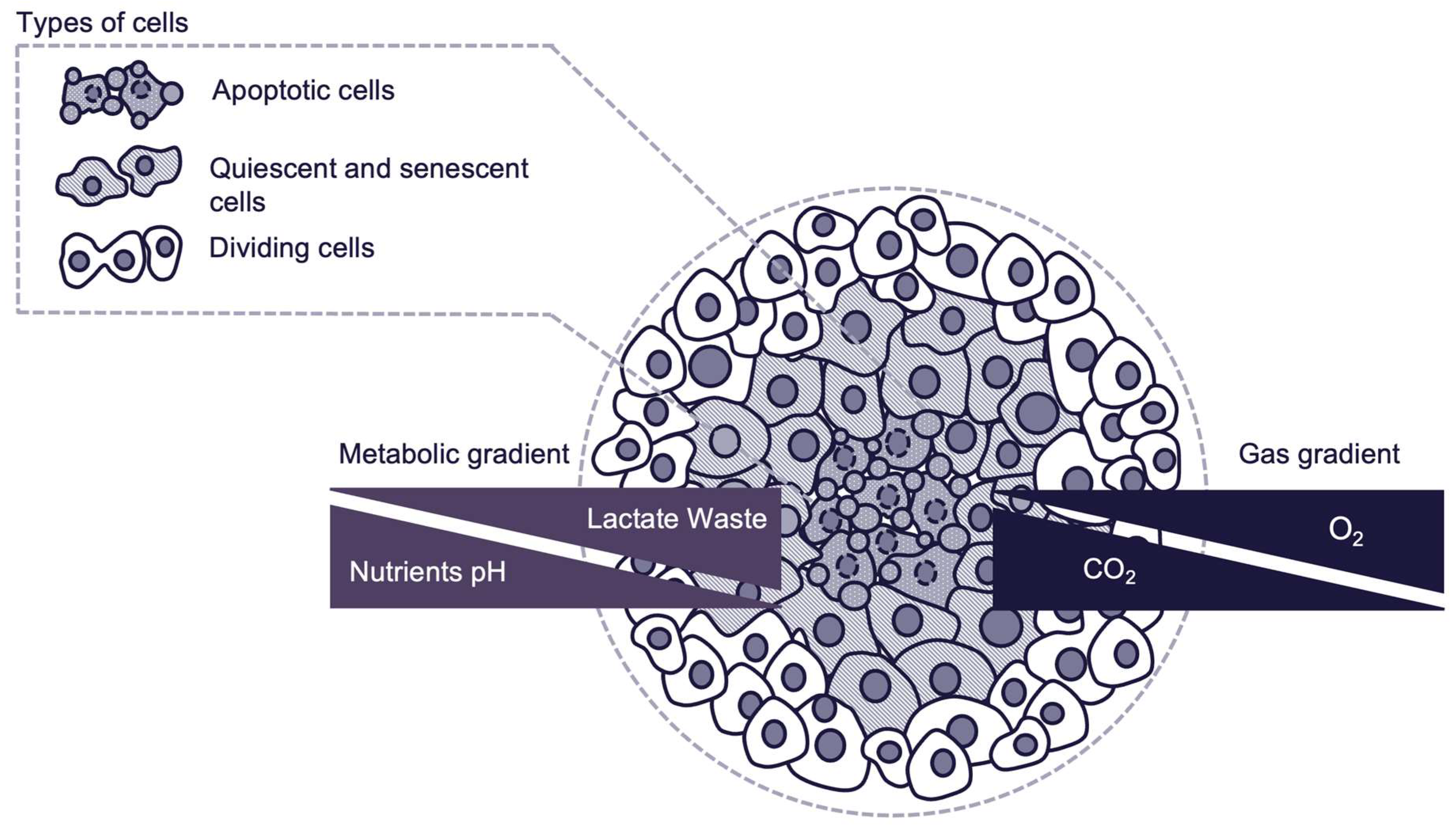
Figure 2.
Selection Process.
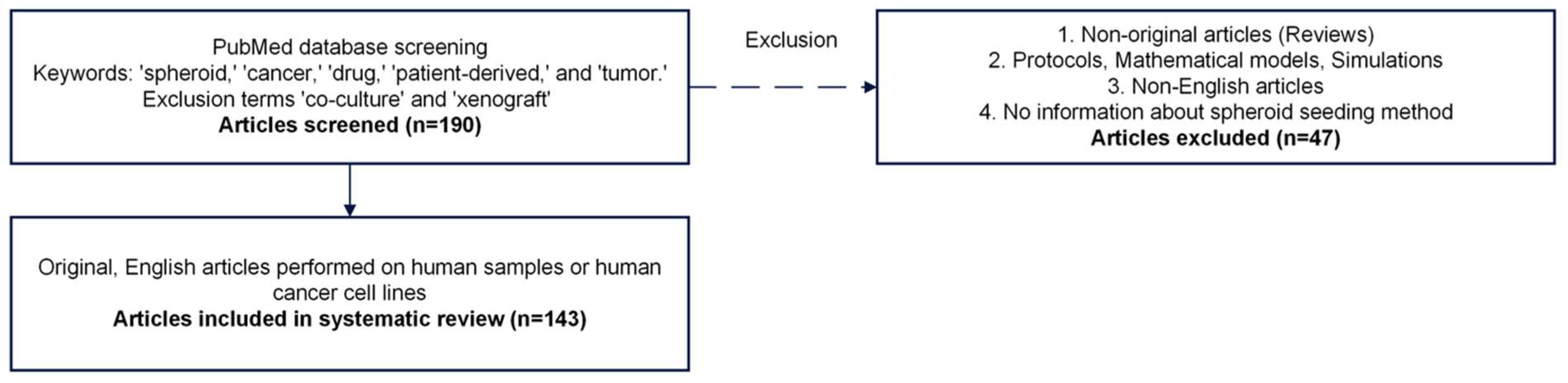
Figure 3.
Summary of spheroid sources.
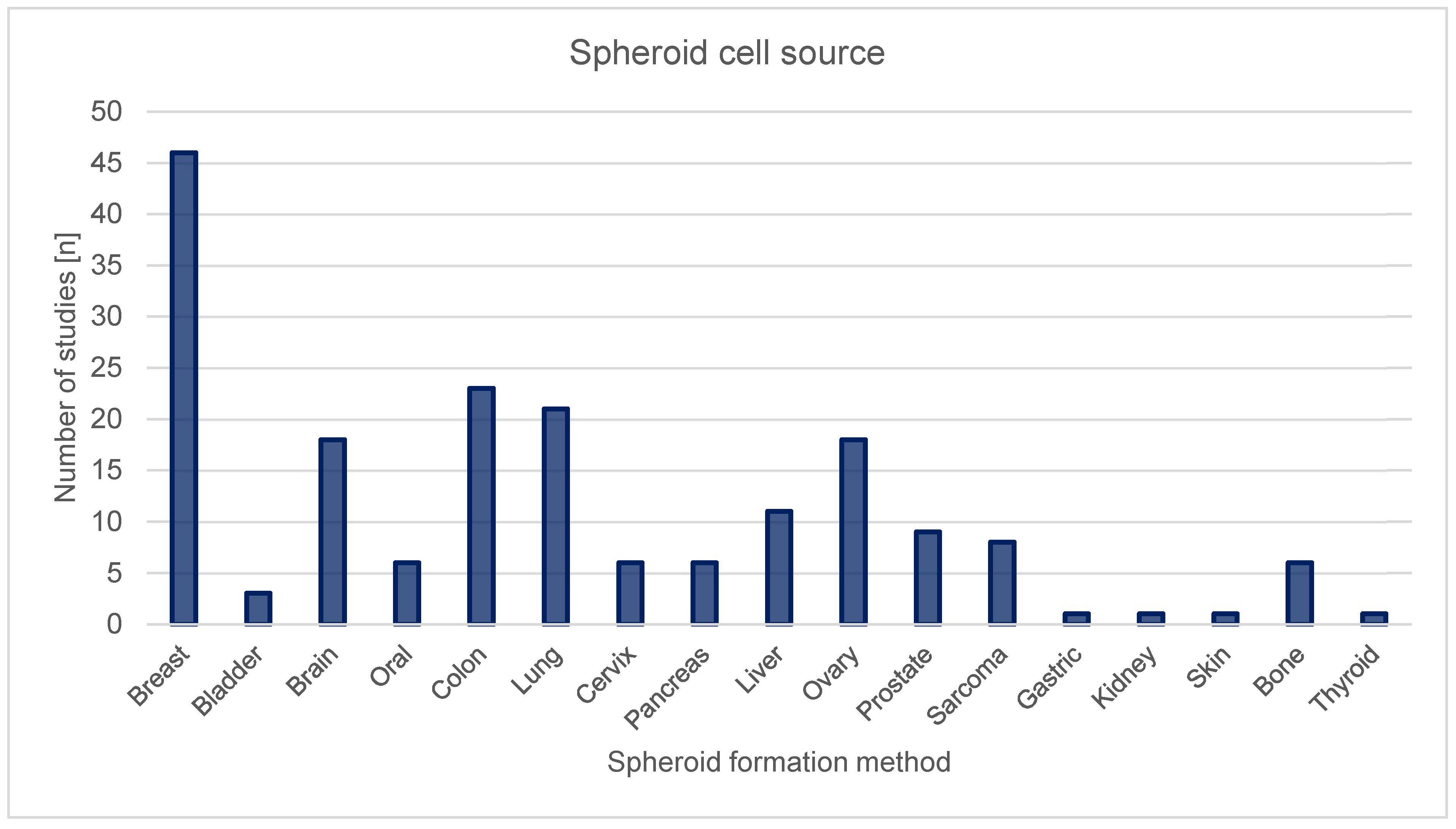
Figure 4.
Summary of spheroid seeding methods.
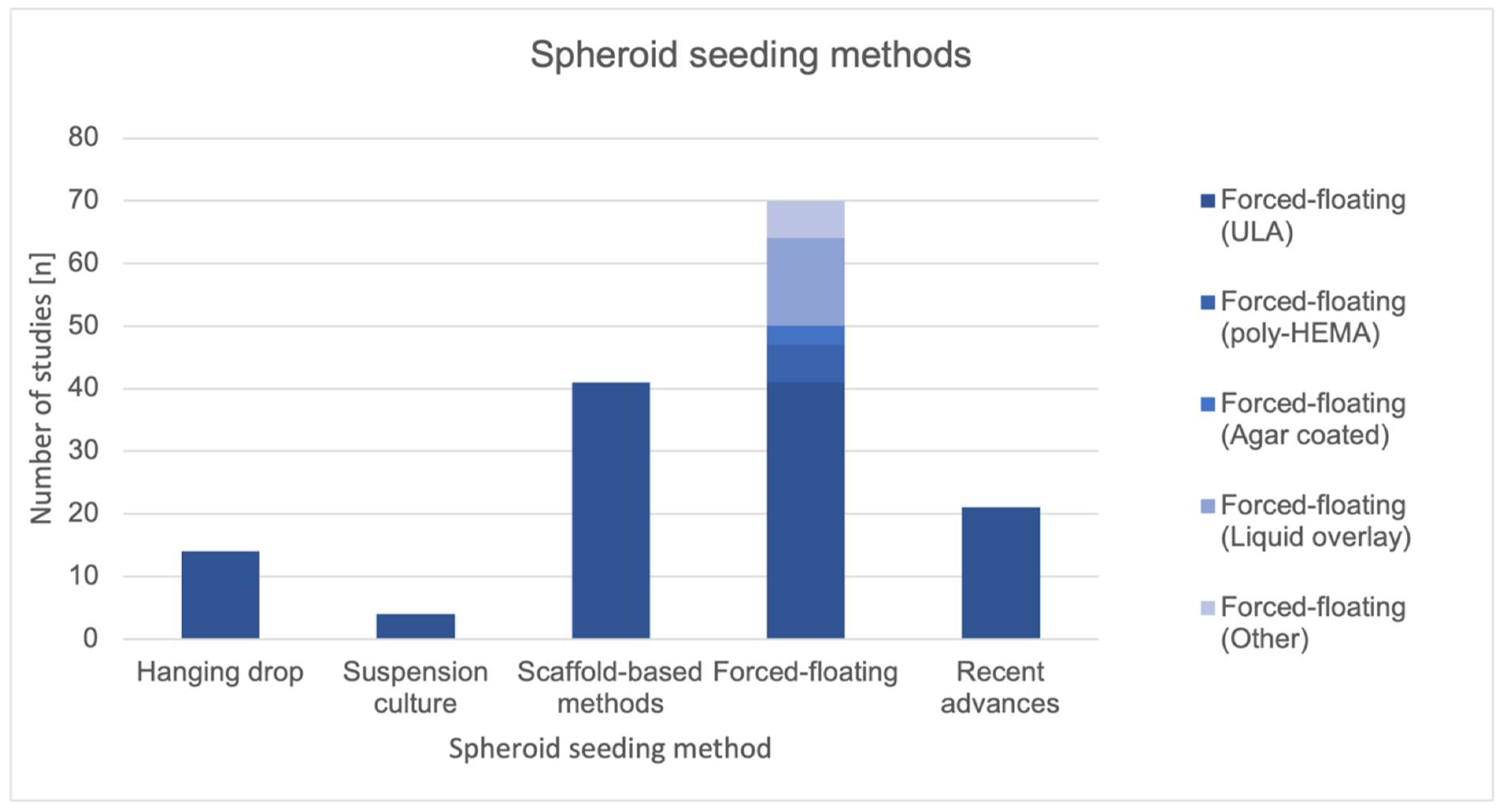
Figure 5.
Techniques for creation of spheroids.
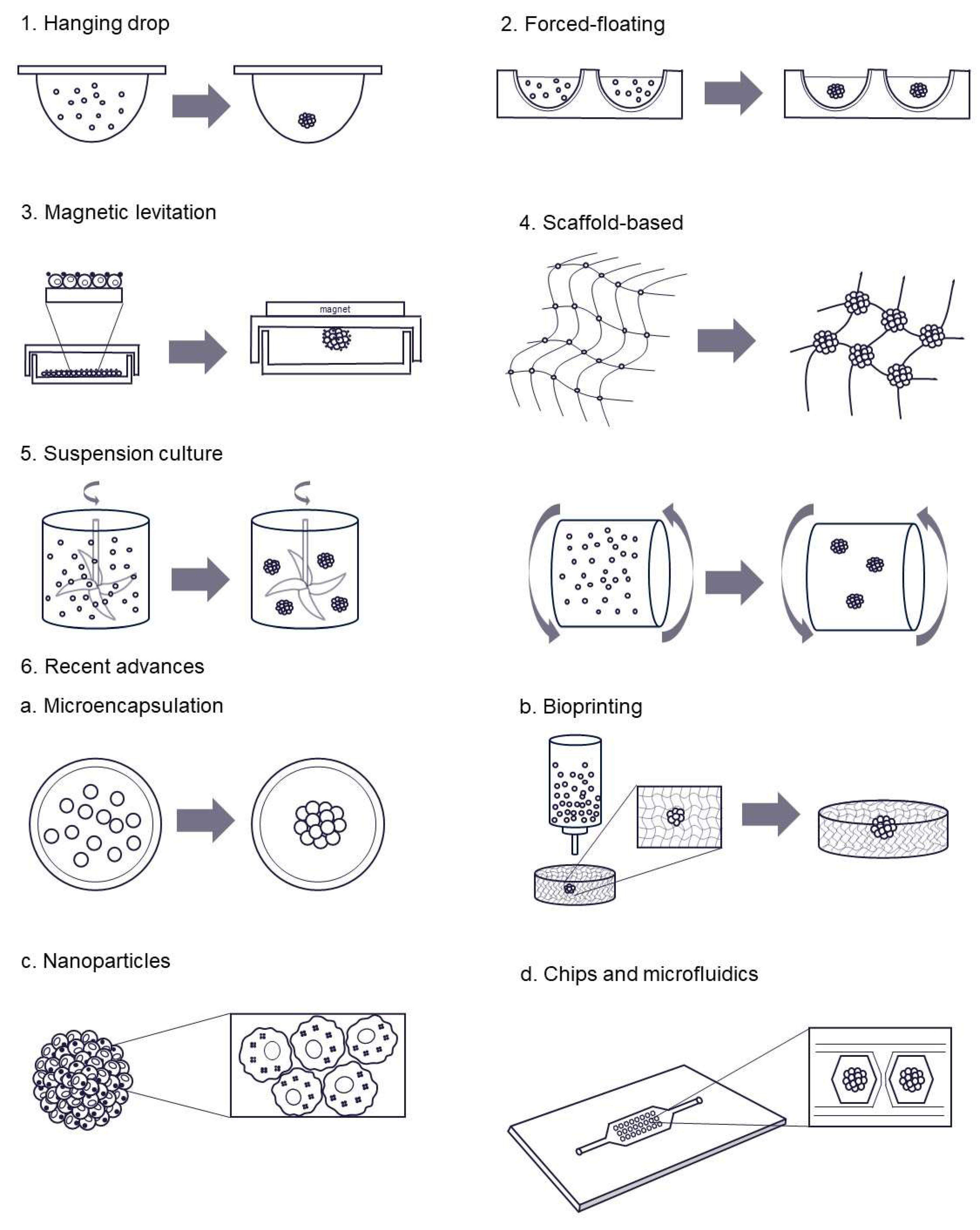

Table 1.
Advantages, limitations and potentials of 2D and 3D models.
| 2D models | 3D models | |
|---|---|---|
| Advantages |
|
|
| Limitations |
|
|
| Potentials |
|
|
Table 2.
Articles selected for systematic review.
| Reference | Cancer primary site (histological type) | Cell line | Spheroid formation method |
|---|---|---|---|
| Agastin S, 2011 [17] | Colon (adenocarcinoma), Breast (adenocarcinoma) | Colo205, MDA-MB-231 | Hanging drop |
| Alhasan L, 2016 [18] | Breast (carcinoma) | BT-474 | Scaffold-based methods |
| An HJ, 2020 [19] | Kidney (carcinoma) | A498 | Scaffold-based methods |
| Árnadóttir SS, 2018 [20] | Colon | Patient-derived | Forced-floating (not defined) |
| Baek N, 2016 [21] | Prostate (carcinoma), Bone (neuroblastoma), Lung (carcinoma), Cervix (adenocarcinoma), Bone (osteosarcoma) | DU145, SH-SY5Y, A549, HeLa, HEp2, 0U-2OS | Forced-floating (agar coated plates) |
| Barone RM, 1981 [22] | Colon (adenocarcinoma) | HT-29 | Suspension culture |
| Bartholomä P, 2005 [23] | Breast (carcinoma) | T-47D | Suspension culture |
| Boo L, 2020 [24] | Breast (adenocarcinoma) | MCF-7 | Forced-floating (agar coated plates) |
| Brooks EA, 2019 [25] | Ovary (adenocarcinoma) | Patient-derived, AU565, BT549, SKOV-3 | Scaffold-based methods |
| Bruns J, 2022 [26] | Brain (glioblastoma) | Patient-derived, U87 | Scaffold-based methods |
| Calori IR, 2022 [27] | Brain (glioblastoma, medulloblastoma) | U87, T98G, A172, UW473 | Forced-floating (ULA) |
| Chang S, 2022 [28] | Breast (adenocarcinoma) | MCF-7 | Recent advances |
| Chen G, 2022 [29] | Breast (adenocarcinoma) | MCF-7 | Forced-floating (ULA) |
| Chen MC, 2010 [30] | Breast (melanoma) | LCC6/Her-2 | Recent advances |
| Chen Z, 2021 [31] | Breast (adenocarcinoma) | MDA-MB-231 | Forced-floating (ULA) |
| Cheng V, 2015 [32] | Brain (glioblastoma) | U87, U251 | Forced-floating (ULA) |
| Close DA, 2022 [33] | Oral (squamous cell carcinoma) | Cal33, FaDu, UM-22B, OSC-19 | Forced-floating (ULA) |
| Das T, 2013 [34] | Ovary (adenocarcinoma) | TOV112D | Hanging drop |
| Das V, 2016 [35] | Colon (carcinoma), Colon (adenocarcinoma), Bone (osteosarcoma), Cervix (adenocarcinoma), Colon (adenocarcinoma), Liver (carcinoma) | HCT116, HT29, U-2OS, HeLa, Caco-2, HepG2 | Forced-floating (liquid overlay) |
| Das V, 2017 [36] | Colon (carcinoma) | HCT116 | Forced-floating (liquid overlay) |
| De Angelis ML, 2018 [37] | Colon | Patient-derived | Forced-floating (ULA) |
| Dhamecha D, 2021 [38] | Lung (carcinoma), Bone (osteosarcoma) | A549, MG-63 | Scaffold-based methods |
| Dias DR, 2016 [39] | Cervix (adenocarcinoma) | HeLa | Recent advances |
| Domenici G, 2021 [40] | Bone (sarcoma) | Patient-derived | Forced-floating (ULA) |
| Dufau I, 2012 [41] | Pancreas (adenocarcinoma) | Capan-2 | Forced-floating (poly-HEMA) |
| Eetezadi S, 2018 [42] | Ovary (carcinoma), Ovary (adenocarcinoma) | UWB1.289, UWB1.289+BRCA1, OV-90, SKOV3, PEO1, PEO4, COV362 | Forced-floating (ULA) |
| Eguchi H, 2022 [43] | Lung (carcinoma) | A549 | Forced-floating (ULA) |
| Eimer S, 2012 [44] | Brain (glioblastoma) | Patient-derived | Forced-floating (ULA) |
| El-Sadek IA, 2021 [45] | Breast (adenocarcinoma) | MCF-7 | Forced-floating (ULA) |
| Enmon RM Jr, 2001 [46] | Prostate (carcinoma) | DU 145 | Forced-floating (agar plates) |
| Flørenes VA, 2019 [47] | Skin (Melanoma) | Patient-derived | Forced-floating (ULA) |
| Fu J, 2020 [48] | Liver (carcinoma), Prostate (carcinoma), Lung (carcinoma), Breast (adenocarcinoma) | HepG2, DU 145, A549, MCF-7, MDA-MB-231 | Scaffold-based methods |
| Fu JJ, 2018 [49] | Prostate (carcinoma) | DU 145, LNCap | Scaffold-based methods |
| Gao Y, 2022 [50] | Lung (carcinoma) | A549 | Recent advances |
| Gencoglu MF, 2018 [51] | Breast (adenocarcinoma), Breast (carcinoma), Prostate (carcinoma), Prostate (adenocarcinoma), Ovary (adenocarcinoma) | AU565, BT549, BT474, HCC 1419, HCC 1428, HCC 1806, HCC 1954, HCC 202, HCC 38, ZR75 1, HCC 70, LNCaPcol, PC3, SKOV3 | Scaffold-based methods, Microwells, Suspension culture |
| Gendre DAJ, 2021 [52] | Lung (mesothelioma), Lung (adenocarcinoma) | H2052, H2052/484, H2452, LuCa1, LuCa61, LuCa62 | Scaffold-based methods |
| Gheytanchi E, 2021 [53] | Colon (adenocarcinoma) | HT-29, Caco-2 | Hanging drop, Forced-floating (Poly-HEMA) |
| Goisnard A, 2021 [54] | Breast (carcinoma), Breast (adenocarcinoma) | SUM1315, MDA-MB-231, HCC1937, SW527, DU4475 | Forced-floating (ULA) |
| Guo X, 2019 [55] | Ovary (adenocarcinoma), Colon (adenocarcinoma), Pancreas (carcinoma), Prostate (adenocarcinoma) | OVCAR3, SW620, PANC-1, PC3 | Scaffold-based methods |
| Hagemann J, 2017 [56] | Oral (carcinoma) | FaDu, Cal27, UPCI-SCC-154 | Forced-floating (ULA), Hanging drop |
| Han S, 2022 [57] | Liver | Patient-derived | Forced-floating (ULA) |
| Harmer J, 2019 [58] | Brain (glioblastoma) | U251, KNS42 | Scaffold-based methods |
| Herter S, 2017 [59] | Colon (adenocarcinoma) | LS174T, LoVo | Hanging drop |
| Ho WY, 2012 [60] | Breast (adenocarcinoma) | MCF-7 | Forced-floating (liquid overlay) |
| Ho WY, 2021 [61] | Breast (adenocarcinoma) | MCF-7 | Scaffold-based methods |
| Hofmann S, 2022 [62] | Breast | Patient-derived | Forced-floating (ULA) |
| Hornung A, 2016 [63] | Colon (adenocarcinoma) | HT-29 | Scaffold-based methods |
| Huang Z, 2020 [64] | Breast (adenocarcinoma) | MDA-MB-231 | Scaffold-based methods |
| Jove M, 2019 [65] | Breast (adenocarcinoma) Colorectal (adenocarcinoma) | MCF-7, DLD-1 | Scaffold-based methods |
| Ju FN, 2023 [66] | Brain (glioblastoma) | U87 | Recent advances |
| Karamikamkar S, 2018 [67] | Breast (adenocarcinoma) | MCF-7 | Scaffold-based methods |
| Karlsson H, 2012 [68] | Colon (carcinoma) | HCT-116 | Forced-floating (ULA) |
| Karshieva SS, 2022 [69] | Colon (carcinoma), Liver (carcinoma) | HCT-116, Huh7 | Forced-floating (ULA) |
| Kato EE, 2021 [70] | Lung (carcinoma) | A549 | Hanging drop |
| Kim CH, 2020 [71] | Liver (carcinoma) | HepG2 | Recent advances |
| Ko J, 2019 [72] | Brain (glioblastoma) | U87 | Scaffold-based methods |
| Kochanek SJ, 2019 [73] | Oral (carcinoma) | Cal33, Cal27, FaDu, UM-22B, BICR56, OSC-19, PCI-13, PCI-52, Detroit-562, UM-SCC-1, and SCC-9 | Forced-floating (ULA) |
| Kochanek SJ, 2020 [74] | Oral (carcinoma) | Cal33, FaDu, UM-22B, BICR56, OSC-19 | Forced-floating (ULA) |
| Koshkin V, 2016 [75] | Breast (adenocarcinoma) | MCF-7 | Scaffold-based methods |
| Kroupová J, 2022 [76] | Colon (adenocarcinoma) | HT-29 | Forced-floating (not defined) |
| Kudláčová J, 2020 [77] | Brain (glioblastoma) | U87 | Forced-floating (ULA) |
| Kumari P, 2017 [78] | Cervix (adenocarcinoma), Lung (carcinoma) | HeLa, A549 | Scaffold-based methods |
| Lal-Nag M, 2017 [79] | Ovary (adenocarcinoma) | Hey-A8–GFP | Forced-floating (ULA) |
| Lama R, 2013 [80] | Lung (carcinoma) | H292 | Scaffold-based methods |
| Landgraf L, 2022 [81] | Prostate (adenocarcinoma), Brain (glioblastoma) | PC-3, U87 | Forced-floating (liquid overlay) |
| Le VM, 2016 [82] | Lung (carcinoma), Colon (carcinoma), Brain (glioblastoma) | 95-D, HCT-116, U87 | Scaffold-based methods |
| Lee SW, 2019 [83] | Lung (carcinoma) | A549 | Recent advances |
| Lee Y, 2022 [84] | Lung (carcinoma) | H460, A549 | Forced-floating (ULA) |
| Lemmo S, 2014 [85] | Breast (adenocarcinoma) | MDA-MB-231 | Scaffold-based methods |
| Li M, 2019 [86] | Cervix (carcinoma) | C-33-A, DoTC2 4510 | Forced-floating (ULA) |
| Lim W, 2018 [87] | Colon (carcinoma), Brain (glioblastoma) | HCT-116, U87 | Recent advances |
| Lin ZT, 2021 [88] | Breast (adenocarcinoma) | MDA-MB-436 | Scaffold-based methods |
| Liu X, 2021 [89] | Sarcoma | HS-SY-II | Recent advances |
| Lorenzo C, 2011 [90] | Pancreas (adenocarcinoma) | Capan-2 | Forced-floating (poly-HEMA) |
| Luan Q, 2022 [91] | Lung (adenocarcinoma), Lung (carcinoma) | HCC4006, H1975, A549 | Scaffold-based methods, Forced-floating (ULA) |
| Madsen NH, 2021 [92] | Breast (adenocarcinoma), Colon (adenocarcinoma), Pancreas (carcinoma) | MCF-7, HT-29, PANC-1, MIA PaCa-2 | Forced-floating (ULA) |
| Marshall SK, 2022 [93] | Bone (osteosarcoma) | MG-63 | Forced-floating (ULA) |
| Maruhashi R, 2018 [94] | Lung (carcinoma) | A549 | Forced-floating (ULA) |
| Melnik D, 2020 [95] | Thyroid (carcinoma) | FTC-133 | Suspension culture |
| Molyneaux K, 2021 [96] | Brain (glioblastoma) | LN229, U87, Gli36 | Forced-floating (not defined) |
| Monazzam A, 2007 [97] | Breast (adenocarcinoma) | MCF-7 | Forced-floating (agar plates) |
| Morimoto T, 2023 [98] | Gastric | Patient-derived | Scaffold-based methods |
| Mosaad EO, 2018 [99] | Prostate (cancer), Prostate (carcinoma) | C42B, LNCaP | Recent advances |
| Mueggler A, 2023 [100] | Lung | Patient-derived | Scaffold-based methods |
| Nashimoto Y, 2020 [101] | Breast (adenocarcinoma) | MCF-7 | Recent advances |
| Nigjeh SE, 2018 [102] | Breast (adenocarcinoma) | MDA-MB-231 | Forced-floating (agar plates), Forced-floating (ULA) |
| Nittayaboon K, 2022 [103] | Colon (carcinoma) | PMF-k014 | Forced-floating (poly-HEMA) |
| Ohya S, 2021 [104] | Prostate (carcinoma) | LNCaP | Forced-floating (ULA) |
| Oliveira MS, 2016 [105] | Breast (adenocarcinoma), Ovary (adenocarcinoma) | MCF-7/Adr, NCI/Adr | Forced-floating (liquid overlay) |
| Ono K, 2022 [106] | Oral (carcinoma) | SAS, HSC-3, HSC-4, OSC-19 | Forced-floating (ULA) |
| Pampaloni F, 2017 [107] | Brain (glioblastoma) | U343 | Forced-floating (liquid overlay) |
| Park MC, 2016 [108] | Brain (glioblastoma) | Patient-derived and PC14PE6, PC14PE6_LvBr3, D54, LN428, LN751, U251E4, U87E4, SN-12C, SNU-119, SNU-216, SNU-668, SNU-719, HCC1171, HCC1195, HCC15, HCC1588, HCC2108, HCC44 | Forced-floating (not defined) |
| Pattni BS, 2016 [109] | Ovary (adenocarcinoma) | NCI/ADR-RES | Forced-floating (liquid overlay) |
| Perche F, 2012 [110] | Ovary (adenocarcinoma) | NCI/ADR-RES | Forced-floating (liquid overlay) |
| Preda P, 2023 [111] | Breast (adenocarcinoma), Brain (glioblastoma) | MDA-MB-231, U87 | Scaffold-based methods |
| Pulze L, 2020 [112] | Breast (adenocarcinoma) | MCF-7 | Forced-floating (ULA) |
| Raghavan S, 2016 [113] | Breast (adenocarcinoma), Ovary (adenocarcinoma) | MCF-7, OVCAR8 | Hanging drop, Forced-floating (liquid overlay) |
| Raghavan S, 2019 [114] | Ovary (adenocarcinoma) | A2780, OVCAR3 | Hanging drop |
| Ralph ACL, 2020 [115] | Breast (adenocarcinoma), Breast (carcinoma) | MCF-7, MDA-MB-231, T47D | Hanging drop |
| Roering P, 2022 [116] | Ovary (adenocarcinoma) | Patient-derived, CAOV3, OVCAR8 | Forced-floating (ULA) |
| Roudi R, 2016 [117] | Lung (carcinoma) | A549 | Forced-floating (poly-HEMA) |
| Rouhani M, 2014 [118] | Breast (carcinoma) | T47D | Forced-floating (liquid overlay) |
| Sakumoto M, 2018 [119] | Sarcoma | Patient-derived | Forced-floating (ULA) |
| Salehi F, 2020 [120] | Breast (adenocarcinoma), Breast (carcinoma) | MDA-MB-231, T47D, MCF-7 | Forced-floating (liquid overlay), Hanging drop |
| Sambi M, 2020 [121] | Breast (adenocarcinoma) | MDA-MB-231 | Scaffold-based methods |
| Sankar S, 2021 [122] | Lung (carcinoma) | A549 | Recent advances |
| Särchen V, 2022 [123] | Sarcoma | RH30 | Forced-floating (ULA) |
| Sarıyar E, 2023 [124] | Liver (carcinoma) | Huh7 | Hanging drop |
| Sauer SJ, 2017 [125] | Breast (carcinoma), Breast (adenocarcinoma) | SUM149, SUM190, T47D, MCF-7 | Forced-floating (ULA) |
| Shaheen S, 2016 [126] | Colon (carcinoma) | HCT-116 | Forced-floating (not defined) |
| Shen K, 2014 [127] | Breast (adenocarcinoma) | MDA-MB-231 | Scaffold-based methods |
| Sheth DB, 2019 [128] | Breast (adenocarcinoma) | MCF-7 | Recent advances |
| Shortt RL, 2023 [129] | Colon (carcinoma) | HCT-116 | Scaffold-based methods |
| Singh A, 2020 [130] | Breast (adenocarcinoma) | MCF-7 | Scaffold-based methods |
| Suhito IR, 2021 [131] | Bone (neuroblastoma), Brain (glioblastoma) | SH-SY5Y, U-87 | Recent advances |
| Tanenbaum LM, 2017 [132] | Ovary (adenocarcinoma) | UCI101, A2780 | Forced-floating (not defined) |
| Tang S, 2017 [133] | Colon (adenocarcinoma), Ovary (adenocarcinoma) | HT-29, SKOV-3 | Hanging drop |
| Taubenberger AV, 2019 [134] | Breast (adenocarcinoma) | MCF-7 | Scaffold-based methods |
| Terrones M, 2024 [135] | Lung (adenocarcinoma) | HCC78 | Forced-floating (ULA) |
| Tevis KM, 2017 [136] | Breast (adenocarcinoma) | MDA-MB-231 | Scaffold-based methods |
| To HTN, 2022 [137] | Stomach (carcinoma) | SNU-216, SNU-484, SNU-601, SNU-638, SNU-668, and SNU-719 | Forced-floating (ULA) |
| Torisawa YS, 2007 [138] | Breast (adenocarcinoma), Liver (carcinoma) | MCF-7, HepG2 | Recent advances |
| Uematsu N, 2018 [139] | Breast (adenocarcinoma) | MCF-7 | Recent advances |
| Varan G, 2021 [140] | Lung (carcinoma), Liver (carcinoma) | A549, HepG2 | Forced-floating (poly-HEMA) |
| Vinci M, 2012 [141] | Brain (glioblastoma), Oral (carcinoma), Breast (adenocarcinoma) | U87, KNS42, LICR-LON-HN4, MDA-MB-231 | Forced-floating (ULA), Agarose plates |
| Wan X, 2016 [142] | Colon (adenocarcinoma), Ovary (adenocarcinoma) | DLD-1, NCI/ADR | Scaffold-based methods |
| Wang Y, 2014 [143] | Cervix (adenocarcinoma) | HeLa | Scaffold-based methods |
| Ware MJ, 2016 [144] | Pancreas (carcinoma) | PANC-1, AsPc-1, BxPC-3, Capan-1, MIA PaCa-2 cells | Hanging drop |
| Wen Z, 2013 [145] | Pancreas (carcinoma) | MIAPaCa-2, PANC-1 | Scaffold-based methods |
| Wenzel C, 2014 [146] | Breast (carcinoma) | T47D | Forced-floating (liquid overlay) |
| Weydert Z, 2020 [147] | Ovary (adenocarcinoma) | HEY, SKOV-3 | Hanging drop |
| Wu G, 2019 [148] | Liver (carcinoma) | HepG2, Huh7 | Scaffold-based methods |
| Wu KW, 2020 [149] | Bladder (carcinoma), Lung (carcinoma), Liver (carcinoma) | T24, A549, Huh-7 | Recent advances |
| Xia H, 2020 [150] | Brain (glioblastoma) | LN229, U87 | Scaffold-based methods |
| Xiong Q, 2023 [151] | Bladder | Patient-derived | Forced-floating (ULA) |
| Yamawaki K, 2021 [152] | Ovary | Patient-derived | Forced-floating (ULA) |
| Yoshida T, 2019 [153] | Bladder | Patient-derived | Scaffold-based methods |
| Yu L, 2015 [154] | Breast (adenocarcinoma) | MCF-7 | Recent advances |
| Yu Q, 2021 [155] | Breast (adenocarcinoma) | MDA-MB-436, MDB-MB-231 | Scaffold-based methods |
| Zhang JZ, 2012 [156] | Colon (adenocarcinoma), Ovary (teratocarcinoma) | DLD-1, PA-1 ovarian cancer cells | Forced-floating (liquid overlay) |
| Zhang JZ, 2012 [157] | Colon (adenocarcinoma) | DLD-1 | Forced-floating (liquid overlay) |
| Zhang X, 2005 [158] | Breast (adenocarcinoma) | MCF-7 | Recent advances |
| Zuchowska A, 2017 [159] | Liver (carcinoma) | HepG2 | Recent advances |
Table 3.
Summary of spheroid sources.
| Spheroid formation method | Number of studies | Percentage of total number of studies |
|---|---|---|
| Forced-floating | 70 | 31.8% |
| Scaffold-based | 41 | 18.6% |
| Recent advances | 21 | 9.5% |
| Hanging drop | 14 | 6.4% |
| Suspension culture | 4 | 1.8% |
| Magnetic levitation | 0 | 0.0% |
Table 4.
Summary of spheroid seeding methods.
| Spheroid formation method | Number of studies | Percentage of total number of studies |
|---|---|---|
| Forced-floating | 70 | 31.8% |
| Scaffold-based | 41 | 18.6% |
| Recent advances | 21 | 9.5% |
| Hanging drop | 14 | 6.4% |
| Suspension culture | 4 | 1.8% |
| Magnetic levitation | 0 | 0.0% |
Table 5.
Summary of advantages and limitations of different seeding methods.
| Method | Advantages | Limitations |
|---|---|---|
| Forced floating method |
|
|
| Scaffold-based |
|
|
| Hanging drop method |
|
|
| Suspension culture |
|
|
| Microencapsulation |
|
|
| Bioprinting |
|
|
| Nanoparticle-assisted techniques |
|
|
| Microfluidics and lab-on-a-chip |
|
|
| Magnetic levitation |
|
|
Disclaimer/Publisher’s Note: The statements, opinions and data contained in all publications are solely those of the individual author(s) and contributor(s) and not of MDPI and/or the editor(s). MDPI and/or the editor(s) disclaim responsibility for any injury to people or property resulting from any ideas, methods, instructions or products referred to in the content. |
© 2025 by the authors. Licensee MDPI, Basel, Switzerland. This article is an open access article distributed under the terms and conditions of the Creative Commons Attribution (CC BY) license (http://creativecommons.org/licenses/by/4.0/).
Copyright: This open access article is published under a Creative Commons CC BY 4.0 license, which permit the free download, distribution, and reuse, provided that the author and preprint are cited in any reuse.


