
Submitted:
28 March 2025
Posted:
29 March 2025
You are already at the latest version
Abstract
Background/ Objectives: The growing number of households with companion dogs raises concerns. Co-living environments between companion dogs and their owners are linked to a heightened risk of cross-infections from strains such as Escherichia coli (E. coli), Staphylococcus aureus (S. aureus), Salmonella, and faecal coliforms. Therefore, this study aims to propose measures for healthy cohabitation by analysing the faecal microbiota of puppies and adult dogs. Methods: We isolated lactic acid bacteria (LAB) from their faeces and assessed their potential to inhibit E. coli, S. aureus, and Salmonella spp. Faecal samples from puppies (< 2 months old) and adult dogs (> 12 months old) were analysed and compared. Results: The analysis revealed that Lactobacillus dominated puppy faeces, while Bacteroidetes were more abundant in adult dogs. In total, 109 primary LAB candidates were isolated from faecal samples. These isolates underwent secondary screening for acid tolerance, bile salt resistance, acid production, heat resistance, protease activity, and antimicrobial activity against E. coli, S. aureus, and Salmonella spp. Five secondary LAB candidates with probiotic potential were further characterised via morphological and genetic analysis. All five strains were Lactobacillus reuteri, with reuteri JJ37 and JJ69 emerging as the final probiotic candidates. Conclusions: It promoting healthier cohabitation between dogs and their owners.
Keywords:
dogs
; lactic acid bacteria
; microbiota
; probiotic
1. Introduction
Gastrointestinal health in companion animals, particularly dogs, remains a critical concern for veterinarians and pet owners worldwide. Salmonella spp. and Escherichia coli (E. coli) are among the most significant bacterial pathogens affecting canine gut health, often causing severe gastroenteritis and systemic infections, posing risks of zoonotic transmission [1]. Conventionally, these infections have been managed with antibiotics, but the increasing prevalence of antimicrobial resistance has necessitated alternative therapeutic approaches. In response, the World Health Organization has advocated for restricted antibiotic use since 1997, recommending administration only in cases of definitive bacterial infections. Excessive antibiotic use not only eradicates pathogens but also disrupts beneficial gut microbiota, leading to dysbiosis and secondary health complications [2]. Furthermore, the emergence of extended-spectrum β-lactamase-producing E. coli in dogs and cats highlights the urgent need for alternative infection management approaches that minimise antibiotic resistance risks [3,4].
Among these alternatives, probiotics have emerged as a promising strategy for maintaining gut health and preventing infections in dogs. Lactobacillus species, in particular, enhance gut microbiota composition, support immune function, and inhibit pathogenic bacterial growth [5]. A study shows that probiotic supplementation improves microbial diversity, increases beneficial bacteria, and suppresses pathogenic pathways, particularly in dogs with gastrointestinal disturbances such as diarrhoea [6]. Additionally, probiotics mitigate antibiotic-induced dysbiosis, thereby preserving gut microbial homeostasis [7]. Therefore, maintaining the gut microbiome using probiotics could provide significant benefits in managing gastrointestinal diseases and promoting overall canine health.
Beyond gastrointestinal health, probiotics offer potential benefits in systemic health conditions, such as chronic kidney disease, where they help maintain nutritional status and improve renal function parameters [8]. The synergistic effects of probiotics, prebiotics, and antioxidants have been explored as therapeutic interventions for chronic diseases in dogs, further underscoring the broad applications of probiotics in veterinary medicine. The rising demand for effective, targeted probiotic formulations highlights the need for continued research into their specific mechanisms and benefits [6]. However, conventional antibiotic therapy remains limited, and the growing demand for alternative treatments highlights the need for targeted probiotics, representing a critical advancement in veterinary medicine. The ability of Lactobacillus strains to inhibit the growth of Salmonella and E. coli highlights their potential as effective probiotics for canine gastrointestinal health. Therefore, this study aims to identify and characterise Lactobacillus strains with antimicrobial activity against key canine pathogens. A two-step screening process was employed: microbiome analysis in healthy puppies and adult dogs was conducted to isolate primary Lactobacillus candidate strains; second, these strains were evaluated for their probiotic potential, focusing on their antimicrobial activity against Salmonella and E. coli. Limosilactobacillus reuteri JJ37 and JJ69 were identified as the final probiotic candidate. These strains exhibited strong antibacterial activity against major pathogenic bacteria affecting dogs, positioning them as a promising candidate for probiotic development to enhance canine gut health and prevent gastrointestinal infections.
2. Materials and Methods
2.1. Chemicals and Reagents
To investigate the distribution of cultivable gut microbiota in canine faecal samples, various media were used. These included Plate Count Agar (PCA) with Bromocresol purple (BCP), Transgalactosylated Oligosaccharides (TOS)-MUP (TOS), Potato Dextrose Agar (PDA), Tryptone sulfite Neomycin (TSN), MacConkey (MC), Salmonella-Shigella (SS), Mannitol salt (MS), Brilliant Green (BG), and MacConkey Sorbitol (MACS), all purchased from Difco BD (Franklin Lakes, NJ, USA). De Man, Rogosa, and Sharpe (MRS) and Blood Agar were obtained from Kisan Bio (Seoul, Korea), while Bacto Agar was purchased from Difco BD (Franklin Lakes, NJ, USA). For the acid resistance assay, 10 M HCl was obtained from DC Chemical Co., Ltd (Seoul, Korea), and for the bile salt resistance assay, bile salts were purchased from Difco BD (Franklin Lakes, NJ, USA). Agarose for PCR was obtained from Yeong Science (Gyeonggi-do, Korea). All reagents used were of analytical grade or higher.
2.2. Faecal Sample Collection
To select candidate probiotic strains and analyse microbiomes, fresh faecal samples from healthy puppies and adult dogs were obtained from a Gyeongsan animal shelter (Gyeongsangbuk-do, Korea). The samples came from 30 healthy adult dogs (5–15 kg, mixed breed, 1–8 years old) and 10 puppies (1–2 kg ± 0.5 kg, mixed breed, 2–3 months old) administered full veterinary care (Supplementary Table S1). The fresh faecal samples were collected using a standardised protocol through induced defecation or rectal swab sampling in healthy dogs [9]. Figure 1 shows the process for selecting probiotic candidates from dogs.
2.3. DNA Extraction and 16S rRNA Gene Sequencing
Genomic DNA was extracted from the faecal samples of puppies and adult dogs using the G-spinTM Genomic DNA Extraction Kit (Qiagen Inc., Hilden, Germany) following the instructions of the manufacturer. DNA quality control was performed on the filtered supernatant using a Take 3 plate with an Agilent microplate reader (Agilent, California, USA). A DNA concentration of at least 15 ng/µL and an A260/280 ratio of at least 1.8 were obtained to ensure sample quality. DNA extracts that passed quality control underwent PCR analysis to confirm their genetic profiles. For this purpose, the AccuPower® ETEC-Toxin 4-Plex PCR kit (Bioneer, Daejeon, Korea) was used according to the instructions of the manufacturer. The purified DNA was quantified, and 1 ng was used as the template for PCR amplification. The V4 region of the 16S rRNA gene was amplified with universal primers (515F and 806R) using the Pacific Biosciences RS1 system (CJ Bioscience Inc., Seoul, Korea). Next-generation sequencing was performed using the EzBioCloud MTP pipeline and the EzBioCloud 16S database PKSSU4.0 to analyse the microbiome taxonomic profile. For diversity and statistical analysis, normalised operational taxonomic units were utilised. The biodiversity (alpha-diversity) within samples was determined using Chao1 and Shannon indices, while the similarities between the samples (beta-diversity) were determined through weighted UniFrac calculations. Principal coordinate analysis was conducted via Curtis similarity clustering analysis.
2.4. Distribution of Microorganisms in Puppies and Adult Dogs and Primary Selection of Candidate Probiotic Strains
Fresh faecal samples from puppies and adult dogs were collected through induced defecation or rectal swabs. The samples were immediately placed in an icebox and transported to the laboratory under anaerobic conditions at temperatures below 4°C. To isolate LAB, selective media such as PCA with BCP, MRS, and TOS-MUP (selective for Bifidobacterium) were used. Moreover, these selective media were also applied to analyse the gut microbiota of puppies and adult dogs.
We used Blood agar to assess total microbial counts, while PDA was utilized to identify fungi and yeast. TSN was used for detecting anaerobic sulfite-reducing bacteria, MC for E. coli, and SS for isolating Salmonella and Shigella species. MS was used for the selective cultivation of Staphylococcus species, BG for Salmonella spp., and MACS for enterohaemorrhagic E. coli.
Samples were serially diluted 10-fold (10-1, 10-3, 10-5, and 10-7) using sterilised 0.1% agar saline and spread-plated onto selective media. Blood agar, PDA, TSN, MAC, MS, BG, MACS, and SS were incubated aerobically at 37°C for 24 h. MRS, PCA with BCP, and TOS-MUP plates were incubated anaerobically in an Oxoid Anaerobic Jar and replaced with O2-free CO2 at 37°C for 48 h. Colonies that developed on the media were selected as primary probiotic candidates following the Mitsuoka method [10] and stored at -20°C for further analysis.
2.5. Secondary Selection of the Primary Candidate Strains
2.5.1. Acid Tolerance Test
To identify candidate probiotic strains, the primary selection focused on acid-tolerant LAB. Candidate strains were initially cultured in MRS broth at 37°C for 24 h. The cultured strains were then inoculated at 1% (v/v) into MRS broth and adjusted to pH 2 and pH 4 using 1M HCl. Then, the bacteria were incubated with shaking for 1.5 h, and LAB survival was assessed by plating the cultures onto MRS agar plates and counting the resulting colonies. Strains that formed colonies under these conditions were considered acid-tolerant and selected as primary candidates.
2.5.2. Lactic Acid Production Test
To identify acid-producing probiotic candidates, 119 LAB were isolated from 57 plates containing primary acid-tolerant strains. These isolates were inoculated into MRS broth and incubated at 37°C for 24 h for initial cultivation. The cultured strains were then streaked onto MRS agar supplemented with 1% CaCO3 and incubated at 37°C for 48−72 h. Strains that formed clear halos around their colonies were classified as acid-producing and selected as secondary candidates.
2.5.3. Bile Tolerance Test
In total, 40 isolates identified as LAB through secondary screening were further assessed for bile tolerance. These isolates were inoculated in MRS broth and incubated at 37°C for 24 h for primary cultivation. Subsequently, the candidate strains were streaked evenly onto the surface of MRS agar plates containing 1% oxgall (DifcoTM Oxgall, BD) using a sterilised loop and incubated at 37°C for 48 h. Strains that formed colonies on the MRS agar plates were selected as bile-tolerant strains.
2.5.4. Heat Tolerance Test
In the tertiary screening, to identify candidate probiotic strains with heat tolerance, 39 bile-tolerant isolates were tested. The candidate strains were inoculated into MRS broth and incubated at 37°C for 24 h for primary cultivation. The primary cultures were then transferred into fresh MRS broth at a 1% inoculum ratio and subjected to shaking incubation at 40°C, 50°C, and 60°C for 1 h. The survival of the strains was evaluated by streaking the cultures onto MRS agar plates and counting colony formations. Strains that formed colonies were selected as heat-tolerant for the quaternary screening.
2.5.5. Selection of Protease-Producing Strains
In the quaternary screening, to identify protease-producing candidate probiotic strains, 28 heat-tolerant strains were analysed. The isolates were spotted onto MRS agar plates supplemented with 2% skim milk using a sterilised loop. The plates were then incubated at 37°C for 24 h, and strains that formed a clear zone (halo) around their colonies were identified as protease-producing strains. Lactobacillus acidophilus (L. acidophilus) KCTC 3111 was used as the control strain.
2.5.6. Selection of Lactobacillus spp. and Antibacterial Activity Against E. coli and Salmonella
The colony morphology, Gram staining, microscopic cell shape, and catalase activity (H2O2 test) were analysed. Strains demonstrating antibacterial activity against E. coli ATCC 35218, Salmonella typhimurium KCTC 2515, and Staphylococcus aureus (S. aureus) ATCC 29213 were selected as primary antibacterial candidates. A single colony from each strain was isolated from the plate and diluted in saline. Pathogenic strains were adjusted to 103 CFU/mL, while candidate probiotic strains were adjusted to 108 CFU/mL. S. aureus ATCC 29213 and E. coli ATCC 35218 were co-cultured with the candidate strains in 5 mL Iso-Sentitest–MRS broth (9:1), whereas S. typhimurium KCTC 2515 was co-cultured in 5 mL 2 × MRS-MHB broth for 24 h at 37°C. After incubation, 100 µL of each culture was sampled, serially diluted (10-fold), and plated on MHB agar (S. aureus ATCC 29213 and E. coli ATCC 35218) and SS agar (S. typhimurium KCTC 2515). A 20 µL aliquot from each dilution was spread onto the respective agar plate and incubated at 37°C for 24 h. For controls, cultures in Iso-Sentitest–MRS (9:1) or 2 × MRS-MHB broth without treatment served as negative controls, and cultures treated with gentamicin (0.1 mg/mL) were used as positive controls. Subsequently, the bacterial colonies were enumerated.
2.6. Identification of the Secondary Candidate Strains
2.6.1. Identification of Lactobacillus spp. JJ37, 68, 69, 71, and 77
The Lactobacillus spp. strains JJ37, 68, 69, 71, and 77 were identified through the screening process and confirmed through 16S rRNA gene sequencing. In brief, genomic DNA was extracted from the isolates using a Qiagen genomic DNA extraction kit (Qiagen Inc., Hilden, Germany). Consequently, polymerase chain reaction (PCR) was performed using the forward primer (5’-AGGTAACGGCTTACCAAGGC-3’) and the reverse primer (5’-CCACCGCTACACATGGAGTT-3’). The PCR reaction mixture contained 50 pmol of primers, 50 ng of template DNA, 5 μL of 10 × Taq DNA polymerase buffer, and 1 U of Taq DNA polymerase (Takara, Japan). The PCR protocol included an initial denaturation at 94°C for 5 min, followed by 35 cycles of denaturation at 95°C for 30 s, annealing at 56°C for 30 s, and elongation at 72°C for 30 s, with a final extension step at 72°C for 7 min. The resulting sequences were compared to closely related sequences in the GenBank database using BLAST software.
2.7. Statistical Analysis
Statistical analyses, including data processing and graph creation, were performed using GraphPad Prism 8.0 (San Diego, CA, USA). One-way analysis of variance (ANOVA) followed by Tukey’s post hoc test was used for group comparisons. Results were expressed as mean ± standard error of the mean (SEM), with statistical significance set as p < 0.05.
3. Results
3.1. DNA Quality and Purity Assessment of Gut Microbiota for Metagenomic Analysis
The quality of DNA was verified through spectrophotometry, gel electrophoresis, and PCR amplification. Quality control parameters, including sample concentrations, volumes, and DNA purity, were assessed based on the following criteria: a minimum concentration of 15 ng/μL, volume of at least 20 μL, and DNA purity ratio (A260/280) within the range of 1.8–2.1. DNA purity was determined by calculating the absorbance ratio at 260 nm (indicative of DNA) to 280 nm (indicative of proteins). All samples (A2, A4, A10, B3, B5, and B7) met the quality control criteria and they were confirmed via PCR analysis (Supplementary Table S2). The successful amplification of the target DNA in each sample was validated by observing the presence of distinct bands on the gel (Supplementary Figure S1).
3.2. Metagenomic Analysis of the Gut Microbiota in Puppies and Adult Dogs
Figure 2 illustrates the comparison of the gut microbiota composition between healthy puppies and adult dogs. At the phylum level, puppy B3 showed the highest proportion of Proteobacteria (78.18%), while puppy B5 was dominated by Firmicutes (62.03%) and puppy B7 by Fusobacteria (62.08%). No single dominant phylum was shared among the puppies. In contrast, adult dogs A2, A4, and A10 showed a consistent prevalence of Bacteroidetes (> 40%) across all samples. Overall, Proteobacteria, Firmicutes, Fusobacteria, and Bacteriodetes were the dominant phyla (> 5%) in both puppies and adult dogs. However, no commonly dominant phylum was observed between the two groups.
At the class level, puppy B3 was dominated by Gammaproteobacteria (76.91%), followed by Clostridia (17.22%). Puppy B5 showed the highest proportion of Negativicutes (38.77%) and Bacteroidia (20.62%), while puppy B7 was primarily composed of Fusobacteria (62.09%), followed by Gammaproteobacteria (15.81%). The predominant classes (> 30%) in each puppy were Gammaproteobacteria, Negativicutes, and Fusobacteria, respectively, while no shared dominant class among the puppies. In adult dogs, A2 was the dominant class identified among the puppies. Moreover, A2 was dominated by Bacterodia (59.48%) and Clostridia (20.34%), A4 by Bacteroidia (45.61%) and Fusobacteria (28.88%), and A10 by Bacteroidia (40.9%) and Clostridia (25.97%). A consistent dominance of Bacteroidia (> 40%) was observed across all adult dogs, but no common dominant class was identified between puppies and adult dogs.
At the order level, puppy B3 had the highest proportion of Enterobacterales (75.33%), followed by Clostridiales (17.22%) and Bacterodiales (2.79%). Puppy B5 was primarily composed of Veillonellales (36.96%), followed by Bacteroidales (20.62%) and Lactobacillales (10.28%). Puppy B7 showed the highest abundance of Fusobacteriales (62.09%), followed by Pseudomonadale (7.54%) and Campylobacterales (6.66%). Each puppy was dominated by different orders—Enterobacterales, Veillonellales, and Fusobacteriales, respectively—with no shared dominant order observed. However, similar to the phylum and class levels, no common dominant order was identified between puppies and adult dogs.
3.3. Differences in Gut Microbial Composition Between Puppies and Adult Dogs
The differences in gut microbiota composition between puppies and adult dogs were analysed by comparing correlations at the phylum, class, and order levels (Figure 3). At the phylum level, puppies showed higher relative abundances of Firmicutes, Fusobacteria, Proteobacteria, Spirochaetes, and Verrucomicrobia, while adult dogs had higher proportions of Actinobacteria, Bacteroidetes, Cyanobacteria, Deferribacteres, Synergistetes, Tenericutes, and Thermotogae. The phyla with more than a 1.5-fold difference between the groups included Bacteroidetes, Fusobacteria, and Proteobacteria. Bacteroidetes accounted for 7.98% of the microbiota in puppies and 48.66% in adult dogs, representing an approximately 6-fold difference. The Fusobacteria constituted 21.82% in puppies and 14.23% in adult dogs, a 1.53-fold difference. Proteobacteria comprised 6.49% of puppies and 38.21% in adult dogs, showing a 5.88-fold difference. No other phyla exhibited differences exceeding 1.5-fold.
At the class level, puppies showed higher relative abundances of Acidimicrobiia, Alphaproteobacteria, Bacilli, Betaproteobacteria, Deltaproteobacteria, Epsilonproteobacteria, Fusobacteria_C, Gammaproteobacteria, Negativicutes, Spirochaetia, and Verrucomicrobiae. In contrast, adult dogs had higher proportions of Actinobacteria_c, Bacterodia, Chroobacteria, Clostridia, Coriobacteriia, Deferribacters_c, Erysipelotrichi, Flavobacteria, Mollicutes, Synerfistia, Tissierellia, Thermotogae_c, and Vampirovibrio_c. Classes with differences (> 1.5 fold) between puppies and adult dogs included Bacilli, Bacterodia, Clostridia, Eplionproteobacteia, Fusobacteria_c, Gammaproteobacteria, and Negativicutes. For instance, Bacilli represented 4.79% of the microbiota in puppies and 0.39% in adult dogs (12.28-fold difference). Bacteroidia accounted for 7.89% in puppies and 48.66% in adult dogs (6.09-fold difference). Clostridia was present at 9.46% in puppies and 17.45% in adult dogs (1.84-fold difference). Epsilonproteobacteria accounted for 3.14% in puppies and 5.25% in adult dogs (1.67%-fold difference). Fusobacteria_c constituted 21.82% in puppies and 14.23% in adult dogs (1.53-fold difference). Gammaproteobacteria comprised 32.16% in puppies and 3.21% in adult dogs (10.01-fold difference). Negativicutes accounted for 13.01% in puppies and 5.24% in adult dogs (2.48-fold difference).
At the order level, puppies showed higher relative abundances of Aeromonadales, Alteromonadales, Bacillales, Brachyspirales, Burkholderiales, Campylobacterales, Caulobacterales, Enterobacterales, Fusobacteriales, Lactobacillales, Mycoplasmatales, Pseudomonadlaes, Spirochaetales, Sphingomonadales, Veillonellales, Verrucomicrobiales, and Vibrionales. In contrast, adult dogs exhibited higher proportion of Acidaminoccocales, Acidimicrobiales, Acholeplasmatales, Actinomycetales, Bacteroidales, Bifidobacteriales, Clostridiales, Chroococcales, Coriobacteriales, Corynebacteriales, Kosmotogales, Pasteurellales, Rhodocyclales, Selenomonadales, Synergistales, and Tissierellales. Orders with differences (> 1.5 fold) between puppies and adult dogs included Acidaminococcales, Bacteroidales, Clostridiales, Enterobacterales, Fusobacteriales, Lactobacillales, Pseudomonadales, and Veillonellales. For example, Acidaminococcales accounted for 0.15% of the microbiota in puppies and 4.05% in adult dogs (25.8-fold difference). Clostridiales was present at 9.46% in puppies and 17.45% in adult dogs (1.84-fold difference). Enterobacterales accounted for 26.06% in puppies and 14.23% in adult dogs (1.53-fold difference). Lactobacillales comprised 4.76% in puppies and 0.39% in adult dogs (12.24-fold difference). Pseudomonadales constituted 2.54% in puppies but were absent in adult dogs. Veillonellales accounted for 12.33% of puppies and 0.02% in adult dogs (456-fold difference). No other order exhibited a significant difference (>1.5 fold).
3.4. F/B Ratio in Adult Dogs and Puppies
To investigate the Firmicutes/Bacteroidetes (F/B) ratio and its potential effect, the gut microbiota composition in adult dogs and puppies was analysed. Genomic DNA was extracted, and 16S rRNA gene sequencing was performed to profile the gut microbiota. The relative abundances of Firmicutes and Bacteroidetes at the phylum level were compared. In puppies, Firmicutes accounted for 34.54% on average (range: 18.66−62.03%), while in adult dogs, it was 28.35% on average (range: 16.48−39.35%. Conversely, Bacteroidetes was significantly less abundant in puppies, with an average of 7.99% (range: 0.55−20.63%), compared to that in adult dogs, which exhibited an average of 48.66% (range: 40.9−59.48%). These findings indicate that puppies had a higher proportion of Firmicutes and a lower proportion of Bacteroidetes than those of adult dogs (Figure 4). The F/B ratio was calculated as the relative abundance of Firmicutes divided by that of Bacteroidetes. The F/B ratio in puppies was 11.12 on average (range: 3.01−23.67), whereas in adult dogs, it was 0.6 (range: 0.36−23.67). This suggests that the F/B ratio is significantly higher in puppies than in adult dogs.
3.5. Differences in Gut Microbiota Diversity Between Puppies and Adult Dogs
In total, 263,479 V4 16S rRNA sequence reads were collected from six samples, with an average of 41,472 reads per sample. Species richness and diversity were assessed using ACE, Chao 1, Jackknife, Shannon, and Simpson indices. The ACE, Chao 1, and Jackknife indices were significantly higher in adult dogs than in puppies (p < 0.05) (Figure 5).
3.6. Comparison of Faecal Microbial Distribution Between Puppies and Adult Dogs
To compare the faecal microbial distribution between healthy puppies and adult dogs, colony counts were obtained via direct culture using selective media. Figure 6 shows the microbial distribution of faecal samples from 10 puppies and 30 adult dogs on each medium. On blood agar, the colony count for puppies was 3.98 × 10⁸ CFU/mL, while for adult dogs, it was 4.86 × 10⁶ CFU/mL. This showed that puppies had 100 times more colonies than those of adult dogs (Figure 6a). On TSN, the colony count was 3.19 × 10⁵ CFU/mL for puppies and 1.1 × 10⁵ CFU/mL for adult dogs. This showed that puppies had approximately 3.16 times more colonies than those of adult dogs (Figure 6b). On PDA, puppies had 8.85 × 10⁶ CFU/mL, and adult dogs had 2.34 × 10⁶ CFU/mL, showing that puppies had 10 times more colonies than those of adult dogs (Figure 6c). On MAC, both puppies and adult dogs had colony counts of 3.3 × 10⁴ CFU/mL (Figure 6d). On SS, puppies had 5.85 × 101CFU/mL and adult dogs had 2.71 × 10³ CFU/mL, showing that adult dogs had 100 times more colonies than those of puppies (Figure 6e). On MS, puppies had 2.48 × 10⁷ CFU/mL, and adult dogs had 2.19 × 10³ CFU/mL, confirming that puppies had 10,000 times more colonies than those of adult dogs (Figure 6f). On BG, puppies had 4.46 × 10⁶ CFU/mL, and adult dogs had 2.32 × 10⁵ CFU/mL, showing that puppies had 10 times more colonies than that of adult dogs (Figure 6g). On MACS, both puppies and adult dogs had colony counts of 3.4 × 10⁴ CFU/mL (Figure 6h). On MRS, colony counts were 2.87 × 10⁷ CFU/mL for puppies and 6.84 × 10⁶ CFU/mL for adult dogs (Figure 6i). On TOS, puppies and adult dogs had 2.68 × 10⁶CFU/mL and 1.71 × 10⁶CFU/mL, respectively (Figure 6j). On PCA, puppies had 1.98 × 10⁸CFU/mL and adult dogs had 3.47 × 10⁷ CFU/mL (Figure 6k). Across PCA, MRS, and TOS media, puppies had approximately 3.16 times more colonies than those of adult dogs. No significant differences were observed between puppies and adult dogs in all media except for the MS medium.
3.7. Selection of Lactic Acid Bacteria Strains Through Multi-Step Screenings
3.7.1. Acid Tolerance Test
Forty faecal samples from dogs were diluted 10-fold and spread on PCA with BCP agar to select 109 lactic acid-producing strains. To identify strains with high probiotic potential, an acid tolerance test was performed. The survival rates at pH 2 and pH 4 were measured (Table 1 and Figure 7a). At pH 2, 57 out of 109 strains (52.3%) survived, while at pH 4, 107 out of 109 strains (98.17%) survived. The control strain, L. acidophilus KCTC 3111, survived at both pH levels, confirming its acid tolerance.
3.7.2. Lactic Acid Production Test
A test was performed to isolate lactic acid-producing strains. Among the 119 strains, 40 (33.61%) produced lactic acid, while 79 (66.39%) did not (Table 1 and Figure 7b). The lactic acid-producing strains were categorised by colony size: small, medium, or large. Among the 40 strains, 30 produced the smallest colonies, 7 produced medium-sized colonies, and 3 produced the largest colonies. The control strain, L. acidophilus KCTC 3111, produced medium-sized colonies and was identified as a lactic acid-producing strain.
3.7.3. Bile Tolerance Test
To identify bile-tolerant strains, a bile tolerance test was conducted on the 40 strains that had previously demonstrated acid resistance and lactic acid production. All strains, except for one, grew on plates containing 1% Oxgall (DifcoTM Oxgall, BD) (Table 1 and Figure 7c). The control strain, L. acidophilus KCTC3111, also grew on MRS agar plates, confirming its bile tolerance.
3.7.4. Heat Resistance Test
A heat resistance test was conducted on 39 strains that exhibited acid tolerance, lactic acid production, and bile tolerance (Table 1 and Figure 7d). Among these, 33 (84.61%), 32 (82.05%), and 28 strains (71.79%) survived at 40°C, 50°C, and 60°C, respectively. The control strain, L. acidophilus KCTC 3111, also survived at all three temperatures, confirming its heat resistance.
3.7.5. Dietary Enzyme of Protease
A protease activity test was conducted on 28 strains that exhibited acid tolerance, lactic acid production, bile tolerance, and heat resistance. The experiment aimed to identify strains capable of producing the digestive enzyme protease (Table 1 and Figure 7e). Among these 28 strains, 20 (71.43%) formed clear zones around their colonies, indicating protease production, while 8 strains (28.57%) did not. Among the 20 protease-producing strains, 12 (60%) produced large clear zones, indicating strong protease activity.
3.8. Identification of Candidate Probiotic Strains
In total, 109 candidate strains (JJ001−JJ109) were isolated from 40 canine faecal samples. Among these, 12 strains were selected for phenotypic characterisation (Table 2). All strains were Gram-positive, rod-shaped, and catalase-negative. To identify final probiotic candidates, an antibacterial activity test was conducted on the 12 selected strains (Figure 8). After co-culturing the candidate strains with three pathogenic bacteria—E. coli ATCC 35218, S. aureus ATCC 29213, and S. typhimurium—KCTC 2515 for 24 h. The results showed that five strains (JJ37, JJ68, JJ69, JJ71, and JJ77) exhibited bacterial counts below 108 CFU/mL against E. coli and S. aureus. The control strain, L. reuteri KCTC3594, had a bacterial count of 108 CFU/ mL against the pathogenic strains, while the remaining strains exceeded 108 CFU/mL. Against S. typhimurium, all strains inhibited bacterial growth to below 101 CFU/mL. Based on their antibacterial activity after 24 h of co-culture, the five strains (JJ37, JJ68, JJ69, JJ71, and JJ77) were identified as probiotic candidates.
3.9. Identification of L. reuteri JJ 37, 68, 69, 71, and 77
The five selected strains were identified as L. reuteri JJ37, JJ68, JJ69, JJ71, and JJ77. Supplementary Figure S2 shows the 16S rRNA gene sequences L. reuteri JJ37 and JJ69. Microgen analysis confirmed that these strains belonged to L. reuteri with over 99.9% similarity (Figure 9).
4. Discussion
In this study, the gut microbiota of healthy puppies and adult dogs were analysed. Cultured microorganisms were then compared to identify and select Lactobacillus strains with probiotic potential. Faecal samples were collected from 40 companion dogs, with only one exhibiting signs of illness. Overall, clinical symptoms in both puppies and adult dogs were generally stable. The puppies weighed less than 2 kg (≤ 2 months old), while the adult dogs had a weight range of 2–7 kg (≥ 2 months old). Haematochezia was observed in one adult dog, but overall health remained stable. This study highlights the differences in gut microbiota composition between puppies and adult dogs and evaluates the potential for selecting Lactobacillus strains with antibacterial activity against pathogenic bacteria. This analysis is significant as it improves our understanding of gut microbiota changes related to age, weight, and health status. It can serve as foundational data for developing targeted probiotics for companion dogs.
In the phylum classification, no bacterial taxa were consistently abundant among puppies. However, in adult dogs, Bacteroides was the most prevalent taxon. The gut microbiota comprises over 1,000 bacterial species, with Bacteroidetes and Firmicutes being the dominant phyla. In this analysis of three adult dogs and three puppies, the predominant bacterial taxa were Proteobacteria, Firmicutes, Bacteroidetes, and Fusobacteria. You and Kim et al. [11] examined the gut microbiota composition of 96 healthy dogs. Their phylum classification identifies Proteobacteria, Firmicutes, Bacteroidetes, and Fusobacteria as the predominant taxa, indicating that the results are statistically significant.
At the class level, no bacterial taxa were commonly abundant in puppies and adult dogs. Additionally, no bacterial taxa were consistently distributed among puppies. However, in adult dogs, Bacteroidia was the most abundant taxon, with dominant taxa including Bacteroidales, Fusobacteria_C, and Clostridia. In the phylum classification, no common taxa were observed in puppies, while adult dogs exhibited significant similarities. The bacterial taxa present in puppies included Enterobacterales, Veillonellales, and Fusobacteriales, but no single taxon was consistently abundant. In contrast, Bacteroidales was the most prevalent bacterial taxon in adult dogs. Puppies showed considerable variability in gut microbiota composition, whereas adult dogs maintained a more stable microbial composition across the phylum, class, and order levels. The gut microbiota composition of adult dogs aligns closely with findings from a previous study[11], reinforcing the consistency of these findings with other research.
This study was conducted to examine the changes in faecal microbiota composition over time across different growth stages in dogs. In this research, dogs younger than 2 months and those older than 12 months were categorised into two groups to observe variations in faecal microbiota. Previous studies confirm the faecal microbiota composition in both puppies and adult dogs. At the phylum level, bacterial taxa exhibited a significant difference of more than 1.5 fold (>1.5%), including Bacteroidetes, Fusobacteria, and Proteobacteria. At the class level, the identified taxa were Bacilli, Bacteroidia, Clostridia, Epsilonproteobacteria, Fusobacteria_C, Gammaproteobacteria, and Negativicutes. The order level classification included Acidaminococcales, Bacteroidales, Clostridiales, Enterobacterales, Fusobacteriales, Lactobacillales, Pseudomonadales, and Veillonellales. Significant differences in microbial diversity and abundance were observed between pre-weaning and post-weaning stages, as well as among puppies, adult dogs, and elderly dogs [12,13]. Previous studies show that faecal microbial composition changes with developmental stages in both humans and pigs. In humans, the faecal microbiota of infants becomes similar to that of adults once they transition to an adult-like diet, indicating that microbial communities are influenced by diet and environmental factors [14]. In pigs, the microbial population increases until approximately 20 weeks after birth, after which no major changes are observed [12].
In puppies, the microbial composition is predominantly characterised by Lactobacillales, while adult dogs exhibit a predominance of Bacteroidetes. The genus Lactobacillus, a lactic acid bacterium, is more prevalent in puppies. It plays a crucial role in lactose digestion during the milk consumption phase [15]. However, following weaning and dietary transition over the first year, its relative abundance decreases, as indicated by taxonomic classification. In contrast, adult dogs show a higher abundance of Bacteroidetes, a dominant bacterial group in the gastrointestinal tracts of animals and are primarily involved in polysaccharide metabolism [16]. These findings suggest that as dogs mature, their carbohydrate intake increases, leading to a proportional rise in Bacteroidetes abundance.
The gut microbiota maintains a stable community by balancing beneficial and harmful bacteria. Beneficial bacteria stimulate the immune function of the host and prevent various bacterial infections, while harmful bacteria induce putrefaction and produce carcinogens or toxins. For optimal gut health, beneficial bacteria must predominate [17]. Within the biological classification system, Firmicutes and Bacteroidetes are the primary constituents of the bacterial species present. A previous study shows that probiotic supplementation improves health status, resulting in a decrease in Firmicutes from 46–31% and an increase in Bacteroidetes from 22–28% [18]. These findings suggest that improved health is associated with higher Bacteroidetes and lower Firmicutes.
An analysis of the gut microbiota composition in puppies and adult dogs reveals that puppies have a lower relative abundance of Bacteroidetes and a higher relative abundance of Firmicutes than that of adult dogs. This suggests that adult dogs have a more balanced gut microbiome. The proportional changes in Firmicutes and Bacteroidetes, along with the F/B ratio, serve as indicators of gut microbial health [19]. The lower F/B ratio observed in adult dogs indicates that they maintain a more stable gut microbiota.
Blood agar was utilised to isolate a broad spectrum of pathogens that are challenging to culture and to evaluate the haemolytic activity of isolates [20]. Haemolytic activity was detected in only two adult dogs, with no haemolytic isolates identified in other samples. TSN agar was employed to culture and isolate Clostridium perfringens [21]. This bacterium secretes toxins and enzymes responsible for various gastrointestinal diseases, including food poisoning, non-food borne diarrhoea, and enteritis in humans and animals [22]. PDA was used for the cultivation of fungi and moulds [23]. MC facilitated the isolation and differentiation of pathogenic enteric bacteria. SS agar was employed to selectively isolate and differentiate Salmonella and Shigella. MS agar enables the selective cultivation and isolation of S. aureus. BG agar was used for the selective isolation of Salmonella spp., excluding S. typhimurium. Additionally, MACS was specifically used to culture enterohaemorrhagic E. coli. MRS agar supported the isolation of Lactobacillus spp., while TOS selectively promoted the growth of Bifidobacterium spp. by inhibiting Lactobacillus growth. PCA was employed to quantify LAB. PCA, MRS, and TOS, which are associated with LAB, exhibited higher microbial counts in puppies. However, no statistically significant differences were observed between puppies and adult dogs in any medium except MS agar. This finding aligns with that of previous research, which reports no significant differences in faecal microbial community distribution patterns between puppies and adult dogs across various media types [24].
LAB must survive passage through the stomach, where the pH is ≤ 3, and reach the small intestine to exert their physiological functions [25]. A previous study reports a 40.6% survival was observed when 101 candidate LAB strains isolated from dogs were cultured at pH 2.5 for 3 h [26]. In the present study, survival was 51.4% when cultured at pH 2 for 1.5 h. Although this difference was not statistically significant, the survival rate consistently remained approximately 50%.
LAB naturally produce lactic acid [27], which exerts bactericidal effects against susceptible bacteria, thus inhibiting harmful gut microorganisms [28]. Therefore, acid-producing strains are expected to have a positive effect.
To reach the intestine, ingested LAB must pass through the stomach, pancreas, and duodenum. Bile tolerance in these areas is a critical characteristic [29]. In this study, when cultured for 2 days in media with 1% bile, all strains, except for one, survived. Similarly, a previous study reports a 99% survival rate when LAB was cultured for 2 h in media with 1% bile, with only one strain failing to survive[26], which aligns with our findings.
Probiotics are typically administered directly or mixed with feed in the form of powder, pellets, granules, pills, or paste. This processing usually involves heat treatment at temperatures ranging from 50–80°C [30]. A previous study shows that probiotic strains isolated from dogs exhibit a 92.5% survival rate after being cultured at 50°C for 5 min, while survival decreases to 80% when cultured at 60°C for 5 min [26]. In the present study, the survival rate was 71.8% when cultured at 50°C for 1 h, and the same survival rate was observed at 60°C for 1 h. The lower survival rate in this study is attributed to the extended culturing time compared to that of the previous study.
Probiotics have a positive effect on protein digestion, influencing digestion and nutrient absorption in the gastrointestinal tract. They induce the activity of digestive proteolytic enzymes and peptidases in the host, and some strains also release enzymes involved in protein breakdown [31]. A previous study characterised LAB strains selected for probiotic use, identifying them as Gram-positive properties, catalase-negative, non-motile, non-spore-forming, and rod-shaped bacilli [32]. The findings of this present study are similar to those of the previous research, supporting the assessment of candidate probiotic strains. LAB produce lactic acid as a metabolic by-product, lowering pH [33], which effectively inhibits the growth of pathogenic bacteria through various antimicrobial mechanisms [34]. Although studies show that increasing LAB populations reduce the growth of pathogenic bacteria such as S. aureus, E. coli, and Salmonella, research on their specific effects remains limited [35].
With the increase in pet ownership in Korea, pets have become integral family members, raising concerns about health issues, particularly the transmission of infectious diseases between pets and their owners. As of 2023, the number of pet-owning households in Korea has increased approximately 2.8 times compared to that of 2020, with dogs comprising 75.6% of the total pet population [36]. However, the shared living environment between pets and humans increases the risk of the transmission of infectious diseases such as pathogenic bacteria. A representative zoonotic disease is ringworm, a fungal infection [37] that spreads from pets to humans through direct contact with fur or skin, posing a higher risk to vulnerable groups such as the elderly and children. Additionally, Salmonella can be transmitted through pet faeces, causing severe gastroenteritis and, in some cases, septicemia [38]. Salmonella infection is a significant public health issue, posing a direct risk to both pet and human health. Moreover, E. coli infection is another concern, as pathogenic E. coli from pets can be transmitted to humans, causing symptoms such as gastroenteritis, abdominal pain, and diarrhoea [39]. The emergence of antibiotic-resistant E. coli strains exacerbates treatment difficulties and is recognised as a critical public health concern. Other bacterial infections, such as campylobacteriosis, can also spread between pets and humans, typically causing diarrhoea, fever, and abdominal pain, resulting in severe complications in immunocompromised individuals [40]. To address the issue of infectious disease transmissions between pets and their owners, probiotics have gained attention as a potential alternative to antibiotics. L. reuteri is a well-studied lactic acid bacterium with demonstrated antimicrobial activity, especially in inhibiting Salmonella and E. coli [41].
5. Conclusions
In this study, through 16S rRNA gene sequencing and phylogenetic analysis, we confirmed that strains such as JJ37 and JJ69 exhibit significant potential in inhibiting pathogenic bacteria. These findings provide an essential foundation for zoonotic disease prevention and health management. Moreover, the growth of the pet market has led to the commercialisation of probiotics in specialised pet foods and canine health supplements. This contributes to the improved quality of life for pets and their owners. Future research should focus on developing customised probiotics tailored to breed, health status, and disease specificity in dogs. Such advancements may contribute to the prevention of pathogenic bacteria infections and improve the balance of gut microbiota in pets.
Supplementary Materials
The following supporting information can be downloaded at the website of this paper posted on Preprints.org, Table S1: Signalment information of study population; Table S2: Quality of DNA extracted from dog faeces; Figure S2: Electrophoresis results from DNA extracted from dog faeces (faeces from puppy dog B3, B5, and B7; adult dog A2, A4, and A10); Figure S2: The base sequence of L. reuteri analysed via 16S rRNA a) L. reuteri JJ 37, b) L. reuteri JJ 69.
Author Contributions
Conceptualization, G.-Y.L. and S.-C.P.; methodology, G.-Y.L., H.-Y.J., L.G. and M.A.A.; software, L.G. and M.S.A.; validation, G.-Y.L., M.S.A. and S.-C.P.; formal analysis, G.-Y.L., H.-Y.J., L.G. and M.S.A.; investigation, G.-Y.L., L.G. S.-J.K. and M.A.A.; resources, S.-C.P.; data curation, G.-Y.L., H.-Y.J, S.-J. K, S.-C.P. and M.S.A.; writing—original draft preparation, G.-Y.L., H.-Y. J.; writing—review and editing, M.S.A., S.-J. K. and S.-C.P.; visualization, S.-C.P.; supervision, S.-C.P.; project administration, S.-C.P.; funding acquisition, S.-C.P. All authors have read and agreed to the published version of the manuscript.
Funding
This research was supported by a National Research Foundation of Korea (NRF) grant (RS-2023-00240204), Republic of Korea.
Institutional Review Board Statement
Not applicable.
Informed Consent Statement
Not applicable.
Data Availability Statement
All data generated for this study are contained within the article/Supplementary Materials.
Acknowledgments
The manuscript is based on a part of the first author’s doctoral dissertation conducted at Kyungpook National University.
Conflicts of Interest
The authors declare no conflicts of interest.
References
- Ducarmon, Q.R.; Zwittink, R.D.; Hornung, B.V.H.; Van Schaik, W.; Young, V.B.; Kuijper, E.J. Gut Microbiota and Colonization Resistance against Bacterial Enteric Infection. Microbiol. Mol. Biol. Rev. 2019, 83, 10–1128. [Google Scholar] [CrossRef] [PubMed]
- Wang, H.; Zhou, Y.; Zhang, L. Antimicrobial Resistance, Gut Microbiota, and Health. Food Microbiol. Fundam. Front. 2019, 902–926. [Google Scholar]
- Zhou, Y.; Ji, X.; Liang, B.; Jiang, B.; Li, Y.; Yuan, T.; Zhu, L.; Liu, J.; Guo, X.; Sun, Y. Antimicrobial Resistance and Prevalence of Extended Spectrum β-Lactamase-Producing Escherichia Coli from Dogs and Cats in Northeastern China from 2012 to 2021. Antibiotics 2022, 11, 1506. [Google Scholar] [CrossRef] [PubMed]
- Moon, B.-Y.; Ali, M.S.; Kwon, D.-H.; Heo, Y.-E.; Hwang, Y.-J.; Kim, J.-I.; Lee, Y.J.; Yoon, S.-S.; Moon, D.-C.; Lim, S.-K. Antimicrobial Resistance in Escherichia Coli Isolated from Healthy Dogs and Cats in South Korea, 2020–2022. Antibiotics 2023, 13, 27. [Google Scholar] [CrossRef]
- Yang, Q.; Wu, Z. Gut Probiotics and Health of Dogs and Cats: Benefits, Applications, and Underlying Mechanisms. Microorganisms 2023, 11, 2452. [Google Scholar] [CrossRef]
- Xu, H.; Zhao, F.; Hou, Q.; Huang, W.; Liu, Y.; Zhang, H.; Sun, Z. Metagenomic Analysis Revealed Beneficial Effects of Probiotics in Improving the Composition and Function of the Gut Microbiota in Dogs with Diarrhoea. Food Funct. 2019, 10, 2618–2629. [Google Scholar] [CrossRef]
- Pilla, R.; Suchodolski, J.S. The Role of the Canine Gut Microbiome and Metabolome in Health and Gastrointestinal Disease. Front. Vet. Sci. 2020, 6, 498. [Google Scholar] [CrossRef] [PubMed]
- Meineri, G.; Saettone, V.; Radice, E.; Bruni, N.; Martello, E.; Bergero, D. The Synergistic Effect of Prebiotics, Probiotics and Antioxidants on Dogs with Chronic Kidney Disease. Ital. J. Anim. Sci. 2021, 20, 1079–1084. [Google Scholar] [CrossRef]
- Joubran, P.; Roux, F.A.; Serino, M.; Deschamps, J.-Y. Gut Microbiota Comparison in Rectal Swabs Versus Stool Samples in Cats with Kidney Stones. Microorganisms 2024, 12, 2411. [Google Scholar] [CrossRef]
- Mitsuoka, T. Establishment of Intestinal Bacteriology. Biosci. microbiota, food Heal. 2014, 33, 99–116. [Google Scholar] [CrossRef]
- You, I.; Kim, M.J. Comparison of Gut Microbiota of 96 Healthy Dogs by Individual Traits: Breed, Age, and Body Condition Score. Animals 2021, 11, 2432. [Google Scholar] [CrossRef] [PubMed]
- Lim, M.Y.; Song, E.-J.; Kang, K.S.; Nam, Y.-D. Age-Related Compositional and Functional Changes in Micro-Pig Gut Microbiome. GeroScience 2019, 41, 935–944. [Google Scholar] [CrossRef] [PubMed]
- Guard, B.C.; Mila, H.; Steiner, J.M.; Mariani, C.; Suchodolski, J.S.; Chastant-Maillard, S. Characterization of the Fecal Microbiome during Neonatal and Early Pediatric Development in Puppies. PLoS One 2017, 12, e0175718–e0175718. [Google Scholar] [CrossRef] [PubMed]
- Robertson, R.C.; Manges, A.R.; Finlay, B.B.; Prendergast, A.J. The Human Microbiome and Child Growth – First 1000 Days and Beyond. Trends Microbiol. 2019, 27, 131–147. [Google Scholar] [CrossRef]
- Łubiech, K.; Twarużek, M. Lactobacillus Bacteria in Breast Milk. Nutrients 2020, 12, 3783. [Google Scholar] [CrossRef]
- Choi, S.-Y.; Choi, B.-H.; Cha, J.-H.; Lim, Y.-J.; Sheet, S.; Song, M.-J.; Ko, M.-J.; Kim, N.-Y.; Kim, J.-S.; Lee, S.-J.; et al. Insight into the Fecal Microbiota Signature Associated with Growth Specificity in Korean Jindo Dogs Using 16S RRNA Sequencing. Animals 2022, 12, 2499. [Google Scholar] [CrossRef]
- Sekirov, I.; Russell, S.L.; Antunes, L.C.M.; Finlay, B.B. Gut Microbiota in Health and Disease. Physiol. Rev. 2010, 90, 859–904. [Google Scholar] [CrossRef]
- Ciaravolo, S.; Martínez-López, L.M.; Allcock, R.J.N.; Woodward, A.P.; Mansfield, C. Longitudinal Survey of Fecal Microbiota in Healthy Dogs Administered a Commercial Probiotic. Front. Vet. Sci. 2021, 8, 664318. [Google Scholar] [CrossRef]
- Koliada, A.; Syzenko, G.; Moseiko, V.; Budovska, L.; Puchkov, K.; Perederiy, V.; Gavalko, Y.; Dorofeyev, A.; Romanenko, M.; Tkach, S.; et al. Association between Body Mass Index and Firmicutes/Bacteroidetes Ratio in an Adult Ukrainian Population. BMC Microbiol. 2017, 17, 120. [Google Scholar] [CrossRef]
- Mogrovejo, D.C.; Perini, L.; Gostinčar, C.; Sepčić, K.; Turk, M.; Ambrožič-Avguštin, J.; Brill, F.H.H.; Gunde-Cimerman, N. Prevalence of Antimicrobial Resistance and Hemolytic Phenotypes in Culturable Arctic Bacteria. Front. Microbiol. 2020, 11, 570. [Google Scholar] [CrossRef]
- Yamagishi, T.; Serikawa, T.; Morita, R.; Nakamura, S.; Nishida, S. Persistent High Numbers of Clostridium Perfringens in the Intestines of Japanese Aged Adults. Jpn. J. Microbiol. 1976, 20, 397–403. [Google Scholar] [CrossRef] [PubMed]
- Kiu, R.; Hall, L.J. An Update on the Human and Animal Enteric Pathogen Clostridium Perfringens. Emerg. Microbes Infect. 2018, 7, 141. [Google Scholar] [CrossRef] [PubMed]
- Le Goff, G.; Lopes, P.; Arcile, G.; Vlachou, P.; Van Elslande, E.; Retailleau, P.; Gallard, J.-F.; Weis, M.; Benayahu, Y.; Fokialakis, N.; et al. Impact of the Cultivation Technique on the Production of Secondary Metabolites by Chrysosporium Lobatum TM-237-S5, Isolated from the Sponge Acanthella Cavernosa. Mar. Drugs 2019, 17, 678. [Google Scholar] [CrossRef] [PubMed]
- Garrigues, Q.; Apper, E.; Chastant, S.; Mila, H. Gut Microbiota Development in the Growing Dog: A Dynamic Process Influenced by Maternal, Environmental and Host Factors. Front. Vet. Sci. 2022, 9, 964649. [Google Scholar] [CrossRef]
- Shokryazdan, P.; Sieo, C.C.; Kalavathy, R.; Liang, J.B.; Alitheen, N.B.; Faseleh Jahromi, M.; Ho, Y.W. Probiotic Potential of Lactobacillus Strains with Antimicrobial Activity against Some Human Pathogenic Strains. Biomed Res. Int. 2014, 2014, 927268. [Google Scholar] [CrossRef]
- Park, S.-M.; Park, H.-E.; Lee, W.-K. Selection and Immunomodulatory Evaluation of Lactic Acid Bacteria Suitable for Use as Canine Probiotics. Korean J. Vet. Res. 2015, 55, 81–88. [Google Scholar] [CrossRef]
- Meurman, J.H. Probiotics: Do They Have a Role in Oral Medicine and Dentistry? Eur. J. Oral Sci. 2005, 113, 188–196. [Google Scholar] [CrossRef]
- De Vuyst, L.; Leroy, F. Bacteriocins from Lactic Acid Bacteria: Production, Purification, and Food Applications. Microb. Physiol. 2007, 13, 194–199. [Google Scholar] [CrossRef]
- Gilliland, S.E.; Staley, T.E.; Bush, L.J. Importance of Bile Tolerance of Lactobacillus Acidophilus Used as a Dietary Adjunct. J. Dairy Sci. 1984, 67, 3045–3051. [Google Scholar] [CrossRef]
- Aguinaga Bósquez, J.P.; Oǧuz, E.; Cebeci, A.; Majadi, M.; Kiskó, G.; Gillay, Z.; Kovacs, Z. Characterization and Viability Prediction of Commercial Probiotic Supplements under Temperature and Concentration Conditioning Factors by NIR Spectroscopy. Fermentation 2022, 8, 66. [Google Scholar] [CrossRef]
- Wang, J.; Ji, H. Influence of Probiotics on Dietary Protein Digestion and Utilization in the Gastrointestinal Tract. Curr. Protein & Pept. Sci. 2018, 20, 125–131. [Google Scholar] [CrossRef]
- Mokoena, M.P. Lactic Acid Bacteria and Their Bacteriocins: Classification, Biosynthesis and Applications against Uropathogens: A Mini-Review. Molecules 2017, 22, 1255. [Google Scholar] [CrossRef]
- Abedi, E.; Hashemi, S.M.B. Lactic Acid Production - Producing Microorganisms and Substrates Sources-State of Art. Heliyon 2020, 6, e04974–e04974. [Google Scholar] [CrossRef] [PubMed]
- Gao, Z.; Daliri, E.B.-M.; Wang, J.; Liu, D.; Chen, S.; Ye, X.; Ding, T. Inhibitory Effect of Lactic Acid Bacteria on Foodborne Pathogens: A Review. J. Food Prot. 2019, 82, 441–453. [Google Scholar] [CrossRef] [PubMed]
- Gilliland, S.E.; Speck, M.L. Antagonistic Action of Lactobacillus Acidophilus Toward Intestinal and Foodborne Pathogens in Associative Cultures. J. Food Prot. 1977, 40, 820–823. [Google Scholar] [CrossRef]
- HWANG, Y.; PARK, K.; PARK, S.; PARK, J. Policy Suggestions for Fostering the Pet Food Industry as a New Growth Engine of the Agro-Food Industry.
- Raval, H.S.; Nayak, J.B.; Patel, B.M.; Bhadesiya, C.M. Zoonotic Importance of Canine Scabies and Dermatophytosis in Relation to Knowledge Level of Dog Owners. Vet. World 2015, 8, 763–767. [Google Scholar] [CrossRef] [PubMed]
- Askari, N.; Rafiee, M.; Amini, K. A Case Control Study of Salmonella SPP. Infection in Stray Dogs in Tehran Shelters and the Correlation between Paraclinical Tests Results and Clinical Findings. Arch. Razi Inst. 2020, 75, 93. [Google Scholar]
- Nam, E.-H. Characterization and Zoonotic Potential of Uropathogenic Escherichia Coli Isolated from Dogs. J. Microbiol. Biotechnol. 2013, 23, 422–429. [Google Scholar] [CrossRef]
- Liyanagama, I.; Oh, S.; Choi, J.H.; Yi, M.-H.; Kim, M.; Yun, S.; Kang, D.; Kim, S.L.; Ojeda Ayala, M.G.; Odua, F.; et al. Metabarcoding Study of Potential Pathogens and Zoonotic Risks Associated with Dog Feces in Seoul, South Korea. PLoS Negl. Trop. Dis. 2024, 18, e0012441–e0012441. [Google Scholar] [CrossRef]
- Yoo, Y.; Lee, J.; Cho, J.; Yoon, Y. Antimicrobial Properties of Limosilactobacillus Reuteri Strains for Control of Escherichia Coli and Salmonella Strains, Diarrhoea Cause in Weaning Pigs. Vet. Med. (Praha). 2023, 68, 191–199. [Google Scholar] [CrossRef]
Figure 1.
Process of the experiment for probiotic candidate selection. Abbreviation: LAB, lactic acid bacteria.
Figure 1.
Process of the experiment for probiotic candidate selection. Abbreviation: LAB, lactic acid bacteria.
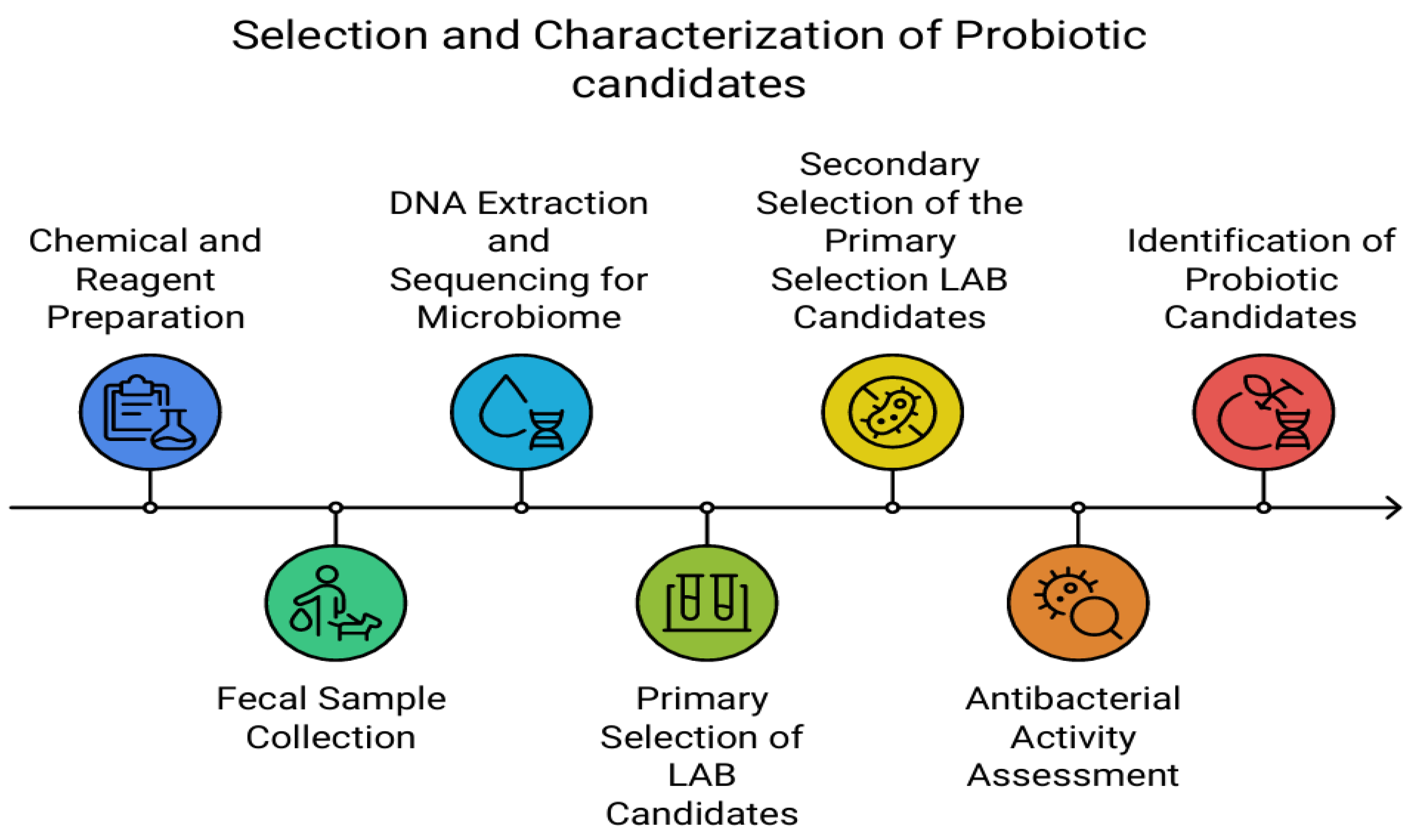
Figure 2.
Taxonomic classification of 40 faecal samples from healthy dogs, including adult dogs and puppies. (a−b) phylum level, (c−d) class level, and (e−f) order level.
Figure 2.
Taxonomic classification of 40 faecal samples from healthy dogs, including adult dogs and puppies. (a−b) phylum level, (c−d) class level, and (e−f) order level.
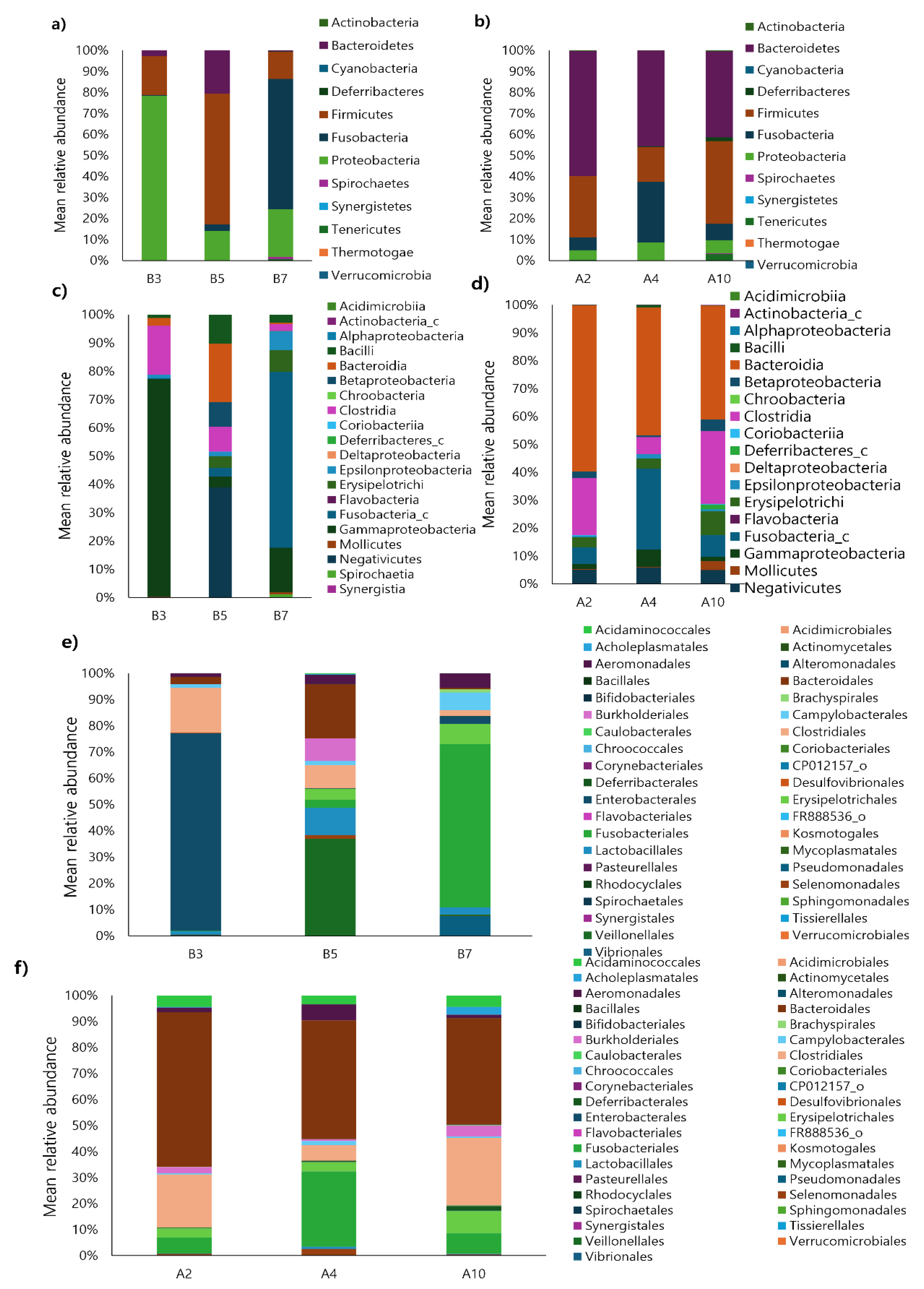
Figure 3.
Correlation between bacterial taxa and age group (adult dogs vs. puppies) at the phylum level (a), class level (b), and order level (c).
Figure 3.
Correlation between bacterial taxa and age group (adult dogs vs. puppies) at the phylum level (a), class level (b), and order level (c).
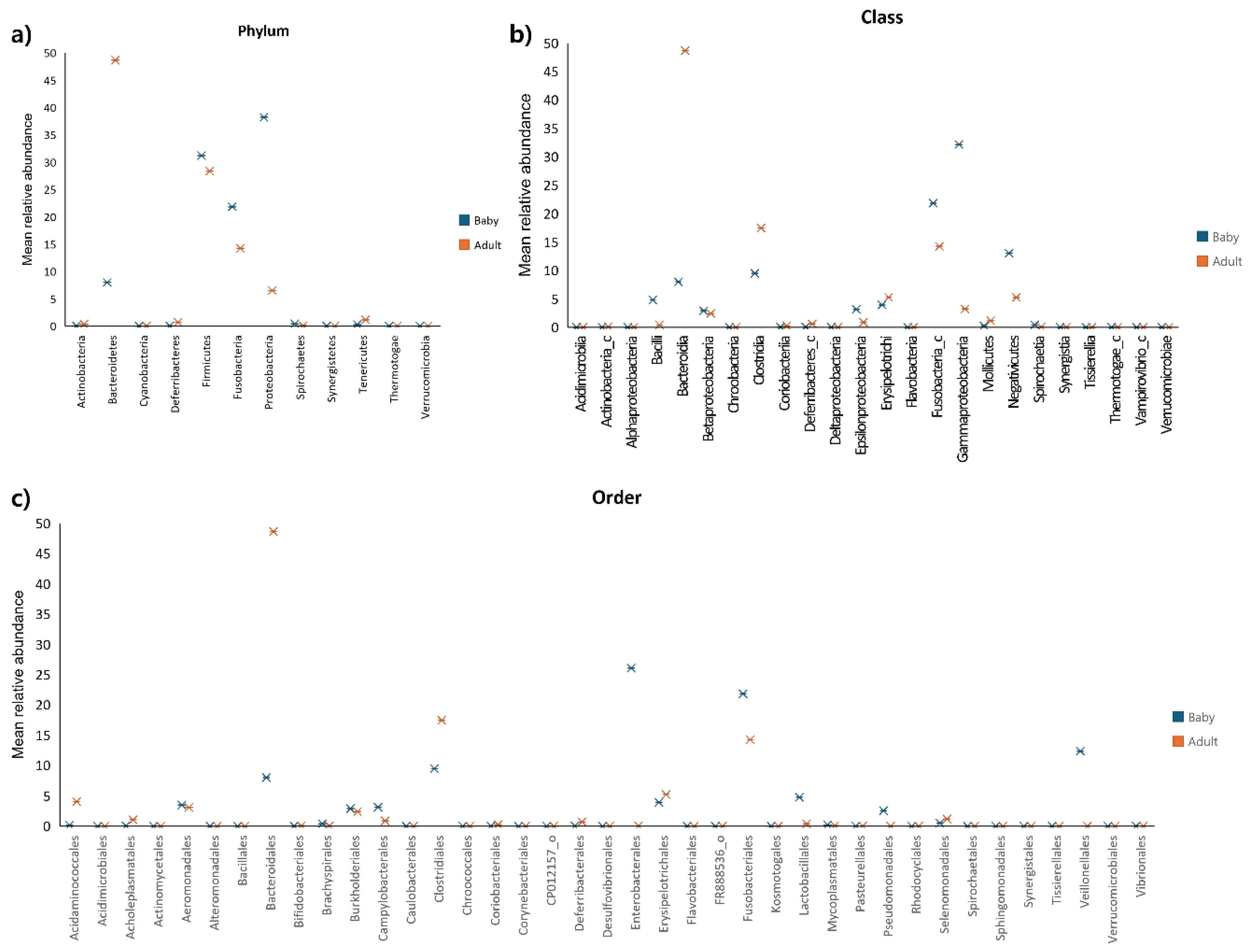
Figure 4.
Variability in the F/B ratio (a) and the relative abundances of Firmicutes (b) and Bacteroidetes (c) in the gut microbiota of adult dogs and puppies. Box plots were constructed using R.*p < 0.05 and **p < 0.01. Abbreviation: F/B, Firmicutes/Bacteroidetes; ns, non-significant.
Figure 4.
Variability in the F/B ratio (a) and the relative abundances of Firmicutes (b) and Bacteroidetes (c) in the gut microbiota of adult dogs and puppies. Box plots were constructed using R.*p < 0.05 and **p < 0.01. Abbreviation: F/B, Firmicutes/Bacteroidetes; ns, non-significant.
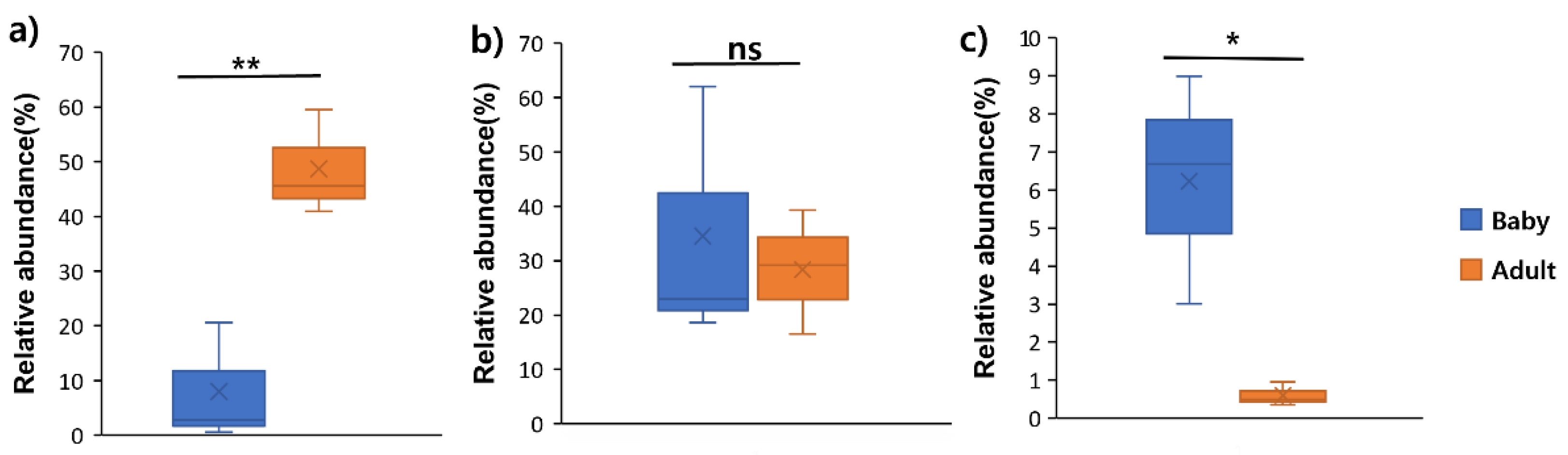
Figure 5.
Differences in alpha diversity between puppies (n = 3) and adult dogs (n = 3). Species richness reflected via a) ACE, b) Chao1, c) Jackknife, d) Shannon, and e) Simpson indices. *p < 0.05 and **p < 0.01. Abbreviation: ns, non-significant.
Figure 5.
Differences in alpha diversity between puppies (n = 3) and adult dogs (n = 3). Species richness reflected via a) ACE, b) Chao1, c) Jackknife, d) Shannon, and e) Simpson indices. *p < 0.05 and **p < 0.01. Abbreviation: ns, non-significant.
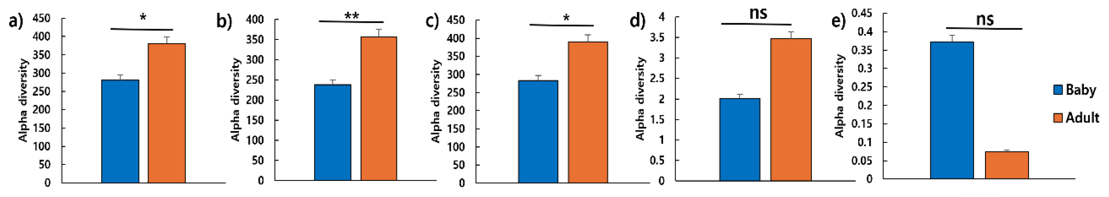
Figure 6.
Diversity and strain-specific characteristics of gut microbiota, as revealed by culturing faecal samples on various media. Bacterial colony counts on a) Blood agar, b) TSN, c) PDA, d) MC, e) SS, f) MS, g) BG, h) MACS, i) MRS, j) TOS, and k) PCA with BCP media of adult dogs and puppies. **p < 0.01 and ***p < 0.001. Abbreviations: TSN, tryptone sulfite neomycin; PDA, potato dextrose agar; MC, MacConkey; SS, salmonella-shigella; MS, mannitol salt; BG, brilliant green; MACS, MacConkey sorbitol; MRS, De Man, Rogosa, and Sharpe; TOS, transgalactosylated oligosaccharides with MUP; PCA, plate count agar; BCP, bromocresol purple; ns, non-significant.
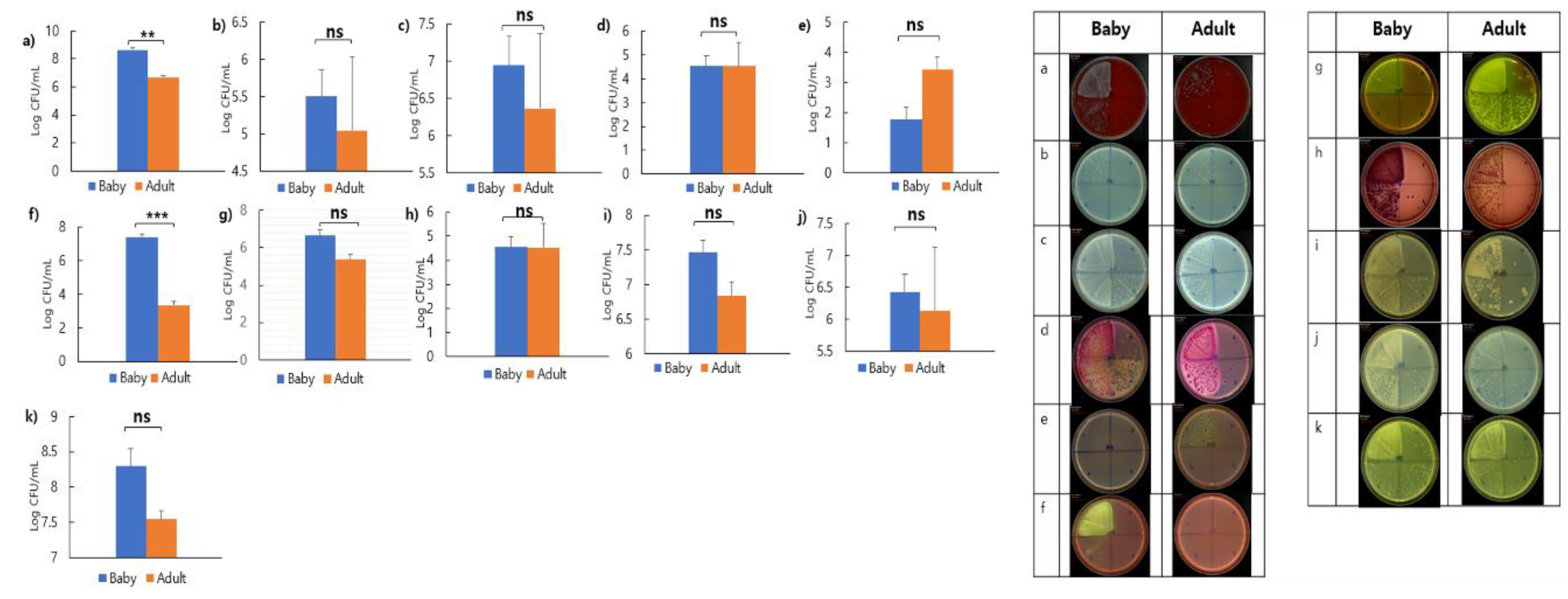
Figure 7.
Probiotic characteristics of selected LAB from the faeces of dogs a) Acid tolerance, b) Lactic acid production, c) Bile resistance, d) Heat resistance, and e) Dietary enzyme of protease. Abbreviation: LAB, lactic acid bacteria.
Figure 7.
Probiotic characteristics of selected LAB from the faeces of dogs a) Acid tolerance, b) Lactic acid production, c) Bile resistance, d) Heat resistance, and e) Dietary enzyme of protease. Abbreviation: LAB, lactic acid bacteria.
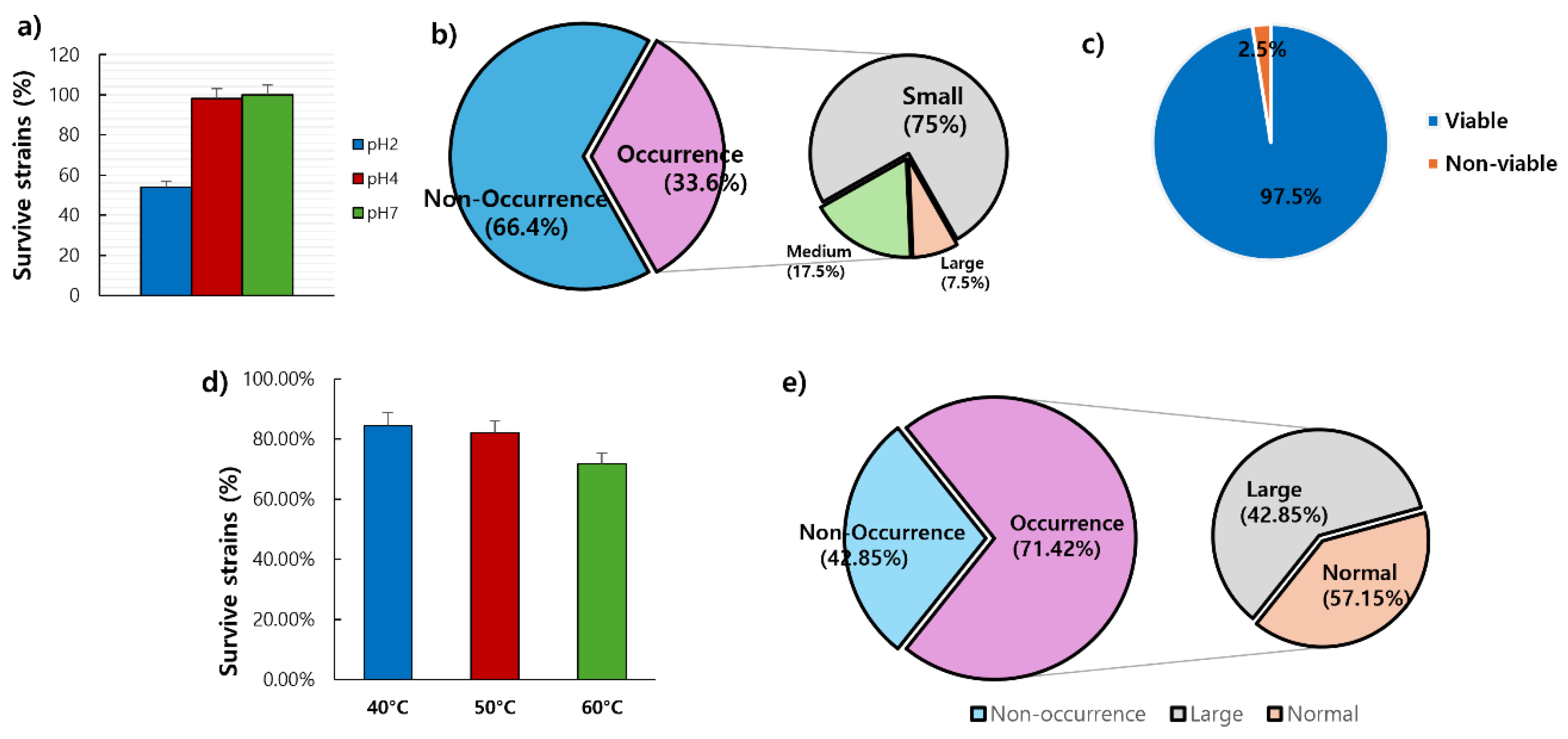
Figure 8.
Co-culture of Lactobacillus with enterotoxigenic pathogen a) E. coli ATCC 35218, b) S. aureus ATCC 29213, and c) Salmonella typhimurium KCTC 2515. The CFU of enterotoxigenic pathogens at 103 CFU/mL after co-culture with Lactobacillus (109 CFU/mL) for 24 h. Abbreviations: E. coli, Escherichia coli; S. aureus, Staphylococcus aureus; CFU, colony-forming units; CT, control.
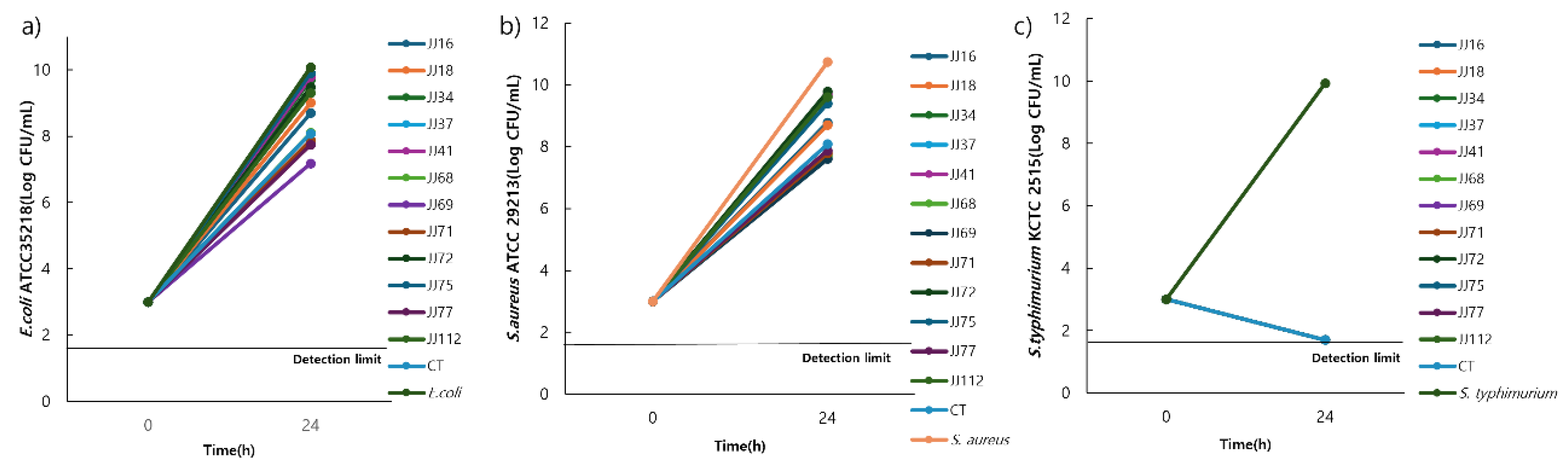
Figure 9.
Phylogenetic tree of L. reuteri strain a) L. reuteri JJ 37 and b) L. reuteri JJ 69 Abbreviation: L. reuteri, Lactobacillus reuteri.
Figure 9.
Phylogenetic tree of L. reuteri strain a) L. reuteri JJ 37 and b) L. reuteri JJ 69 Abbreviation: L. reuteri, Lactobacillus reuteri.
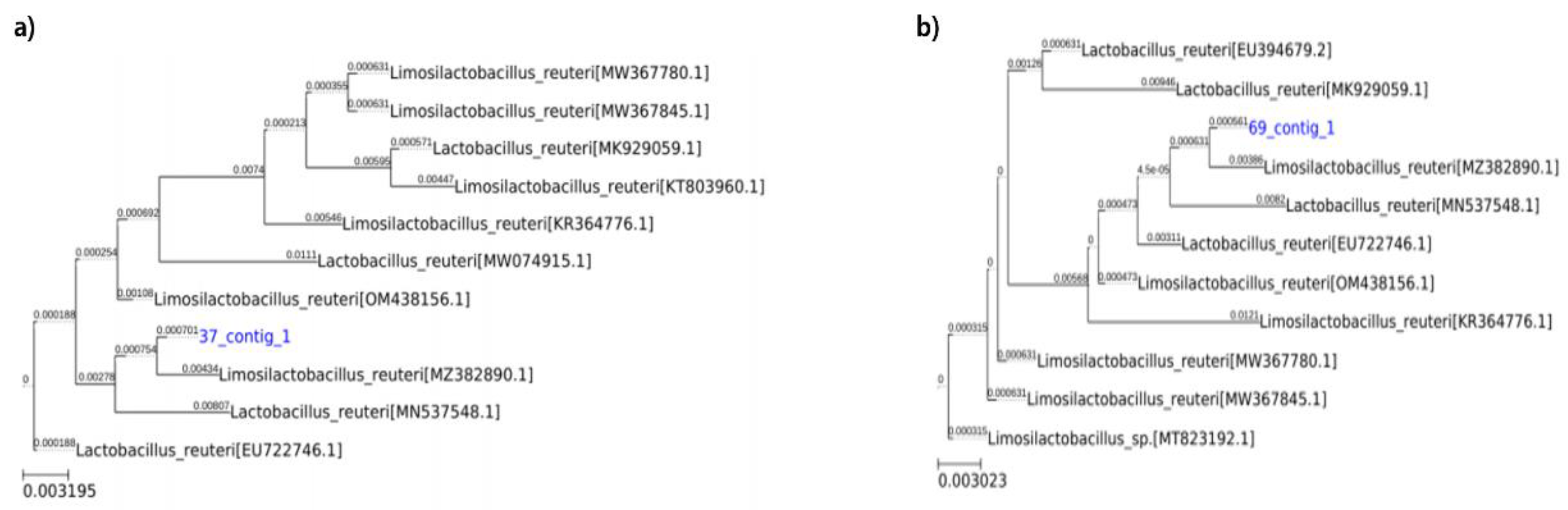

Table 1.
Probiotic characteristics of selected lactic acid bacteria from the faeces of dogs.
| Characteristics | Number of isolates (%) | |
| Acid tolerance | pH 2, 1.5 h | 59/109 (51.4) |
| pH 4, 1.5 h | 107/109 (98.17) | |
| pH 7, 1.5 h | 109/109 (100.0) | |
| Lactic acid production | + | 30/119 (25.21) |
| ++ | 7/119 (5.88) | |
| +++ | 3/119 (2.52) | |
| Bile tolerance | 1.0% Oxgall, 48 h | 39/40 (97.5) |
| Heat resistance | 40°C, 1 h | 33/39 (84.61) |
| 50°C, 1 h | 32/39 (82.05) | |
| 60°C, 1 h | 28/39 (71.79) | |
| Dietary enzyme of protease | + | 8/28 (28.57) |
| ++ | 12/28 (60.0) |
+, Minimum; ++, Medium; +++, Maximum. Classified on lactic acid production (brightness: weak, moderate, and strong translucent ring) and protease (diameter: 0.5−1cm, 1.0−1.5cm, and ≥1.5cm).
Table 2.
Characterisation of lactobacilli isolated from dog faeces.
| Isolates | Gram staining | Cell morphology | Catalase | Antibacterial activity |
| JJ16 | + | Rod | − | + |
| JJ18 | + | Rod | − | + |
| JJ34 | + | Rod | − | + |
| JJ37 | + | Rod | − | +++ |
| JJ41 | + | Rod | − | + |
| JJ68 | + | Rod | − | +++ |
| JJ69 | + | Rod | − | +++ |
| JJ71 | + | Rod | − | +++ |
| JJ72 | + | Rod | − | + |
| JJ75 | + | Rod | − | + |
| JJ77 | + | Rod | − | +++ |
| JJ112 | + | Rod | − | + |
| CT | + | Rod | − | + |
+, Positive;−, Negative; +++, Maximum; ++, Medium; +, Minimum. CT, Control.
Disclaimer/Publisher’s Note: The statements, opinions and data contained in all publications are solely those of the individual author(s) and contributor(s) and not of MDPI and/or the editor(s). MDPI and/or the editor(s) disclaim responsibility for any injury to people or property resulting from any ideas, methods, instructions or products referred to in the content. |
© 2025 by the authors. Licensee MDPI, Basel, Switzerland. This article is an open access article distributed under the terms and conditions of the Creative Commons Attribution (CC BY) license (http://creativecommons.org/licenses/by/4.0/).
Copyright: This open access article is published under a Creative Commons CC BY 4.0 license, which permit the free download, distribution, and reuse, provided that the author and preprint are cited in any reuse.


