
Submitted:
21 March 2025
Posted:
25 March 2025
You are already at the latest version
Abstract
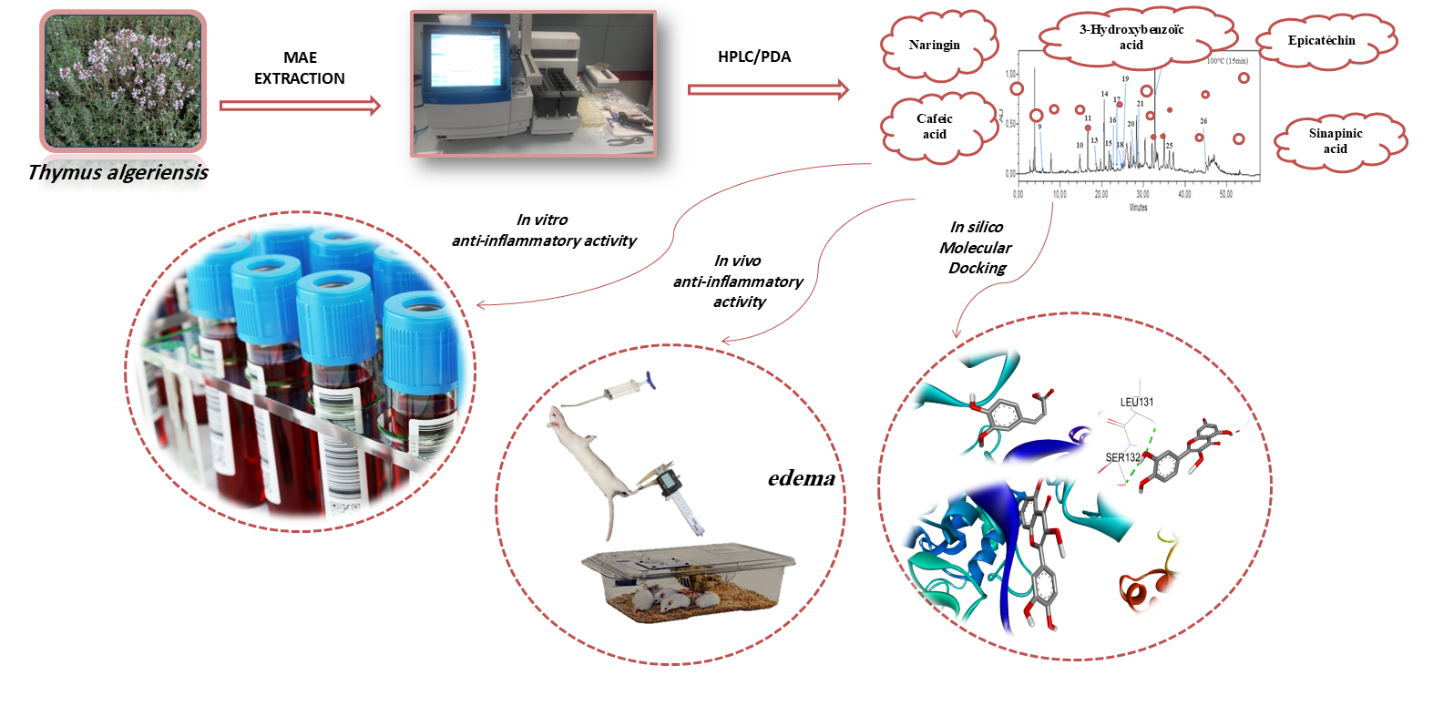
The objective of this work is to study the anti-inflammatory effect in vitro and in vivo of microwave extracts (MW) of Thymus algeriensis. The in vitro study was performed by the human red blood cell protection test, while the in vivo study used the carragee-nan-induced rat leg edema model. The experimental results were confirmed by a mo-lecular docking calculation. The results indicated that all microwave extracts have a moderate anti-inflammatory effect, depending on their richness in phenolic com-pounds. Among the extracts studied, the one obtained at 100°C for 15 minutes showed the most pronounced anti-inflammatory effect, with an inhibition of 78.52%, which is attributed to its richness in flavonoids. In particular, the flavonoids Naringine and Catechin showed the best affinity for the target protein, with values of -10.3 kcal/mol and -9.2 kcal/mol, respectively, as well as low inhibition constants of 0.028 μM and 0.18 μM. These results indicate that these flavonoids generate interactions that enhance the stability of the target ligand-protein complex, thus contributing to the observed an-ti-inflammatory effect.
Keywords:
Thymus algeriensis
; anti-inflammatory activity
; phenolic compounds
; micro-wave-assisted extraction
; molecular docking
1. Introduction
Medicinal plants contain various types of antioxidants, mostly carotenoids, phenolic compounds, benzoic acid derivatives, flavonoids, proanthocyanidins, stilbenes, coumarins, lignans, and lignins, which exhibit high antioxidant activity [1,2]. The recommendations based on epidemiological studies assessed that fruits, vegetables and less processed staple foods can ensure the best protection against the development of diseases caused by oxidative stress, such as cancer, coronary heart disease, obesity, type 2 diabetes, hypertension and cataract [1]. The explanation relies on the beneficial health effect, due to their antioxidant agents. Indeed, among 50 analysed food products with high antioxidant content, 13 were spices, 8 were fruits and vegetables, 5 were berries, 5 were chocolate-based food, 5 were breakfast cereals, and 4 were nuts or seeds [3].
Inflammation is a complex biological response of vascular tissue to harmful stimuli and pathogens, characterized by redness, warmth, swelling, and pain [4]. Prolonged inflammation leads to rheumatoid arthritis, atherosclerosis, hay fever, ischemic heart diseases, and is also a common manifestation of infectious diseases [5,6].
In the plant kingdom, the genus Thymus belongs to the Lamiaceae family which comprises about 215 to 400 species according to previous studies [7]. Generally, this genus consists of perennial plants and subshrubs native mainly in Europe, Western Asia and the Mediterranean countries [8]. Historically, the volatile constituents-rich aerial parts of Thymus species have been commonly used as herbal teas, condiments, and spices. In addition, they have shown many ethnomedicinal properties such as tonic, carminative, digestive, antispasmodic, antimicrobial, antioxidant, anti-inflammatory [9,10,11,12,13,14,15]. T. algeriensis is the most pervasive North African species, endemic to Morocco, Tunisia, Algeria, and Libya [16,17,18,19,20]. Its essential oil was proposed as antimicrobial, acridicidal, antioxidant, anti-inflammatory, and neuroprotective after extensive knowledge of safety, chemical composition, and stability [21,22,23,24,25,26,27,28,29,30,31]. Recent invetigations were focused on the polyphenolic and flavonoid contents isolated by classical extraction from the aerial parts of this species as alternative components to exert a valuable pharmacological activity [32,33,34,35,36,37].
To continue our investigations the biological activities of Thymus [38], this study was designed to investigate the anti-inflammatory activity of microwave-assisted extracts of Algerian T. algeriensis aerial parts. Microwave-assisted extraction (MW) was applied to recover a high-benefit composition from this endemic species.
Furthermore, to support the in vivo results and gain deeper insights into the molecular mechanisms behind the anti-inflammatory activity of the extracts, we have employed in silico studies, specifically molecular docking simulations. Molecular docking allows for the prediction of the binding affinity and interaction of bioactive compounds with specific proteins or receptors involved in inflammatory pathways. This computational technique can provide valuable information regarding the potential mechanisms of action of the compounds extracted from T. algeriensis, and help identify key targets involved in their anti-inflammatory effects. Recent studies have shown that in silico approaches, including molecular docking, are highly effective in understanding the bioactivity of plant-derived compounds, aiding the drug discovery process by identifying promising candidates for further experimental validation [39,40]. By simulating the binding interactions of compounds with key receptors or enzymes involved in inflammation, molecular docking can provide insights into the mechanisms underlying their therapeutic effects [41,42]. Moreover, this method has been widely used in drug discovery to streamline the identification of promising candidates for further experimental validation.
2. Materials and Methods
2.1. Plant Material and Preparation of Extracts
The plant material was collected from Biskra region (coordinates: 34°51′01″ N / 5°43′40″ E), Algeria. The plant was identified by Prof. M. Kaabeche (Biology Department, University of Setif 1, Algeria). A relative specimen voucher has been registered in the Herbarium of Frères Mentouri University, Algeria. Microwave-assisted extraction (MW) was carried out by means of a single-mode microwave reactor automatic Biotage Initiator™ 2.0 (Biotage AB, Uppsala, Sweden) characterized by a power range of 0–300 W and 2.45 GHz microwaves. MW was performed at discrete temperatures (T = 40, 60, 80, 100, or 120°C) for 5, 10, or 15 min in water as the solvent as previously reported.
2.2. Profile of Bioactive Compounds
We measured the total amount of phenolic acid and flavonoids in T. algeriensis extracts that had been microwave-irradiated. We also looked for chlorophylls and carotenoids. We wrote more about the exact steps we took in our previous paper [38]. High-quality standard chemicals for the chromatographic analyses (gallic acid, catechin, chlorogenic acid, epicatechin, 4-hydroxybenzoic acid, t-ferulic acid, naringin, vanillic acid, p-coumaric acid, rutin, syringic acid, benzoic acid, o-coumaric acid, 3-hydroxybenzoic acid, sinapinic acid, 2,3-dimethoxybenzoic acid, quercetin, isovanillin) were purchased by Merck (Milan, Italy). The HPLC-PDA method was used for the quantitative characterization of the compounds in these extracts.
2.3. In Vitro Anti-Inflammatory Activity
The anti-inflammatory activity of MW extracts was evaluated using the human red blood cell (RBC) membrane stabilization method [43]. The blood was collected from a healthy human volunteer who had not taken any NSAIDs for 2 weeks prior to the experiment. The collected blood was then mixed with an equal volume of Alsever solution. (2% dextrose, 0.8% sodium citrate, 0.5% citric acid, and 0.42% NaCl) and centrifuged at 1730 g. The packed cells were washed with isosaline, and a 10% suspension was made. Different concentrations of extracts were prepared (250, 500, and 1000 mg/mL) using distilled water. To each concentration, 1 mL of phosphate buffer, 2 mL of hyposaline, and 0.5 mL of HRBC (human red blood cell) suspension were added. Each sample was incubated at 37°C for a duration of 30 min, followed by centrifugation at 1730 g for 20 min. The supernatant solution was then analyzed spectrophotometrically at a wavelength of 560 nm to estimate the hemoglobin content.
2.4. Inflammatory Paw Edema Test in Rats
In this part of the experiments, the anti-inflammatory activity of each microwave-assisted extract was investigated on carrageenan-induced inflammatory paw edema [44]. Female Wistar rats (80-125 g) were obtained from the Pasteur Institute (Algeria). Rats were housed in standard polypropylene cages, four per cage. The extracts were dissolved and dispersed in physiological saline (0.9%) and administered orally to a pretreated group of rats at a dosage of 50 mg/kg. The control group received physiological saline (0.9%) at the same volume as the vehicle used for administering the extracts. One hour after administration, 0.1 mL of 1% carrageenan solution was injected into the footpad of the hind paws of each rat in all groups. Prior to carrageenan injection, the rat paw volume was measured. Increase of carrageenan-induced inflammatory paw volume was measured at 1, 2, and 3 h over the injection. The anti-inflammatory activity of T. algeriensis extracts was compared with that of 50 mg/kg diclofenac. The percentages of inhibition were calculated according to the following formula:
where V is the diameter of injected paw, V0 is the average inflammation (hind paw edema) of the control group at a given time 0; and VT is the average of diameters of the hind paw edema of the drug-treated rat at the same time.
This test was approved by the ethics committee (The committee of the Algerian Association of Sciences in Animal Experimentation (N°. 8808/1988), associated with veterinary medical activities and animal health protection N° JORA :004/1988). All experimental procedures were conducted in accordance with established ethical principles to ensure the respect of participants' rights and well-being.
2.5. Molecular Docking Study
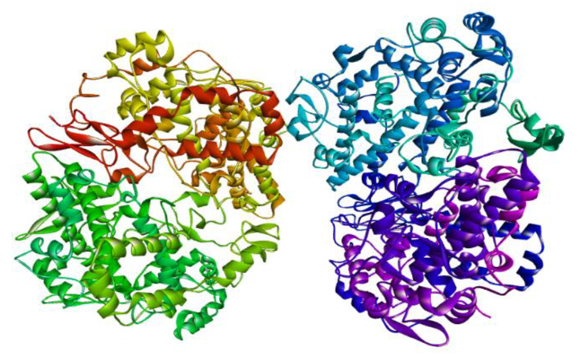
The study of molecular docking of the organic molecules that have been extracted from the plant was performed to complement and confirm the results in the experimental section for the in vitro and in vivo activities using AutoDock Vina program. The protein used to simulate this activity, named COX2, has been obtained from the RCSB database with the code [3LN0] [45] .The active site of the protein used is delimited by a box of dimensions: a volume of 40 × 40 × 40 Å3 and a center x, y, z: 41, 35 and 47, respectively.
To ensure an effective simulation, the ligands (19 molecules: gallic acid, catechin, chlorogenic acid, epicatechin, 4-hydroxy-benzoic acid, t-ferulic acid, naringin, vanillic acid, p-coumaric acid, rutin, syringic acid, benzoic acid, o-coumaric acid, 3-hydroxy-benzoic acid, sinapinic acid, 2,3-dimethoxy-benzoic acid, quercetin, isovanillin and dichlofenac were initially prepared by optimizing them using the Chem3D 16.0 to achieve a stable geometry with minimum energy. Next, the receptors (proteins) for the two activities were prepared using Discovery Studio. This involved removing water molecules and heteroatoms, and adding polar hydrogen atoms. Afterwards, molecular docking simulations were conducted using the AutoDock Vina program [46]
Also, the molecular interactions between protein targets and compounds were viewed using BIOVIA Discovery Studio. Amino acids engaging with the ligand, hydrogen bonds (H-bonds), hydrophobic interactions, and the individual atoms involved were examined in each ligand cluster (Supplementary Table S2).
2.6. Statistical Analysis
Statistical analysis was performed using IBM SPSS Statistics 27. Comparisons were made using one-way analysis of variance (ANOVA), followed by Tukey’s test. The results of both in-vitro and in-vivo experiments are presented as mean ± standard deviation (SD) with (n = 3), when the p-value was less than 0.05.
3. Results and Discussion
3.1. In Vitro Anti-Inflammatory Activity in Human RBCs (HBRC)
The microwave-assisted extracts of the T. algeriensis were then studied for in vitro anti-inflammatory activity by the HRBC membrane stabilization method. The results are reported in Table 1. The in vitro anti-inflammatory activity of the extracts was concentration-dependent, and again, the higher protection of HRBC in hypotonic solution was attributed to extracts obtained at higher temperature (120°C) or time of extraction (15 min). All the results were compared with standard diclofenac, which showed 96.67 ± 0.15% protection at the highest concentration.
The results show that the in vitro anti-inflammatory activity increases with the treatment temperature, reaching its maximum at 120°C for all tested concentrations, with more pronounced effects at 0.5 mg/ml and 1 mg/ml. The treatment at 100°C for 15 minutes also shows a significant improvement compared to a 5-minute exposure. Although the effectiveness of the tested treatments is close to that of diclofenac, it remains slightly lower, but the differences are not always statistically significant. These results suggest that combining high temperature with extended treatment time optimizes the anti-inflammatory activity, offering potential comparable to that of reference anti-inflammatory drugs.
Microwave-assisted extracts exhibited a membrane stabilization effect by inhibiting hypotonicity-induced lysis of the erythrocyte membrane. The erythrocyte membrane is analogous to the lysosomal membrane [47], and its stabilization implies that the extract may as well stabilize lysosomal membranes. Stabilization of lysosomal membrane is important in limiting the inflammatory response by preventing the release of lysosomal constituents of activated neutrophils such as bactericidal enzymes and proteases, which cause further tissue inflammation and damage upon extracellular release [48].
The exact mechanism for the membrane-stabilizing effect of these extracts of T. algeriensis and the chemical constituents responsible for this effect is hitherto not known. However, a number of studies have shown that flavonoids exhibit analgesic and anti-inflammatory effects as a result of their membrane stabilizing ability in various experimental models [49]. The extract may inhibit the processes, which may stimulate or enhance the efflux of these intracellular components [50]. It has also been shown that microwave-assisted extracts of T. algeriensis contain flavonoids such as rutin, catechin, and epicatechin [38]. Thus, these extracts showed significant in vitro anti-inflammatory activity as compared to the standard. On the basis of the above results, it can be concluded that these MW extracts of T. algeriensis have a potential anti-inflammatory activity.
3.2. Carrageenan-Induced Paw Oedema
Paw oedema thickness was measured at 1, 2, and 3 hours post-inflammation induction in animals pre-treated with the MW-extract. The percentage of oedema inhibition by the MW-extract was measured. The inflammation induced by carrageenan develops into two distinct phases. The initial phase, occurring within one hour of exposure, is identified by the release of histamine, serotonin, and bradykinin. The late phase, occurring after more than an hour, results from the release of prostaglandin-like substances [51,52]. Research has demonstrated the involvement of free radicals in the initial phase of acute inflammation induced by carrageenan [53].
Table 2 shows the effect of microwave-assisted extracts of T. algeriensis on the carrageenin-induced rat paw edema test. After carrageenan induction there was a significant (ρ<0,001) increase in edema formation in normal rats. Carrageenan-induced hind paw edema is the standard experimental model of acute inflammation. Moreover, the experimental model exhibits a high degree of reproducibility [54].
Figure 1 shows the inhibition percentages of microwave-treated extracts of Thymus algeriensis and diclofenac. One hour after carrageenan injection, all microwave extracts showed a moderate anti-inflammatory effect comparable to that of the reference drug (diclofenac), with inhibition percentages ranging from 24% to 29% for extracts and 23% for diclofenac. These results suggest that microwave-treated extracts of Thymus algeriensis have similar anti-inflammatory activity to diclofenac, although moderate.
After three hours of experimentation, diclofenac showed a marked anti-inflammatory effect, reaching 83.74%, which confirms the efficacy of non-steroidal anti-inflammatories, which act by inhibition of COX-1 and COX-2 enzymes [55]. On the other hand, microwave-treated extracts showed significant inhibition, although lower than diclofenac, with percentages ranging between 51% and 70%. However, the extract treated at 100°C for 15 minutes showed an anti-inflammatory effect similar to that of diclofenac, with a percentage of 78.52%, suggesting that this treatment could be an interesting alternative with efficacy comparable to the reference drug.
Some studies have indicated that the flavonoids contained in plant extracts have anti-inflammatory properties and that they are able to modulate the functioning of the immune system by inhibiting the activity of enzymes which are responsible for inflammation; they may also modulate monocyte adhesion during atherosclerotic inflammation by inhibiting the expression of inflammatory mediators [50]. Our previous phytochemical study of T. algeriensis aerial parts revealed the presence of some of these compounds (epicatechin, catechin, rutin, quercetin and naringin), which could be responsible of this anti-inflammatory activity of the extracts. It has been also demonstrated that various biologically active flavonoids (such as rutin and quercetin ) produced significant antinociceptive and/or anti-inflammatory activities [56,57].
The extract treated at 100°C for 15 minutes (MW 100°C 15 min) had the most significant anti-inflammatory effects compared to the other extracts tested. The higher efficacy is due to the extract's availability of flavonoids, including quercetin, rutin, catechin, epicatechin, and naringin, which are compounds known for their ability to reduce inflammation. These flavonoids, by their action on pro-inflammatory enzymes and their ability to modulate inflammatory mediators, play a key role in reducing inflammatory processes.
The microwave extraction process at 100°C for 15 minutes seems to have optimized the extraction of these compounds, which explains the higher efficiency of this extract. Indeed, this method allows a faster and more complete release of flavonoids while preserving their structure and bioactive activity. These results reinforce the idea that microwave extraction can be an effective method to obtain plant extracts rich in anti-inflammatory compounds.
Several researchers have also demonstrated the anti-inflammatory effect of Thymus algeriensis extracts, showing that they can reduce inflammation markers and attenuate inflammatory responses in various experimental models.
According to Mensour et al. (2020), and Mahdi et al.2020, Thymus algeriensis has significant anti-inflammatory activities. Extracts from the leaves of Thymus algeriensis were evaluated for their ability to inhibit the enzymes cyclooxygenase-1 (COX-1) and cyclooxygenase-2 (COX-2), as well as lipoxygenase (5-LOX). The results indicated that the extract of Thymus algeriensis is more selective for COX-2 than for COX-1, with an index of selectivity similar to celecoxib, a selective inhibitor of COX-2. The extract also showed anti-inflammatory effects in living things, like rats with leg swelling caused by carrageenan and mice with white blood cells moving into the peritoneal cavity [22,58].
3.3. Molecular Docking
Docking of molecules is a modern bioinformatics method that predicts the probable experimental orientation and the binding affinity needed to produce a stable complex structure between a ligand and a target [59]. The AutoDock Vina tool was used to do molecular docking tests to see if each of the identified compounds had an anti-inflammatory effect compared to the standard drug, dichlofenac.
The findings allowed the assessment of the binding energy between the protein and different ligand positions (Table 3). A negative binding energy indicates a potential for binding between the ligand and the receptor. The inhibition constant (Ki) was calculated using the formula: Ki = exp(ΔG/RT), where ΔG indicates the binding energy, R is the gas constant (1.9872036×10⁻³ kcal. Mol-1), and T represents the ambient temperature (298.15 K) [60]. A smaller inhibition constant signifies a more effective drug derived from the title molecule.
It was found that Naringin and Catechin have the best affinity, with values of -10.3 kcal. Mol-1 and -9.2 kcal. Mol-1, respectively, as well as a low inhibition constant of 0.028 μM and 0.18 μM, respectively. Additionally, we performed molecular docking using a commercialized drug named diclofenac. According to Table 2, Catechin, Chlorogenic acid, Epicatechin, Naringin, Quercetin and Rutin have shown favorable results compared to diclofenac (Table 3).
Figure 2.
3D structure of the inflammatory protein COX-2 [PDB ID: 3LN0].
The binding affinity of the fixed ligands to the target proteins in this investigation was affected by non-covalent interactions, including hydrogen bonds and hydrophobic interactions. Several investigations have demonstrated that binding affinity increases when ligands can participate in hydrophobic interactions with a significant amount of hydrophobic residues of amino acids within the binding region of a target protein [61,62]. Stojanovi and Zari [63] emphasized the significance of hydrophobic interactions in various systems characterized by powerful intermolecular forces [63] .This mechanism could be responsible for the significant binding affinity of the active chemicals associated with the proteins selected in this study. The significance of hydrogen bonds in stabilizing molecular interactions between ligands and proteins must not be ignored, because they play a crucial role in enzymatic catalysis and the formation of protein-substrate and protein-inhibitor complexes, contributing to the overall stability of multiple biological molecules [63,64].
For this purpose, the analysis of molecular interactions of the active compounds, carried out on the basis of their binding energy, hydrogen bonds, and hydrophobic interactions with surrounding amino acids in selected protein targets, revealed a significant impact of the ligand-protein complex status. In particular, it was seen that flavonoids taken from Thymus algeriensis have incredibly low free binding energy values compared to other polyphenolic compounds being studied. Low free energy indicates a more stable interaction and the strong affinity of flavonoids for their protein targets.
Also, the inhibition constant values (Ki) for these flavonoids were lower than those for other polyphenolic compounds. This means that they had a very strong affinity for binding to the target proteins. Low Ki values are synonymous with a better ability of these flavonoids to effectively inhibit the activity of target enzymes, thus enhancing their role in the modulation of inflammatory pathways. This evidence suggests that flavonoids, including quercetin, rutin, catechin, epicatechin, and naringin, are able to bind stably to the active sites of proteins, which may be the main reason for their anti-inflammatory symptoms noted in this study.
In the in vivo test, it was found that the extract treated by microwave at 100°C for 15 minutes (MW 100°C 15 min) revealed the most powerful anti-inflammatory effect. This result is directly related to the presence of a maximum level of flavonoids in this extract. Extracts with the highest concentrations of flavonoids showed significant inhibition of inflammatory markers, which confirms that the richness in flavonoids plays a decisive role in the anti-inflammatory efficacy observed. These results are consistent with data from the study of molecular interaction between different ligands and their target proteins, where flavonoids showed a strong affinity for the active sites of the target proteins, due to low Ki values and particularly low bonding energies.
Figure 3 and Figure 4 show numerous hydrogen bonds between each ligand and the target protein, illustrating the stability and efficacy of these interactions. In addition, non-covalent interactions such as hydrophobic and electrostatic interactions were also observed, further enhancing the stability of ligand-protein complexes. These non-covalent interactions are crucial for the stability of complexes and increase their effectiveness by inhibiting the activity of proteins responsible for inflammation.
Based on the results obtained, it appears that each flavonoid (ligand) has the potential to be an effective molecule in the fight against inflammation, thanks to its ability to interact stably with target proteins. The combined effect of the 18 ligands tested could still produce improved results, suggesting that the use of these flavonoids in combination therapies may have a stronger impact on inflammatory processes.
In addition, the in-silico results, which were obtained by simulating ligand-protein interactions, fully corroborate the experimental results obtained during in vivo tests. This concordance between the molecular analyses and the experimental results reinforces the validity of the data and highlights the reducing action of the inflammation of the extracts of Thymus algeriensis, validating the efficacy of flavonoids as potential therapeutic agents.
4. Conclusions
After the characterization of Thymus algeriensis extracts obtained by microwave, we focused our study on the anti-inflammatory activity of these extracts (in vitro and in vivo), evaluating the inhibition of inflammation according to their composition in secondary metabolites. All extracts showed moderate results, but the extract treated at 100°C for 15 minutes had the most pronounced anti-inflammatory effect, effectively reducing inflammatory edema in rats due to its high content of flavonoids, compared to other extracts. These results were corroborated by an in silico analysis, which revealed that the flavonoids present in microwave-processed extracts have a strong affinity for the target protein, thus explaining the efficacy of the extract in reducing inflammation.
Supplementary Materials
The following supporting information can be downloaded at the website of this paper posted on Preprints.org.
Author Contributions
“Conceptualization, N.B. and M.L.; methodology, N.B. and M.A.; software, N.B. and N.L.; validation, N.B., M.A. and M.L.; formal analysis, N.B. and N.L.; investigation, R.L.; resources, R.L. and N.B.; data curation, N.B.; writing—original draft preparation, N.B.; writing—review and editing, C.C. and F.M.; visualization, N.B. and N.L.; supervision, M.L.; project administration, N.B. All authors have read and agreed to the published version of the manuscript.
Funding
This research received no external funding.
Data Availability Statement
No data are available.
Acknowledgments
The authors would like to thank Laboratory of Pharmacy Department, University “G. d’Annunzio” of Chieti-Pescara, for the preparation and characterization of MAE extracts.
Conflicts of Interest
The authors declare no conflicts of interest.
Abbreviations
| The following abbreviations are used in this manuscript: | . |
| MW | Microwave extracts |
| RBC | Red Blood Cell |
| NSAIDs | Non-steroidal anti-inflammatory drugs |
References
- Halvorsen, B.L., et al., Content of redox-active compounds (ie, antioxidants) in foods consumed in the United States. The American journal of clinical nutrition, 2006. 84(1): p. 95-135. [CrossRef]
- Halvorsen, B.L., et al., A systematic screening of total antioxidants in dietary plants. The Journal of nutrition, 2002. 132(3): p. 461-471. [CrossRef]
- Lindsay, D.G. and S.B. Astley, European research on the functional effects of dietary antioxidants-EUROFEDA. Molecular Aspects of Medicine, 2002. 1(23): p. 1-38. [CrossRef]
- Chen, L., et al., Inflammatory responses and inflammation-associated diseases in organs. Oncotarget, 2018. 9(6): p. 7204. [CrossRef]
- Fioranelli, M., et al., Regulation of inflammatory reaction in health and disease. International Journal of Molecular Sciences, 2021. 22(10): p. 5277. [CrossRef]
- Pezone, A., et al., Inflammation and DNA damage: Cause, effect or both. Nature Reviews Rheumatology, 2023. 19(4): p. 200-211. [CrossRef]
- Cronquist, A., The evolution and classification of flowering plants. Bronx: New York Botanical Garden, 1988. 556. [CrossRef]
- Bourlière, F., Mabberley, DJ—The Plant Book. A Portable Dictionary of the Higher Plants. Cambridge University Press, Cambridge, 1987. Revue d'Écologie (La Terre et La Vie), 1988. 43(2): p. 198-199.
- Benjilali, B., et al., Essential oil composition of different Moroccan thyme varieties. 2. Principal component analysis. Sciences des Aliments (France), 1987.
- Guesmi, F., et al., Thymus hirtus Sp. algeriensis Boiss. and Reut. volatile oil enhances Trail/Apo2l induced apoptosis and inhibits colon carcinogenesis through upregulation of death receptor pathway. Aging (Albany NY), 2021. 13(18): p. 21975. [CrossRef]
- Guesmi, F., et al., Histopathological and biochemical effects of thyme essential oil on H2O2 stress in heart tissues. Heart, Lung and Circulation, 2020. 29(2): p. 308-314.
- Bukvicki, D., et al., Cheese supplemented with Thymus algeriensis oil, a potential natural food preservative. Journal of dairy science, 2018. 101(5): p. 3859-3865. [CrossRef]
- Zaïri, A., et al., Phytochemical analysis and assessment of biological properties of essential oils obtained from Thyme and Rosmarinus species. Current pharmaceutical biotechnology, 2020. 21(5): p. 414-424. [CrossRef]
- Fatma, G., B.H.A. Sami, and L. Ahmed, Investigation of extracts from Tunisian ethnomedicinal plants as antioxidants, cytotoxins, and antimicrobials. Biomedical and Environmental Sciences, 2017. 30(11): p. 811-824.
- Ouakouak, H., et al., Biological properties of essential oils from Thymus algeriensis Boiss. Plants, 2021. 10(4): p. 786. [CrossRef]
- Zouari, N., et al., Variation of chemical composition of essential oils in wild populations of Thymus algeriensis Boiss. et Reut., a North African endemic Species. Lipids in health and disease, 2012. 11(1): p. 1-12. [CrossRef]
- Maissa, B.J. and H. Walid, Antifungal activity of chemically different essential oils from wild Tunisian Thymus spp. Natural product research, 2015. 29(9): p. 869-873. [CrossRef]
- Guesmi, F., et al., Effects of Thymus hirtus sp. algeriensis Boiss. et Reut.(Lamiaceae) essential oil on healing gastric ulcers according to sex. Lipids in Health and Disease, 2014. 13(1): p. 1-14. [CrossRef]
- Ait-Ouazzou, A., et al., Chemical composition and antimicrobial activity of essential oils of Thymus algeriensis, Eucalyptus globulus and Rosmarinus officinalis from Morocco. Journal of the Science of Food and Agriculture, 2011. 91(14): p. 2643-2651.
- Ahmed, S.B.H., et al., Evaluation of antileishmanial, cytotoxic and antioxidant activities of essential oils extracted from plants issued from the leishmaniasis-endemic region of Sned (Tunisia). Natural Product Research, 2011. 25(12): p. 1195-1201. [CrossRef]
- Rezq, S., et al., Thymus algeriensis and Thymus fontanesii exert neuroprotective effect against chronic constriction injury-induced neuropathic pain in rats. Scientific Reports, 2020. 10(1): p. 1-15. [CrossRef]
- Sobeh, M., et al., Thymus algeriensis and Thymus fontanesii: chemical composition, in vivo antiinflammatory, pain killing and antipyretic activities: a comprehensive comparison. Biomolecules, 2020. 10(4): p. 599. [CrossRef]
- Fatma, G., B. Houda, and L. Ahmed, H2O2-Induced Oxidative Stress, AChE inhibition and mediated brain injury attenuated by Thymus algeriensis. 2018.
- Kouache, B., et al., Chemical composition and acaricidal activity of Thymus algeriensis essential oil against Varroa destructor. Natural product communications, 2017. 12(1): p. 1934578X1701200138. [CrossRef]
- Fatma, G., et al., Antioxidant machinery related to decreased MDA generation by Thymus algeriensis essential oil-induced liver and kidney regeneration. Biomedical and Environmental Sciences, 2016. 29(9): p. 639-649.
- Guesmi, F., et al., Prevention of H2O2 induced oxidative damages of rat testis by Thymus algeriensis. Biomedical and Environmental Sciences, 2016. 29(4): p. 275-285. [CrossRef]
- Fatma, G., et al., In-vitro assessment of antioxidant and antimicrobial activities of methanol extracts and essential oil of Thymus hirtus sp. algeriensis. Lipids in Health and Disease, 2014. 13(1): p. 1-12. [CrossRef]
- Jayari, A., et al., Nanoencapsulation of thyme essential oils: Formulation, characterization, storage stability, and biological activity. Foods, 2022. 11(13): p. 1858. [CrossRef]
- Nedjimi, B., Trace element quantification in two Algerian thymes (Thymus algeriensis Boiss & Reut. and Thymus capitatus (L.) Hoffm. & Link) using EDXRF spectrometry. Biological Trace Element Research, 2023. 201(1): p. 455-463. [CrossRef]
- El Ouahdani, K., et al., Thymus algeriensis and Artemisia herba-alba essential oils: chemical analysis, antioxidant potential and in vivo anti-inflammatory, analgesic activities, and acute toxicity. Molecules, 2021. 26(22): p. 6780.
- Rezzoug, M., et al., Chemical composition and bioactivity of essential oils and Ethanolic extracts of Ocimum basilicum L. and Thymus algeriensis Boiss. & Reut. from the Algerian Saharan Atlas. BMC complementary and alternative medicine, 2019. 19(1): p. 1-10. [CrossRef]
- Mahdi, I., et al., Unraveling the phytochemistry, traditional uses, and biological and pharmacological activities of Thymus algeriensis Boiss. & Reut. Oxidative Medicine and Cellular Longevity, 2022. 2022. [CrossRef]
- Bouafia, M., et al., Ethnobotanical and ethnomedicinal analysis of wild medicinal plants traditionally used in Naâma, southwest Algeria. Vegetos, 2021. 34: p. 654-662.
- Righi, N., et al., Thymus algeriensis Bioss & Reut: Relationship of phenolic compounds composition with in vitro/in vivo antioxidant and antibacterial activity. Food Research International, 2020. 136: p. 109500. [CrossRef]
- Jaouadi, R., et al., Differentiation of phenolic composition among tunisian thymus algeriensis boiss. Et reut.(lamiaceae) populations: correlation to bioactive activities. Antioxidants, 2019. 8(11): p. 515.
- Ziani, B.E., et al., Phenolic compounds characterization by LC-DAD-ESI/MSn and bioactive properties of Thymus algeriensis Boiss. & Reut. and Ephedra alata Decne. Food Research International, 2019. 116: p. 312-319.
- Zaïri, A., et al., Antioxidant, antimicrobial and the phenolic content of infusion, decoction and methanolic extracts of Thyme and Rosmarinus species. Current Pharmaceutical Biotechnology, 2018. 19(7): p. 590-599. [CrossRef]
- Boutaoui, N., et al., Qualitative and quantitative phytochemical analysis of different extracts from Thymus algeriensis aerial parts. Molecules, 2018. 23(2): p. 463. [CrossRef]
- Herowati, R. and G.P. Widodo, Molecular docking analysis: Interaction studies of natural compounds to anti-inflammatory targets. Quantitative structure-activity relationship, 2017. 63(10.5772). [CrossRef]
- Thamaraiselvi, L., et al., In-silico molecular docking analysis of some plant derived molecules for anti-inflammatory inhibitory activity. Curr. Bot, 2021. 12: p. 22-27. [CrossRef]
- Zhang, Q., et al., Mechanism of anti-inflammatory and antibacterial effects of QingXiaoWuWei decoction based on network pharmacology, molecular docking and in vitro experiments. Frontiers in Pharmacology, 2021. 12: p. 678685. [CrossRef]
- Laloo, D., J.M. Kalita, and S.K. Prasad, Molecular docking studies of plant-derived bioactive compounds: a comprehensive in silico standardization approach. Evidence Based Validation of Traditional Medicines: A comprehensive Approach, 2021: p. 371-404.
- Brown, J., H. Mackey, and D. Riggilo, A novel in vitro assay for anti-inflammatory agents based on stabilization of erythrocytes. Proceedings of the Society for Experimental Biology and Medicine, 1967. 125(3): p. 837-843. [CrossRef]
- Trovato, A., et al., Anti-inflammatory and analgesic activity of Hypericum empetrifolium Willd.(Guttiferae). Il Farmaco, 2001. 56(5-7): p. 455-457. [CrossRef]
- Wang, J.L., et al., The novel benzopyran class of selective cyclooxygenase-2 inhibitors. Part 2: The second clinical candidate having a shorter and favorable human half-life. Bioorganic & medicinal chemistry letters, 2010. 20(23): p. 7159-7163. [CrossRef]
- Trott, O. and A.J. Olson, AutoDock Vina: improving the speed and accuracy of docking with a new scoring function, efficient optimization, and multithreading. Journal of computational chemistry, 2010. 31(2): p. 455-461. [CrossRef]
- Chou, C.T., The Antiinflammatory Effect of an Extract of Tripterygium wilfordii Hook F on Adjuvant-induced Paw Oedema in Rats and Inflammatory Mediators Release. Phytotherapy Research: An International Journal Devoted to Medical and Scientific Research on Plants and Plant Products, 1997. 11(2): p. 152-154. [CrossRef]
- Murugesh, N., S. Vembar, and C. Damodaran, Studies on erythrocyte membrane IV: in vitro haemolytic activity of oleander extract. Toxicology letters, 1981. 8(1-2): p. 33-38. [CrossRef]
- David, S., Studies force new view on biology of flavonoids. Biol Med, 2007. 541: p. 737-87.
- González-Gallego, J., et al., Fruit polyphenols, immunity and inflammation. British journal of nutrition, 2010. 104(S3): p. S15-S27.
- Rosa, M.D. and D. Willoughby, Screens for anti-inflammatory drugs. Journal of Pharmacy and Pharmacology, 1971. 23(4): p. 297-298.
- Salvemini, D., et al., Evidence of peroxynitrite involvement in the carrageenan-induced rat paw edema. European journal of pharmacology, 1996. 303(3): p. 217-220. [CrossRef]
- Rayego-Mateos, S., et al., Pathogenic pathways and therapeutic approaches targeting inflammation in diabetic nephropathy. International journal of molecular sciences, 2020. 21(11): p. 3798. [CrossRef]
- Morris, C., Carrageenan-induced paw edema in the rat and mouse. Inflammation Protocols. Edited by: Winyard PG, Wiloughby DA, 2003, Totowa, New Jersey: Humana Press Inc.
- Tai, F.W.D. and M.E. McAlindon, Non-steroidal anti-inflammatory drugs and the gastrointestinal tract. Clinical Medicine, 2021. 21(2): p. 131-134. [CrossRef]
- Srivastava, S., et al., Antiinflammatory, analgesic and antipyretic activities of aerial parts of Costus speciosus Koen. Indian journal of pharmaceutical sciences, 2013. 75(1): p. 83.
- Silva, G.N., et al., Investigation of anti-inflammatory and antinociceptive activities of Lantana trifolia. Journal of ethnopharmacology, 2005. 100(3): p. 254-259. [CrossRef]
- Mahdi, I., et al., Unraveling the phytochemistry, traditional uses, and biological and pharmacological activities of Thymus algeriensis Boiss. & Reut. Oxidative Medicine and Cellular Longevity, 2022. 2022(1): p. 6487430. [CrossRef]
- Kumar, S. and S. Kumar, Molecular docking: a structure-based approach for drug repurposing, in In silico drug design2019, Elsevier. p. 161-189.
- Yung-Chi, C. and W.H. Prusoff, Relationship between the inhibition constant (KI) and the concentration of inhibitor which causes 50 per cent inhibition (I50) of an enzymatic reaction. Biochemical pharmacology, 1973. 22(23): p. 3099-3108.
- Salentin, S., et al., PLIP: fully automated protein–ligand interaction profiler. Nucleic acids research, 2015. 43(W1): p. W443-W447.
- Stojanović, S. and S. Zarić, Hydrogen bonds and hydrophobic interactions of porphyrins in porphyrin-containing proteins. The Open Structural Biology Journal, 2009. 3: p. 34-41. [CrossRef]
- Omoboyowa, D.A., et al., Identification of terpenoids from Abrus precatorius against Parkinson’s disease proteins using in silico approach. Bioinformatics and Biology Insights, 2021. 15: p. 11779322211050757. [CrossRef]
- Mohapatra, S., et al., In silico investigation of black tea components on α-amylase, α-glucosidase and lipase. Journal of Applied Pharmaceutical Science, 2015. 5(12): p. 042-047.
Figure 1.
Percentage of inhibition inflammation of MW extracts in rats with carrageenan-induced ear edema (values presented as means ±SD (n=3), ns: not significant difference, *P<0.05 **P<0.01, ***P<0.001 considered significant when compared with diclofenac).
Figure 1.
Percentage of inhibition inflammation of MW extracts in rats with carrageenan-induced ear edema (values presented as means ±SD (n=3), ns: not significant difference, *P<0.05 **P<0.01, ***P<0.001 considered significant when compared with diclofenac).
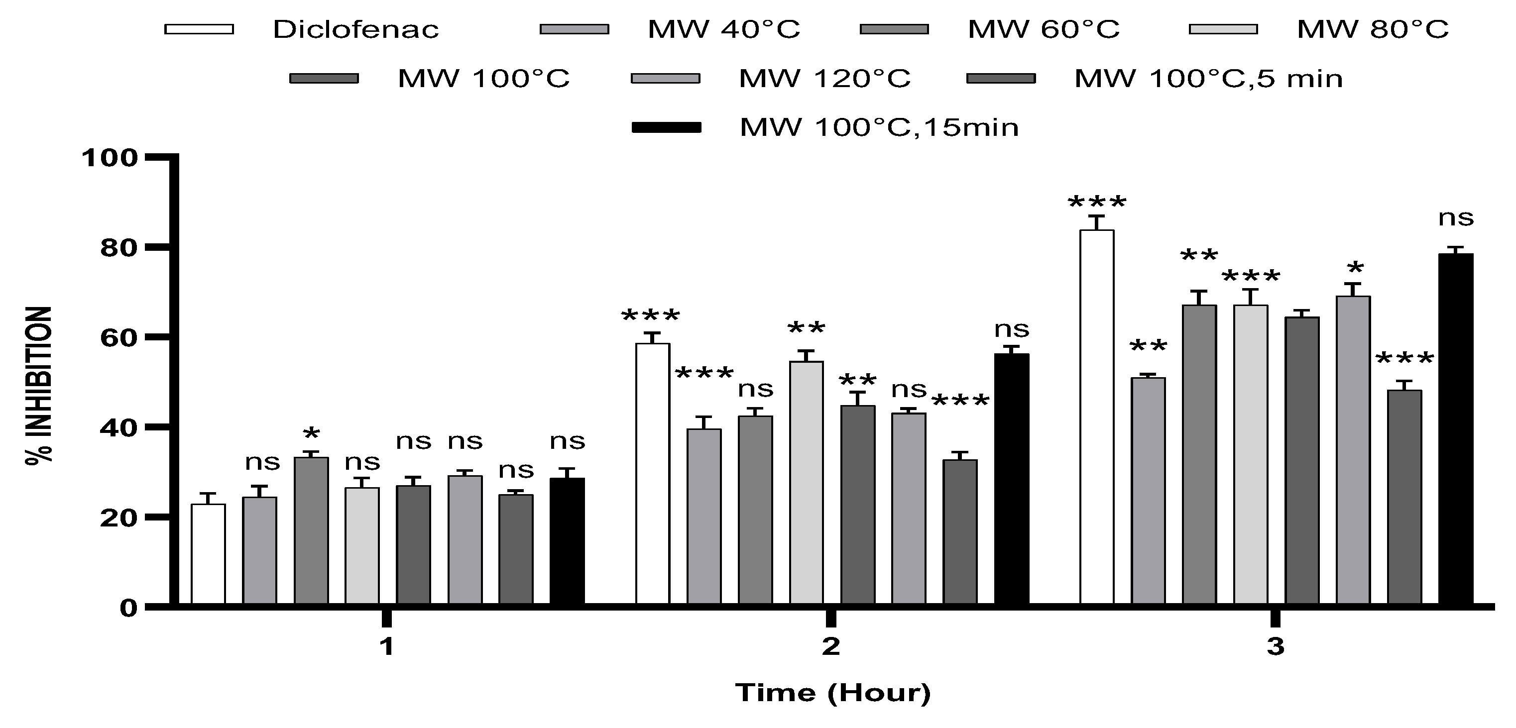
Figure 3.
2D detailed binding sites of each ligand into the receptor [PDB ID: 3LN0]. A: 2,3-dimethoxybenzoic acid; B: 3-hydroxybenzoic acid; C: 4-hydrobenzoic acid; D: syringic acid; E: gallic acid; F: benzoic acid; G: catechin; H: chlorogenic acid; I: epicatechin; J: isovanillin; K: naringin; L: o-coumaric acid; M: p-coumaric acid; N: quercetin; O: rutin; P: sinapinic acid; Q: t-ferulic acid; R: vanillic acid; S: diclofenac.
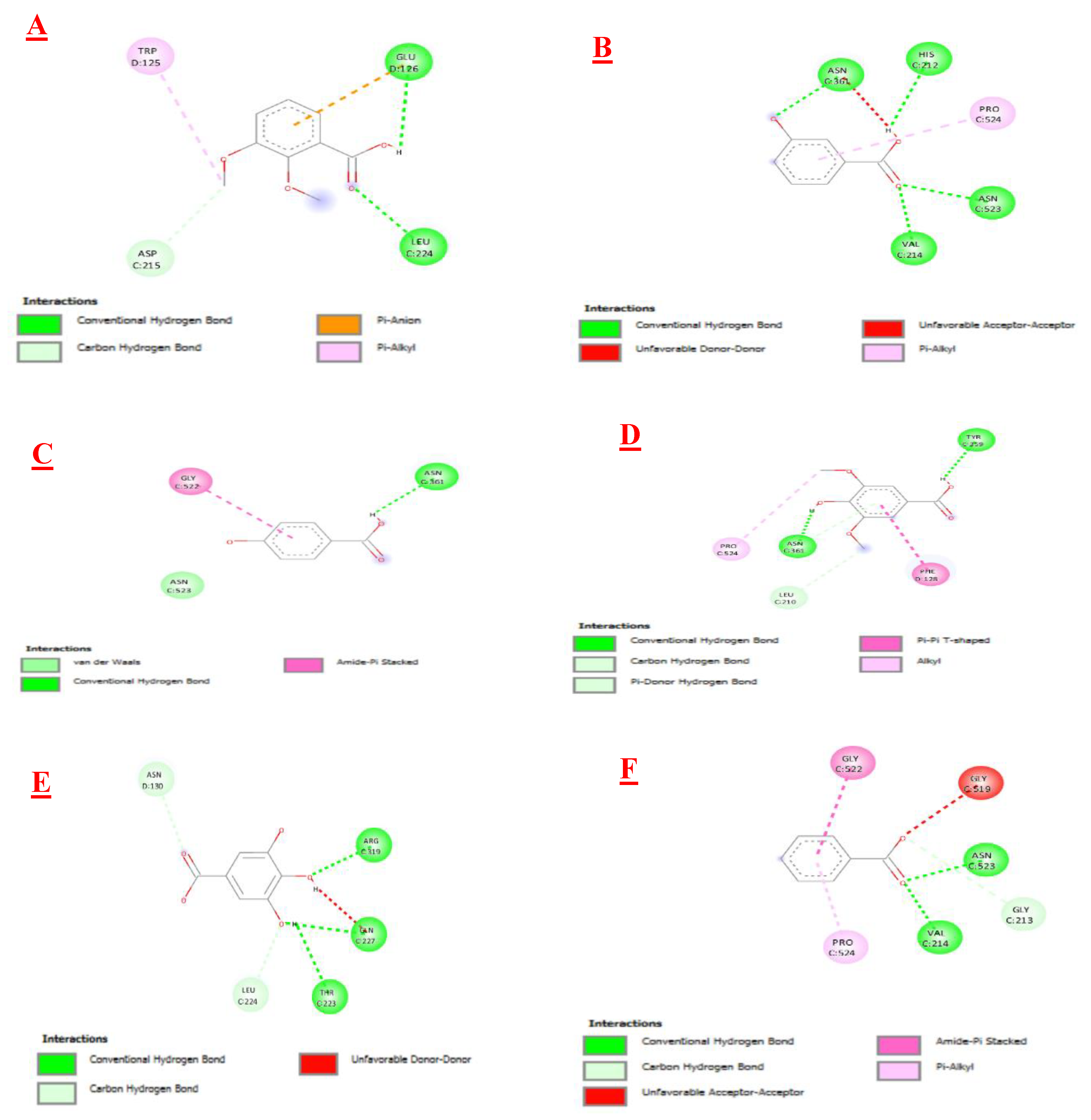
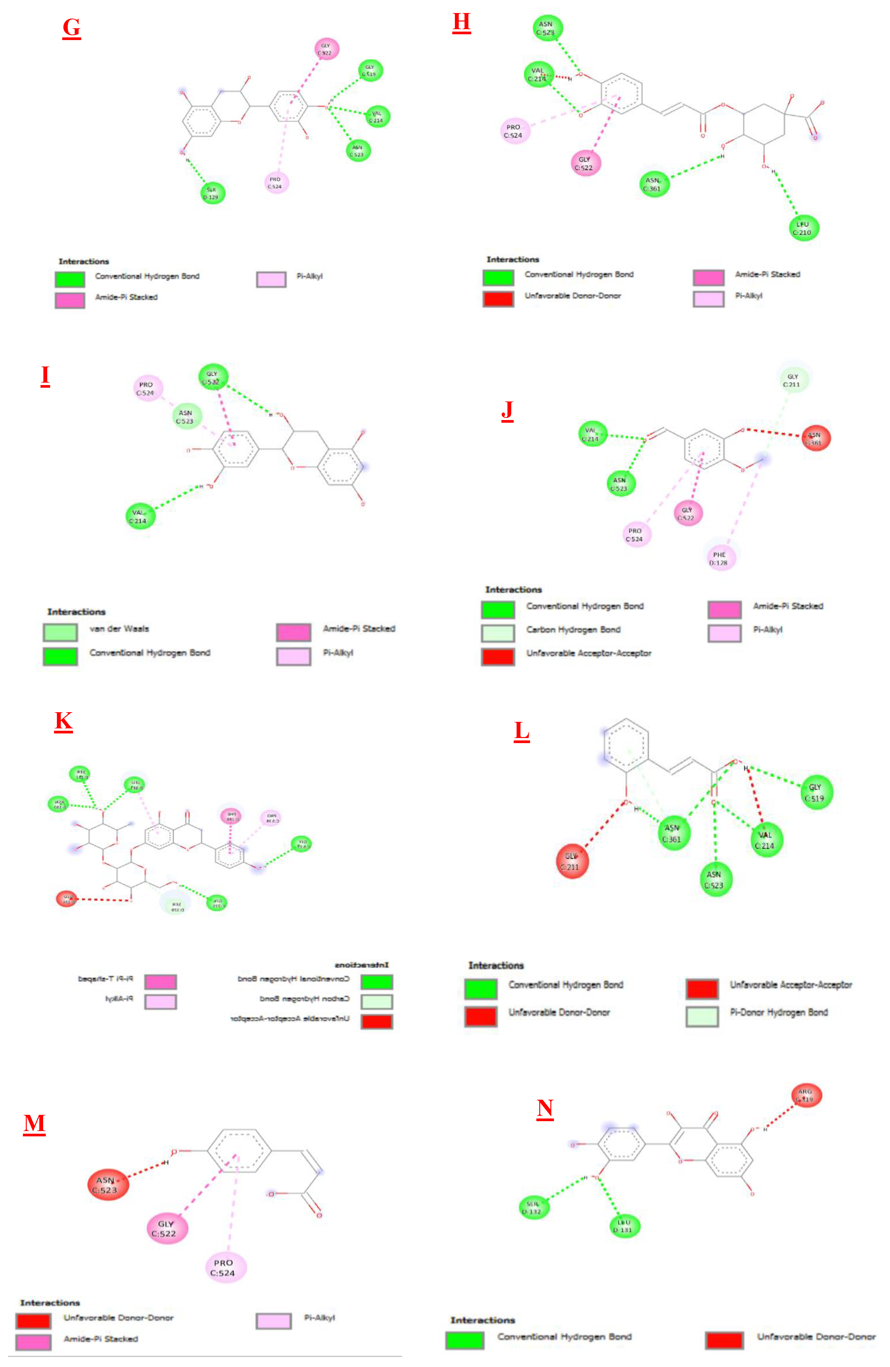
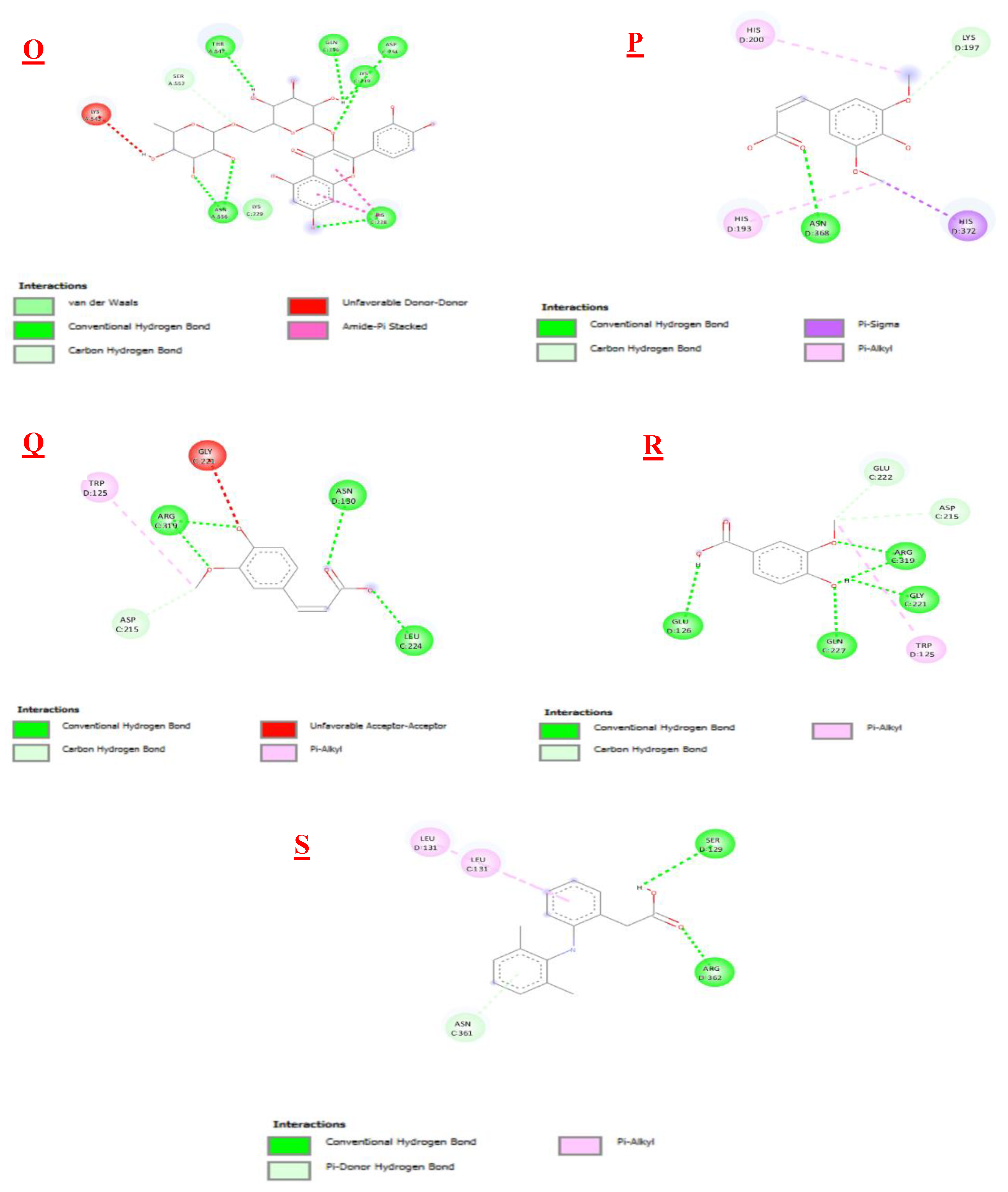
Figure 4.
3D detailed binding sites of each ligand into the receptor [PDB ID: 3LN0]. A: 2,3-dimethoxybenzoic acid; B: 3-hydroxybenzoic acid; C: 4-hydrobenzoic acid; D: syringic acid; E: gallic acid; F: benzoic acid; G: catechin; H: chlorogenic acid; I: epicatechin; J: isovanillin; K: naringin; L: o-coumaric acid; M: p-coumaric acid; N: quercetin; O: rutin; P: sinapinic acid; Q: t-ferulic acid; R: vanillic acid; S: dichlofenac.
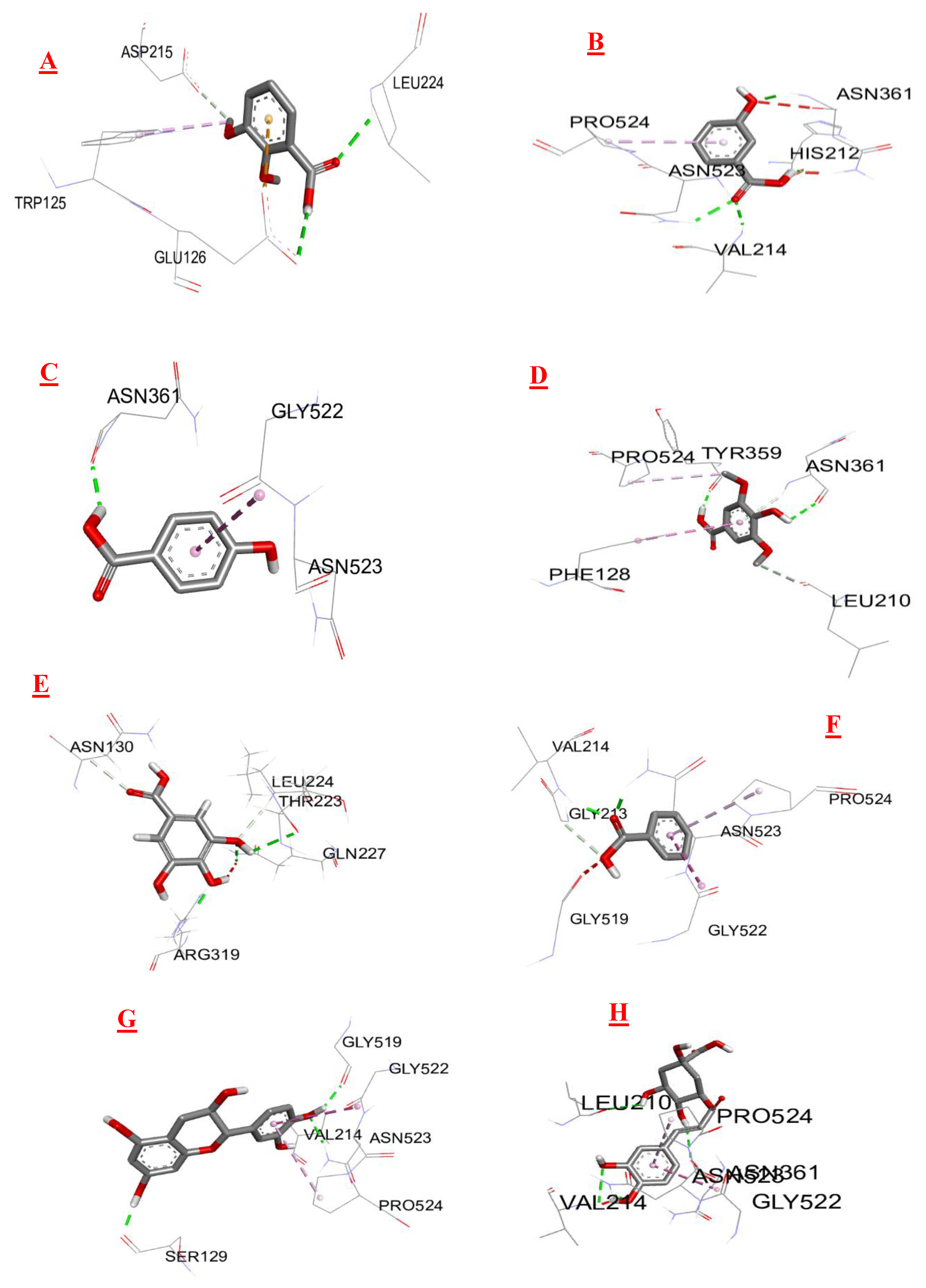
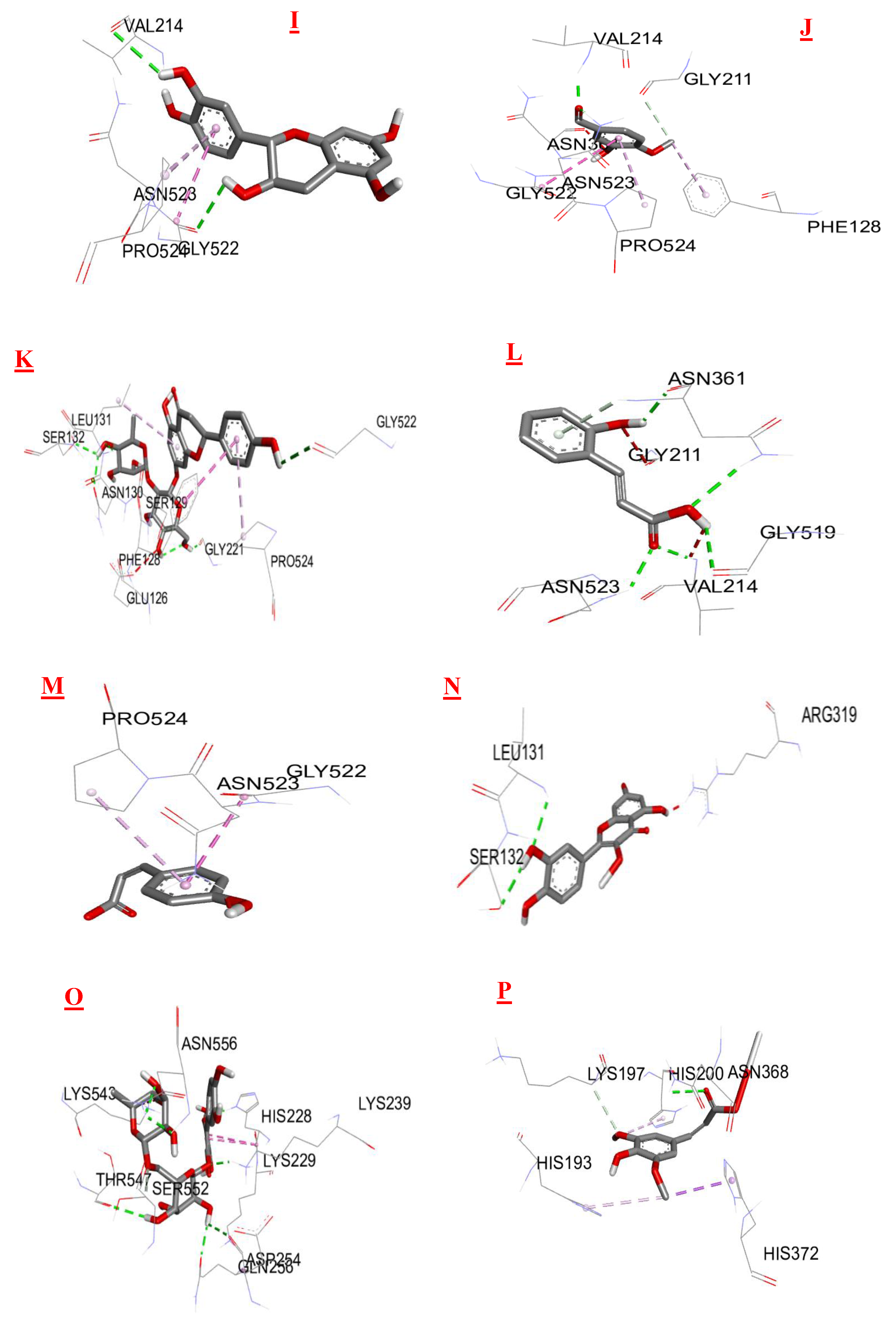
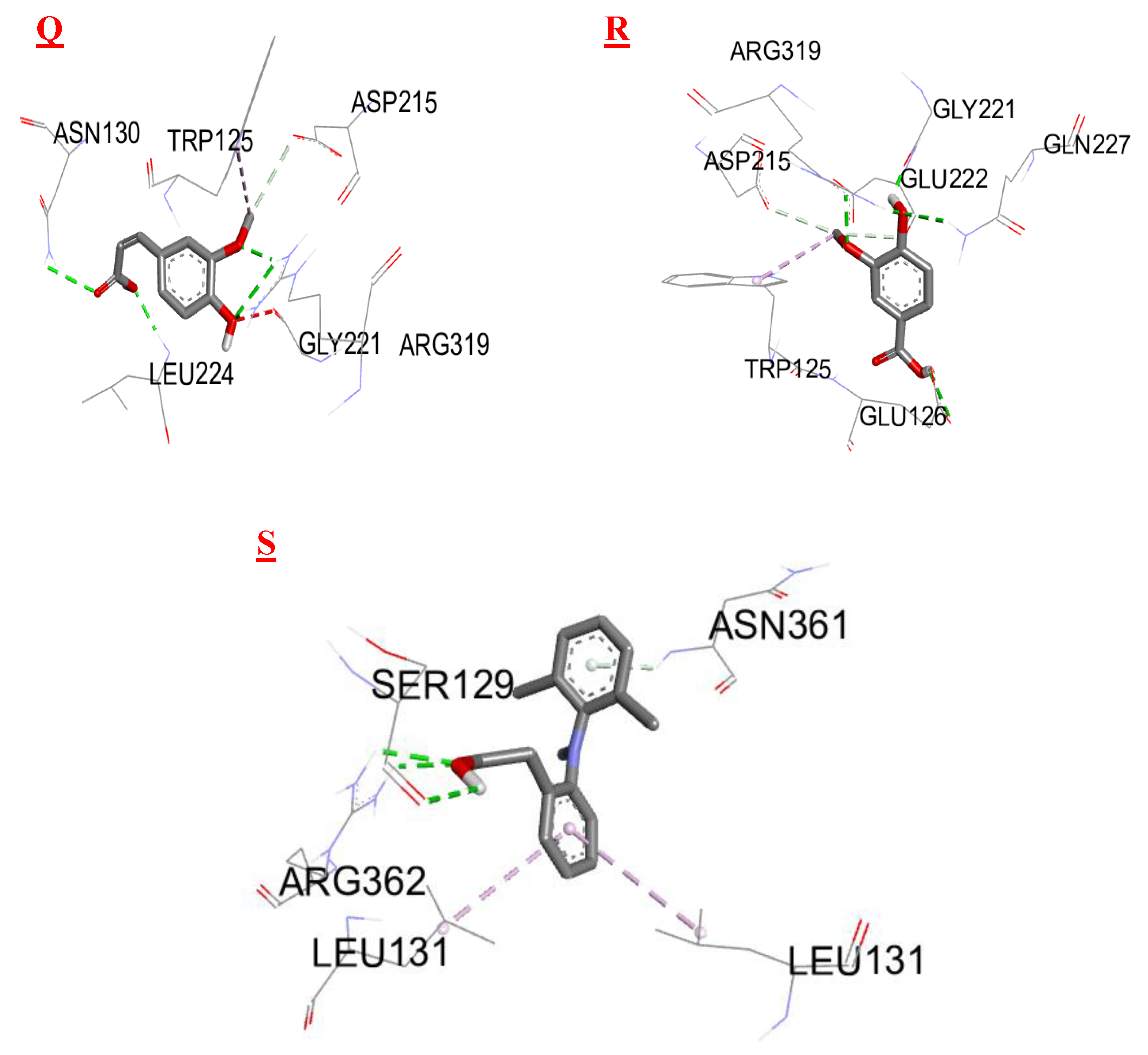

Table 1.
In vitro anti-inflammatory activity by HRBC method.
| 0,25 mg/ml | 0,5 mg/ml | 1 mg/ml | |
|---|---|---|---|
| MW 40°C | 77,18±0,17 *** | 84,92±0,30 *** | 89,28±0,20 *** |
| MW 60°C | 78,59±0,18 *** | 87,43±0,22*** | 90,67±0,10 *** |
| MW 80°C | 80,53±0,20 *** | 88,12±0,13 *** | 91,97±0,15 *** |
| MW 100°C | 77,06±3,04 *** | 88,99±0,31 *** | 94,94±0,25 *** |
| MW 120°C | 86,25±0,12 ns | 91,13±0,25 ns | 96,88±0,15 ns |
| MW 100°C, 5min | 76,92±0,20 *** | 85,09±0,22 *** | 88,64±0,18 *** |
| MW 100°C,15min | 88,96± 0,17 ns | 90,95±0,15 ns | 96,85±0,17 ns |
| Diclofenac | 89,91±0,17 | 91,91±0,2 | 96,67±0,15 |
Results are presented as mean ± SD (n=3); ns: no significant difference, *P<0.05, **P<0.01, ***P<0.001 compared with diclofenac.
Table 2.
Effect of microwave-assisted T. algeriensis extracts on carrageenan-induced paw edema in rats.
Table 2.
Effect of microwave-assisted T. algeriensis extracts on carrageenan-induced paw edema in rats.
| Mean increase in paw thickness (mm) ± SD | |||
|---|---|---|---|
| Group | 1h | 2h | 3h |
| Control | 1,92 ±0,04 | 1,74 ±0,09 | 1,49 ± 0,07 |
| Diclofenac | 1,48 ±0,04*** | 0,72 ± 0,04*** | 0,24 ± 0,04*** |
| MW 40°C | 1,45 ±0,04*** | 1,05 ±0,04*** | 0,73 ±0,01*** |
| MW 60°C | 1,28 ±0,02*** | 1 ±0,02*** | 0,49 ±0,04*** |
| MW 80°C | 1,41 ±0,04*** | 0,79 ±0,04*** | 0,49 ±0,05*** |
| MW 100°C | 1,4 ±0,03*** | 0,96 ±0,05*** | 0,53 ±0,02*** |
| MW 120°C | 1,36 ±0,02*** | 0,99 ±0,01*** | 0,46 ±0,04*** |
| MW 100°C 5min | 1,44 ±0,01*** | 1,17 ±0,02*** | 0,77 ±0,02*** |
| MW 100°C 15min | 1,37 ±0,04*** | 0,76 ±0,02*** | 0,32 ±0,02*** |
Results are presented as mean ± SD (n=3); ns: no significant difference, *P<0.05, **P<0.01, ***P<0.001 compared with control.
Table 3.
Results for affinity and inhibition constant of ligands with the same protein.
| Biomolecules | Anti-inflammatory activity | |
|---|---|---|
| Protein : COX-2 (3LN0) | ||
| ΔG (Kcal/mol1-) | Ki (µmol) | |
| 2,3- dimethoxybenzoic acid | -6,0 | 39,95 |
| 3-hydroxybenzoic acid | -6,1 | 33,74 |
| 4-hydroxybenzoic acid | -5,7 | 66,28 |
| Syringic acid | -5,9 | 47,29 |
| Gallic acid | -6,4 | 20,33 |
| Benzoic acid | -5,5 | 92,90 |
| Catechin | -9,2 | 0,18 |
| Chlorogenic acid | -8,5 | 0,58 |
| Epicatechin | -8,9 | 0,29 |
| Isovanillin | -5,9 | 47,29 |
| Naringin | -10,3 | 0,028 |
| o-coumaric acid | -6,3 | 24,07 |
| p-coumaric acid | -6,1 | 33,74 |
| Quercetin | -7,9 | 1,61 |
| Rutin | -8,8 | 0,35 |
| sinapinic acid | -5,8 | 55,99 |
| t-ferulic acid | -6,1 | 33,74 |
| Vannilic acid | -6,2 | 28,50 |
| Dichlofenac | -7,1 | 6,23 |
Disclaimer/Publisher’s Note: The statements, opinions and data contained in all publications are solely those of the individual author(s) and contributor(s) and not of MDPI and/or the editor(s). MDPI and/or the editor(s) disclaim responsibility for any injury to people or property resulting from any ideas, methods, instructions or products referred to in the content. |
© 2025 by the authors. Licensee MDPI, Basel, Switzerland. This article is an open access article distributed under the terms and conditions of the Creative Commons Attribution (CC BY) license (http://creativecommons.org/licenses/by/4.0/).
Copyright: This open access article is published under a Creative Commons CC BY 4.0 license, which permit the free download, distribution, and reuse, provided that the author and preprint are cited in any reuse.


