
Submitted:
21 March 2025
Posted:
24 March 2025
You are already at the latest version
Abstract
Yellow fever virus (YFV) is an endemic arbovirus in parts of Africa and the Americas. In Brazil, following the eradication of the urban transmission cycle, YFV is maintained in a sylvatic cycle involving several species of neotropical primates and mosquitoes of the genera Haemagogus and Sabethes, which serve as primary and secondary vectors, respectively. During the 2016–2019 outbreak in São Paulo State, a total of 3,731 mosquito pools were collected from sites with ongoing epizootic events in 192 municipalities. RT-qPCR analysis detected YFV in 46 pools (1.4%) across nine mosquito species, including both primary and secondary vectors, as well as species from the genera Aedes and Psorophora. Differences in viral loads were observed among species. While Aedes aegypti was not found to be positive, the detection of natural YFV infection in other Aedes species raises concerns about potential virus reurbanization. Further studies are needed to clarify the role of additional mosquito species in YFV transmission in Brazil.
Keywords:
yellow fever virus
; surveillance
; Brazil
; RT-qPCR
; vector transmission
1. Introduction
Yellow Fever (YF) is a severe disease caused by the arbovirus Orthoflavivirus flavi (former Yellow Fever Virus (YFV), a member of the Flaviviridae family, and remains a significant public health concern in parts of Africa and the Americas [1]. YF may cause significant morbidity and mortality rates in the human populations, as well as impact the neotropical primates population [2]. Despite the availability of the live attenuated 17-DD vaccine, a high case fatality rate (CFR) of 40% to 60% persists, particularly in South America [1,3]. In Brazil, after the eradication of the urban YFV cycle in 1942 transmitted by Aedes aegypti mosquitoes, YFV is maintained by a sylvatic transmission cycle involving several species of neotropical primates (NTPs) and forest canopy-dwelling mosquitoes, mainly Haemagogus-spp and Sabethes-spp, and human cases are caused by a spillover process in green areas [4]. YF surveillance is based on confirmation of epizootic events through virus detection by RT-qPCR and or immunohistochemistry in accordance with the Ministry of Health Guidelines [5].
Seasonal climatic variations significantly influence YFV transmission by affecting mosquito population dynamics and viral amplification. During the rainy season, abundant precipitation creates numerous larval habitats while elevated temperatures and high humidity accelerate mosquito development and viral replication, leading to surges in sylvatic vectors such as Haemagogus and Sabethes spp. These conditions enhance virus amplification among non-human primates and elevate the risk of spillover to humans [6]. Conversely, in the dry season, reduced rainfall limits breeding sites and diminishes vector densities, though desiccation-resistant eggs permit a low-level virus circulation that can rapidly rebound once rains return [7].
From mid-2016 until late 2018, Brazil faced one of the largest YF outbreaks in recent decades, mainly in the southeastern region [8,9,10,11]. São Paulo state, located in southeast Brazil, is the most densely populated state of the country, containing one of the world’s largest urban conurbations [12]. A total of 875 cases of YFV in NTPs between July 2016 and November 2019 and 624 cases of YFV in humans between January 2017 and 18th November 2019 were reported. This outbreak was caused by the 1E lineage belonging to South American I (SA-I) genotype that originated in the Amazon basin which has later disseminated from northern São Paulo into geographically neighboring areas of western MG and into the south of the state [14]. Some epizootic events in Callithrix monkeys were confirmed in large urbanized cities in proximity of urban green areas where Haemagogus and Sabethes mosquitoes were not found [15], indicating that synanthropic mosquitoes were likely involved in viral transmission in these areas. Considering that entomological investigation is a complementary tool to better understand eco-epidemiological aspects of YF after notification of suspected epizootic events, here we describe different Aedini and Sabethini mosquitoes found positive with YFV by RT-qPCR and its ecological factors, showing the continuous threat of reurbanization of YFV in Brazil.
2. Materials and Methods
2.1. Study Area
The study was conducted in the state of São Paulo, Brazil, which comprises 645 municipalities organized into 15 administrative regions. The state spans approximately 248,196.960 square kilometers and has a population of 44,749,699 inhabitants, primarily concentrated in the coastal region. São Paulo encompasses two distinct biomes: the Cerrado and the Atlantic Forest, both of which have suffered significant deforestation in recent years.
2.2. Epizootic Events and Mosquito Collection
Between November 2016 and June 2019, a total of 3,731 mosquito pools from the Aedini Tribe and Sabethes genus were collected in 192 municipalities with ongoing epizootic events and adjacent cities. Briefly, frozen carcasses of NTPs were sent to Adolfo Lutz Institute for YFV detection, according to the Brazilian Ministry of Health Guidelines as previously described [8]. Mosquitoes were captured at ground level between 9 a.m and 3 p.m using entomologic nets and bottle-type manual vacuums in forested and green areas, and Nasci Aspirator in urban dwellings. After sampling, mosquitoes were frozen, transferred to cryogenic tubes, and stored in liquid nitrogen containers for transport. Identification was performed based on morphological characteristics by the Pasteur Institute (formerly the Superintendence for Control of Endemic Diseases - SUCEN). The mosquitoes were subsequently sorted into pools containing 1 to 50 individuals per pool, according to species, collection date, and location. Molecular detection for YFV was carried out in non-engorged mosquitoes (n=3,376) at the local reference laboratory for arthropod-borne viruses at Instituto Adolfo Lutz (IAL) in São Paulo. Pools were triturated in FastPrep-24 5G Instrument (MP Biomedicals, Ohio, USA) and in Magna Lyser (Roche) in 1 mL of phosphate-buffered saline solution with 0.75% bovine albumin, penicillin (100 units/mL) and streptomycin (100 µg/mL). The resultant suspension was centrifuged at 1800×g for 15 min, and the supernatant was withdrawn and frozen at -70ºC until further use.
2.3. YFV RNA Detection and Statistical Analysis
Viral RNA was extracted using QIAamp Viral RNA Mini Kit following the manufacturer’s instructions (QIAGEN, Hilden, Germany). Detection of YFV RNA was performed using an RT-qPCR protocol [11]. Results with Cycle Threshold (CT) values ≥ 35 were retested. If the new result had a CT value ≤ 38, the pool was considered positive for YFV. The Kruskal-Wallis test was conducted exclusively among YFV-positive mosquito pools to evaluate differences in viral load by mosquito species, as indicated by their CT values. To assess differences in Yellow Fever Virus (YFV) viral loads among different mosquito species, a Generalized Linear Model (GLM) was performed with Ct value as the dependent variable. Ct values were used as a proxy for viral load, where lower Ct values indicate higher viral loads.
The primary YVF vector Hg. leucocelaenus was set as the reference category to compare viral loads across species. The model was specified as Ct ~ Species, where Ct value was assumed to follow a Gaussian (normal) distribution with an identity link function. The analysis reports estimated mean differences in Ct values (β coefficients) for each species compared to Hg. leucocelaenus. To evaluate whether seasonal variation (rainy vs. dry) influenced Ct values, season was included as an additional predictor in the GLM. An interaction term (Species × Season) was also tested to assess potential species-specific seasonal effects. All p-values <0.05 were considered significant. All analysis were performed using Rstudio v.2023.12.1, ggplot2 package [16].
3. Results
A total of 3,731 mosquito pools were collected during the outbreak (Table 1), of which 46 pools (1.4%) from 9 mosquitos species tested positive for yellow fever virus (YFV), representing 22 municipalities (8.7%) (Table 2). Additionally, epizootic events were confirmed by RT-qPCR in 82 cities (Supplementary Material 1). The Ct values of YFV-positive pools ranged from 16 to 38, with a median of 32 (Figure 1).
Among the species collected, Aedes scapularis accounted for 26.46% of all mosquitoes, with 0.67% of pools testing positive, followed by Aedes albopictus (21.66%, 0.41% positive) and Psorophora ferox (11.20%, 1.32% positive). Haemagogus leucocelaenus represented 8.09% of the total, with 5.83% of its pools testing positive, while Haemagogus janthinomys/capricornii comprised 3.4%, with 5.51% positive. Other species testing positive for YFV included Aedes serratus (5.72%, 2.07% positive), Sabethes albiprivus (2.67%, 15.78% positive), Sabethes purpureus (0.80%, 2.08% positive), and Sabethes identicus (0.74%, 1.75% positive).
Analysis of Ct values among YFV-positive mosquito pools revealed significant differences in viral loads (Figure 1). Haemagogus species consistently exhibited the lowest Ct values, indicating higher viral loads, while Sa. albiprivus, Ae. albopictus, Ae. serratus, and Ps. ferox had higher Ct values, suggesting lower viral loads. The distribution of Ct values varied across species, with some species displaying a wider range, indicating heterogeneity in infection levels within the same species. While Ae. scapularis pools generally showed high Ct values, two pools recorded Ct values of 25 and 28, suggesting moderate viral loads. The Kruskal-Wallis test confirmed a statistically significant difference in Ct values among species (p = 0.0002). These differences are visually represented in Figure 1.
The Generalized Linear Model (GLM) analysis identified significant differences in Ct values among mosquito species. Ha. leucocelaenus exhibited the lowest Ct values and was used as the reference species. Compared to Ha. leucocelaenus, Ha. janthinomys-capricornii showed a moderate increase in Ct values (β = 4.71, p = 0.039).
Mosquito species from the Aedes, Psorophora, and Sabethes genera exhibited significantly higher Ct values, indicating lower viral loads. Ae. scapularis had a β coefficient of 11.83 (p < 0.001), while Ps. ferox and Ae. albopictus showed β values of 13.80 and 15.67, respectively (p < 0.001). Among Sabethes species, Sa. purpureus, Sa. albiprivus, and Sa. identicus exhibited the highest Ct values (β = 14.00 to 16.00, p < 0.01).
These results indicate species-specific differences in YFV viral loads, with Haemagogus species displaying lower Ct values compared to other genera. A full summary of the GLM estimates is presented in Table 3. The effect of season (rainy vs. dry) on Ct values was not statistically significant (p = 0.173). The interaction between mosquito species and season also did not significantly influence Ct values (p > 0.3 for all species).
Out of the 46 positive mosquito pools, 24 (52.2%) were collected during the rainy season (18th October – 4th April), and 22 (47.8%) during the dry season (Supplementary Material 2). Aedes species were predominantly collected during the rainy season, whereas Haemagogus spp. and Psorophora ferox were mostly collected during the dry season. Notably, Sa. albiprivus and Sa. identicus tested positive exclusively in the dry season. All YFV-positive mosquito pools were collected within the Atlantic Forest biome (Supplementary Material 3).
4. Discussion
Brazil is an endemic country for YFV, with the Amazon region acting as a source for viral diversity and dipersal across the country. Although YFV circulation has been documented in southeastern Brazil since the early 21st century, the 2016-2018 outbreak caused by the SA-I genotype, particularly in São Paulo state, was unexpected due to the high number of positive cases reported in both humans and animals [14,17]. Notably, during this outbreak, nine different species of Culicidae, including mosquitoes from the Aedes, Psorophora and Sabethes genera, tested positive for YFV. All these mosquitos were collected in the Atlantic Forest biome, where Haemagogus leucocelaenus act as the primary vector [18,19,20]. While Sabethes spp. are traditionally considered secondary vectors limited information is available regarding their role in YFV transmission in this region.
Our findings confirm that YFV viral loads varied accross Culicidae species, with Hg.janthinomys/capricornii. and Hg. leucocelaenus having the highest viral loads. Notably, two pools of Ae. scapularis also had viral loads comparable to those of Hg.janthinomys/capricornii., indicating that this species may play a more relevant role in YFV transmission than previously thought. These pools were collected in Urupês on February 15, 2017, and in Araçatuba on November 25, 2016, at the municipal Zoo, yet neither location reported epizootic events at the time. The presence of YFV in these areas could be attributed by different susceptibility of NTP, as some Callithryx sp. may be less susceptible to the disease [2]. Ae. scapularis, which was the most abundant specie collected in this study, is considered a generalist in its use of habitats, occurring in both sylvatic and human-dominated areas. Adult females are opportunistic in their behaviour, feeding especially on mammals [21,22]. Considering the wide host breadth and feeding habitats, coupled with synanthropic adaptions, it is possible that Ae. scapularis may be an important bridge vector for human and animal viruses. Thus, our data suggest that this species may have played a secondary role in the YF outbreak.
Sabethes mosquitoes were observed in low abundance, distribution, and infection rates, suggesting a local or secondary role during the 2016–2018 outbreak in the Brazilian Southeastern region [23]. In our study, this genus accounted for 11.5% (n = 430 pools) of the collected mosquitoes, with 10% (n = 5) of positive pools, all exhibiting high Ct values, indicative of low viral loads. Similarly, during the 2009 YF outbreak in São Paulo, YFV was only isolated from a single pool of Hg. leucocelaenus in Buri, despite the collection of Sa. chloropterus, Sa. purpureus, and Sa. undosus in the same area [19]. However, the absence of RT-qPCR analysis in that study may explain the lack of positive detections among Sabethini mosquitoes. Conversely, during a YF epidemic and epizootic in Misiones, a northeastern province of Argentina, YFV was successfully isolated in cell culture from pools of Sabethes albiprivus [24]. This viral isolation indicates high viral loads, contrasting with the low viral loads observed in Sabethes specimens from the Atlantic Forest.
Sa. chloropterus has been identified as the primary YF vector during the dry season in the Cerrado biome of Minas Gerais [25]. In Espírito Santo, where the sylvatic YF cycle was first described in Brazil, Sa. chloropterus, Sa. soperi, Sa. identicus, Aedes aureolineatus, and Shannoniana fluviatilis were noted for their secondary roles in YFV transmission [26]. Additionally, Sa. albiprivus from Rio de Janeiro demonstrated high vector competence when inoculated with Brazilian YFV strains [27]. To better elucidate the role of Sabethes mosquitoes in the YF transmission cycle within São Paulo state, where the virus has now been established [28], additional studies are required.
Considering the Aedes genus, earlier studies suggested that Brazilian Ae. aegypti mosquitoes might not favor the establishment of an urban cycle of YF [29]. However, a more recent study demonstrated that both anthropophilic mosquitoes, Ae. aegypti and Ae. albopictus, are highly susceptible to American and African YFV strains [27]. In 2018, in Minas Gerais state, a single Ae. albopictus mosquito pool tested positive for YFV [30]. In our surveillance study, Ae. albopictus was the second most frequent species collected, accounting for 21.66% of the total, with three pools testing positive for YFV, all of which exhibited low viral loads. No Ae. aegypti mosquitoes were found positive. Despite the high number of human infections during the outbreak, no urban YF cases were reported. Given that YFV has demonstrated potential for adaptation to Ae. albopictus and can be transmitted between NTP [31,32] our findings underscore a potential threat to endemic areas in South America where these mosquitoes are present. With their widespread distribution and ecological plasticity, Ae. albopictus could serve as a bridge vector, facilitating virus transmission between urban environments and rural areas.
One objective of this study was to assess whether seasonal variation (rainy vs. dry) influenced YFV viral loads in mosquitoes. Despite previous reports showing seasonal peaks in mosquito abundance and transmission during rainy periods our results indicate that season was not a significant predictor of Ct values, suggesting that once a mosquito is infected, viral replication remains stable. Sacchetto and collaborators reported viral persistence during the non-epidemic dry season in NTP collected in Belo Horizonte, Minas Gerais state [34]. These results show the importance of continuous surveillance, regardless of seasonal variations.
Our study has some limitations. Specifically, our study involved triturating whole mosquitoes instead of processing solely the salivary gland. Additionally, the contents of the mosquitoes' digestive systems—whether engorged or not—were assessed solely through visual examination, and some of the positive results could came from a residual blood feeding. Nevertheless, the data obtained in the present study is relevant, as monitoring of virus circulation and characterizing vectors are fundamental elements for understanding the dynamics of vector-borne viruses, providing new insights for the establishement of control strategies and to prevent the risk of re-urbanization of YFV. More, new studies of vectorial competence, mainly in Ae. scapularis, are needed.
Supplementary Materials
The following supporting information can be downloaded at the website of this paper posted on Preprints.org, Figure S1: title; Table S1: title; Video S1: title.
Author Contributions
Manuscript preparation: MSC, GSC, LOV. Obtained funding and study supervision: MSC, ELLA; Mosquitoes collection and identification: RMT, RMTM, LFM, JTD, ESB. Experiments of viral detection: GSC, KB; Statical analysis: MSC, LOV. All authors reviewed, contributed to, and approved the final version of the manuscript.
Funding
This research was funded by Secretaria de Estado de Saúde de São Paulo (SES). GSC was sponsored by Fesima project (#GAPS/NATO 479/2020). KMBN was sponsored with a Fedial (Programa de Formação para Investigação Científica) scholarship from Instituto Adolfo Lutz.
Acknowledgments
we thank Pasteur team (former SUCEN) from São Paulo state for collecting mosquitoes. We also thank Mariza Pereira from Pasteur for her help with local authorities, and Elizabeth Kelvin, from Cuny Graduate School of Public Health for her suggestions.
Conflicts of Interest
The authors declare no conflicts of interest.
References
- Vasconcelos PFC, Monath TP. Yellow Fever Remains a Potential Threat to Public Health. Vector-Borne and Zoonotic Diseases 2016; 16: 566–567. [CrossRef]
- de Azevedo Fernandes NCC, Guerra JM, Díaz-Delgado J, et al. Differential Yellow Fever Susceptibility in New World Nonhuman Primates, Comparison with Humans, and Implications for Surveillance. Emerg Infect Dis 2021; 27: 47–56. [CrossRef]
- Monath TP, Vasconcelos PFC. Yellow fever. Journal of Clinical Virology 2015; 64: 160–173. [CrossRef]
- Vasconcelos PFC, Costa ZG, Travassos Da Rosa ES, et al. Epidemic of jungle yellow fever in Brazil, 2000: Implications of climatic alterations in disease spread. J Med Virol 2001; 65: 598–604. [CrossRef]
- Ministerio da Saúde. Brasília-DF 2017 GUIA DE VIGILÂNCIA DE EPIZOOTIAS EM PRIMATAS NÃO HUMANOS E ENTOMOLOGIA APLICADA À VIGILÂNCIA DA FEBRE AMARELA MINISTÉRIO DA SAÚDE 2 a edição atualizada. https://www.gov.br/saude/pt-br (2017).
- Li SL, Acosta AL, Hill SC, et al. Mapping environmental suitability of Haemagogus and Sabethes spp. mosquitoes to understand sylvatic transmission risk of yellow fever virus in Brazil. PLoS Negl Trop Dis 2022; 16: e0010019. [CrossRef]
- Silva-Inacio CL, Ximenes M de FF de M. Haemagogus spegazzinii Brèthes, 1912 (Diptera: Culicidae) in Brazilian semiarid: resistance in eggs and scale color variation in adults. Rev Bras Entomol; 65. Epub ahead of print 2021. [CrossRef]
- Cunha MS, da Costa AC, de Azevedo Fernandes NCC, et al. Epizootics due to Yellow Fever Virus in São Paulo State, Brazil: viral dissemination to new areas (2016–2017). Sci Rep 2019; 9: 5474. [CrossRef]
- Fernandes NCC de A, Cunha MS, Guerra JM, et al. Outbreak of Yellow Fever among Nonhuman Primates, Espirito Santo, Brazil, 2017. Emerg Infect Dis 2017; 23: 2038–2041. [CrossRef]
- Mello M De, Rezende D, Adelino R, et al. Persistence of Yellow fever virus outside the Amazon Basin , causing epidemics in Southeast Brazil , from 2016 to 2018. 2018; 1–12. [CrossRef]
- Moreira-Soto A, Torres MC, Lima de Mendonça MC, et al. Evidence for multiple sylvatic transmission cycles during the 2016–2017 yellow fever virus outbreak, Brazil. Clinical Microbiology and Infection 2018; 24: 1019.e1-1019.e4. [CrossRef]
- Instituto Brasileiro de Geografia e Estatística. Sao Paulo, https://www.ibge.gov.br/cidades-e-estados/sp/sao-paulo.html (2022, accessed 5 March 2025).
- Faria NR, Kraemer MUG, Hill SC, et al. Genomic and epidemiological monitoring of yellow fever virus transmission potential. Science (1979) 2018; 361: 894–899. [CrossRef]
- Hill SC, de Souza R, Thézé J, et al. Genomic Surveillance of Yellow Fever Virus Epizootic in São Paulo, Brazil, 2016 – 2018. PLoS Pathog 2020; 16: e1008699. [CrossRef]
- Cunha MS, Tubaki RM, de Menezes RMT, et al. Possible non-sylvatic transmission of yellow fever between non-human primates in São Paulo city, Brazil, 2017–2018. Sci Rep 2020; 10: 15751. [CrossRef]
- Wickham H. ggplot2: Elegant Graphics for Data Analysis.
- Figueiredo PDO, Gabriella A, Costa GB, et al. Re-Emergence of Yellow Fever in Brazil during 2016–2019: Challenges, Lessons Learned, and Perspectives. [CrossRef]
- Abreu FVS de, Ribeiro IP, Ferreira-de-Brito A, et al. Haemagogus leucocelaenus and Haemagogus janthinomys are the primary vectors in the major yellow fever outbreak in Brazil, 2016–2018. Emerg Microbes Infect 2019; 8: 218–231. [CrossRef]
- Souza RP de, Petrella S, Coimbra TLM, et al. Isolation of yellow fever virus (YFV) from naturally infectied Haemagogus (Conopostegus) leucocelaenus (diptera, cukicudae) in São Paulo State, Brazil, 2009. Rev Inst Med Trop Sao Paulo 2011; 53: 133–139. [CrossRef]
- Vasconcelos PFC, Sperb AF, Monteiro HAO, et al. Isolations of yellow fever virus from Haemagogus leucocelaenus in Rio Grande do Sul State, Brazil. Trans R Soc Trop Med Hyg 2003; 97: 60–62. [CrossRef]
- de Carvalho GC, dos Santos Malafronte R, Miti Izumisawa C, et al. Blood meal sources of mosquitoes captured in municipal parks in São Paulo, Brazil. Journal of Vector Ecology 2014; 39: 146–152. [CrossRef]
- Forattini OP, Gomes A de C, Natal D, et al. Preferências alimentares e domiciliação de mosquitos Culicidae no Vale do Ribeira, São Paulo, Brasil, com especial referência a Aedes scapularis e a Culex (Melanoconion). Rev Saude Publica 1989; 23: 9–19. [CrossRef]
- Abreu FVS de, Ribeiro IP, Ferreira-de-Brito A, et al. Haemagogus leucocelaenus and Haemagogus janthinomys are the primary vectors in the major yellow fever outbreak in Brazil, 2016–2018. Emerg Microbes Infect 2019; 8: 218–231. [CrossRef]
- Goenaga S, Fabbri C, Dueñas JCR, et al. Isolation of Yellow Fever Virus from Mosquitoes in Misiones Province, Argentina. Vector-Borne and Zoonotic Diseases 2012; 12: 986–993. [CrossRef]
- de Oliveira CH, Andrade MS, Campos FS, et al. Yellow Fever Virus Maintained by Sabethes Mosquitoes during the Dry Season in Cerrado, a Semiarid Region of Brazil, in 2021. Viruses 2023; 15: 757. [CrossRef]
- Stanzani LM de A, Motta M de A, Erbisti RS, et al. Back to Where It Was First Described: Vectors of Sylvatic Yellow Fever Transmission in the 2017 Outbreak in Espírito Santo, Brazil. Viruses 2022; 14: 2805. [CrossRef]
- Couto-lima D, Madec Y, Bersot MI, et al. Potential risk of re-emergence of urban transmission of Yellow Fever virus in Brazil facilitated by competent Aedes populations. 2017; 1–12. [CrossRef]
- Fernandes NCC de A, Cunha MS, Suarez PEN, et al. Phylogenetic analysis reveals a new introduction of Yellow Fever virus in São Paulo State, Brazil, 2023. Acta Trop 2024; 251: 107110.
- Lourenço-de-Oliveira R, Vazeille M, Filippis AMB de, et al. Oral Susceptibility to Yellow Fever Virus of Aedes aegypti from Brazil. Mem Inst Oswaldo Cruz 2002; 97: 437–439. [CrossRef]
- Laurindo Barbosa G, Sterlino Bergo E, Pereira M, et al. Presença de Aedes aegypti e Aedes albopictus em ambientes urbanos adjacentes às áreas silvestres que apresentam potencial para a circulação do vírus da febre amarela no estado de São Paulo. BEPA Boletim Epidemiológico Paulista 2022; 16: 25–30. [CrossRef]
- Damasceno-Caldeira R, Nunes-Neto JP, Aragão CF, et al. Vector Competence of Aedes albopictus for Yellow Fever Virus: Risk of Reemergence of Urban Yellow Fever in Brazil. Viruses 2023; 15: 1019. [CrossRef]
- Amraoui F, Pain A, Piorkowski G, et al. Experimental Adaptation of the Yellow Fever Virus to the Mosquito Aedes albopictus and Potential risk of urban epidemics in Brazil, South America. Sci Rep 2018; 8: 14337. [CrossRef]
- Hamlet A, Jean K, Perea W, et al. The seasonal influence of climate and environment on yellow fever transmission across Africa. PLoS Negl Trop Dis 2018; 12: e0006284. [CrossRef]
- Sacchetto L, Silva NIO, Rezende IM de, et al. Neighbor danger: Yellow fever virus epizootics in urban and urban-rural transition areas of Minas Gerais state, during 2017-2018 yellow fever outbreaks in Brazil. PLoS Negl Trop Dis 2020; 14: e0008658. [CrossRef]
Figure 1.
Boxplot of YFV Ct values for different Culicidae species.
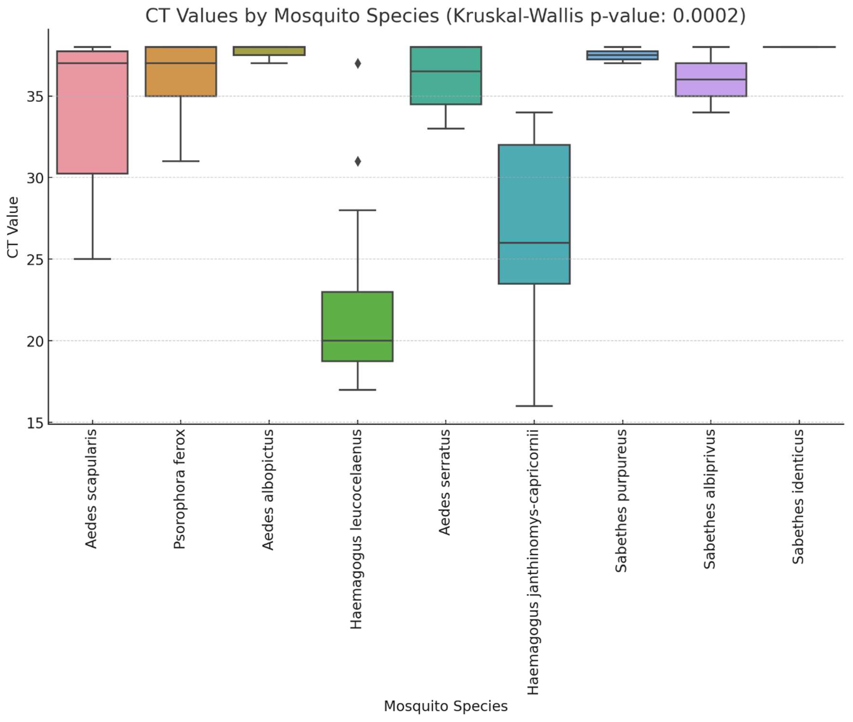

Table 1.
Culicidae pools tested for YFV.
| Species | N | % | Positive | %_Pos |
|---|---|---|---|---|
| Aedes scapularis | 893 | 26.46 | 6 | 0.67 |
| Aedes albopictus | 731 | 21.66 | 3 | 0.41 |
| Psorophora ferox | 378 | 11.20 | 5 | 1.32 |
| Haemagogus leucocelaenus | 274 | 8.09 | 16 | 5.83 |
| Aedes serratus | 193 | 5.72 | 4 | 2.07 |
| Aedes aegypti | 148 | 4.39 | 0 | 0 |
| Haemagogus janthinomys/capricornii | 127 | 3.4 | 7 | 5.51 |
| Sabethes purpureus | 96 | 2.84 | 2 | 2.08 |
| Sabethes glaucodaemon | 94 | 2.79 | 0 | 0 |
| Aedes terrens | 72 | 2.13 | 0 | 0 |
| Sabethes identicus | 57 | 1.69 | 1 | 1.75 |
| Sabethes albiprivus | 47 | 1.39 | 2 | 4.26 |
| Sabethes imperfectus | 35 | 1.04 | 0 | 0 |
| Psorophora albigenu | 31 | 0.83 | 0 | 0 |
| Sabethes intermedius | 28 | 0.83 | 0 | 0 |
| Psorophora albipes | 27 | 0.80 | 0 | 0 |
| Psorophora (Jan.) sp | 17 | 0.50 | 0 | 0 |
| Sabethes belisarioi | 16 | 0.47 | 0 | 0 |
| Aedes argyrothorax | 13 | 0.39 | 0 | 0 |
| Psorophora sp | 11 | 0.33 | 0 | 0 |
| Sabethes chloropterus | 11 | 0.33 | 0 | 0 |
| Sabethes sp | 11 | 0.30 | 0 | 0 |
| Aedes sp | 9 | 0.27 | 0 | 0 |
| Sabethes undosus | 9 | 0.27 | 0 | 0 |
| Sabethes tridentatus | 8 | 0.24 | 0 | 0 |
| Psorophora lutzii | 6 | 0.18 | 0 | 0 |
| Sabethes soperi | 6 | 0.18 | 0 | 0 |
| Sabethes whitmani | 6 | 0.18 | 0 | 0 |
| Howardina fulvithorax | 6 | 0.18 | 0 | 0 |
| Culex sp. | 4 | 0.12 | 0 | 0 |
| Sabethes undosus aff. | 3 | 0.09 | 0 | 0 |
| Aedes fluviatilis | 2 | 0.06 | 0 | 0 |
| Culex quinquefaciatus | 2 | 0.06 | 0 | 0 |
| Limatus sp. | 1 | 0.03 | 0 | 0 |
| Psorophora lanei | 1 | 0.03 | 0 | 0 |
| Sabethes belisarioi aff. | 1 | 0.03 | 0 | 0 |
| Sabethes petrochiae | 1 | 0.03 | 0 | 0 |
| Sabethes shannoni | 1 | 0.03 | 0 | 0 |
Table 2.
YFV positive mosquitoes collected in São Paulo State, 2016-2018.
| Pool number |
Species | Local | CT_value | Date | Season |
|---|---|---|---|---|---|
| 443 | Aedes scapularis | Urupes | 25 | 26/11/2016 | Rainy |
| 465 | Psorophora ferox | Pontalinda | 38 | 21/08/2018 | Dry |
| 732 | Aedes albopictus | Jundiaí | 38 | 28/08/2018 | Dry |
| 1415 | Aedes scapularis | Araçatuba | 28 | 25/11/2016 | Rainy |
| 2152 | Haemagogus leucocelaenus | Caieras | 23 | 16/04/2019 | Dry |
| 2163 | Haemagogus leucocelaenus | Guarulhos | 21 | 14/12/2018 | Rainy |
| 2198 | Aedes serratus | Jarinu | 33 | 12/02/2019 | Rainy |
| 2322 | Haemagogus leucocelaenus | Jarinu | 37 | 03/05/2018 | Dry |
| 2348 | Aedes scapularis | Sao Paulo | 37 | 19/02/2018 | Rainy |
| 2377 | Haemagogus leucocelaenus | Jarinu | 20 | 30/01/2018 | Rainy |
| 2438 | Haemagogus janthinomys-capricornii | Mairipora | 33 | 23/01/2018 | Rainy |
| 2572 | Haemagogus janthinomys-capricornii | Valinhos | 31 | 04/09/2018 | Dry |
| 2577 | Haemagogus janthinomys-capricornii | Valinhos | 34 | 17/09/2018 | Dry |
| 3268 | Haemagogus leucocelaenus | Sao Paulo | 19 | 20/12/2017 | Rainy |
| 3269 | Haemagogus leucocelaenus | Sao Paulo | 18 | 20/12/2017 | Rainy |
| 3318 | Haemagogus leucocelaenus | Sao José dos Campos | 28 | 10/10/2018 | Dry |
| 3491 | Sabethes purpureus | Sao Miguel Arcanjo | 37 | 04/09/2018 | Dry |
| 3514 | Haemagogus leucocelaenus | Piedade | 31 | 10/10/2018 | Dry |
| 3521 | Haemagogus leucocelaenus | Jacarei | 20 | 04/09/2018 | Dry |
| 3530 | Haemagogus leucocelaenus | Jacarei | 18 | 28/05/2018 | Dry |
| 3541 | Haemagogus leucocelaenus | Igarata | 19 | 28/05/2018 | Dry |
| 3542 | Sabethes albiprivus | Igarata | 38 | 28/05/2018 | Dry |
| 3543 | Sabethes identicus | Igarata | 38 | 28/05/2018 | Dry |
| 3551 | Haemagogus janthinomys-capricornii | Igarata | 16 | 23/05/2018 | Dry |
| 3552 | Haemagogus leucocelaenus | Igarata | 20 | 23/05/2018 | Dry |
| 3687 | Haemagogus leucocelaenus | Sao José dos Campos | 23 | 04/07/2018 | Dry |
| 3689 | Haemagogus janthinomys-capricornii | Sao José dos Campos | 25 | 04/07/2018 | Dry |
| 3766 | Haemagogus janthinomys-capricornii | Caçapava | 22 | 25/06/2018 | Dry |
| 3769 | Sabethes albiprivus | Caçapava | 34 | 25/06/2018 | Dry |
| 3777 | Aedes albopictus | Itariri | 38 | 16/01/2019 | Rainy |
| 4188 | Psorophora ferox | Jacupiranga | 31 | 10/12/2018 | Rainy |
| 4231 | Aedes albopictus | Pereira Barreto | 37 | 16/01/2019 | Rainy |
| 4232 | Aedes scapularis | Pereira Barreto | 37 | 16/01/2019 | Rainy |
| 4233 | Aedes serratus | Pereira Barreto | 38 | 16/01/2019 | Rainy |
| 4234 | Aedes scapularis | Pereira Barreto | 38 | 16/01/2019 | Rainy |
| 4238 | Aedes scapularis | Sao Paulo | 38 | 16/01/2019 | Rainy |
| 4272 | Haemagogus leucocelaenus | Monteiro Lobato | 17 | 16/01/2019 | Rainy |
| 4273 | Psorophora ferox | Monteiro Lobato | 37 | 16/01/2019 | Rainy |
| 4275 | Haemagogus leucocelaenus | Monteiro Lobato | 17 | 16/01/2019 | Rainy |
| 4276 | Aedes serratus | Monteiro Lobato | 35 | 16/01/2019 | Rainy |
| 4279 | Sabethes purpureus | Monteiro Lobato | 38 | 16/01/2019 | Rainy |
| 4297 | Haemagogus leucocelaenus | Monteiro Lobato | 21 | 25/03/2019 | Rainy |
| 4298 | Haemagogus janthinomys-capricornii | Monteiro Lobato | 26 | 25/03/2019 | Rainy |
| 4449 | Aedes serratus | Iguape | 38 | 25/03/2019 | Rainy |
| 4568 | Psorophora ferox | Sarapui | 38 | 21/05/2018 | Dry |
| 5077 | Psorophora ferox | Iporanga | 35 | 25/04/2019 | Dry |
Table 3.
GLM statistics for YFV positive mosquitoes by species.
| Species | Estimate (β) | SE | 95% CI | p-value |
|---|---|---|---|---|
| Intercept (Hg. leucocelaenus) | 22 | 1.26 | (19.53, 24.47) | <0.001 |
| Aedes scapularis | 11.83 | 2.41 | (7.11, 16.56) | <0.001 |
| Psorophora ferox | 13.8 | 2.58 | (8.74, 18.86) | <0.001 |
| Aedes albopictus | 15.67 | 3.17 | (9.46, 21.88) | <0.001 |
| Aedes serratus | 14 | 2.81 | (8.48, 19.52) | <0.001 |
| Haemagogusjanthinomys/capricornii | 4.71 | 2.28 | (0.24, 9.19) | 0.039 |
| Sabethes purpureus | 15.5 | 3.78 | (8.10, 22.90) | <0.001 |
| Sabethes albiprivus | 14 | 3.78 | (6.60, 21.40) | <0.001 |
| Sabethes identicus | 16 | 5.19 | (5.83, 26.17) | 0.002 |
Disclaimer/Publisher’s Note: The statements, opinions and data contained in all publications are solely those of the individual author(s) and contributor(s) and not of MDPI and/or the editor(s). MDPI and/or the editor(s) disclaim responsibility for any injury to people or property resulting from any ideas, methods, instructions or products referred to in the content. |
© 2025 by the authors. Licensee MDPI, Basel, Switzerland. This article is an open access article distributed under the terms and conditions of the Creative Commons Attribution (CC BY) license (http://creativecommons.org/licenses/by/4.0/).
Copyright: This open access article is published under a Creative Commons CC BY 4.0 license, which permit the free download, distribution, and reuse, provided that the author and preprint are cited in any reuse.


