Submitted:
11 March 2025
Posted:
14 March 2025
You are already at the latest version
Abstract
The intraosseous environment is a dynamic and intricate system integral to bone health, encompassing vascular, cellular, and biochemical interactions that drive key processes such as hematopoiesis, bone remodeling, and systemic mineral regulation. This review examines the structural composition of the bone matrix, the diverse cellular landscape, and the interconnected vascular and nervous networks, highlighting their roles in preserving bone function and responding to pathological changes. Recent studies reveal regulatory mechanisms involving oxygen tension, ionic balance, signaling molecules, and mechanotransduction pathways that shape bone metabolism and its adaptation to mechanical forces. Insights into the bone microenvironment’s metabolic shifts in cancer and its interaction with inflammation underscore its pivotal role in disease progression and therapeutic innovation. Additionally, advances in imaging techniques and biomaterials fuel progress in bone regeneration and understanding its microenvironment. Exploring the intricate physiochemical dynamics and regulatory networks within the intraosseous system unlocks potential clinical applications in bone diseases, tissue engineering, and systemic metabolic disorders. This comprehensive review bridges fundamental science with translational research, offering insights into the complex yet essential intraosseous environment.
Keywords:
Intraosseous therapy
; Hematopoiesis
; Regeneration
; Bone marrow environment
; Mesenchymal stem cells
; Mechanotransduction
1. Introduction
The intraosseous environment refers to the internal space within bones, encompassing the bone marrow, blood vessels, and the specialized microenvironment that supports bone cells like osteoblasts, osteoclasts, and osteocytes. In medical emergencies, intraosseous access is often used to deliver fluids and medications directly into the bone marrow, offering a rapid pathway to the bloodstream. This technique can save valuable time for critically ill patients, significantly reducing delays in medication delivery. In prehospital settings, traditional manual intraosseous infusion devices have shown higher success rates and faster insertion times compared to semi-automatic devices.
Beyond its medical applications, the intraosseous environment is vital for maintaining skeletal health and function. Far from being a passive structure, it actively supports key processes such as bone remodeling and blood cell production (hematopoiesis). Bone tissue is richly supplied with blood vessels, enabling the exchange of nutrients, hormones, and waste. increase studies highlight the critical relationship between vascular health and bone density, showing how impaired blood flow can weaken bones and increase the risk of fractures. This intricate system is essential for bone integrity and overall physiological health [1]. Additionally, the presence of pain associated with metastatic disease in the bone highlights the critical role of the intraosseous environment in cancer-related conditions. Investigating the peripheral mechanisms influencing intraosseous pathways, particularly the role of bone marrow afferent neurons, offers valuable insights into pain management. Understanding these pathways could lead to innovative therapeutic approaches, aiming to alleviate pain and improve the quality of life for patients dealing with bone metastases [2]. The intraosseous environment must be recognized as a dynamic system essential to both skeletal physiology and clinical outcomes. Research indicates that the bone microenvironment is integral to bone regeneration, maintaining homeostasis, immune responses, and cancer progression. Bone serves as a supportive microenvironment for diverse cell types that collectively orchestrate vital skeletal functions, including energy metabolism, mineral regulation, bone formation, and blood cell production.
Endothelial cells create a complex vascular network within the bone, sustaining and organizing distinct microenvironments. Recent discoveries of vascular heterogeneity within the bone marrow highlight the presence of multiple specialized vascular niches. These niches, defined by unique combinations of cells and signaling factors, provide specific regulatory cues that mediate critical physiological functions. This intricate interplay underscores the importance of the intraosseous environment in maintaining bone health and responding to systemic and local challenges [3]. Glycosaminoglycans play a pivotal role in shaping the bone microenvironment by interacting with key mediators of various signaling pathways. These interactions can significantly influence the activity and function of bone-remodeling cells, such as osteoblasts, osteoclasts, and osteocytes. By modulating these pathways, glycosaminoglycans contribute to the regulation of bone remodeling, impacting processes like bone formation, resorption, and overall skeletal homeostasis[4]. Bone provides a unique metabolic microenvironment, home to highly energy-intensive processes such as bone resorption and bone formation, which are dysregulated in the presence of cancer. Approaches such as metabolomics demonstrate metabolic plasticity in patients with advanced diseases. Metabolic crosstalk between tumor cells and the surrounding stroma supports disease pathogenesis. Increasing evidence suggests that metabolic reprogramming within the tumor-bone microenvironment plays a key role in driving disease progression. Furthermore, understanding these metabolic adaptations will reveal new therapeutic targets and approaches [2].
The intraosseous environment is essential for understanding key physiological processes that impact health and disease. Found within the bone marrow and surrounding bone cortex, these specialized spaces play a central role in blood cell production (hematopoiesis) and the storage of critical resources like calcium and phosphorus. Recent advances in medical research have shed light on the intricate interactions between cellular activity, regulatory systems, and external factors that shape these environments, sparking new questions about their role in overall body physiology. This review explores the core factors influencing the intraosseous environment, including oxygen levels, nutrient availability, and signaling molecules. By examining these elements, we can gain deeper insight into their contributions to bone health and systemic functions, paving the way for a more thorough understanding of the importance of the intraosseous environment [5].
2. Structural Composition of the Intraosseous Environment
The intraosseous environment is a critical hub for processes like blood cell production (hematopoiesis), bone remodeling, and pain regulation. A major factor shaping this environment is the composition of bone marrow, which includes both hematopoietic and mesenchymal stem cells. Recent research has revealed changes in gene expression related to pain pathways in the peripheral nervous system, highlighting the role these cells play in managing pain within the bone. Molecular signaling further adds to this complexity, with osteoclasts being closely linked to bone pain. Interestingly, the balance of osteoclast activity appears finely tuned only when their activity surpasses a certain threshold triggering inflammation and pain. This delicate regulation underscores the intricate dynamics of the intraosseous environment. Gaining a deeper understanding of these factors is crucial for designing targeted therapies for conditions like osteoarthritis and cancer-related bone pain, ultimately improving patient outcomes [6].
2.1. Bone Matrix Components
The bone matrix is made up of several key components, including organic proteins (primarily type I collagen), inorganic minerals like hydroxyapatite, and a variety of extracellular matrix proteins that help regulate bone remodeling and regeneration. Bone mineralization is a complex process that begins with calcium phosphate-loaded vesicles inside cells, which are thought to serve as precursors for the formation of carbonated hydroxyapatite. This intricate system ensures the strength, structure, and adaptability of bones [1]. Bone minerals are connected to the organic matrix through protein-bound phosphate bonds [7]. These bonds are an integral part of both the structural organic matrix and the inorganic calcium phosphate crystals, creating a strong and cohesive framework that supports bone strength and function. The cellular composition of two microenvironments ( spongy bone and compact bone) are different in Figure 1. Despite extensive research, some aspects of the composition and structure of mature bone mineral particles remain unclear. These particles are often described as calcium-deficient and hydroxyl-deficient carbonated hydroxyapatite, with some of the PO₄³⁻ lattice sites occupied by HPO₄²⁻ ions. Using advanced solid-state nuclear magnetic resonance techniques, researchers have closely examined the hydrogen-bearing components in bone minerals, particularly focusing on the presence of HPO₄²⁻ ions, to gain deeper insights into their role in bone structure [3].
2.2. Organic Phase (Collagen and Non-Collagenous Proteins).
Bone consists of a specialized calcified extracellular matrix, which is a primary source of connective tissue components in the body. The organic matrix is predominantly composed of type I collagen (90%), which is distinct from type I collagen in other connective tissues due to unique post-translational modifications. The remaining 10% of the organic matrix is composed of non-collagenous proteins, which have been extensively studied over the past 15 years, aided by advancements in protein biochemistry and molecular biology, as reviewed by Gehron-Robey (1989).
Through the use of dissociating agents (e.g., EDTA) and non-dissociating buffers, researchers have been able to differentiate the mineral phase (hydroxyapatite crystals) from the organic phase (collagenous matrix) and analyze the distribution of proteins between these phases. The proteins in the bone matrix are classified as either exogenous or endogenous. Exogenous proteins are synthesized in other organs, circulate in the blood and tissue fluids, and become incorporated into the bone matrix due to their affinity for hydroxyapatite. Endogenous proteins, produced by osteoblasts, are directly integrated into the three-dimensional structure of the bone matrix during its formation. These bone matrix proteins are of significant interest due to their critical roles in regulating various aspects of bone physiology and remodeling [4].
2.3. Cellular Landscape
The cellular composition of bones includes several key cell types, each contributing uniquely to bone formation, remodeling, and maintenance. These include osteoblasts, osteoclasts, osteocytes, and bone marrow stromal cells. Osteoblasts are specialized cells responsible for synthesizing the mineralized matrix of bones. They arise from multiple sources, such as chondrocytes within the growth plate, bone marrow stromal cells, quiescent bone lining cells, and specific fibroblasts in craniofacial regions. With a finite lifespan, osteoblasts require continuous renewal by preosteoblasts, their direct precursors. The differentiation of osteoblasts from skeletal stem cells is tightly regulated by signaling pathways, including the non-canonical Notch molecule Delta-like 1/preadipocyte factor 1 (Dlk1/Pref-1) and the Wnt co-receptor Lrp5 [8].
Bone cell differentiation osteoclastogenesis and osteoblastogenesis are illustrated in Figure 2. Osteoclasts are large, multinucleated cells specialized in bone resorption. They degrade bone tissue by secreting acids and enzymes that dissolve the mineralized matrix and collagen. This process plays a vital role in bone remodeling and the regulation of calcium levels in the body. Osteoclast activity is meticulously controlled by osteocytes and osteoblasts through diverse signaling molecules, ensuring the maintenance of bone homeostasis [9]. Osteocytes are osteoblasts that become embedded within the mineralized bone matrix. These dynamic and multifunctional cells act as central regulators, integrating hormonal and mechanical signals to coordinate the activity of osteoclasts and osteoblasts. Osteocytes are essential for maintaining bone homeostasis and influence factors such as bone marrow fat, body composition, and energy metabolism through both paracrine and endocrine signaling. Additionally, they play a key role in the development of various bone diseases and serve as promising targets for emerging therapeutic strategies [10].
Bone marrow stromal cells also referred to as skeletal stem cells, are multipotent cells located within the bone marrow stroma. These cells can differentiate into osteoblasts, chondrocytes, and adipocytes. BMSCs are critical for bone formation and regeneration, with their differentiation into osteoblasts being regulated by various intracellular signaling pathways. Additionally, under specific genetic conditions, these cells are linked to the development of osteosarcoma, a primary malignant bone tumor [11]. All of them are summarized in Table 1.
2.4. Vascular and Nervous Network Within Bone
The vascular and nervous networks within bone are essential for its development, maintenance, and repair. These systems deliver critical nutrients, oxygen, and regulatory signals that support bone health and regeneration. The bone vasculature comprises a dense network of blood vessels, including arteries, arterioles, and capillaries, which provide oxygen, nutrients, and growth factors to bone cells. This intricate network plays a vital role in bone development, remodeling, and regeneration [12]. The endothelial niche plays a crucial role in maintaining hematopoietic stem cells (HSCs) by regulating their quiescence, proliferation, and differentiation through complex molecular and cellular interactions. Recent studies emphasize the role of specialized vascular structures, including sinusoidal and arteriolar networks, in creating distinct microenvironments for HSC regulation. Arteriolar vessels are associated with quiescent HSCs, while sinusoidal vessels facilitate HSC mobilization and differentiation. Furthermore, the interaction between endothelial cells, perivascular stromal cells, and extracellular matrix components provides key regulatory signals through pathways such as VEGF, Notch, and CXCL12. These molecular cues coordinate HSC maintenance, mobilization, and immune function. Disruptions in endothelial niche function have been implicated in hematological disorders, impaired immune responses, and defective bone regeneration, underscoring its physiological significance [13,14]. The pathological implications of vascular network dysfunction include the development of conditions such as ischemia, arthritis, osteonecrosis, and osteoporosis. Adequate vascularization is essential for successful outcomes in bone grafting and tissue engineering, underscoring the importance of a healthy vascular system in maintaining bone integrity and supporting repair processes [15].
Nerve fibers are widely distributed throughout the bone, with a particular concentration in the periosteum, cortical bone, and cancellous bone. These fibers play a vital role in bone formation, remodeling, and repair by closely interacting with the vascular network, highlighting their importance in maintaining bone health and facilitating regeneration [12]. The interaction between nerve fibers and blood vessels, termed neurovascular coupling, enhances the development and function of both systems. This coupling is essential for effective bone regeneration and integration of bone implants [16]. Nerve fibers are most densely concentrated above bone remodeling surfaces and within cortical pores, emphasizing their critical role in the bone remodeling process. Their close association with vascular structures, particularly capillaries and arterioles, highlights the intricate interplay between the nervous and vascular systems in regulating bone health and remodeling [17].
Integrating vascular and nervous networks into bone biomaterials presents significant challenges but is essential for replicating the natural bone environment and enhancing bone regeneration. Recent advancements in this field include the development of innovative hydrogels and polyhedron-like scaffolds designed to promote both angiogenesis and neurogenesis, offering promising solutions for improving bone repair and regeneration [18], as indicated in Figure 3. Hydrogels exhibit excellent biocompatibility and can be tailored to support both vascularization and innervation. This adaptability makes them highly promising candidates for applications in bone regeneration, as they effectively mimic the native bone environment and facilitate repair processes [19]. Polyhedron-like scaffolds, which are 3D-printed to replicate the structure of cancellous bone, have demonstrated the ability to enhance osteogenesis, angiogenesis, and neurogenesis. By closely mimicking the native bone architecture, these scaffolds significantly improve bone regeneration outcomes, making them a valuable innovation in tissue engineering.
2.5. Bone Biomaterial
Bone biomaterials are engineered materials designed to support, replace or enhance bone tissue, playing an important role in bone regeneration, specifically in cases of fractures, bone defects and orthopedic surgeries [20]. These materials can be classified into various types, including autografts that involve using the patient’s bone, allografts from donors, xenografts derived from other species, and synthetic substitutes like hydroxyapatite bioactive glass and calcium phosphate. However, polymeric biomaterials like collagen based, chitosan and PLGA scaffolds are widely explored for their biocompatibility [21]. An ideal bone biomaterial should exhibit several key properties that include biocompatibility to avoid immune response, osteo-conductivity to support bone cell attachment and growth osteo-inductivity to promote progenitor cell differentiation into osteo-blasts, adequate mechanical strength to match or support native bone and biodegradability to ensure gradual degradation at a rate compatible with new bone formation [22]. Recent advancement in bone biomaterials have introduced cutting edge approaches including 3D printing with customized architecture tailored for specific bone defects, nanomaterial-based biomaterials that enhance cell adhesion and differentiation, growth factor loaded materials incorporating bioactive molecules such as BMPs and VEGF to accelerate bone healing. Smart biomaterials with controlled drug release mechanisms are also being developed to improve post-surgical recovery [23]. These advancements have led to significant improvements in clinical applications which includes dental implants, spinal fusion and trauma surgeries however several challenges remain such as potential rejection, infection risks and cost related limitations which continue to drive research toward the development of more efficient and affordable biomaterials for bone regeneration [24].
3. Physiochemical Parameters of the Intraosseous Environment
The intraosseous environment, crucial for sustaining bone health and function, is shaped by key physiochemical factors, including pH regulation, oxygen levels, ionic composition, calcium dynamics, phosphate balance, and trace mineral concentrations. These elements work in concert to regulate bone mineralization and support various cellular activities. Among these, the pH of the bone interstitial fluid (ISF) is particularly important, as it influences ion availability and the formation of hydroxyapatite, a primary mineral component of bone. Importantly, pH tightly controls phosphate levels, which directly affect hydroxyapatite precipitation and, in turn, the bone mineralization process [25].
The interstitial fluid (ISF) within bone contains a complex mixture of ions, including calcium, phosphate, carbonate, sodium, potassium, magnesium, and chloride, which interact to form various chemical species. Maintaining a delicate balance among these ions is essential for preserving the proper ionic strength of the ISF and preventing the unintended formation of mineral deposits. This balance ensures optimal conditions for bone health and function. Although detailed data on oxygen tension in the intraosseous environment is limited, oxygen levels are widely recognized as crucial for cellular metabolism and tissue function, including in bone. Variations in oxygen tension can have a significant impact on bone cell activity and the mineralization process. Calcium ions, present in high concentrations in bodily fluids, are critical for maintaining bone density. However, their precise regulation is equally important to prevent undesired calcification in soft tissues. Proteins such as fetuin-A play a vital role in this regulation by stabilizing nascent mineral particles and inhibiting calcium phosphate sedimentation, thereby ensuring controlled mineralization and healthy bone formation [26]. Phosphate, in conjunction with calcium, is integral to the formation of hydroxyapatite, a fundamental component of bone mineralization. Its concentration is tightly regulated by pH, which plays a crucial role in influencing the mineralization process. This precise regulation ensures proper bone formation, maintenance, and overall skeletal health.
3.1. Extracellular Matrix Microenvironment
The extracellular matrix (ECM) of bone is a complex and dynamic environment that plays an essential role in bone formation, regeneration, and maintaining overall tissue integrity. It is composed of both organic and inorganic components, which together provide structural support and regulate cellular functions. The organic portion of the bone ECM includes type I collagen, which imparts tensile strength, while the inorganic component, primarily hydroxyapatite crystals, provides the rigidity and hardness characteristic of bone [27]. Glycosaminoglycans are also present in the extracellular matrix (ECM) of bone, where they interact with various signaling pathways to influence bone remodeling. The ECM plays a central role in regulating the behavior of bone cells, including osteoblasts, osteoclasts, and mesenchymal stromal cells (MSCs). It affects key cellular processes such as adhesion, proliferation, differentiation, and responses to growth factors. These interactions are vital for maintaining bone homeostasis and facilitating remodeling, ensuring the continuous adaptation and repair of bone tissue [28]. ECM-based scaffolds are widely utilized in bone tissue engineering to facilitate bone regeneration. These scaffolds can be engineered to improve their osteoinductive, osteoconductive, and osteogenic properties, enhancing their effectiveness in repairing bone defects and supporting the regeneration of functional bone tissue [29].
The extracellular matrix (ECM) provides critical biochemical and biophysical cues that influence bone cell behavior. For example, the incorporation of hydroxyapatite within the ECM can enhance its angiogenic properties, which are essential for developing vascularized bone tissue models. This highlights the ECM's role in supporting not only cellular activities but also the integration of vascularization in bone regeneration efforts [30]. In diseases such as cancer and hematologic malignancies, the extracellular matrix (ECM) plays a pivotal role in disease progression. By interacting with cancer cells, the ECM can influence their proliferation, survival, and behavior within the bone microenvironment, thereby shaping the dynamics of the disease and its impact on bone tissue [31]. ECM-modified biomaterial scaffolds and decellularized ECM scaffolds are being designed to replicate the natural bone environment. These advanced scaffolds aim to enhance integration with surrounding tissues and improve functionality, making them highly effective tools in promoting bone repair and regeneration [32]. A deeper understanding of the extracellular matrix (ECM) and its role in bone physiology and pathology can pave the way for more effective therapies for conditions such as osteoporosis and bone metastases. Additionally, this knowledge can improve the design and efficacy of bone grafts and regenerative treatments, offering significant advancements in the management and repair of bone-related conditions [33].
4. Regulatory Mechanisms
Bone health and development are shaped by a complex interplay of regulatory mechanisms, hormones, cytokines, and signaling pathways. Parathyroid Hormone (PTH) plays a pivotal role in maintaining calcium balance and facilitating bone remodeling. It promotes the secretion of monocyte chemoattractant protein-1 (MCP-1) by osteoblasts, which amplifies transforming growth factor-β (TGF-β) signaling, thereby contributing to its anabolic effects on bone. Although the specific roles of calcitonin and growth hormone were not detailed in the provided abstracts, these hormones are generally recognized for their influence on bone metabolism by regulating calcium levels and supporting bone growth. Additionally, sex hormones such as estrogen and testosterone are well-documented for their critical contributions to bone density and strength. Bone Morphogenetic Proteins (BMPs) and TGF-β are essential for bone formation and homeostasis. Figure 4 illustrated signaling pathways that regulate osteoblast differentiation and promote bone formation, with disruptions in BMP signaling being linked to disorders affecting bone mass [34].
4.1. Mechanical Stress and Mechanotransduction
Mechanical stress and mechanotransduction are key concepts for understanding how bones adapt to physical forces. Mechanical stress encompasses forces such as compression, tension, and shear, which act on bone tissue. Mechanotransduction refers to the process by which bone cells detect these mechanical forces and translate them into biochemical signals, triggering cellular responses that influence bone formation and remodeling. Bones are subjected to different types of mechanical stress, with fluid shear stress being particularly significant in influencing the behavior of bone cells [35]. Mechanical stress plays a vital role in preserving bone architecture and strength. It promotes bone formation while also regulating the delicate balance between bone formation and resorption [36]. Bone cells, especially osteocytes, possess mechanoreceptors that sense mechanical stimuli. These receptors initiate signaling pathways, such as the Wnt signaling cascade, which play a crucial role in the bone's adaptive response to mechanical loading [37].
Mechanosensitive channels, such as Piezo1 and Piezo2, are essential for bone development and the differentiation of osteoblasts. These channels mediate the response to mechanical forces by activating signaling pathways that involve key transcription factors, including NFAT, YAP1, and β-catenin [38]. Mechanotransduction triggers the release of signaling molecules that regulate the activity of osteoblasts and osteoclasts, playing a critical role in bone remodeling and adaptation [39]. Impairments in mechanotransduction can result in conditions such as osteoporosis, characterized by reduced bone mass and strength. These dysfunctions often stem from disrupted signaling pathways and increased osteocyte apoptosis. A deeper understanding of mechanotransduction pathways provides promising therapeutic targets for improving bone regeneration and addressing bone loss disorders [40].
4.2. Neuronal and Endocrine Interactions
Interactions between the nervous and endocrine systems with bone involve intricate regulatory mechanisms, where each influences bone health and overall metabolic balance. These interactions are vital for maintaining bone homeostasis as indicated in Figure 5 [41]. The nervous system, through neurotransmitters such as serotonin and norepinephrine, plays a key role in regulating bone health. Specifically, the sympathetic nervous system can inhibit bone formation, while the parasympathetic system has the opposite effect, promoting bone formation [42]. Bone can detect mechanical loading and adapt its structure accordingly to minimize the risk of fractures. This adaptive process is significantly influenced by neuronal signals [43]. Bone secretes hormones like osteocalcin and lipocalin-2, which exert systemic effects. Osteocalcin plays a role in regulating insulin secretion and sensitivity, while lipocalin-2 influences appetite control by acting on the brain [44].
Hormones such as leptin, produced by adipocytes, influence bone metabolism through signaling via the hypothalamic relay, underscoring the endocrine system's contribution to bone health. The neuroendocrine system, integrating both neuronal and hormonal pathways, plays a pivotal role in regulating bone homeostasis by modulating the processes of bone formation and resorption [42]. Pituitary disorders can impact bone remodeling and metabolism, demonstrating the significant role of pituitary hormones in maintaining bone health [45]. Thus, neuronal and endocrine interactions with bone are essential for sustaining bone health and maintaining systemic metabolic balance. The nervous system impacts bone through neurotransmitters, while bone functions as an endocrine organ by secreting hormones that regulate diverse physiological processes. Exploring these interactions offers promising avenues for developing new therapeutic strategies to address bone-related disorders.
5. Pathological Alterations of the Intraosseous Environment
Pathological changes in the intraosseous environment caused by metabolic bone diseases like osteoporosis, Osteomalacia, and Paget's disease result in specific alterations in bone mass, structure, and turnover as presented in Figure 6. Osteoporosis is marked by reduced bone mass and compromised bone microarchitecture, which increases bone fragility and the risk of fractures [46]. Osteoporosis arises from an imbalance between bone resorption and formation, where osteoclastic activity exceeds osteoblastic activity, leading to a net loss of bone mass [47]. Similarly, Osteomalacia stems from defective mineralization of newly formed osteoid, resulting in softened bones and the development of characteristic looser zones. This condition can arise from nutritional deficiencies, genetic disorders such as X-linked hypophosphatemia, or other metabolic abnormalities [48]. Furthermore, Paget's disease is characterized by excessive and disorganized bone remodeling, involving heightened osteoclast-mediated resorption followed by compensatory osteoblastic activity, ultimately leading to structurally compromised bone [49].
Paget's disease has a significant genetic component, often associated with mutations in genes such as SQSTM1, and may also be influenced by environmental factors [50]. Paget's disease can result in bone pain, deformities, arthritis, and fractures, with rare potential for neoplastic transformations A thorough understanding of these changes is essential for accurate diagnosis and effective management [51]. Inflammatory conditions, malignant transformations, and age-related changes have a profound impact on bone health. Inflammation disrupts bone remodeling by shifting the balance toward increased bone resorption. This process is driven by inflammatory cytokines and peptides, which modulate the expression of RANK and RANKL, key regulators of osteoclast and osteoblast activity [52]. Chronic inflammatory conditions such as rheumatoid arthritis and inflammatory bowel diseases are linked to bone loss or osteopenia. This is largely attributed to prolonged activation of the immune system, which exacerbates inflammation and disrupts bone homeostasis [53]. Both acute and chronic inflammation influence bone repair, but dysregulated inflammation can result in heightened bone resorption and impaired bone formation[54].
Chronic inflammation in the bone marrow can cause DNA damage in hematopoietic stem cells, potentially leading to bone marrow failure or leukemia. The inflammatory microenvironment plays a critical role in the development and progression of hematopoietic malignancies [55]. Inflammatory signals in the bone marrow can drive clonal selection, which, combined with aging and toxic insults, may contribute to malignant transformations. Aging is associated with chronic low-grade inflammation, termed inflammaging, which impairs osteoblastogenesis while promoting osteoclastogenesis, resulting in bone loss. The accumulation of senescent cells and their secretory phenotype (SASP) exacerbates chronic inflammation and bone aging, linking this process to both aging and cancer pathways. Aging also impacts the monocyte-macrophage-osteoclast and mesenchymal stem cell-osteoblast lineages, diminishing osteogenic potential and complicating bone healing [56]. Table 2 summarizes all these disorders.
Inflammation, whether driven by chronic conditions or aging, significantly impacts bone health by enhancing bone resorption and inhibiting bone formation. This imbalance can result in conditions such as osteopenia and may also facilitate malignant transformations within the bone marrow. A deeper understanding of these processes is vital for devising strategies to prevent bone loss and promote bone health in aging populations.
6. Emerging Technologies and Research Techniques
Emerging technologies and research techniques in bone studies are progressing rapidly, providing valuable insights into bone structure, function, and potential therapeutic approaches. Advanced methods such as high-resolution computed tomography (CT), synchrotron-based imaging, and ultra-high-field magnetic resonance imaging (MRI) offer detailed visualization of bone's 3D macrostructure and microstructure, enhancing the understanding of bone health and disease [57]. Techniques such as SPECT/CT, PET/CT, and whole-body MRI enhance the accuracy of diagnosing bone metastases and improve the monitoring of therapeutic responses [58]. Furthermore, techniques such as RT-PCR, FISH, and NGS play a vital role in diagnosing bone sarcomas and advancing personalized treatment strategies. These methods facilitate the identification of genetic alterations and support the development of targeted therapies, though further research is essential to address challenges related to therapeutic resistance [59].
Three-dimensional culture models for bone microenvironment modeling provide a valuable tool for simulating the bone microenvironment, enabling the study of tumor interactions and mechanisms of drug resistance. These models represent a promising alternative to animal studies in cancer research, with the potential to advance precision medicine approaches [60]. This technology has shown promising results in both human and animal studies for craniofacial bone repair. It enables the creation of 3D volumetric structures designed to support and enhance bone regeneration [61]. Advances in imaging technologies are enhancing the assessment of tissue-engineered bone grafts (TEBGs), which play a vital role in craniofacial bone repair. These innovations offer detailed insights into the dynamic healing process and the interactions between transplanted cells and host tissues [62].
7. Conclusion
The intraosseous environment is integral to bone physiology, encompassing complex interactions between mechanical, vascular, and cellular components. Mechanosensitive ion channels, such as the TRP family and Piezo1/2, play a pivotal role in bone metabolism by responding to mechanical stimuli and regulating anabolic or catabolic processes. Bone perfusion, maintained by intraosseous pressure (IOP), is critical for sustaining bone health and is implicated in conditions like osteoarthritis, where vascular changes are evident. Dysfunctions in the intraosseous environment can compromise bone's mechanical properties and self-regulatory capabilities, contributing to pathological sensitization.
Current techniques for measuring IOP provide localized data, limiting a comprehensive understanding of bone-wide conditions. Advancing imaging and modeling methods to study bone perfusion and IOP holistically, alongside the bone’s systemic interactions, is essential. Research into ion channel modulation offers promising therapeutic avenues for bone-related diseases. Integrative approaches that address the dynamic and interconnected nature of bone physiology will be crucial for advancing treatments and enhancing our understanding of bone health and disease.
Author Contributions
Conceptualization, M.Y.A. and E.S.; methodology, I.M.; software, L.K..; validation, M.Y.A., E.S. and L.K..; formal analysis, M.Y.A. investigation, I.M..; resources, M.Y.A.; data curation, I.M.; writing—original draft preparation, M.Y.A.; writing—review and editing, I.M.; visualization, M.Y.A.; supervision, E.S.; project administration, L.K. All authors have read and agreed to the published version of the manuscript.
Funding
This research received no external funding.
Data Availability Statement
No new data were created or analyzed in this study.
Conflicts of Interest
The authors declare no conflicts of interest.
References
- Mahamid, J.; Sharir, A.; Gur, D.; Zelzer, E.; Addadi, L.; Weiner, S. Bone mineralization proceeds through intracellular calcium phosphate loaded vesicles: a cryo-electron microscopy study. Journal of structural biology 2011, 174, 527–535. [Google Scholar] [CrossRef] [PubMed]
- Glimcher, M. Recent studies of the mineral phase in bone and its possible linkage to the organic matrix by protein-bound phosphate bonds. Philosophical transactions of the Royal Society of London B biological sciences 1984, 304, 479–508. [Google Scholar] [PubMed]
- Von Euw, S.; Wang, Y.; Laurent, G.; Drouet, C.; Babonneau, F.; Nassif, N.; Azaïs, T. Bone mineral: new insights into its chemical composition. Scientific reports 2019, 9, 8456. [Google Scholar] [CrossRef] [PubMed]
- Doherty, S.P.; Collins, M.J.; Harris, A.J.; Sistiaga, A.; Newton, J.; Alexander, M.M. A modern baseline for the paired isotopic analysis of skin and bone in terrestrial mammals. Royal Society Open Science 2022, 9, 211587. [Google Scholar] [CrossRef] [PubMed]
- Martusevich, A.; Surovegina, A.; Popovicheva, A.; Didenko, N.; Artamonov, M.; Nazarov, V. Some beneficial effects of inert gases on blood oxidative metabolism: In vivo study. BioMed Research International 2022, 2022, 5857979. [Google Scholar] [CrossRef] [PubMed]
- Artamonov, M.Y.; Pyatakovich, F.A.; Minenko, I.A. Synergistic Antioxidant Effects of Molecular Hydrogen and Cold Atmospheric Plasma in Enhancing Mesenchymal Stem Cell Therapy. Antioxidants 2024, 13, 1584. [Google Scholar] [CrossRef] [PubMed]
- Artamonov, M.Y.; LeBaron, T.W.; Pyatakovich, F.A.; Minenko, I.A. Mesenchymal Stem Cell Priming: Potential Benefits of Administration of Molecular Hydrogen. Pharmaceuticals 2024, 17, 469. [Google Scholar] [CrossRef] [PubMed]
- Mizoguchi, T.; Ono, N. The diverse origin of bone-forming osteoblasts. Journal of Bone and Mineral Research 2020, 36, 1432–1447. [Google Scholar] [CrossRef]
- Abdallah, B.M.; Jafari, A.; Zaher, W.; Qiu, W.; Kassem, M. Skeletal (stromal) stem cells: an update on intracellular signaling pathways controlling osteoblast differentiation. Bone 2015, 70, 28–36. [Google Scholar] [CrossRef]
- Delgado-Calle, J.; Bellido, T. The osteocyte as a signaling cell. Physiological reviews 2022, 102, 379–410. [Google Scholar] [CrossRef]
- Otani, S.; Ohnuma, M.; Ito, K.; Matsushita, Y. Cellular dynamics of distinct skeletal cells and the development of osteosarcoma. Frontiers in Endocrinology 2023, 14, 1181204. [Google Scholar] [CrossRef] [PubMed]
- Marrella, A.; Lee, T.Y.; Lee, D.H.; Karuthedom, S.; Syla, D.; Chawla, A.; Khademhosseini, A.; Jang, H.L. Engineering vascularized and innervated bone biomaterials for improved skeletal tissue regeneration. Materials Today 2018, 21, 362–376. [Google Scholar] [CrossRef] [PubMed]
- Huang, J.; Liao, C.; Yang, J.; Zhang, L. The role of vascular and lymphatic networks in bone and joint homeostasis and pathology. Frontiers in Endocrinology 2024, 15, 1465816. [Google Scholar] [CrossRef] [PubMed]
- Ramasamy, S.K. Structure and functions of blood vessels and vascular niches in bone. Stem cells international 2017, 2017, 5046953. [Google Scholar] [CrossRef] [PubMed]
- Filipowska, J.; Tomaszewski, K.A.; Niedźwiedzki, Ł.; Walocha, J.A.; Niedźwiedzki, T. The role of vasculature in bone development, regeneration and proper systemic functioning. Angiogenesis 2017, 20, 291–302. [Google Scholar] [CrossRef]
- Li, X.; Cui, Y.; He, X.; Mao, L. Hydrogel-Based Systems in Neuro-Vascularized Bone Regeneration: A Promising Therapeutic Strategy. Macromolecular Bioscience 2024, 24, 2300484. [Google Scholar] [CrossRef] [PubMed]
- Sayilekshmy, M.; Hansen, R.B.; Delaissé, J.-M.; Rolighed, L.; Andersen, T.L.; Heegaard, A.-M. Innervation is higher above bone remodeling surfaces and in cortical pores in human bone: lessons from patients with primary hyperparathyroidism. Scientific reports 2019, 9, 5361. [Google Scholar] [CrossRef] [PubMed]
- Zhang, H.; Zhang, M.; Zhai, D.; Qin, C.; Wang, Y.; Ma, J.; Zhuang, H.; Shi, Z.; Wang, L.; Wu, C. Polyhedron-like biomaterials for innervated and vascularized bone regeneration. Advanced Materials 2023, 35, 2302716. [Google Scholar] [CrossRef]
- Dos Santos, B.P.; Garbay, B.; Fenelon, M.; Rosselin, M.; Garanger, E.; Lecommandoux, S.; Oliveira, H.; Amédée, J. Development of a cell-free and growth factor-free hydrogel capable of inducing angiogenesis and innervation after subcutaneous implantation. Acta biomaterialia 2019, 99, 154–167. [Google Scholar] [CrossRef] [PubMed]
- Ananth, K.P.; Jayram, N.D. A comprehensive review of 3D printing techniques for biomaterial-based scaffold fabrication in bone tissue engineering. Annals of 3D printed medicine 2024, 13, 100141. [Google Scholar] [CrossRef]
- Dai, K.; Geng, Z.; Zhang, W.; Wei, X.; Wang, J.; Nie, G.; Liu, C. Biomaterial design for regenerating aged bone: materiobiological advances and paradigmatic shifts. National Science Review 2024, 11, nwae076. [Google Scholar] [CrossRef] [PubMed]
- Shah, S.A.; Sohail, M.; Nakielski, P.; Rinoldi, C.; Zargarian, S.S.; Kosik-Kozioł, A.; Ziai, Y.; Haghighat Bayan, M.A.; Zakrzewska, A.; Rybak, D. Integrating micro-and nanostructured platforms and biological drugs to enhance biomaterial-based bone regeneration strategies. Biomacromolecules 2024, 26, 140–162. [Google Scholar] [CrossRef] [PubMed]
- Bai, L.; Li, J.; Li, G.; Zhou, D.; Su, J.; Liu, C. Skeletal interoception and prospective application in biomaterials for bone regeneration. Bone Research 2025, 13, 1. [Google Scholar] [CrossRef] [PubMed]
- Artamonov, M.Y.; Sokov, E.L. Intraosseous Delivery of Mesenchymal Stem Cells for the Treatment of Bone and Hematological Diseases. Current Issues in Molecular Biology 2024, 46, 12672–12693. [Google Scholar] [CrossRef]
- Poorhemati, H.; Komarova, S.V. Mathematical model of physicochemical regulation of precipitation of bone hydroxyapatite. Frontiers in Applied Mathematics and Statistics 2023, 9, 1294540. [Google Scholar] [CrossRef]
- Chang, J.C.; Miura, R.M. Regulatory inhibition of biological tissue mineralization by calcium phosphate through post-nucleation shielding by fetuin-A. The Journal of chemical physics 2016, 144. [Google Scholar] [CrossRef] [PubMed]
- Kolb, A.D.; Bussard, K.M. The bone extracellular matrix as an ideal milieu for cancer cell metastases. Cancers 2019, 11, 1020. [Google Scholar] [CrossRef] [PubMed]
- Lin, X.; Patil, S.; Gao, Y.-G.; Qian, A. The bone extracellular matrix in bone formation and regeneration. Frontiers in pharmacology 2020, 11, 757. [Google Scholar] [CrossRef] [PubMed]
- Dong, C.; Qiao, F.; Chen, G.; Lv, Y. Demineralized and decellularized bone extracellular matrix-incorporated electrospun nanofibrous scaffold for bone regeneration. Journal of Materials Chemistry B 2021, 9, 6881–6894. [Google Scholar] [CrossRef]
- LiáJeon, N. Microfluidic vascularized bone tissue model with hydroxyapatite-incorporated extracellular matrix. Lab on a Chip 2015, 15, 3984–3988. [Google Scholar]
- Sidhu, I.; Barwe, S.P.; Gopalakrishnapillai, A. The extracellular matrix: A key player in the pathogenesis of hematologic malignancies. Blood Reviews 2021, 48, 100787. [Google Scholar] [CrossRef] [PubMed]
- Hanetseder, D.; Levstek, T.; Teuschl-Woller, A.H.; Frank, J.K.; Schaedl, B.; Redl, H.; Marolt Presen, D. Engineering of extracellular matrix from human iPSC-mesenchymal progenitors to enhance osteogenic capacity of human bone marrow stromal cells independent of their age. Frontiers in Bioengineering and Biotechnology 2023, 11, 1214019. [Google Scholar] [CrossRef]
- Baroncelli, M. The Extracellular Matrix for Bone Regeneration: Interplay between mesenchymal stromal cells and the bone microenvironment. 2018.
- Rahman, M.S.; Akhtar, N.; Jamil, H.M.; Banik, R.S.; Asaduzzaman, S.M. TGF-β/BMP signaling and other molecular events: regulation of osteoblastogenesis and bone formation. Bone research 2015, 3, 1–20. [Google Scholar] [CrossRef]
- Alfieri, R.; Vassalli, M.; Viti, F. Flow-induced mechanotransduction in skeletal cells. Biophysical reviews 2019, 11, 729–743. [Google Scholar] [CrossRef] [PubMed]
- Carina, V.; Della Bella, E.; Costa, V.; Bellavia, D.; Veronesi, F.; Cepollaro, S.; Fini, M.; Giavaresi, G. Bone's response to mechanical loading in aging and osteoporosis: molecular mechanisms. Calcified tissue international 2020, 107, 301–318. [Google Scholar] [CrossRef] [PubMed]
- Stewart, S.; Darwood, A.; Masouros, S.; Higgins, C.; Ramasamy, A. Mechanotransduction in osteogenesis. Bone & joint research 2020, 9, 1–14. [Google Scholar]
- Zhou, T.; Gao, B.; Fan, Y.; Liu, Y.; Feng, S.; Cong, Q.; Zhang, X.; Zhou, Y.; Yadav, P.S.; Lin, J. Piezo1/2 mediate mechanotransduction essential for bone formation through concerted activation of NFAT-YAP1-ß-catenin. Elife 2020, 9, e52779. [Google Scholar] [CrossRef]
- Klein-Nulend, J.; van Oers, R.F.; Bakker, A.D.; Bacabac, R.G. Bone cell mechanosensitivity, estrogen deficiency, and osteoporosis. Journal of biomechanics 2015, 48, 855–865. [Google Scholar] [CrossRef] [PubMed]
- Liu, Z.; Wang, Q.; Zhang, J.; Qi, S.; Duan, Y.; Li, C. The mechanotransduction signaling pathways in the regulation of osteogenesis. International Journal of Molecular Sciences 2023, 24, 14326. [Google Scholar] [CrossRef] [PubMed]
- Zhou, R.; Guo, Q.; Xiao, Y.; Guo, Q.; Huang, Y.; Li, C.; Luo, X. Endocrine role of bone in the regulation of energy metabolism. Bone Research 2021, 9, 25. [Google Scholar] [CrossRef] [PubMed]
- Zhao, Y.; Peng, X.; Wang, Q.; Zhang, Z.; Wang, L.; Xu, Y.; Yang, H.; Bai, J.; Geng, D. Crosstalk between the neuroendocrine system and bone homeostasis. Endocrine Reviews 2024, 45, 95–124. [Google Scholar] [CrossRef] [PubMed]
- Streeten, E.A. Bone as a classic endocrine organ: Interactions with non-bone tissues. Reviews in Endocrine and Metabolic Disorders 2015, 16, 77–78. [Google Scholar] [CrossRef] [PubMed]
- Mera, P.; Ferron, M.; Mosialou, I. Regulation of energy metabolism by bone-derived hormones. Cold Spring Harbor perspectives in Medicine 2018, 8, a031666. [Google Scholar] [CrossRef] [PubMed]
- Mazziotti, G.; Frara, S.; Giustina, A. Pituitary diseases and bone. Endocrine reviews 2018, 39, 440–488. [Google Scholar] [CrossRef]
- Chang, C.Y.; Rosenthal, D.I.; Mitchell, D.M.; Handa, A.; Kattapuram, S.V.; Huang, A.J. Imaging findings of metabolic bone disease. Radiographics 2016, 36, 1871–1887. [Google Scholar] [CrossRef] [PubMed]
- Nagaratnam, N.; Nagaratnam, K.; Cheuk, G.; Nagaratnam, N.; Nagaratnam, K.; Cheuk, G. Metabolic Bone Disorders in the Elderly. Diseases in the Elderly: Age-Related Changes and Pathophysiology 2016, 247–261. [Google Scholar]
- Arboleya, L.; Braña, I.; Pardo, E.; Loredo, M.; Queiro, R. Osteomalacia in adults: a practical insight for clinicians. Journal of Clinical Medicine 2023, 12, 2714. [Google Scholar] [PubMed]
- Ralston, S.H.; Corral-Gudino, L.; Cooper, C.; Francis, R.M.; Fraser, W.D.; Gennari, L.; Guanabens, N.; Javaid, M.K.; Layfield, R.; O'Neill, T.W. Diagnosis and management of Paget's disease of bone in adults: a clinical guideline. Journal of bone and mineral research 2019, 34, 579–604. [Google Scholar] [PubMed]
- Tuck, S.P.; Walker, J. Adult Paget's disease of bone. Clinical Medicine 2020, 20, 568–571. [Google Scholar] [PubMed]
- Chen, X.; Hua, W.; Huang, X.; Chen, Y.; Zhang, J.; Li, G. Regulatory role of RNA N6-methyladenosine modification in bone biology and osteoporosis. Frontiers in endocrinology 2020, 10, 911. [Google Scholar] [CrossRef] [PubMed]
- Epsley, S.; Tadros, S.; Farid, A.; Kargilis, D.; Mehta, S.; Rajapakse, C.S. The effect of inflammation on bone. Frontiers in physiology 2021, 11, 511799. [Google Scholar] [CrossRef] [PubMed]
- Straub, R.H.; Cutolo, M.; Pacifici, R. Evolutionary medicine and bone loss in chronic inflammatory diseases—a theory of inflammation-related osteopenia. In Proceedings of the Seminars in arthritis and rheumatism; 2015; pp. 220–228. [Google Scholar]
- Loi, F.; Córdova, L.A.; Pajarinen, J.; Lin, T.-h.; Yao, Z.; Goodman, S.B. Inflammation, fracture and bone repair. Bone 2016, 86, 119–130. [Google Scholar] [CrossRef]
- Leimkühler, N.B.; Schneider, R.K. Inflammatory bone marrow microenvironment. Hematology 2014, the American Society of Hematology Education Program Book 2019, 2019, 294–302. [Google Scholar]
- Gibon, E.; Lu, L.Y.; Nathan, K.; Goodman, S.B. Inflammation, ageing, and bone regeneration. Journal of orthopaedic translation 2017, 10, 28–35. [Google Scholar] [CrossRef] [PubMed]
- Grüneboom, A.; Kling, L.; Christiansen, S.; Mill, L.; Maier, A.; Engelke, K.; Quick, H.H.; Schett, G.; Gunzer, M. Next-generation imaging of the skeletal system and its blood supply. Nature Reviews Rheumatology 2019, 15, 533–549. [Google Scholar] [CrossRef] [PubMed]
- Cook, G.J.; Goh, V. Molecular imaging of bone metastases and their response to therapy. Journal of Nuclear Medicine 2020, 61, 799–806. [Google Scholar] [CrossRef] [PubMed]
- Akhter, M.; Recker, R. High resolution imaging in bone tissue research-review. Bone 2021, 143, 115620. [Google Scholar] [CrossRef] [PubMed]
- Cortini, M.; Baldini, N.; Avnet, S. New advances in the study of bone tumors: a lesson from the 3D environment. Frontiers in Physiology 2019, 10, 814. [Google Scholar] [CrossRef] [PubMed]
- Maroulakos, M.; Kamperos, G.; Tayebi, L.; Halazonetis, D.; Ren, Y. Applications of 3D printing on craniofacial bone repair: A systematic review. Journal of Dentistry 2019, 80, 1–14. [Google Scholar] [CrossRef] [PubMed]
- Rindone, A.N.; Grayson, W.L. Illuminating the Regenerative Microenvironment: Emerging Quantitative Imaging Technologies for Craniofacial Bone Tissue Engineering. ACS Biomaterials Science & Engineering 2022, 8, 4610–4612. [Google Scholar]
Figure 1.
Structure of a long bone showing spongy and compact bone with their anatomical components.
Figure 1.
Structure of a long bone showing spongy and compact bone with their anatomical components.
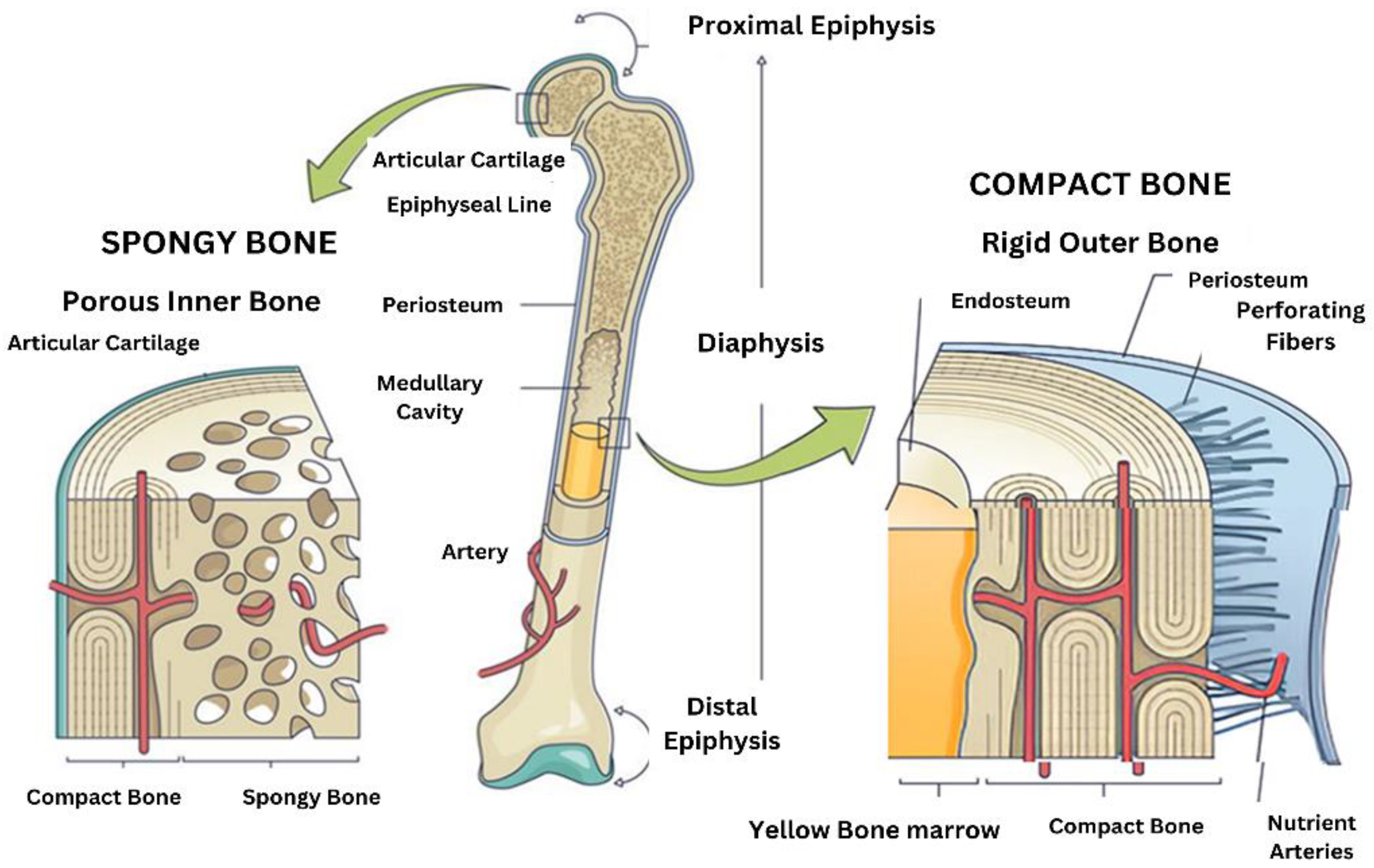
Figure 2.
Illustration of bone cell differentiation a) Osteoclastogenesis from hematopiotic stem cells leading to bone resorbing osteoclasts b) Osteoblastogenesis from mesenchymal precursors forming mature osteoblasts which further differentiation into lining cells or osteocytes.
Figure 2.
Illustration of bone cell differentiation a) Osteoclastogenesis from hematopiotic stem cells leading to bone resorbing osteoclasts b) Osteoblastogenesis from mesenchymal precursors forming mature osteoblasts which further differentiation into lining cells or osteocytes.
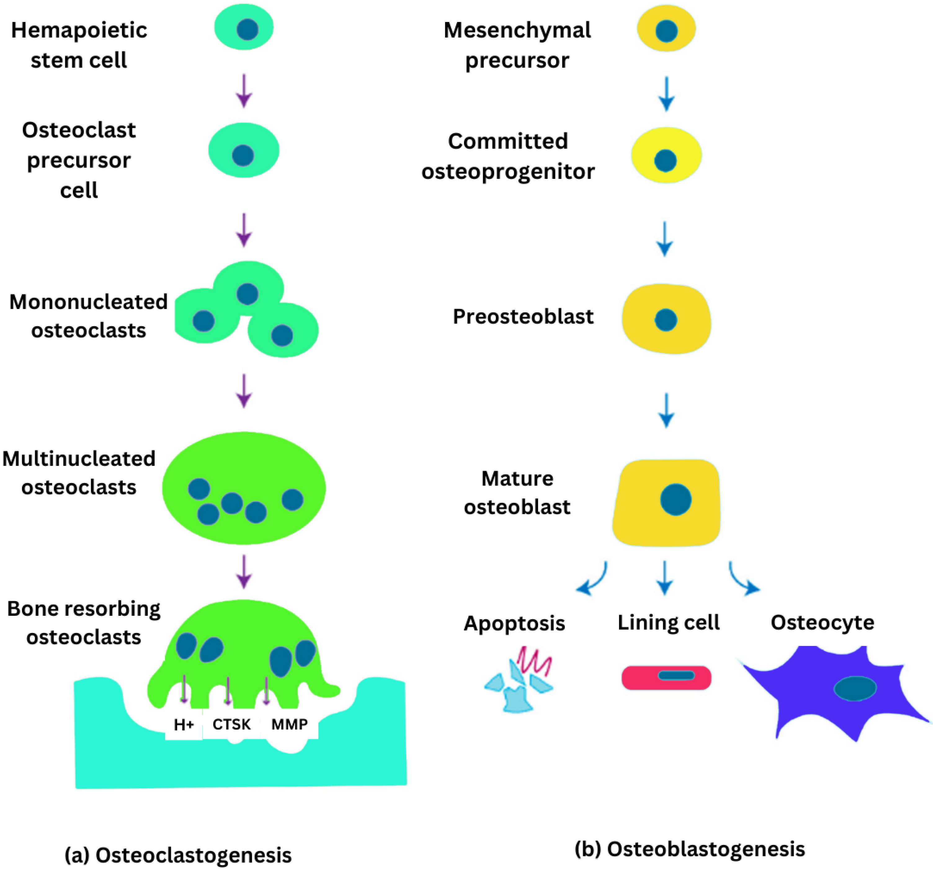
Figure 3.
3D schematic representation of advanced bone biomaterials: Polyhedron-like scaffolds integrated with vascular and nervous networks supported by hydrogel matrices, illustrating their role in promoting osteogenesis, angiogenesis, and neurogenesis for enhanced bone regeneration.
Figure 3.
3D schematic representation of advanced bone biomaterials: Polyhedron-like scaffolds integrated with vascular and nervous networks supported by hydrogel matrices, illustrating their role in promoting osteogenesis, angiogenesis, and neurogenesis for enhanced bone regeneration.
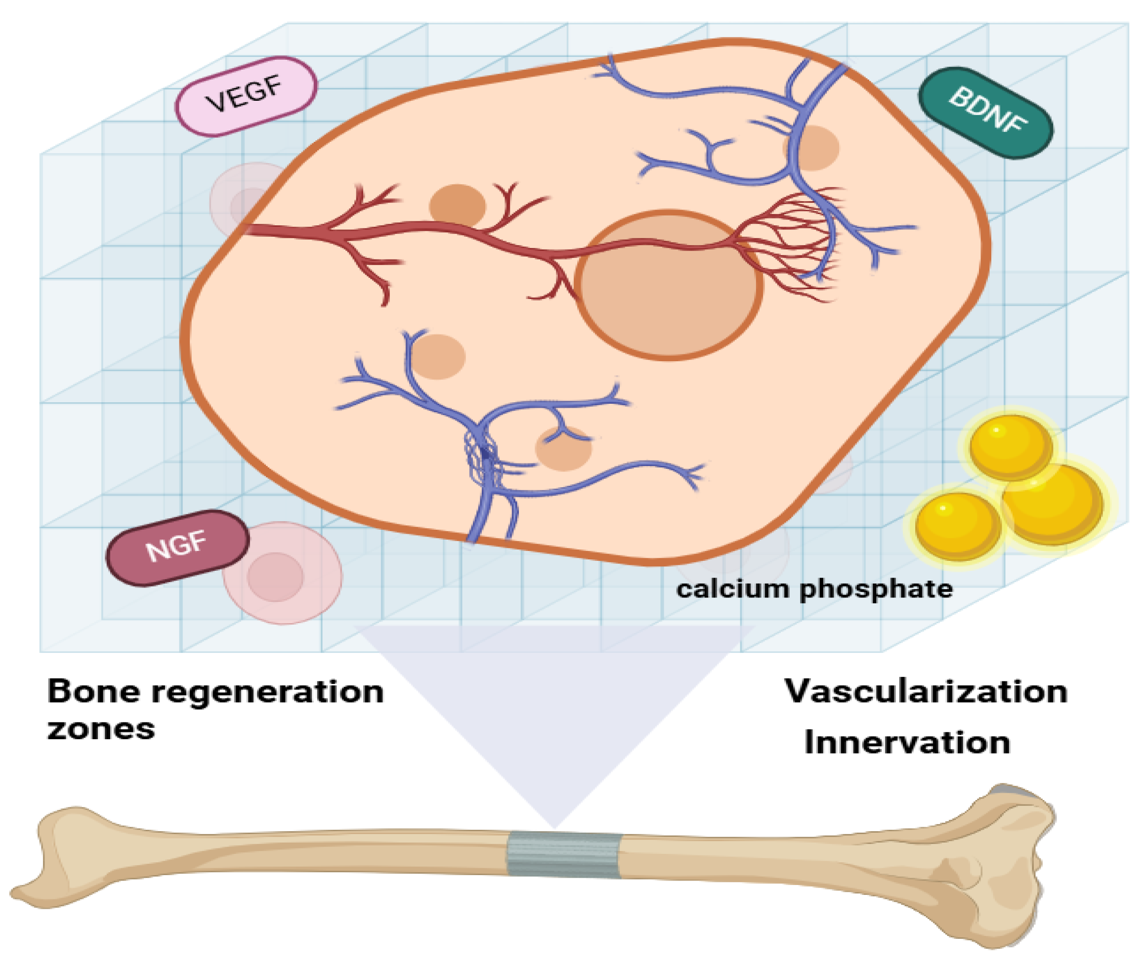
Figure 4.
Regulation of bone formation and resorption: The upper panel illustration osteoblast differentiation and maturation while the lower panel depicts osteoclastogenesis regulated by signaling molecules RANK/RANKL interaction and osteoblastic cell mediation.
Figure 4.
Regulation of bone formation and resorption: The upper panel illustration osteoblast differentiation and maturation while the lower panel depicts osteoclastogenesis regulated by signaling molecules RANK/RANKL interaction and osteoblastic cell mediation.
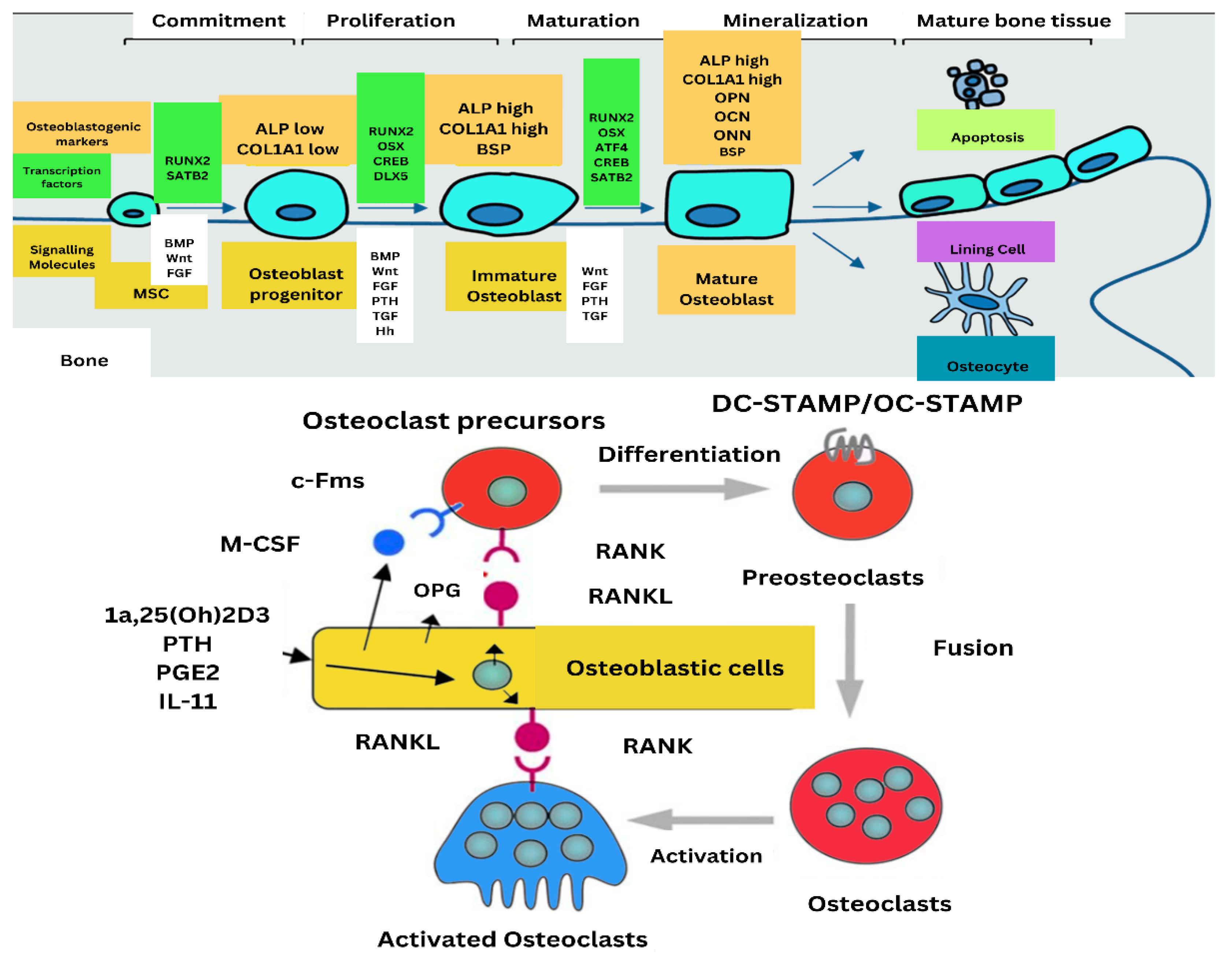
Figure 5.
Illustration of Neuronal and Endocrine Interactions with Bone: Highlighting the roles of the nervous system (via neurotransmitters and mechanical loading), the endocrine system (via hormones such as leptin, osteocalcin, and lipocalin-2), and their integration in regulating bone homeostasis and systemic metabolic balance [41].
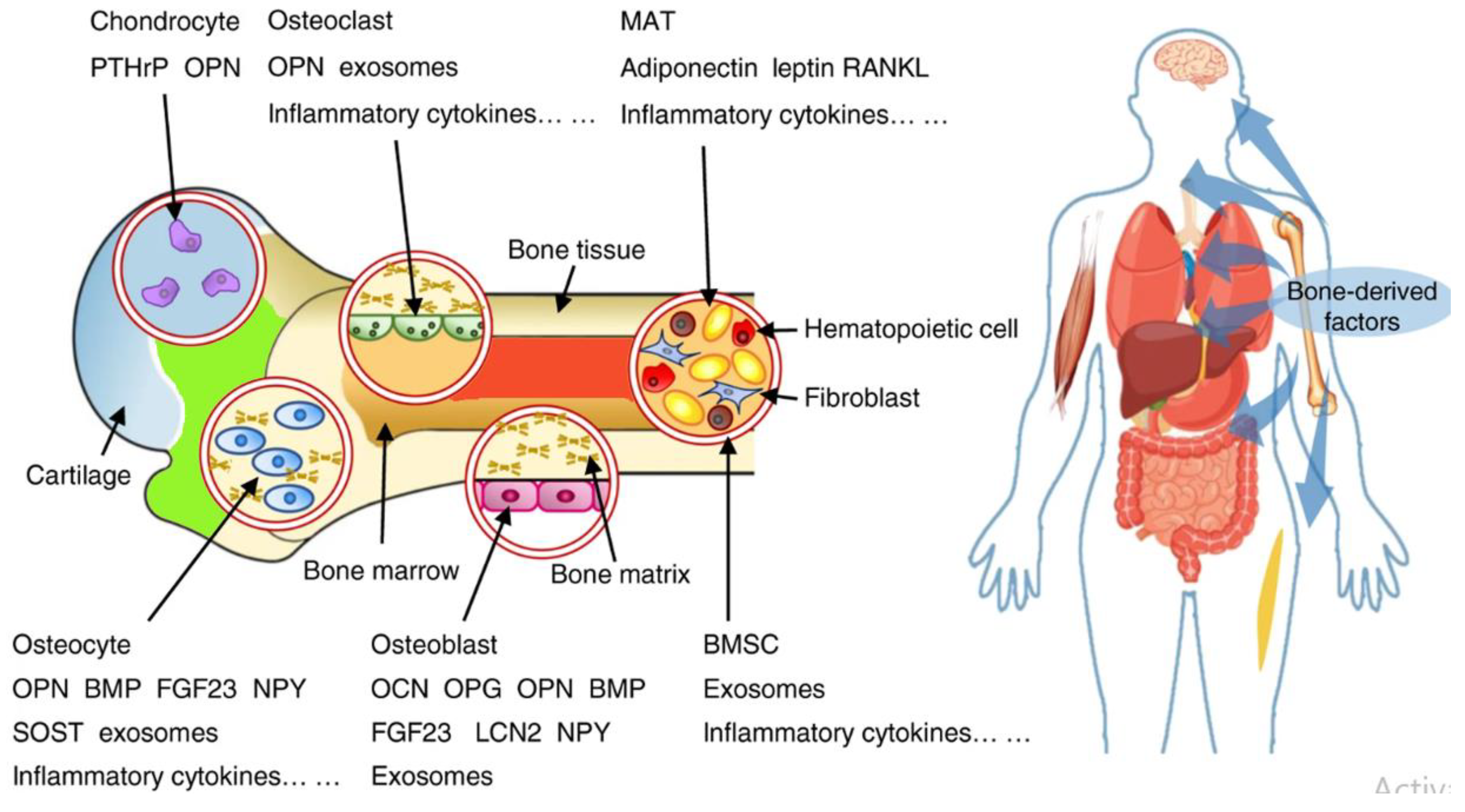
Figure 6.
A visual overview illustrating the progression of pathological changes in bone health caused by metabolic disorders, inflammation, and aging. Healthy bone serves as the baseline, with pathways diverging to conditions such as osteoporosis, Osteomalacia, Paget's disease, chronic inflammation-related bone damage, and age-associated bone loss. The summary highlights distinct mechanisms and outcomes, emphasizing how each condition disrupts bone structure and function.
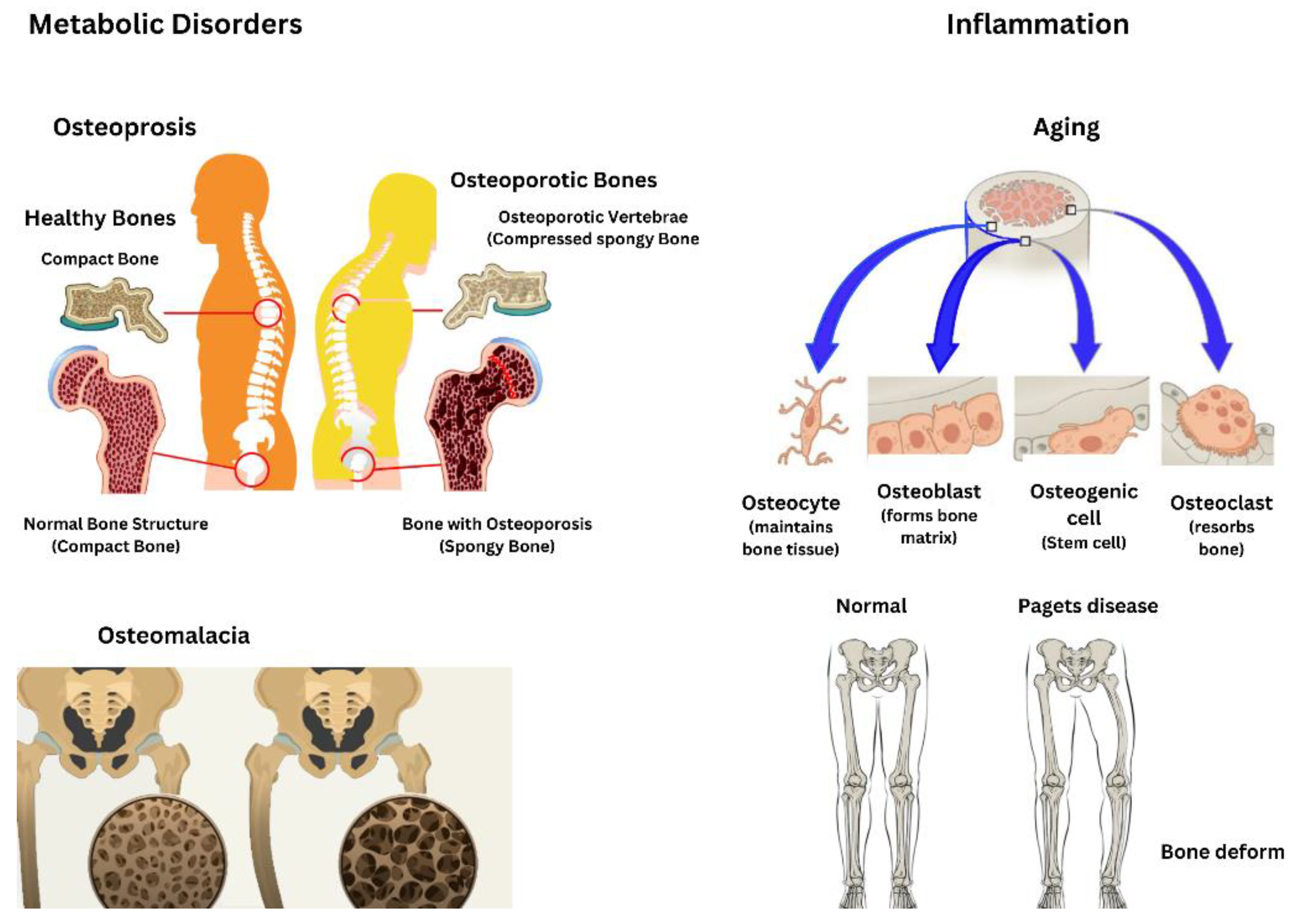

Table 1.
Explains different types of bone cells along with their origin and functions.
| Cell Type | Function | Origin/Source | Key Signaling Pathways | Additional Notes |
|---|---|---|---|---|
| Osteoblasts | Synthesize the mineralized bone matrix; key role in bone formation. | - Chondrocytes within the growth plate. - Bone marrow stromal cells. - Quiescent bone lining cells. - Specific fibroblasts in craniofacial regions. | Non-canonical Notch molecule Delta-like 1/preadipocyte factor 1 (Dlk1/Pref-1). - Wnt co-receptor Lrp5. | Short lifespan; requires renewal by preosteoblasts. |
| Osteoclasts | Specialized in bone resorption; degrades mineralized matrix and collagen by secreting acids and enzymes. | Derived from hematopoietic progenitors. | Regulated by signaling molecules from osteoblasts and osteocytes to maintain bone homeostasis. | The key for bone remodeling and calcium regulation. |
| Osteocytes | Central regulators of bone remodeling; coordinate osteoblast and osteoclast activity. | Differentiated osteoblasts embedded in mineralized bone matrix. | Paracrine and endocrine signaling pathways. | Influence bone marrow fat, body composition, and energy metabolism; involved in bone diseases and therapeutic targets. |
| Bone Marrow Stromal Cells (BMSCs) | Multipotent cells capable of differentiating into osteoblasts, chondrocytes, and adipocytes; are critical for bone regeneration. | Found in bone marrow stroma. | Regulated by various intracellular signaling pathways. | Linked to the development of osteosarcoma under specific genetic conditions. |
Table 2.
Key Bone Conditions: Features, Causes, and Consequences.
| Condition | Key Features | Causes | Consequences |
|---|---|---|---|
| Osteoporosis | Reduced bone mass and compromised bone microarchitecture, increasing bone fragility and fracture risk. | Imbalance between bone resorption and formation; osteoclastic activity exceeds osteoblastic activity | Increased fracture risk due to fragile bone microarchitecture |
| Osteomalacia | Defective mineralization of osteoid leads to softened bones and characteristic Looser zones. | Nutritional deficiencies, genetic disorders like X-linked hypophosphatemia, or other metabolic abnormalities | Softened bones and the development of looser |
| Paget's Disease | Excessive and disorganized bone remodeling with elevated osteoclast and osteoblast activity, leading to structural compromise | Genetic mutations (e.g., SQSTM1) and environmental factors | Bone pain, deformities, arthritis, fractures, and rare neoplastic transformations |
| Aging | Chronic low-grade inflammation (inflammation) impairs osteoblastogenesis and promotes osteoclastogenesis, causing bone loss. | Senescent cells and their secretory phenotype (SASP), diminished osteogenic potential, and inflammation. | Bone loss, impaired bone healing, and links to cancer pathways |
Disclaimer/Publisher’s Note: The statements, opinions and data contained in all publications are solely those of the individual author(s) and contributor(s) and not of MDPI and/or the editor(s). MDPI and/or the editor(s) disclaim responsibility for any injury to people or property resulting from any ideas, methods, instructions or products referred to in the content. |
© 2025 by the authors. Licensee MDPI, Basel, Switzerland. This article is an open access article distributed under the terms and conditions of the Creative Commons Attribution (CC BY) license (http://creativecommons.org/licenses/by/4.0/).
Copyright: This open access article is published under a Creative Commons CC BY 4.0 license, which permit the free download, distribution, and reuse, provided that the author and preprint are cited in any reuse.


