
Submitted:
10 March 2025
Posted:
11 March 2025
You are already at the latest version
Abstract
This systematic review and network meta-analysis aimed to evaluate the bond strength of artificial caries-affected dentin (ACAD) of permanent human teeth with and without biomimetic remineralization (BR), assessed in in vitro studies. Following PRISMA guidelines, we conducted a systematic search until June 2023, identifying 82 eligible articles for full-text analysis. We assessed the study characteristics, methodological quality, and summary results. Bond strength was examined immediately and after artificial aging using three bond strength tests. We performed meta-regressions (using OpenBUGS software) to explore the relationship between the independent variable’s adhesive application technique (Etch-and-Rinse or Self-Etch) and ACAD protocol (chemical or biological) and the dependent variable bond strength. Additionally, we conducted random-effect NMAs (using CINEMA software) to compare the effect of multiple interventions per application technique and ACAD protocol simultaneously. Among the included studies that compared various BR strategies. Most studies (19 out of 22) presented a medium risk of bias. In some comparisons, meta-regression results revealed a significant association between bond strength at 24h and both the adhesive application technique and the ACAD protocol. Our findings indicate the potential of BR to enhance bond strength in human ACAD in in vitro settings .
Keywords:
Adhesives
; Biomimetic Material
; Dentin-Bonding Agents
; Tooth Demineralization
; Tooth Remineralization
1. Introduction
Dentin-bonding procedures pose persistent challenges in Operative Dentistry despite the currently significant successes achieved in enamel bonding [1]. A well-documented issue in the literature is the gradual deterioration of the adhesive systems' bond strength to dentin over time, primarily due to hybrid layer degradation [2]. This compromise in dentin bonding significantly limits the lifespan of adhesive restorations [3].
The ideal dentin-bonding process involves exposing the collagen network and facilitating the penetration of chelating agents or acidic functional monomers to form the crucial hybrid layer [4]. However, a portion of the exposed collagen matrix remains unfilled with resin monomers, rendering it susceptible to hydrolytic degradation over time, thus jeopardizing the longevity of dentin bonding due to nanoleakage. The incomplete water removal within hydrophilic resin monomers also creates a weak point in resin-dentin bonds [5,6]. These phenomena have led to the exploration of an innovative approach to improve dentin adhesion: the biomimetic remineralization (BR) of collagen fibrils exposed during biomineralization [7,8].
There are two primary BR strategies: incorporating mineral-promoting agents into adhesives or restorative materials and applying pre-treating solutions before adhesive systems [9,10]. For the first strategy, researchers have developed experimental adhesive systems or restorative materials containing bioactive components like calcium phosphate or other inorganic materials that supply mineral ions to remineralize the resin-dentin interface [11,12]. The second strategy involves solutions containing non-collagenous proteins or template analogs to stimulate intra/extra-fibrillar mineralization [13,14]. These remineralizing agents facilitate the formation of nanometric apatite crystals, which replace excess water, mimicking physiological remineralization [14], thus enhancing the structural integrity of dentin and extending the longevity of the dentin-composite resin bonding interface [7,15,16]. Some studies have also suggested that these agents can inhibit the degradation of exposed collagen by attracting calcium to it [17].
Therefore, it is essential to analyze the challenges posed by dentin-bonding procedures and the potential advantages of BR procedures. This systematic review uses a comprehensive network meta-analysis (NMA) to assess and compare the bond strength of human artificial caries-affected dentin (ACAD) with and without BR evaluated in in vitro studies.
2. Materials and Methods
2.1. Search Strategy
The systematic review was registered in PROSPERO and performed according to the PRISMA statement [41]. On June 2023, PubMed, ISI Web of Science, and SCOPUS were searched to identify potentially relevant studies. In addition to electronic databases, reference lists of included studies and relevant systematic reviews were also searched. Complete search strategies are available in Appendix 1.
2.2. Outcomes
The primary outcome of this systematic review was the mean difference between the bond strength of ACAD with and without BR by different adhesive application techniques —etch-and- rinse (ER) or self-etch (SE)— and ACAD protocols —chemical or biological.
2.3. Eligibility Criteria
The following inclusion criteria were established: experimental or quasi-experimental in vitro studies investigating the influence of any BR procedure on the ACAD-adhesive interface’s bond strength; having a control group (dentin without BR) for comparison; ACAD protocols in which agents were applied immediately prior to bonding; outcomes measured by shear, micro-shear, or micro-tensile bond strength (SBS, µSBS, µTBS) tests. Exclusion criteria included studies with doped materials or modified adhesive systems.
The terms “caries-affected dentin,” “demineralized dentin,” and “artificial eroded dentin” were considered as references to ACAD. ACAD consists of human dentin tissue artificially demineralized to mimic the characteristics of dentin affected by carious changes. It is created by exposing dentin tissue to acidic or demineralizing solutions to remove mineral content, leading to softening and structural alterations like those observed in natural caries-affected dentin. [46,53,54] This demineralization process is performed in a laboratory setting to replicate the conditions and properties of carious dentin.
2.4. Data Extraction and Collection
Firstly, two authors (RC and JP) independently reviewed titles and abstracts to select articles for further assessment per their consensus. Disagreements were resolved by discussion until a consensus was reached. Full texts of the selected articles were retrieved, and the same two authors further evaluated and independently extracted data from them. The reference lists of included full texts were also screened and cross-referred.
In case of missing/unclear items (e.g., missing bond strength measurements, missing standard deviation values, uncertain number of samples used) or inconsistent data within or between sources (e.g., differences in data between text and figures, bond strength measurements only in figures), authors of the respective studies were contacted via e-mail. Two follow-up e-mails were sent with a one-week interval.
Search results from online databases were imported to Endnote20 (Clarivate, Philadelphia, USA), where duplicates were removed. The Rayyan app[55] was used to keep records and assist in abstract screening, full-text review, and data extraction. Data for the systematic review and NMA were extracted using a custom-made Excel worksheet.
The following items were extracted from each source: authors; year of publication; study randomization; risk of bias; means and standard deviations; number of samples; ACAD protocol (chemical or biological); BR procedure; adhesive type used (ER, SE, or universal) and adhesive application technique; method of bond strength assessment; outcome measurement time point (24h or after artificial aging method).
The authors classified and grouped the treatments by active substance into nine groups: fluorine, calcium phosphate, peptide, silica, hydroxyapatite, flavonoids, calcium, and 2-hydroxyethyl methacrylate/ethylene glycol dimethacrylate (HEMA/EDGMA).
2.5. Risk of Bias Assessment
Two authors (RC and JP) independently assessed the risk of bias in the included in vitro studies according to the QUIN tool [56]. Disagreements were resolved by discussion until a consensus was reached. Each study was graded accordingly as having high, medium, or low risk based on the final score of the tool: low risk of bias if >70%, medium risk of bias if 50–70%, and high risk of bias if <50%.
2.6. Data Synthesis and Statistical Analysis
2.6.1. Qualitative Synthesis
Qualitative evidence synthesis was performed by descriptive analysis of the studies’ characteristics, methodologic quality, and summary results, using a narrative description and summary tables providing a clear overview of the individual study characteristics, main findings, and methodological assessments.
2.6.2. Quantitative Synthesis
Quantitative syntheses were performed by random-effects NMA of the mean difference between the intervention and control groups. NMAs were conducted using the CINEMA software, based on R software packages meta an netmeta [57,58], by adhesive application technique and ACAD protocol and included all possible pair-wise comparisons based on direct and indirect evidence. In accordance with Cochrane guidelines [59], when trials had more than two arms, we combined interventions into a single group if they belonged to the same intervention category. When more than one independent treatment-comparator pair existed in each study, we treated them as if they pertained to independent studies. Following Cochrane guidelines[59] standard deviations were imputed from other included studies in cases where they were not available in the manuscript and could not be obtained upon contact with the authors.
The rating of confidence in the results was assessed following the CINEMA approach by evaluating the domains: within-study bias, reporting bias, indirectness, imprecision, heterogeneity, and incoherence. The minimal clinically important difference was established by consensus of the authors as 7 megapascals (MPa).
In addition, since it has been reported that the adhesive application technique (ER vs. SE) [8,25,26,27] and the ACAD protocol (chemical vs. biological) [7,28] might influence BR treatment’s effect, we explored effects of these two covariates in NMA effects estimates by random-effects Bayesian meta-regressions using the OpenBUGS software (Code in Appendix 1). Within a random-effects Bayesian framework, the OpenBUGS software [60] was also used to estimate each intervention's posterior median ranks and probability to be the best.
Finally, to assess the robustness of the results obtained from NMAs, as assumptions change, we conducted the following two sensitivity analyses:
- Random selection of one treatment intervention: Instead of combining interventions belonging to the same intervention category, as in the main analysis, we randomly selected only one.
- Removal of SBS test results: Instead of including all bond strength tests, as in the main analysis, we included only results from µSBS and µTBS tests.
3. Results and Discussion
3.1. Search Results
In the electronic search, 1874 records were identified after eliminating duplicates. Only 82 were selected for full-text screening. Reasons for the exclusion of screened full texts are shown in Appendix Table 1. After critical appraisal, 23 remaining articles were included in our systematic review and 22 in the NMA. A PRISMA flow diagram of the complete process is illustrated in Figure 1.
3.2. Characteristics of Included Studies
Table 1 displays the characteristics of the included studies, interventions, and outcomes. Of the 23 studies in the systematic review, 16 were experimental [8,15,18,19,20,21,22,23,24,25,26,27,28,29,30,31] and seven were quasi-experimental [7,14,32,33,34,35,36]. One study was excluded from the NMA because it lacked reporting data, which could not be obtained upon direct contact with the authors. (Appendix Table A2)
All 22 studies in the NMA performed immediate (24h) bond strength measurements. Of those studies, 13 investigated the ER technique associated with the chemical ACAD protocol [7,8,18,19,20,22,23,27,29,31,35,37], five the ER with the biological ACAD [7,15,21,28,33], 13 the SE with the chemical ACAD [8,14,20,22,23,25,26,32,34,36,37,38,39], and only one the SE with the biological ACAD [28]; the latter was insufficient to perform an NMA. In turn, 11 studies measured bond strength after artificial aging of the specimens: four used thermocycling [14,21,25,40], and seven stored them in a fluid solution for months [15,18,29,31,32,33,36].
Overall, both immediate and aged bond strength in the ACAD benefited from BR. The artificial aging method globally diminished bond strength values, and thermocycling caused the lowest bond strength.
3.3. Meta-Regressions
3.3.1. Influence of the Adhesive Technique on NMA Effect Estimates
Meta-regression results showed that the ER technique performed better than the SE in four NMA comparisons: control vs. calcium phosphate, control vs. peptide, fluorine vs. calcium phosphate, and fluorine vs. peptide. On the contrary, the SE technique performed better in the NMA comparison peptide vs. hydroxyapatite. In all other comparisons, both techniques demonstrated similar performance (Appendix Table A5).
3.3.2. Influence of the ACAD Protocol on NMA Effect Estimates
Regarding the influence of different ACAD protocols on NMA effect estimates, the chemical ACAD protocol resulted in higher bond strength values than the biological ACAD protocol in nine NMA comparisons: control vs. fluorine, control vs. calcium phosphate, control vs. peptide, control vs. HEMA, control vs. flavonoids, control vs. calcium, control vs. hydroxyapatite, fluorine vs. calcium phosphate, and fluorine vs. peptide. In all other comparisons, both protocols performed similarly (Appendix Table A6).
3.4. Network Meta-Analysis
Plots for the three performed NMAs are shown in Table 2.
Table 3 shows NMA results from the BR intervention network.
3.4.1. ER Technique with Chemical ACAD Protocol
The results of this NMA suggested that no statistically significant differences exist between any BR interventions in all the network comparisons
3.4.2. ER Technique with Biological ACAD Protocol
When the ER technique and the biological ACAD protocol were used together, eight of the 10 BR intervention network’s comparisons achieved statistically significant results: the calcium phosphate intervention compared to control (MD: -21.209, 95%CI: -25.954, -16.463), flavonoids (MD: -12.771, 95%CI: -20.538, -5.003), and fluorine (MD: -17.012, 95%CI: -22.103, -11.920); the flavonoids intervention compared to control (MD:8.438, 95%CI: 2.289, 14.587); the peptide intervention compared to control (MD:18.295, 95%CI:14.418, 22.172), flavonoids (MD: 9.857, 95%CI: 2.588, 17.126), and fluorine (MD: 14.098, 95%CI: 9.684, 18.512); and the fluorine intervention compared to control (MD:4.197, 95%CI:1.080, 7.314).
3.4.3. SE Technique with Chemical ACAD Protocol
When the SE technique and the chemical ACAD protocol were used together, only two of the 36 BR intervention network’s comparisons achieved statistically significant results: the calcium phosphate (MD: -4.455, 95%CI: -8.857, -0.053) and the flavonoids (MD: -7.520, 95%CI: -14.758, -0.281) interventions compared to hydroxyapatite.
3.5. NMA Confidence Ratings
3.5.1. ER Technique with Chemical ACAD Protocol
In this NMA, two direct comparisons (calcium vs. control and control vs. fluorine) and one indirect comparison (hydroxyapatite vs. peptide) presented very low confidence, mainly due to major imprecision, heterogeneity, or incoherence concerns. The remaining indirect and direct comparisons presented a low or moderate confidence rating.
3.5.2. ER Technique with Biological ACAD Protocol
In this NMA, all the direct and indirect comparisons presented a moderate confidence rating.
3.5.3. SE Technique with Chemical ACAD Protocol
A low confidence rating was observed for six direct comparisons (calcium phosphate vs. peptide, calcium vs. fluorine, control vs. HEMA, control vs. SiO2, fluorine vs. HEMA, and fluorine vs. peptide) and two indirect ones (calcium vs. hydroxyapatite and HEMA vs. peptide), mostly due to major concerns in heterogeneity, incoherence, and within-study bias. The remaining comparisons presented a moderate confidence rating.
3.6. Rankings
Treatment rankings and probability to rank best are displayed in Table 4.
3.6.1. ER Technique with Chemical ACAD Protocol
Among all the treatments in the NMA, hydroxyapatite achieved the highest probability of being the best treatment (46.10%), closely followed by peptide (41.55%).
3.6.2. ER Technique with Biological ACAD Protocol
In this NMA, calcium phosphate ranked first, with an 85.24% probability of being the best BR treatment.
3.6.3. SE Technique with Chemical ACAD Protocol
Compared to the other treatments in the NMA, flavonoids achieved the highest probability of being best (46.36%), followed by HEMA (17.49%).
3.7. Sensitivity Analyses
3.7.1. ER Technique with Chemical ACAD Protocol
Both sensitivity analyses showed results like those of the main analysis.
3.7.2. ER Technique with Biological ACAD Protocol
In this NMA, the sensitivity analysis where studies measuring the outcome with SBS tests were excluded was impossible because none used this test to assess the outcome. In the sensitivity analysis where we randomly selected one treatment intervention instead of combining interventions from the same category, the flavonoids vs. peptide comparison result lost statistical significance due to the loss of precision.
3.7.3. SE Technique with Chemical ACAD Protocol
When we excluded studies using SBS tests from the NMA, the flavonoids vs. hydroxyapatite comparison ceased to show differences between the two interventions due to a loss of precision. When we randomly selected one treatment intervention instead of combining interventions from the same category, eight of the 36 NMA comparison conclusions changed from not showing differences between the interventions to favoring one of them.
3.8. Discussion
This systematic review aimed to unravel the intricate interactions among different BR procedures and their influence on bond strength in human ACAD by analyzing and comparing bond strength from various in vitro studies through NMA. NMA allows the integration of data from direct and indirect comparisons, enabling a more precise estimation of treatment effects and a deeper understanding of optimal treatment options. Ultimately, this systematic review and NMA aspires to contribute to the existing knowledge on dentin-bonding procedures and offer valuable insights into the effectiveness of BR. The findings may help clinicians make informed decisions regarding dentin-bonding strategies for improved treatment outcomes [43].
This study’s systematic review and NMA have shed light on the potential benefits of BR for bond strength in human ACAD, measured both immediately and after artificial aging. Its findings indicate that BR protocols are promising in enhancing restorative materials’ bonding performance on demineralized dentin surfaces.
ACAD’s compromised nature negatively affects bond strength, and its surface is more challenging for bonding due to the incomplete infiltration of adhesives into the exposed collagen matrix [44]. Furthermore, the low pH associated with ACAD promotes the activation and activity of proteolytic enzymes, accelerating the breakdown of non-infiltrated collagen and the hybrid layer [28,38].
Our NMA findings highlighted differences between chemical and biological ACAD protocols. Chemical protocols consistently yielded higher bond strength results than biological, agreeing with previous research [45]. This difference may derive from the thicker demineralization layer associated with chemical protocols and the excessive softness of the primary dentine resulting from microbiological approaches [45].
The NMA also revealed variations in bond strength depending on the adhesive application technique. With their additional acid-etching stage, ER techniques proved more efficient in dissolving the smear layer than SE methods, which have a less acidic composition and are more sensitive [46]. Additionally, SE relies on chemical interactions with calcium ions, often found in lower concentrations in ACAD. Consequently, ER techniques yielded significantly higher bond strength values than SE, in line with the existing literature [20,22,44,47]. Moreover, when considering the ACAD surface, ER consistently demonstrated higher bond strength than SE materials [44].
This systematic review’s 23 in vitro studies showed medium heterogeneity, reflecting variations in ACAD protocols, aging methods, and bond strength tests. Thus, random-effects models were employed throughout the NMA investigation. Artificial aging methods, such as thermocycling and months of storage, generally reduce bond strength. Thermocycling promoted the most extreme breakdown of the bond interface and caused the lowest bond strength, even with associated BR, which is consistent with other studies [48]. However, different bond strength tests were used in the included investigations, which could affect the measurement results, and aspects such as specimen preparation and geometry, loading configuration, and material characteristics were not considered [3,49,50].
BR overall increased the bond strength values, even after artificial aging methods [10,51]. Nonetheless, the limited availability of studies reporting BR associated with bond strength restricts the exploration of these relationships [52]. Incorporating these BR methods into dental treatments can potentially enhance the durability and quality of the resin-dentin interface, offering promising avenues for improving clinical outcomes in restorative dentistry. In the NMA on ER with chemical ACAD, hydroxyapatite was the most effective treatment (46.10%), closely followed by peptide (41.55%), despite the low confidence in some comparisons. In the NMA on ER with biological ACAD, calcium phosphate emerged as the top-ranking BR (85.24%), significantly surpassing the control, flavonoids, and fluoride treatments. However, the NMA on SE with chemical ACAD showed low confidence in various comparisons, with flavonoids having the highest probability (46.36%) of being more effective, followed by HEMA (17.49%). These findings highlight the nuanced effectiveness of BR, influenced by different protocols and compositions. Most investigations on BR have shown its ability to remineralize ACAD in a basic manner. However, because they were carried out in vitro, their application in clinical contexts remains unexplored [52].
This study has some limitations. Most notably, in vitro studies lack the complexity of the oral environment, including oral biofluids and microbial interactions [3,44,49,50,52]. The absence of real dental caries development processes in the ACAD models is also a limitation. Future studies should address these shortcomings for a more comprehensive understanding of the clinical applicability of BR.
Another limitation is related to the sensitivity analysis for the NMA on SE with chemical ACAD. In this network, when we randomly selected one treatment intervention instead of combining interventions from the same category, eight out of the 36 NMA comparisons changed their conclusions from not showing differences between the interventions to favoring one of them. Despite this, we are confident that combining multiple arms related to the same intervention yields more reliable estimates because it does not waste useful data and evidence, as outlined and in accordance with the Cochrane recommendations. Moreover, regardless of the strategy used to cope with multiple-arm trials, six of the eight comparisons that had their conclusions changed in the sensitivity analysis were based solely on indirect evidence, which inherently carries less confidence than scenarios where direct evidence is also available.
Despite these limitations, our findings suggest that BR can enhance bond strength in ACAD, offering potential benefits for clinical practice. Dental professionals can use this knowledge to optimize treatment approaches, improve patient outcomes, and extend the longevity of adhesive bonding materials [3,44,49,50,52].
5. Conclusions
In conclusion, through a systematic review and NMAs we showed that bond strength degraded after biological or chemical ACAD protocols. As a result, surface preparation with BR procedures prior to bonding is advised to increase the bonding of ER and SE adhesives.
Supplementary Materials
A supplemental Appendix 1 to this article is available .The following supporting information can be downloaded at: www.mdpi.com/xxx/s1
Author Contributions
Rosário Costa contributed to concepts, design, the definition of intellectual content, literature search, data acquisition, and article preparation. Joana Reis-Pardal contributed to concepts, design, the definition of intellectual content, literature search, data acquisition, statistical analysis, and article preparation. João Cardoso Ferreira contributed to the definition of intellectual content and article preparation. Sofia Arantes-Oliveira contributed to the definition of intellectual content and article preparation. Luís Filipe Azevedo contributed to concepts, design, the definition of intellectual content, statistical analysis and critically revised the manuscript. Paulo Ribeiro de Melo contributed to concepts, design, the definition of intellectual content and critically revised the manuscript. All authors gave their final approval and agreed to be accountable for all aspects of the work.
Institutional Review Board Statement
The study was approved by local “Comissão de Ética para a Saúde da Faculdade de Medicina Dentária da Universidade do Porto”.
Data Availability Statement
The data that support the findings of this study are available from the corresponding author, Rosário Costa, upon reasonable request.
Conflicts of Interest
The authors declare no conflicts of interest.
Abbreviations
The following abbreviations are used in this manuscript:
| NMA | Network Meta-analysis |
| ACAD | Artificial caries-affected dentin |
| BR | Biomimetic remineralization |
Appendix 1
1. Search Strategies
PubMed: 1121 retrieved records
#1 Light-Curing of Dental Adhesives [MeSH] OR Self-Curing of Dental Resins [MeSH] OR adhesi*[tw] OR (bond*[tw] AND strength[tw])
#2 biomimetic*[tw] OR Biomimetics [MeSH] OR mineraliz*[tw] OR biomineraliz*[tw] OR Biomineralization [MeSH] OR remineraliz*[tw] OR Tooth Remineralization [MeSH] OR ((Dental Caries [MeSH] OR cari*[tw] OR eroded[tw] OR desensitized[tw]) AND (pre-treat*[tw] OR pretreat*[tw] OR treat*[tw] OR therap*[tw]))
#3 Dentin-Bonding Agents [MeSH] OR (dentin[tw] AND bond*[tw])
#1 AND #2 AND #3
ISI Web of Science: 1176 retrieved records
#1 adhesi* OR (bond* AND strength) (All Fields)
#2 biomimetic* OR mineraliz* OR biomineraliz* OR remineraliz* OR ((cari* OR eroded OR desensitized) AND (pre-treat* OR pretreat* OR treat* OR therap*)) (All Fields)
#3 dentin AND bond* (All Fields)
#1 AND #2 AND #3
SCOPUS: 1052 retrieved records
#1 TITLE-ABS-KEY (adhesi* OR (bond* AND strength))
#2 TITLE-ABS-KEY (biomimetic* OR mineraliz* OR biomineraliz* OR remineraliz* OR ((cari* OR eroded OR desensitized) AND (pre-treat* OR pretreat* OR treat* OR therap*)))
#3 TITLE-ABS-KEY (dentin AND bond*)
#1 AND #2 AND #3

Table A1.
Reasons for excluding studies after accessing full-texts.
| Studies | Reason for exclusion |
|---|---|
| Doozandeh et al. (2015)[61] | 1-Without ACAD |
| Bergamin et al. (2016)[62] | 1-Without ACAD |
| Ghani et al. (2017)[63] | 1-Without ACAD |
| Komori et al. (2009) [64] | 1-Without ACAD |
| Leal et al. (2017)[65] | 1-Without ACAD |
| Luong et al. (2020)[66] | 1-Without ACAD |
| Meraji et al. (2018)[67] | 1-Without ACAD |
| Prasansuttiporn et al. (2020)[68] | 1-Without ACAD |
| Sajjad et al. (2022)[69] | 1-Without ACAD |
| Yilmaz et al. (2017)[70] | 1-Without ACAD |
| Castellan et al. (2010)[71] | 2- Without remineralization procedures |
| Okuyama et al. (2011)[72] | 2- Without remineralization procedures |
| Wang et al. (2012)[73] | 2- Without remineralization procedures |
| de-Melo et al. (2013)[74] | 2- Without remineralization procedures |
| Carvalho et al. (2016)[75] | 2- Without remineralization procedures |
| Deari et al. (2017)[76] | 2- Without remineralization procedures |
| Giacomini et al. (2017)[77] | 2- Without remineralization procedures |
| Rodrigues et al. (2017)[78] | 2- Without remineralization procedures |
| Imiolczyk et al. (2017)[79] | 2- Without remineralization procedures |
| Stape et al. (2021)[80] | 2- Without remineralization procedures |
| Hartz et al. (2022)[81] | 2- Without remineralization procedures |
| Wang et al. (2016)[82] | 3-Modified Materials |
| Moda et al (2018)[83] | 3-Modified Materials |
| Choi et al. (2020)[84] | 3-Modified Materials |
| Abdelshafi et al. (2021) [85] | 3-Modified Materials |
| Al-Qahtani et al. (2021)[86] | 3-Modified Materials |
| Khor et al. (2022)[87] | 3-Modified Materials |
| Adebayo et al. (2010)[88] | 4- Without bond strength measurement |
| Liu et al. (2011)[89] | 4- Without bond strength measurement |
| Chen et al. (2016)[90] | 4- Without bond strength measurement |
| Bortolotto et al. (2017)[91] | 4- Without bond strength measurement |
| Liang et al. (2017)[92] | 4- Without bond strength measurement |
| Wang et al. (2021)[93] | 4- Without bond strength measurement |
| Zhou et al. (2016) [94] | 5- Modified adhesive |
| Flury et al (2017)[95] | 5- Modified adhesive |
| Ye et al. (2017)[96] | 5- Modified adhesive |
| Liang et al. (2018) [97] | 5- Modified adhesive |
| Cardenas et al. (2021) [38] | 5- Modified adhesive |
| Hasegawa et al. (2021)[98] | 5- Modified adhesive |
| Bridi et al (2012)[99] | 6- Not biomimetic remineralization agents |
| Castellan et al (2013)[100] | 6- Not biomimetic remineralization agents |
| Monteiro et al (2013)[101] | 6- Not biomimetic remineralization agents |
| Abu Nawareg et al (2016)[102] | 6- Not biomimetic remineralization agents |
| Lee et al (2017)[103] | 6- Not biomimetic remineralization agents |
| Prasansuttiporn et al (2017)[104] | 6- Not biomimetic remineralization agents |
| Ramezanian Nik et al (2017)[105] | 6- Not biomimetic remineralization agents |
| Costa et al (2019)[106] | 6- Not biomimetic remineralization agents |
| Fialho et al (2019)[107] | 6- Not biomimetic remineralization agents |
| Landmayer et al (2020) [108] | 6- Not biomimetic remineralization agents |
| Costa et al (2021)[109] | 6- Not biomimetic remineralization agents |
| Giacomini et al (2021)[110] | 6- Not biomimetic remineralization agents |
| Shioya et al (2021)[111] | 6- Not biomimetic remineralization agents |
| Xu et al (2021)[112] | 6- Not biomimetic remineralization agents |
| Atay et al (2022)[113] | 6- Not biomimetic remineralization agents |
| Lemos et al (2022)[114] | 6- Not biomimetic remineralization agents |
| Zhang et al (2015)[115] | 7-Non-Roman Alphabet language after unsuccessful contact with authors |
| Wang et al (2017)[116] | 7-Non-Roman Alphabet language after unsuccessful contact with authors |
| Meng et al (2022)[117] | 7-Non-Roman Alphabet language after unsuccessful contact with authors |
| Kim et al (2020)[13] | 8- Missing control group |
Table A2.
Reasons for excluding studies from Network Meta-Analyses.
| Study | Reason for exclusion |
|---|---|
| Atomura et al (2018)[32] | Standard Deviation and sample size (N) missing and authors didn´t respond to the various emails. |
Table A3.
Data Information.
| Study | Data Information |
|---|---|
| Zumstein et al. (2018)[42] | Missing data obtained from another Meta-Analysis by Wiegand et al.2021 [50]. Authors didn´t respond to the various emails. |
Table A4.
Authors providing data via email, upon request.
| Study | Data Information |
|---|---|
| Barbosa-Martins et al. (A) (2018)[8] | Unit of statistical analysis |
| Barbosa-Martins et al. (B) (2018)[7] | Unit of statistical analysis |
| de Sousa et al. (2019)[33] | Unit of statistical analysis |
| Moreira et al. (2021)[15] | Unit of statistical analysis |
| Meng et al. (2021) [24] | Mean and SD values |
| Pei et al. (2019)[26] | Unit of statistical analysis |
| Pulidindi et al. (2021) [40] | Unit of statistical analysis |
| Yang et al. (2018)[29] | Mean and SD values and Unit of statistical analysis |
| Zang et al. (2018)[30] | Mean and SD values and Unit of statistical analysis |
2. OpenBUGS Code for Random Effects Meta-Regression Model with a Subgroup Indicator Covariate
# Normal likelihood, identity link, subgroup
# Random effects model for multi-arm trials
model{ # *** PROGRAM STARTS
for(i in 1:ns){ # LOOP THROUGH STUDIES
w[i,1] <- 0 # adjustment for multi-arm trials is zero for control arm
delta[i,1] <- 0 # treatment effect is zero for control arm
mu[i] ~ dnorm(0,.0001) # vague priors for all trial baselines
for (k in 1:na[i]) { # LOOP THROUGH ARMS
var[i,k] <- pow(se[i,k],2) # calculate variances
se[i,k] ~ dunif(0,10) # vague prior for SE
prec[i,k] <- 1/var[i,k] # set precisions
y[i,k] ~ dnorm(theta[i,k],prec[i,k]) # binomial likelihood
theta[i,k] <- mu[i] + delta[i,k] + (beta[t[i,k]]-beta[t[i,1]]) * x[i]# model for linear predictor, covariate effect relative to treat in arm 1
#Deviance contribution
dev[i,k] <- (y[i,k]-theta[i,k])*(y[i,k]-theta[i,k])*prec[i,k]
}
# summed residual deviance contribution for this trial
resdev[i] <- sum(dev[i,1:na[i]])
for (k in 2:na[i]) { # LOOP THROUGH ARMS
# trial-specific LOR distributions
delta[i,k] ~ dnorm(md[i,k],taud[i,k])
# mean of LOR distributions, with multi-arm trial correction
md[i,k] <- d[t[i,k]] - d[t[i,1]] + sw[i,k]
# precision of LOR distributions (with multi-arm trial correction)
taud[i,k] <- tau *2*(k-1)/k
# adjustment, multi-arm RCTs
w[i,k] <- (delta[i,k] - d[t[i,k]] + d[t[i,1]])
# cumulative adjustment for multi-arm trials
sw[i,k] <- sum(w[i,1:k-1])/(k-1)
}
}
totresdev <- sum(resdev[]) #Total Residual Deviance
d[1]<-0 # treatment effect is zero for control arm
beta[1] <- 0 # covariate effect is zero for reference treatment
# vague priors for treatment effects
for (k in 2:nt){ # LOOP THROUGH TREATMENTS
d[k] ~ dnorm(0,.0001) # vague priors for treatment effects
beta[k] <- B # common covariate effect
}
B ~ dnorm(0,.0001) # vague prior for covariate effect
sd ~ dunif(0,5) # vague prior for between-trial SD
tau <- pow(sd,-2) # between-trial precision = (1/between-trial variance)
# treatment effect when covariate = z[j]
for (k in 1:nt){ # LOOP THROUGH TREATMENTS
for (j in 1:nz) { dz[j,k] <- d[k] + (beta[k]-beta[1])*z[j] }
}
# All pairwise comparisons, if nt>2
for (c in 1:(nt-1)) {
for (k in (c+1):nt) {
# when covariate is zero
diff[c,k] <- (d[c] - d[k])
#at covariate=z[j]
for (j in 1:nz) {
diff.j[c,k] <- (dz[j,c] - dz[j,k])
}}}
} # *** PROGRAM ENDS
3. Meta-Regression
Table A5.
Meta-Regression results evaluating the influence of Adhesive application type (er vs se) on treatment effects at 24h.
Table A5.
Meta-Regression results evaluating the influence of Adhesive application type (er vs se) on treatment effects at 24h.
| NMA comparison | Mean | 95% CrI |
|---|---|---|
| CTRL:F | 0.8846 | (-1.72; 3.52) |
| CTRL:CaP | -3.351 | (-6.664; -0.03009) |
| CTRL:Pept. | -5.384 | (-9.103; -1.65) |
| CTRL:SiO2 | -0.8296 | (-10.72; 9.049) |
| CTRL:HEMA | -1.728 | (-10.22; 6.768) |
| CTRL:FLs | -4.982 | (-12.35; 2.382) |
| CTRL:Ca | 0.3152 | (-5.575; 6.211) |
| CTRL:HAp | 1.223 | (-2.536; 4.98) |
| F:CaP | -4.236 | (-7.499; -0.9842) |
| F:Pept. | -6.268 | (-9.996; -2.552) |
| F:SiO2 | -1.714 | (-11.95; 8.521) |
| F:HEMA | -2.613 | (-11.1; 5.868) |
| F:FLs | -5.867 | (-13.45; 1.726) |
| F:Ca | -0.5694 | (-6.352; 5.196) |
| F:HAp | 0.3381 | (-3.942; 4.631) |
| CaP:Pept. | -2.032 | (-5.601; 1.525) |
| CaP:SiO2 | 2.522 | (-7.906; 12.94) |
| CaP:HEMA | 1.623 | (-7.297; 10.54) |
| CaP:FLs | -1.631 | (-9.428; 6.172) |
| CaP:Ca | 3.666 | (-2.196; 9.529) |
| CaP:HAp | 4.574 | (-0.07638; 9.248) |
| Pept.:SiO2 | 4.554 | (-6.013; 15.1) |
| Pept.:HEMA | 3.656 | (-5.425; 12.76) |
| Pept.:FLs | 0.4015 | (-7.578; 8.394) |
| Pept.:Ca | 5.699 | (-0.6984; 12.08) |
| Pept.:HAp | 6.606 | (1.658; 11.56) |
| SiO2:HEMA | -0.8984 | (-13.94; 12.13) |
| SiO2:FLs | -4.153 | (-16.46; 8.18) |
| SiO2:Ca | 1.145 | (-10.37; 12.64) |
| SiO2:HAp | 2.052 | (-8.496; 12.66) |
| HEMA:FLs | -3.254 | (-14.41; 7.904) |
| HEMA:Ca | 2.043 | (-8.051; 12.17) |
| HEMA:HAp | 2.951 | (-6.264; 12.18) |
| FLs:Ca | 5.297 | (-3.932; 14.52) |
| FLs:HAp | 6.205 | (-1.944; 14.31) |
| Ca:HAp | 0.9075 | (-5.872; 7.675) |
1 Note: Negative mean values favor ER application type. Statistically significant results are highlighted in bold. Legend: Control (CTRL), Fluorine(F), Calcium Phosphate (CaP), Peptide (Pept.), Silica (SiO2), Flavonoids (FLs), Calcium (Ca), Hydroxyapatite (HAp), Credible Interval (Crl).
Table A6.
Meta-Regression results evaluating the influence of acad protocol type (chemical vs biological) on treatment effects at 24h.
Table A6.
Meta-Regression results evaluating the influence of acad protocol type (chemical vs biological) on treatment effects at 24h.
| NMA comparison | Mean | 95% CrI |
|---|---|---|
| CTRL:F | -7.588 | (-11.3; -3.877) |
| CTRL:CaP | -12.97 | (-17.32; -8.628) |
| CTRL:Pept. | -14.2 | (-18.64; -9.77) |
| CTRL:SiO2 | -10.15 | (-20.78; 0.5214) |
| CTRL:HEMA | -10.62 | (-19.71; -1.499) |
| CTRL:FLs | -11.2 | (-18.69; -3.706) |
| CTRL:Ca | -9.321 | (-15.94; -2.717) |
| CTRL:HAp | -9.208 | (-14.65; -3.78) |
| F:CaP | -5.381 | (-8.621; -2.139) |
| F:Pept. | -6.617 | (-10.3; -2.935) |
| F:SiO2 | -2.563 | (-12.68; 7.6) |
| F:HEMA | -3.031 | (-11.43; 5.39) |
| F:FLs | -3.61 | (-11.2; 3.981) |
| F:Ca | -1.734 | (-7.48; 3.999) |
| F:HAp | -1.62 | (-5.972; 2.749) |
| CaP:Pept. | -1.236 | (-4.793; 2.332) |
| CaP:SiO2 | 2.818 | (-7.433; 13.12) |
| CaP:HEMA | 2.35 | (-6.401; 11.14) |
| CaP:FLs | 1.77 | (-6.08; 9.585) |
| CaP:Ca | 3.647 | (-2.149; 9.428) |
| CaP:HAp | 3.761 | (-0.8584; 8.391) |
| Pept.:SiO2 | 4.055 | (-6.382; 14.52) |
| Pept.:HEMA | 3.586 | (-5.386; 12.55) |
| Pept.:FLs | 3.007 | (-4.964; 10.97) |
| Pept.:Ca | 4.884 | (-1.454; 11.21) |
| Pept.:HAp | 4.997 | (-0.001725; 10.01) |
| SiO2 :HEMA | -0.4682 | (-13.38; 12.48) |
| SiO2:FLs | -1.048 | (-13.4; 11.32) |
| SiO2:Ca | 0.829 | (-10.56; 12.18) |
| SiO2:HAp | 0.9427 | (-9.562; 11.42) |
| HEMA:FLs | -0.5796 | (-11.75; 10.58) |
| HEMA:Ca | 1.297 | (-8.708; 11.3) |
| HEMA:HAp | 1.411 | (-7.759; 10.56) |
| FLs:Ca | 1.877 | (-7.369; 11.12) |
| FLs:HAp | 1.991 | (-6.31; 10.31) |
| Ca:HAp | 0.1137 | (-6.606; 6.846) |
1 Note: Negative mean values favor Chem ACAD protocol type. Statistically significant results are highlighted in bold.. Legend: Control (CTRL), Fluorine(F), Calcium Phosphate (CaP), Peptide (Pept.), Silica (SiO2), Flavonoids (FLs), Calcium (Ca), Hydroxyapatite (HAp), Credible Interval (Crl).
4. Contribution Tables
Table A7.
Per-comparison contribution matrix for the ER with Chemical Network.
| NMA treatment effect/ comparisons | Ca:CaP | Ca:CTRL | Ca:F | CaP:CTRL | CaP:F | CaP:Pept. | CTRL:F | CTRL:HAp | CTRL:Pept. | F:Pept. |
|---|---|---|---|---|---|---|---|---|---|---|
| Mixed estimates | ||||||||||
| CaP:CTRL | 2.935 | 2.375 | 0.56 | 63.32 | 7.4 | 7.4783 | 8.4533 | 0 | 6.985 | 0.4933 |
| CaP:F | 4.195 | 0.0675 | 4.2625 | 22.975 | 31.11 | 6.1242 | 25.1317 | 0 | 2.2242 | 3.9 |
| CaP:Pept. | 1.1317 | 0.6967 | 0.435 | 16.795 | 5.04 | 52.39 | 0.535 | 0 | 18.0267 | 4.94 |
| Ca:CaP | 38.27 | 15.92 | 11.94 | 17.4467 | 7.655 | 2.7583 | 3.2517 | 0 | 1.725 | 1.0333 |
| Ca:CTRL | 15.095 | 36.58 | 15.3917 | 13.445 | 0.125 | 1.775 | 14.73 | 0 | 2.3117 | 0.5367 |
| Ca:F | 12.5817 | 15.78 | 38.56 | 3.1633 | 7.905 | 1.5133 | 18.9033 | 0 | 0.04 | 1.5533 |
| CTRL:F | 0.5775 | 2.515 | 3.0925 | 8.505 | 8.155 | 0.2275 | 70.38 | 0 | 3.3875 | 3.16 |
| CTRL:HAp | 0 | 0 | 0 | 0 | 0 | 0 | 0 | 100 | 0 | 0 |
| CTRL:Pept. | 0.8633 | 0.8783 | 0.015 | 14.86 | 1.5317 | 17.255 | 7.2017 | 0 | 51.7 | 5.685 |
| F:Pept. | 1.5167 | 0.5925 | 2.1092 | 3.9225 | 10.465 | 15.9042 | 25.565 | 0 | 22.235 | 17.69 |
| Indirect estimates | ||||||||||
| CaP:HAp | 2.0033 | 1.5833 | 0.42 | 31.66 | 4.9333 | 5.0267 | 5.7233 | 43.6233 | 4.6567 | 0.37 |
| Ca:HAp | 10.2008 | 18.29 | 10.3225 | 8.9633 | 0.1 | 1.3375 | 9.82 | 38.8133 | 1.74 | 0.4025 |
| Ca:Pept. | 17.22 | 16.965 | 12.4708 | 0.7075 | 1.7067 | 19.6342 | 4.2742 | 0 | 20.5317 | 6.49 |
| F:HAp | 0.4445 | 1.6767 | 2.1212 | 5.6992 | 5.4367 | 0.182 | 35.19 | 44.8545 | 2.2887 | 2.1067 |
| HAp:Pept. | 0.6475 | 0.6595 | 0.012 | 9.9067 | 1.1495 | 11.7037 | 4.9275 | 41.3437 | 25.85 | 3.79 |
* Note: Columns refer to comparisons with direct data and rows to NMA treatment effects. Data in each cell show how much (in %) each direct comparison contributes to the NMA treatment effects. Values in bold and grey identifies the percentage each direct comparison contributes to the corresponding NMA comparison treatment effect. Legend: Control (CTRL), Fluorine(F), Calcium Phosphate (CaP), Peptide (Pept.), Calcium (Ca), Hidroxiapatite (HAp).
Table A8.
Per-comparison contribution matrix for the ER with Biological Network.
| NMA treatment effect/ comparisons | CaP:CTRL | CaP:F | CaP:Pept. | CTRL:FLs | CTRL:F | CTRL:Pept. | F:Pept. |
|---|---|---|---|---|---|---|---|
| Mixed estimates | |||||||
| CaP:CTRL | 47.08 | 13.7 | 12.3317 | 0 | 14.5567 | 11.475 | 0.8567 |
| CaP:F | 17.525 | 40.42 | 11.1833 | 0 | 19.6783 | 2.1533 | 9.03 |
| CaP:Pept. | 16.035 | 11.5217 | 43.84 | 0 | 1.0367 | 17.0717 | 10.485 |
| CTRL:FLs | 0 | 0 | 0 | 100 | 0 | 0 | 0 |
| CTRL:F | 6.015 | 6.345 | 0.33 | 0 | 71.49 | 8.075 | 7.745 |
| CTRL:Pept. | 7.505 | 1.1033 | 8.6083 | 0 | 12.8783 | 58.13 | 11.775 |
| F:Pept. | 0.9933 | 8.17 | 9.1633 | 0 | 21.8183 | 20.825 | 39.04 |
| Indirect estimates | |||||||
| CaP:FLs | 23.54 | 9.1333 | 8.2925 | 40.9658 | 9.7758 | 7.65 | 0.6425 |
| FLs:F | 4.01 | 4.2575 | 0.2475 | 45.1658 | 35.745 | 5.4108 | 5.1633 |
| FLs:Pept. | 5.0033 | 0.8275 | 5.8308 | 42.7458 | 8.6775 | 29.065 | 7.85 |
* Note: Columns refer to comparisons with direct data and rows to NMA treatment effects. Data in each cell show how much (in %) each direct comparison contributes to the NMA treatment effects. Values in bold and grey identifies the percentage each direct comparison contributes to the corresponding NMA comparison treatment effect. Legend: Control (CTRL), Fluorine(F), Calcium Phosphate (CaP), Peptide (Pept.), Calcium (Ca), Flavonoids (FLs).
Table A9.
Per-comparison contribution matrix for the SE with Chemical Network.
| NMA treatment effect/ comparisons | Ca:CaP | Ca:CTRL | Ca:F | CaP:CTRL | CaP:F | CaP:Pept. | CTRL: FLS | CTRL:F | CTRL: HEMA | CTRL:HAp | CTRL:Pept. | CTRL:SiO2 | F:HEMA | F:Pept. |
|---|---|---|---|---|---|---|---|---|---|---|---|---|---|---|
| Mixed estimates | ||||||||||||||
| CaP:CTRL | 5.215 | 3.955 | 1.26 | 51.94 | 9.345 | 8.4575 | 0 | 10.425 | 0.4725 | 0 | 8.165 | 0 | 0.4725 | 0.2925 |
| CaP:F | 6.4433 | 0.7333 | 5.71 | 20.78 | 29.56 | 5.3175 | 0 | 23.9333 | 1.1025 | 0 | 3.5225 | 0 | 1.1025 | 1.795 |
| CaP:Pept. | 1.9783 | 1.3433 | 0.635 | 14.09 | 4.5933 | 54.07 | 0 | 2.3683 | 0.13 | 0 | 17.932 | 0 | 0.13 | 2.73 |
| Ca:CaP | 41.85 | 14.96 | 11.2705 | 15.37 | 8.04 | 2.8205 | 0 | 2.5325 | 0.168 | 0 | 2.2905 | 0 | 0.168 | 0.53 |
| Ca:CTRL | 13.2408 | 37.83 | 16.005 | 10.395 | 0.6525 | 2.1933 | 0 | 15.825 | 0.6925 | 0 | 2.3333 | 0 | 0.6925 | 0.14 |
| Ca:F | 11.2397 | 16.065 | 39.64 | 3.365 | 6.74 | 1.1347 | 0 | 19.125 | 0.773 | 0 | 0.468 | 0 | 0.773 | 0.6667 |
| CTRL:FLs | 0 | 0 | 0 | 0 | 0 | 0 | 100 | 0 | 0 | 0 | 0 | 0 | 0 | 0 |
| CTRL:F | 0.4375 | 2.85 | 3.2875 | 5.19 | 5.3533 | 0.6008 | 0 | 72.96 | 3.15 | 0 | 1.8108 | 0 | 3.15 | 1.21 |
| CTRL:HEMA | 0.174 | 0.9567 | 1.1307 | 1.74 | 1.8025 | 0.2365 | 0 | 18.35 | 52.86 | 0 | 0.6432 | 0 | 21.6898 | 0.4067 |
| CTRL:HAp | 0 | 0 | 0 | 0 | 0 | 0 | 0 | 0 | 0 | 100 | 0 | 0 | 0 | 0 |
| CTRL:Pept. | 1.553 | 1.29 | 0.263 | 11.88 | 2.5467 | 15.9797 | 0 | 5.8717 | 0.248 | 0 | 56.8 | 0 | 0.248 | 3.31 |
| CTRL:SiO2 | 0 | 0 | 0 | 0 | 0 | 0 | 0 | 0 | 0 | 0 | 0 | 100 | 0 | 0 |
| F:HEMA | 0.174 | 0.9433 | 1.1173 | 1.72 | 1.78 | 0.234 | 0 | 18.125 | 21.4223 | 0 | 0.634 | 0 | 53.44 | 0.4 |
| F:Pept. | 2.31 | 0.455 | 2.765 | 3.8833 | 9.255 | 15.4483 | 0 | 28.2333 | 1.265 | 0 | 26.07 | 0 | 1.265 | 9.05 |
| Indirect estimates | ||||||||||||||
| CaP:FLs | 3.5907 | 2.6367 | 0.954 | 25.97 | 6.23 | 5.6773 | 41.468 | 7.04 | 0.378 | 0 | 5.4433 | 0 | 0.378 | 0.234 |
| CaP:HEMA | 4.1842 | 1.6142 | 2.57 | 23.36 | 12.08 | 4.2917 | 0 | 3.6925 | 24.7767 | 0 | 3.495 | 0 | 19.1392 | 0.7967 |
| CaP:HAp | 3.5907 | 2.6367 | 0.954 | 25.97 | 6.23 | 5.6773 | 0 | 7.04 | 0.378 | 41.468 | 5.4433 | 0 | 0.378 | 0.234 |
| CaP:SiO2 | 3.5907 | 2.6367 | 0.954 | 25.97 | 6.23 | 5.6773 | 0 | 7.04 | 0.378 | 0 | 5.4433 | 41.468 | 0.378 | 0.234 |
| Ca:FLs | 9.097 | 18.915 | 10.685 | 6.93 | 0.522 | 1.645 | 38.697 | 10.55 | 0.552 | 0 | 1.75 | 0 | 0.552 | 0.105 |
| Ca:HEMA | 8.9795 | 17.48 | 17.97 | 5.1542 | 2.6933 | 1.132 | 0 | 1.0295 | 22.5367 | 0 | 0.932 | 0 | 21.8928 | 0.2 |
| Ca:HAp | 9.097 | 18.915 | 10.685 | 6.93 | 0.522 | 1.645 | 0 | 10.55 | 0.552 | 38.697 | 1.75 | 0 | 0.552 | 0.105 |
| Ca:Pept. | 17.575 | 16.975 | 11.148 | 0.668 | 1.6767 | 19.9197 | 0 | 5.6533 | 0.298 | 0 | 22.258 | 0 | 0.298 | 3.52 |
| Ca:SiO2 | 9.097 | 18.915 | 10.685 | 6.93 | 0.522 | 1.645 | 0 | 10.55 | 0.552 | 0 | 1.75 | 38.697 | 0.552 | 0.105 |
| FLs:F | 0.35 | 1.9 | 2.25 | 3.46 | 3.5825 | 0.4725 | 45.2192 | 36.48 | 2.1 | 0 | 1.2792 | 0 | 2.1 | 0.8067 |
| FLs:HEMA | 0.145 | 0.7175 | 0.8625 | 1.305 | 1.355 | 0.195 | 41.1858 | 12.2333 | 26.43 | 0 | 0.5 | 0 | 14.7558 | 0.305 |
| FLs:HAp | 0 | 0 | 0 | 0 | 0 | 0 | 50 | 0 | 0 | 50 | 0 | 0 | 0 | 0 |
| FLs:Pept. | 1.1862 | 0.9675 | 0.2187 | 7.92 | 1.91 | 11.0162 | 41.6228 | 4.1287 | 0.2067 | 0 | 28.4 | 0 | 0.2067 | 2.2067 |
| FLs:SiO2 | 0 | 0 | 0 | 0 | 0 | 0 | 50 | 0 | 0 | 0 | 0 | 50 | 0 | 0 |
| F:HAp | 0.35 | 1.9 | 2.25 | 3.46 | 3.5825 | 0.4725 | 0 | 36.48 | 2.1 | 45.2192 | 1.2792 | 0 | 2.1 | 0.8067 |
| F:SiO2 | 0.35 | 1.9 | 2.25 | 3.46 | 3.5825 | 0.4725 | 0 | 36.48 | 2.1 | 0 | 1.2792 | 45.2192 | 2.1 | 0.8067 |
| HEMA:HAp | 0.145 | 0.7175 | 0.8625 | 1.305 | 1.355 | 0.195 | 0 | 12.2333 | 26.43 | 41.1858 | 0.5 | 0 | 14.7558 | 0.305 |
| HEMA:Pept. | 1.4625 | 0.2 | 1.2625 | 4.635 | 4.37 | 10.4675 | 0 | 5.7817 | 25.81 | 0 | 26.757 | 0 | 15.3342 | 3.92 |
| HEMA:SiO2 | 0.145 | 0.7175 | 0.8625 | 1.305 | 1.355 | 0.195 | 0 | 12.2333 | 26.43 | 0 | 0.5 | 41.1858 | 14.7558 | 0.305 |
| HAp:Pept. | 1.1862 | 0.9675 | 0.2187 | 7.92 | 1.91 | 11.0162 | 0 | 4.1287 | 0.2067 | 41.623 | 28.4 | 0 | 0.2067 | 2.2067 |
| HAp:SiO2 | 0 | 0 | 0 | 0 | 0 | 0 | 0 | 0 | 0 | 50 | 0 | 50 | 0 | 0 |
| Pept.:SiO2 | 1.1862 | 0.9675 | 0.2187 | 7.92 | 1.91 | 11.0162 | 0 | 4.1287 | 0.2067 | 0 | 28.4 | 41.6228 | 0.2067 | 2.2067 |
* Note: Columns refer to comparisons with direct data and rows to NMA treatment effects. Data in each cell show how much (in %) each direct comparison contributes to the NMA treatment effects. Values in bold and grey identifies the percentage each direct comparison contributes to the corresponding NMA comparison treatment effect. Legend: Control (CTRL), Fluorine(F), Calcium Phosphate (CaP), Peptide (Pept.), Calcium (Ca), Flavonoids (FLs), Hidroxiapatite (HAp), Silica (SiO2),.
5. Confidence Ratings Output of CINeMA Software
Table A10.
Confidence ratings Table for the ER with Chem network meta-analysis.
| Comparison | Number of studies | Within-study bias | Reporting bias | Indirectness | Imprecision | Heterogeneity | Incoherence | Confidence rating |
|---|---|---|---|---|---|---|---|---|
| Mixed estimates | ||||||||
| CaP:CTRL | 1 | Some concerns | Low risk | No concerns | Major concerns | No concerns | No concerns | Low |
| CaP:F | 1 | Some concerns | Low risk | No concerns | Major concerns | No concerns | No concerns | Low |
| CaP:Pept. | 1 | Some concerns | Low risk | No concerns | Major concerns | No concerns | No concerns | Low |
| Ca:CaP | 7 | Some concerns | Low risk | No concerns | No concerns | Major concerns | No concerns | Low |
| Ca:CTRL | 3 | Some concerns | Low risk | No concerns | No concerns | Major concerns | Major concerns | Very low |
| Ca:F | 4 | Some concerns | Low risk | No concerns | Some concerns | Some concerns | No concerns | Moderate |
| CTRL:F | 8 | Some concerns | Low risk | No concerns | No concerns | Major concerns | Major concerns | Very low |
| CTRL:HAp | 3 | Some concerns | Low risk | No concerns | Some concerns | No concerns | Major concerns | Low |
| CTRL:Pept. | 4 | Some concerns | Low risk | No concerns | Some concerns | No concerns | No concerns | Moderate |
| F:Pept. | 2 | Some concerns | Low risk | No concerns | Some concerns | Some concerns | No concerns | Moderate |
| Indirect estimates | ||||||||
| CaP:HAp | 0 | Some concerns | Low risk | No concerns | Some concerns | Some concerns | Major concerns | Low |
| Ca:HAp | 0 | Some concerns | Low risk | No concerns | Some concerns | Some concerns | Major concerns | Low |
| Ca:Pept. | 0 | Some concerns | Low risk | No concerns | Some concerns | Some concerns | Major concerns | Low |
| F:HAp | 0 | Some concerns | Low risk | No concerns | Some concerns | Some concerns | Major concerns | Low |
| HAp:Pept. | 0 | Some concerns | Low risk | No concerns | Major concerns | No concerns | Major concerns | Very low |
* Legend: Control (CTRL), Fluorine(F), Calcium Phosphate (CaP), Peptide (Pept.), Calcium (Ca), Hidroxiapatite (HAp).
Table A11.
Confidence ratings Table for the ER with Biol network meta-analysis.
| ER with Biological | ||||||||
|---|---|---|---|---|---|---|---|---|
| Comparison | Number of studies | Within-study bias | Reporting bias | Indirectness | Imprecision | Heterogeneity | Incoherence | Confidence rating |
| Mixed estimates | ||||||||
| CaP:CTRL | 2 | Some concerns | Low risk | No concerns | No concerns | No concerns | No concerns | Moderate |
| CaP:F | 2 | Some concerns | Some concerns | No concerns | No concerns | No concerns | No concerns | Moderate |
| CaP:Pept. | 2 | Some concerns | Low risk | No concerns | Some concerns | No concerns | No concerns | Moderate |
| CTRL:FLs | 1 | Some concerns | Some concerns | No concerns | No concerns | Some concerns | No concerns | Moderate |
| CTRL:F | 4 | Some concerns | Low risk | No concerns | No concerns | Some concerns | No concerns | Moderate |
| CTRL:Pept. | 3 | Some concerns | Low risk | No concerns | No concerns | No concerns | No concerns | Moderate |
| F:Pept. | 2 | Some concerns | Some concerns | No concerns | No concerns | No concerns | No concerns | Moderate |
| Indirect estimates | ||||||||
| CaP:FLs | 0 | Some concerns | Low risk | No concerns | No concerns | No concerns | No concerns | Moderate |
| FLs:F | 0 | Some concerns | Low risk | No concerns | Some concerns | No concerns | No concerns | Moderate |
| FLs:Pept. | 0 | Some concerns | Low risk | No concerns | No concerns | Some concerns | No concerns | Moderate |
* Legend: Control (CTRL), Fluorine(F), Calcium Phosphate (CaP), Peptide (Pept.), Flavonoids (FLs).
Table A12.
Confidence ratings Table for the SE with Chem network meta-analysis.
| SE with Chemical | ||||||||
|---|---|---|---|---|---|---|---|---|
| Comparison | Number of studies | Within-study bias | Reporting bias | Indirectness | Imprecision | Heterogeneity | Incoherence | Confidence rating |
| Mixed estimates | ||||||||
| CaP:CTRL | 1 | Some concerns | Low risk | No concerns | Some concerns | No concerns | No concerns | Moderate |
| CaP:F | 1 | Some concerns | Low risk | No concerns | No concerns | Some concerns | No concerns | Moderate |
| CaP:Pept. | 1 | Some concerns | Low risk | No concerns | No concerns | Major concerns | No concerns | Low |
| Ca:CaP | 4 | Some concerns | Low risk | No concerns | No concerns | Some concerns | Some concerns | Moderate |
| Ca:CTRL | 2 | Some concerns | Low risk | No concerns | No concerns | Some concerns | No concerns | Moderate |
| Ca:F | 3 | Some concerns | Low risk | No concerns | No concerns | Major concerns | No concerns | Low |
| CTRL:FLs | 1 | Some concerns | Low risk | No concerns | Some concerns | No concerns | Some concerns | Moderate |
| CTRL:F | 8 | Some concerns | Low risk | No concerns | No concerns | Some concerns | No concerns | Moderate |
| CTRL:HEMA | 1 | Major concerns | Low risk | No concerns | Some concerns | No concerns | No concerns | Low |
| CTRL:HAp | 5 | Some concerns | Low risk | No concerns | No concerns | Some concerns | Some concerns | Moderate |
| CTRL:Pept. | 3 | Some concerns | Low risk | No concerns | No concerns | Some concerns | Some concerns | Moderate |
| CTRL:SiO2 | 1 | Some concerns | Low risk | No concerns | No concerns | Major concerns | Some concerns | Low |
| F:HEMA | 1 | Major concerns | Low risk | No concerns | Some concerns | No concerns | No concerns | Low |
| F:Pept. | 1 | Some concerns | Low risk | No concerns | No concerns | Some concerns | Major concerns | Low |
| Indirect estimates | ||||||||
| CaP:FLs | 0 | Some concerns | Low risk | No concerns | Some concerns | No concerns | Some concerns | Moderate |
| CaP:HEMA | 0 | Some concerns | Low risk | No concerns | Some concerns | No concerns | Some concerns | Moderate |
| CaP:HAp | 0 | Some concerns | Low risk | No concerns | Some concerns | No concerns | Some concerns | Moderate |
| CaP:SiO2 | 0 | Some concerns | Low risk | No concerns | Some concerns | No concerns | Some concerns | Moderate |
| Ca:FLs | 0 | Some concerns | Low risk | No concerns | Some concerns | Some concerns | Some concerns | Moderate |
| Ca:HEMA | 0 | Some concerns | Low risk | No concerns | Some concerns | Some concerns | Some concerns | Moderate |
| Ca:HAp | 0 | Some concerns | Low risk | No concerns | No concerns | Major concerns | Some concerns | Low |
| Ca:Pept. | 0 | Some concerns | Low risk | No concerns | No concerns | Some concerns | Some concerns | Moderate |
| Ca:SiO2 | 0 | Some concerns | Low risk | No concerns | Some concerns | Some concerns | Some concerns | Moderate |
| FLs:F | 0 | Some concerns | Low risk | No concerns | Some concerns | No concerns | Some concerns | Moderate |
| FLs:HEMA | 0 | Some concerns | Low risk | No concerns | Some concerns | Some concerns | Some concerns | Moderate |
| FLs:HAp | 0 | Some concerns | Low risk | No concerns | No concerns | Some concerns | Some concerns | Moderate |
| FLs:Pept. | 0 | Some concerns | Low risk | No concerns | Some concerns | Some concerns | Some concerns | Moderate |
| FLs:SiO2 | 0 | Some concerns | Low risk | No concerns | Some concerns | Some concerns | Some concerns | Moderate |
| F:HAp | 0 | Some concerns | Low risk | No concerns | No concerns | Some concerns | Some concerns | Moderate |
| F:SiO2 | 0 | Some concerns | Low risk | No concerns | Some concerns | Some concerns | Some concerns | Moderate |
| HEMA:HAp | 0 | Some concerns | Low risk | No concerns | Some concerns | No concerns | Some concerns | Moderate |
| HEMA:Pept. | 0 | Some concerns | Low risk | No concerns | No concerns | Major concerns | Some concerns | Low |
| HEMA:SiO2 | 0 | Some concerns | Low risk | No concerns | Some concerns | Some concerns | Some concerns | Moderate |
| HAp:Pept. | 0 | Some concerns | Low risk | No concerns | Some concerns | No concerns | Some concerns | Moderate |
| HAp:SiO2 | 0 | Some concerns | Low risk | No concerns | Some concerns | No concerns | Some concerns | Moderate |
* Legend: Control (CTRL), Fluorine(F), Calcium Phosphate (CaP), Peptide (Pept.), Silica (SiO2), Flavonoids (FLs), Calcium (Ca), Hidroxiapatite (HAp).
6. Sensitivity Analyses
Table A13.
Results of Sensitivity analysis for ER with Chemical network meta-analysis.
| RANDOM | Calcium | |||||
| 1.106 ( -7.711, 9.922) | CaP | |||||
| -0.123 ( -8.738, 8.491) | -1.229 ( -5.371, 2.913) | Control | ||||
| -1.291 (-10.094, 7.512) | -2.397 ( -7.595, 2.802) | -1.168 ( -5.227, 2.892) | Fluorine | |||
| 5.813 ( -4.953, 16.580) | 4.707 ( -2.965, 12.380) | 5.937 ( -0.522, 12.395) | 7.104 ( -0.524, 14.733) | HAp | ||
| 5.028 ( -4.929, 14.984) | 3.922 ( -1.860, 9.704) | 5.151 ( -0.520, 10.822) | 6.319 ( -0.223, 12.860) | -0.786 ( -9.381, 7.809) | Peptide | |
| WITHOUT SB | Calcium | |||||
| 1.849 (-10.377, 14.074) | CaP | |||||
| -1.065 (-12.941, 10.810) | -2.914 ( -9.377, 3.549) | Control | ||||
| -1.323 (-13.357, 10.710) | -3.172 (-10.460, 4.116) | -0.258 ( -5.532, 5.016) | Fluorine | |||
| 3.777 (-10.759, 18.313) | 1.928 ( -8.656, 12.513) | 4.842 ( -3.540, 13.225) | 5.100 ( -4.804, 15.004) | HAp | ||
| 5.947 ( -8.203, 20.096) | 4.098 ( -4.949, 13.145) | 7.012 ( -1.747, 15.771) | 7.270 ( -2.143, 16.683) | 2.170 ( -9.954, 14.293) | Peptide |
1 Note: Data in each cell are Mean Difference (MD) with 95% confidence intervals (CI) for the network comparison of row-defining treatment versus column-defining treatment. Negative values favour the intervention in the column. Legend: Calcium Phosphate (CaP), Hydroxyapatite (HAp).
Table A14.
Results of Sensitivity analysis for ER with Biological network meta-analysis.
| RANDOM | Calcium phosphate | ||||
| -21.320 (-26.341, -16.299) | Control | ||||
| -11.160 (-20.061, -2.258) | 10.160 (2.810, 17.510) | Flavonoids | |||
| -17.063 (-22.451, -11.675) | 4.257 (0.806, 7.708) | -5.903 (-14.023, 2.217) | Fluorine | ||
| -2.924 ( -8.500, 2.652) | 18.396 (14.256, 22.535) | 8.236 ( -0.200, 16.672) | 14.139 (9.408, 18.870) | Peptide |
1 Note: Data in each cell are Mean Difference (MD) with 95% confidence intervals (CI) for the network comparison of row-defining treatment versus column-defining treatment. Negative values favour the intervention in the column. In blue results reaching a different conclusion from the main analysis.
Table A15.
Results of Sensitivity analysis for SE with Chemical network meta-analysis.
| RANDOM | Calcium | ||||||||
| 1.938 ( -2.927, 6.803) | CaP | ||||||||
| 1.437 ( -3.182, 6.057) | -0.500 (-3.617, 2.616) | Control | |||||||
| 10.407 (1.928, 18.887) | 8.470 (0.706, 16.233) | 8.970 (1.859, 16.081) | FLs | ||||||
| 0.937 ( -3.774, 5.649) | -1.000 (-4.507, 2.507) | -0.500 (-2.804, 1.804) |
-9.470 (-16.945, -1.995) |
Fluorine | |||||
| 0.768 ( -5.753, 7.290) | -1.169 (-6.806, 4.467) | -0.669 (-5.510, 4.172) |
-9.639 (-18.241, -1.036) |
-0.169 (-5.004, 4.667) | HEMA | ||||
| -3.124 ( -8.605, 2.357) | -5.061 (-9.352, -0.771) | -4.561 (-7.511, -1.611) |
-13.531 (-21.230,-5.832) |
-4.061 (-7.804, -0.318) | -3.892 (-9.561, 1.776) | HAp | |||
| 4.256 ( -1.421, 9.932) | 2.318 (-1.565, 6.201) | 2.818 (-0.994, 6.632) |
-6.152 (-14.220, 1.917) |
3.318 (-0.931, 7.568) | 3.487 (-2.601, 9.576) | 7.380 (2.559, 12.200) | Peptide | ||
| 2.277 ( -5.235, 9.789) | 0.340 (-6.354, 7.033) | 0.840 (-5.084, 6.764) |
-8.130 (-17.385, 1.125) |
1.340 (-5.016, 7.696) | .509 (-6.141, 9.159) | 5.401 (-1.217, 12.019) | -1.978 (-9.024, 5.066) | Silica | |
| WITHOUT SB | Calcium | ||||||||
| 3.937 ( -3.398, 11.273) | CaP | ||||||||
| 0.117 ( -6.760, 6.994) | -3.820 ( -9.111, 1.471) | Control | |||||||
| 4.721 ( -6.357, 15.800) | 0.784 ( -9.386, 10.954) | 4.604 ( -4.082, 13.290) | FLs | ||||||
| 0.254 ( -6.695, 7.203) | -3.684 ( -9.251, 1.884) | 0.137 ( -3.243, 3.516) | -4.467 (-13.787, 4.853) | Fluorine | |||||
| -2.875 (-10.771, 5.021) | -6.812 (-13.373, -0.252) | -2.992 ( -6.871, 0.887) | -7.596 (-17.108, 1.916) | -3.129 ( -8.273, 2.016) | HAp | ||||
| -2.811 (-11.987, 6.365) | -6.748 (-13.994, 0.498) | -2.928 ( -9.722, 3.866) | -7.532 (-18.559, 3.495) | -3.065 (-10.240, 4.111) | 0.064 ( -7.759, 7.888) | Peptide |
1 Note: Data in each cell are Mean Difference (MD) with 95% confidence intervals (CI) for the network comparison of row-defining treatment versus column-defining treatment. Negative values favour the intervention in the column. In blue results reaching a different conclusion from the main analysis Legend: Calcium Phosphate (CaP), Hydroxyapatite (HAp), Flavonoids (FLs).
References
- Jr., S.E., Dentin/enamel adhesives: review of the literature. Pediatr Dent. , 2002. 24: p. 456–461.
- Tjaderhane, L., et al., Optimizing dentin bond durability: control of collagen degradation by matrix metalloproteinases and cysteine cathepsins. Dent Mater, 2013. 29(1): p. 116-35. [CrossRef]
- Yang, Y., et al., Mineralization strategy on dentin bond stability: a systematic review of in vitro studies and meta-analysis. Journal of Adhesion Science and Technology, 2021: p. 1-15. [CrossRef]
- Pashley DH, T.F., Yiu, C., et al. , Collagen degradation by host-derived enzymes during aging. . J Dent Res, 2004. 83: p. 216–221.
- Pashley, D.H., et al., State of the art etch-and-rinse adhesives. Dent Mater, 2011. 27(1): p. 1-16. [CrossRef]
- Carrilho MRO, G.S., Tay FR, de Goes MF, Carvalho RM, Tjäderhane L, Reis AF, Hebling J, Mazzoni A, Breschi L, Pashley DH, In vivo preservation of the hybrid layer by chlorhexidine. J Dent Res J, 2007. 86: p. 529–533.
- Barbosa-Martins, L.F., et al., Biomimetic Mineralizing Agents Recover the Micro Tensile Bond Strength of Demineralized Dentin. Materials (Basel), 2018. 11(9). [CrossRef]
- Barbosa-Martins, L.F., et al., Enhancing bond strength on demineralized dentin by pre-treatment with selective remineralising agents. J Mech Behav Biomed Mater, 2018. 81: p. 214-221. [CrossRef]
- Xu, A.-W., Y. Ma, and H.; Cölfen, Biomimetic mineralization. J. Mater. Chem., 2007. 17(5): p. 415-449.
- Cao, C.Y., et al., Methods for biomimetic remineralization of human dentine: a systematic review. Int J Mol Sci, 2015. 16(3): p. 4615-27. [CrossRef]
- Osorio, R., C.I., Medina-Castillo AL, et al., Zinc-modified nanopolymers improve the quality of resin-dentin bonded interfaces. Clin Oral Invest. , 2016. 20: p. 2411–2420.
- Abuna, G., et al., Bonding performance of experimental bioactive/biomimetic self-etch adhesives doped with calcium-phosphate fillers and biomimetic analogs of phosphoproteins. J Dent, 2016. 52: p. 79-86. [CrossRef]
- Kim, H., et al., Effect of Remineralized Collagen on Dentin Bond Strength through Calcium Phosphate Ion Clusters or Metastable Calcium Phosphate Solution. Nanomaterials (Basel), 2020. 10(11). [CrossRef]
- Chen, R., et al., Biomimetic remineralization of artificial caries dentin lesion using Ca/P-PILP. Dent Mater, 2020. [CrossRef]
- Moreira, K.M., et al., Impact of biomineralization on resin/biomineralized dentin bond longevity in a minimally invasive approach: An "in vitro" 18-month follow-up. Dent Mater, 2021. [CrossRef]
- Poggio, C., et al., Analysis of dentin/enamel remineralization by a CPP-ACP paste: AFM and SEM study. Scanning, 2013. 35(6): p. 366-74. [CrossRef]
- Padovano, J.D., et al., DMP1-derived peptides promote remineralization of human dentin. J Dent Res, 2015. 94(4): p. 608-14. [CrossRef]
- Bauer, J.S., A.S.E.; Carvalho, E.M.; Carvalho, C.N.; Carvalho, R.M.; Manso, A.P.;, A niobophosphate bioactive glass suspension for rewetting dentin: Effect on antibacterial activity, pH and resin-dentin bonding durability. Journal: International Journal of Adhesion and Adhesives 2018. 84: p. 178-183. [CrossRef]
- Cardenas, A.F.M., et al., Influence of silver diamine fluoride on the adhesive properties of interface resin-eroded dentin. International Journal of Adhesion and Adhesives, 2021. 106. [CrossRef]
- Cifuentes-Jimenez, C.A.-L., P.; Benavides-Reyes, C.; Gonzalez-Lopez, S.; Rodriguez-Navarro, A.B.; Bolaños-Carmona, M.V.;, Physicochemical and Mechanical Effects of Commercial Silver Diamine Fluoride (SDF) Agents on Demineralized Dentin. J Adhes Dent 2021. 23,(6): p. 557-567.
- Dávila-Sánchez, A.G., M.F.; Bermudez, J.P.; Méndez-Bauer, M.L.; Hilgemberg, B.; Sauro, S.; Loguercio, A.D.; Arrais, C.A.G.;, Influence of flavonoids on long-term bonding stability on caries-affected dentin. Dent Mater, 2020. 36(9): p. 1151-1160.
- Gungormus, M. and F. Tulumbaci, Peptide-assisted pre-bonding remineralization of dentin to improve bonding. J Mech Behav Biomed Mater, 2021. 113: p. 104119. [CrossRef]
- Krithi, B.V., S.; Mahalaxmi, S.;, Microshear bond strength of composite resin to demineralized dentin after remineralization with sodium fluoride, CPP-ACP and NovaMin containing dentifrices. J Oral Biol Craniofac Res 2020. 10( 2): p. 122-127. [CrossRef]
- Meng, Y.H., F.; Wang,S.; Li, M.; Lu, Y.; Pei, D.; Li, A., Bonding Performance of Universal Adhesives Applied to Nano-Hydroxyapatite Desensitized Dentin Using Etch-and-Rinse or Self-Etch Mode. Materials, 2021. 14: p. 4746.
- Paik, Y.K., J.H.; Yoo, K.H.; Yoon, S.Y.; Kim, Y.I.;, Dentin Biomodification with Flavonoids and Calcium Phosphate Ion Clusters to Improve Dentin Bonding Stability. Materials (Basel) -, 2022. 15.
- Pei, D.M., Y.; Li, Y.; Liu, J.; Lu, Y.;, Influence of nano-hydroxyapatite containing desensitizing toothpastes on the sealing ability of dentinal tubules and bonding performance of self-etch adhesive. J Mech Behav Biomed Mater, 2019. 91: p. 38-44.
- Pulidindi, H., M.J.B.R.R.R.A.P.P.P., Effect of remineralizing agents on resin-dentin bond durability of adhesive restorations: An in vitro. J Int Oral Health, 2021. 13(470-7). [CrossRef]
- Siqueira, F.S.F.M., L.A.R.; Granja, M.C.P.; de Melo, B.O.; Monteiro-Neto, V.; Reis, A.; Cardenas, A.F.M.; Loguercio, A.D.;, Effect of Silver Diamine Fluoride on the Bonding Properties to Caries-affected Dentin. J Adhes Dent, 2020. 22: p. 161-172. [CrossRef]
- Yang, H.Y.C., Z.Y.; Yan, H.Y.; Huang, C.;, Effects of calcium-containing desensitizers on the bonding stability of an etch-and-rinse adhesive against long-term water storage and pH cycling. Dental Materials Journal 2018. 37( 1): p. 122-129.
- Zhang, L.S., H.L.; Yu, J.; Yang, H.Y.; Song, F.F.; Huang, C.;, Application of electrophoretic deposition to occlude dentinal tubules in vitro. Journal of Dentistry, 2018. 71: p. 43-48.
- Altinci, P., et al., Microtensile bond strength to phosphoric acid-etched dentin treated with NaF, KF and CaF 2. International Journal of Adhesion and Adhesives, 2018. 85: p. 337-343. [CrossRef]
- Atomura, J.I., G.; Nikaid, T.; Yamanaka, K.; Uo, M.; Tagami, J.;, Influence of FCP-COMPLEX on bond strength and the adhesive artificial caries-affected dentin interface. Dental Materials Journal 2018. 37( 5): p. 775-782.
- de Sousa, J.P., et al., The Self-Assembling Peptide P11-4 Prevents Collagen Proteolysis in Dentin. J Dent Res, 2019. 98(3): p. 347-354. [CrossRef]
- Priya, C.H.L.N., S.B.; Kumar, N.K.; Merwade, S.; Brigit, B.; Prabakaran, P.;, Evaluation of the bond strength of posterior composites to the dentin, treated with four different desensitizing agents - An In vitro study. Journal of the International Clinical Dental Research Organization 2020. 12(1): p. 38-41.
- Van Duker, M.H., J.; Chan, D.C.; Tagami, J.; Sadr, A.;, Effect of silver diamine fluoride and potassium iodide on bonding to demineralized dentin. Am J Dent 2019. 32( 3): p. 143-146.
- Zumstein, K., et al., The Effect of SnCl2/AmF Pretreatment on Short- and Long-Term Bond Strength to Eroded Dentin. Biomed Res Int, 2018. 2018: p. 3895356.
- Meng, Y., et al., Bonding Performance of Universal Adhesives Applied to Nano-Hydroxyapatite Desensitized Dentin Using Etch-and-Rinse or Self-Etch Mode. Materials (Basel), 2021. 14(16). [CrossRef]
- Cardenas, A.F.M.A., L.C.R.; Szesz, A.L.; de Jesus Tavarez, R.R.; Siqueira, F.S.F.; Reis, A.; Loguercio, A.D.;, Influence of Application of Dimethyl Sulfoxide on the Bonding Properties to Eroded Dentin. J Adhes Dent, 2021. 23(6): p. 589-598.
- Zhang, L., et al., Application of electrophoretic deposition to occlude dentinal tubules in vitro. J Dent, 2018. 71: p. 43-48. [CrossRef]
- Pulidindi, H.M., J.; Borugadda, R.; Ravi, R.; Angadala, P.; Penmatsa, P.;, Effect of remineralizing agents on resin-dentin bond durability of adhesive restorations: An in vitro study. Journal of International Oral Health 2021. 13: p. 470-477.
- Page JM, e.a., The PRISMA 2020 statement: an updated guideline for reporting systematic reviews. BMJ, 2021. 371(71): p. 1-9.
- Zumstein, K.P., A.; Lussi, A.; Flury, S.;, The Effect of SnCl(2)/AmF Pretreatment on Short- and Long-Term Bond Strength to Eroded Dentin. Biomed Res Int 2018-. 2018: p. 3895356.
- Cipriani, A., H.J., Geddes JR, Salanti, G., Conceptual and technical challenges in network meta-analysis. Ann Intern Med, 2013. 159: p. 130-7.
- Isolan CP, S.-O.R., Lima GS, Moraes RR., Bonding to Sound and Caries-Affected Dentin: A Systematic Review and Meta-Analysis. J Adhes Dent, 2018. 20: p. 7-18.
- Marquezan, M., et al., Artificial methods of dentine caries induction: A hardness and morphological comparative study. Arch Oral Biol, 2009. 54(12): p. 1111-7. [CrossRef]
- Erhardt, M.C., et al., Histomorphologic characterization and bond strength evaluation of caries-affected dentin/resin interfaces: effects of long-term water exposure. Dent Mater, 2008. 24(6): p. 786-98. [CrossRef]
- Ceballos, L., et al., Microtensile bond strength of total-etch and self-etching adhesives to caries-affected dentine. J Dent, 2003. 31(7): p. 469-77. [CrossRef]
- Teixeira, G.S., Pereira, G.K.R. ,Susin,A.H., Aging Methods—An Evaluation of Their Influence on Bond Strength Eur J Dent, 2021. 15:: p. 448–453.
- Hardan, L., et al., Bond Strength of Universal Adhesives to Dentin: A Systematic Review and Meta-Analysis. Polymers (Basel), 2021. 13(5). [CrossRef]
- Wiegand, A., C. Lechte, and P. Kanzow, Adhesion to eroded enamel and dentin: systematic review and meta-analysis. Dent Mater, 2021. 37(12): p. 1845-1853. [CrossRef]
- Niu, L.N., et al., Biomimetic remineralization of dentin. Dent Mater, 2014. 30(1): p. 77-96. [CrossRef]
- Dawasaz, A.A.T., R.A.; Mahmood, Z.; Ahmad, A.; Thirumulu Ponnuraj, K. , Remineralization of Dentinal Lesions Using Biomimetic Agents: A Systematic Review and Meta-Analysis. Biomimetics, 2023. 8: p. 159.
- Haj-Ali, R., et al., Histomorphologic characterization of noncarious and caries-affected dentin/adhesive interfaces. J Prosthodont, 2006. 15(2): p. 82-8. [CrossRef]
- Joves GJ, I.G., Nakashima S, Sadr A, Nikaido T, Tagami, J., Mineral density, morphology and bond strength of natural versus artificial caries-affected dentin. Dent Mater J, 2013. 32: p. 138-43.
- Mourad Ouzzani, H.H., Zbys Fedorowicz, and Ahmed Elmagarmid., Rayyan — a web and mobile app for systematic reviews. Systematic Reviews 2016. 5: p. 210.
- Sheth VH, S.N., Jain R, Bhanushali N, Bhatnagar, V., Development and validation of a risk-of-bias tool for assessing in vitro studies conducted in dentistry: The QUIN. J Prosthet Dent., 2022. 22(S0022-3913): p. 00345-6.
- Nikolakopoulou, A., H.J., Papakonstantinou T, Chaimani A, Del Giovane, C., Egger, M., et al., CINeMA: An approach for assessing confidence in the results of a network meta-analysis. PLoS Med, 2020. 17: p. e1003082. [CrossRef]
- Papakonstantinou, T., N.A., Higgins JPT, Egger, M., Salanti, G. , CINeMA: Software for semiautomated assessment of the confidence in the results of network meta-analysis. Campbell Systematic Reviews., 2020. 16: p. e1080.
- Higgins JPT, T.J., Chandler J, Cumpston M, Li T, Page MJ, Welch VA, Cochrane Handbook for Systematic Reviews of Interventions version 6.3, in Chapter 6: Choosing effect measures and computing estimates of effect. , L.T. Higgins JPT, Deeks JJ, Editor. 2022, Cochrane.
- Jonas DE, W.T., Bangdiwala, S., et al., Appendix A, WinBUGS Code Used in Bayesian Mixed Treatment Comparisons Meta-Analysis., in Findings of Bayesian Mixed Treatment Comparison Meta-Analyses: Comparison and Exploration Using Real-World Trial Data and Simulation A.f.H.R.a.Q. (US), Editor. 2013: Rockville (MD).
- Doozandeh, M.F., M.; Mirmohammadi, M., The Simultaneous Effect of Extended Etching Time and Casein Phosphopeptide-Amorphous Calcium Phosphate containing Paste Application on Shear Bond Strength of Etch-and-rinse Adhesive to Caries-affected Dentin. J Contemp Dent Pract 2015. 16(10): p. 794-9. [CrossRef]
- Bergamin, A.C.B., E.C.; Amaral, F.L.; Turssi, C.P.; Basting, R.T.; Aguiar, F.H.; França, F.M., Influence of an arginine-containing toothpaste on bond strength of different adhesive systems to eroded dentin. Gen Dent, 2016. 64(1): p. 67-73.
- Ghani, S.K., M.H.; Jindal, M.K.; Chaudhary, S.; Manuja, N., Comparative Evaluation Of The Influence Of Pre-Treatment With Cpp-Acp And Novamin On Dentinal Shear Bond Strength With Composite- An In Vitro Study. Annals of Dental Specialty 2017. 5(4): p. 140-145.
- Komori, P.C.P., D.H.; Tjäderhane, L.; Breschi, L.; Mazzoni, A.; de Goes, M.F.; Wang, L.; Carrilho, M.R.;, Effect of 2% chlorhexidine digluconate on the bond strength to normal versus caries-affected dentin. Oper Dent 2009. 34( 2): p. 157-65.
- Leal, A.C., C.; Maia, E.; Monteiro-Neto, V.; Carmo, M.; Maciel, A.; Bauer, J, Airborne-particle abrasion with niobium phosphate bioactive glass on caries-affected dentin-effect on the microtensile bond strength. Journal of Adhesion Science and Technology, 2017. 31(22): p. 2410-2423.
- Luong, M.N., et al., In Vitro Study on the Effect of a New Bioactive Desensitizer on Dentin Tubule Sealing and Bonding. J Funct Biomater, 2020. 11(2). [CrossRef]
- Meraji, N., et al., Bonding to caries affected dentine. Dent Mater, 2018. 34(9): p. e236-e245. [CrossRef]
- Prasansuttiporn, T., et al., Effect of antioxidant/reducing agents on the initial and long-term bonding performance of a self-etch adhesive to caries-affected dentin with and without smear layer-deproteinizing. International Journal of Adhesion and Adhesives, 2020. 102. [CrossRef]
- Sajjad, M.M., N.; Inayat, N.; Qaiser, A.; Wajahat, M.; Khan, M.W., Shear Bond Strength Of Etch And Rinse Adhesives To Dentin- Comparison Of Bond Strength After Acid And Papacarie Pre-Treatment. J Ayub Med Coll Abbottabad 2022. 34(1): p. 45-48.
- Yilmaz, N.A., E. Ertas, and H. Orucoglu, Evaluation of Five Different Desensitizers: A Comparative Dentin Permeability and SEM Investigation In Vitro. Open Dent J, 2017. 11: p. 15-33. [CrossRef]
- Castellan, C.S., et al., Mechanical characterization of proanthocyanidin-dentin matrix interaction. Dent Mater, 2010. 26(10): p. 968-73. [CrossRef]
- Okuyama, K., et al., Fluorine analysis of human dentin surrounding resin composite after fluoride application by μ-PIGE/PIXE analysis. Nuclear Instruments and Methods in Physics Research Section B: Beam Interactions with Materials and Atoms, 2011. 269(20): p. 2269-2273. [CrossRef]
- Wang, Y.L., S.Y.; Pei, D.D.; Du, X.J.; Ouyang, X.B.; Huang, C., Effect of an 8.0 arginine and calcium carbonate in-office desensitizing paste on the microtensile bond strength of self-etching dental adhesives to human dentin. American Journal of Dentistry, 2012. 25(5): p. 281-286.
- de-Melo, M.A.G.D., C.; de-Moraes, M.D.; Santiago, S.L.; Rodrigues, L.K., Effect of chlorhexidine on the bond strength of a self-etch adhesive system to sound and demineralized dentin. Braz Oral Res, 2013. 27(3): p. 218-24.
- Carvalho, C., et al., Effect of green tea extract on bonding durability of an etch-and-rinse adhesive system to caries-affected dentin. J Appl Oral Sci, 2016. 24(3): p. 211-7. [CrossRef]
- Deari, S., et al., Influence of Different Pretreatments on the Microtensile Bond Strength to Eroded Dentin. J Adhes Dent, 2017. 19(2): p. 147-155. [CrossRef]
- Giacomini, M.C., et al., Role of Proteolytic Enzyme Inhibitors on Carious and Eroded Dentin Associated With a Universal Bonding System. Oper Dent, 2017. 42(6): p. E188-E196. [CrossRef]
- Rodrigues, R.V., et al., Effect of conditioning solutions containing ferric chloride on dentin bond strength and collagen degradation. Dent Mater, 2017. 33(10): p. 1093-1102. [CrossRef]
- Imiolczyk, S.M., et al., The Influence of Cold Atmospheric Plasma Irradiation on the Adhesive Bond Strength in Non-Demineralized and Demineralized Human Dentin: An In Vitro Study. The Open Dentistry Journal, 2018. 12(1): p. 960-968. [CrossRef]
- Stape, T.H.S., et al., The pursuit of resin-dentin bond durability: Simultaneous enhancement of collagen structure and polymer network formation in hybrid layers. Dent Mater, 2021. 37(7): p. 1083-1095. [CrossRef]
- Hartz, J.J., et al., Influence of pretreatments on microtensile bond strength to eroded dentin using a universal adhesive in self-etch mode. International Journal of Adhesion and Adhesives, 2022. 114. [CrossRef]
- Wang, A.S., et al., Effects of silver diammine fluoride on microtensile bond strength of GIC to dentine. International Journal of Adhesion and Adhesives, 2016. 70: p. 196-203. [CrossRef]
- Moda, M.D., et al., Analysis of the bond interface between self-adhesive resin cement to eroded dentin in vitro. PLoS One, 2018. 13(11): p. e0208024.
- Choi, Y.J., et al., Effects of microsurface structure of bioactive nanoparticles on dentinal tubules as a dentin desensitizer. PLoS One, 2020. 15(8): p. e0237726. [CrossRef]
- Abdelshafi, M.A., et al., Bond strength of demineralized dentin after synthesized collagen/hydroxyapatite nanocomposite application. J Mech Behav Biomed Mater, 2021. 121: p. 104590. [CrossRef]
- Al-Qahtani, Y.M., Impact of graphene oxide and silver diamine fluoride in comparison to photodynamic therapy on bond integrity and microleakage scores of resin modified glass ionomer cement to demineralized dentin. Photodiagnosis Photodyn Ther, 2021. 33: p. 102163. [CrossRef]
- Khor, M.M., et al., SMART: Silver diamine fluoride reduces microtensile bond strength of glass ionomer cement to sound and artificial caries-affected dentin. Dent Mater J, 2022. 41(5): p. 698-704. [CrossRef]
- Adebayo, O.A., M.F. Burrow, and M.J. Tyas, Resin-dentine interfacial morphology following CPP-ACP treatment. J Dent, 2010. 38(2): p. 96-105.
- Liu, Y., et al., Differences between top-down and bottom-up approaches in mineralizing thick, partially demineralized collagen scaffolds. Acta Biomater, 2011. 7(4): p. 1742-51. [CrossRef]
- Chen, C., et al., Glutaraldehyde-induced remineralization improves the mechanical properties and biostability of dentin collagen. Mater Sci Eng C Mater Biol Appl, 2016. 67: p. 657-665. [CrossRef]
- Bortolotto, T.R., A.; Nerushay, I.; Kling, S.; Hafezi, F.; Garcia-Godoy, F.; Krejci, I., Effects of riboflavin, calcium-phosphate layer and adhesive system on stress-strain behavior of demineralized dentin. American Journal of Dentistry, 2017. 30(4): p. 179-184.
- Liang, K., et al., Poly (amido amine) and nano-calcium phosphate bonding agent to remineralize tooth dentin in cyclic artificial saliva/lactic acid. Mater Sci Eng C Mater Biol Appl, 2017. 72: p. 7-17. [CrossRef]
- Wang, Y., et al., Cranberry Juice Extract Rapidly Protects Demineralized Dentin against Digestion and Inhibits Its Gelatinolytic Activity. Materials (Basel), 2021. 14(13). [CrossRef]
- Zhou, J., et al., Cross-linked dry bonding: A new etch-and-rinse technique. Dent Mater, 2016. 32(9): p. 1124-32. [CrossRef]
- Flury, S., A. Lussi, and A. Peutzfeldt, Long-Term Bond Strength of Two Benzalkonium Chloride-Modified Adhesive Systems to Eroded Dentin. Biomed Res Int, 2017. 2017: p. 1207208. [CrossRef]
- Ye, Q.S., P.; Yuca, E.; Tamerler, C., Engineered Peptide Repairs Defective Adhesive-Dentin Interface. Macromol Mater Eng, 2017. 302(5).
- Liang, K., et al., Poly (amido amine) dendrimer and dental adhesive with calcium phosphate nanoparticles remineralized dentin in lactic acid. J Biomed Mater Res B Appl Biomater, 2018. 106(6): p. 2414-2424. [CrossRef]
- Hasegawa, M.T., A.; Hosaka, K.; Kuno, Y.; Ikeda, M.; Nozaki, K.; Chiba, A.; Nakajima, M.; Tagami, J., Degree of conversion and dentin bond strength of light-cured multi-mode adhesives pretreated or mixed with sulfinate agents. Dental Materials Journal, 2021. 40(4): p. 877-884.
- Bridi, E.C., et al., Influence of storage time on bond strength of self-etching adhesive systems to artificially demineralized dentin after a papain gel chemical–mechanical agent application. International Journal of Adhesion and Adhesives, 2012. 38: p. 31-37. [CrossRef]
- Castellan, C.S., et al., Effect of dentin biomodification using naturally derived collagen cross-linkers: one-year bond strength study. Int J Dent, 2013. 2013: p. 918010. [CrossRef]
- Monteiro, T.M.A., et al., Influence of natural and synthetic metalloproteinase inhibitors on bonding durability of an etch-and-rinse adhesive to dentin. International Journal of Adhesion and Adhesives, 2013. 47: p. 83-88. [CrossRef]
- Abu Nawareg, M., et al., Is chlorhexidine-methacrylate as effective as chlorhexidine digluconate in preserving resin dentin interfaces? J Dent, 2016. 45: p. 7-13. [CrossRef]
- Lee, J.S., C., Glutaraldehyde collagen cross-linking stabilizes resin-dentin interfaces and reduces bond degradation. European Journal of Oral Sciences 2017. 125(1): p. 63-71. [CrossRef]
- Prasansuttiporn, T., et al., Bonding Durability of a Self-etch Adhesive to Normal Versus Smear-layer Deproteinized Dentin: Effect of a Reducing Agent and Plant-extract Antioxidant. J Adhes Dent, 2017. 19(3): p. 253-258. [CrossRef]
- Ramezanian Nik, I., et al., Effect of Chlorhexidine and Ethanol on Microleakage of Composite Resin Restoration to Dentine. Chin J Dent Res, 2017. 20(3): p. 161-168. [CrossRef]
- Costa, C.A.G., et al., Effect of Metalloproteinase Inhibitors on Bond Strength of a Self-etching Adhesive on Erosively Demineralized Dentin. J Adhes Dent, 2019. 21(4): p. 337-344. [CrossRef]
- Fialho, M.P.N., et al., Effect of epigallocatechin-3- gallate solutions on bond durability at the adhesive interface in caries-affected dentin. J Mech Behav Biomed Mater, 2019. 91: p. 398-405. [CrossRef]
- Landmayer, K., et al., Could applying gels containing chlorhexidine, epigallocatechin-3-gallate, or proanthocyanidin to control tooth wear progression improve bond strength to eroded dentin? J Prosthet Dent, 2020. 124(6): p. 798 e1-798 e7.
- Costa, A.R.N., L.Z.; Garcia-Godoy, F.; Tsuzuki, F.M.; Correr, A.B.; Correr-Sobrinho, L.; Puppin-Rontani, R.M., CHX Stabilizes the Resin-demineralized Dentin Interface. Braz Dent J 2021. 32(4): p. 106-115.
- Giacomini, M.C., et al., Performance of MDP-based system in eroded and carious dentin associated with proteolytic inhibitors: 18-Month exploratory study. J Mech Behav Biomed Mater, 2021. 114: p. 104177. [CrossRef]
- Shioya, Y., et al., Sodium p-Toluenesulfinate Enhances the Bonding Durability of Universal Adhesives on Deproteinized Eroded Dentin. Polymers (Basel), 2021. 13(22). [CrossRef]
- Xu, J.C., Y.; Li, X.; Lei, Y.; Shu, C.; Luo, Q.; Chen, L.; Li, X., Reconstruction of a Demineralized Dentin Matrix via Rapid Deposition of CaF(2) Nanoparticles In Situ Promotes Dentin Bonding. ACS Appl Mater Interfaces, 2021. 13(43): p. 51775-51789.
- Tekbas Atay, M., et al., Long-term effect of curcuminoid treatment on resin-to-dentin bond strength. Eur J Oral Sci, 2022. 130(1): p. e12837. [CrossRef]
- Lemos, M., et al., Evaluation of Novel Plant-derived Monomers-based Pretreatment on Bonding to Sound and Caries-affected Dentin. Oper Dent, 2022. 47(1): p. E12-E21. [CrossRef]
- Zhang, Y.L., Y.H.; Zhou, Y.S.; Chung, K.H., Influence of carbodiimide-ethanol solution surface treatment on dentin microtensile bond strength. Beijing Da Xue Xue Bao Yi Xue Ban, 2015. 47(5): p. 825-8.
- Wang, H., et al., Oriented and Ordered Biomimetic Remineralization of the Surface of Demineralized Dental Enamel Using HAP@ACP Nanoparticles Guided by Glycine. Sci Rep, 2017. 7: p. 40701. [CrossRef]
- Meng, Y.C., et al., [Effect of hydroxyapatite based agents on the bonding properties of universal adhesives]. Zhonghua Kou Qiang Yi Xue Za Zhi, 2022. 57(2): p. 173-181.
Figure 1.
PRISMA 2020 flow diagram of literature search for new systematic reviews[41].
Figure 1.
PRISMA 2020 flow diagram of literature search for new systematic reviews[41].
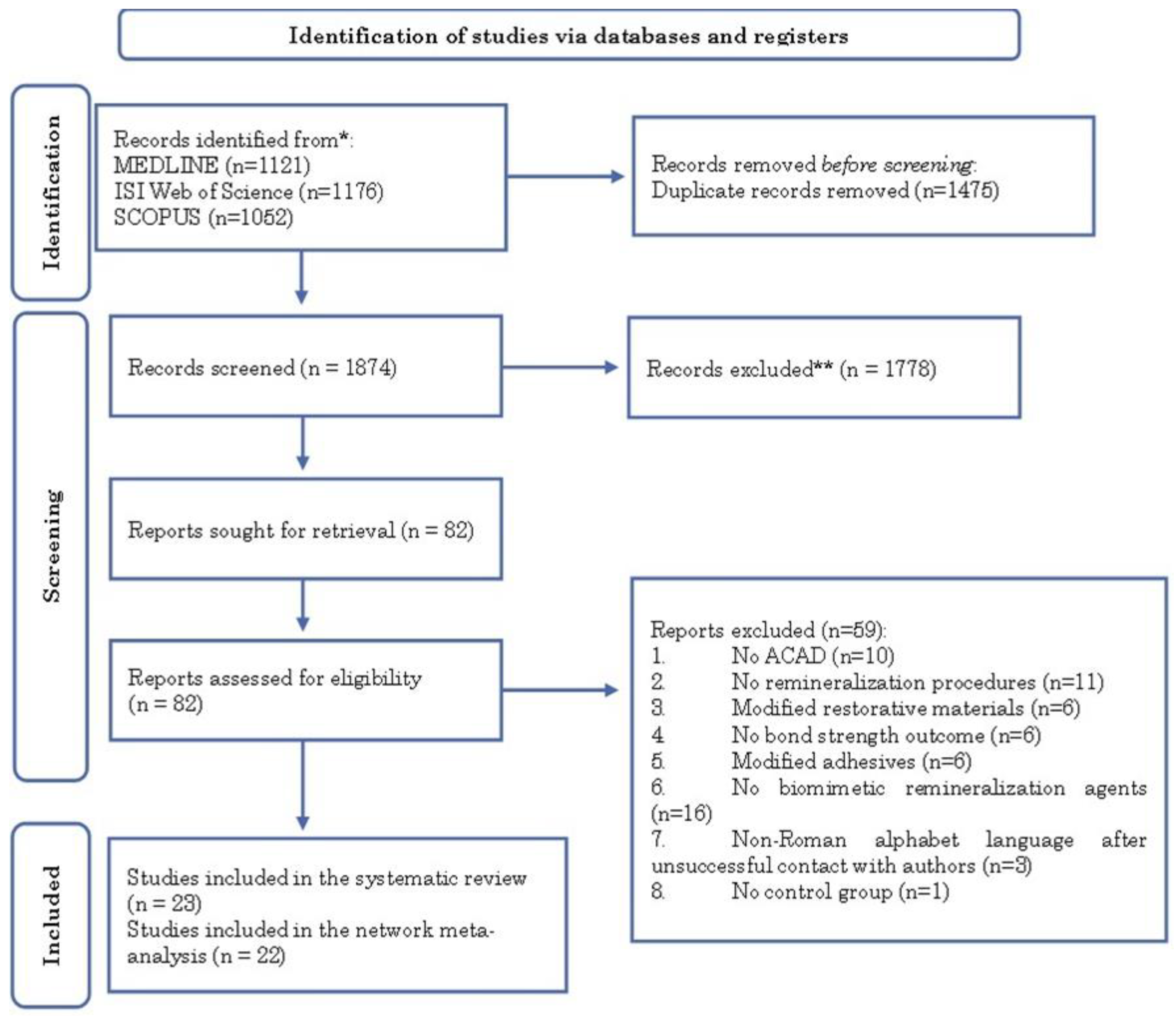
Table 1.
Characteristics of the included studies, interventions, and outcomes.
| Study/Year | RoB (score) | Study type | ACAD | BRP | Groups | N (teeth) | Mean (SD) | AT | OM Test | |
|---|---|---|---|---|---|---|---|---|---|---|
| 24-hour measurement | ||||||||||
| ER + C | Altinci et al. 2018 [31] | M(50) | Exp. | 32% phosphoric acid | Control | Control | 9 | 35.27 (4.63)a | ER | µTBS |
| F | NaF + 6mM F | 34.7 (4.63) a | ||||||||
| NaF + 24mM F | 54.66 (4.63) a | |||||||||
| NaF+179mM F | 47.11 (4.63) a | |||||||||
| KF + 6mM F | 51.8 (4.63) a | |||||||||
| KF + 24mM F | 48.56 (4.63) a | |||||||||
| KF + 179mM F | 47.58 (4.63) a | |||||||||
| CaF2 + 6mM F | 36.34 (4.63) a | |||||||||
| CaF2+24mM F | 39.49 (4.63) a | |||||||||
| CaF2+179mM F | 48.47 (4.63) a | |||||||||
| Excite F | 48.84 (4.63) a | |||||||||
| Barbosa-Martins et al. (A) 2018[8] | M(54) | Exp. | 6% CMC | Control | Control | 6 | 26.38 (8.64) | ER | µTBS | |
| F | NaF | 33.43 (10.41) | ||||||||
| CaP | CPP-ACP | 45.25 (8.82) | ||||||||
| Pept. | P11-4 | 46.42 (12.03) | ||||||||
| Barbosa-Martins et al. (B) 2018[7] | M(54) | Quasi-Exp. | 6% CMC | Control | Control | 6 | 21.96 (5.92) | ER | µTBS | |
| F | NaF | 33.43 (10.42) | ||||||||
| CaP | CPP-ACP | 45.25 (8.83) | ||||||||
| Pept. | P11-4 | 46.42 (12.03) | ||||||||
| Bauer et al. 2018[18] | M(50) | Exp. | 35% phosphoric acid | Control | Control | 13 | 17 (4.1) | ER | SBS | |
| CaP | 5% NbG | 17.9 (5) | ||||||||
| 10%NbG | 15.8 (6.4) | |||||||||
| 20%NbG | 16.6 (4.4) | |||||||||
| 40%NbG | 15.8 (4.1) | |||||||||
| Cardenas et al. 2021[19] | M(63) | Exp. | pH cycling | Control | Control | 5 | 33.74 (3.6) | Univ. | µTBS | |
| F | SDF 12% | 38.03 (3.5) | ||||||||
| SDF 38% | 39.68 (2.7) | |||||||||
| SDF 38% without KI | 39.38 (2.5) | |||||||||
| Control | Control | 34.9 (3.3) | ||||||||
| F | SDF 12% | 42.45 (2.9) | ||||||||
| SDF 38% | 40.47 (4.2) | |||||||||
| SDF 38% without KI | 41.3 (2.5) | |||||||||
| Chen et al. 2020[14] | M(54) | Quasi-Exp. | pH cycling | Control | Control | 4 | 13.8 (3.35) a | Univ. | µTBS | |
| CaP | Ca/P-PILP | 23.8 (3.35) a | ||||||||
| Pept. | PAA-PASP | 14 (3.35) a | ||||||||
| CaP | Ca/P | 11.9 (3.35) a | ||||||||
| Cifuentes-Jimenez et al. 2021[20] | M(50) | Exp. | pH cycling | Control | Control | 5 | 31.4 (4.63) a | ER | µTBS | |
| F | Cariestop | 15.1 (4.63) a | ||||||||
| RivaStar1 | 10.1 (4.63) a | |||||||||
| RivaStar2 | 7.5 (4.63) a | |||||||||
| Saforide | 23.2 (4.63) a | |||||||||
| Gungormus et al. 2021[22] | M(50) | Exp. | 37% phosphoric acid | Control | Control | 10 | 15.38 (1.3) | ER | SBS | |
| CaP | NPR 60min | 15.85 (1.44) | ||||||||
| Pept. | PR 10 min | 20.81 (1.74) | ||||||||
| PR 30 min | 20 (1.68) | |||||||||
| PR 60 min | 16.21 (1.1) | |||||||||
| Krithi et al. 2020[23] | M(54) | Exp. | 0.5% citric acid | Control | Control | 15 | 11.83 (0.43) | ER | µSBS | |
| F | NaF | 11.56 (0.15) | ||||||||
| CaP | CPP-ACP | 12.12 (0.57) | ||||||||
| Novamin | 11.66 (0.28) | |||||||||
| Ca | Non-Fidated | 11.94 (0.27) | ||||||||
| Meng et al. 2021[24] | M(50) | Exp. | 1% citric acid | Control | Control | 8 | 46.8b (4.63) a | Univ. | µTBS | |
| Hap | Biorepair | 50.72 b (4.63) a | ||||||||
| Dontodent Sensitive | 50.71 b (4.63) a | |||||||||
| nHAp | 51.24 b (4.63) a | |||||||||
| Control | Control | 50.41 b (4.63) a | ||||||||
| Hap | Biorepair | 53.38 b (4.63) a | ||||||||
| Dontodent Sensitive | 54.5 b (4.63) a | |||||||||
| nHAp | 55.63 b (4.63) a | |||||||||
| Control | Control | 46.85 b (4.63) a | ||||||||
| Hap | Biorepair | 50.77 b (4.63) a | ||||||||
| Dontodent Sensitive | 53.82 b (4.63) a | |||||||||
| nHAp | 55 b (4.63) a | |||||||||
| Pulidindi et al. 2021[40] | M(63) | Exp. | 37% phosphoric acid | Control | Control | 15 | 48.84 (4.63) a | ER | µTBS | |
| Pept. | P11-4 | 38.66 (4.63) a | ||||||||
| CaP | CPP-ACP | 34.07 (4.63) a | ||||||||
| Control | Control | 22.63 (4.63) a | ||||||||
| Pept. | P11-4 | 25.37 (4.63) a | ||||||||
| CaP | CPP-ACP | 23.62 (4.63) a | ||||||||
| Van Duker et al. 2019[35] | H(46) | Quasi-Exp. | 7 days in ADS | Control | Control | 10 | 23.5 (10.7) | Univ. | µTBS | |
| F | SDF 38% | 19.8 (8.4) | ||||||||
| SDF 38% without KI | 7.9 (6.6) | |||||||||
| Yang et al. 2018[29] | M(50) | Exp. | 1% citric acid | Control | Control | 10 | 46.5 b (4.63)a | ER | µTBS | |
| CaP | CPP-ACP | 42.6 b (4.63) a | ||||||||
| Novamin | 43.3 b (4.63) a | |||||||||
| Control | Control | 22.3 b (4.63) a | ||||||||
| CaP | CPP-ACP | 41.2 b (4.63) a | ||||||||
| Novamin | 31.4 b (4.63) a | |||||||||
| ER + B | Barbosa-Martins et al. (B) 2018 | M(54) | Quasi-Exp. | BHI+ S.Mutans | Control | Control | 6 | 22.89 (2.68) | ER | µTBS |
| F | NaF | 26.94 (6.7) | ||||||||
| CaP | CPP-ACP | 47.95 (6.69) | ||||||||
| Pept. | P11-4 | 42.07 (7.83) | ||||||||
| Dávila-Sánchez et al. 2020[21] | M(54) | Exp. | Cariogenic+ S. Mutans | Control | Control | 7 | 14.42 (4.43) | Univ. | µTBS | |
| Fls. | QUE | 24.58 (4.9) | ||||||||
| HES | 18.41 (5.3) | |||||||||
| RUT | 26 (5.51) | |||||||||
| NAR | 24.64 (3.7) | |||||||||
| PRO | 20.66 (3.92) | |||||||||
| de Sousa et al. 2019[33] | M(50) | Quasi-Exp. | Cariogenic+ S. Mutans | Control | Control | 8 | 21.07 (3.24) | ER | µTBS | |
| Pept. | P11-4 | 42.07 (7.83) | ||||||||
| Moreira et al. 2021[15] | M(54) | Exp. | Cariogenic+ S. Mutans | Control | Control | 8 | 25.4 (2.45) | ER | µTBS | |
| F | NaF | 25.47 (4.8) | ||||||||
| CaP | CPP-ACP | 41.79 (5.85) | ||||||||
| Pept. | P11-4 | 40.12 (3.62) | ||||||||
| Siqueira et al. 2020[28] | M(63) | Exp. | Cariogenic+ S. Mutans | Control | Control | 5 | 16.81 (3.5) | Univ. | µTBS | |
| F | SDF 12% | 21.11 (4.1) | ||||||||
| SDF 38% | 24.36 (3.4) | |||||||||
| Control | Control | 19.89 (2.4) | ||||||||
| F | SDF 12% | 24.47 (3.4) | ||||||||
| SDF 38% | 26.32 (2) | |||||||||
| SE + C | Atomura et al. 2018[32] | H(46) | Quasi-Exp. | 7 days in ADS | Control | Control | unknown | 48.3 (13) | SE | µTBS |
| F | NaF | 47.7 (8.6) | ||||||||
| FCP complex | 43.9 (14.3) | |||||||||
| Barbosa-Martins et al. (A) 2018 | M(54) | Exp. | 48h 6% CMC | Control | Control | 6 | 25.38 (8.58) | SE | µTBS | |
| F | NaF | 35.59 (9.18) | ||||||||
| CaP | CPP-ACP | 48.11 (11.71) | ||||||||
| Pept. | P11-4 | 25.7 (8.95) | ||||||||
| Cardenas et al. 2021 | M(63) | Exp. | pH cycling | Control | Control | 5 | 33.74 (3.6) | Univ. | µTBS | |
| F | SDF 12% | 39.53 (4.2) | ||||||||
| SDF 38% | 41.31 (2) | |||||||||
| SDF 38% without KI | 40.55 (2.9) | |||||||||
| Control | Control | 36.56 (4.1) | ||||||||
| F | SDF 12% | 39.98 (1.7) | ||||||||
| SDF 38% | 41.08 (3) | |||||||||
| SDF 38% without KI | 41.57 (2.4) | |||||||||
| Chen et al. 2020 | M(54) | Quasi-Exp. | pH cycling | Control | Control | 4 | 13.8 (3.35) a | Univ. | µTBS | |
| CaP | Ca/P-PILP | 23.8 (3.35) a | ||||||||
| Pept. | PAA-PASP | 14 (3.35) a | ||||||||
| CaP | Ca/P | 11.9 (3.35) a | ||||||||
| Control | Control | 9.2 (3.35) a | ||||||||
| CaP | Ca/P-PILP | 15.1 (3.35) a | ||||||||
| Pept. | PAA-PASP | 9.3 (3.35) a | ||||||||
| CaP | Ca/P | 9.8 (3.35) a | ||||||||
| Cifuentes-Jimenez et al. 2021 | M(50) | Exp. | pH cycling | Control | Control | 5 | 31.4 (3.35) a | SE | µTBS | |
| F | Cariestop | 9.6 (3.35) a | ||||||||
| Saforide | 8.03 (3.35) a | |||||||||
| Gungormus et al. 2021 | M(50) | Exp. | 37% phosphoric acid | Control | Control | 10 | 15.38 (1.3) | SE | SBS | |
| CaP | NPR 60min | 15.49 (1.17) | ||||||||
| Pept. | PR 10 min | 18.93 (0.99) | ||||||||
| PR 30 min | 19.62 (0.9) | |||||||||
| PR 60 min | 21.73 (1.57) | |||||||||
| Krithi et al. 2020 | M(54) | Exp. | 0.5% citric acid | Control | Control | 15 | 11.83 (0.43) | SE | µSBS | |
| F | NaF | 12.4 (0.18) | ||||||||
| CaP | CPP-ACP | 11.97 (0.39) | ||||||||
| Novamin | 11.97 (0.17) | |||||||||
| Ca | Non-Fidated | 10.62 (0.11) | ||||||||
| Meng et al. 2021 | M(50) | Exp. | 1% citric acid | Control | Control | 8 | 46.8 b (3.35) a | Univ. | µTBS | |
| Hap | Biorepair | 47.62 b (3.35) a | ||||||||
| Dontodent Sensitive | 51.89 b (3.35) a | |||||||||
| nHAp | 51.89 b (3.35) a | |||||||||
| Control | Control | 56.3 b (3.35) a | ||||||||
| Hap | Biorepair | 51.62 b (3.35) a | ||||||||
| Dontodent Sensitive | 57.47 b (3.35) a | |||||||||
| nHAp | 58.39 b (3.35) a | |||||||||
| Control | Control | 56.8 b (3.35) a | ||||||||
| Hap | Biorepair | 52.25 b (3.35) a | ||||||||
| Dontodent Sensitive | 50.8 b (3.35) a | |||||||||
| nHAp | 56.1 b (3.35) a | |||||||||
| Paik et al. 2022[25] | M(50) | Exp. | 35% phosphoric acid | Control | Control | 4 | 21.66 (3.35) a | Univ. | µTBS | |
| Fls. | ICT | 24.4 (3.35) a | ||||||||
| FIS | 26.81 (3.35) a | |||||||||
| SIB | 25.65 (3.35) a | |||||||||
| CPIC | 25.97 (3.35) a | |||||||||
| ICT+ C | 30.63 (3.35) a | |||||||||
| FIS+ C | 25.63 (3.35) a | |||||||||
| SIB+ C | 24.76 (3.35) a | |||||||||
| Pei et al. 2019[26] | M(50) | Exp. | 1% citric acid | Control | Control | 4 | 43.61 (3.35) a | SE | µTBS | |
| Hap | Biorepair | 33.16 (3.35) a | ||||||||
| Dontodent Sensit. | 35.41 (3.35) a | |||||||||
| nHAp | 46.92 (3.35) a | |||||||||
| Control | Control | 47.47 (3.35) a | ||||||||
| Hap | Biorepair | 43.47 (3.35) a | ||||||||
| Dontodent Sensit. | 42.3 (3.35) a | |||||||||
| nHAp | 41.24 (3.35) a | |||||||||
| Priya et al. 2020[34] | H(46) | Quasi-Exp. | 37% phosphoric acid | Control | Control | 13 | 6.677 (1.254) | Univ. | SBS | |
| F | VivaSens | 3.332 (0.78) | ||||||||
| MS Coat F | 3.127 (0.478) | |||||||||
| HEMA | GLUMA Desensit. | 4.572 (0.718) | ||||||||
| Systemp | 9.697 (1.127) | |||||||||
| Zang et al. 2018[30] | M(50) | Exp. | 37% phosphoric acid | Control | Control | 6 | 19.73 b (2.108) | Univ. | SBS | |
| SiO2 | Charged mesoporous | 20.57 b (2.244) | ||||||||
| Zumstein et al. 2018[42] | M(50) | Quasi-Exp. | pH cycling | Control | Control | 20 | 24.7 (8.1)c | SE | µTBS | |
| F | SnCl2/ AmF4 | 23.3 (8.2) c | ||||||||
| Control | Control | 23.73 (8) c | Univ. | |||||||
| F | SnCl2/ AmF4 | 21.39 (6.8) c | ||||||||
| SE + B | Siqueira et al. 2020 | M(63) | Exp. | Cariogenic+ S. Mutans | Control | Control | 5 | 16.81 (3.5) | Univ. | µTBS |
| F | SDF 12% | 20.02 (4.6) | ||||||||
| SDF 38% | 25.21 (3) | |||||||||
| Control | Control | 19.61 (3.3) | ||||||||
| F | SDF 12% | 23.82 (4.4) | ||||||||
| SDF 38% | 27.16 (3.6) | |||||||||
| TMC measurement | ||||||||||
| ER + C | Pulidindi et al. 2021 | M(63) | Exp. | 37% phosphoric acid | Control | Control | 15 | 48.84 (4.63) a | ER | µTBS |
| Pept. | P11-4 | 25.37 (4.63) a | ||||||||
| CaP | CPP-ACP | 23.62 (4.63) a | ||||||||
| ER + B | Dávila-Sánchez et al. 2020 | M(54) | Exp. | Cariogenic+ S. Mutans | Control | Control | 7 | 14.42 (4.43) | Univ. | µTBS |
| Fls. | QUE | 12.02 (5.21) | ||||||||
| HES | 15.73 (6.07) | |||||||||
| RUT | 21.08 (4.75) | |||||||||
| NAR | 22.12 (2.92) | |||||||||
| PRO | 17.2 (2.72) | |||||||||
| SE + C | Chen et al. 2020 | M(54) | Quasi-Exp. | pH cycling | Control | Control | 4 | 13.8 (3.35) a | Univ. | µTBS |
| CaP | Ca/P-PILP | 15.1 (3.35) a | ||||||||
| Pept. | PAA-PASP | 9.3 (3.35) a | ||||||||
| CaP | Ca/P | 9.8 (3.35) a | ||||||||
| Paik et al. 2022 | M(50) | Exp. | 35% phosphoric acid | Control | Control | 4 | 21.66 (3.35) a | Univ. | µTBS | |
| Fls. | ICT | 20.53 (3.35) a | ||||||||
| FIS | 19.4 (3.35) a | |||||||||
| SIB | 22.04 (3.35) a | |||||||||
| CPIC | 23.43 (3.35) a | |||||||||
| ICT+ C | 26.74 (3.35) a | |||||||||
| FIS+ C | 23.42 (3.35) a | |||||||||
| SIB+ C | 25.17 (3.35) a | |||||||||
| Storage in a fluid solution for 3-month measurement | ||||||||||
| ER + C | Bauer et al. 2018 | M(50) | Exp. | 35% phosphoric acid | Control | Control | 13 | 17 (4.1) | ER | SBS |
| CaP | 5% NbG | 11.8 (3.7) | ||||||||
| 10%NbG | 13.9 (3.2) | |||||||||
| 20%NbG | 13.2 (2.7) | |||||||||
| 40%NbG | 14.7 (2.9) | |||||||||
| SE + C | Atomura et al. 2018 | H(46) | Quasi-Exp. | 7 days in ADS | Control | Control | unknown | 48.3 (13) | SE | µTBS |
| F | NaF | 42.6 (12.1) | ||||||||
| FCP complex | 47.4 (9.2) | |||||||||
| Storage in a fluid solution for 6-month measurement | ||||||||||
| ER + C | Altinci et al. 2018 | M(50) | Exp. | 32% phosphoric acid | Control | Control | 9 | 35.27 (4.63) a | ER | µTBS |
| F | NaF + 6mM F | 50.31 (4.63) a | ||||||||
| NaF + 24mM F | 49.28 (4.63) a | |||||||||
| NaF+179mM F | 47.73 (4.63) a | |||||||||
| KF + 6mM F | 41.95 (4.63) a | |||||||||
| KF + 24mM F | 51.53 (4.63) a | |||||||||
| KF + 179mM F | 54.29 (4.63) a | |||||||||
| CaF2 + 6mM F | 52.25 (4.63) a | |||||||||
| CaF2+24mM F | 41.1 (4.63) a | |||||||||
| CaF2+179mM F | 40.85 (4.63) a | |||||||||
| Excite F |
46.22 (4.63) a | |||||||||
| de Sousa et al. 2019 | M(50) | Quasi-Exp. | Cariogenic+ S. Mutans | Control | Control | 8 | 21.07 (3.24) | ER | µTBS | |
| Pept. | P11-4 | 31.98 (3.44) | ||||||||
| Moreira et al. 2021 | M(54) | Exp. | Cariogenic+ S. Mutans | Control | Control | 8 | 25.4 (2.45) | ER | µTBS | |
| F | NaF | 18.36 (5.5) | ||||||||
| CaP | CPP-ACP | 36.55 (4.27) | ||||||||
| Storage in a fluid solution for 12-month measurement | ||||||||||
| ER+C | Altinci et al. 2018 | M(50) | Exp. | 32% phosphoric acid | Control | Control | 9 | 35.27 (4.63) a | ER | µTBS |
| F | NaF + 6mM F | 51.63 (4.63) a | ||||||||
| NaF + 24mM F | 45.56 (4.63) a | |||||||||
| NaF+179mM F | 39.31 (4.63) a | |||||||||
| KF + 6mM F | 40.01 (4.63) a | |||||||||
| KF + 24mM F | 51.85 (4.63) a | |||||||||
| KF + 179mM F | 36.48 (4.63) a | |||||||||
| CaF2 + 6mM F | 33.06 (4.63) a | |||||||||
| CaF2+24mM F | 38.24 (4.63) a | |||||||||
| CaF2+179mM F | 0.88 (4.63) a | |||||||||
| Excite F | 42.4 (4.63) a | |||||||||
| Yang et al. 2018 | M(50) | Exp. | 1% citric acid | Control | Control | 10 | 46.5 b (4.63) a | ER | µTBS | |
| CaP | CPP-ACP | 41.2 b (4.63) a | ||||||||
| Novamin | 31.4 b (4.63) a | |||||||||
| SE+C | Zumstein et al. 2018 | M(50) | Quasi-Exp. | pH cycling | Control | Control | 20 | 24.7 (8.1)c | SE | µTBS |
| F | SnCl2/ AmF4 | 16.3 (6.36)c | ||||||||
| Control | Control | 15.43 (6.53)c | Univ. | |||||||
| F | SnCl2/ AmF4 | 14.12 (7.12)c | ||||||||
| Storage in a fluid solution for 18-month measurement | ||||||||||
| ER+B | Moreira et al. 2021 | M(54) | Exp. | Cariogenic+ S. Mutans | Control | Control | 8 | 25.4 (2.45) | ER | µTBS |
| F | NaF | 7.81 (4.48) | ||||||||
| CaP | CPP-ACP | 26.01 (3.28) | ||||||||
| Pept. | P11-4 | 25.24 (3.98) | ||||||||
| a- Input SD Values; b- Information given by authors; c- Information from another meta-analysis. | ||||||||||
| Legend: B- Biological; C- Chemical; RoB- Risk of bias; ACAD- Artificial caries-affected dentin; BRP- Biomimetic remineralization procedure; SD- Standard deviation; AT- Adhesive technique; OM-Outcome measurement; ADS- Artificial demineralization solution; M- Medium; H- High; Exp.-Experimental; ER- Etch-and-rinse; SE- Self-etch; Univ.- Universal; F- Fluorine; Ca-Calcium; CaP- Calcium phosphate; Pept.- Peptide; FLs- Flavonoids; SiO2- Silica Hap- Hidroxiapatite; HEMA- 2-hydroxyethyl methacrylate; TMC- Thermocycling; µTBS- microtensile bond strengh; SBS- shear bond strength; µSBS- microshear bond strength. | ||||||||||
Table 2.
Network meta-analysis plots.
| Plot of the NMA ER + chemical ACAD |
Plot of the NMA ER + biological ACAD |
Plot of the NMA SE + chemical ACAD |
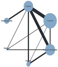 |
 |
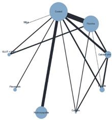 |
1 Note: Black lines connect biomimetic remineralization interventions that were compared head-to-head. The size of each node (circle) provides a measure of the sample size. The thickness of the line provides a measure of the number of direct comparisons between two interventions. Legend: ACAD- Artificial caries-affected dentin; ER- Etch-and-Rinse; NMA- Network meta-analysis; SE- Self-etch.
Table 3.
Network meta-analysis results from the network of biomimetic remineralization interventions.
Table 3.
Network meta-analysis results from the network of biomimetic remineralization interventions.
 |
* Note: Data in each cell are the mean difference with 95% confidence intervals for the network comparison of row-defining treatment versus column-defining treatment. Negative values favor the intervention in the column. Statistically significant results are in bold and gray. Legend: ER- Etch-and-Rinse; SE- Self-etch; HAp- Hydroxyapatite; HEMA- 2-hydroxyethyl methacrylate.
Table 4.
Treatment rankings and probability of ranking best.
| NMA | Ranks and probability of ranking best |
|---|---|
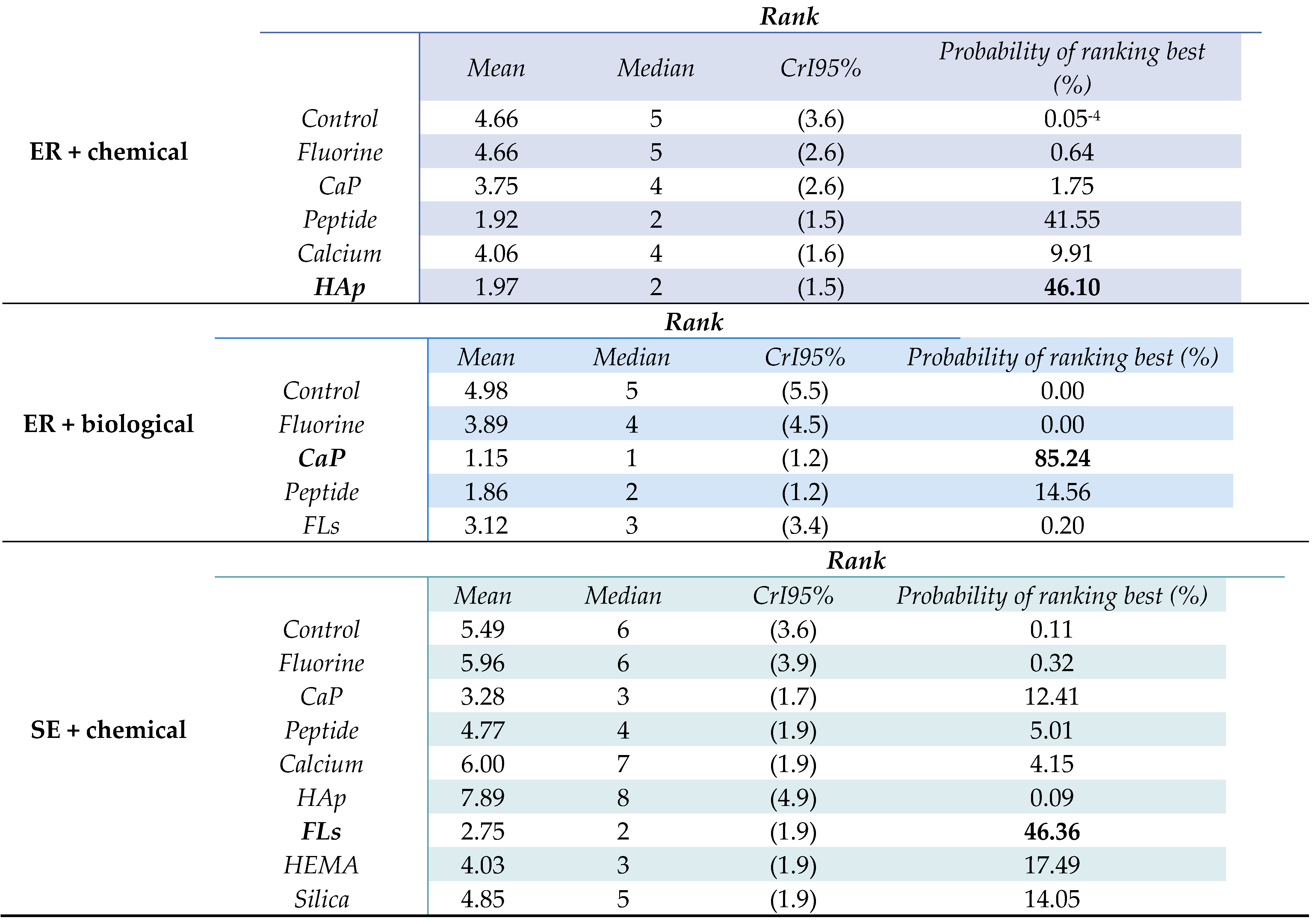 | |
* Note: Interventions ranked best are highlighted in bold. Legend: ER- Etch-and-Rinse; SE- Self-etch; CrI- Credible interval; CaP- Calcium phosphate; FLs- Flavonoids, HAp- Hydroxyapatite; HEMA- 2-hydroxyethyl methacrylate.
Disclaimer/Publisher’s Note: The statements, opinions and data contained in all publications are solely those of the individual author(s) and contributor(s) and not of MDPI and/or the editor(s). MDPI and/or the editor(s) disclaim responsibility for any injury to people or property resulting from any ideas, methods, instructions or products referred to in the content. |
© 2025 by the authors. Licensee MDPI, Basel, Switzerland. This article is an open access article distributed under the terms and conditions of the Creative Commons Attribution (CC BY) license (http://creativecommons.org/licenses/by/4.0/).
Copyright: This open access article is published under a Creative Commons CC BY 4.0 license, which permit the free download, distribution, and reuse, provided that the author and preprint are cited in any reuse.


