

Submitted:
10 March 2025
Posted:
11 March 2025
You are already at the latest version
Abstract
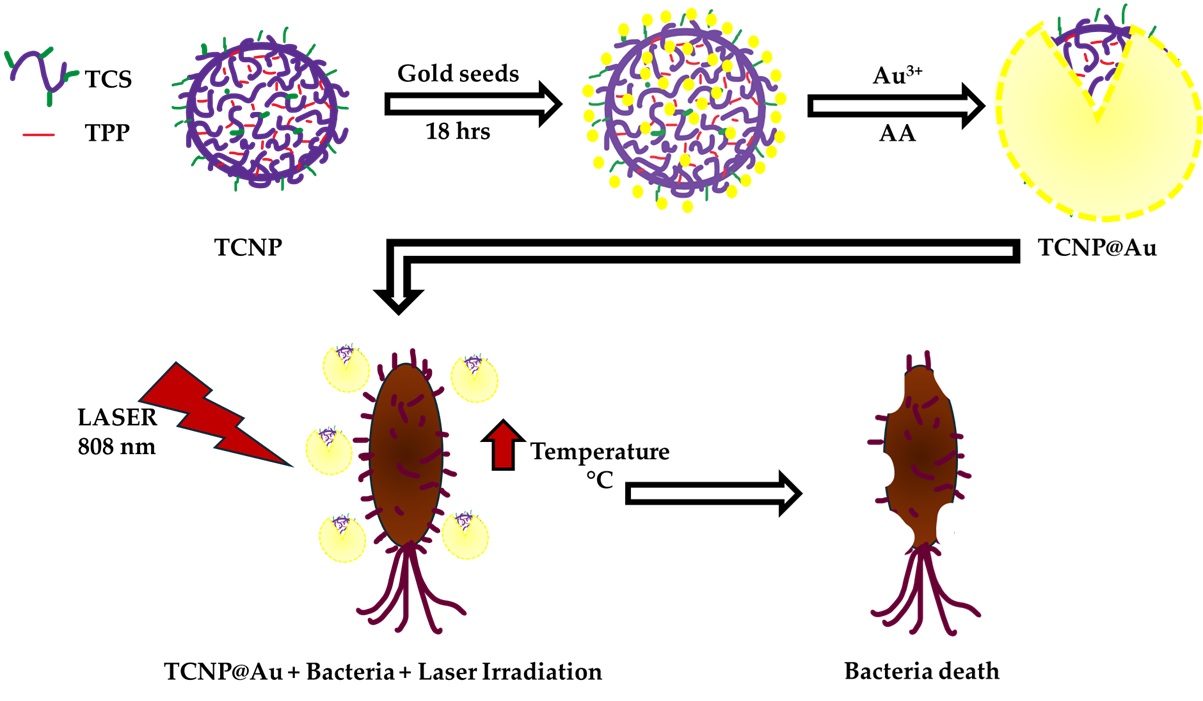
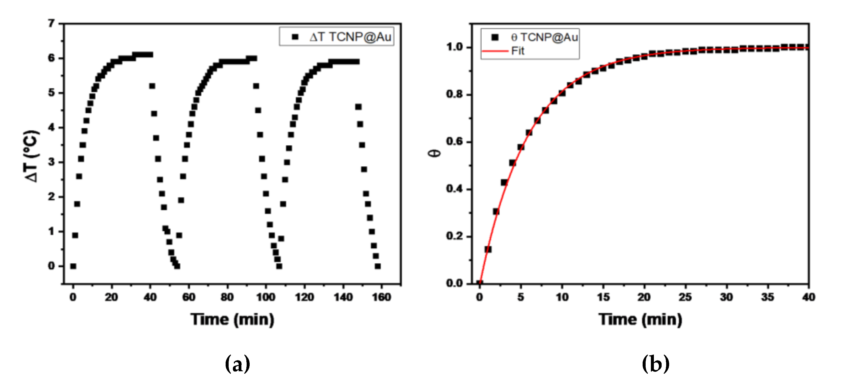
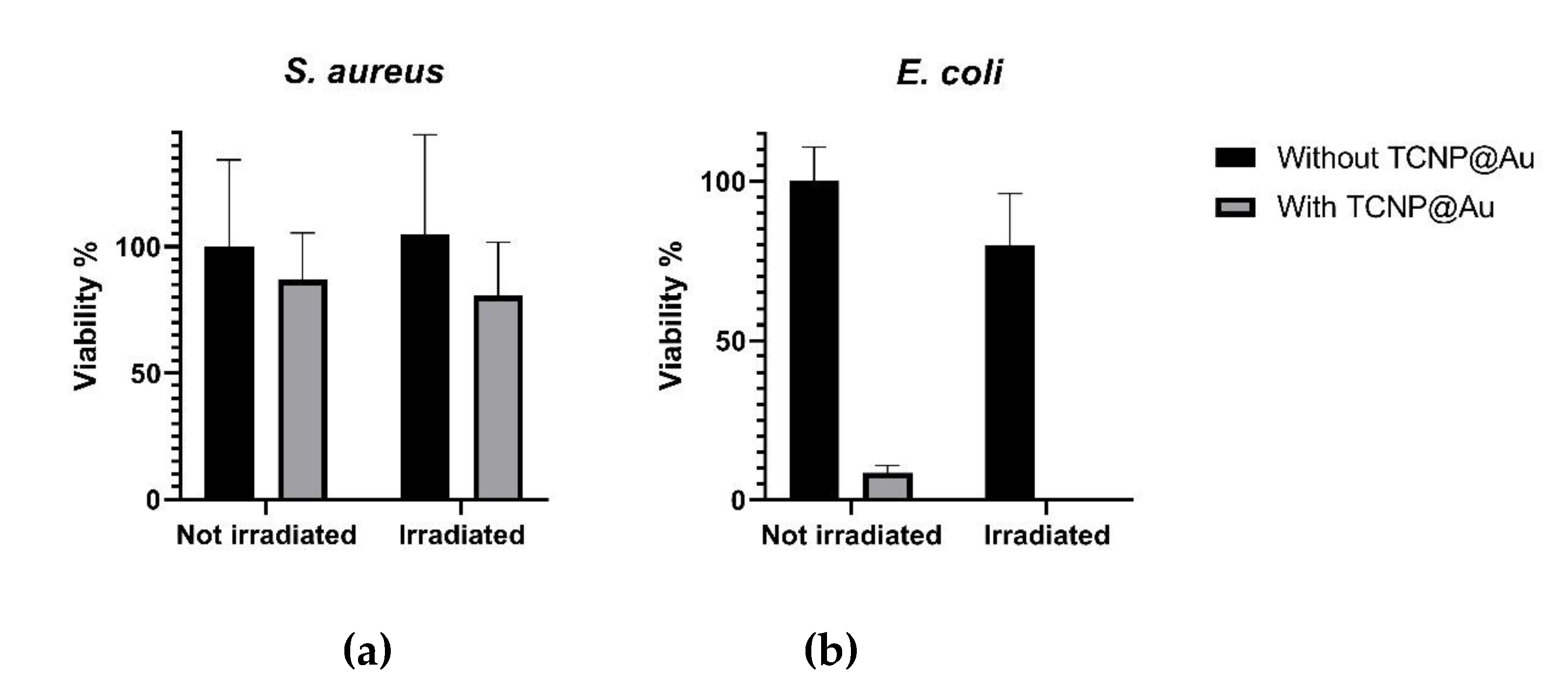
Bacterial infections remain among the top ten major public health concerns, contributing to a high number incidence of disease and mortality worldwide. This issue has been exacerbated by the rise of multidrug-resistant bacteria (MDRB). Consequently, it is crucial to develop novel antimicrobial strategies, including the use of functional nanoparticles. Gold nanoparticles (GNPs) have emerged as promising candidates due to their unique optical properties, particularly their ability to efficiently convert absorbed light into heat through the photothermal (PT) effect, which can be harnessed for bacteria eradication. In this study, gold nanoshells (GNSs) were synthesized and proposed as photothermal devices to bacteria clearance via PT. First, chitosan was modified with 3-mercaptopropionic acid to introduce sulfur groups, facilitating gold deposition onto chitosan nanoparticle surface. Thiolated chitosan nanoparticles (TCNPs), with a size of 178 nm and spherical morphology were synthesized using the ionic gelation method. The gold shell was subsequently formed via a seed-mediated method, wherein gold seeds were adsorbed onto TCNPs and further grown to form the shell. The resulting TCNP@Au exhibited a photothermal conversion efficiency of 31%, making them a promising photothermal agent for bacterial clearance. Notably, the viability of Escherichia coli was significantly reduced in the presence of TCNP@Au and was almost eradicated upon PT treatment, with viability dropping to 0.3 %. In contrast, TCNP@Au were non-toxic for Staphylococcus aureus. Interestingly, S. aureus exhibited a reduced susceptibility to the PT effect, maintaining a viability of 76 % after the laser irradiation treatment. Despite these results, TCNP@Au demonstrated favorable photothermal properties, presenting a novel nanoplatform for antibacterial applications, particularly against Gram-negative bacteria. However, further investigation is required to optimize the PT-based strategies against Gram-positive bacteria, such as S. aureus.
Keywords:
1. Introduction
2. Results
2.1. Chitosan Modification and Synthesis of Chitosan Nanoparticles (TCNP)
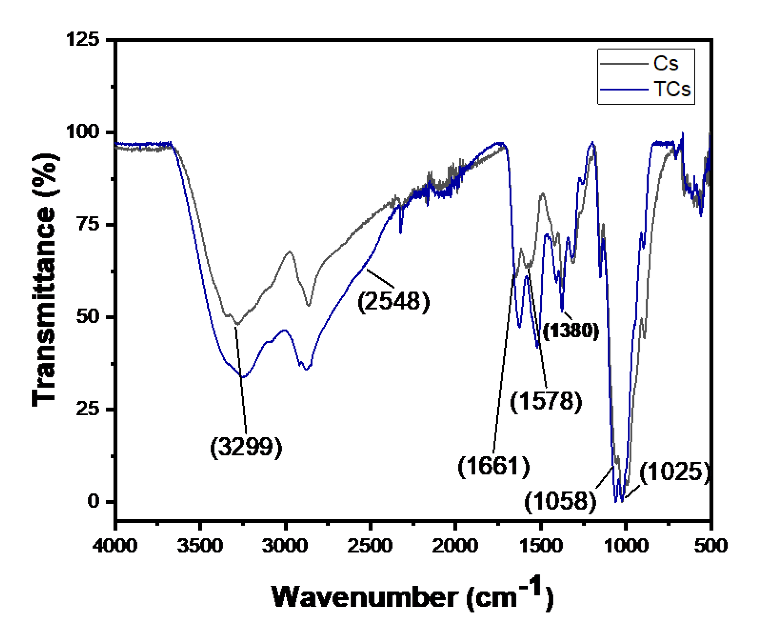
2.1.1. Chitosan Modification
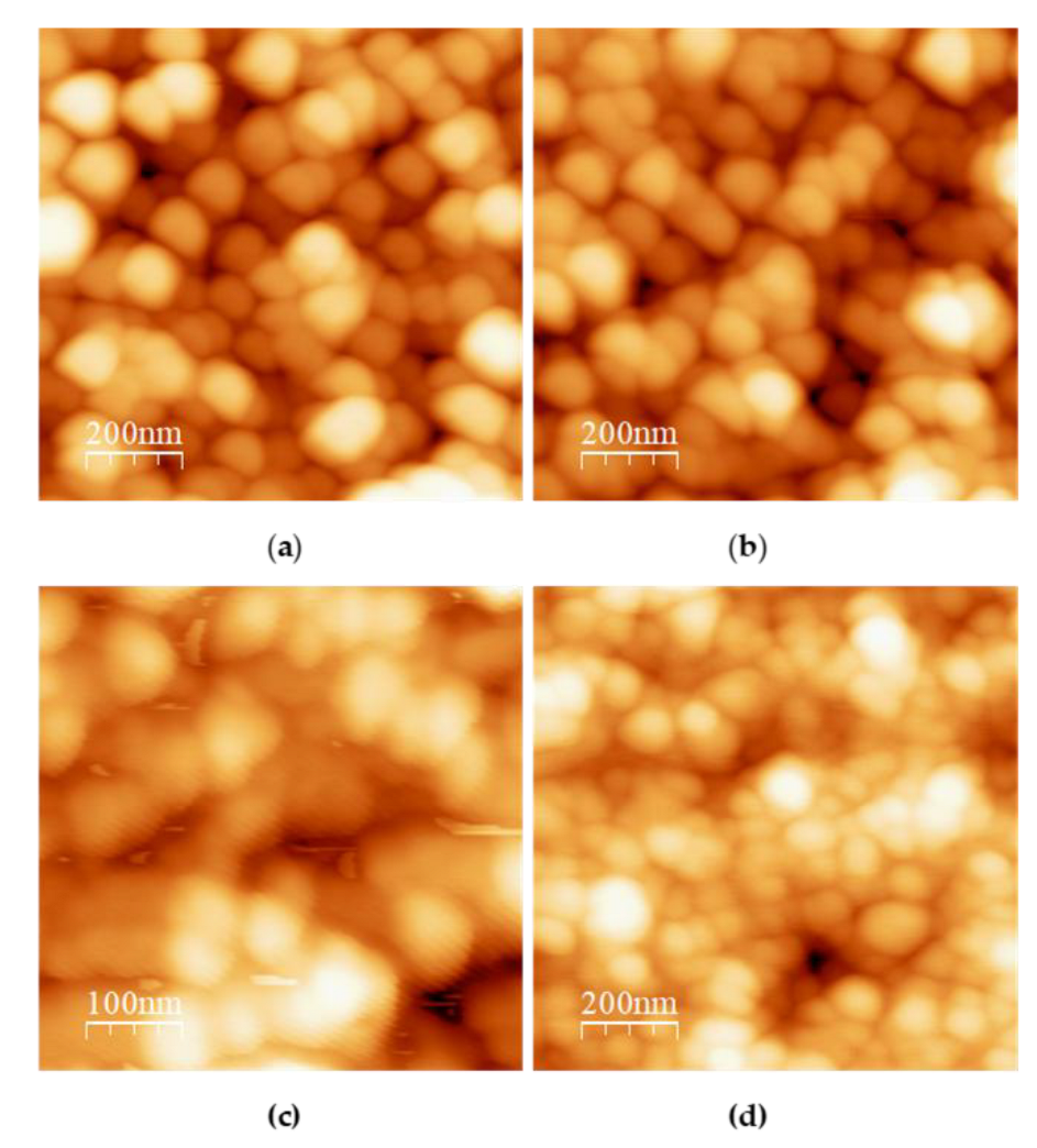
2.1.1. Synthesis of TCNPs
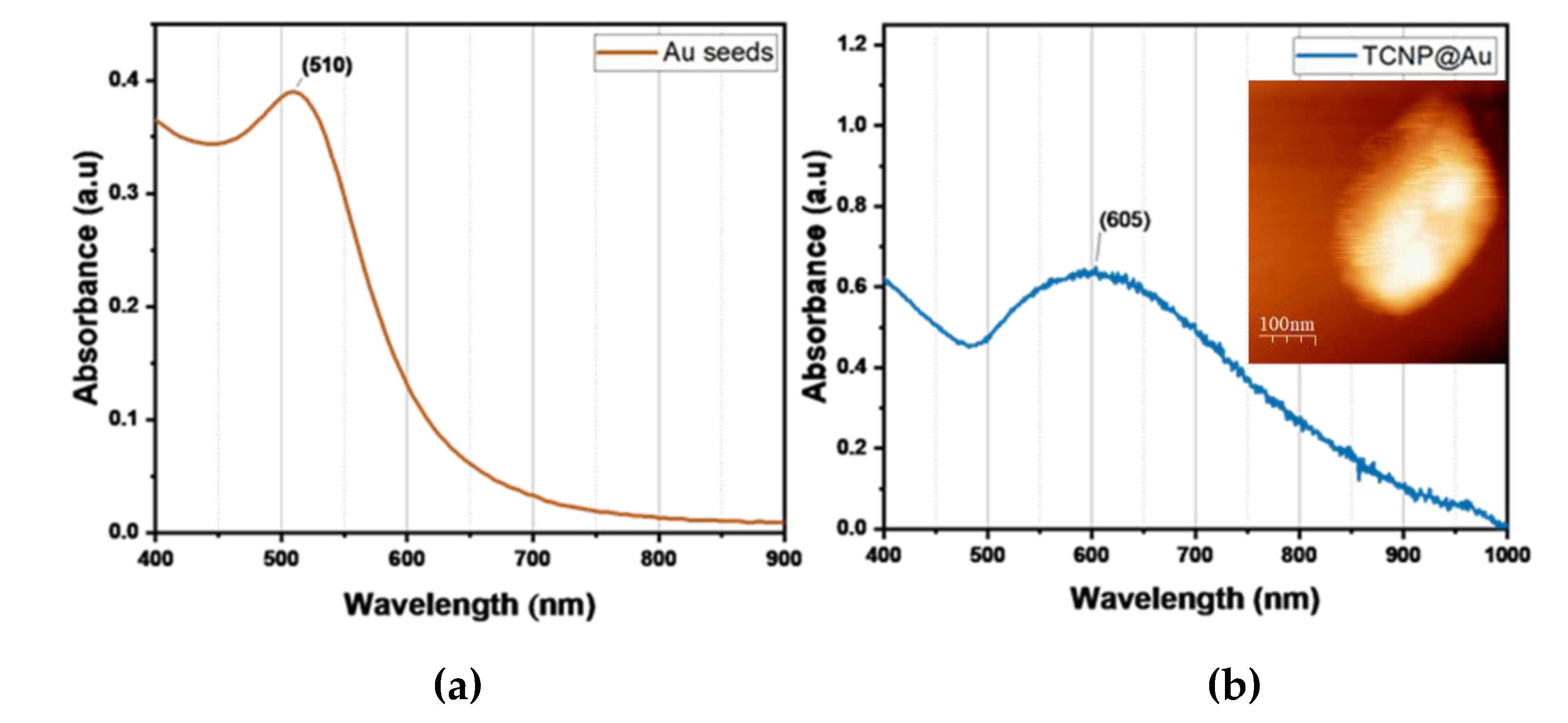
2.2. Core-Shell Chitosan-Gold Nanoparticles
2.2.1. Photothermal Conversion Efficiency of TCNP@Au
2.3. Photothermal Effect of TCNP@Au on the Viability of Gram-Positive and Gram-Negative Bacteria
3. Materials and Methods
3.1. Thiolated Chitosan (TCs)
3.1.1. Fourier Transform Infrared Spectroscopy-Attenuated Total Reflectance (FTIR-ATR)
3.2. Synthesis of Thiolated Chitosan Nanoparticles (TCsNp)
3.3. Synthesis of Gold-Shell on TCSNPs
3.4. Characterization
3.4.1. Nanoparticles Size Analysis by Dynamic Light Scattering (DLS)
3.4.2. Atomic Force Microscopy
3.4.3. UV-Vis Spectroscopy
3.5. Photothermal Conversion of TCNP@Au
4.5. Photothermal Effect on the Bacterial Growth
5. Conclusions
Author Contributions
Funding
Data Availability Statement
Acknowledgments
Conflicts of Interest
Abbreviations
| Cs | Chitosan |
| TPP | Sodium Triphosphate Pentabasic |
| 3-MPA | 3-mercaptopropionic acid |
| TCs | Thiolated Chitosan |
| FTIR-ATR | Fourier Transform Infrared Spectroscopy-Attenuated Total Reflectance |
| TCsNPs | Thiolated Chitosan Nanoparticles |
| TCs@AuNp | Core-Shell Chitosan-Gold Nanoparticles |
| AFM | Atomic Force Microscopy |
| AuSD | Gold Seeds |
| AA | Ascorbic Acid |
| LSPR | Localized Surface Plasmonic Resonance |
| PDI | Polydispersity Index |
References
- A. Karnwal, A. Y. A. Karnwal, A. Y. Jassim, A. A. Mohammed, A. R. Mohammad Said Al-Tawaha, M. Selvaraj, and T. Malik, “Addressing the global challenge of bacterial drug resistance : insights, strategies, and future directions,” no. 2025. [Google Scholar] [CrossRef]
- S. Ajulo and B. Awosile, “Global antimicrobial resistance and use surveillance system ( GLASS 2022 ): Investigating the relationship between antimicrobial resistance and antimicrobial consumption data across the participating countries,” no. Glass 2022, pp. 2024; 21. [CrossRef]
- A. McDonnell et al. E: “Forecasting the Fallout from AMR, 2024. [CrossRef]
- X. Hu, Y. X. Hu, Y. Zhang, T. Ding, J. Liu, and H. Zhao, “Multifunctional Gold Nanoparticles: A Novel Nanomaterial for Various Medical Applications and Biological Activities,” Front. Bioeng. Biotechnol., vol. 8, no. August, pp. 2020; 17. [Google Scholar] [CrossRef]
- H. Hassan, P. H. Hassan, P. Sharma, M. R. Hasan, S. Singh, D. Thakur, and J. Narang, “Gold nanomaterials – The golden approach from synthesis to applications,” Mater. Sci. Energy Technol., vol. 5, pp. 2022. [Google Scholar] [CrossRef]
- P. P. P. Kumar and D. K. Lim, “Photothermal Effect of Gold Nanoparticles as a Nanomedicine for Diagnosis and Therapeutics,” Pharmaceutics, vol. 15, no. 2023; 9. [CrossRef]
- J. Milan, K. J. Milan, K. Niemczyk, and M. Kus-Liśkiewicz, “Treasure on the Earth—Gold Nanoparticles and Their Biomedical Applications,” Materials (Basel)., vol. 15, no. 2022; 9. [Google Scholar] [CrossRef]
- O. Tatsuno, Y. O. Tatsuno, Y. Niimi, M. Tomita, H. Terashima, T. Hasegawa, and T. Matsumoto, “Mechanism of transient photothermal inactivation of bacteria using a wavelength-tunable nanosecond pulsed laser,” Sci. Rep., vol. 11, no. 1, pp. 2021; 14. [Google Scholar] [CrossRef]
- J. R. Cole, N. A. Mirin, M. W. Knight, G. P. Goodrich, and N. J. Halas, “Photothermal efficiencies of nanoshells and nanorods for clinical therapeutic applications,” J. Phys. Chem. C, vol. 113, no. 28, pp. 12090–1 2094, 2009. [CrossRef]
- A. Topete et al., “Simple control of surface topography of gold nanoshells by a surfactant-less seeded-growth method,” ACS Appl. Mater. Interfaces, vol. 6, no. 14, pp. 11142–1 1157, 2014. [CrossRef]
- Y. Liu et al., “Photothermal conversion of gold nanoparticles for uniform pulsed laser warming of vitrified biomaterials,” Nanoscale, vol. 12, no. 23, pp. 12346–1 2356, 2020. [CrossRef]
- N. De Torre-miranda, L. N. De Torre-miranda, L. Reilly, P. Eloy, C. Poleunis, and S. Hermans, “Thiol functionalized activated carbon for gold thiosulfate recovery, an analysis of the interactions between gold and sulfur functions,” Carbon N. Y., vol. 204, no. 22, pp. 20 December 2023. [Google Scholar] [CrossRef]
- M. S. Inkpen, Z. M. S. Inkpen, Z. –F Liu, H. Li, L. M. Campos, J. B. Neaton, and L. Venkataraman, “Non-chemisorbed gold–sulfur binding prevails in self-assembled monolayers,” Nat. Chem., vol. 11, no. 4, pp. 2019. [Google Scholar] [CrossRef]
- M. A. M. Hussein, M. Grinholc, A. S. A. Dena, I. M. El-Sherbiny, and M. Megahed, “Boosting the antibacterial activity of chitosan–gold nanoparticles against antibiotic–resistant bacteria by Punicagranatum L. extract,” Carbohydr. Polym., vol. 256, no. December 2020, p. 11 7498, 2021. [CrossRef]
- C. Pan, J. Qian, C. Zhao, H. Yang, X. Zhao, and H. Guo, “Study on the relationship between crosslinking degree and properties of TPP crosslinked chitosan nanoparticles,” Carbohydr. Polym., vol. 241, no. April, p. 11 6349, 2020. [CrossRef]
- T. Hahn, E. Tafi, A. Paul, R. Salvia, P. Falabella, and S. Zibek, “Current state of chitin purification and chitosan production from insects,” J. Chem. Technol. Biotechnol., vol. 95, no. 11, pp. 2775– 2795, 2020. [CrossRef]
- S. A. Algharib et al., “Preparation of chitosan nanoparticles by ionotropic gelation technique: Effects of formulation parameters and in vitro characterization,” J. Mol. Struct., vol. 1252, p. 13 2129, 2022. [CrossRef]
- D. J. Sullivan, M. D. J. Sullivan, M. Cruz-Romero, T. Collins, E. Cummins, J. P. Kerry, and M. A. Morris, “Synthesis of monodisperse chitosan nanoparticles,” Food Hydrocoll., vol. 83, pp. 2018. [Google Scholar] [CrossRef]
- T. Mudalige, H. T. Mudalige, H. Qu, D. Van Haute, S. M. Ansar, A. Paredes, and T. Ingle, Characterization of Nanomaterials : Tools and Challenges. Elsevier Inc., 2019.
- P. Yadav and A. B. Yadav, “Preparation and characterization of BSA as a model protein loaded chitosan nanoparticles for the development of protein-/peptide-based drug delivery system,” Futur. J. Pharm. Sci., vol. 7, no. 2021; 1. [CrossRef]
- T. T. Ha Pham et al., “The structural transition of bimetallic Ag-Au from core/shell to alloy and SERS application,” RSC Adv., vol. 10, no. 41, pp. 24577–2 4594, 2020. [CrossRef]
- A. S. Dikkumbura et al., “Growth Dynamics of Colloidal Silver-Gold Core-Shell Nanoparticles Studied by in Situ Second Harmonic Generation and Extinction Spectroscopy,” J. Phys. Chem. C, vol. 125, no. 46, pp. 25615–2 5623, 2021. [CrossRef]
- M. S. Alwhibi, K. M. O. Ortashi, A. A. Hendi, M. A. Awad, D. A. Soliman, and M. El-Zaidy, “Green synthesis, characterization and biomedical potential of Ag@Au core–shell noble metal nanoparticles,” J. King Saud Univ. - Sci., vol. 34, no. 4, p. 10 2000, 2022. [CrossRef]
- M. Gordel-Wójcik, M. M. Gordel-Wójcik, M. Pietrzak, R. Kołkowski, and E. Zych, “Silica-coated gold nanoshells: Surface chemistry, optical properties and stability,” J. Lumin., vol. 270, no. 2024. [Google Scholar] [CrossRef]
- S. Lee, S. S. Lee, S. Lee, S. Hwang, and S.-J. Park, “Gold nanoshells with varying morphologies through templated surfactant-assisted.pdf. 2023. [Google Scholar] [CrossRef]
- M. A. Dheyab, A. A. M. A. Dheyab, A. A. Aziz, M. S. Jameel, O. A. Noqta, and B. Mehrdel, “Synthesis and coating methods of biocompatible iron oxide/gold nanoparticle and nanocomposite for biomedical applications,” Chinese J. Phys., vol. 64, no. 19, pp. 20 November 2020. [Google Scholar] [CrossRef]
- O. Sci. Instrum. 2007.
- H. Liu et al., “Photothermal therapy of Lewis lung carcinoma in mice using gold nanoshells on carboxylated polystyrene spheres,” Nanotechnology, vol. 19, no. 2008; 45. [CrossRef]
- A. Sood, V. A. Sood, V. Arora, J. Shah, R. K. Kotnala, and T. K. Jain, “Ascorbic acid-mediated synthesis and characterisation of iron oxide/gold core–shell nanoparticles.,” J. Exp. Nanosci., vol. 11, no. 5, pp. 2016. [Google Scholar] [CrossRef]
- A. Grabowska-Jadach, D. Kalinowska, M. Drozd, and M. Pietrzak, “Synthesis, characterization and application of plasmonic hollow gold nanoshells in a photothermal therapy—New particles for theranostics,” Biomed. Pharmacother., vol. 111, no. January, pp. 1147– 1155, 2019. [CrossRef]
- V. P. Pattani and J. W. Tunnell, “Nanoparticle-mediated photothermal therapy: A comparative study of heating for different particle types,” Lasers Surg. Med., vol. 44, no. 8, pp. 2012. [CrossRef]
- O. A. Savchuk, J. J. O. A. Savchuk, J. J. Carvajal, J. Massons, M. Aguiló, and F. Díaz, “Determination of photothermal conversion efficiency of graphene and graphene oxide through an integrating sphere method,” Carbon N. Y., vol. 103, pp. 2016. [Google Scholar] [CrossRef]
- C. Ayala-orozco et al., “Au Nanomatryoshkas as E ffi cient Transducers for Cancer Treatment : Benchmarking against Nanoshells,” no. 6, pp. 6372–6381, 2014.
- H. Hassan, C. F. Iskandar, R. Hamzeh, N. J. Malek, A. El Khoury, and M. G. Abiad, “Heat resistance of Staphylococcus aureus, Salmonella sp., and Escherichia coli isolated from frequently consumed foods in the Lebanese market,” Int. J. Food Prop., vol. 25, no. 1, pp. 2435– 2444, 2022. [CrossRef]
- Y. Liu et al., “Multifunctional Magnetic Copper Ferrite Nanoparticles as Fenton-like Reaction and Near-Infrared Photothermal Agents for Synergetic Antibacterial Therapy,” ACS Appl. Mater. Interfaces, vol. 11, no. 35, pp. 31649–3 1660, 2019. [CrossRef]
- S. Zhang et al., “Gold-Platinum Nanodots with High-Peroxidase-like Activity and Photothermal Conversion Efficiency for Antibacterial Therapy,” ACS Appl. Mater. Interfaces, vol. 13, no. 31, pp. 37535–3 7544, 2021. [CrossRef]
- C. Mao et al., “Local Photothermal/Photodynamic Synergistic Therapy by Disrupting Bacterial Membrane to Accelerate Reactive Oxygen Species Permeation and Protein Leakage,” ACS Appl. Mater. Interfaces, vol. 11, no. 19, pp. 17902–1 7914, 2019. [CrossRef]
- D. K. Roper, W. Ahn, and M. Hoepfner, “Microscale heat transfer transduced by surface plasmon resonant gold nanoparticles,” J. Phys. Chem. C, vol. 111, no. 9, pp. 3636– 3641, 2007. [CrossRef]

| pH | Ratio TPP:TCS | Size (nm) | PDI | Zeta Potential (mV) |
| 4.5 | 1.2:1 | 298±84 | 0.228 | +15±1.2 |
| 1:1 | 248±82 | 0.381 | +16±1.3 | |
| 0.8:1YIYANG0.6:1 | 215±63YIYANG204±31 | 0.460YIYANG0.481 | +20±1.5YIYANG+25±2 | |
| 4.8 | 1.2:1YIYANG1:1YIYANG0.8:1YIYANG0.6:1 | 691±142YIYANG236±7YIYANG178±3YIYANG205±34 | 0.377YIYANG0.173YIYANG0.208YIYANG0.365 | +14±1.1YIYANG+17±1.3YIYANG+14±0.5YIYANG+24±1.9 |
| 5.0 | 1.2:1 | 1079±123 | 0.390 | +12±0.4 |
| 1:1 | 382±6 | 0.247 | +14±0.3 | |
| 0.8:1YIYANG0.6:1 | 215±36YIYANG167±4 | 0.280YIYANG0.423 | +15±0.3YIYANG+17±1 | |
| 5.2 | 1.2:1 | 3501±439 | 0.407 | +11±1.5 |
| 1:1YIYANG0.8:1YIYANG0.6:1 | 1616±248YIYANG201±25YIYANG166±6YIYANG | 0.853YIYANG0.219YIYANG0.325 | +12±0.6YIYANG+14±1.2YIYANG+17±0.6 |
Disclaimer/Publisher’s Note: The statements, opinions and data contained in all publications are solely those of the individual author(s) and contributor(s) and not of MDPI and/or the editor(s). MDPI and/or the editor(s) disclaim responsibility for any injury to people or property resulting from any ideas, methods, instructions or products referred to in the content. |
© 2025 by the authors. Licensee MDPI, Basel, Switzerland. This article is an open access article distributed under the terms and conditions of the Creative Commons Attribution (CC BY) license (http://creativecommons.org/licenses/by/4.0/).


