Preprint
Review

This version is not peer-reviewed.

A Disintegrin and Metalloprotease with Thrombospondin Motif, Member 13 and Von Willebrand Factor in Relation to the Duality of Pre-eclampsia and HIV- Infection

A peer-reviewed article of this preprint also exists.

Submitted:

10 March 2025

Posted:

11 March 2025

You are already at the latest version

Abstract

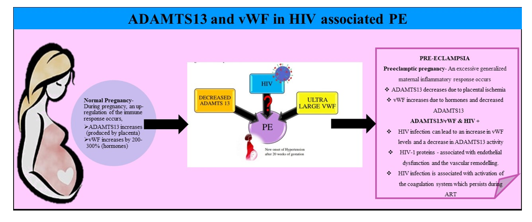
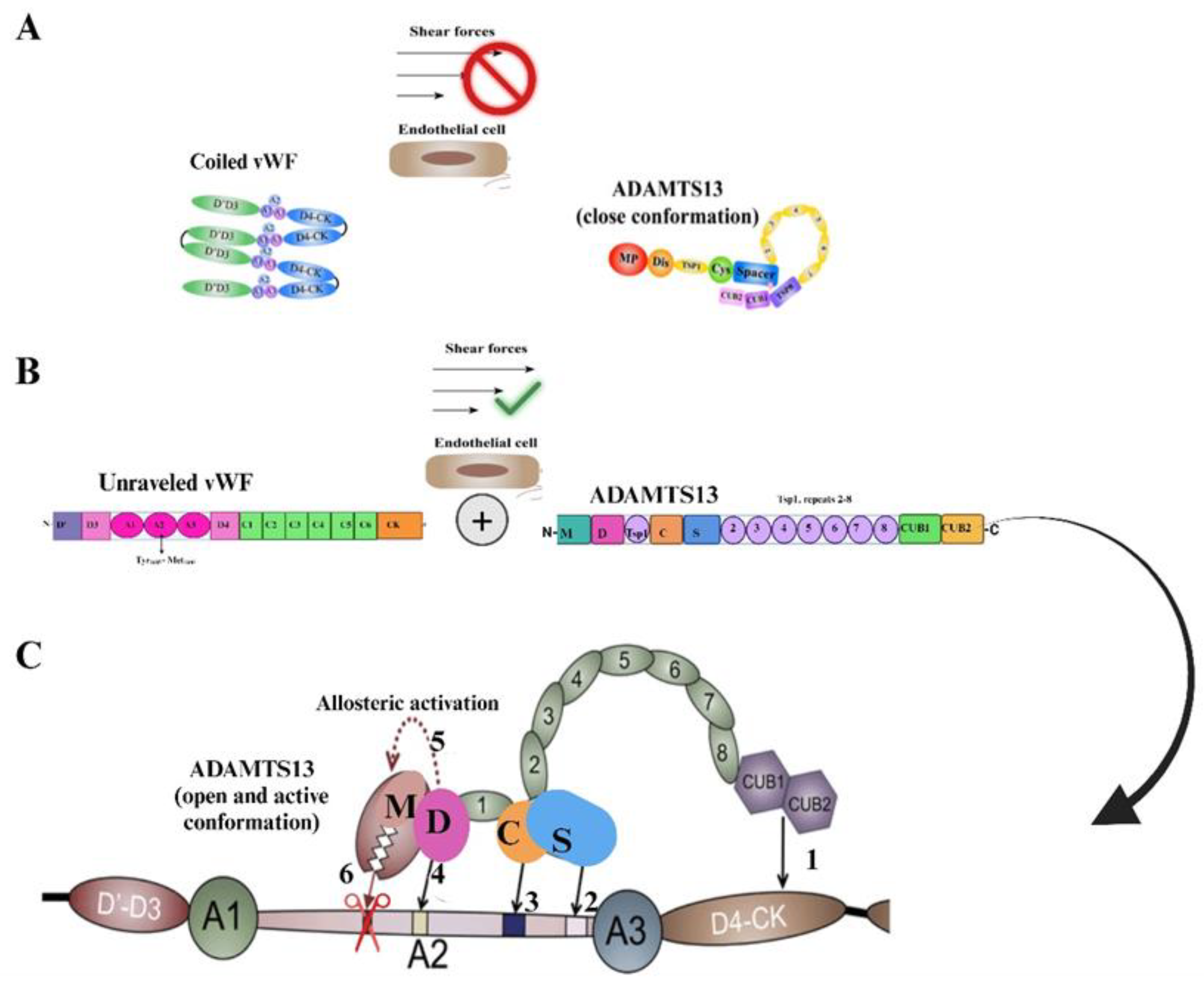
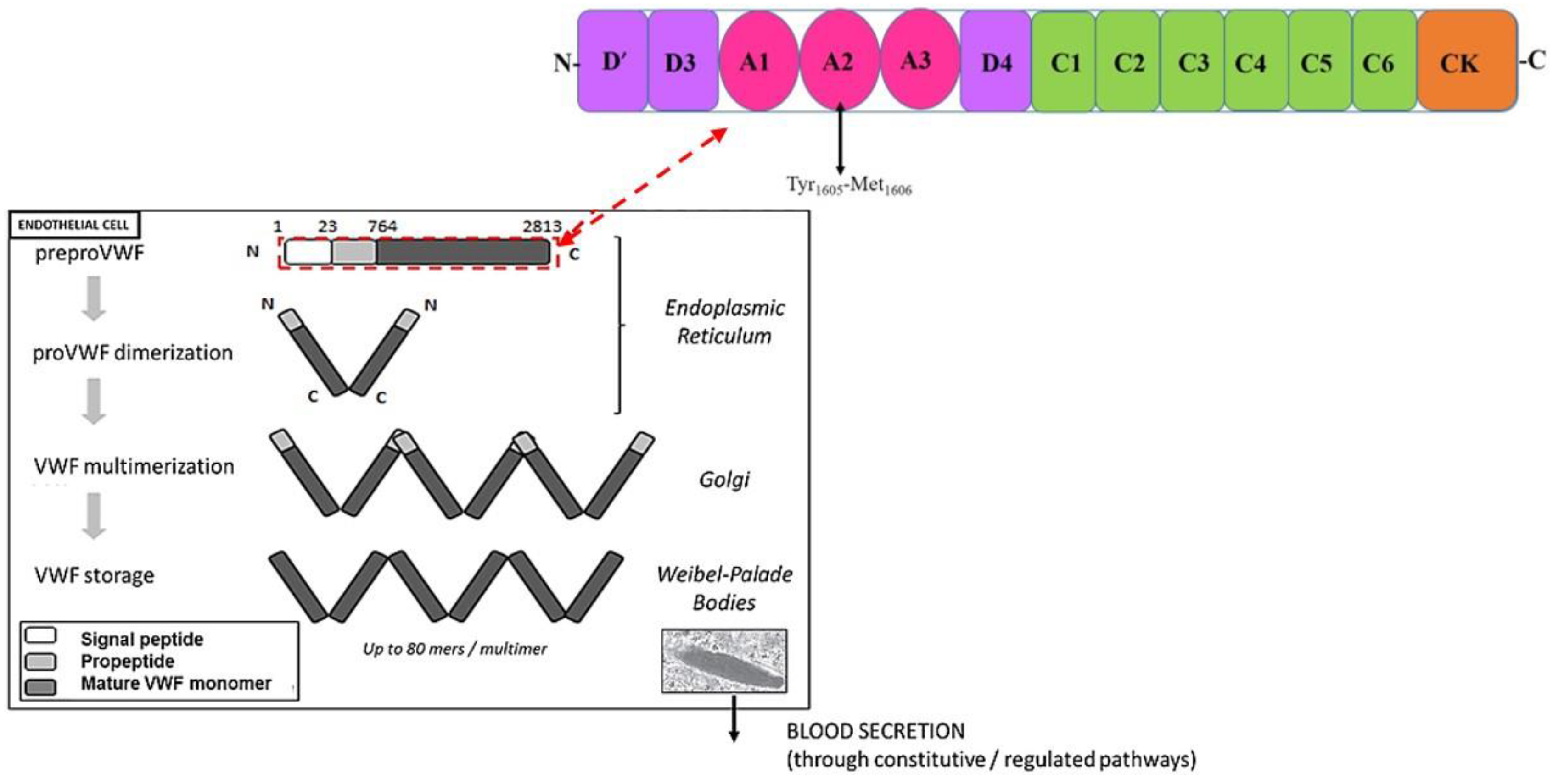
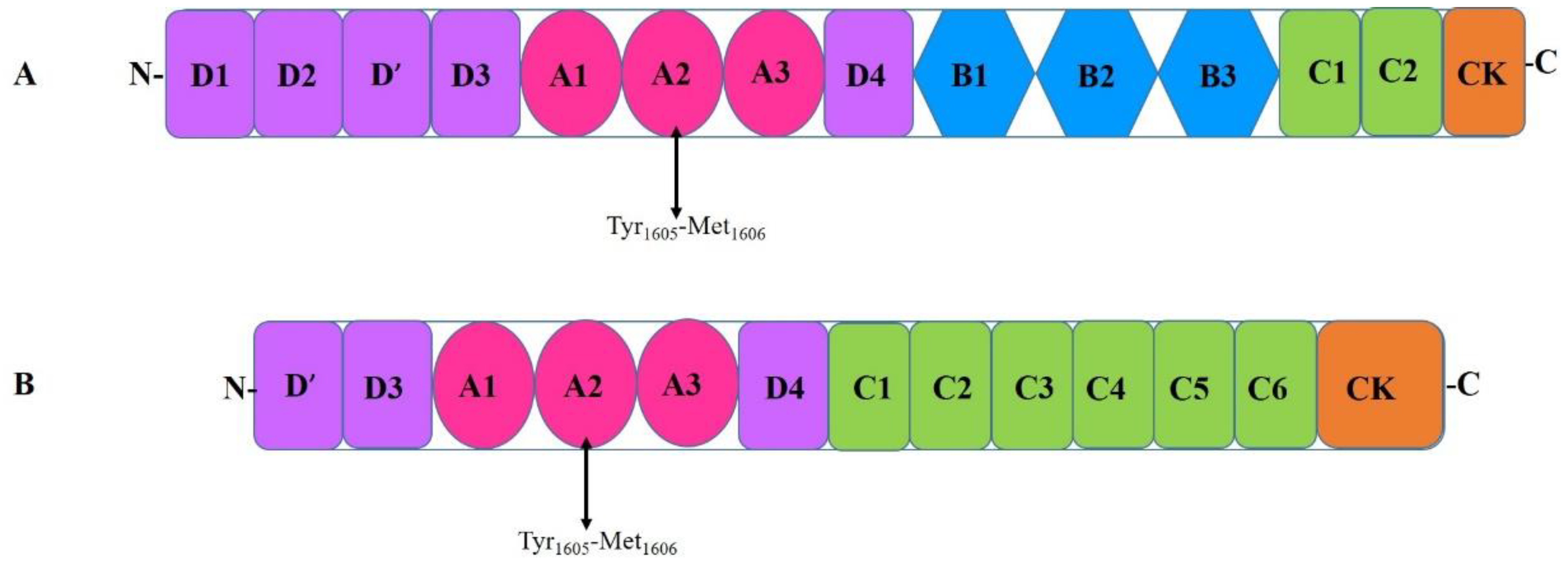
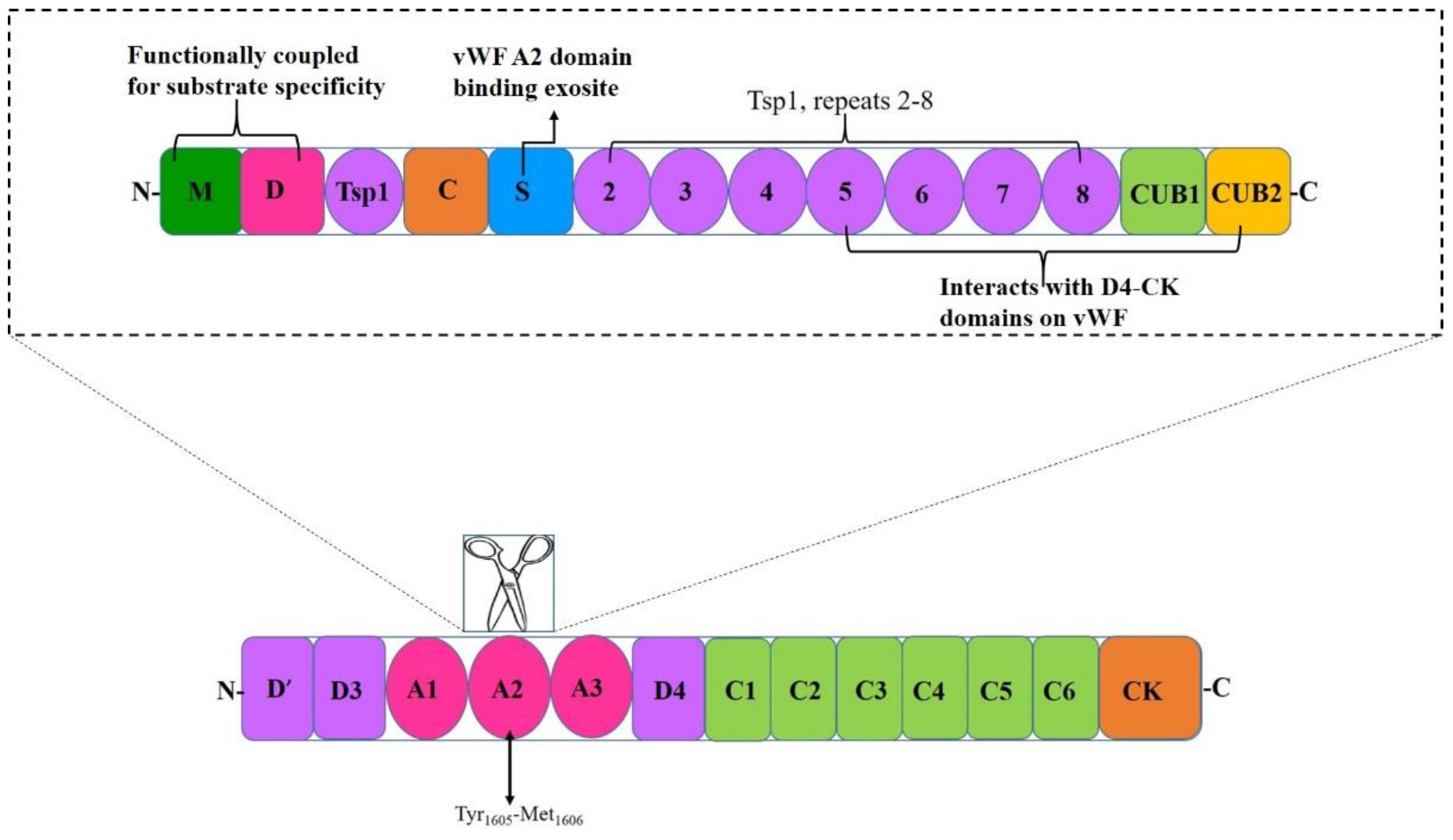
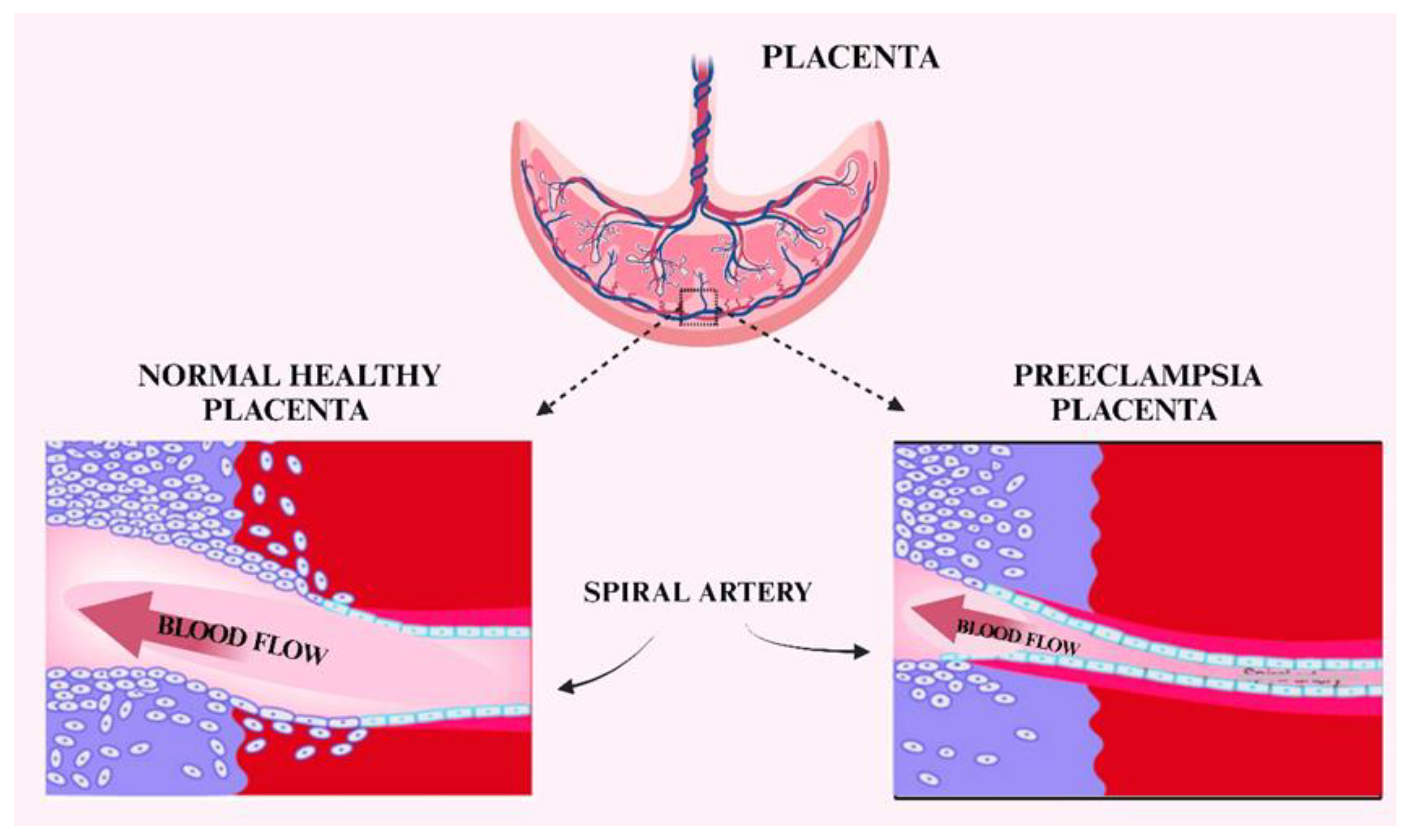
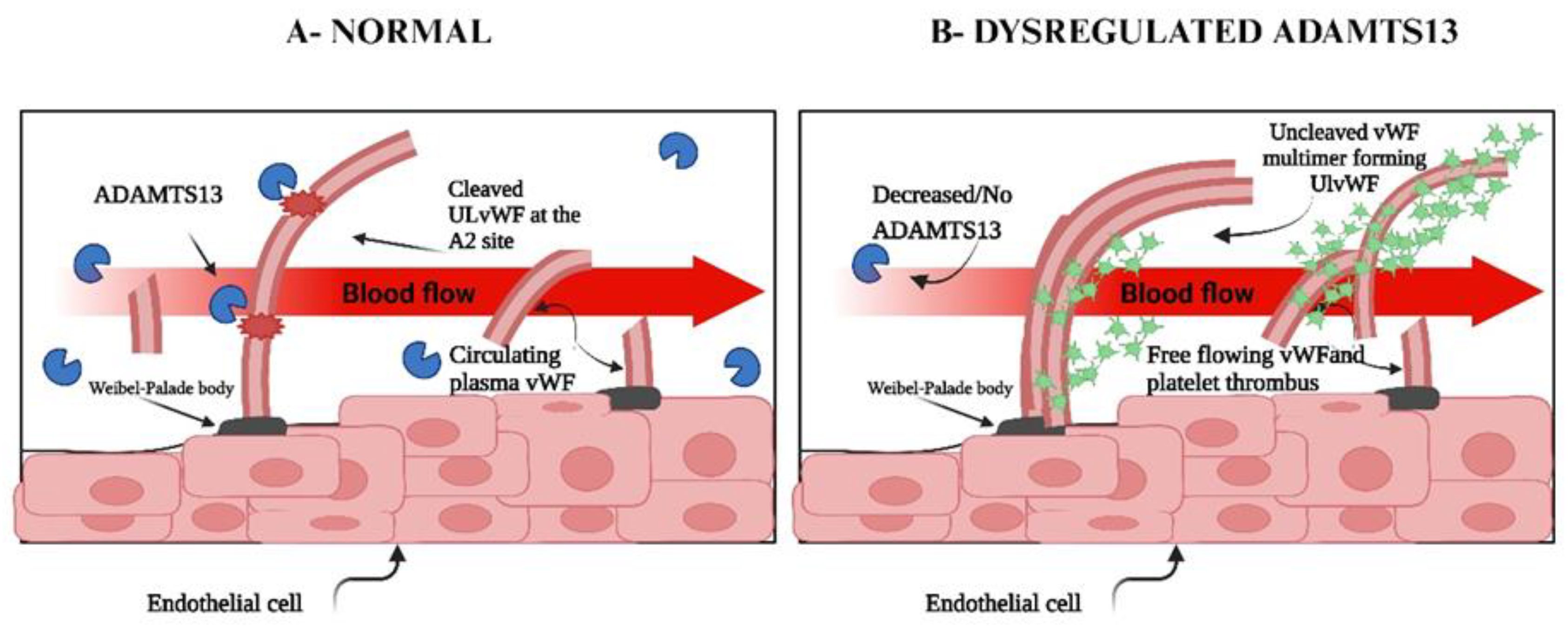
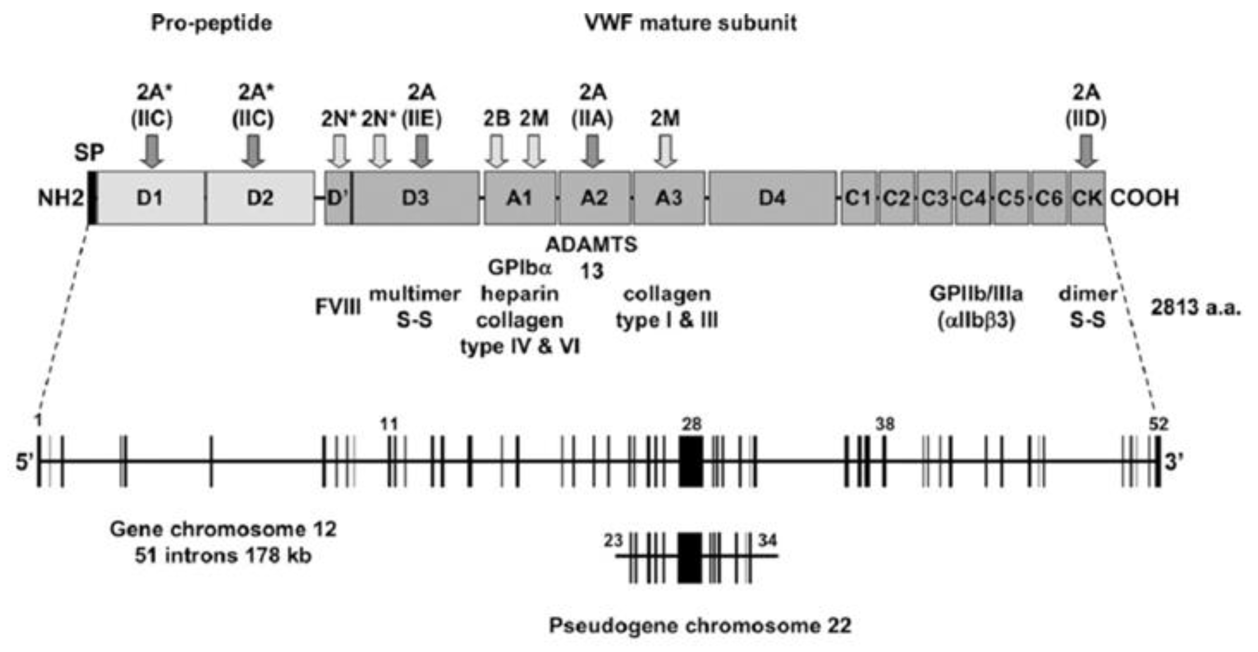
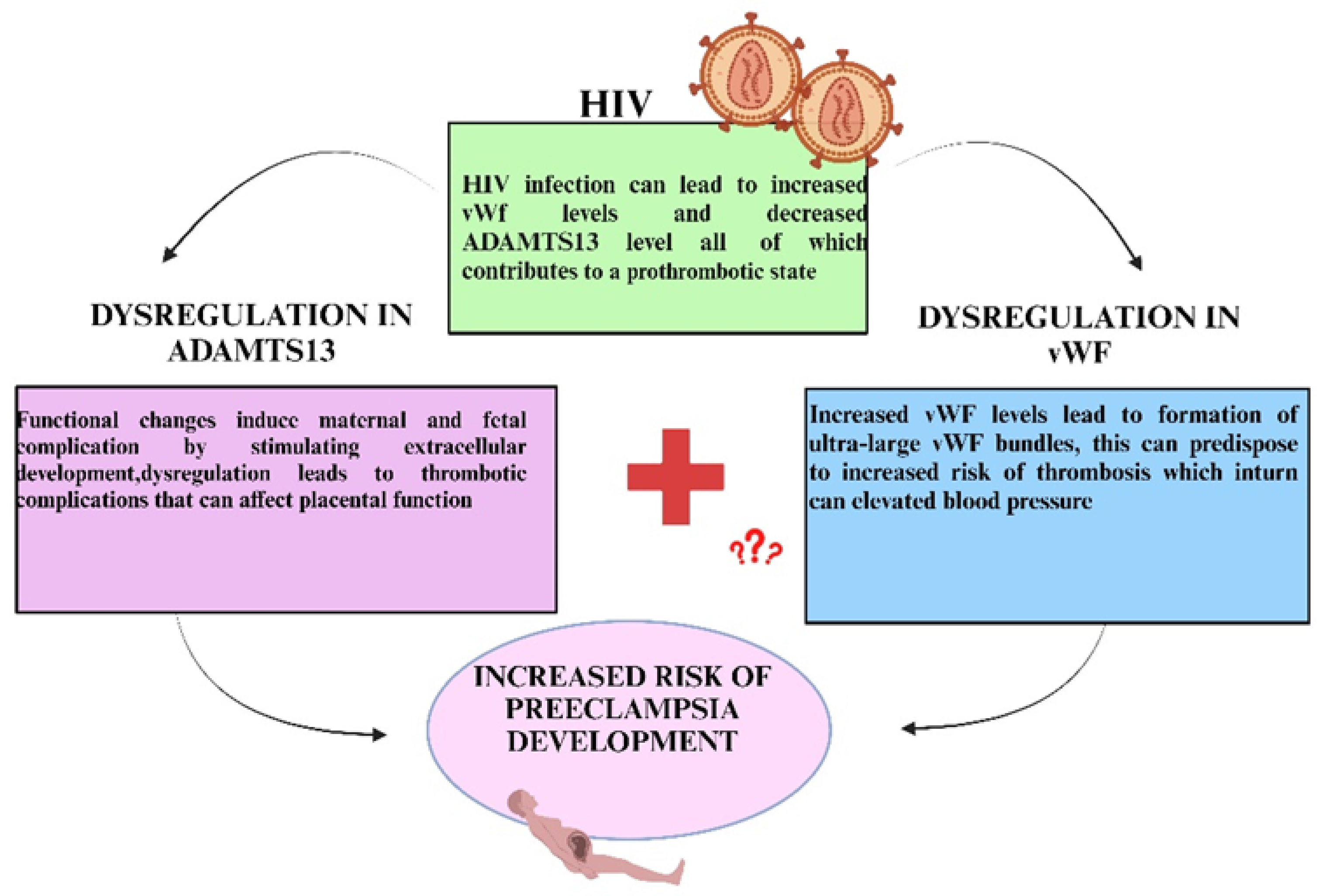
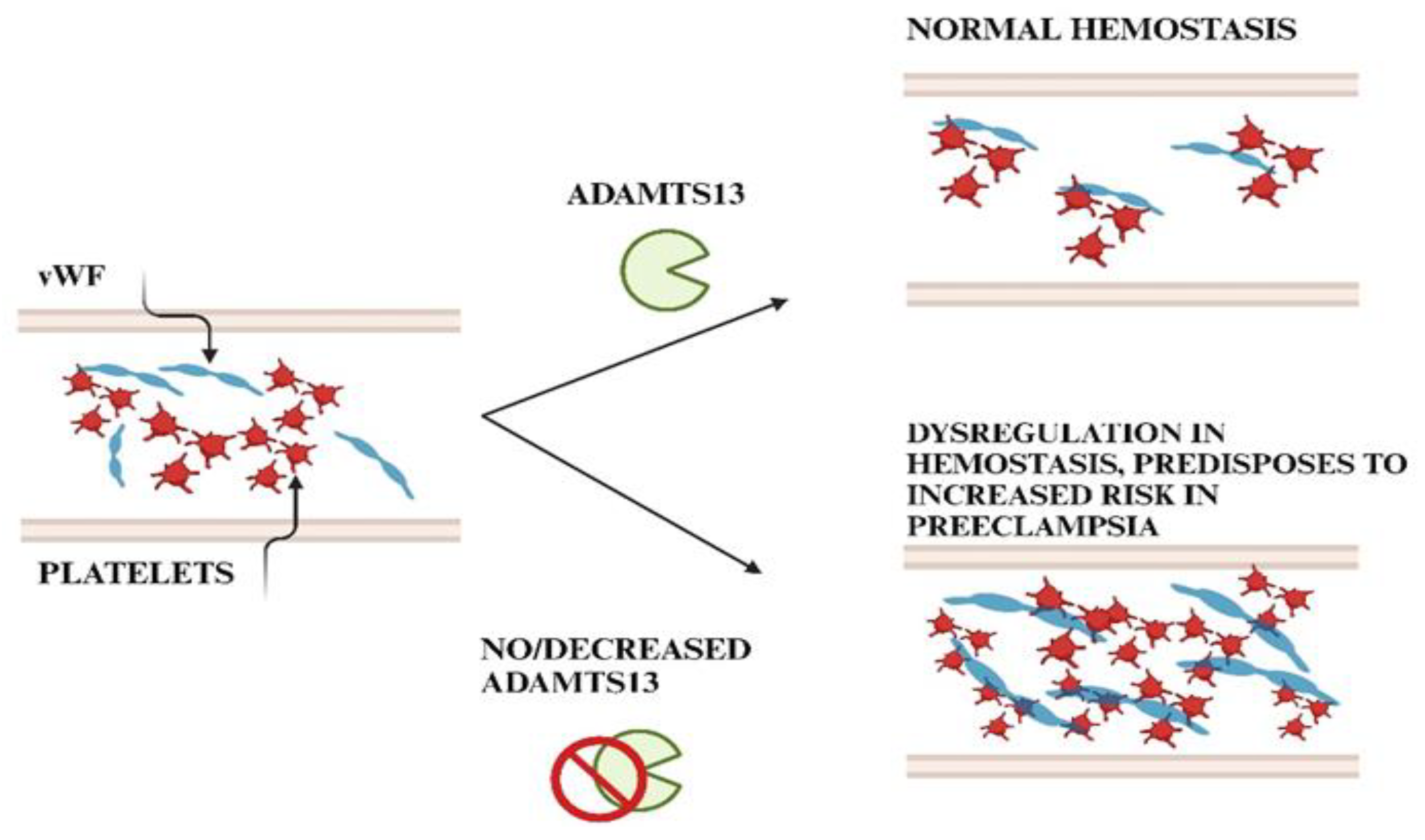
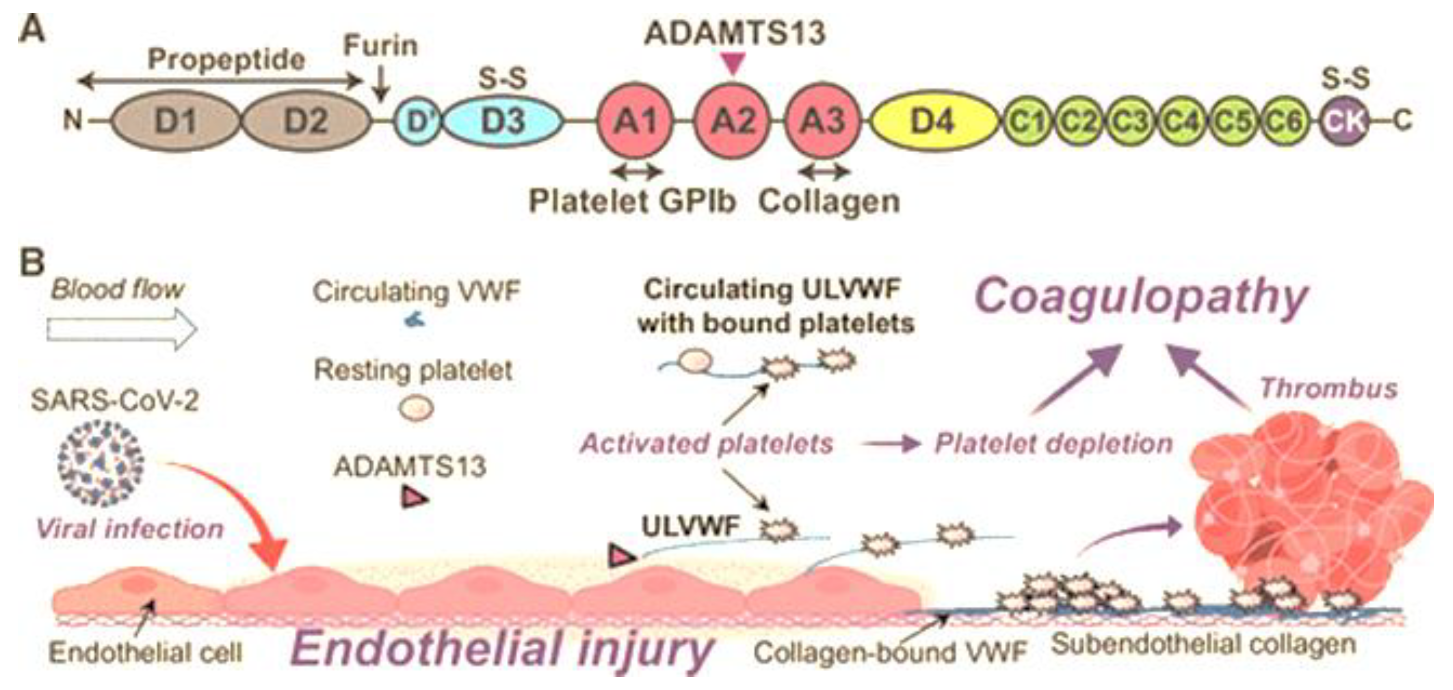
Normal pregnancy is associated with multiple changes of the coagulation and the fibrinolytic system. In contrast to a non-pregnant state, pregnancy is a hypercoagulable state where the level of vWF increases by 200–375% affecting coagulation activity. Moreover, in this hypercoagulable state of pregnancy, preeclampsia is exacerbated. ADAMTS13 cleaves the bond between Tyr1605 and Met1606 in the A2 domain of vWF, thereby reducing its molecular weight. A deficiency of ADAMTS13 originates from mutations in gene or autoantibodies formed against the protease, leading to defective enzyme production. Von Willebrand protein is critical for hemostasis and thrombosis, promoting thrombus formation by mediating adhesion of platelets and aggregation at high shear stress conditions within the vessel wall. Mutations in vWF disrupts multimer assembly, secretion and/or catabolism thereby influencing bleeding. The release of even small amounts of active ADAMTS13 protease has a profound inhibitory effect on thrombosis and inflammation, making vWF the major regulator of plasma ADAMTS13 concentration. Endothelial activation caused by HIV infection leads to the release of vWF. The SARS-CoV-2 infection promotes circulating proinflammatory cytokines, in-creasing endothelial secretion of ultra large vWF that causes an imbalance in vWF/ADAMTS13. Raised vWF levels corresponds with greater platelet adhesiveness, promoting a thrombotic tendency in stenotic vessels, leading to increased shear stress conditions. Keywords: HIV; Preeclampsia; ADAMTS13; vWF; Pregnancy.

Keywords: 
;  ;  ;  ;  

1. Introduction

Maternal mortality is the annual number of maternal deaths emanating from any cause connected to or aggravated by pregnancy or its management during pregnancy and childbirth or within 42 days of the termination of pregnancy [1]. Of note, 95% of all maternal deaths predominated in low- and middle-income countries [1]. In the period 2000-2020, the maternal mortality ratio (MMR) (number of maternal deaths per 100 000 live births) decreased across the world by 34% [2].
In 2020-2021 the MMR in South Africa (SA) was 120.9 maternal deaths per 100 000 live births. There has been a slight decrease in MMR (119.1 deaths per 100 000 live births) during the period 2021/2022 in SA. However, the MMR currently in SA is 125 deaths per 100 000 live births [3,4]. The leading cause of maternal deaths in SA is non-pregnancy related infections such as HIV, tuberculosis, pneumonia etc., followed by haemorrhage, medical and surgical disorders [4]. The fourth and direct leading cause of maternal deaths in SA is hypertensive disorders of pregnancy (HDP), of which, 83% is due to preeclampsia (PE) and eclampsia [5].
Hypertensive disorders of pregnancy affects 4-8% of all pregnancies worldwide [6]. Globally, it is also responsible for over 500 000 fetal and neonatal deaths and over 70 000 maternal deaths [7]. In SA, the prevalence of PE is 12% and is associated with 15% of preterm births [8]. Notably, a previous history of PE, affects one’s life expectancy due to an increased risk of cardiovascular disease, diabetes and development of stroke later-in-life [9].
Preeclampsia is a pregnancy specific condition characterised by new onset hypertension at ≥20 weeks of gestation and is associated with either/or proteinuria; maternal end-organ dysfunction, including neurological complications; pulmonary oedema; placental abruption; angiogenic imbalance; fetal growth restriction and intrauterine fetal death [7]. It has a variable clinical presentation associated with decreased utero-placental flow emanating from defective placentation [10] and coagulation abnormalities that may/may not result in a myriad of clinical abnormalities of the fetus [11,12]. Definitive treatment for PE is delivery of the placenta [13] and effective treatment is premature delivery or termination of pregnancy [14].
Pregnancy is often referred to as a hypercoagulable state characterized by increased levels of coagulation factors, decreased fibrinolysis, and increased platelet activation [15]. These changes are primarily due to the production of procoagulant molecules by the placenta, which serve to protect the mother from excessive blood loss during childbirth [16]. The hormonal changes of pregnancy also contribute to this hypercoagulable state [17]. Normal pregnancy is associated with dysregulation of coagulation [15] and fibrinolytic activity across trimesters [18]. The levels of Von Willebrand Factor (vWF) may increase by 200–375% in pregnancy, often resulting in doubling of coagulation compared to a non-pregnant state [15]. Notably, platelet count often decreases because of its consumption by the uteroplacental unit [15]. The dysregulation of coagulation in pregnancy may lead to various complications such as disseminated intravascular coagulation, a condition characterized by excessive clotting throughout the body, leading to organ failure [19]. As a typical physiological reaction, vWF rises and a disintegrin and metalloprotease with thrombospondin type 1 motif 13 (ADAMTS13) falls during pregnancy [20]. Abnormal persistence of ultra-large vWF (ULvWF) multimers is caused by severe ADAMTS13 deficiency, which triggers thrombotic thrombocytopenic purpura (TTP), thereby predisposing microvascular thrombosis [21].
In PE the hypercoagulable state is accentuated [18].The intrinsic coagulation pathway is activated and the common pathway appears to be hypercoagulable in PE [18]. More specifically, prothrombin times are accelerated in PE, linked to alterations in fibrinogen and factors II, V, and XI [22]. In PE, tissue type plasminogen activator, synthesized by endothelial cells, are released during endothelial injury thereby triggering the fibrinolytic system [23,24]. Hematological abnormalities are indicative of intravascular coagulation and less frequently erythrocyte destruction in PE [18].
There is a complex interplay between HIV infection and coagulation processes. HIV not only affects the immune system but also induces a pro-inflammatory state that can lead to hypercoagulability [25]. Patients with HIV are at a higher risk for thrombo-embolic events, which is exacerbated by factors such as chronic inflammation and antiretroviral therapy (ART) side effects [26]. Antiretroviral therapies, whilst essential for managing HIV infection, can also influence coagulation profiles, thus complicating patient care [27]. Additionally, emerging evidence suggests that the presence of HIV can alter platelet function and increase levels of clotting factors, further complicating the coagulation cascade [27].

2. A Disintegrin and Metalloprotease with A Thrombospondin Motif Type 1 Member 13 (ADAMTS13)

All mammalian genomes contain nineteen genes of A Disintegrin And Metalloprotease ThromboSpondin type motif (ADAMTS) [28]. This gene belongs to the metzincin protease superfamily, and it lacks both epidermal growth factor-like transmembrane and cytoplasmic modules [28]. ADAMTS13 has a number of unique properties for a circulating protease: 1. ADAMTS13 is constitutively secreted as an active protease [29]; 2. Relative to other coagulation and ADAMTS proteases, ADAMTS13 has a prolonged circulating half-life (two to four days), due in part to a lack of physiological inhibitors [30] and 3. The only known substrate of ADAMTS13 is vWF, and this protease displays no known off-target proteolysis [29]
Localisation: ADAMTS13 is localized on chromosome 9q34 which contains twenty-nine exons spanning 37kb in the genomic sequence [31,32]. It is synthesized primarily in the liver [32]. Its messenger RNA and translated proteins are localized to hepatic stellate cells [33].
Function: ADAMTS13 is known to process the large multimeric vWF precursor protein under fluid shear stress conditions [28]. An extensive exosite interaction between these domains and vWF-A2 domain governs substrate specificity and cleavage efficiency [31,33,34]. The cleavage of a single peptide bond (Tyr1605-Met1606) on the A2 domain of the vWF molecule generates vWF proteins required for the maintenance of haemostasis [31].
Structure: Structure-function analysis has established that the ADAMTS13 fragment consists of the signal peptide (Sp), a propeptide (P; pro-domain), metalloprotease domain (M), disintegrin domain (D), first thrombospondin 1 repeat domain (Tsp1), Cys-rich domain (C), spacer domain (S) at the N-terminal with seven additional Tsp1 repeat domains and 2 CUB domains (Complement c1r/c1s, sea Urchin epidermal growth factor, and Bone morphogenetic 1 and 2 proteins) [31,32] (Figure 1). Mutations in the highly variable regions of these domains significantly impair ADAMTS13 function [35,36].
Notably, the Sp component serves to guide the enzyme to the endoplasmic reticulum, where it undergoes post-translational modification and folding before being transported to the Golgi apparatus for further processing and eventual secretion outside the cell [37,38]. During this process, the Sp is cleaved off, thereby releasing the mature form of ADAMTS13 into the extracellular space where it can perform its enzymatic function [37]. The P component is involved in maintaining the enzyme in an inactive state while inside the cell and upon secretion into the bloodstream, proteolytic cleavage removes this P component, thereby activating ADAMTS13 and allowing it to cleave vWF efficiently [39].
The M domain on ADAMTS13 alone does not have the ability to bind with specificity to vWF or to cleave at the vWF cleavage site [29]. This non-catalytic domain is necessary for substrate specificity [40]. ADAMTS13 activity includes the binding of three calcium ions which is essential for the structural integrity of the proteolytic site [41]. Moreover, it has several hydrophobic residues that ensure the protease binds to vWF [42]. This M domain contains no crystal structures but is characterized by the adamalysin/reprolysin type, zinc-binding sequence (HEXXHXXGXXHD), where H denotes histidine, E glutamic acid, G glycine, D aspartic acid, and X variable amino acid [37,43]. Zinc cations must be bound for enzymatic activity since zinc-coordinating and calcium-binding residues control the cleavage activity of ADAMTS13 [44]. Also, leu1603 is a residue present on vWF molecule that interacts with residues near the zinc ion allowing for proper cleavage of vWF [32].
The next domain on ADAMTS13 is the D domain which lacks the cysteine signature or the arginine, glycine, asparagine motif but contains a crystal structure. The M and D domains are functionally coupled [28,32]. Addition of this short D domain restores full substrate binding specificity and specific proteolytic activity to the cleavage site of the M domain [42]. Within this domain, two hydrophobic residues and one charged residue have been identified and binds to vWF [42].
The first Tsp1 repeat domain on ADAMTS13 has three anti-parallel strands, the whole domain is capped by disulfide bonds on each end [32]. These strands are stabilized by a cysteine, tryptophan, and arginine layered core [45]. The Tsp1 domain engages in substrate recognition and functions in facilitating ligand interaction [31,46].
The C domain is structurally similar to the D-domain. This domain has a short α-helix and two pairs of double stranded anti-parallel β-sheets stabilized by six disulfide linkages [32]. De Groot, hypothesized that this domain may contribute specifically to ADAMTS13 function [47], however other studies have only implicated its role in the interaction with vWF [48,49]. Nonetheless, this domain is homologous with others in the ADAMTS family; however, the non-conserved V-loop forms a hydrophobic pocket to favour interactions with the vWF A2 domain [50]. The residues Gly471–Val474 at the base of the variable loop within the cysteine-rich domain forms a hydrophobic pocket involved in the binding to hydrophobic residues on vWF [42,47]. The absence of these hydrophobic interactions result in a 75-to-200-fold decrease in proteolysis [47].
The S domain differs across members of the ADAMTS family and is critical for substrate recognition; this domain has the highest binding affinity for the A2 site in vWF [12,32,42]. Residues Leu621-Asp632 in the spacer domain form a loop that interacts with the proximal portion of the cysteine-rich domain [48]. Both the C and S domains have been suggested to function closely and similarly to each other [42]. The S domain acts as a connector between the functional domains, potentially influencing enzyme activity, while the M domain plays a direct role in substrate recognition and binding [42,48].
The seven additional Tsp1 repeats at the C-terminal have no available crystal structure; they are required largely for flexibility and supporting the two CUB domains and S- domain interaction which maintains latency [50]. Of note, the deletion of Tsp1(7) and Tsp1(8) disrupts allosteric regulation [42].
The two CUB domains at the C-terminus is unique to ADAMTS13. The crystal structure of the CUB domains have approximately 110 residues in anti-parallel stranded β-sheets, characteristic of a β-sandwich structure [51]. Furthermore, ADAMTS13 CUB domains lack calcium-binding, and N-linked glycan [51,52]. Linker regions exist between Tsp1(2-5), and Tsp1(8)-CUB domains, which provide the necessary flexibility to the Tsp1 repeats in the tail allowing ADAMTS13 to adopt the closed conformation [53]. The two CUB domains are involved in the initial binding between ADAMTS13 and the D4-CK domain of vWF [54]. This results in conformational changes in both proteins, resulting in the linearizing of vWF and unfolding of ADAMTS13 through disruption of the Spacer–CUB interaction [50]. Mutations in both CUB domains affects protein secretion rather than directly affecting protease activity [50]. The two CUB domain is also involved in developmental regulation [51].

3. Von Willebrand Factor

Von Willebrand factor is a large multimeric glycoprotein that controls platelet adhesion and aggregation [34] with an average plasma concentration of ~10 μg/ml [32,42]. Interestingly, another protease in plasma, plasmin, is able to cleave vWF multimers [55]. Plasmin cleaves the bond between amino acid residues K1491-R1492 in the polypeptide chain region linking domains A1 and A2 [56]. Desmopressin, the analogue of vasopressin, substitutes D-arginine for L-arginine via V2 receptors, causing an immediate 2-fold increase in the level of vWF antigen in human blood plasma [55].
Localisation and storage: Von Willebrand factor is present as a series of repeating sub-units, ranging in size from ~500 kDa to ~20 000 kDa, each containing 2050 amino acid residues [32]. It is produced primarily in vascular endothelial cells and α-granules of platelets and is stored in either the golgi apparatus of endothelial cells or within endothelial cytoplasmic granules referred to as Weibel-Palade bodies (WPBs) and is also found within sub-endothelial connective tissue [57]. Following synthesis, vWF is transported to storage organelles in both megakaryocytes/platelets (α-granules) and endothelial cells (Figure 2) [58].
The majority of vWF is secreted constitutively, whereas the remainder is stored in WPBs that are specific for endothelium [60]. The WPBs and α-granules differ from each other in their dependency on vWF for their formation: α-granules can form in the absence of vWF, whereas the generation of WPBs is strictly vWF dependent [61]. Circulating vWF in plasma is predominantly endothelial cell derived, as platelets release vWF from α-granules only when activated [62]. There are some key differences between vWF originating from endothelial cells versus megakaryocyte. Endothelial cell-vWF is constitutively secreted and undergoes proteolytic processing by ADAMTS13, whereas platelet-vWF is not constitutively secreted and does not undergo significant proteolysis [63].
Function: Under shear stress conditions, the vWF protein promotes thrombus formation by mediating adhesion of platelets and aggregation within vessels [34]. Moreover, it serves as the primary adhesive link between platelets and sub-endothelium whilst also carrying and stabilizing coagulation factor VIII (FVIII) in circulation [31]. The vWF subunit is capable of interacting with a variety of plasma and matrix proteins, including coagulation factor VIII and platelet glycoprotein 1b which interacts with the A1 domain on vWF and collagen matrix with the A3 domain on vWF respectively [32]. Also, vWF carries a pro-coagulant FVIII, which protects it from rapid proteolytic degradation and delivery to sites of vascular damage [64]. Of note, vWF is secreted into blood and provides platelet attachment to the damaged vascular wall via its binding with collagen, as well as performs the function of carrier protein for blood clotting factor VIII [61,65].
The multimeric size of vWF is central to its platelet-tethering function, with larger multimers conferring greater hemostatic potential than smaller forms [47]. The length and thickness of vWF multimers strongly correlate with physiological hemostatic potential, making its cleavage by ADAMTS13 critical for balanced hemostasis [32,33]. Secretion of vWF multimers are induced by thrombin, histamine, fibrin, complement protein C5b-9 complexes and bacterial Shiga toxin at the site of vascular injury [33].
Structure: The vWF sequence contains an unusually high content of cysteine residues paired by disulfide bonds in all domains except in the A domains [32,42,66]. Cysteine plays a critical role in stabilizing domain structure [67]. The mature sub-unit is extensively glycosylated with 12 N-linked and 10 O-linked oligosaccharides, whilst the pro-peptide has three more potential N-glycosylation sites [32,42].
The domains of vWF are arranged in the following sequence: D1-D2-D’-D3-A1- A2-A3-D4-B1-B2-B3-C1-C2-CK [52]. The initial vWF monomer is formed by domains D1-D2-D'-D3-A1-A2-A3-D4-C1-C2-C3-C4-C5-C6-CK [55]. The mature human vWF subunit includes the following structural domains, D'-D3-A1-A2-A3-D4-C1-C2-C3-C4-C5-C6-CK [68] (Figure 3). Domains D1, D2, D3 include the following modules: von Willebrand D domain (VWd), cysteine-8 (C8), trypsin inhibitor-like (TIL), and E modules (E) [52]. Domain D' does not contain the VWd domain and C8 module, whilst D4 lacks the E-module and has a D4N subdomain [55]. The D1, D2, and D’ and D3 domains mediate the assembly of disulfide linkage of vWF dimers in the acidic environment of the Golgi apparatus, C-terminal cysteine knot (CK) domain dimerizes within the endoplasmic reticulum [69]
The key element of the vWF monomer is formed by triplicated A domains. The N- and C-terminal of the A1 and A3 domains form a disulfide bridge that fixes each of these domains in a rather rigid configuration. In contrast, the A2 domain is flexible and susceptible to proteolysis because it stretches in high shear stress situations due to its non-rigid structure [55,70]. Of note, vWF ultra-large multimers are more thrombogenic [55].
The primary function of the A1 domain of vWF is to capture platelets via GPIbα, resulting in the formation of the platelet plug [55,71]. In addition to platelet binding, the A1 domain can also bind heparin, this can competitively inhibit platelets from binding to vWF, thereby affecting platelet plug formation during heparin administration [72]. The A1 domain has also been shown to interact with collagen IV in the basement membrane [73]. The A2 domain contains a calcium-binding loop in the α3-β4 loop, which protects it against cleavage by ADAMTS13 by promoting rapid refolding of the domain [70].The Tyr1605-Met1606 cleavage site for ADAMTS13 is buried in the hydrophobic core of the β4-sheet [70]. Under shear force, the unfolding of the A2 domain begins at the C-terminus and proceeds through the β4-sheet, which contains the ADAMTS13 cleavage site [74]. The A3 domain facilitates the binding of vWF to the exposed sub-endothelial collagen upon vascular injury [72]. Moreover, the A3 domain also interacts with collagen sequences containing positively charged and hydrophobic residues [75].
The C-terminal domains of vWF play an important role in binding to ADAMTS13. More specifically, D4, C1-C6, and CK domains of vWF interact with the C-terminal domains of ADAMTS13 [Tsp1 (5-8) and CUB domains (CUB1 and CUB2)] [67]. These C-terminal domains of vWF allow ADAMTS13 to bind and form a complex with vWF, and circulate in plasma [67]. The formation of this complex is a critical step in the proteolysis of vWF by ADAMTS13 in circulation [67]. The CK domain plays an important role in the dimerization of vWF, which is required for the formation of long multimers [76]. In each monomer the CK domains flank the inter-chain disulfide bonds and the backbone of hydrogen bonds of the β-sheet, creating a rigid cross-linked structure that is highly resistant to hydrodynamic forces, which may contribute to the efficient transmission of force between monomeric subunits in the vWF multimer [76].

4. Interactions Between Adamts13 and Von Willebrand Factor

When vWF is unwinding, the exosite that binds to the S domain is initially exposed, this allows the S domain to recognize the vWF exosite [31,32]. ADAMTS13 holds a closed conformation maintained by the interaction between the S domain and C-terminal domains. Once the vWF D4-CK domains bind to the CUB1 and CUB2 domains of ADAMTS13, a conformational change occurs on ADAMTS13, transforming it from a closed to an open state. This exposes the S domain exosite therefore facilitating binding to the unfolded vWF A2 domain [77]. The S domain and the C domain function closely with and similarly to one another. When the Tsp1(2-8) repeats and the CUB domains are truncated, the remaining domains still cleave vWF substrates [32,34]. The CUB domains alone have no measurable affinity for vWF however, in the presence of shear stress, the CUB1 peptide will inhibit proteolysis of vWF [32]. Five thiol groups within Tsp1 repeats 2–8 and CUB-1 domain form disulfide bonds with vWF, therefore anchoring the enzyme; these free thiol interactions of the distal regions have anti-thrombotic activity independent of the proteolytic functions of ADAMTS13 [42]. The CUB domains have a negative regulatory function of ADAMTS13 activity however, may also have regulatory functions entirely unrelated to the proteolytic activity [32] (Figure 4 and Figure 5).
Also, platelet vWF that is found in a high-molecular-weight form, lacks N-linked sialylation hence is resistant to proteolysis by ADAMTS13 [78]. The binding of calcium to the A2 domain protects against unfolding by denaturants whilst also promoting refolding under a tensile force, thereby reducing the susceptibility of vWF to cleavage by ADAMTS13 [79]. Nonetheless, small amounts of active ADAMTS13 released has profound inhibitory effects on thrombosis and inflammation [32]. The major regulator of plasma ADAMTS13 concentration is vWF [32].
ADAMTS13 binds soluble vWF adsorbed onto the cell surface with a dissociation constant (KD) of ~20 nM resulting in efficient cleavage of ULvWF bundles [32]. The cleavage of vWF can occur in the absence of flow but is modestly enhanced by fluid shear stress, therefore cell bound ULvWF is in its “open” conformation [33,42]. Released ULvWF in solution exhibits a “closed” conformation which is not sensitive to ADAMTS13 cleavage until arterial shear is applied [28,31,32,33,80]. Under shear stress conditions, the binding of platelet glycoprotein 1b and/or FVIII to soluble vWF dramatically increases its rate of proteolysis by ADAMTS13 [33,45]. Rate-enhancing effect of GP1b and FVIII on vWF proteolysis is mediated by their alteration of domain–domain interactions and destabilization of the cleavage site in the vWF-A2 domain [32]. The release of even a small amount of active ADAMTS13 protease has a profound inhibitory effect on thrombosis and inflammation [32], thus the vWF is the major regulator of plasma ADAMTS13 concentration. In the absence of ADAMTS13 activity, endothelium anchored ULvWF strings are capable of gathering platelets, leading to uncontrolled thrombosis in terminal arterioles and capillaries [32]. The closed conformation is relieved when distal C-terminal domains of ADAMTS13 interact with the distal domains of vWF (D4-CK), resulting in exposure of the spacer domain, which in turn engages the A2 domain of vWF (Figure 4 and Figure 5)[54].
Figure 5. Schematic diagram showing the interactions between ADAMTS13 and vWF: A- Under normal circumstances, multimeric vWF circulates in the plasma in a globular conformation. ADAMTS13 circulates in a “closed” conformation stabilized through the interaction of CUB domains with the Spacer domain, no shear forces are present. B- With shear forces present, the unravelling of vWF reveals the binding site (A2) and cleavage site (Tyr1605-Met1606) for ADAMTS13. C- ADAMTS13 recognizes unfolded vWF through multiple interactions in the sequence from 1-6. M-Metalloprotease domain; D- Disintegrin domain; C-cysteine rich domain; S-Spacer domain; Tsp- Thrombospondin motif domain (1-8) -Adapted from- [45,81].
Preprints 151834 g005

5. Preeclampsia

The highest prevalence of HDPs predominates in South Asia (3.84 million), western sub-Saharan Africa (3.71 million) and eastern sub-Saharan Africa (3.12 million) [82]. These women from low-income countries such as South Asia and sub-Saharan Africa [83] are at a higher risk of developing PE compared to those in high-income countries [84]. Of note, sub-Saharan Africa (56%) and Southern Asia account for 85% of the global burden of deaths [82].
Based on the onset of clinical signs and symptoms, PE may be categorized into two sub-types viz., early- onset PE (EOPE) occurring <34 weeks of gestation and late-onset PE (LOPE) that occurs >34 weeks of gestation [9,85]. The EOPE is characterized by defective placentation and is associated with severe clinical manifestations to both mother and baby [85,86]. In contrast, LOPE is referred to as a maternal disorder characterized with endothelial injury [85]. Of note, intrauterine growth restriction dominates in EOPE [87]. Severe PE predisposes to intravascular coagulation, increasing the risk of bleeding [18].
Preeclampsia has a multifactorial pathogenesis where the placenta is the main organ affected [11]. Its pathophysiology is linked to vascular, immunologic, and genetic factors that culminate in multiple-organ injury, including the liver and kidney [88]. Placental villous lesions are found in 45.2% and 14.6% of PE and normotensive pregnancies, respectively [24]. The risk factors associated with PE includes maternal age over 35 years, obesity, history of PE [89], race, increased body mass index (BMI) [90], primiparity and underlying medical conditions, such as chronic hypertension, diabetes mellitus, and cardiac and renal diseases [91]
In normal pregnancies, the low resistance muscular arteries are converted to a tortuous high flow low resistance system, with a 4 to 6 fold increase in arterial diameter, to meet the demands of the growing fetus [92]. In PE, however, trophoblast cell invasion is defective and the remodelling of the spiral arteries is limited to the decidua [58,93] (Figure 6). The pathogenesis of PE may be divided into two stages-Stage one is the feto-placental stage or the preclinical stage involving defective placentation occurring in the first and second trimester [94]. Stage two is referred to as the maternal stage or the clinical stage which occurs during the second and third trimester [94]. In stage 2, the poor placental perfusion triggers excessive release of inflammatory and anti-angiogenic factors into the maternal bloodstream [95]. An imbalance between the generation, release, and response to vasodilators in favor of vasoconstrictors by the endothelium results in a heightened inflammatory response [94]. Notably, an increased release of the soluble form of the endothelial growth factor receptor type 1 (soluble fms–like tyrosine kinase, sFlt-1) and the soluble endoglin (sEng) into the maternal circulation predominates [96]. More specifically, sFlt-1 prevents vascular endothelial growth factor receptor type 2 (VEGFR2) activation in the maternal and foetoplacental tissues, while sEng exerts pro-migratory and pro-angiogenic stimuli in endothelial cells [14].

6. Human Immunodeficiency Virus Infection

Infections such as malaria, human immunodeficiency virus (HIV), tuberculosis, and syphilis account for more than one third of all maternal deaths worldwide [97]. HIV infection is a major contributor to poor global public health, affecting 40.1 million lives [98]. The estimated HIV prevalence rate in the South African population is approximately 14% [99]. The total number of people living with HIV (PLWH) in SA is estimated at 8.45 million in 2022. Of note, 19.6% of adults aged 15–49 years are HIV positive. More importantly, young girls in their reproductive ages of 15-24 remain at substantial risk of acquiring HIV [100]. The overall HIV prevalence in pregnancy at national level is 27.5% with the highest overall HIV prevalence occurring in the province of KwaZulu-Natal (37.1%) [101].
HIV infection is linked with a chronic inflammatory process [102]. The most advanced stage of HIV infection is acquired immunodeficiency syndrome [103]. The rate at which HIV infection progresses to AIDS depends on viral, host and environmental factors [95,104]. Furthermore, a range of host proteins interact with HIV proteins or DNA to either restrict or promote virus replication in specific cell types and transmission of the founder virus is followed by a rapid increase in HIV replication and then a striking induction of inflammatory cytokines and chemokines [105]. When the concentration of the uninfected CD4+ T-cells goes below 200 cell/mm3, then the infection progresses to AIDS [106].
HIV treatment involves the use of combined antiretroviral therapy (ART) to effectively suppress the viral load and to preserve/improve immune function and therefore reduces the risk of opportunistic infections and cancers [1]. Of note, ART also decreases inflammation caused by immune activation contributing to increased occurrence of cardiovascular, renal, neurological, and other end-organ diseases [1]. The use of ART has decreased both vertical and sexual transmission [88]. The development and widespread use of potent ARTs such as highly active ART (HAART) in which a concomitant use of at least three antiretroviral drugs, has transformed HIV infection from a near death sentence to a chronic manageable condition [88]. The increased access to ART has significantly reduced the risk of mother-to-child transmission among HIV infected pregnant women worldwide, however studies proposed that the risk of PE development is heightened among treated HIV infected women [88,107]. Despite the receipt of ART, PLWH display a high rate of non-AIDS related comorbidities. Additionally, they present with increased systemic oxidative stress due to suppression of endogenous antioxidant enzymatic mechanisms, leading to increased systemic immune activation; analogous to a PE milieu [108,109]. In a large cohort of 1038 pregnancies, the risk of new-onset HDP was similar by ART class, but those initiating ART after 20 weeks’ gestation had a greater risk compared with those receiving ART at conception [110]. Initiating ART during pregnancy rather than before pregnancy is associated with a lower likelihood of receiving a viral load test during pregnancy [101]. The World Health Organisation recommends that all pregnant and breastfeeding women with HIV irrespective of CD4 cell count, viral load, and clinical stage should have triple ART regimen[1,111].
There are four classes of ARTs namely, nucleoside/nucleotide reverse-transcriptase inhibitors (NRTIs) with examples being azidothymidine, zidovudine, lamivudine, abacavir, emtricitabine, stavudine, and tenofovir; the non-nucleoside reverse-transcriptase inhibitors (NNRTIs) with examples being efavirenz, nevirapine and etravirine; the protease inhibitors (PIs) with examples being lopinavir, ritonavir, atazanavir, indinavir, saquinavir, nelfinavir and the integrase strand transfer inhibitors (INSTIs) with examples being raltegravir, dolutegravir [111,112]. Drug toxicities are associated with the different classes of ART and may manifest as anaemia, mitochondrial toxicity, hyperlipidaemia, fat redistribution, and insulin resistance [111]. Table 1 highlights some of the adverse effects of the ART classes.

7. The Synergy of PE and HIV Infection

HIV infection may reduce fertility, irrespective of the stage of infection [115]. The management of HIV infection during pregnancy is complex as perinatal transmission of HIV can occur in utero, during labour and delivery, or postnatally through breastfeeding [115]. HIV infection is associated with varying rates of adverse pregnancy outcomes, such as increased spontaneous abortion, still-birth, perinatal and infant mortality, intrauterine growth retardation, low birth weight, and chorioamnionitis [72,111,116].
HIV infected individuals display increased systemic oxidative stress due to suppression of endogenous anti-oxidant enzymatic mechanisms [108,109]. The comorbidity of HIV infection and PE remains a considerable challenge to maternal health worldwide, with the main target being low-middle income countries [117]. HIV infection causes high maternal mortality rates in communities with high HIV prevalence [118]. HIV infected women have a significantly higher risk of PE compared to HIV naïve women, the increased access to HAART has however significantly reduced the risk of mother-to-child transmission among HIV-infected pregnant women worldwide [88,107]. However the risk of PE development is heightened among HIV-infected women receiving HAART [88].
A systemic inflammatory response and an up-regulation of the immune response occurs in all pregnancies, but is significantly amplified in PE [8,119]. This is evident in the excessively heightened immune reactions to the activation of the innate immune system and other proinflammatory factors [119,120]. This immune imbalance consists of increased pro-inflammatory immune cells and cytokines, together with a decline of regulatory immune cells and cytokines that eventuates in a state of inflammation [121]. As a result, inadequate invasion of trophoblasts into the myometrium and insufficient remodelling of spiral arteries leading to placental ischemia by exposing the placental site to vasoconstriction culminating in the development of PE [122]. Of note, EOPE is a proinflammatory placental state, whilst in LOPE systemic maternal inflammation occurs [123]. Immunological deficiency, caused by HIV, may lower the incidence of PE development by reducing immune hyper-reactivity [121]. A breakdown of immune tolerance or immunological incompatibility between the mother and foetus may be associated with PE development [124]. Pre-eclampsia is dominated by a proinflammatory Th1 response; however, in combination with HIV infection and ART, the Th1 response is exacerbated [125]. The immune reconstitution associated with ART use and the toxic mechanism involving endothelial inflammation and liver damage result in the loss of the protective effect which elevates PE risk [8].
Chronic immunological activation and inflammation occur in HIV infection, and is prognostic of the disease development [126]. The hallmarks of chronic inflammation include immune cell metabolic dysregulation, cellular fatigue, and malfunction. Antiretroviral therapy significantly lowers immunological activation and systemic inflammation, but not to levels that are consistent with HIV naïve individuals [127]. Table 2 highlights the mechanism of action of ARTs. The use of ART leads to immune reconstitution, which increases the risk of PE development compared to treatment naïve women [88]. Notably, the HIV accessory protein, Tat mimics vascular endothelial growth factors (VEGF), preventing VEGFR-2 signaling by the VEGF-A ligand [128]. More specifically, VEGF is unable to bind to its receptor preventing its effector function, resulting in abnormal angiogenesis. This also anticipates endothelial injury thereby increasing the risk of PE development [128].

8. Dysregulation of Adamts13

Genetic mutation changes the protein building blocks within the ADAMTS13 enzyme. Of note, a deficiency of the vWF-cleaving protease (ADAMTS13) originates in such mutations in the ADAMTS13 gene [142]. This dysregulation leads to the production of an abnormally small version of the enzyme that cannot function properly. This defective vWF processing predisposes to the formation of UlvWF multimers that attract platelets thereby promoting microthrombi formation [32]. If vWF is not cut into smaller fragments by the ADAMTS13 enzyme, it promotes the formation of abnormal clots throughout the body. The multimeric version of vWF induce platelets to stick together, even in the absence of injury, leads to clinical signs and symptoms of TTP [143]. Of note, these clots cause serious pathologies when they block vessels and restrict blood flow to organs such as the placenta, brain, kidneys and heart.
Two mechanisms that cause aberrant ADAMTS13 activity has been identified [144]. The most frequent mechanism is caused by circulating anti-ADAMTS13 autoantibodies which neutralize enzymatic activity and/or accelerate protease removal from circulation, secondly, mutations in the ADAMTS13 gene, may cause congenital TTP in which ADAMTS13 activity is < 10% of normal levels [145]. This is also termed Upshaw-Schulman syndrome, a rare autosomal recessive disorder caused by homozygous or double-heterozygous mutation of ADAMTS13 gene [146,147]. This predisposes to the formation of unusually large forms of vWF multimers within circulation (Figure 7) culminating in intravascular platelet clumping and thrombotic microangiopathies [146]. Certain combinations of polymorphisms or mutations may predispose severe ADAMTS13 deficiency, however mutation screening has revealed considerable genetic heterogeneity, with the majority being confined to a single family [145]. Also, in transformed vascular endothelial cells, ULvWF multimers are produced at sites of liver injury and a decreased ADAMTS13 activity may be involved in sinusoidal microcirculatory disturbances and the progression of liver injury that eventually leads to multi-organ failure [148].
Synonymous single nucleotide variants (sSNVs) may cause protein deficiency or dysfunction pre-empting diseases [149]. Synonymous variants may alter constitutive splice sites or activate cryptic splice sites, which results in unstable mRNA or defective protein [150]. In humans, approximately 200 disease-causative SNVs of ADAMTS13 have been identified in patients with TTP all of which were non-synonymous and detected as haplotypes [81,151].
In a recent study by Jsnkwoska et al.,2022, a total of 376 naturally occurring sSNVs of ADAMTS13, including 357 from a healthy population and 19 from patients with USS, were identified in the SNP [149]. These sSNVs affect almost 9% of nucleotides in the mRNA of ADAMTS13 with most sSNVs being identified in exon 25. They have also reported that many sSNVs have been found within the M domain and C-terminal region of ADAMTS13, within exons 24 to 29, which encode the Tsp1(7-8) and CUB1-2 domains [149]. These SNVs leads to downstream reduced plasma ADAMTS13 activity and disrupted ADAMTS13-vWF interactions by changing untranslated regions, splice regulatory regions, or the coding sequence [149,152]. In addition, various truncated forms of ADAMTS13 are detectable in plasma, and several alternatively spliced mRNA variants have been characterized [149]. Deficiency or dysfunction of ADAMTS13 due to SNVs may lead to thrombotic pathologies, including TTP, USS, myocardial infarction, and ischemic stroke [153].

9. Dysregulation of Von Willebrand Factor

Of the inherited coagulopathies, vWD is the most common, followed by hemophilia B which is a factor IX deficiency [154]. Acquired coagulation disorders can result from drug actions or side effects, or underlying systemic disease [155]. Tadu, found that platelet counts were considerably lower in PE where bleeding times were significantly greater allied to D-dimer levels being significantly higher in PE compared to normotensive pregnant women [18].
Mutations in vWF disrupt multimer assembly, secretion and/or catabolism thereby influencing bleeding [156]. Mutations in vWF, result in deficiencies of vWF protein predisposing mild to severe bleeding, a disorder known as Von Willebrand disease (vWD) (Figure 8) [157]. This is an autosomal inherited mucocutaneous bleeding disorder [64]. Defects in the secretion or intravascular clearance of vWF multimers results in dysregulation of vWD type 1 [64,156], whilst defects in the assembly or intravascular proteolysis of vWF multimers influence vWD type 2A or 2B [156]. Missense mutations affecting platelet binding or FVIII-binding are responsible for the four sub-types; 2A, 2B, 2M and 2N, whilst mutations resulting in a lack of vWF expression predominate in recessive type 3 vWD [64]. Increased vWF levels predispose atherothrombotic complications [157]. Apart from its function in haemostasis, vWF may induce downstream cell-signaling pathways associated with angiogenesis, inflammation, cell apoptosis and metastasis as well as vascular wall thickening [157].

10. Adamts13 in HIV Infection

In HIV-associated TTP, the dysfunction of vascular endothelial cells due to HIV infection can lead to local thrombin generation and consumption of ADAMTS13 [158]. Notably, HIV-associated TTP occurs in the setting of profound CD4 deficiency with altered ADAMTS13 protease activity complicated by myocardial infarction and stroke [159,160]. ADAMTS13 response may be impaired in HIV infection. In a study by Graham, HIV viral load correlated with both ADAMTS13 antigen and activity [160]. More specifically, men with acute HIV infection had significantly higher levels of ADAMTS13 activity compared to HIV naïve controls [160]. Additionally, both acute and chronic untreated HIV infection exhibited higher ADAMTS13 activity compared to chronic treated infection [160]. ADAMTS13 levels are lower in HIV-infected compared to HIV naïve individuals [147]. Autoantibodies forming against ADAMTS13 are present in PLWH because of an impaired immune system [147]. HIV infection itself can lead to an increase in vWF levels and a decrease in ADAMTS13 activity, which may contribute to a pro-thrombotic state [33].

11. Von Willebrand Factor in HIV Infection

A significant increase in vWF levels is linked to the advancement of HIV illness; there is a positive association between vWF levels and plasma viral load, and higher levels of vWF antigen are associated with a higher risk of mortality [160]. During HIV infection, vWF antigen quantity and adhesive activity are elevated, despite an apparent corresponding increase in the quantity and activity of its regulating protease (ADAMTS13) [160]. The coagulation and the immune systems are intricately linked and activate each other therefore HIV-1 infection results in a procoagulant state [161]. Increased endothelial activation, inflammation and coagulation are present in untreated HIV infected patients [162]. Activation of endothelial cells is accompanied by vWF release and persistent attachment to the vessel wall and self-assembles into strings and fibers, enabling platelet adhesion [160]. A marked rise in vWF levels is associated with HIV disease progression, with a positive correlation between vWF levels and plasma viral load [160]. The vWF antigen levels decrease after effective ART, as do levels of other endothelial activation biomarkers [160]. Moreover, the elevated vWF concentration may contribute to endothelial cell injury in PLWH of African ancestry [163].

12. Von Willebrand Factor and HIV-Treatment

HIV infection is associated with inflammation and activation of the coagulation system and which persists during ART [160]. Endothelial activation caused by HIV infection leads to the release of vWF, which enters the circulation or attaches to vessel walls and self-assembles into strings and fibers, enabling platelet adhesion [164]. The study by Graham, Chen [160], noted that plasma viral load positively correlated with vWF adhesive activity, whilst elevated levels of circulating vWF occurs in treated HIV infected individuals, however this study included a male population. Of note they had also demonstrated that vWF antigen levels decrease post ART [160]. Notably, patients receiving abacavir did not have increased levels of plasma coagulation markers such as vWF compared to patients who did not receive an abacavir containing regimen [165]. Combination ART, includes regimens that contain protease inhibitors that reduces but do not normalize levels of vWF [165]. Of note, over time the effect of vWF on the intact endothelium with allied platelet adhesion promotes atherosclerosis, and an increased cardiovascular risk in PLWH [166].

13. Adamts13 in Pregnancy

ADAMTS13 concentration is dependent on age, being lowest in neonates and in individuals above 65 years of age in physiological conditions [34]. Pregnancy is characterized by deep placentation; physiological placentation is characterized by the invasion of the uterine spiral arteries by extravillous trophoblast cells arising from anchoring villi. [167]. Notably, the differentiation and invasive activity of the trophoblast cells is tightly controlled spatially and temporally also regulated by multiple factors to enable proper placentation, thus defects in trophoblast invasion are associated with severe pregnancy-related complications [168].
Full length and proteolytically active ADAMTS13 is expressed in trophoblast cells and in villous endothelial cells of human placentae at the highest levels in the first trimester and declines in the second and third trimester of gestation [169]. ADAMTS13 mRNA and protein are expressed in human normal placenta and decidua throughout the pregnancy, as well on trophoblast and fetal blood vessel endothelium [169]. For a cell to migrate, proteolysis of the extracellular matrix is mediated by the matrix metalloproteases [170,171]. A decreased plasma ADAMTS13 activity occurs at 12 to 16 weeks gestation and by the third trimester it decreases progressively to 23% with an increased sensitivity to thrombotic microa[170,171ngiopathies [TMAs; the presence of hemolytic anaemia (destruction of red blood cells), low platelets, and organ damage due to blood clots in capillaries and small arteries] occurring within the second trimester of pregnancy [34,172]. TMAs result in abnormalities in the blood vessel walls of arterioles and capillaries with resultant microvascular thrombosis. Hemolytic uremic syndrome and TTP are primary forms of TMAs [147]. Thrombotic thrombocytopenic purpura is caused by severe deficiency of ADAMTS13 due to acquired autoantibodies or, genetic mutations [159]. The most common form of HUS (typical HUS) follows a diarrheal illness caused by Shiga toxin–producing Escherichia coli; whereas atypical HUS is associated with abnormal host susceptibility to complement-mediated damage. Moreover, TTP is three times more common in females, and half of those affected are pregnant or postpartum [173]. ADAMTS13 may be either pro- or anti-angiogenic depending on its local microenvironment [174].
Throughout gestation, proper regulation of vWF by ADAMTS13 is critical for maintaining normal blood flow in the placental vasculature. Imbalances in this regulatory mechanism may lead to excessive platelet adhesion and aggregation within the placental circulation, resulting in thrombotic complications such as placental infarction or abruption [175]. These conditions can impair fetal growth and development by compromising the exchange of nutrients and oxygen between the mother and fetus [93]. Dysregulation of this enzyme can lead to thrombotic complications and contribute to pregnancy-related disorders that affect placental function and fetal well-being [32].

14. Adamts13 in Pre-Eclampsia

Placental villi express significantly lower levels of ADAMTS13 protein in PE compared to normotensive pregnancies [169]. A reduction of ADAMTS13 synthesis and secretion within the placentae of women with severe PE may be associated with hypertension-related placental ischemia and tissue hypoxia [169]. Functional changes in ADAMTS13 proteases induce maternal and fetal complications by stimulating extracellular matrix development [176]. Aref and Goda [177]reported that plasma ADAMTS13 activity was significantly reduced in PE compared to normal pregnant women and non-pregnant women [178]. Partial deficiencies of ADAMTS13 have been observed in diseases sharing inflammatory states, including cardiovascular diseases, severe sepsis, and septic shock [178,179]. There is significant association between ADAMTS13 activity levels and EOPE, whereas severe PE was associated with increased levels of P-selectin [34]. ADAMTS13 has begun to be identified as a prognostic and/or diagnostic marker of other diseases, such as those related to inflammatory processes, liver damage, metastasis of malignancies, sepsis, and different disorders related to angiogenesis [21] (Figure 9), hence may be a useful predictor marker for severe PE development.
ADAMTS13 protein is proteolytically active and placental ADAMTS13 is produced at the highest levels in the first trimester (when placentation occurs) and declines in the second and third trimester of gestation [169]. ADAMTS13 mRNA are expressed in human normal placentae and deciduae throughout the pregnancy, as well on trophoblast and fetal blood vessel endothelium [169]. ADAMTS13 levels are significantly reduced in placental villous tissues in severe PE, emanating from the hypoxic microenvironment [143,169]. This hypothesis is supported by the reduction in ADAMTS13 synthesis after placental explant are exposed to hypoxic conditions [169]. In pregnancy, ADAMTS13 promotes proliferation, migration, invasion and network formation of trophoblasts cells [169], suggesting a role of ADAMTS13 protease in normal pregnancy and in the defective placentation of PE.
Recently ADAMTS13 has been identified as a prognostic and/or diagnostic marker of other diseases, such as those related to inflammatory processes, liver damage, metastasis of malignancies, sepsis, and different disorders related to angiogenesis [21], hence may be a useful predictor marker for PE development since it reflects an antiangiogenic milieu. Pre-eclampsia is associated with an early decline of ADAMTS13 activity independently of vWF concentration, thus resulting in an increase of circulating vWF with high placental microthrombotic risk [12,34]. Notably, ADAMTS13 activity that is lower than 10% leads to microvascular thrombosis, accounting for the placental dysfunction and the possible underlying pathophysiology of PE [180].

15. Von Willebrand Factor in Pregnancy

Hormonal influences across gestation contributes to an increase in vWF shifting haemostasis to a procoagulant state to compensate for the anticipated haemorrhage during parturition [181]. Von Willebrand disease is caused by either a quantitative or qualitative defect in vWF secretion [182]. During pregnancy, significant changes occur in the hemostatic system of several plasma proteins, especially at term gestation [183]. The levels of vWF antigen increases across the trimesters of pregnancy [184]. Although vWF levels rise and peak during the third trimester, women with vWD are at risk of early pregnancy bleeding, as well as postpartum haemorrhage [181]. The vWF and FVIII attain high levels during normal pregnancies, while in VWD patients the pattern is variable [185].

16. Von Willebrand Factor Levels in Pre-Eclampsia

The vWF antigen levels are significantly higher in PE compared to normal pregnant and non-pregnant women [177]. The source of increased vWF levels in PE is likely to be the endothelium [184]. Significantly higher levels of vWF antigen and activity from the endothelium were also noted in PE compared to normotensive pregnancy [184]. The presence of increased amounts of active vWF in PE emanates from the decreased levels of ADAMTS13 activity. This reduction of ADAMTS13 activity causes biologically active ULvWF multimers to circulate in patients with PE [177]. In a study conducted by Deng, Bremme [186], the levels of vWF were higher in PE than in normal pregnancy within the second and third trimesters. Of note, patients with severe PE had elevated levels of vWF five weeks postpartum [186].
Hemolysis, elevated liver enzymes, and low platelet count syndrome, otherwise known as HELLP syndrome, is considered a severe complication of pregnancy that coexists in many in cases with PE [187,188]. Maternal mortality due to HELLP is reported to be between 1 and 30% [188]. The HELLP syndrome belongs to the broad spectrum of TMAs characterized by hemolytic anemia, TTP, and organ damage as a result of micro-clots [189]. Of note, the HELLP syndrome may develop in pregnant women without pre-existing hypertension and proteinuria [189]. During the preliminary stages of pregnancy HELLP syndrome impairs placentation and is associated with hepatic and coagulation cascade involvement [187]. The complete form of HELLP syndrome is characterized by intravascular hemolysis (H), the elevation of liver enzymes (EL) and lower platelet count (LP), including lactate dehydrogenase >600 U/L, aspartate aminotransferase ≥70 U/L and thrombocytopenia <100.0 G/L [189]. It mostly occurs between the 27th and 37th week of gestation, but 30% of cases are postpartum, with the most common symptoms being right upper abdominal quadrant or epigastric pain, nausea, and vomiting [188].
HELLP syndrome impairs placentation during the preliminary stages of pregnancy and is associated with the involvement of hepatic and coagulation cascades [187]. Placental ischemia causes maternal vascular endothelium activation, leading to increased production of anti-angiogenic factors into systemic circulation [190]. These anti-angiogenic factors cause widespread endothelial injury [191]. Platelet adhesion on damaged endothelium activates the coagulation cascade; leading to vasospasm, platelet aggregation and exaggerated endothelial damage [188]. Damage of vascular endothelial cells emanate from of anti-angiogenic factors together with elevated levels of active vWF [187]. In women with severe HELLP and multi-organ failure, the serum concentrations of thrombin–inhibitor complexes are high, resulting in an exacerbated activation of coagulation [187]. A coexistence of HELLP and placental abruption has been reported [187]. In patients with the HELLP syndrome, systemic endothelial damage, complement dysregulation, and elevated serum levels of active multimeric vWF leads to TMAs and multi-organ microvascular injury [188].

17. Adamts13 in The Duality of PE and HIV Infection

Decreased ADAMTS13 levels occur in the pathological states of diabetes, TTP and PE [192]. Notably, a deficiency of ADAMTS13 occurs in other inflammatory conditions such as cardiovascular diseases [193], severe sepsis and septic shock [172], myocardial infarction, severe Plasmodium falciparum malaria [194], alcoholic hepatitis and anti-phospholipid syndrome [34]. P-selectin (P-sel) and Tsp1 are involved in the regulation of ADAMTS13 protease activity towards vWF production by colocalizing with vWF in WPBs and platelet α-granules. [34]. P-selectin levels are increased in women with PE [195]. Inflammatory cytokines such as interleukin-4, interleukin-6, interleukin-1β, interferon-γ and tumour necrosis factor-α may inhibit ADAMTS13 proteolytic activity and/or its expression in HSCs and endothelial cells [34]
The study conducted by Funderburg, investigated ADAMTS13 levels in HIV-infected women and found that they were significantly lower in PLWH compared to healthy controls [165]. Bashir, report similar findings[147]. In contrast, Graham, observed that ADAMTS13 antigen and activity were significantly higher in PLWH than uninfected people however pregnant women were excluded from this population [160]. Autoantibodies to ADAMTS13 are additionally present in PLWH because of an impaired immune system [147]. HIV infection itself can lead to an increase in vWF levels and a decrease in ADAMTS13 activity, which may contribute to a pro-thrombotic state [33].
When compared to normotensive pregnant women, PE is associated with an early decline of ADAMTS13 activity independently of vWF concentration, thus resulting in an increase of circulating vWF with high placental microthrombotic risk [12,34]. Notably, ADAMTS13 activity lower than 10% could lead to microvascular thrombosis, which could explain placental dysfunction and the possible underlying pathophysiology of PE [180]. Women with obstetric anti-phospholipid syndrome can develop placental diseases, such as PE, a diagnosis associated with reduced ADAMTS13 levels [12]. Additionally, Venou, Varelas [196]reported that women with PE had decreased ADAMTS13 activity [196]. The mRNA expression of ADAMTS13 in the placenta with its protein abundance and proteolytic activity are higher at the first trimester of pregnancy but are lower at term in normal pregnancies [14]. In PE, ADAMTS13 expression in the placental villous tissue is reduced, possibly emanating from placental ischemia, oxidative stress and endotheliosis [14], implicating a role of ADAMTS13 protease in the pathogenesis of pregnancy-associated vascular remodeling [14].
The protein level of the proteoglycanases viz., ADAMTS1, ADAMTS4, ADAMTS12, and ADAMTS13 are also reported to be lower in maternal and umbilical cord blood but all except ADAMS13 are higher in the placental tissue of preeclamptic women compared to normal pregnancies [14]. The maternal systemic signs arise from soluble factors which are released from the placenta in response to oxidative stress [12]. The relationship between ARTs and ADAMTS13 levels is sparse, however, understanding this interaction is important for managing the overall health of women receiving ART

18. Von Willebrand Factor in The Duality of PE and HIV Infection

In contrast to normal pregnancy, women with PE exhibit heightened platelet activation including consumptive thrombocytopenia, increased mean platelet volume and generation of platelet microparticles [197]. In PE platelets are dysfunctional, hyper-activated and prothrombotic although they become less able to aggregate [197]. Therefore it is likely that platelet activation contributes to the prothrombotic state of PE [197]. It is plausible that the increased maternal inflammatory response in PE predisposes to extensive endothelial cell activation which is known to elevate soluble thrombomodulin, E-selectin, and vWF levels [177].
HIV-1 proteins are associated with endothelial dysfunction and vascular remodelling [198]. Notably, vWF is considered to be a marker of endothelial dysfunction [199]. Raised vWF levels corresponds with greater platelet adhesiveness thereby promoting a thrombotic tendency especially in stenotic vessels, leading to increased shear stress [200]. Evidence suggests that HIV infection promotes chronic arterial inflammation and injury that promotes endothelial dysfunction, atherosclerosis and thrombosis in the HIV treatment naïve population [201]. It is possible that in this chronic inflammatory state, vWF levels could parallel that of the extent of endothelial dysfunction, further increasing thrombosis. Platelets and HIV infection are linked, as platelets are capable of internalising HIV particles, leading to platelet-bound HIV-1 infected permissive cells [200]. The possible mechanisms that lead to the increase platelet reactivity are multifactorial and may be related to endothelial dysfunction and chronic inflammatory states [202,203]. This elevation may also be attributable to the varying metabolic effects of the different ART drug classes such as the direct damage of endothelial cells by ART [200]. Of note vWF levels also correlates with viral load, CD4 count and overall immune status; higher viral load and lower CD4 count correlate with elevated vWF levels [200]. The presence of ULvWF multimers with or without severely reduced levels of ADAMTS13 has been reported in patients with sepsis, DIC, liver diseases, infection with Plasmodium falciparum, transplantation and immunosuppression with cyclosporin and sickle cell disease [204].
The amount of vWF that can be activated by Ristocetin is a measure of the functionality of vWF and can be determined using the RCo assay (vWF:RCo) [205]. Notably, plasma vWF levels and the vWF:RCo are significantly higher in PE compared to healthy pregnant and non-pregnant women [177]. In contrast, Hulstein, Heimel [205], found that vWF:RCo was not significantly increased in PE compared to a healthy pregnant group. ADAMTS13 activity is also significantly lower in severe PE compared to mild PE, whilst vWF antigen levels and vWF:RCo are significantly elevated in severe PE compared to mild PE [177]. Molvarec, Rigó Jr [206]concluded that the plasma ADAMTS13 activity is normal in PE despite the increased vWF:Ag levels [206]. In contrast, the study by Stepanian, Cohen-Moatti [34]had shown that individuals with the lowest levels of ADAMTS13 had a significantly increased risk of PE development, independent of vWF:Ag level [34].
Other forms of TMAs have been linked to inflammation and complement activation, causing endothelial dysfunction and excessive release of ULvWF multimers [207]. It is plausible to assume that since PE is also associated with a complement dysregulation [208,209], enhanced inflammation [119] and endothelial dysfunction [108], ADAMTS13 and vWF levels may also be dysregulated. Of note, patients receiving abacavir, a NNRTI have elevated vWF levels whilst combination ART regimens containing PIs reduces but does not normalize levels of vWF [165]. Additionally, short-term treatment with ART reduces markers of endothelial dysfunction such as the vWF, with no differences between PIs and NNRTIs [210].

19. Coupled Adamts13 and Von Willebrand Factor in HIV Associated Pre-Eclampsia.

Pregnancy-related processes require marked uteroplacental and vascular remodeling by proteolytic enzymes and metalloproteinase [14]. Of note, HIV infection causes an imbalance of ADAMTS13/vWF homeostasis. In women where there is a decrease of the ADAMTS13 enzyme due to either a mutation in the gene or autoantibodies formed against ADAMTS13, vWF is unable to break up into smaller molecules, thus resulting in endothelium anchored ULvWF chains precipating uncontrolled thrombosis, increasing blood pressure and the risk of PE development [32]. Figure 10 illustrates the possible relationship between ADAMTS13 and vWF in PLWH and the development of PE.
During hypoxia, endothelial cells are activated, promoting the release of WPBs which facilitate blood coagulation by initiating thrombus formation [211]. P-selectin is stored in WPBs and is increased during hypoxia [212]. Vascular injury and inflammation induce WPBs to simultaneously release vWF [213] and P sel translocation [214]. P-Selectin dysregulation occurs in the pathogenesis of HIV-associated coagulopathy [215]. Also, P-selectin levels are attenuated in PE compared to normotensive pregnancies [216]. Therefore, it is plausible that vWF and P-selectin release is altered in the hypoxic state of PE where endothelial cells injury and heightened inflammation predominates. Moreover, P-selectin is an adhesion receptor hence it may alter placentation.
The deposition of vWF occurs in venous and arterial subendothelial matrices implicating their role in thrombogenacity [217]. Of note, microthrombi may also occur in capillaries. The fine balance between vWF and ADAMTS13 ensures that circulating vWF is hemostatically active but not prothrombotic [218]. It is however disrupted in PE where vWF binds to the GP Ib-IX-V-complex to activate platelets predisposing to a pro-coagulative activity [63,219]. Of note, in PE with associated hypercoagulability, placental-derived extracellular vesicles of platelet and endothelial cells are significantly elevated [220] (Figure 11).
Additionally, studies have found that PE is associated with early decreased levels of ADAMTS13, independently of vWF, contributing to the increase of circulating vWF in PE and possibly enhancing the placental microthrombotic risk [12]. The association of dysregulated ADAMTS13 and ART in HIV infection may predispose to the risk of PE development; whereby the ULvWF multimers increase clotting which in turn up-regulate blood pressure, hence it is necessary to investigate these effects in the synergy of HIV infection and PE. The combined effect of a decrease in ADAMTS13 and being HIV positive would have fatal effects, further increasing maternal mortality and morbidity. Identification of pregnant women who have the HIV virus as well as a dysregulation in the ADAMTS13 protease could potentially affect pregnancy outcomes, thus research into these proteins will enable better management of this comorbidity.

20. COVID-19 In The Synergy of HIV and Preeclampsia in Relation to Adamts13 and Von Willebrand Factor

COVID-19 is caused by the highly contagious novel coronavirus SARS-CoV-2 [221]. The SARS-CoV-2 infection directly causes endothelial damage and dysregulation of the immune response [222]. A high incidence of thrombotic events has been observed in severe cases of COVID-19 [223]. SARS-CoV-2 infection was reported to induce hypertension and preeclampsia-like symptoms in pregnant women [224]. Intrauterine infection caused by COVID-19 can alter ACE2 expression, promoting a preeclamptic state [225]. The SARS-CoV-2 infection promotes circulating proinflammatory cytokines with induction of endothelial secretion of ULvWF that causes an imbalance in vWF/ADAMTS13. Zhang, Bignotti [226], noted significantly elevated plasma levels of vWF in COVID-19 patients compared to healthy controls concomitant with lower plasma ADAMTS13 activity in patients with critical COVID-19 [226]. The insufficiency of ADAMTS13 to cleave UlvWF may result in hypercoagulability, including spontaneous thrombus formation in blood vessels and vWF adhesion onto sub-endothelial collagen exposed during endothelial injury (Figure 12) [223]. Increased endothelial cell activation and Weibel-Palade body exocytosis in severe COVID-19 lead to markedly increased plasma vWF levels [68]. In keeping with the concept that severe COVID-19 is associated with marked endothelial cell activation, markedly elevated plasma VWF antigen (VWF:Ag) and activity (VWF:RCo and VWF:CB) levels have been observed in patients with severe COVID-19. SARS-CoV-2 infection exploits ACE2 to induce endothelial dysfunction and hypertension, thereby mimicking angiotensin II-mediated PE in severe cases of infection [108,117]. Upregulated ACE2 in pregnancy is a possible risk factor for SARS-CoV-2 infection and subsequent PE development [117].

21. Conclusions

This review highlights the conceptual framework underlying the hemostatic balance involving ADAMTS13 and vWF dysregulation in PLWH and PE. ADAMTS13 downregulation predisposes PE development, independent of vWF:Ag levels. An ADAMTS13 activity lower than 10% could lead to microvascular thrombosis, possibly explaining placental dysfunction in PE. Moreover, P-sel and Tsp1 are involved in the regulation of ADAMTS13 protease activity required for the formation of vWF. Additionally, inflammatory cytokines inhibit ADAMTS13 proteolytic activity/expression in endothelial cells. The increased maternal inflammatory response in PE predisposes extensive endothelial cell activation thereby up regulating vWF levels. The plasma concentration of vWF in PE is significantly increased compared to normotensive pregnancies reducing ADAMTS13 expression within the ischemic placental microenvironment, suggesting a role of ADAMTS13 protease in the pathogenesis of pregnancy-associated vascular remodeling in PE. ART may be involved in increasing platelet reactivity, such as the direct damage of endothelial cells by ART. It was noted that HIV infection promotes induction of endothelial secretion of UlvWF that causes an imbalance in vWF/ADAMTS13, this raised vWF levels coincides with greater platelet adhesiveness, promoting a thrombotic tendency, which may predispose maternal mortality.
The involvement of ADAMTS13 and vWF and/or HIV infection may predispose an increased risk of PE development, hence it is necessary to investigate these effects in the synergy of HIV infection and PE. Women with HIV infection require ART and if left untreated, the results would be fatal to both mother and fetus. The combined effect of a decrease in ADAMTS13 and increased vWF while being HIV positive would have fatal effects, further increasing maternal mortality and morbidity. Identification of pregnant women who are HIV infected and have a dysregulation in the ADAMTS13 enzyme could potentially have an effect on pregnancy outcomes.

22. Future Directions

We recommend conducting a large-scale study investigating the allelic and genotypic differences of single nucleotide polymorphisms of ADAMTS13 and vWF in pregnant women of African ancestry. Furthermore, future investigations should include correlations between ADAMTS13 and vWF together with their downstream signaling effects. Additionally, further research is warranted to explore the interplay between HIV infection, ART and the pathogenesis of PE as it may guide the development of safer ART regimes during pregnancy.
Further research involving single nucleotide polymorphisms of vWF and ADAMTS13 should be carried out on a large sample cohort in women of different ethnicities and ancestry

Author Contributions

P. Naidoo- original draft preparation. T. Naicker- Conceptualization, review and editing. All authors have read and agreed to the published version of the manuscript.

Acknowledgments

My professor T naicker for her guidance during the write up.The optics and imaging department for providing resources required for the write up of this review paper.

Conflicts of Interest

None of the authors have any conflict of interest to declare.

References

  1. World Health Organization. Maternal mortality. 2023. Available online: https://www.who.int/news-room/fact-sheets/detail/maternal-mortality (accessed on 16 May 2023).
  2. UNICEF Data. Maternal mortality rates and statistics. 21 June 2023. [Google Scholar]
  3. Pillay, Y.; Moodley, J. Will South Africa meet the Sustainable Development Goals target for maternal mortality by 2030? SAMJ South Afr. Med. J. 2024, 114, 1–3. [Google Scholar] [CrossRef] [PubMed]
  4. Health, N.D.o. Saving Mothers: Executive Summary 2020 - 2022: Includes data for COVID-19 pandemic. Pretoria. NDoH 2023. [Google Scholar]
  5. Moodley, J. Deaths from hypertensive disorders of pregnancy during 2017-2019: declining trends in South Africa. In Obstetrics and Gynaecology Forum; In House Publications, 2020. [Google Scholar]
  6. Booker, W.A. Hypertensive disorders of pregnancy. Clin. Perinatol. 2020, 47, 817–833. [Google Scholar]
  7. A Magee, L.; Brown, M.A.; Hall, D.R.; Gupte, S.; Hennessy, A.; Karumanchi, S.A.; Kenny, L.C.; McCarthy, F.; Myers, J.; Poon, L.C.; et al. The 2021 International Society for the Study of Hypertension in Pregnancy classification, diagnosis & management recommendations for international practice. Pregnancy Hypertens. 2022, 27, 148–169. [Google Scholar] [CrossRef]
  8. Sikhosana, M.L.; Suchard, M.; Kuonza, L.; Cutland, C.; Slogrove, A.; Otwombe, K.; Motaze, N.V. Association between preeclampsia and HIV: a case-control study in urban South Africa. AJOG Glob. Rep. 2022, 2, 100056. [Google Scholar] [CrossRef]
  9. Dimitriadis, E.; et al. Pre-eclampsia. Nat. Rev. Dis. Primers 2023, 9, 8. [Google Scholar]
  10. Naicker, T.; et al. Quantitative analysis of trophoblast invasion in preeclampsia. Acta Obstet. Et Gynecol. Scand. 2003, 82, 722–729. [Google Scholar]
  11. Naljayan, M.V.; Karumanchi, S.A. New Developments in the Pathogenesis of Preeclampsia. Adv. Chronic Kidney Dis. 2013, 20, 265–270. [Google Scholar] [CrossRef]
  12. Bitsadze, V.; Bouvier, S.; Khizroeva, J.; Cochery-Nouvellon, É.; Mercier, É.; Perez-Martin, A.; Makatsariya, A.; Gris, J.-C. Early ADAMTS13 testing associates with pre-eclampsia occurrence in antiphospholipid syndrome. Thromb. Res. 2021, 203, 101–109. [Google Scholar] [CrossRef]
  13. Bremner, L.; Gill, C.; Seed, P.T.; Conti-Ramsden, F.; Webster, L.; Fleminger, J.; Chappell, L.C.; Shennan, A.; Bramham, K. Rule-in and rule-out of pre-eclampsia using DELFIA Xpress PlGF 1-2-3 and sFlt-1: PlGF ratio. Pregnancy Hypertens. 2021, 27, 96–102. [Google Scholar] [CrossRef]
  14. Qu, H.; Khalil, R.A. Role of ADAM and ADAMTS disintegrin and metalloproteinases in normal pregnancy and preeclampsia. Biochem. Pharmacol. 2022, 206, 115266. [Google Scholar] [CrossRef]
  15. Katz, D.; Beilin, Y. Disorders of coagulation in pregnancy. BJA: Br. J. Anaesth. 2015, 115 (suppl_2), ii75–ii88. [Google Scholar]
  16. Forstner, D.; Guettler, J.; Gauster, M. Changes in Maternal Platelet Physiology during Gestation and Their Interaction with Trophoblasts. Int. J. Mol. Sci. 2021, 22, 10732. [Google Scholar] [CrossRef]
  17. Gangakhedkar, G.R.; Kulkarni, A.P. Physiological changes in pregnancy. Indian J. Crit. Care Med. 2021, 25 (Suppl 3). [Google Scholar]
  18. Tadu, S.; Yerroju, K.; Gudey, S. A Comparative Study of Coagulation Profile in Normal Pregnancy, Mild Preeclampsia, and Severe Preeclampsia Patients. J. South Asian Fed. Obstet. Gynaecol. 2023, 15, 71–75. [Google Scholar]
  19. Erez, O.; et al. DIC in pregnancy–pathophysiology, clinical characteristics, diagnostic scores, and treatments. J. Blood Med. 2022, 21–44. [Google Scholar]
  20. Neave, L.; Thomas, M.; de Groot, R.; Doyle, A.J.; Singh, D.; Adams, G.; David, A.L.; Maksym, K.; Scully, M. Alterations in the von Willebrand factor/ADAMTS-13 axis in preeclampsia. J. Thromb. Haemost. 2023, 22, 455–465. [Google Scholar] [CrossRef]
  21. Woods, A.I.; Paiva, J.; Dos Santos, C.; Alberto, M.F.; Sánchez-Luceros, A. From the Discovery of ADAMTS13 to Current Understanding of Its Role in Health and Disease. Semin. Thromb. Hemost. 2022, 49, 284–294. [Google Scholar] [CrossRef]
  22. Grover, S.P.; Mackman, N. Anticoagulant SERPINs: Endogenous Regulators of Hemostasis and Thrombosis. Front. Cardiovasc. Med. 2022, 9, 878199. [Google Scholar] [CrossRef]
  23. Pinheiro, M.; Gomes, K.; Dusse, L. Fibrinolytic system in preeclampsia. Clin. Chim. Acta 2013, 416, 67–71. [Google Scholar] [CrossRef]
  24. Han, C.; Huang, P.; Lyu, M.; Dong, J. Oxidative Stress and Preeclampsia-Associated Prothrombotic State. Antioxidants 2020, 9, 1139. [Google Scholar] [CrossRef] [PubMed]
  25. Bloch, M.; John, M.; Smith, D.; Rasmussen, T.; Wright, E. Managing HIV-associated inflammation and ageing in the era of modern ART. HIV Med. 2020, 21, 2–16. [Google Scholar] [CrossRef] [PubMed]
  26. Perkins, M.V.; Joseph, S.B.; Dittmer, D.P.; Mackman, N. Cardiovascular Disease and Thrombosis in HIV Infection. Arter. Thromb. Vasc. Biol. 2023, 43, 175–191. [Google Scholar] [CrossRef]
  27. Obeagu, E.; Obeagu, G. Platelet Dysfunction in HIV Patients: Assessing ART Risks. Elite J. Sci. Res. Rev. 2024, 2, 1–16. [Google Scholar]
  28. Kelwick, R.; Desanlis, I.; Wheeler, G.N.; Edwards, D.R. The ADAMTS (A Disintegrin and Metalloproteinase with Thrombospondin motifs) family. Genome Biol. 2015, 16, 113. [Google Scholar] [CrossRef]
  29. Gao, W.; Anderson, P.J.; Majerus, E.M.; Tuley, E.A.; Sadler, J.E. Exosite interactions contribute to tension-induced cleavage of von Willebrand factor by the antithrombotic ADAMTS13 metalloprotease. Proc. Natl. Acad. Sci. 2006, 103, 19099–19104. [Google Scholar] [CrossRef]
  30. DeYoung, V.; Singh, K.; Kretz, C.A. Mechanisms of ADAMTS13 regulation. J. Thromb. Haemost. 2022, 20, 2722–2732. [Google Scholar]
  31. Lancellotti, S.; Basso, M.; De Cristofaro, R. Proteolytic processing of von Willebrand factor by adamts13 and leukocyte proteases. Mediterr. J. Hematol. Infect. Dis. 2013, 5, e2013058. [Google Scholar] [CrossRef]
  32. Zheng, X.L. ADAMTS13 and von Willebrand Factor in Thrombotic Thrombocytopenic Purpura. Annu. Rev. Med. 2015, 66, 211–225. [Google Scholar] [CrossRef]
  33. Khemisi, M.M. Characterization of autoantibodies to ADAMTS13 in HIV-associated thrombotic thrombocytopenic purpura; University of the Free State, 2020. [Google Scholar]
  34. Stepanian, A.; et al. Von Willebrand factor and ADAMTS13: a candidate couple for preeclampsia pathophysiology. Arterioscler. Thromb. Vasc. Biol. 2011, 31, 1703–1709. [Google Scholar] [CrossRef]
  35. Peyvandi, F.; Lavoretano, S.; Palla, R.; Valsecchi, C.; Merati, G.; De Cristofaro, R.; Rossi, E.; Mannucci, P.M. Mechanisms of the interaction between twoADAMTS13 gene mutations leading to severe deficiency of enzymatic activity. Hum. Mutat. 2006, 27, 330–336. [Google Scholar] [CrossRef] [PubMed]
  36. Joly, B.S.; Boisseau, P.; Roose, E.; Stepanian, A.; Biebuyck, N.; Hogan, J.; Provot, F.; Delmas, Y.; Garrec, C.; Vanhoorelbeke, K.; et al. ADAMTS13 Gene Mutations Influence ADAMTS13 Conformation and Disease Age-Onset in the French Cohort of Upshaw–Schulman Syndrome. Thromb. Haemost. 2018, 118, 1902–1917. [Google Scholar] [CrossRef] [PubMed]
  37. Zhong, S.; Khalil, R.A. A Disintegrin and Metalloproteinase (ADAM) and ADAM with thrombospondin motifs (ADAMTS) family in vascular biology and disease. Biochem. Pharmacol. 2019, 164, 188–204. [Google Scholar] [CrossRef] [PubMed]
  38. Lumangtad, L.A.; Bell, T.W. The signal peptide as a new target for drug design. Bioorganic Med. Chem. Lett. 2020, 30, 127115. [Google Scholar] [CrossRef]
  39. Shelat, S.G.; Ai, J.; Zheng, X.L. Molecular Biology of ADAMTS13 and Diagnostic Utility of ADAMTS13 Proteolytic Activity and Inhibitor Assays. Semin. Thromb. Hemost. 2005, 31, 659–672. [Google Scholar] [CrossRef]
  40. Gao, W.; Zhu, J.; Westfield, L.A.; Tuley, E.A.; Anderson, P.J.; Sadler, J.E. Rearranging Exosites in Noncatalytic Domains Can Redirect the Substrate Specificity of ADAMTS Proteases. J. Biol. Chem. 2012, 287, 26944–26952. [Google Scholar] [CrossRef]
  41. Lynch, C.J.; Lane, D.A.; Luken, B.M. Control of VWF A2 domain stability and ADAMTS13 access to the scissile bond of full-length VWF. Blood 2014, 123, 2585–2592. [Google Scholar] [CrossRef]
  42. Zander, C.B.; Cao, W.; Zheng, X.L. ADAMTS13 and von Willebrand factor interactions. Curr. Opin. Hematol. 2015, 22, 452–459. [Google Scholar] [CrossRef]
  43. Zheng, X.; Chung, D.; Takayama, T.K.; Majerus, E.M.; Sadler, J.E.; Fujikawa, K. Structure of von Willebrand Factor-cleaving Protease (ADAMTS13), a Metalloprotease Involved in Thrombotic Thrombocytopenic Purpura. J. Biol. Chem. 2001, 276, 41059–41063. [Google Scholar] [CrossRef]
  44. Anderson, P.J.; Kokame, K.; Sadler, J.E. Zinc and Calcium Ions Cooperatively Modulate ADAMTS13 Activity. Journal of Biological Chemistry 2006, 281, 850–857. [Google Scholar] [CrossRef]
  45. Yang, J.; Wu, Z.; Long, Q.; Huang, J.; Hong, T.; Liu, W.; Lin, J. Insights Into Immunothrombosis: The Interplay Among Neutrophil Extracellular Trap, von Willebrand Factor, and ADAMTS13. Front. Immunol. 2020, 11, 610696. [Google Scholar] [CrossRef] [PubMed]
  46. Adil, S.N.; Karim, F. Thrombotic microangiopathies: role of ADAMTS-13. JPMA-J. Pak. Med. Assoc. 2012, 62, 91. [Google Scholar]
  47. de Groot, R.; Lane, D.A.; Crawley, J.T.B. The role of the ADAMTS13 cysteine-rich domain in VWF binding and proteolysis. Blood 2015, 125, 1968–1975. [Google Scholar] [CrossRef] [PubMed]
  48. Akiyama, M.; Takeda, S.; Kokame, K.; Takagi, J.; Miyata, T. Crystal structures of the noncatalytic domains of ADAMTS13 reveal multiple discontinuous exosites for von Willebrand factor. Proc. Natl. Acad. Sci. 2009, 106, 19274–19279. [Google Scholar] [CrossRef] [PubMed]
  49. Gao, W.; Anderson, P.J.; Sadler, J.E. Extensive contacts between ADAMTS13 exosites and von Willebrand factor domain A2 contribute to substrate specificity. Blood 2008, 112, 1713–1719. [Google Scholar] [CrossRef]
  50. Markham-Lee, Z.; Morgan, N.V.; Emsley, J. Inherited ADAMTS13 mutations associated with Thrombotic Thrombocytopenic Purpura: a short review and update. Platelets 2022, 34, 2138306. [Google Scholar] [CrossRef]
  51. Kim, H.J.; Xu, Y.; Petri, A.; Vanhoorelbeke, K.; Crawley, J.T.B.; Emsley, J. Crystal structure of ADAMTS13 CUB domains reveals their role in global latency. Sci. Adv. 2021, 7, eabg4403. [Google Scholar] [CrossRef]
  52. Singh, K.; Sparring, T.; Madarati, H.; Kretz, C.A. Review of our Current Understanding of ADAMTS13 and Von Willebrand Factor in Sepsis and Other Critical Illnesses. In Biomarkers in Trauma, Injury and Critical Care; Springer: Cham, 2022. [Google Scholar]
  53. Deforche, L.; Roose, E.; Vandenbulcke, A.; Vandeputte, N.; Feys, H.B.; Springer, T.A.; Mi, L.Z.; Muia, J.; Sadler, J.E.; Soejima, K.; et al. Linker regions and flexibility around the metalloprotease domain account for conformational activation of ADAMTS-13. J. Thromb. Haemost. 2015, 13, 2063–2075. [Google Scholar] [CrossRef]
  54. South, K.; et al. Conformational activation of ADAMTS13. Proc. Natl. Acad. Sci. 2014, 111, 18578–18583. [Google Scholar] [CrossRef]
  55. Avdonin, P.; et al. Von Willebrand Factor in Health and Disease. Biochem. (Mosc.) Suppl. Ser. A Membr. Cell Biol. 2021, 15, 201–218. [Google Scholar] [CrossRef]
  56. van der Vorm, L.N.; et al. Effects of plasmin on von Willebrand factor and platelets: a narrative review. TH Open 2018, 2, e218–e228. [Google Scholar] [PubMed]
  57. von Krogh, A.-S.; et al. ADAMTS13 gene variants and function in women with preeclampsia: a population-based nested case-control study from the HUNT study. Thromb. Res. 2015, 136, 282–288. [Google Scholar]
  58. Gyselaers, W. Preeclampsia Is a Syndrome with a Cascade of Pathophysiologic Events. J. Clin. Med. 2020, 9, 2245. [Google Scholar] [CrossRef]
  59. Rauch, A.; Susen, S.; Zieger, B. Acquired von Willebrand Syndrome in Patients With Ventricular Assist Device. Front. Med. 2019, 6, 7. [Google Scholar] [CrossRef]
  60. Zenner, H.L. The biogenesis of Weibel-Palade bodies; University of London: London, UK, 2007. [Google Scholar]
  61. Harris, N.S.; Pelletier, J.P.; Marin, M.J.; Winter, W.E. Von Willebrand factor and disease: a review for laboratory professionals. Crit. Rev. Clin. Lab. Sci. 2021, 59, 241–256. [Google Scholar] [CrossRef] [PubMed]
  62. Ruggeri, Z. Von Willebrand factor, platelets and endothelial cell interactions. J. Thromb. Haemost. 2003, 1, 1335–1342. [Google Scholar]
  63. Chen, J.; Chung, D.W. Inflammation, von Willebrand factor, and ADAMTS13. Blood J. Am. Soc. Hematol. 2018, 132, 141–147. [Google Scholar]
  64. Goodeve, A.C. The genetic basis of von Willebrand disease. Blood Rev. 2010, 24, 123–134. [Google Scholar] [CrossRef]
  65. Atiq, F.; O’donnell, J.S. Novel functions for von Willebrand factor. Blood 2024, 144, 1247–1256. [Google Scholar] [CrossRef]
  66. Lenting, P.J.; Christophe, O.D.; Denis, C.V. von Willebrand factor biosynthesis, secretion, and clearance: connecting the far ends. Blood J. Am. Soc. Hematol. 2015, 125, 2019–2028. [Google Scholar] [CrossRef]
  67. Crawley, J.T.B.; de Groot, R.; Xiang, Y.; Luken, B.M.; Lane, D.A. Unraveling the scissile bond: how ADAMTS13 recognizes and cleaves von Willebrand factor. Blood 2011, 118, 3212–3221. [Google Scholar] [CrossRef]
  68. Fujimura, Y.; Holland, L.Z. COVID-19 microthrombosis: unusually large VWF multimers are a platform for activation of the alternative complement pathway under cytokine storm. Int. J. Hematol. 2022, 115, 457–469. [Google Scholar] [CrossRef] [PubMed]
  69. Dong, X.; Leksa, N.C.; Chhabra, E.S.; Arndt, J.W.; Lu, Q.; Knockenhauer, K.E.; Peters, R.T.; Springer, T.A. The von Willebrand factor D′D3 assembly and structural principles for factor VIII binding and concatemer biogenesis. Blood 2019, 133, 1523–1533. [Google Scholar] [CrossRef] [PubMed]
  70. Zhang, X.; Halvorsen, K.; Zhang, C.Z.; Wong, W.P.; Springer, T.A. Mechanoenzymatic Cleavage of the Ultralarge Vascular Protein von Willebrand Factor. Science 2009, 324, 1330–1334. [Google Scholar] [CrossRef] [PubMed]
  71. Auton, M.; Zhu, C.; Cruz, M.A. The Mechanism of VWF-Mediated Platelet GPIbα Binding. Biophys. J. 2010, 99, 1192–1201. [Google Scholar] [CrossRef]
  72. Cortes, G.A.; Moore, M.J.; El-Nakeep, S. Physiology, von Willebrand factor. 2020. [Google Scholar]
  73. Flood, V.H.; Schlauderaff, A.C.; Haberichter, S.L.; Slobodianuk, T.L.; Jacobi, P.M.; Bellissimo, D.B.; Christopherson, P.A.; Friedman, K.D.; Gill, J.C.; Hoffmann, R.G.; et al. Crucial role for the VWF A1 domain in binding to type IV collagen. Blood 2015, 125, 2297–2304. [Google Scholar] [CrossRef]
  74. Petri, A.; Kim, H.J.; Xu, Y.; de Groot, R.; Li, C.; Vandenbulcke, A.; Vanhoorelbeke, K.; Emsley, J.; Crawley, J.T.B. Crystal structure and substrate-induced activation of ADAMTS13. Nat. Commun. 2019, 10, 1–16. [Google Scholar] [CrossRef]
  75. Romijn, R.A.; Westein, E.; Bouma, B.; Schiphorst, M.E.; Sixma, J.J.; Lenting, P.J.; Huizinga, E.G. Mapping the Collagen-binding Site in the von Willebrand Factor-A3 Domain. J. Biol. Chem. 2003, 278, 15035–15039. [Google Scholar] [CrossRef]
  76. Zhou, Y.-F.; Springer, T.A. Highly reinforced structure of a C-terminal dimerization domain in von Willebrand factor. Blood 2014, 123, 1785–1793. [Google Scholar] [CrossRef]
  77. Miyata, T. Autoantibody-resistant ADAMTS13 variant. Blood J. Am. Soc. Hematol. 2021, 137, 2575–2576. [Google Scholar]
  78. Shahidi, M. Thrombosis and von Willebrand factor. In Thrombosis and Embolism: from Research to Clinical Practice: Volume 1; Springer: Cham, 2017; pp. 285–306. [Google Scholar]
  79. Jakobi, A.J.; Mashaghi, A.; Tans, S.J.; Huizinga, E.G. Calcium modulates force sensing by the von Willebrand factor A2 domain. Nat. Commun. 2011, 2, 385. [Google Scholar] [CrossRef]
  80. Lynch, C. Unfolding of the von Willebrand factor A2 domain. 2017. [Google Scholar]
  81. Sukumar, S.; BLämmle; Cataland, S.R. Thrombotic thrombocytopenic purpura: pathophysiology, diagnosis, and management. J. Clin. Med. 2021, 10, 536. [Google Scholar] [CrossRef] [PubMed]
  82. Jikamo, B.; Adefris, M.; Azale, T.; Alemu, K. Incidence, trends and risk factors of preeclampsia in sub-Saharan Africa: a systematic review and meta-analysis. PAMJ One Heal. 2023, 11. [Google Scholar] [CrossRef]
  83. Feroz, A.S.; Afzal, N.; Seto, E. Exploring digital health interventions for pregnant women at high risk for pre-eclampsia and eclampsia in low-income and-middle-income countries: a scoping review. BMJ Open 2022, 12, e056130. [Google Scholar] [CrossRef]
  84. Poon, L.C.; Shennan, A.; Hyett, J.A.; Kapur, A.; Hadar, E.; Divakar, H.; McAuliffe, F.; da Silva Costa, F.; von Dadelszen, P.; McIntyre, H.D.; et al. The International Federation of Gynecology and Obstetrics (FIGO) initiative on pre-eclampsia: A pragmatic guide for first-trimester screening and prevention. Int. J. Gynaecol. Obstet. Off. Organ Int. Fed. Gynaecol. Obstet. 2019, 145 (Suppl. S1), 1–33. [Google Scholar] [CrossRef]
  85. Yang, N.; et al. Expression profiles and functions of ferroptosis-related genes in the placental tissue samples of early-and late-onset preeclampsia patients. BMC Pregnancy and Childbirth 2022, 22, 87. [Google Scholar] [PubMed]
  86. Aziz, A.; Mose, J.C. The Differences of Characteristic, Management, Maternal and Perinatal Outcomes among Early and Late Onset Preeclampsia. OALib 2016, 3, 1–7. [Google Scholar] [CrossRef]
  87. Redman, C.W.; Staff, A.C. Preeclampsia, biomarkers, syncytiotrophoblast stress, and placental capacity. Am. J. Obstet. Gynecol. 2015, 213, S9.e1–S9.e4. [Google Scholar] [CrossRef]
  88. Sansone, M.; Sarno, L.; Saccone, G.; Berghella, V.; Maruotti, G.M.; Migliucci, A.; Capone, A.; Martinelli, P. Risk of Preeclampsia in Human Immunodeficiency Virus–Infected Pregnant Women. Obstet. Gynecol. 2016, 127, 1027–1032. [Google Scholar] [CrossRef]
  89. Meazaw, M.W.; Chojenta, C.; Muluneh, M.D.; Loxton, D. Systematic and meta-analysis of factors associated with preeclampsia and eclampsia in sub-Saharan Africa. PLOS ONE 2020, 15, e0237600. [Google Scholar] [CrossRef]
  90. Ari, N.C.; Novembriany, Y.E.; Norlina, S.; Mariyana, M.; Sari, D.P.; Intarti, W.D. Preeclampsia and the Associated Risk Factors Among Pregnant Women in Indonesia: a Literature Review. Path Sci. 2024, 10, 1001–1012. [Google Scholar] [CrossRef]
  91. Demissie, M.; Molla, G.; Tayachew, A.; Getachew, F. Risk factors of preeclampsia among pregnant women admitted at labor ward of public hospitals, low income country of Ethiopia; case control study. Pregnancy Hypertens. 2022, 27, 36–41. [Google Scholar] [CrossRef]
  92. Brosens, I.; Puttemans, P.; Benagiano, G. Placental bed research: I. The placental bed: from spiral arteries remodeling to the great obstetrical syndromes. Am. J. Obstet. Gynecol. 2019, 221, 437–456. [Google Scholar] [CrossRef]
  93. Gathiram; Moodley, J. Pre-eclampsia: its pathogenesis and pathophysiolgy: review articles. Cardiovasc. J. Afr. 2016, 27, 71–78. [Google Scholar]
  94. Valenzuela, F.J.; Pérez-Sepúlveda, A.; Torres, M.J.; Correa, P.; Repetto, G.M.; Illanes, S.E. Pathogenesis of Preeclampsia: The Genetic Component. J. Pregnancy 2011, 2012, 1–8. [Google Scholar] [CrossRef] [PubMed]
  95. Boyajian, T. Preeclampsia in HIV Positive Pregnant Women on Highly Active Anti-Retroviral Therapy: A matched cohort study. 2010. [Google Scholar]
  96. Karumanchi, S.A. Angiogenic factors in preeclampsia: from diagnosis to therapy. Hypertension 2016, 67, 1072–1079. [Google Scholar] [PubMed]
  97. Fowkes, F.J.I.; Draper, B.L.; Hellard, M.; Stoové, M. Achieving development goals for HIV, tuberculosis and malaria in sub-Saharan Africa through integrated antenatal care: barriers and challenges. BMC Med. 2016, 14, 1–10. [Google Scholar] [CrossRef]
  98. Paudel, S.; et al. Comorbidities and factors associated with health-related quality of life among people living with HIV/AIDS in Gandaki Province of Nepal. 2024. [Google Scholar]
  99. Zuma, K.; Simbayi, L.; Zungu, N.; Moyo, S.; Marinda, E.; Jooste, S.; North, A.; Nadol, P.; Aynalem, G.; Igumbor, E.; et al. The HIV Epidemic in South Africa: Key Findings from 2017 National Population-Based Survey. Int. J. Environ. Res. Public Heal. 2022, 19, 8125. [Google Scholar] [CrossRef]
  100. Murewanhema, G.; Musuka, G.; Moyo, P.; Moyo, E.; Dzinamarira, T. HIV and adolescent girls and young women in sub-Saharan Africa: A call for expedited action to reduce new infections. IJID Reg. 2022, 5, 30–32. [Google Scholar] [CrossRef]
  101. Woldesenbet, S.; et al. Recent HIV infection among pregnant women in the 2017 antenatal sentinel cross–sectional survey, South Africa: Assay–based incidence measurement. PloS ONE 2021, 16, e0249953. [Google Scholar]
  102. Mthembu, M.H. The role of Endothelin-1 in HIV associated pre-eclampsia. 2019. [Google Scholar]
  103. UNAIDS. Global HIV & AIDS statistics — Fact sheet. 2023. Available online: https://www.unaids.org/en/resources/fact-sheet (accessed on 17 July 2023).
  104. Huang, G.; Takeuchi, Y.; Korobeinikov, A. HIV evolution and progression of the infection to AIDS. J. Theor. Biol. 2012, 307, 149–159. [Google Scholar] [CrossRef] [PubMed]
  105. Maartens, G.; Celum, C.; Lewin, S.R. HIV infection: epidemiology, pathogenesis, treatment, and prevention. Lancet 2014, 384, 258–271. [Google Scholar] [CrossRef] [PubMed]
  106. Zayas, J.P.; Mamede, J.I. HIV infection and spread between Th17 cells. Viruses 2022, 14, 404. [Google Scholar] [CrossRef] [PubMed]
  107. Schapkaitz, E.; Libhaber, E.; Jacobson, B.F.; Meiring, M.; Büller, H.R. von Willebrand factor propeptide-to-antigen ratio in HIV-infected pregnancy: Evidence of endothelial activation. J. Thromb. Haemost. 2021, 19, 3168–3176. [Google Scholar] [CrossRef]
  108. Naicker, T.; Govender, N.; Abel, T.; Naidoo, N.; Moodley, M.; Pillay, Y.; Singh, S.; Khaliq, O.P.; Moodley, J. HIV Associated Preeclampsia: A Multifactorial Appraisal. Int. J. Mol. Sci. 2021, 22, 9157. [Google Scholar] [CrossRef]
  109. Margaritis, M. Endothelial dysfunction in HIV infection: experimental and clinical evidence on the role of oxidative stress. Ann. Res. Hosp. 2019, 3, 7. [Google Scholar] [CrossRef]
  110. Yee, L.M.; Jacobson, D.L.; Haddad, L.B.; Jao, J.; Powis, K.M.; Kacanek, D.; Zash, R.; Diperna, A.; Chadwick, E.G. Evaluating the association of antiretroviral therapy and immune status with hypertensive disorders of pregnancy among people with HIV. AIDS 2023, 37, 1715–1723. [Google Scholar] [CrossRef]
  111. Chilaka, V.N.; Konje, J.C. HIV in pregnancy–An update. Eur. J. Obstet. Gynecol. Reprod. Biol. 2021, 256, 484–491. [Google Scholar]
  112. Schank, M.; Zhao, J.; Moorman, J.P.; Yao, Z.Q. The Impact of HIV- and ART-Induced Mitochondrial Dysfunction in Cellular Senescence and Aging. Cells 2021, 10, 174. [Google Scholar] [CrossRef]
  113. Montessori, V.; Press, N.; Harris, M.; Akagi, L.; Montaner, J.S.G. Adverse effects of antiretroviral therapy for HIV infection. Cmaj 2004, 170, 229–238. [Google Scholar]
  114. Nel, J.; Dlamini, S.; Meintjes, G.; Burton, R.; Black, J.M.; Davies, N.E.; Hefer, E.; Maartens, G.; Mangena, P.M.; Mathe, M.T.; et al. Southern African HIV Clinicians Society guidelines for antiretroviral therapy in adults: 2020 update. South. Afr. J. HIV Med. 2020, 21, 39. [Google Scholar] [CrossRef]
  115. Gray, G.E.; McIntyre, J.A. HIV and pregnancy. Bmj 2007, 334, 950–953. [Google Scholar] [PubMed]
  116. Cerveny, L.; Murthi; Staud, F. HIV in pregnancy: Mother-to-child transmission, pharmacotherapy, and toxicity. Biochim. Et Biophys. Acta (BBA)-Mol. Basis Dis. 2021, 1867, 166206. [Google Scholar] [CrossRef] [PubMed]
  117. Naidoo, N.; Moodley, J.; Naicker, T. Maternal endothelial dysfunction in HIV-associated preeclampsia comorbid with COVID-19: a review. Hypertens. Res. 2021, 44, 386–398. [Google Scholar] [CrossRef]
  118. Alkema, L.; Chou, D.; Hogan, D.; Zhang, S.; Moller, A.-B.; Gemmill, A.; Fat, D.M.; Boerma, T.; Temmerman, M.; Mathers, C.; et al. Global, regional, and national levels and trends in maternal mortality between 1990 and 2015, with scenario-based projections to 2030: a systematic analysis by the UN Maternal Mortality Estimation Inter-Agency Group. Lancet 2016, 387, 462–474. [Google Scholar] [CrossRef]
  119. Michalczyk, M.; Celewicz, A.; Celewicz, M.; Woźniakowska-Gondek, P.; Rzepka, R. The Role of Inflammation in the Pathogenesis of Preeclampsia. Mediat. Inflamm. 2020, 2020, 1–9. [Google Scholar] [CrossRef]
  120. Aneman, I.; Pienaar, D.; Suvakov, S.; Simic, T.P.; Garovic, V.D.; McClements, L. Mechanisms of Key Innate Immune Cells in Early- and Late-Onset Preeclampsia. Front. Immunol. 2020, 11. [Google Scholar] [CrossRef]
  121. Pillay, Y.; Moodley, J.; Naicker, T. The role of the complement system in HIV infection and preeclampsia. Inflamm. Res. 2019, 68, 459–469. [Google Scholar] [CrossRef]
  122. Geldenhuys, J.; Rossouw, T.M.; Lombaard, H.A.; Ehlers, M.M.; Kock, M.M. Disruption in the Regulation of Immune Responses in the Placental Subtype of Preeclampsia. Front. Immunol. 2018, 9, 1659. [Google Scholar] [CrossRef]
  123. Valencia-Ortega, J.; Zárate, A.; Saucedo, R.; Hernández-Valencia, M.; Cruz, J.G.; Puello, E. Placental Proinflammatory State and Maternal Endothelial Dysfunction in Preeclampsia. Gynecol. Obstet. Investig. 2018, 84, 12–19. [Google Scholar] [CrossRef]
  124. Govender, S. The role of complement components C2 and C5a in HIV associated preeclampsia. 2020. [Google Scholar]
  125. Naicker, T.; Phoswa, W.N.; Onyangunga, O.A.; Gathiram, P.; Moodley, J. Angiogenesis, Lymphangiogenesis, and the Immune Response in South African Preeclamptic Women Receiving HAART. Int. J. Mol. Sci. 2019, 20, 3728. [Google Scholar] [CrossRef] [PubMed]
  126. Mu, W.; et al. Examining chronic inflammation, immune metabolism, and T cell dysfunction in HIV infection. Viruses 2024, 16, 219. [Google Scholar] [CrossRef] [PubMed]
  127. Hileman, C.O.; Funderburg, N.T. Inflammation, immune activation, and antiretroviral therapy in HIV. Curr. Hiv/Aids Rep. 2017, 14, 93–100. [Google Scholar] [PubMed]
  128. Abel, T.; Moodley, J.; Khaliq, O.P.; Naicker, T. Vascular Endothelial Growth Factor Receptor 2: Molecular Mechanism and Therapeutic Potential in Preeclampsia Comorbidity with Human Immunodeficiency Virus and Severe Acute Respiratory Syndrome Coronavirus 2 Infections. Int. J. Mol. Sci. 2022, 23, 13752. [Google Scholar] [CrossRef]
  129. Jamaluddin, M.; Lin, P.; Yao, Q.; Chen, C. QS419. Non-Nucleoside Reverse Transcriptase Inhibitor Efavirenz Increases Monolayer Permeability of Human Coronary Artery Endothelial Cells. J. Surg. Res. 2009, 151, 301. [Google Scholar] [CrossRef]
  130. Singh, A.; et al. In silico Studies on N-(Pyridin-2-yl) Thiobenzamides as NNRTIs against Wild and Mutant HIV-1 Strains. Philipp. J. Sci. 2018, 147, 37–46. [Google Scholar]
  131. Franzese, O.; Barbaccia, M.L.; Bonmassar, E.; Graziani, G. Beneficial and Detrimental Effects of Antiretroviral Therapy on HIV-Associated Immunosenescence. Chemotherapy 2018, 63, 64–75. [Google Scholar] [CrossRef]
  132. Korencak, M.; Byrne, M.; Richter, E.; Schultz, B.T.; Juszczak, P.; Ake, J.A.; Ganesan, A.; Okulicz, J.F.; Robb, M.L.; Reyes, B.d.L.; et al. Effect of HIV infection and antiretroviral therapy on immune cellular functions. J. Clin. Investig. 2019, 4. [Google Scholar] [CrossRef]
  133. Herschhorn, A.; Hizi, A. Retroviral reverse transcriptases. Cell. Mol. Life Sci. 2010, 67, 2717–2747. [Google Scholar]
  134. Bastos, M.M.; Costa, C.C.; Bezerra, T.C.; Silva, F.d.C.d.; Boechat, N. Efavirenz a nonnucleoside reverse transcriptase inhibitor of first-generation: Approaches based on its medicinal chemistry. Eur. J. Med. Chem. 2016, 108, 455–465. [Google Scholar] [CrossRef]
  135. Pillay, S.; Naicker, T. Morphometric image analysis of vascular endothelial growth factor receptor-3 in preeclamptic, HIV infected women. Eur. J. Obstet. Gynecol. Reprod. Biol. 2020, 253, 304–311. [Google Scholar] [CrossRef] [PubMed]
  136. Voshavar, C. Protease Inhibitors for the Treatment of HIV/AIDS: Recent Advances and Future Challenges. Curr. Top. Med. Chem. 2019, 19, 1571–1598. [Google Scholar] [CrossRef]
  137. Castro-Guillén, J.L.; García-Gasca, T.; Blanco-Labra, A. Protease inhibitors as anticancer agents. New Approaches Treat. Cancer 2010, 91–124. [Google Scholar]
  138. E Dunk, C.; Serghides, L. Protease inhibitor-based antiretroviral therapy in pregnancy: effects on hormones, placenta, and decidua. Lancet HIV 2022, 9, e120–e129. [Google Scholar] [CrossRef]
  139. Kala, S.; Dunk, C.; Acosta, S.; Serghides, L. Periconceptional exposure to lopinavir, but not darunavir, impairs decidualization: a potential mechanism leading to poor birth outcomes in HIV-positive pregnancies. Hum. Reprod. 2020, 35, 1781–1796. [Google Scholar] [CrossRef]
  140. Trivedi, J.; Mahajan, D.; Jaffe, R.J.; Acharya, A.; Mitra, D.; Byrareddy, S.N. Recent Advances in the Development of Integrase Inhibitors for HIV Treatment. Curr. HIV/AIDS Rep. 2020, 17, 63–75. [Google Scholar] [CrossRef] [PubMed]
  141. Marchetti, G.; Merlini, E.; Sinigaglia, E.; Iannotti, N.; Bai, F.; Savoldi, A.; Tincati, C.; Carpani, G.; Bini, T.; Monforte, A.D. Immune Reconstitution in HIV+ Subjects on Lopinavir/Ritonavir-Based HAART According to the Severity of Pre-Therapy CD4+. Curr. HIV Res. 2012, 10, 597–605. [Google Scholar] [CrossRef]
  142. Assink, K.; Schiphorst, R.; Allford, S.; Karpman, D.; Etzioni, A. Mutation analysis and clinical implications of von Willebrand factor–cleaving protease deficiency. Kidney Int. 2003, 63, 1995–1999. [Google Scholar] [CrossRef] [PubMed]
  143. Feys, H. Deckmyn, and K. Vanhoorelbeke, Insights into thrombotic thrombocytopenic purpura by monoclonal antibody-based analysis of the Von Willebrand factor cleaving protease, ADAMTS-13. 2006. [Google Scholar]
  144. Ferrari, B.; Cairo, A.; Pontiggia, S.; Mancini, I.; Masini, L.; Peyvandi, F. Congenital and acquired ADAMTS13 deficiency: Two mechanisms, one patient. J. Clin. Apher. 2014, 30, 252–256. [Google Scholar] [CrossRef]
  145. Pérez-Rodríguez, A.; Lourés, E.; Rodríguez-Trillo, Á.; Costa-Pinto, J.; García-Rivero, A.; Batlle-López, A.; Batlle, J.; López-Fernández, M.F. Inherited ADAMTS13 deficiency (Upshaw-Schulman syndrome): A short review. Thromb. Res. 2014, 134, 1171–1175. [Google Scholar] [CrossRef]
  146. Rank, C.U.; Hovinga, J.K.; Taleghani, M.M.; Lämmle, B.; Gøtze, J.P.; Nielsen, O.J. Congenital thrombotic thrombocytopenic purpura caused by new compound heterozygous mutations of theADAMTS13gene. Eur. J. Haematol. 2013, 92, 168–171. [Google Scholar] [CrossRef]
  147. Bashir, B.A.; Mohamed, M.H.; Hussain, M.A.; Osman, W.; Mothana, R.A.; Hasson, S. Trends of Coagulation Parameters in Human Immunodeficiency Virus Patients. Medicina 2023, 59, 1826. [Google Scholar] [CrossRef]
  148. Uemura, M.; Fujimura, Y.; Ko, S.; Matsumoto, M.; Nakajima, Y.; Fukui, H. Pivotal role of ADAMTS13 function in liver diseases. Int. J. Hematol. 2010, 91, 20–29. [Google Scholar] [CrossRef] [PubMed]
  149. Jankowska, K.I.; Meyer, D.; Holcomb, D.D.F.; Kames, J.; Hamasaki-Katagiri, N.; Katneni, U.K.; Hunt, R.C.; Ibla, J.C.; Kimchi-Sarfaty, C. Synonymous ADAMTS13 variants impact molecular characteristics and contribute to variability in active protein abundance. Blood Adv. 2022, 6, 5364–5378. [Google Scholar] [CrossRef]
  150. Gaither, J.B.S.; E Lammi, G.; Li, J.L.; Gordon, D.M.; Kuck, H.C.; Kelly, B.J.; Fitch, J.R.; White, P. Synonymous variants that disrupt messenger RNA structure are significantly constrained in the human population. GigaScience 2021, 10. [Google Scholar] [CrossRef] [PubMed]
  151. Ma, Q.; Jacobi, P.M.; Emmer, B.T.; Kretz, C.A.; Ozel, A.B.; McGee, B.; Kimchi-Sarfaty, C.; Ginsburg, D.; Li, J.Z.; Desch, K.C. Genetic variants in ADAMTS13 as well as smoking are major determinants of plasma ADAMTS13 levels. Blood Adv. 2017, 1, 1037–1046. [Google Scholar] [CrossRef] [PubMed]
  152. Jiang, Y.; Huang, D.; Kondo, Y.; Jiang, M.; Ma, Z.; Zhou, L.; Su, J.; Bai, X.; Ruan, C.; Wang, Z.; et al. Novel mutations in ADAMTS13 CUB domains cause abnormal pre-mRNA splicing and defective secretion of ADAMTS13. J. Cell. Mol. Med. 2020, 24, 4356–4361. [Google Scholar] [CrossRef]
  153. Alwan, F.; Vendramin, C.; Liesner, R.; Clark, A.; Lester, W.; Dutt, T.; Thomas, W.; Gooding, R.; Biss, T.; Watson, H.G.; et al. Characterization and treatment of congenital thrombotic thrombocytopenic purpura. Blood 2019, 133, 1644–1651. [Google Scholar] [CrossRef]
  154. Batsuli, G.; Kouides, P. Rare Coagulation Factor Deficiencies (Factors VII, X, V, and II). Hematol. Clin. North Am. 2021, 35, 1181–1196. [Google Scholar] [CrossRef]
  155. Patton, L.L. Bleeding and clotting disorders. Burket’s oral medicine: diagnosis and treatment, 10th ed.; Hamilton (ON): BC Decker, 2003; pp. 454–477. [Google Scholar]
  156. Sadler, J.E. von Willebrand factor: two sides of a coin. J. Thromb. Haemost. 2005, 3, 1702–1709. [Google Scholar] [CrossRef]
  157. Casari, C.; Berrou, E.; Lebret, M.; Adam, F.; Kauskot, A.; Bobe, R.; Desconclois, C.; Fressinaud, E.; Christophe, O.D.; Lenting, P.J.; et al. von Willebrand factor mutation promotes thrombocytopathy by inhibiting integrin αIIbβ3. J. Clin. Investig. 2013, 123, 5071–5081. [Google Scholar] [CrossRef] [PubMed]
  158. Meiring, M.; Webb, M.; Goedhals, D.; Louw, V. HIV-associated Thrombotic Thrombocytopenic Purpura — What We Know So Far. Eur. Oncol. Haematol. 2012, 8, 89. [Google Scholar] [CrossRef]
  159. Plautz, W.E.; et al. ADAMTS13: origins, applications, and prospects. Transfusion 2018, 58, 2453–2462. [Google Scholar] [CrossRef]
  160. Graham, S.M.; Chen, J.; Le, J.; Ling, M.; Chung, D.W.; Liles, W.C.; López, J.A. Von Willebrand Factor Adhesive Activity and ADAMTS13 Protease Activity in HIV-1-Infected Men. Int. J. Med Sci. 2019, 16, 276–284. [Google Scholar] [CrossRef]
  161. Younas, M.; et al. Immune activation in the course of HIV-1 infection: causes, phenotypes and persistence under therapy. HIV Med. 2016, 17, 89–105. [Google Scholar] [PubMed]
  162. Arildsen, H.; et al. Endothelial dysfunction, increased inflammation, and activated coagulation in HIV-infected patients improve after initiation of highly active antiretroviral therapy. HIV Med. 2013, 14, 1–9. [Google Scholar]
  163. Singh, S.; Moodley, J.; Naicker, T. Differential expression of the angiotensin receptors (AT1, AT2, and AT4) in the placental bed of HIV-infected preeclamptic women of African ancestry. Hypertens. Res. 2023, 46, 1970–1982. [Google Scholar] [CrossRef] [PubMed]
  164. Pretorius, E. Platelets in HIV: a guardian of host defence or transient reservoir of the virus? Front. Immunol. 2021, 12, 649465. [Google Scholar]
  165. Funderburg, N.T. Markers of coagulation and inflammation often remain elevated in ART-treated HIV-infected patients. Curr. Opin. HIV AIDS 2014, 9, 80–86. [Google Scholar] [CrossRef]
  166. Ceccarelli, G.; et al. What happens to cardiovascular system behind the undetectable level of HIV viremia? AIDS Res. Ther. 2016, 13. [Google Scholar]
  167. Lunghi, L.; et al. Control of human trophoblast function. Reprod. Biol. Endocrinol. 2007, 5, 1–14. [Google Scholar] [CrossRef] [PubMed]
  168. Sharma, S.; Godbole, G.; Modi, D. Decidual control of trophoblast invasion. Am. J. Reprod. Immunol. 2016, 75, 341–350. [Google Scholar] [CrossRef] [PubMed]
  169. Xiao, J.; Feng, Y.; Li, X.; Li, W.; Fan, L.; Liu, J.; Zeng, X.; Chen, K.; Chen, X.; Zhou, X.; et al. Expression of ADAMTS13 in Normal and Abnormal Placentae and Its Potential Role in Angiogenesis and Placenta Development. Arter. Thromb. Vasc. Biol. 2017, 37, 1748–1756. [Google Scholar] [CrossRef]
  170. Bonnans, C.; Chou, J.; Werb, Z. Remodelling the extracellular matrix in development and disease. Nat. Rev. Mol. Cell Biol. 2014, 15, 786–801. [Google Scholar] [CrossRef]
  171. Niland, S.; Riscanevo, A.X.; Eble, J.A. Matrix Metalloproteinases Shape the Tumor Microenvironment in Cancer Progression. Int. J. Mol. Sci. 2021, 23, 146. [Google Scholar] [CrossRef]
  172. Martin, K.; Borgel, D.; Lerolle, N.; Feys, H.B.; Trinquart, L.; Vanhoorelbeke, K.; Deckmyn, H.; Legendre, P.; Diehl, J.-L.; Baruch, D. Decreased ADAMTS-13 (A disintegrin-like and metalloprotease with thrombospondin type 1 repeats) is associated with a poor prognosis in sepsis-induced organ failure*. Crit. Care Med. 2007, 35, 2375–2382. [Google Scholar] [CrossRef]
  173. Scully, M.; Thomas, M.; Underwood, M.; Watson, H.; Langley, K.; Camilleri, R.S.; Clark, A.; Creagh, D.; Rayment, R.; Mcdonald, V.; et al. Thrombotic thrombocytopenic purpura and pregnancy: presentation, management, and subsequent pregnancy outcomes. Blood 2014, 124, 211–219. [Google Scholar] [CrossRef] [PubMed]
  174. Lee, M.; Rodansky, E.S.; Smith, J.K.; Rodgers, G.M. ADAMTS13 promotes angiogenesis and modulates VEGF-induced angiogenesis. Microvasc. Res. 2012, 84, 109–115. [Google Scholar] [CrossRef]
  175. Urra, M.; Lyons, S.; Teodosiu, C.G.; Burwick, R.; Java, A. Thrombotic Microangiopathy in Pregnancy: Current Understanding and Management Strategies. Kidney Int. Rep. 2024, 9, 2353–2371. [Google Scholar] [CrossRef]
  176. Oglak, S.C.; Obut, M. Expresión de ADAMTS13 y PCNA en las Placentas de Madres Diabéticas Gestacionales. Int. J. Morphol. 2021, 39, 38–44. [Google Scholar] [CrossRef]
  177. Aref, S.; Goda, H. Increased VWF antigen levels and decreased ADAMTS13 activity in preeclampsia. Hematology 2013, 18, 237–241. [Google Scholar] [CrossRef]
  178. Claus, R.; et al. The balance between von-Willebrand factor and its cleaving protease ADAMTS13: biomarker in systemic inflammation and development of organ failure? Curr. Mol. Med. 2010, 10, 236–248. [Google Scholar]
  179. Karim, F.; Adil, S.N.; Afaq, B.; Haq, A.U. Deficiency of ADAMTS-13 in pediatric patients with severe sepsis and impact on in-hospital mortality. BMC Pediatr. 2013, 13, 44. [Google Scholar] [CrossRef] [PubMed]
  180. Dap, M.; Romiti, J.; Dolenc, B.; Morel, O. Thrombotic thrombocytopenic purpura and severe preeclampsia: a clinical overlap during pregnancy and a possible coexistence. J. Gynecol. Obstet. Hum. Reprod. 2022, 51, 102422. [Google Scholar] [CrossRef] [PubMed]
  181. Berube, C.; Dietrich, B. Ask the Hematologists: Von Willebrand Disease and Pregnancy. Hematol. 2017, 14. [Google Scholar] [CrossRef]
  182. Pacheco, L.D.; Costantine, M.M.; Saade, G.R.; Mucowski, S.; Hankins, G.D.; Sciscione, A.C. von Willebrand disease and pregnancy: a practical approach for the diagnosis and treatment. Am. J. Obstet. Gynecol. 2010, 203, 194–200. [Google Scholar] [CrossRef]
  183. Warren, B.B.; Moyer, G.C.; Manco-Johnson, M.J. Hemostasis in the pregnant woman, the placenta, the fetus, and the newborn infant. In Seminars in thrombosis and hemostasis; Thieme Medical Publishers, Inc., 2023. [Google Scholar]
  184. Prochazka, M.; Procházková, J.; Lubušký, M.; Pilka, R.; Úlehlová, J.; Michalec, I.; Polák, P.; Kacerovský, M.; Slavik, L. Markers of endothelial activation in preeclampsia. Clin. Lab. 2015, 61, 39–46. [Google Scholar]
  185. Sadler, B.; Castaman, G.; O'Donnell, J.S. von Willebrand disease and von Willebrand factor. Haemophilia 2022, 28, 11–17. [Google Scholar]
  186. Deng, L.; Bremme, K.; O Hansson, L.; Blombäck, M. Plasma levels of von Willebrand factor and fibronectin as markers of persisting endothelial damage in preeclampsia. Obstet. Gynecol. 1994, 84, 941–945. [Google Scholar]
  187. Petca, A.; Miron, B.C.; Pacu, I.; Dumitrașcu, M.C.; Mehedințu, C.; Șandru, F.; Petca, R.-C.; Rotar, I.C. HELLP Syndrome—Holistic Insight into Pathophysiology. Medicina 2022, 58, 326. [Google Scholar] [CrossRef]
  188. Boustani, P.; Eslamian, L.; Nurzadeh, M.; Marsosi, V.; Ghaemi, M. HELLP syndrome complicated by ischemic colitis: A case report. Clin. Case Rep. 2023, 11, e7557. [Google Scholar] [CrossRef]
  189. Lewandowska, M.; et al. A Rare Case of HELLP Syndrome with Hematomas of Spleen and Liver, Eclampsia, Severe Hypertension and Prolonged Coagulopathy—A Case Report. Int. J. Environ. Res. Public Health 2022, 19, 7681. [Google Scholar] [CrossRef] [PubMed]
  190. Bakrania, B.A.; et al. Preeclampsia: linking placental ischemia with maternal endothelial and vascular dysfunction. Compr. Physiol. 2020, 11, 1315. [Google Scholar]
  191. Shah, D.A.; Khalil, R.A. Bioactive factors in uteroplacental and systemic circulation link placental ischemia to generalized vascular dysfunction in hypertensive pregnancy and preeclampsia. Biochem. Pharmacol. 2015, 95, 211–226. [Google Scholar] [CrossRef] [PubMed]
  192. Levy, G.G.; Nichols, W.C.; Lian, E.C.; Foroud, T.; McClintick, J.N.; McGee, B.M.; Yang, A.Y.; Siemieniak, D.R.; Stark, K.R.; Gruppo, R.; et al. Mutations in a member of the ADAMTS gene family cause thrombotic thrombocytopenic purpura. Nature 2001, 413, 488–494. [Google Scholar] [CrossRef] [PubMed]
  193. Bongers, T.; de Bruijne, E.; Dippel, D.; de Jong, A.; Deckers, J.; Poldermans, D.; de Maat, M.; Leebeek, F. Lower levels of ADAMTS13 are associated with cardiovascular disease in young patients. Atherosclerosis 2009, 207, 250–254. [Google Scholar] [CrossRef]
  194. Larkin, D.; de Laat, B.; Jenkins, P.V.; Bunn, J.; Craig, A.G.; Terraube, V.; Preston, R.J.S.; Donkor, C.; Grau, G.E.; van Mourik, J.A.; et al. Severe Plasmodium falciparum Malaria Is Associated with Circulating Ultra-Large von Willebrand Multimers and ADAMTS13 Inhibition. PLOS Pathog. 2009, 5, e1000349. [Google Scholar] [CrossRef]
  195. Rafaqat, S.; Khalid, A.; Riaz, S. Irregularities of Coagulation in Hypertension. Curr. Hypertens. Rep. 2023, 25, 271–286. [Google Scholar] [CrossRef]
  196. Venou, T.M.; Varelas, C.; Gardikioti, A.; Vetsiou, E.; Klonizakis, P.; Daniilidis, A.; Christodoulou, I.; Koravou, E.-E.; Koutra, M.; Mavrikou, I.; et al. P1698: increased complement activation and decreased adamts13 activity in patients with pre-eclampsia compared to healthy pregnancies. HemaSphere 2022, 6, 1579–1580. [Google Scholar] [CrossRef]
  197. Agbani, E.O.; Skeith, L.; Lee, A. Preeclampsia: Platelet procoagulant membrane dynamics and critical biomarkers. Res. Pr. Thromb. Haemost. 2023, 7, 100075. [Google Scholar] [CrossRef]
  198. Marincowitz, C.; Genis, A.; Goswami, N.; De Boever, P.; Nawrot, T.S.; Strijdom, H. Vascular endothelial dysfunction in the wake of HIV and ART. FEBS J. 2019, 286, 1256–1270. [Google Scholar] [CrossRef] [PubMed]
  199. Balta, S. Endothelial dysfunction and inflammatory markers of vascular disease. Curr. Vasc. Pharmacol. 2021, 19, 243–249. [Google Scholar]
  200. Allie, S. The role of von Willebrand factor and its cleaving protease, ADAMTS13, in young patients with HIV-related stroke; University of Cape Town, 2013. [Google Scholar]
  201. Fourie, C.; Van Rooyen, J.; Pieters, M.; Conradie, K.; Hoekstra, T.; Schutte, A. Is HIV-1 infection associated with endothelial dysfunction in a population of African ancestry in South Africa? Cardiovasc. J. Afr. 2011, 22, 134–140. [Google Scholar] [CrossRef]
  202. Schneider, D.J. Factors Contributing to Increased Platelet Reactivity in People with Diabetes. Diabetes Care 2009, 32, 525–527. [Google Scholar] [CrossRef] [PubMed]
  203. Lordan, R.; Tsoupras, A.; Zabetakis, I. Platelet activation and prothrombotic mediators at the nexus of inflammation and atherosclerosis: Potential role of antiplatelet agents. Blood Rev. 2021, 45, 100694. [Google Scholar] [CrossRef]
  204. Tsai, H.-M. Pathophysiology of thrombotic thrombocytopenic purpura. Int. J. Hematol. 2010, 91, 1–19. [Google Scholar] [CrossRef] [PubMed]
  205. Hulstein, J.J.J.; Heimel, P.J.V.R.; Franx, A.; Lenting, P.J.; Bruinse, H.W.; Silence, K.; DE Groot, P.G.; Fijnheer, R. Acute activation of the endothelium results in increased levels of active von Willebrand factor in hemolysis, elevated liver enzymes and low platelets (HELLP) syndrome. J. Thromb. Haemost. 2006, 4, 2569–2575. [Google Scholar] [CrossRef]
  206. Jr, J.R.; Bõze, T.; Derzsy, Z.; Cervenak, L.; Makó, V.; Gombos, T.; Udvardy, M.L.; Hársfalvi, J.; Prohászka, Z.; Molvarec, A. Increased plasma von Willebrand factor antigen levels but normal von Willebrand factor cleaving protease (ADAMTS13) activity in preeclampsia. Thromb. Haemost. 2009, 101, 305–311. [Google Scholar] [CrossRef]
  207. Louw, S.; Gounden, R.; Mayne, E.S. Thrombotic thrombocytopenic purpura (TTP)-like syndrome in the HIV era. Thromb. J. 2018, 16, 1–7. [Google Scholar] [CrossRef]
  208. David, M.; Naicker, T. The complement system in preeclampsia: a review of its activation and endothelial injury in the triad of COVID-19 infection and HIV-associated preeclampsia. Obstet. Gynecol. Sci. 2023, 66, 253–269. [Google Scholar] [CrossRef]
  209. Govender, S.; David, M.; Naicker, T. Is the Complement System Dysregulated in Preeclampsia Comorbid with HIV Infection? Int. J. Mol. Sci. 2024, 25, 6232. [Google Scholar] [CrossRef] [PubMed]
  210. Francisci, D.; et al. HIV type 1 infection, and not short-term HAART, induces endothelial dysfunction. Aids 2009, 23, 589–596. [Google Scholar] [CrossRef]
  211. Brill, A.; Suidan, G.; Wagner, D. Hypoxia, such as encountered at high altitude, promotes deep vein thrombosis in mice. J. Thromb. Haemost. 2013, 11, 1773–1775. [Google Scholar] [CrossRef] [PubMed]
  212. Novoyatleva, T.; Kojonazarov, B.; Owczarek, A.; Veeroju, S.; Rai, N.; Henneke, I.; Böhm, M.; Grimminger, F.; Ghofrani, H.A.; Seeger, W.; et al. Evidence for the Fucoidan/P-Selectin Axis as a Therapeutic Target in Hypoxia-induced Pulmonary Hypertension. Am. J. Respir. Crit. Care Med. 2019, 199, 1407–1420. [Google Scholar] [CrossRef]
  213. Ruggeri, Z.M. The role of von Willebrand factor in thrombus formation. Thromb. Res. 2007, 120, S5–S9. [Google Scholar] [CrossRef] [PubMed]
  214. Denis, C.V.; André, P.; Saffaripour, S.; Wagner, D.D. Defect in regulated secretion of P-selectin affects leukocyte recruitment in von Willebrand factor-deficient mice. Proc. Natl. Acad. Sci. 2001, 98, 4072–4077. [Google Scholar] [CrossRef] [PubMed]
  215. Obeagu, E.; Obeagu, G. P-Selectin Expression in HIV-Associated Coagulopathy: Implications for Treatment. Elite J. Haematol. 2024, 2, 25–41. [Google Scholar]
  216. Palalioglu, R.M.; Erbiyik, H.I. Evaluation of maternal serum SERPINC1, E-selectin, P-selectin, RBP4 and PP13 levels in pregnancies complicated with preeclampsia. J. Matern. Neonatal Med. 2023, 36, 2183472. [Google Scholar] [CrossRef]
  217. Cho, J.-S.; Ouriel, K. Differential Thrombogenicity of Artery and Vein: the Role of von Willebrand Factor. Ann. Vasc. Surg. 1995, 9, 60–70. [Google Scholar] [CrossRef]
  218. Mancini, I.; et al. The ADAMTS13-von Willebrand factor axis in COVID-19 patients. J. Thromb. Haemost. 2021, 19, 513–521. [Google Scholar] [CrossRef]
  219. Theilen, L.H.; Campbell, H.D.; Mumford, S.L.; Purdue-Smithe, A.C.; Sjaarda, L.A.; Perkins, N.J.; Radoc, J.G.; Silver, R.M.; Schisterman, E.F. Platelet activation and placenta-mediated adverse pregnancy outcomes: an ancillary study to the Effects of Aspirin in Gestation and Reproduction trial. Am. J. Obstet. Gynecol. 2020, 223, 741.e1–741.e12. [Google Scholar] [CrossRef] [PubMed]
  220. Chen, Y.; Huang, P.; Han, C.; Li, J.; Liu, L.; Zhao, Z.; Gao, Y.; Qin, Y.; Xu, Q.; Yan, Y.; et al. Association of placenta-derived extracellular vesicles with pre-eclampsia and associated hypercoagulability: a clinical observational study. BJOG: Int. J. Obstet. Gynaecol. 2020, 128, 1037–1046. [Google Scholar] [CrossRef]
  221. Barbera, L.K.; Kamis, K.F.; Rowan, S.E.; Davis, A.J.; Shehata, S.; Carlson, J.J.; Johnson, S.C.; Erlandson, K.M. HIV and COVID-19: review of clinical course and outcomes. HIV Clin. Trials 2024, 22, 102–118. [Google Scholar] [CrossRef]
  222. Tran, M.; Alessandrini, V.; Lepercq, J.; Goffinet, F. Risk of preeclampsia in patients with symptomatic COVID-19 infection. J. Gynecol. Obstet. Hum. Reprod. 2022, 51, 102459. [Google Scholar] [CrossRef] [PubMed]
  223. Seth, R.; McKinnon, T.A.J.; Zhang, X.F. Contribution of the von Willebrand factor/ADAMTS13 imbalance to COVID-19 coagulopathy. Am. J. Physiol. Circ. Physiol. 2021, 322, H87–H93. [Google Scholar] [CrossRef]
  224. Serrano, B.; Bonacina, E.; Garcia-Ruiz, I.; Mendoza, M.; Garcia-Manau, P.; Garcia-Aguilar, P.; Gil, J.; Armengol-Alsina, M.; Fernández-Hidalgo, N.; Sulleiro, E.; et al. Confirmation of preeclampsia-like syndrome induced by severe COVID-19: an observational study. Am. J. Obstet. Gynecol. MFM 2022, 5, 100760. [Google Scholar] [CrossRef]
  225. Coronado-Arroyo, J.C.; Concepción-Zavaleta, M.J.; Zavaleta-Gutiérrez, F.E.; Concepción-Urteaga, L.A. Is COVID-19 a risk factor for severe preeclampsia? Hospital experience in a developing country. Eur. J. Obstet. Gynecol. Reprod. Biol. 2020, 256, 502–503. [Google Scholar] [CrossRef]
  226. Zhang, Q.; Bignotti, A.; Yada, N.; Ye, Z.; Liu, S.; Han, Z.; Zheng, X.L. Dynamic Assessment of Plasma von Willebrand Factor and ADAMTS13 Predicts Mortality in Hospitalized Patients with SARS-CoV-2 Infection. J. Clin. Med. 2023, 12, 7174. [Google Scholar] [CrossRef]
Figure 1. Schematic diagram of A-ADAM, B- ADAMTS, and C-ADAMTS13 with propeptide attached and D- Mature ADAMTS13 structure. The structural domains are indicated: signal peptide (Sp), propeptide (P), metalloprotease (M), disintegrin domain (D), first thrombospondin type 1 (Tsp1), cysteine-rich domain (C), spacer domain the second to eighth Tsp1 repeats (2) through (8) and two CUB domains (CUB1 and CUB2).
Preprints 151834 g001
Figure 2. Schematic illustration representing vWF storage and structure. The prepro-vWF undergoes dimerization followed by multimerization and is finally stored in Weibel-Palade bodies. (Adapted from Rauch., 2019[59]).
Preprints 151834 g002
Figure 3. Von Willebrand Factor structural arrangement, A- vWF with propeptide, B-Mature vWF structure. The Tyr-Met bond in the A2 domain is cleaved by ADAMTS13. Domains of vWF are arranged in the following sequence: D1-D2-D’-D3-A1- A2-A3-D4-B1-B2-B3-C1-C2-CK.
Preprints 151834 g003
Figure 4. Illustration of ADAMTS13 cleaving vWF in the A2 domain. M-Metalloprotease domain; D- Disintegrin domain; C-cysteine rich domain; S-Spacer domain; Tsp- Thrombospondin motif domain (1-8).
Preprints 151834 g004
Figure 6. The spiral artery in normal pregnancies vs the spiral artery in preeclampsia. Normally, placental cells colonize the lining of blood vessels in the uterus, thereby expanding vessels to ensure they carry sufficient blood for the fetus. In PE, however, this colonization process is only partially complete, and the vessels remain narrow, and the blood supply is reduced. Created in BioRender. Naidoo, P. (2025).
Preprints 151834 g006
Figure 7. A schematic illustration depicting deficiency of ADAMTS13. A- vWF multimers are produced and stored in the Weibel-Palade bodies of endothelial cells. Under high shear stress conditions, vWF in the platelet-rich thrombus is in a stretched conformation and is cleaved by ADAMTS13. B- When ADAMTS13 is absent or inhibited by autoantibodies, vWF-dependent platelet accumulation is uncontrolled leading to accumulation of ULvWF bundles. (Adapted from Lancelotti 2013 and Created in BioRender. Naidoo, P. (2025).
Preprints 151834 g007
Figure 8. Mutations occurring at different points in vWF structure, results in distinct types of vWD. The structure of the vWF precursor, its gene and pseudogene are shown above. The schematic structure of the VWF precursor (prepro-VWF) consists of a signal peptide (residues 1–22), propeptide (residues 23–763) and mature sub-unit (residues 764–2813). The pro-vWF is organized into repeats of homologous structural domains (A, C and D). vWF binding sites for factor VIII, platelet glycoprotein Iba, collagen, platelet GPIIb/IIIa (aIIbb3) and ADAMTS13 cleavage sites are shown. Arrows show the positions of mutations that cause VWD type 2. (Adapted from Baronciani et al., 2017(Baronciani, 2017 #158)).
Preprints 151834 g008
Figure 9. Illustration of the expression of different ADAMs and ADAMTS in hypertensive pregnancy, ADAMTS13 role in development of preeclampsia. (Adapted from Qu., 2022 [14]).
Preprints 151834 g009
Figure 10. The possible relationship between HIV infection, ADAMTS13 and vWF. Created in BioRender. Naidoo, P. (2025).
Preprints 151834 g010
Figure 11. Schematic diagram illustrating relationship between ADAMTS13 and vWF molecules. vWF- Von Willebrand factor, PE-pre-eclampsia, ADAMTS13-a disintegrin and metalloprotease with thrombospondin type motif 13. Created in BioRender. Naidoo, P. (2025).
Preprints 151834 g011
Figure 12. Schematic illustration of viral infection, endothelial injury, the VWF/ADAMTS13 axis, and COVID-19 coagulopathy A: illustration of vWF domain arrangement, with A-domains key functions. B: coagulopathy because of the combined effects of viral infection with endothelial injury, (Adapted from Seth., 2022 [223]).
Preprints 151834 g012
Table 1. ADVERSE EFFECTS OF ART CLASSES.
ART CLASS EFFECTS REFERENCE
Nucleoside/Nucleotide Reverse Transcriptase Inhibitors (NRTIs) Zidovudine and Stavudine, have been shown to induce mitochondrial toxicity and oxidative stress in platelets, leukopenia, elevation of liver enzyme levels, elevation of lactic acid level,
Abacavir- Hypersensitivity reactions such as fever, rash, myalgia, arthralgia, malaise
[113].
[114].
[27].
Non-nucleoside Reverse Transcriptase Inhibitors (NNRTIs) Efavirenz and Nevirapine- induce hepatic enzyme induction, alter platelet metabolism, central nervous system toxicity, and psychosis, rash. [113].
[114].
[27].
Protease Inhibitors (PIs) Impairing platelet function
Induce endothelial dysfunction
Alter the balance of pro- and anti-thrombotic factors
Gastrointestinal upset, rash
Indinavir- nephrolithiasis, hypertension
[113].
[114].
[27].
Integrase strand transfer Inhibitors (INSTIs) Raltegravir and Dolutegravir is associated with changes in lipid metabolism, endothelial function, gastrointestinal upset, hepatitis. [114].
[27].
Table 2. MECHANISM OF ACTION OF DIFFERENT ARTs ON PREECLAMPSIA DEVELOPMENT.
ART CLASS MECHANISM OF ACTION REFERENCE
Non-nucleoside reverse transcriptase inhibitor (NNRTIs) They bind in a non-competitive way to HIV-1 reverse transcriptase enzyme and inhibit the conversion of viral RNA into DNA.
Restores immune response and are elevated during oxidative stress.
Dysregulates NF-κB transcription factors hence decrease MMP-9 and VEGF expression.
Dysregulates immunoexpression of angiopoietin, endoglin and PlGF.
Decreases tight junction proteins such as claudin-1, occludin, zonula occluden-1 and junctional adhesion molecule-1 which increases vascular permeability.
[129]
[130]
[131]
[132]
[117].
Nucleoside/nucleotide reverse transcriptase inhibitors (NRTIs) These directly block HIV-1 reverse transcriptase enzyme from converting viral RNA into DNA.
They reconstitute immune response.
Decreases endothelial cell proliferation, migration via defective tyrosine kinase receptor and VEGFR-2 signalling.
Exacerbates mitochondrial oxidative stress, and this increase in ROS generation pre-empts trophoblast apoptosis and thus predisposing PE and/or IUGR development.
[133]
[134]
[135]
[117]
Protease Inhibitors (PIs) Inhibit HIV-1 protease, inhibiting the transformation of immature HIV particles to mature HIV particles.
They restore immune response
They deplete uNK cells.
However, they lower progesterone in trophoblast cells hence impeding invasion following decreased expression of the transcription factor STAT3.
This leads to a dysregulated uterine decidualization, incomplete trophoblast cell invasion and defective spiral artery remodelling. They also decrease VEGF, PlGF, angiopoietin-2, interferon-gamma, and MMP-9 in decidual cells
They decrease endothelial cell proliferation, migration and causes defective tyrosine kinase receptor and VEGFR-2 signalling.
Moreover, PIs also elevate mitochondrial oxidative stress which leads to increased ROS generation elevating trophoblast apoptosis and predisposing PE and/or IUGR development.
[136]
[137].
[117]
[138]
[139]
Integrase strand transfer Inhibitors (INSTIs) Prevents HIV replication by blocking integrase which is used to insert viral DNA into the host CD4 cell. [140]
HAART Based on immune reconstitution.
HAART dysregulates NF-κB transcription factors hence decreases MMP and VEGF expression.
HAART also is implicated in an increase in sFlt-1 and sEng with concomitant decrease in PlGF and VCAM-1 expression.
[141]
[125]
Disclaimer/Publisher’s Note: The statements, opinions and data contained in all publications are solely those of the individual author(s) and contributor(s) and not of MDPI and/or the editor(s). MDPI and/or the editor(s) disclaim responsibility for any injury to people or property resulting from any ideas, methods, instructions or products referred to in the content.
Copyright: This open access article is published under a Creative Commons CC BY 4.0 license, which permit the free download, distribution, and reuse, provided that the author and preprint are cited in any reuse.
Prerpints.org logo

Preprints.org is a free preprint server supported by MDPI in Basel, Switzerland.

Subscribe

Disclaimer

Terms of Use

Privacy Policy

Privacy Settings

© 2025 MDPI (Basel, Switzerland) unless otherwise stated