Preprint
Technical Note

This version is not peer-reviewed.

Robotic Approach for Regional Lymphadenectomy and Liver Bisegmentectomy in Gallbladder Cancer

Submitted:

10 March 2025

Posted:

11 March 2025

You are already at the latest version

Abstract

Gallbladder cancer (GBC) is an aggressive malignancy with poor prognosis. Due to the generally asymptomatic course of the disease in its early stages, patients are most often diagnosed incidentally (iGBC), following cholecystectomy for presumed benign disease. Thorough staging to predict oncologic and technical resectability is mandatory. Robotic surgery has emerged as a promising approach for the treatment of complex hepatic malignancies, and it can be performed in almost all types of liver resections. However, due to limited data, the minimally invasive approach in GBC remains controversial. Positive regional lymph nodes (LNs) and therefore the quality of lymphadenectomy have been proven to be significant predictors of survival after surgery. This study aims to report our approach for regional lymphadenectomy and robotic liver resection (RLR) in patients with GBC or iGBC diagnosed after laparoscopic cholecystectomy (LC), focusing on the surgical technique, adequacy of regional lymphadenectomy and outcomes.

Keywords: 
;  ;  ;  ;  

1. Introduction

Gallbladder cancer (GBC) is the fifth most common gastrointestinal cancer and the most common cancer of the biliary tract, accounting for 165,000 annual tumour-related deaths worldwide [1]. The likelihood of being affected is four times greater for women than men and the main risk factors for GBC development are cholelithiasis, polyps and gallbladder adenomyoma [2]. GBC is an aggressive malignancy with poor prognosis. Due to the generally asymptomatic course of the disease in its early stages, patients are most often diagnosed incidentally (iGBC), following cholecystectomy for presumed benign disease. Just 25% of these will have prospectively radical surgery, and only 16% of them will survive longer than 5 years [3,4]. Careful staging to predict oncologic and technical resectability is mandatory. Status of portal lymph nodes (LNs), peritoneal implants and vascular invasion are the most faithful parameters to define resectability.
Robotic surgery has emerged as a promising approach for the treatment of complex hepatic malignancies. In 2018, an multinational consensus statement on robotic hepatectomy gived recommendations for robotic liver resection (RLR), introducing to the standardization of these surgical procedures [5]. This approach can be employed in nearly all liver resections, including minor hepatectomy, major hepatectomy, donor hepatectomy and complex liver resections [6,7,8,9,10]. In 2023, the international consensus statement on RLR updated recommendations and provided guidance to further standardize robotic hepatectomy and to improve the outcomes in RLR clinical practice [11]. In the majority of currently available studies, RLR is associated with less intraoperative blood loss, shorter postoperative hospital stay, fewer overall complications and less postoperative pain, compared to open liver resection (OLR) and laparoscopic liver resection (LLR) [11].
However, due to limited data, the minimally invasive approach in GBC remains controversial. Positive regional LNs and therefore the quality of lymphadenectomy have been proven to be essential predictors of survival after surgery.
This descriptive study aims to report our surgical approach for regional lymphadenectomy and RLR in patients with GBC, focusing on precision surgery, adequacy of regional lymphadenectomy and outcomes.

2. Description of Surgical Techniques

We describe our current surgical strategy in the case of an iGBC referred to our center after undergoing LC for gallbladder (GB) stones, with negative cystic duct margin. It shows a robotic minimally invasive radical resection of segments 4b and 5 with regional lymphadenectomy, using the Da Vinci Xi Surgical System.

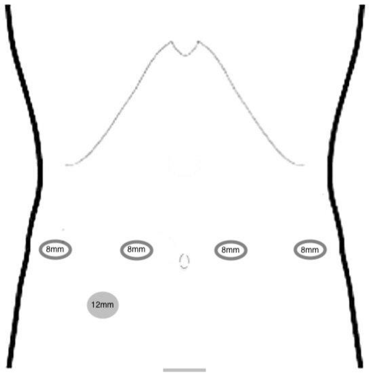
Patient Positioning and Port Placement

The patient is placed in a supine and 30° reverse Trendelenburg position. Robotic surgery is performed with the da Vinci Xi robotic platform (Intuitive Surgical Inc., Sunnyvale, CA) using a five-trocar technique. Pneumoperitoneum is created using a Veress needle via percutaneous puncture at the Palmer point. Standard port placement is used with the Xi System docked from the right side of the patient. An assistant port is placed in the right flank (Figure 1). The camera is inserted through arm 2 and targeting is also performed from arm 2. The surgeon sits at the robotic console while the assistant surgeon stands on the left-side of the patient.

Robotic Instrumentation

Standard robotic instruments used: fenestrated bipolar forceps at arm 1, camera at arm 2, monopolar curved scissors and SynchroSeal (Da Vinci Energy by Intuitive) at arm 3, Cadiere forceps at arm 4.

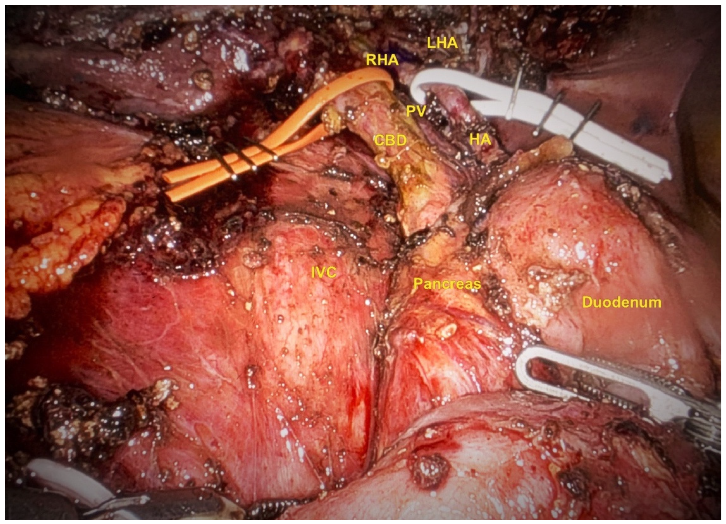
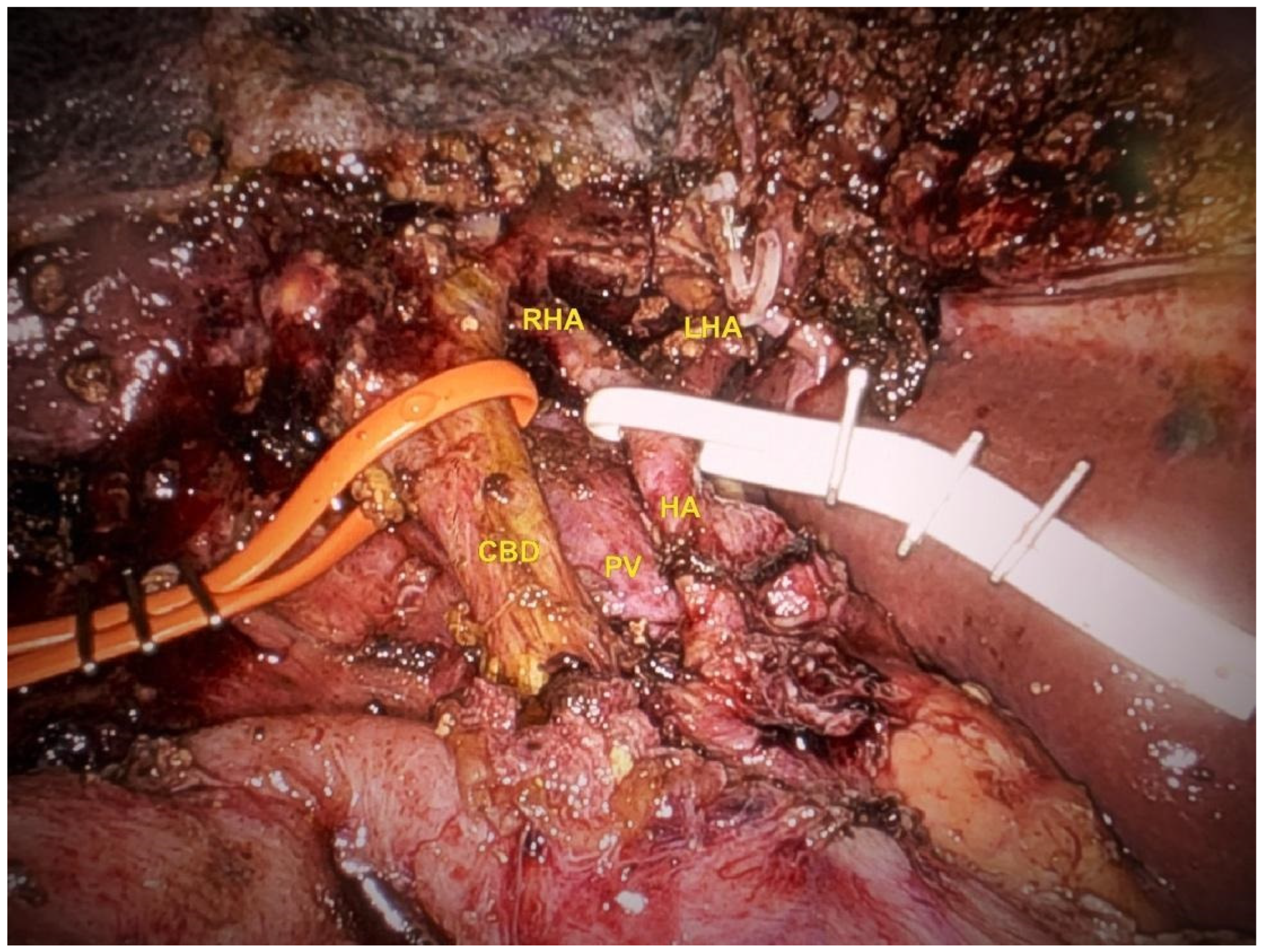
Lymphadenectomy and Hilar Dissection

The abdominal cavity is first inspected for any unknown peritoneal implants. At this time, adhesions from the previous surgery are divided. After a complete Kocher maneuver, intercavo-aortic (station 16), retropancreatic (station 13) and common hepatic artery (station 8) lymphadenectomy is performed. The hepatic hilum is then carefully dissected, to expose the proper hepatic artery (station 12a), the left and right hepatic arteries, the common bile duct (station 12b) and the portal vein (station 12p). The vascular elements are then suspended on tapes for indirect traction and an extensive hilar lymphadenectomy is performed (Figure 2-Figure 3). At this time, a Foley catheter is placed as usual around the hepatic pedicle as a precaution in case of a Pringle maneuver.

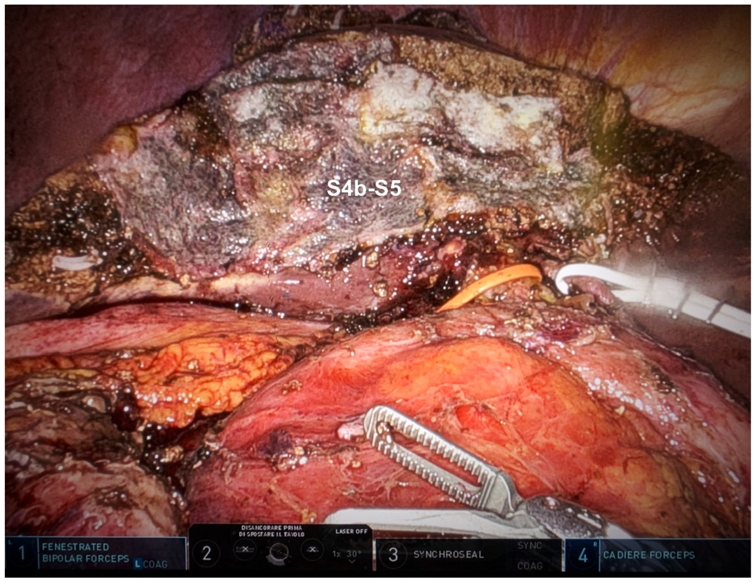
Hepatic Bisegmentectomy (S4b + S5)

Intraoperative ultrasound is performed by inserting the ultrasound probe through the 12 mm assistant port. After identifying the glissonian pedicles for segments 4b and 5 the surgical resection margins are marked on the hepatic surface. The liver parenchyma is then transected using robotic fenestrated bipolar forceps and monopolar curved scissors or SynchroSeal. The glissonian elements for segments 4b and 5 are accessed via the intrahepatic approach and divided between hemoloks. In this case, a single Pringle maneuver is performed throughout the procedure with a total clamping time of 12 minutes (Figure 4). The liver's raw surface is checked for bleeding and bile leakage. Haemostatic dressings are put on and the peritoneal cavity is drained with a 19-F tube. The surgical specimen is removed through a Pfannenstiel incision, after being wrapped in a plastic bag to minimize the risk of intra-abdominal seeding and recurrence at the port sites. The scar from the umbilical port site used for the extraction of the GB during the previous cholecystectomy, is finally removed.

3. Results

From 2023, 4 patients with GBC or iGBC after LC were treated with the same robotic approach to regional lymphadenectomy and liver bisegmentectomy, using the Da Vinci Xi Surgical System (Table 1). The mean operation time was 339 minutes. Estimated blood loss was < 200 ml and no transfusions were required. The postoperative course was uneventful, no postoperative bleeding or biliary fistulas were observed. The mean length of stay was 10 days. No 30 days-mortality was reported. No residual disease was found in the final pathology examination of either the liver specimen or excised scar tissue from the port site. Mean LNs retrieved are 9.5 (range 6 – 12). However, in 1 case, 1 of 12 removed LNs (station 12p) contained a 2 mm subcapsular metastasis. After surgery, the patient started adjuvant chemotherapy with gemcitabine, which is still ongoing. No evidence of disease was seen at the first CT follow-up at 4 months.

4. Discussion

The diagnosis of Stage II (T2) GBC is often made incidentally following LC and necessitates re-resection for adequate treatment and staging. Due to the high propensity of GBCs to spread through the lymphatic system, achieving an adequate lymphadenectomy is essential to improve the outcome of these patients. Effective LN dissection and a higher number of retrieved LNs are indeed associated with better survival, even in node-negative patients [12,13,14,15,16]. LN metastasis is therefore a critical prognostic factor for patient survival after curative resection. Although the 8th edition of the AJCC Cancer Staging Manual recommends at least 6 LNs to be harvested [17], most studies report a median yield of approximately two or three LNs, demonstrating the need for improvement in this area [4].
To achieve this goal, it is often necessary to extend the dissection beyond the portal nodes [4]. The gallbladder's regional LNs are classified into three distinct groups: first-level nodes, second-level nodes, and more distant nodes (Figure 5). The first-echelon nodes are situated on either the cystic duct (CD) or common bile duct (CBD), while the second-echelon nodes are situated posterosuperior to the head of the pancreas or around the portal vein/hepatic arteries [18].
The second-echelon nodes are a challenge to dissect thoroughly, even for experienced hepatobiliary surgeons. In particular, removal of the posterior superior pancreaticoduodenal, retroportal and right celiac nodes requires meticulous technique.
We believe these cases of GBCs were an excellent indication for the use of the robotic system to significantly improve the quality of lymphadenectomy. The superior three-dimensional magnified vision provided by the robotic platform, along with the extreme flexibility of the tremor-filtered robotic arms, allows the surgeon to operate with extreme precision and skill in even the most delicate and complex surgical fields.
Indeed, in the described study, surgical steps such as dissection of the hepatic hilum, axial (aortocaval/celiac) lymphadenectomy, and intraparenchymal access and division of the glissonian pedicle benefited greatly from the utilization of the robotic platform. The exceptional stability of the system, combined with the well-known characteristics of the robotic platform (superior image rendering, increased dexterity, multiple degrees of freedom and high performance instruments) translated into great confidence in the surgeon’s manipulation of even the smallest anatomical structures.
As a result, in the described case the total LN harvest was 12, with 1 single GBC metastasis identified among the retrieved nodes, specifically located along the bile duct (station 12b).
There are a few tricks that we believe can be used to make the lymphadenectomy easier to perform. First, a complete Kocher maneuver: this not only provides direct access to the aortocaval and retropancreatic LNs, but also provides a better view of the retroportal space, which facilitates its subsequent dissection (Figure 5). The easy orientation and much closer view of the surgical field provided by the robotic platform allows for gentle dissection and retraction of even the most delicate retroperitoneal structures, minimizing the risk of inadvertent injury.
When approaching the hepatic pedicle, anatomical landmarks such as the CD (or cystic stump in the case of previous cholecystectomy) and the large LN located on the anterior aspect of the common hepatic artery (station 8), should not be overlooked as they are critical for the proper identification and preparation. Therefore, we believe it is imperative to correctly identify these structures at the beginning of the dissection. Once the hepatic pedicle elements are identified, they should be immediately encircled with rubber bands and any direct manipulation of these fragile structures by the robotic arms should be avoided.
Up to 26% of patients with GBC will present with axial (aortocaval/celiac) nodal invasion that would not benefit them from radical surgery. As stated in the 2015 Expert Consensus on Gallbladder Cancer, it is important to assess the status of these lymphatic stations prior to any further surgical steps [4]. Therefore, after the exploration of the abdominal cavity, dissection of aortocaval and coeliac LNs should be the first step of surgery.
For tumours confined to the GB (T1b–2), cholecystectomy with radical exeresis of adjacent liver parenchyma is recommended. In these cases, the common hepatic duct (CHD)/ CBD resection should be added if gross or microscopic implication of the CD is demonstrated on intraoperative evaluation. For patients with T3 or T4 cancers, the extention of primary exeresis is still controversial; therefore, radical resection of locally advanced GBCs should be reserved just for medically suit patients following pluridisciplinary debate [4].
Minimally invasive surgery provides superior perioperative outcomes in terms of hospital stay, pain control, and cosmesis. According to an international multicenter study of different surgical approaches to GBC by Ielpo et al. [1], the robotic approach was associated with multiple advantages, including a higher number of retrieved LNs, minor Pringle maneuver times, lower valued intraoperative blood loss, and shorter hospital stay when compared to both open and laparoscopic approaches.
However, minimally invasive surgery is not yet routinely used for the treatment of GBC. As elsewhere in oncology, strict adherence to the basic principles of cancer surgery is essential for the safe implementation of robotic surgery in this setting.
The technical challenges to be met when using a minimally invasive approach are 1) appropriate portal and aortocaval lymph node sampling; 2) R0 liver transection margins, and, in the more advanced stages, (3) CHD/ CBD resection or reconstruction [4].
In conclusion, the quality of lymphadenectomy is an important predictor of survival after surgery for GBC. The robotic approach has been shown to be technically safe and feasible when performed in centers with adequate experience in minimally invasive surgery. It can provide adequate lymph node recovery and good oncologic radicality, offering better short-term results and at least non-inferior long-term outcomes.
Author’s Contributions: Conception and design of study: GM, AO.; Acquisition of data: GM.; Analysis and/or interpretation of data: GM, AO.; Drafting the manuscript: GM, AO.; Revising the manuscript critically for important intellectual content: AO, DC, GT, RM.; Supervision: PP, AO.

Funding

This research received no external funding.

Acknowledgments

Neither fund nor grants were received for this research.

Conflicts of Interest

The authors declare no conflicts of interest.

Abbreviations

GBC gallbladder cancer;
iGBC incidentally gallbladder cancer;
RLR robotic liver resection;
OLR open liver resection;
LLR laparoscopic liver resection;
LNs lymph nodes;
LC laparoscopic cholecystectomy;
CBD common bile duct;
CHD common hepatic duct.

References

  1. Ielpo B, Vittoria d'Addetta M, Cremona S, Podda M, Di Martino M, Di Franco G et al. IRON: A retrospective international multicenter study on robotic versus laparoscopic versus open approach in gallbladder cancer. Surgery. 2024 Oct;176(4):1008-1015. [CrossRef]
  2. Singh BP, Khan WF, Rathore YS, Pol MM. Incidental Carcinoma Gallbladder: Incidence, Risk Factors, and Factors Affecting Survival-5-Year Experience from a Tertiary Care Institute. J Gastrointest Cancer. 2020 Sep;51(3):980-987. [CrossRef]
  3. de Savornin Lohman EAJ, van der Geest LG, de Bitter TJJ, Nagtegaal ID, van Laarhoven CJHM, van den Boezem P et al. Re-resection in Incidental Gallbladder Cancer: Survival and the Incidence of Residual Disease. Ann Surg Oncol. 2020 Apr;27(4):1132-1142. [CrossRef]
  4. Aloia TA, Járufe N, Javle M, Maithel SK, Roa JC, Adsay V et al. Gallbladder cancer: expert consensus statement. HPB (Oxford). 2015 Aug;17(8):681-90. [CrossRef]
  5. Liu R, Wakabayashi G, Kim HJ, Choi GH, Yiengpruksawan A, Fong Y et al. International consensus statement on robotic hepatectomy surgery in 2018. World J Gastroenterol. 2019 Mar 28;25(12):1432-1444. [CrossRef]
  6. Stewart C, Wong P, Warner S, Raoof M, Singh G, Fong Y, Melstrom L. Robotic minor hepatectomy: optimizing outcomes and cost of care. HPB (Oxford). 2021 May;23(5):700-706. [CrossRef]
  7. Sucandy I, Luberice K, Lippert T, Castro M, Krill E, Ross S, Rosemurgy A. Robotic Major Hepatectomy: An Institutional Experience and Clinical Outcomes. Ann Surg Oncol. 2020 Dec;27(13):4970-4979. [CrossRef]
  8. Sucandy I, Jacoby H, Crespo K, Syblis C, App S, Ignatius J et al. A Single Institution's Experience With Robotic Minor and Major Hepatectomy. Am Surg. 2023 May;89(5):1387-1391. [CrossRef]
  9. Broering DC, Elsheikh Y, Alnemary Y, Zidan A, Elsarawy A, Saleh Y et al. Robotic Versus Open Right Lobe Donor Hepatectomy for Adult Living Donor Liver Transplantation: A Propensity Score-Matched Analysis. Liver Transpl. 2020 Nov;26(11):1455-1464. [CrossRef]
  10. Rojas AE, Paterakos P, Choi SH. Robotic Central Bisectionectomy for Centrally Located Hepatic Malignant Tumor. Ann Surg Oncol. 2022 Mar 31. [CrossRef]
  11. Liu R, Abu Hilal M, Wakabayashi G, Han HS, Palanivelu C, Boggi U et al. International experts consensus guidelines on robotic liver resection in 2023. World J Gastroenterol. 2023 Aug 28;29(32):4815-4830. [CrossRef]
  12. Tran TB, Nissen NN. Surgery for gallbladder cancer in the US: a need for greater lymph node clearance. J Gastrointest Oncol. 2015 Oct;6(5):452-8. [CrossRef]
  13. Coburn NG, Cleary SP, Tan JC, Law CH. Surgery for gallbladder cancer: a population-based analysis. J Am Coll Surg. 2008 Sep;207(3):371-82. [CrossRef]
  14. Widmann B, Warschkow R, Beutner U, Weitzendorfer M, Ukegjini K, Schmied BM et al. Effect of lymphadenectomy in curative gallbladder cancer treatment: a systematic review and meta-analysis. Langenbecks Arch Surg. 2020 Aug;405(5):573-584. [CrossRef]
  15. Choi SB, Han HJ, Kim CY, Kim WB, Song TJ, Suh SO et al. Surgical outcomes and prognostic factors for T2 gallbladder cancer following surgical resection. J Gastrointest Surg. 2010 Apr;14(4):668-78. [CrossRef]
  16. Vega EA, Newhook TE, Mellado S, Ruzzenente A, Okuno M, De Bellis M et al. Benchmarks and Geographic Differences in Gallbladder Cancer Surgery: An International Multicenter Study. Ann Surg Oncol. 2023 Aug;30(8):4904-4911. [CrossRef]
  17. Chun, Y.S., Pawlik, T.M. & Vauthey, JN. 8th Edition of the AJCC Cancer Staging Manual: Pancreas and Hepatobiliary Cancers. Ann Surg Oncol 25, 845–847 (2018). [CrossRef]
  18. Shirai Y, Wakai T, Sakata J, Hatakeyama K. Regional lymphadenectomy for gallbladder cancer: rational extent, technical details, and patient outcomes. World J Gastroenterol. 2012 Jun 14;18(22):2775-83. [CrossRef]
Figure 1. Robotic port placement for RLR.
Preprints 151836 g001
Figure 2. Surgical field during robotic lymphadenectomy after Kocher maneuver. IVC inferior vena cava; CBD common bile duct; PV portal vein; HA hepatic artery; RHA right hepatic artery; LHA left hepatic artery.
Preprints 151836 g002
Figure 3. Robotic hilar lymphadenectomy. CBD common bile duct; PV portal vein; HA hepatic artery; RHA right hepatic artery; LHA left hepatic artery.
Preprints 151836 g003
Figure 4. Robotic liver bisegmentectomy of S4b-S5.
Preprints 151836 g004
Figure 5. Topographical distribution of the regional lymph nodes of the gallbladder, shown after a Kocher maneuver. GB gallbladder; IVC inferior vena cava; AO aorta; IMA, inferior mesenteric artery.
Preprints 151836 g005
Disclaimer/Publisher’s Note: The statements, opinions and data contained in all publications are solely those of the individual author(s) and contributor(s) and not of MDPI and/or the editor(s). MDPI and/or the editor(s) disclaim responsibility for any injury to people or property resulting from any ideas, methods, instructions or products referred to in the content.
Copyright: This open access article is published under a Creative Commons CC BY 4.0 license, which permit the free download, distribution, and reuse, provided that the author and preprint are cited in any reuse.
Prerpints.org logo

Preprints.org is a free preprint server supported by MDPI in Basel, Switzerland.

Subscribe

Disclaimer

Terms of Use

Privacy Policy

Privacy Settings

© 2025 MDPI (Basel, Switzerland) unless otherwise stated