Submitted:
05 March 2025
Posted:
06 March 2025
You are already at the latest version
Abstract
Abstract: Huntington’s disease (HD) is a detrimental neurodegenerative disease caused by the expansion of a CAG triplet in the HTT gene, ending in the production of abnormal Huntingtin (Htt) protein with toxic gain-of-function. The mutant protein (mHtt) is responsible in several ways for the establishment of an intricate pathogenetic scenario in affected cells, particularly in HD neurons. Among all the HD features, oxidative stress plays a relevant role in the progression of the disease at the cellular level. Mitochondrial dysfunction, bioenergetic deficits, Reactive Oxygen Species (ROS) production, neuroinflammation, and general reduction of antioxidants levels are all involved in the promotion of a toxic oxidative environment, eventually causing cell death. Nonetheless, neuronal cells exert antioxidant molecules to build up defense mechanisms. Key components of these defensive mechanisms are the nuclear factor erythroid 2-related factor 2 (NRF2) and peroxisome proliferator-activated receptor gamma coactivator-1 α (PGC-1α). Thus, aim of this re-view article is to describe the involvement of oxidative stress in HD by exploring the role of the NRF2 and PGC-1α, crucial actors in this play. Finally, antioxidant therapeutic strategies targeting such markers will be discussed.
Keywords:
Huntington’s disease
; oxidative stress
; ROS
; neuroinflammation
; NRF2
; PGC-1α
; PPARs
; neurodegeneration
; antioxidant therapy
1. Introduction
Huntington’s disease (HD) is a detrimental neurodegenerative condition caused by the expansion mutation of the glutamine encoding Cytosine-Adenine-Guanine (CAG) trinucleotide in the exon 1 of the Huntingtin gene (HTT). The HTT gene is located in chromosome 4p16 and is responsible for the production of the Huntingtin (Htt) protein which is involved in many relevant processes within cells [1]. In HD, a prolonged mutant Htt (mHtt) is produced by the expanded gene determining a toxic gain of function for the physiological Htt with an extended polyglutamine (polyQ) tract. Indeed, this feature is considered among the major characteristics of HD pathology. Moreover, the presence of toxic N-terminal fragments containing only exon 1 that are produced by proteolytic cleavage of mHtt or CAG length-dependent aberrant splicing strongly define the HD pathology at the cellular level [2]. From a clinical perspective, HD is characterized by motor, psychiatric, and cognitive symptoms, with loss of functional autonomy over time. Symptoms are expressions of a pathological loss of neurons in the Central Nervous System (CNS), mainly in striatal γ amino-butyric acid (GABA)-ergic medium spiny neurons (MSNs) which are the most susceptible cells to the HD insult [3]. HD is an uncommon autosomal-dominant disorder affecting approximately 5–10 individuals per 100000 across most regions of North and South America, Australia, and Europe. In contrast, its prevalence (0,5/100000) is considerably lower in Asian and African populations [4]. Although juvenile-onset cases exist, HD typically emerges between 35 and 50 years of age. Following symptom onset, life expectancy generally ranges from 15 to 20 years. Interestingly, disease onset is inversely correlated with the number of repeats within the mutated HTT gene. Indeed, adult patients typically show 36–50 repeats compared to the 10-35 of healthy people, whereas juvenile-onset cases exceed 50. The more the HTT gene is expanded and the more the mHtt toxicity will spread fast, aggregating in neurons and driving neurodegeneration locally and beyond. Thus, a complex network of pathophysiological events takes place within cells and tissues, with mitochondrial dysfunction, oxidative stress and neuroinflammation as common thread both in CNS and periphery [5,6]. For instance, oxidative stress involvement in HD is deep: increased levels of antioxidant enzymes such as catalase, superoxide dismutase (SOD), and glutathione peroxidases (GPx), which are products of the antioxidant response element (ARE) genes, have been detected in HD-affected brains [7]. In physiological conditions, oxidative stress is under control with functional mitochondria producing ATP through oxidative phosphorylation, balanced oxidant/antioxidant signaling, and preserving calcium homeostasis, finely tuning the redox homeostasis. However, enormous Reactive Oxygen Species (ROS) accumulation in HD cells brings damage to intracellular molecules. As a basic leucine zipper (bZIP) transcription factor, nuclear factor erythroid 2-related factor 2 (NRF2) binds to ARE sequences, orchestrating the expression of antioxidant, anti-inflammatory, and detoxifying genes [8]. For instance, NRF2 pharmacological activation in rodent HD models has been followed by increased antioxidants expression and ROS reduction [9]. Similarly, peroxisome proliferator-activated receptor (PPAR) γ (PPARγ) coactivator-1 α (PGC-1α), a transcription factor involved in the regulation of several energetic processes in the cells, is downregulated in HD and gives a crucial contribution to the establishment of HD cellular pathology. As PGC-1α plays a pivotal role in the physiological maintenance of the redox balance and the preservation of mitochondrial homeostasis in healthy cells, its downregulation in HD cells is followed by detrimental effects with loss of mitochondrial biogenesis and maintenance [10]. Thus, aim of this review article is to describe the involvement of NRF2 and PGC-1α in HD pathology and their potential in antioxidant therapeutic strategies for HD.
2. HD Pathogenesis
The formation of toxic mHtt monomers promotes the development of oligomers, which act as precursors to fibrils accumulating in cytoplasm and nucleus compromising the protective functions of the normal Htt with organelles dysfunction and cell loss. Indeed, mHtt impairs the Htt control over apoptotic pathways inhibition and neurotrophins such as Brain-Derived Neurotrophic Factor (BDNF) production [11]. The toxic mHtt can affect transcriptional regulation by interacting with DNA regions of numerous genes through its expanded polyglutamine tract [12]. Furthermore, HD disrupts protein degradation systems, including the ubiquitin–proteasome system (UPS) and autophagy, as demonstrated in both animal models and human tissues [13,14]. Starting from the expansion mutation, the mHtt production and accumulation within neurons and beyond can be responsible for gliosis in CNS. The activation states of glial cells initiate intracellular signaling pathways that alter gene expression enhancing cytokine release, downregulating astrocytic glutamate transporter 1 (GLT1), impairing Kir4.1 channels, reducing BDNF levels, and elevating both glutamate and cytokine release from astrocytes, ultimately increasing oxidative stress and mitochondrial dysfunction, ROS production, and neuroinflammation [15]. Given the role of the cited mechanisms in HD pathogenesis and the involvement of NRF2 and PGC-1α in them, the following section will describe oxidative stress and mitochondrial dysfunction, and neuroinflammation in more detail.
2.1. Oxidative Stress and Mitochondrial Dysfunction
Mitochondrial dysfunction represents a crucial aspect in the pathogenesis of HD, with mitochondrial DNA (mtDNA) abnormalities being closely associated with striatal degeneration and impaired respiratory chain complexes function. Studies have highlighted disrupted mitochondrial morphogenesis in HD brains, characterized by enhanced fission, reduced fusion, and overall mitochondrial depletion. Alterations in mitochondrial dynamics induced by mHtt contribute to excessive mitochondrial fragmentation [16]. In fact, the Htt protein plays an essential role in maintaining mitochondrial integrity and function, while mHtt disrupts mitochondrial inner membrane proteins, promoting neuronal dysfunction and cell death. Also, in HD-affected neurons, mHTT interacts with mitofusin 1 (Mfn1) and the mitochondrial fission GTPase dynamin-related protein 1 (Drp1), further reducing mitochondrial fusion and exacerbating mitochondrial fragmentation and neuronal damage. Such pathological alterations have been linked to mtDNA copy number reduction and increased mtDNA damage [16]. Similarly, mtDNA alterations are connected with dopamine altered metabolism. Dopamine and other neurotransmitters such as acetylcholine, glutamate, and GABA are involved in HD pathogenesis. Indeed, mitochondria can produce neurotransmitters and their impairment in HD can be followed by altered neurotransmission [17]. For instance, glutamate synthesis takes place within astrocytic mitochondria, and energy-dependent processes such as ATP generation are crucial for its transfer from astrocytes to neurons and its storage in synaptic vesicles [18]. Mitochondrial dysfunction has been identified as a major contributor to glutamate-induced excitotoxicity, compromising neuronal resistance and increasing mitochondrial and cytosolic Ca2+ accumulation, main features of HD-dependent excitotoxic damage. Dysregulation of GABA-A receptors has been associated with altered mitochondrial membrane potential, intensifying neurodegeneration. Moreover, the breakdown of antioxidant defenses under glutamate excitotoxic conditions amplifies oxidative stress, further impairing mitochondrial dynamics and reinforcing disease progression [19]. Moreover, impaired mitochondrial axonal transport emerges as an early contributor to HD progression [20]. Indeed, in HD the mHtt interferes with mitochondrial dynamics by impairing biogenesis and disrupting both anterograde and retrograde transport, governed by kinesin and dynein motor proteins. This interference results from mHtt binding directly to these motor proteins, impairing mitochondrial attachment, reducing transport efficiency, and inducing cell death through mitochondrial membrane damage [21]. Thus, mitochondrial dysfunction disrupts calcium homeostasis and intensifies ROS production, promoting oxidative stress and neuronal injury in HD. Indeed, the interplay between mitochondrial dysfunction and oxidative stress is pivotal in the establishment of HD pathogenesis [22]. Researchers demonstrated a significant correlation between CAG repeat length and oxidative DNA damage [22]. Furthermore, mHtt has been shown to enhance oxidative stress in both neuronal and non-neuronal cells, thereby intensifying HD pathology. Nuclear accumulation of mHTT represents a hallmark of HD, and oxidative stress has been implicated as a crucial factor for its establishment [23]. HD pathology is primarily characterized by increased oxidative stress and heightened vulnerability of the striatum to mHTT toxicity. Genetic studies have revealed that mHTT expression induces transcriptional alterations, leading to enhanced oxidative stress and upregulation of antioxidant genes associated with the NRF2-ARE pathway. This response likely represents a compensatory mechanism aimed at mitigating oxidative damage [24]. Additionally, mHTT downregulates PGC-1α expression and activity, a crucial co-regulator of mitochondrial biogenesis and antioxidant enzyme synthesis. Reduced PGC-1α levels heighten susceptibility to striatal degeneration and oxidative damage [25]. Collectively, the evidence underscores the critical role of mitochondrial dysfunction and oxidative stress in HD pathogenesis, highlighting potential therapeutic targets aimed at preserving mitochondrial integrity and mitigating oxidative stress-driven neuronal damage.
2.2. Neuroinflammation
Concerning neuroinflammation, HD has been associated with increased levels of inflammatory markers in both the central and peripheral nervous systems [15]. Research indicates that disease progression corresponds with increased pro-inflammatory cytokine concentrations alongside a decline in anti-inflammatory cytokines. Glial cells play a pivotal supportive role to neurons into CNS [26]. However, the establishment of a pro-oxidant and pro-inflammatory environment inside CNS is responsible for glial cells activation [27]. Indeed, astrocyte reactivity has been observed in the early, pre-symptomatic stages of HD and correlates with disease progression [28]. In HD, astrocytes are predominantly neurotoxic and are associated with elevated pro-inflammatory cytokines and ROS at synapses, disrupted potassium balance, and impaired glutamate uptake, resulting in membrane depolarization [29]. On the other hand, few neuroprotective astrocytes resist and contribute to the antioxidant response facilitating the restoration of damaged neuronal circuits [30]. Similarly, microglial activation, triggered by various pro-inflammatory signals, leads to the secretion of both pro- and anti-inflammatory cytokines, such as interleukins (IL) 1β (IL-1β), IL-4, IL-6, and tumor necrosis factor α (TNF-α), as well as growth factors like transforming growth factor β (TGF-β), mannose receptor (CD206), and arginase 1 (Arg1) [27]. Evidence of relevant accumulation of reactive microglia have been found in post-mortem analyses of HD brains, particularly in the frontal cortex and striatum, regions closely associated with HD pathology [31]. Interestingly, HD-related neurodegeneration correlated with the density of activated microglia, underscoring their role in neuronal damage. Mitochondrial dysfunction in microglial cells appears to impair IL-4-driven anti-inflammatory responses, further promoting inflammation [32,33]. This dysfunction may contribute to elevated pro-inflammatory mediator expression and neuronal injury. Interestingly, NRF2 has been recognized for its role in suppressing neuroinflammation by reducing inflammatory responses in neurons [8]. This protein is involved in the assembly of the inflammasome, a protein complex responsible for regulating inflammation. Specifically, NRF2 interacts with the nucleotide-binding oligomerization domain (NOD)-like receptor containing pyrin domain 3 (NLRP3), an inflammasome sensor protein, to downregulate inflammatory signaling. Through this mechanism, NRF2 appears to modulate neuroinflammation, contributing to the mitigation of HD symptoms [34]. Additionally, oxidative stress induced by activated microglia has been linked to neurotoxicity, partly due to increased ROS production, which further amplifies proinflammatory gene expression and microglial and astrocytic activation. In this context, researchers described the modulation of the pro-oxidant and pro-inflammatory environment by anti-inflammatory microglia via PGC-1α pathway, suggesting its therapeutic potential in the HD-driven neuroinflammation [35].
3. Role of NRF2 and PGC1a
After outlining HD and its key pathological features, including oxidative stress and neuroinflammation, the focus will now shift to a detailed examination of NRF2 and PGC-1α, highlighting their roles in both physiological and pathological conditions. The discussion will conclude by exploring potential interactions between these two regulators and therapeutic strategies targeting their pathways.
3.1. NRF2
NRF2 regulates the cellular response to oxidative stress by binding to ARE, which functions as a regulatory enhancer sequence located within the promoter regions of detoxifying genes, modulating the expression of genes involved in antioxidant defense, and inflammation control [36]. Thus, activation of NRF2 signaling leads to the upregulation of antioxidant enzymes, including GPx, SOD, heme oxygenase-1 (HO-1), and NAD(P)H quinone oxidoreductase-1 (NQO1). Furthermore, NRF2 induces anti-inflammatory mediators and protects against mitochondrial dysfunction, underscoring its crucial role in cellular resilience against oxidative stress and inflammation [37]. From a structural perspective NRF2, a member of the cap-and-collar (CNC) bZIP transcription factor family, consists of seven functional domains, designated Neh1 through Neh7. The C-terminal region includes Neh1, Neh3, and Neh6 domains. Within Neh1, which harbors the CNC bZIP motif, NRF2 interacts with DNA by forming heterodimers with Maf proteins [38]. The Neh2 domain, located at the N-terminal, plays a pivotal role in cytoplasmic regulation by facilitating NRF2 binding to the Kelch-like ECH-associated protein-1 (Keap1) [39]. The Neh3 domain recruits chromodomain helicase DNA-binding protein-6 (CHD6), which modulates transcriptional activity, while the Neh6 domain contains DSGIS and DSAPGS motifs, which are essential for β-TrCP-mediated NRF2 degradation [40]. Additionally, the Neh7 domain interacts with the retinoic acid receptor (RAR), further regulating NRF2 activity [41]. Keap1 predominantly controls NRF2 stability under basal conditions by anchoring it in the cytoplasm. Two Keap1 molecules can bind a single NRF2 protein, promoting its ubiquitination and proteasomal degradation. Upon oxidative stress, NRF2 dissociates from Keap1, translocates into the nucleus, and accumulates at higher levels. This nuclear accumulation enhances NRF2 binding to AREs within the promoter regions of target genes involved in antioxidant defense, metabolism, and cellular homeostasis [41]. Building upon the pivotal role of NRF2, various signaling pathways converge to modulate its activity and orchestrate cellular defense. For instance, the mitogen-activated protein kinase (MAPK) cascade, known for regulating cell proliferation, differentiation, and apoptosis, is activated by reactive oxygen species generated during calcium imbalance [42]. While MAPK signaling typically promotes pro-inflammatory responses, NRF2 activation counteracts this effect by upregulating antioxidant enzymes and suppressing inflammatory mediators, thereby restoring redox homeostasis [43,44,45]. In addition, AMP-activated protein kinase (AMPK) senses energy deficits and responds to metabolic stress by activating NRF2 [46]. Early AMPK activation reduces neuroinflammation and works synergistically with NRF2 to elevate antioxidant enzyme expression, thereby safeguarding neuronal function [47,48]. Concurrently, extracellular signal-regulated kinase (ERK) reinforces NRF2 activity, aiding recovery from DNA damage and excitotoxic events [49]. Similarly, the phosphoinositide 3-kinase (PI3K)/ protein kinase B (Akt) pathway, crucial for cell survival and metabolic regulation, enhances NRF2 activity [50]. Stimulated by growth factors and hypoxic stress, Akt promotes NRF2 nuclear translocation and transcription of detoxifying genes, effectively mitigating oxidative damage [51,52,53]. This crosstalk exemplifies how NRF2 serves as a central mediator against cellular stress. Sirtuins (SIRTs) further refine this defense network. In both mice and humans, metabolic processes, including mitochondrial function, gluconeogenesis, and cell survival, are governed by sirtuins, a family of NAD⁺-dependent histone deacetylases. These enzymes orchestrate cellular energy production and overall metabolism and their pivotal role in metabolic regulation is unequivocally recognized [54]. SIRT1-mediated deacetylation stabilizes NRF2, increasing its capacity to drive antioxidant responses, while inhibition of SIRT2 has been linked to neuroprotection in experimental models [55,56,57]. Such modulation underscores NRF2’s role in preserving cellular integrity under oxidative challenges. The interplay extends to the mammalian target of rapamycin (mTOR) pathway, where reduced mTOR activity is associated with neurodegeneration [58]. NRF2 regulates mTOR expression via ARE, mitigating neuronal atrophy. Moreover, modulation of p53 in response to oxidative stress further emphasizes NRF2’s central role [59]. Indeed, lower stress conditions favor NRF2-driven defenses, while higher stress triggers p53-mediated apoptosis. Collectively, these interconnected pathways show NRF2 as the master regulator of cellular resilience against oxidative stress and inflammation. By integrating signals from MAPK, PI3K/Akt, SIRTs, AMPK, ERK, mTOR, and p53, NRF2 coordinates a comprehensive protective response that maintains neuronal viability and offers therapeutic targets for neurodegenerative disorders. Thus, the integrative network governed by NRF2 not only fortifies cellular defenses but also represents a promising focus for innovative, targeted therapeutic strategies.
3.2. PGC-1α
The brain's high metabolic requirements, alongside the critical role of ATP production and mitochondrial integrity in neuronal function, have led to extensive investigations into PGC-1 coactivators as key regulators of energy metabolism. Expression studies have revealed abundant PGC-1α presence in several brain regions, including the cerebral cortex, hippocampus, striatum, thalamic nucleus, and substantia nigra [60]. PGC-1α serves as a versatile transcriptional coactivator, interacting with a broad range of transcription factors involved in various biological processes. These include mitochondrial biogenesis, oxidative phosphorylation (OXPHOS), antioxidant responses, adaptive thermogenesis, and metabolic regulation of glucose and fatty acids [61]. Indeed, the PGC-1 family consists of proteins that are tightly regulated in response to various environmental signals and are essential in modulating signaling pathways to ensure proper cellular and systemic adaptations to changing conditions. PGC-1α, the most thoroughly studied member, was initially identified as a protein interacting with PPARγ in brown adipose tissue [62]. PGC-1β, which closely resembles PGC-1α, shares considerable sequence similarity, while the PGC-related coactivator (PRC) has more limited homology [63]. Regarding PGC-1α, it forms complexes with transcription factors such as NRF1, NRF2, and nuclear receptors including PPARα, PPARδ, PPARγ, and estrogen-related receptor α (ERRα) [25]. These complexes regulate the expression of nuclear-encoded mitochondrial genes like cytochrome c (Cyt c), mitochondrial complexes I-V, and mitochondrial transcription factor A (TFAM), which are crucial for mitochondrial function. Mitochondrial defense against ROS is primarily mediated by antioxidant enzymes such as SOD2, which converts superoxide anions to hydrogen peroxide, along with peroxiredoxins (Prx3 and Prx5), mitochondrial thioredoxin (Trx2), and thioredoxin reductase (TrxR2) [64,65]. PGC-1α is recognized for suppressing oxidative stress by upregulating mitochondrial uncoupling proteins (UCPs) and antioxidant enzymes including SOD1, SOD2, and GPx-1. Regulatory promoter regions of SOD2, UCP-2, and Prx5 are directly bound by PGC-1α, establishing a central role in transcriptional control [66,67]. Additionally, while the nuclear SIRT1 controls PGC-1α activation status via deacetylation regulating the energy levels inside cells, mitochondria express SIRT3 under PGC-1α regulation, which deacetylates and activates SOD2, thereby reducing ROS and enhancing fatty acid oxidation [68]. Therefore, through the activation of mitochondrial gene networks PGC-1α enhances fatty acid β-oxidation, the tricarboxylic acid cycle, and OXPHOS efficiency. Additionally, it promotes the expression of genes associated with ion transport, heme biosynthesis, and mitochondrial protein synthesis, while boosting overall respiratory capacity [69,70]. PGC-1α has also been associated with the maintenance of multiple neurotransmitter systems, including cholinergic, glutamatergic, dopaminergic, and GABAergic synapses [71,72,73]. A deficiency of PGC-1α within specific brain regions, particularly GABAergic neurons, has been linked to hyperactivity, impaired short-term habituation, and heightened startle responses [74]. Conversely, PGC-1α activation or overexpression has demonstrated neuroprotective effects by enhancing mitochondrial function, supporting neuronal survival, reducing neuroinflammation, and promoting protein clearance [35,75]. PGC-1α plays a multifaceted role in HD, impacting mitochondrial function, protein homeostasis, and neuronal integrity [10]. It regulates postnatal myelination by promoting myelin basic protein (MBP) expression and cholesterol synthesis [76]. Both adult HD models and PGC-1α knockout mice show decreased MBP levels and impaired myelination, while PGC-1α overexpression enhances MBP promoter activity, suggesting its critical role in myelin maintenance. At the organelle level, PGC-1α upregulation increases mitochondrial mass, promotes organelle fusion, and restore mitochondrial dynamics [77]. In HD mouse models, brown adipose tissue exhibits a significant reduction in functional mitochondria and a lower ATP/ADP ratio [78]. This mitochondrial dysfunction correlates with decreased expression of PGC-1α target genes involved in energy metabolism, indicating that impaired PGC-1α signaling may drive broader metabolic deficits in HD. On a molecular scale, PGC-1α stimulates transcription factor EB (TFEB), a key regulator of the autophagy-lysosome pathway, facilitating the clearance of mHtt and thereby supporting cellular proteostasis and reducing the accumulation of toxic aggregates commonly observed in HD [79,80]. Given its role in mitochondrial function, protein turnover, and myelination, enhancing PGC-1α activity has emerged as a promising therapeutic strategy for early intervention in HD, offering potential benefits for both neuronal maintenance and metabolic homeostasis.
3.3. INTERACTION BETWeen NRF2 and PGC-1α
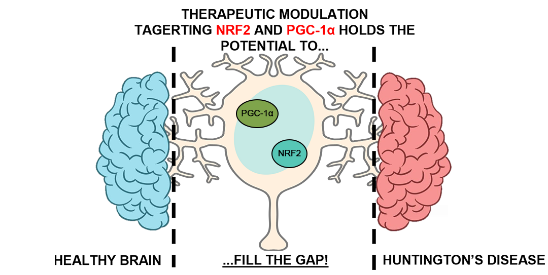
NRF2 accumulates in the cytoplasm bound to Keap1, a negative regulator. Upon exposure to oxidative stress, NRF2 dissociates from Keap1, translocates into the nucleus, and binds to regulatory enhancer sequences ARE in the promoter regions of various detoxifying genes (e.g., GPx, SOD, and NQO1) [7,24]. This nuclear accumulation of NRF2 not only upregulates antioxidant enzymes but also amplifies cellular response to inflammation, thus playing a central role in cellular resilience against stress. Interestingly, NRF2 activation is tightly regulated by various signaling pathways, one of the most crucial being AMPK [81]. As described by Joo and collaborators, AMPK phosphorylates NRF2 at Ser50, which in turn leads to the inactivation of glycogen synthase kinase 3β (GSK3β), which is overexpressed in oxidative stress and inflammation promoting NRF2 degradation, thereby facilitating NRF2's translocation to the nucleus [81,82]. This process is essential for NRF2's function as a transcriptional activator of antioxidant and cytoprotective genes. Moreover, various compounds such as berberine, fidarestat, and pterostilbene have been shown to activate NRF2 via AMPK signaling, highlighting the importance of this pathway in cellular redox regulation [83,84,85]. When AMPK is inhibited, NRF2 activation is abolished, underscoring the interdependent nature of AMPK and NRF2 signaling [86]. Beyond AMPK, NRF2 activity is intricately connected to PGC-1α (Figure 1). It was demonstrated that PGC-1α can promote the transcriptional activation of antioxidant genes via NRF2 [87]. Downregulation of PGC-1α expression in mice led to a significant reduction in the binding of NRF2 to specific genes like GCLC, affecting the levels of SOD2 and glutamate cysteine ligase (GCL) proteins. Further studies in PGC-1α-deficient mice showed disrupted NRF2-dependent mitochondrial biogenesis, pointing to a crucial role of PGC-1α in orchestrating NRF2 activation under stress conditions [88]. The interaction between NRF2 and PGC-1α is thought to be governed by the p38 MAPK pathway, which is positively regulated by PGC-1α and leads to GSK3β inactivation, further promoting NRF2 activation [89]. This interplay suggests that NRF2 and PGC-1α may form a feedback loop. NRF2 itself may regulate the expression of PGC-1α, enhancing mitochondrial function and antioxidative defense. Indeed, the PGC-1α gene promoter contains two ARE sequences (-1723 and -226) which are directly regulated by NRF2 [90]. Experiments involving NRF2 siRNA silencing or NRF2 knockout resulted in a decrease in mitochondrial biogenesis and down-regulation of PGC-1α expression across various cell types, including hepatocytes and skeletal muscles, further supporting the existence of NRF2-PGC-1α feedback regulation [91,92]. Simultaneous activation of the NRF2 and PGC-1α pathways may also be observed. For instance, the ERK1/2 pathway activate both NRF2 and PGC-1α via phosphorylation of liver kinase B1 (LKB1), which subsequently activates AMPK [93]. In the context of HD, NRF2 presents a potent avenue for therapeutic exploration. Indeed, NRF2 activation has been shown to protect neurons from oxidative stress and mitochondrial dysfunction, and overexpression of NRF2 offers neuroprotection from mHtt toxicity [9,94]. Conversely, deletion of NRF2 renders mice more susceptible to neurotoxins like malonate, compound that mimic HD pathophysiology [95]. Alongside NRF2, PGC-1α levels are reduced in HD as previously described, further implicating mitochondrial dysfunction as a central component of HD pathology. Interestingly, direct interactions between PGC-1α and NRF2 may occur in a bidirectional manner to maintain mitochondrial resilience under stress.
4. Antioxidant Therapeutic Strategies Involving NRF2 and PGC-1α Signaling
4.1. Modulation of NRF2 as a Therapy for HD
Therapeutic interventions aimed at modulating the NRF2 pathway have attracted significant attention as potential strategies to combat HD. In various preclinical studies (reviewed below), neuroprotection has been achieved by enhancing NRF2 activity using a range of pharmacological agents that mitigate oxidative stress and inflammation. Animal models of HD have been instrumental in delineating the beneficial effects of these compounds, which include naturally occurring phytochemicals and synthetic compounds. Phytochemicals have emerged as promising candidates in the therapeutic landscape of HD. Naringin, a flavanone abundant in citrus fruits, has been shown to alleviate inflammation and oxidative stress in male Wistar rats exposed to 3-nitropropionic acid (3-NPA), a mitochondrial inhibitor [96]. In these studies, upregulation of the NRF2 pathway by naringin was linked to a reduction in neuroinflammatory markers and oxidative damage. Consistent results have been observed in PC12 cell cultures, where naringin’s activation of NRF2 contributed to a significant decrease in 3-NPA-induced neurotoxicity [52]. Protopanaxatriol, an active constituent derived from ginseng, has been similarly implicated in neuroprotection [97]. When tested in rat models of 3-NPA-induced neurodegeneration, protopanaxatriol activated the NRF2 pathway and was found to mitigate neuronal injury, further supporting the role of NRF2 modulation in countering HD pathology. Similarly, 6-shogaol, a compound contained in ginger, showed remarkable effects in a 3-NPA-induced HD rat model [98]. Indeed, improved behavior and biochemical indices with restored levels of neurotransmitters and decreased neuroinflammatory molecules were achieved via NRF2/Keap1 and NFkB-dependent inflammation modulation caused by 6-shogaol administration. Researchers described in PC12 cells that the expression of the exon 1 in HTT is influenced by NRF2-associated mechanisms [99]. This finding has led researchers to explore compounds that activate NRF2 as a means to modulate HD-associated gene expression. Among these compounds, triterpenoids have been extensively investigated for their ability to upregulate NRF2 activity [2]. For instance, the triterpenoid derivative 2-Cyano-3,12-dioxo-olean-1,9-dien-28-oic acid-methyl-amide (CDDO-MA) exhibits excellent blood–brain barrier permeability and has demonstrated neuroprotective effects in 3-NPA neurotoxic models [100]. In these models, the activation of the NRF2 signaling pathway by CDDO-MA resulted in diminished neuronal damage. In a complementary approach, transgenic mice expressing a 171-amino acid N-terminal fragment of human mHtt with 82 CAG repeats (N171-82Q), sufficient to cause striatal atrophy, were treated with compounds such as CDDO-ethyl amide (CDDO-EA) and CDDO-tri-fluoroethyl amide (CDDO-TFA) [94,101]. In these experiments, the transcription of NRF2-regulated genes was enhanced, which correlated with improved motor performance and increased neuronal survival in the striatum. Another study demonstrated that azilsartan (Azil), an angiotensin II type 1 receptor blocker, exhibited neuroprotective effects in a 3-NPA-induced neurotoxicity rat model [102]. Azil improved motor function, restored neurotransmitter balance, and reduced inflammation, oxidative stress, and apoptosis through modulation of NFkB and NRF2/Keap1 pathways, highlighting its therapeutic potential. Additional evidence supporting the neuroprotective role of NRF2 comes from investigations employing cysteamine [103]. In its reduced form, cysteamine exerts inhibitory effects on several enzymes that are implicated in neurodegenerative processes [104]. When administered in various HD in vitro models, cysteamine conferred protection by promoting NRF2 activation, thereby counteracting the deleterious effects of oxidative stress [103]. The therapeutic portfolio is further broadened by di-methyl fumarate (DMF), which have been evaluated in both the YAC128 and R6/2 mouse models of HD [9,105,106,107]. DMF treatment has been associated with improvements in muscle function, stabilization of body weight, and a marked reduction in neuronal loss in critical brain regions, including the cortex and striatum. These outcomes suggest that DMF’s activation of NRF2-dependent pathways contributes to enhanced neuronal survival and functional recovery. Even though preclinical studies are promising for these substances, there is a lack of clinical evidence. Among these antioxidant agents, DMF is the only approved neurodegenerative therapy (e.g., multiple sclerosis) that acts via NRF2 activation [108]. Nonetheless, resveratrol and epigallocatechin gallate (EGCG) demonstrated antioxidant effects via NRF2 in animals, even though these animals were modeling conditions different from HD [109]. Indeed, starting from these results and from the neuroprotective effects showed in preclinical research in 3-NPA-induced models using male C57BL/6J mice, 3-NPA-treated rats, and N171-82Q transgenic mice, a clinical investigation evaluated resveratrol’s effect on caudate volume in HD patients (ID: NCT02336633). Similarly, a randomized, double-blind phase II trial assessed the safety, tolerability, and cognitive benefits of EGCG, a green tea polyphenol, in HD patients (ID: NCT01357681). Unfortunately, the results of both the trials remain unpublished. Finally, cysteamine in currently tested only in a dose-finding and tolerability trial with HD patients (ID: NCT02101957).
4.2. Modulation of PGC-1α as a Therapy for HD
As mitochondrial dysfunction and oxidative stress remain central to HD progression, researchers explore strategies that enhance mitochondrial quality control as a means of neuroprotection. One promising avenue is the upregulation of PGC-1α. Recent experimental models have demonstrated that elevating PGC-1α levels can not only restore energy production but also promote the clearance of toxic protein aggregates, thereby attenuating neuronal loss. Indeed, in an innovative genetic study, mice engineered to overexpress PGC-1α in a controlled, inducible manner exhibited a striking reduction in the accumulation of mHtt aggregates via activation of TFEB, which is crucial in the autophagy–lysosome system, as mentioned above [79,80]. Activation of TFEB facilitates the degradation of misfolded proteins and damaged organelles, effectively resetting the intracellular protein quality control machinery [80]. Such clearance mechanisms are vital for maintaining neuronal integrity and mitigating the toxic effects of mutant proteins. Moreover, using a lentiviral vector to introduce PGC-1α into the striatum of R6/2 HD mice effectively halted local tissue degeneration, underscoring the promise of gene therapy strategies that enhance mitochondrial regulation [110]. Parallel to genetic approaches, several small molecule therapies are under investigation to indirectly boost PGC-1α activity. For instance, fenofibrate, a well-known agonist of PPARs, has been shown to modulate PGC-1α function in 3-NPA rats [111,112]. By binding to PPARs, fenofibrate initiates a cascade of transcriptional events that enhance mitochondrial oxidative phosphorylation and biogenesis. Recently, a six-month phase 2 clinical trial described the safety and efficacy of fenofibrate administration in HD patients and improved motor and cognitive symptoms compared to placebo, reflecting the translational potential of targeting metabolic regulators to combat neurodegeneration (ID: NCT03515213). Natural compounds have also received considerable attention. Resveratrol, a polyphenol present in red wine and other sources, acts as a potent activator of SIRT1, which by deacetylating PGC-1α enhances its transcriptional effects on mitochondrial genes, leading to improved mitochondrial respiration and overall energy metabolism [113]. Mechanistically, resveratrol inhibits cAMP-degrading phosphodiesterases, thereby increasing intracellular cAMP levels, which in turn raise calcium concentrations and activate the CamKKβ-AMPK pathway. This cascade ultimately promotes the formation of NAD⁺ and further stimulates SIRT1 activity. Interestingly the neuronal inhibition of phosphodiesterase IV (PDE4), a resveratrol’s target, has emerged as a strategy to elevate cAMP levels, thereby indirectly boosting PGC-1α expression [114,115]. For instance, rolipram, a potent PDE4 inhibitor, has demonstrated both antidepressant and neuroprotective effects in preclinical and clinical HD models by increasing protein kinase A (PKA) activity [116,117,118,119]. However, despite its efficacy, rolipram’s narrow therapeutic window and gastrointestinal side effects have hindered its clinical adoption. Nonetheless, preclinical data from models using 3-NPA suggest that resveratrol can ameliorate motor deficits, although its benefits in the striatum appear limited by suboptimal brain penetration [120,121,122]. Notably, resveratrol’s capacity to restore mitochondrial function extends beyond neuronal cultures. In lymphoblasts isolated from HD patients, treatment with resveratrol completely reinstated mitochondrial membrane potential and respiratory activity while upregulating mitochondrial TFAM [123]. Furthermore, enhanced SIRT1 levels correlate with improvements in motor coordination, a reduction in brain atrophy, and mitigation of mHtt–induced metabolic disturbances [123,124,125]. The therapeutic implications extend to the mitochondrial SIRT3, which is intimately involved in maintaining mitochondrial integrity. Located within the mitochondrial matrix, SIRT3 deacetylates components of the respiratory chain like Complex I and enhances fatty acid oxidation and antioxidant defenses by activating enzymes like SOD2 [126]. In HD cellular models, SIRT3 expression is diminished, yet pharmacological induction with viniferin is able to reverse these deficits leading to neuroprotection [126]. Beyond direct modulation of PGC-1α, activation of nuclear receptors such as PPARs offers an alternative route to improve mitochondrial function via PGC-1α. In HD cells, activation of PPARγ with rosiglitazone has been shown to prevent calcium-induced mitochondrial dysfunction and reduce oxidative stress [127]. Additionally, bezafibrate, a pan-PPAR agonist, has recently been reported to elevate PGC-1α expression, ameliorate behavioral deficits, extend survival, reduce striatal atrophy, and diminish oxidative damage in the R6/2 mouse model [128]. While the potential risk of tumorigenesis linked to PPAR agonists remains a concern, the cumulative evidence suggests that modulation of the PGC-1α pathway, whether through direct overexpression or via upstream nuclear receptor activation, represents a multifaceted strategy to restore mitochondrial integrity, mitigate oxidative stress, and ultimately protect neurons in HD [129,130]. This innovative approach may offer a promising avenue for the development of effective therapies aimed at slowing disease progression and improving patient outcomes.

Table 1.
Overview of the antioxidant therapeutic strategies involving NRF2 and PGC-1α signaling.
| THERAPEUTIC STRATEGY | HD MODEL | NRF2activation | PGC-1αactivation | Effects | REF. |
|---|---|---|---|---|---|
| Naringin | 3-NPA-stressed rats | V | Reduced neuroinflammatory markers and oxidative damage | [96] | |
| 3-NPA-stressed PC12 cells | V | Reduced 3-NPA-induced neurotoxicity | [52] | ||
| Protopanaxatriol | 3-NPA-stressed rats | V | Reduced neuronal injury | [97] | |
| 6-Shogaol | 3-NPA-stressed rats | V | Improved behavior and biochemical indices with restored levels of neurotransmitters and decreased neuroinflammatory molecules | [98] | |
| CDDO-MA | 3-NPA-stressed rats | V | Reduced neuronal injury | [100] | |
| CDDO-EA | N171-82Q mice | V | Improved motor performance and increased neuronal survival in striatum | [94] | |
| CDDO-TFA | N171-82Q mice | V | Improved motor performance and increased neuronal survival in striatum | [94] | |
| Azilsartan | 3-NPA-stressed rats | V | Improved motor function, restored neurotransmitter balance, and reduced inflammation, oxidative stress, and apoptosis | [102] | |
| Cysteamine | Mouse primary neurons and human iPSCs | V | Reduced oxidative stress and neuroprotection | [103] | |
| Human patients | V | (Ongoing study) | NCT02101957 | ||
| DMF | YAC128 and R6/2 mice | V | improvements in muscle function, stabilization of body weight, and a marked reduction in neuronal loss | [9] | |
| Resveratrol | Human patients | V | (Unpublished results) | NCT02336633 | |
| Striatal and cortical neurons from YAC128 mice, Human lymphoblasts, and YAC128 mice | V | Rescued mitochondrial membrane potential and respiratory activity with TFAM upregulation | [123] | ||
| EGCG | Human patients | V | (Unpublished results) | NCT01357681 | |
| PGC-1α overexpression | N171-82Q mice | V | Improved neurological function and TFEB-mediated eradication of mHtt aggregates in the brain | [79] | |
| R6/2 mice | V | Halted striatal degeneration | [110] | ||
| Fenofibrate | 3-NPA-stressed rats | V | PPARγ-mediated enhanced mitochondrial oxidative phosphorylation and biogenesis. | [111] | |
| Human patients | V | Improved motor and cognitive symptoms | NCT03515213 | ||
| Rolipram | Quinolinic acid-stressed rats | V | Antidepressant and neuroprotective effects | [117] | |
| R6/2 mice | V | Neuroprotective effects | [118] | ||
| R6/2 mice | V | Improved motor functions and neuroprotection | [119] | ||
| SIRT1 overexpression | N171-82Q mice | V | Improved metabolism and neuroprotection, decreased brain atrophy | [124] | |
| R6/2 mice | V | Improves survival, neuropathology and neutrophins levels | [125] | ||
| Viniferin | Striatal cells and primary cortical neurons from HD mice, N63-148Q PC12, and N2A cells | V | SIRT3-dependent neuroprotection | [126] | |
| Rosiglitazone | Striatal cells from HD mice | V | Reduced mitochondrial dysfunction and oxidative stress | [127] | |
| Benzafibrate | R6/2 mice | V | Improved behavior and survival, decreased striatal atrophy and oxidative stress | [128] |
5. Discussion
Mitochondrial dysfunction, neuroinflammation, and oxidative stress are HD hallmarks that pose significant challenges for effective treatment. As therapy for HD remains challenging, targeting NRF2 offers a novel means of intervention by reducing oxidative stress and preserving mitochondrial function. NRF2’s regulation of mitochondrial biogenesis and its feedback loop with PGC-1α ensure the maintenance of cellular homeostasis, which is compromised during the progression of these diseases. The combined focus on both NRF2 activation and simultaneous upregulation of mitochondrial functions via PGC-1α could substantially slow HD progression and improve patient outcomes. Over the past decades, extensive studies have elucidated the multifaceted role of mitochondria in neuronal survival and degeneration. Mitochondria are not only the powerhouses of the cell but also play critical roles in programmed cell death. Their dynamic nature allows them to alter morphology, replicate, and even fuse or divide in response to metabolic demands or stress signals. Genetic mutations, aberrant mtDNA, and disrupted transcriptional regulation contribute to a decline in mitochondrial performance. Such dysfunction is now recognized as a central player in HD pathophysiology. Recent preclinical investigations have provided robust evidence that targeting mitochondrial deficiencies can be a viable therapeutic avenue. Specifically, strategies aimed at correcting mitochondrial dynamics and intracellular trafficking are gaining attention. However, clinical trials targeting these pathways have often been unsuccessful due to incomplete understanding of the underlying molecular networks, heterogeneous patient populations, and the absence of reliable biomarkers for therapeutic efficacy. Indeed, bridging the gap between preclinical discoveries and clinical application requires rigorous validation of safety, robust proof of efficacy, and an in-depth understanding of HD complexity. Moreover, challenges in translating preclinical successes into clinical benefits extend beyond molecular intricacies. The blood–brain barrier (BBB) remains a formidable obstacle for drug delivery, limiting the effective concentrations of many promising compounds in the CNS [131]. Innovative drug delivery systems, including nanoparticle-based carriers and viral vectors for gene therapy, may provide the means to overcome these obstacles [132,133]. Additionally, the integration of advanced imaging and biochemical biomarkers could facilitate early detection of therapeutic effects, enabling more refined clinical trial designs [134,135]. Notably, while much attention has been focused on pharmacological approaches, non-pharmacological interventions such as physical exercise, dietary modifications, and controlled exposure to mild stressors (like cold or hypoxia) have also been shown to upregulate NRF2 and PGC-1α and enhance mitochondrial function [136,137]. These lifestyle interventions could complement pharmacotherapy by providing a holistic strategy to maintain cellular energy homeostasis and reduce oxidative stress. Combining such non-pharmacological approaches with targeted molecular therapies may yield synergistic effects, offering new hope for HD patients. Furthermore, the complexity of HD necessitates a multifaceted approach that goes beyond single-target interventions. The interplay between various cellular processes—ranging from protein quality control and autophagy to neuroinflammatory signaling—suggests that a combinatorial therapy approach may be more effective. In summary, a comprehensive strategy that integrates NRF2 activation with enhanced PGC-1α–mediated mitochondrial biogenesis represents a promising frontier in HD therapy. By restoring mitochondrial function, reducing oxidative stress, and improving metabolic efficiency, this approach has the potential to mitigate neuronal loss and slow disease progression. Looking forward, advancements in drug delivery, biomarker discovery, and combined therapeutic modalities will be crucial. These innovations not only promise to refine our understanding of HD’s complex molecular landscape but also hold the potential to translate into transformative clinical therapies that enhance quality of life and extend survival in patients afflicted with neurodegenerative disorders.
6. Conclusions
In conclusion, NRF2 and PGC-1α integrate multiple cellular processes critical for stress response, mitochondrial biogenesis, and antioxidant and neuroinflammatory defenses. Indeed, the crosstalk between these two factors is essential for maintaining cellular homeostasis during oxidative stress and neuroinflammation. Their synergistic work in protecting against the cited toxic events positions them as key targets for therapeutic tools in HD and other neurodegenerative disorders.
Author Contributions
Conceptualization, V.C. and F.D.; software, F.A.; writing—original draft preparation, F.D. and V.C.; writing—review and editing, A.C., M.d., S.K., and E.Q.; supervision, M.d. and A.C.. All authors have read and agreed to the published version of the manuscript.
Funding
This research was funded by Dompé farmaceutici S.p.A, by Progetto n. 74 – Piattaforma tecnologica integrata per l’identificazione e lo sviluppo di neurotrofine per il trattamento di patologie neurosensoriali a carico degli organi di vista e udito e patologie del CNS, rare o ad elevato bisogno di cura insoddisfatto (PINNACOLO) - CUP B19J23000180005, by European Union-NextGenerationEU under the Italian University and Research (MUR) National Innovation Ecosystem grant ECS00000041-VITALITY-CUP E13C22001060006, and by PhD funds within Programma Operativo Nazionale Ricerca e Innovazione 2014-2020 (CCI 2014IT16M2OP005), risorse FSE REACT-EU, Azione IV.4 “Dottorati e contratti di ricerca su tematiche dell’innovazione” e Azione IV.5 “Dottorati su tematiche Green” CUP E19J21012890001.
Acknowledgments
Not applicable.
Conflicts of Interest
The authors declare no conflict of interest.
References
- Ross, C.A.; Tabrizi, S.J. Huntington’s Disease: From Molecular Pathogenesis to Clinical Treatment. The Lancet Neurology 2011, 10, 83–98. [Google Scholar] [CrossRef] [PubMed]
- D’Egidio, F.; Castelli, V.; Cimini, A.; d’Angelo, M. Cell Rearrangement and Oxidant/Antioxidant Imbalance in Huntington’s Disease. Antioxidants (Basel) 2023, 12, 571. [Google Scholar] [CrossRef]
- McColgan, P.; Tabrizi, S.J. Huntington’s Disease: A Clinical Review. European Journal of Neurology 2018, 25, 24–34. [Google Scholar] [CrossRef] [PubMed]
- Medina, A.; Mahjoub, Y.; Shaver, L.; Pringsheim, T. Prevalence and Incidence of Huntington’s Disease: An Updated Systematic Review and Meta-Analysis. Movement Disorders 2022, 37, 2327–2335. [Google Scholar] [CrossRef]
- Buendia, I.; Michalska, P.; Navarro, E.; Gameiro, I.; Egea, J.; León, R. Nrf2-ARE Pathway: An Emerging Target against Oxidative Stress and Neuroinflammation in Neurodegenerative Diseases. Pharmacol Ther 2016, 157, 84–104. [Google Scholar] [CrossRef]
- McGarry, A.; Moaddel, R. A Pilot Proteomic Analysis of Huntington’s Disease by Functional Capacity. Brain Sci 2025, 15, 76. [Google Scholar] [CrossRef]
- Sorolla, M.A.; Rodríguez-Colman, M.J.; Vall-llaura, N.; Tamarit, J.; Ros, J.; Cabiscol, E. Protein Oxidation in Huntington Disease. BioFactors 2012, 38, 173–185. [Google Scholar] [CrossRef]
- Tucci, P.; Lattanzi, R.; Severini, C.; Saso, L. Nrf2 Pathway in Huntington’s Disease (HD): What Is Its Role? International Journal of Molecular Sciences 2022, 23, 15272. [Google Scholar] [CrossRef]
- Ellrichmann, G.; Petrasch-Parwez, E.; Lee, D.-H.; Reick, C.; Arning, L.; Saft, C.; Gold, R.; Linker, R.A. Efficacy of Fumaric Acid Esters in the R6/2 and YAC128 Models of Huntington’s Disease. PLoS One 2011, 6, e16172. [Google Scholar] [CrossRef]
- Jamwal, S.; Blackburn, J.K.; Elsworth, J.D. PPARγ/PGC1α Signaling as a Potential Therapeutic Target for Mitochondrial Biogenesis in Neurodegenerative Disorders. Pharmacol Ther 2021, 219, 107705. [Google Scholar] [CrossRef]
- Zuccato, C.; Cattaneo, E. Huntington’s Disease. In Neurotrophic Factors; Lewin, G.R., Carter, B.D., Eds.; Springer: Berlin, Heidelberg, 2014; ISBN 978-3-642-45106-5. [Google Scholar]
- Tabrizi, S.J.; Flower, M.D.; Ross, C.A.; Wild, E.J. Huntington Disease: New Insights into Molecular Pathogenesis and Therapeutic Opportunities. Nat Rev Neurol 2020, 16, 529–546. [Google Scholar] [CrossRef] [PubMed]
- Harding, R.J.; Tong, Y. Proteostasis in Huntington’s Disease: Disease Mechanisms and Therapeutic Opportunities. Acta Pharmacol Sin 2018, 39, 754–769. [Google Scholar] [CrossRef] [PubMed]
- Luo, C.; Yang, J. Age- and Disease-related Autophagy Impairment in Huntington Disease: New Insights from Direct Neuronal Reprogramming. Aging Cell 2024, 23, e14285. [Google Scholar] [CrossRef]
- D’Egidio, F.; Castelli, V.; d’Angelo, M.; Ammannito, F.; Quintiliani, M.; Cimini, A. Brain Incoming Call from Glia during Neuroinflammation: Roles of Extracellular Vesicles. Neurobiology of Disease 2024, 106663. [Google Scholar] [CrossRef]
- Reddy, P.H. Increased Mitochondrial Fission and Neuronal Dysfunction in Huntington’s Disease: Implications for Molecular Inhibitors of Excessive Mitochondrial Fission. Drug discovery today 2014, 19, 951. [Google Scholar] [CrossRef]
- Davis, S.E.; Cirincione, A.B.; Jimenez-Torres, A.C.; Zhu, J. The Impact of Neurotransmitters on the Neurobiology of Neurodegenerative Diseases. Int J Mol Sci 2023, 24, 15340. [Google Scholar] [CrossRef]
- Garret, M.; Du, Z.; Chazalon, M.; Cho, Y.H.; Baufreton, J. Alteration of GABAergic Neurotransmission in Huntington’s Disease. CNS Neurosci Ther 2018, 24, 292–300. [Google Scholar] [CrossRef]
- Rosas-Arellano, A.; Tejeda-Guzmán, C.; Lorca-Ponce, E.; Palma-Tirado, L.; Mantellero, C.A.; Rojas, P.; Missirlis, F.; Castro, M.A. Huntington’s Disease Leads to Decrease of GABA-A Tonic Subunits in the D2 Neostriatal Pathway and Their Relocalization into the Synaptic Cleft. Neurobiol Dis 2018, 110, 142–153. [Google Scholar] [CrossRef]
- Tian, J.; Yan, Y.-P.; Zhou, R.; Lou, H.-F.; Rong, Y.; Zhang, B.-R. Soluble N-Terminal Fragment of Mutant Huntingtin Protein Impairs Mitochondrial Axonal Transport in Cultured Hippocampal Neurons. Neurosci Bull 2013, 30, 74–80. [Google Scholar] [CrossRef]
- Yablonska, S.; Ganesan, V.; Ferrando, L.M.; Kim, J.; Pyzel, A.; Baranova, O.V.; Khattar, N.K.; Larkin, T.M.; Baranov, S.V.; Chen, N.; et al. Mutant Huntingtin Disrupts Mitochondrial Proteostasis by Interacting with TIM23. Proc Natl Acad Sci U S A 2019, 116, 16593–16602. [Google Scholar] [CrossRef]
- Kumar, A.; Ratan, R.R. Oxidative Stress and Huntington’s Disease: The Good, The Bad, and The Ugly. J Huntingtons Dis 2016, 5, 217–237. [Google Scholar] [CrossRef] [PubMed]
- Adegbuyiro, A.; Stonebraker, A.R.; Sedighi, F.; Fan, C.K.; Hodges, B.; Li, P.; Valentine, S.J.; Legleiter, J. Oxidation Promotes Distinct Huntingtin Aggregates in the Presence and Absence of Membranes. Biochemistry 2022, 61, 1517–1530. [Google Scholar] [CrossRef] [PubMed]
- Sharma, V.; Sharma, P.; Singh, T.G. Mechanistic Insights on the Role of Nrf-2 Signalling in Huntington’s Disease. Neurol Sci 2025, 46, 593–604. [Google Scholar] [CrossRef] [PubMed]
- Johri, A.; Chandra, A.; Flint Beal, M. PGC-1α, Mitochondrial Dysfunction, and Huntington’s Disease. Free Radic Biol Med 2013, 62, 37–46. [Google Scholar] [CrossRef]
- Quan, L.; Uyeda, A.; Muramatsu, R. Central Nervous System Regeneration: The Roles of Glial Cells in the Potential Molecular Mechanism Underlying Remyelination. Inflammation and Regeneration 2022, 42, 7. [Google Scholar] [CrossRef]
- Kwon, H.S.; Koh, S.-H. Neuroinflammation in Neurodegenerative Disorders: The Roles of Microglia and Astrocytes. Translational Neurodegeneration 2020, 9, 42. [Google Scholar] [CrossRef]
- Datta, G.; Colasanti, A.; Kalk, N.; Owen, D.; Scott, G.; Rabiner, E.A.; Gunn, R.N.; Lingford-Hughes, A.; Malik, O.; Ciccarelli, O.; et al. 11C-PBR28 and 18F-PBR111 Detect White Matter Inflammatory Heterogeneity in Multiple Sclerosis. Journal of Nuclear Medicine 2017, 58, 1477–1482. [Google Scholar] [CrossRef]
- Liddelow, S.A.; Barres, B.A. Reactive Astrocytes: Production, Function, and Therapeutic Potential. Immunity 2017, 46, 957–967. [Google Scholar] [CrossRef]
- Palpagama, T.H.; Waldvogel, H.J.; Faull, R.L.M.; Kwakowsky, A. The Role of Microglia and Astrocytes in Huntington’s Disease. Frontiers in Molecular Neuroscience 2019, 12. [Google Scholar]
- Kraft, A.D.; Kaltenbach, L.S.; Lo, D.C.; Harry, G.J. Activated Microglia Proliferate at Neurites of Mutant Huntingtin-Expressing Neurons. Neurobiol Aging 2012, 33, 621.e17–33. [Google Scholar] [CrossRef]
- Young, A.P.; Denovan-Wright, E.M. Microglia-Mediated Neuron Death Requires TNF and Is Exacerbated by Mutant Huntingtin. Pharmacological Research 2024, 209, 107443. [Google Scholar] [CrossRef]
- Golpich, M.; Amini, E.; Mohamed, Z.; Azman Ali, R.; Mohamed Ibrahim, N.; Ahmadiani, A. Mitochondrial Dysfunction and Biogenesis in Neurodegenerative Diseases: Pathogenesis and Treatment. CNS Neurosci Ther 2017, 23, 5–22. [Google Scholar] [CrossRef] [PubMed]
- Saha, S.; Buttari, B.; Profumo, E.; Tucci, P.; Saso, L. A Perspective on Nrf2 Signaling Pathway for Neuroinflammation: A Potential Therapeutic Target in Alzheimer’s and Parkinson’s Diseases. Front Cell Neurosci 2022, 15, 787258. [Google Scholar] [CrossRef] [PubMed]
- Yang, X.; Xu, S.; Qian, Y.; Xiao, Q. Resveratrol Regulates Microglia M1/M2 Polarization via PGC-1α in Conditions of Neuroinflammatory Injury. Brain Behav Immun 2017, 64, 162–172. [Google Scholar] [CrossRef]
- Sharma, V.; Kaur, A.; Singh, T.G. Counteracting Role of Nuclear Factor Erythroid 2-Related Factor 2 Pathway in Alzheimer’s Disease. Biomed Pharmacother 2020, 129, 110373. [Google Scholar] [CrossRef]
- Sivandzade, F.; Bhalerao, A.; Cucullo, L. Cerebrovascular and Neurological Disorders: Protective Role of NRF2. Int J Mol Sci 2019, 20, 3433. [Google Scholar] [CrossRef]
- Niture, S.K.; Khatri, R.; Jaiswal, A.K. Regulation of Nrf2—an Update. Free Radical Biology and Medicine 2014, 66, 36–44. [Google Scholar] [CrossRef]
- Fukutomi, T.; Takagi, K.; Mizushima, T.; Ohuchi, N.; Yamamoto, M. Kinetic, Thermodynamic, and Structural Characterizations of the Association between Nrf2-DLGex Degron and Keap1. Mol Cell Biol 2014, 34, 832–846. [Google Scholar] [CrossRef]
- Wang, H.; Liu, K.; Geng, M.; Gao, P.; Wu, X.; Hai, Y.; Li, Y.; Li, Y.; Luo, L.; Hayes, J.D.; et al. RXRα Inhibits the NRF2-ARE Signaling Pathway through a Direct Interaction with the Neh7 Domain of NRF2. Cancer Res 2013, 73, 3097–3108. [Google Scholar] [CrossRef]
- Sandberg, M.; Patil, J.; D’Angelo, B.; Weber, S.G.; Mallard, C. NRF2-Regulation in Brain Health and Disease: Implication of Cerebral Inflammation. Neuropharmacology 2014, 79, 298–306. [Google Scholar] [CrossRef]
- Park, J.-I. MAPK-ERK Pathway. Int J Mol Sci 2023, 24, 9666. [Google Scholar] [CrossRef]
- Kim, E.-J.; Jang, M.; Lee, M.J.; Choi, J.H.; Lee, S.J.; Kim, S.K.; Jang, D.S.; Cho, I.-H. Schisandra Chinensis Stem Ameliorates 3-Nitropropionic Acid-Induced Striatal Toxicity via Activation of the Nrf2 Pathway and Inhibition of the MAPKs and NF-κB Pathways. Front Pharmacol 2017, 8, 673. [Google Scholar] [CrossRef] [PubMed]
- Jang, M.; Cho, I.-H. Sulforaphane Ameliorates 3-Nitropropionic Acid-Induced Striatal Toxicity by Activating the Keap1-Nrf2-ARE Pathway and Inhibiting the MAPKs and NF-κB Pathways. Mol Neurobiol 2016, 53, 2619–2635. [Google Scholar] [CrossRef]
- Jang, M.; Choi, J.H.; Chang, Y.; Lee, S.J.; Nah, S.-Y.; Cho, I.-H. Gintonin, a Ginseng-Derived Ingredient, as a Novel Therapeutic Strategy for Huntington’s Disease: Activation of the Nrf2 Pathway through Lysophosphatidic Acid Receptors. Brain Behav Immun 2019, 80, 146–162. [Google Scholar] [CrossRef]
- Garcia, D.; Shaw, R.J. AMPK: Mechanisms of Cellular Energy Sensing and Restoration of Metabolic Balance. Mol Cell 2017, 66, 789–800. [Google Scholar] [CrossRef] [PubMed]
- Habib, M.Z.; Tadros, M.G.; Abd-Alkhalek, H.A.; Mohamad, M.I.; Eid, D.M.; Hassan, F.E.; Elhelaly, H.; Faramawy, Y.E.; Aboul-Fotouh, S. Harmine Prevents 3-Nitropropionic Acid-Induced Neurotoxicity in Rats via Enhancing NRF2-Mediated Signaling: Involvement of P21 and AMPK. Eur J Pharmacol 2022, 927, 175046. [Google Scholar] [CrossRef]
- Demaré, S.; Kothari, A.; Calcutt, N.A.; Fernyhough, P. Metformin as a Potential Therapeutic for Neurological Disease: Mobilizing AMPK to Repair the Nervous System. Expert Rev Neurother 2021, 21, 45–63. [Google Scholar] [CrossRef]
- Bowles, K.R.; Jones, L. Kinase Signalling in Huntington’s Disease. J Huntingtons Dis 2014, 3, 89–123. [Google Scholar] [CrossRef]
- Rai, S.N.; Dilnashin, H.; Birla, H.; Singh, S.S.; Zahra, W.; Rathore, A.S.; Singh, B.K.; Singh, S.P. The Role of PI3K/Akt and ERK in Neurodegenerative Disorders. Neurotox Res 2019, 35, 775–795. [Google Scholar] [CrossRef]
- Mustafa, A.M.; Rabie, M.A.; Zaki, H.F.; Shaheen, A.M. Inhibition of Brain GTP Cyclohydrolase I Attenuates 3-Nitropropionic Acid-Induced Striatal Toxicity: Involvement of Mas Receptor/PI3k/Akt/CREB/ BDNF Axis. Front Pharmacol 2021, 12, 740966. [Google Scholar] [CrossRef]
- Kulasekaran, G.; Ganapasam, S. Neuroprotective Efficacy of Naringin on 3-Nitropropionic Acid-Induced Mitochondrial Dysfunction through the Modulation of Nrf2 Signaling Pathway in PC12 Cells. Mol Cell Biochem 2015, 409, 199–211. [Google Scholar] [CrossRef] [PubMed]
- Sayed, N.H.; Fathy, N.; Kortam, M.A.; Rabie, M.A.; Mohamed, A.F.; Kamel, A.S. Vildagliptin Attenuates Huntington’s Disease through Activation of GLP-1 Receptor/PI3K/Akt/BDNF Pathway in 3-Nitropropionic Acid Rat Model. Neurotherapeutics 2020, 17, 252–268. [Google Scholar] [CrossRef] [PubMed]
- Neo, S.H.; Tang, B.L. Sirtuins as Modifiers of Huntington’s Disease (HD) Pathology. Prog Mol Biol Transl Sci 2018, 154, 105–145. [Google Scholar] [CrossRef] [PubMed]
- Naia, L.; Rego, A.C. Sirtuins: Double Players in Huntington’s Disease. Biochim Biophys Acta 2015, 1852, 2183–2194. [Google Scholar] [CrossRef]
- Beura, S.K.; Dhapola, R.; Panigrahi, A.R.; Yadav, P.; Kumar, R.; Reddy, D.H.; Singh, S.K. Antiplatelet Drugs: Potential Therapeutic Options for the Management of Neurodegenerative Diseases. Med Res Rev 2023, 43, 1835–1877. [Google Scholar] [CrossRef]
- Chopra, V.; Quinti, L.; Kim, J.; Vollor, L.; Narayanan, K.L.; Edgerly, C.; Cipicchio, P.M.; Lauver, M.A.; Choi, S.H.; Silverman, R.B.; et al. The Sirtuin 2 Inhibitor AK-7 Is Neuroprotective in Huntington’s Disease Mouse Models. Cell Rep 2012, 2, 1492–1497. [Google Scholar] [CrossRef]
- Lee, J.H.; Tecedor, L.; Chen, Y.H.; Monteys, A.M.; Sowada, M.J.; Thompson, L.M.; Davidson, B.L. Reinstating Aberrant mTORC1 Activity in Huntington’s Disease Mice Improves Disease Phenotypes. Neuron 2015, 85, 303–315. [Google Scholar] [CrossRef]
- Gonchar, O.O.; Maznychenko, A.V.; Klyuchko, O.M.; Mankovska, I.M.; Butowska, K.; Borowik, A.; Piosik, J.; Sokolowska, I. C60 Fullerene Reduces 3-Nitropropionic Acid-Induced Oxidative Stress Disorders and Mitochondrial Dysfunction in Rats by Modulation of P53, Bcl-2 and Nrf2 Targeted Proteins. Int J Mol Sci 2021, 22, 5444. [Google Scholar] [CrossRef]
- Tritos, N.A.; Mastaitis, J.W.; Kokkotou, E.G.; Puigserver, P.; Spiegelman, B.M.; Maratos-Flier, E. Characterization of the Peroxisome Proliferator Activated Receptor Coactivator 1 Alpha (PGC 1alpha) Expression in the Murine Brain. Brain Res 2003, 961, 255–260. [Google Scholar] [CrossRef]
- Chen, L.; Qin, Y.; Liu, B.; Gao, M.; Li, A.; Li, X.; Gong, G. PGC-1α-Mediated Mitochondrial Quality Control: Molecular Mechanisms and Implications for Heart Failure. Front Cell Dev Biol 2022, 10, 871357. [Google Scholar] [CrossRef]
- Puigserver, P.; Wu, Z.; Park, C.W.; Graves, R.; Wright, M.; Spiegelman, B.M. A Cold-Inducible Coactivator of Nuclear Receptors Linked to Adaptive Thermogenesis. Cell 1998, 92, 829–839. [Google Scholar] [CrossRef] [PubMed]
- Liu, C.; Lin, J.D. PGC-1 Coactivators in the Control of Energy Metabolism. Acta Biochim Biophys Sin (Shanghai) 2011, 43, 248–257. [Google Scholar] [CrossRef] [PubMed]
- Napolitano, G.; Fasciolo, G.; Venditti, P. Mitochondrial Management of Reactive Oxygen Species. Antioxidants (Basel) 2021, 10, 1824. [Google Scholar] [CrossRef]
- Jomova, K.; Alomar, S.Y.; Alwasel, S.H.; Nepovimova, E.; Kuca, K.; Valko, M. Several Lines of Antioxidant Defense against Oxidative Stress: Antioxidant Enzymes, Nanomaterials with Multiple Enzyme-Mimicking Activities, and Low-Molecular-Weight Antioxidants. Arch Toxicol 2024, 98, 1323–1367. [Google Scholar] [CrossRef]
- Abu Shelbayeh, O.; Arroum, T.; Morris, S.; Busch, K.B. PGC-1α Is a Master Regulator of Mitochondrial Lifecycle and ROS Stress Response. Antioxidants (Basel) 2023, 12, 1075. [Google Scholar] [CrossRef]
- Stanzione, R.; Forte, M.; Cotugno, M.; Bianchi, F.; Marchitti, S.; Busceti, C.L.; Fornai, F.; Rubattu, S. Uncoupling Protein 2 as a Pathogenic Determinant and Therapeutic Target in Cardiovascular and Metabolic Diseases. Curr Neuropharmacol 2022, 20, 662–674. [Google Scholar] [CrossRef]
- Rius-Pérez, S.; Torres-Cuevas, I.; Millán, I.; Ortega, Á.L.; Pérez, S. PGC-1α, Inflammation, and Oxidative Stress: An Integrative View in Metabolism. Oxid Med Cell Longev 2020, 2020, 1452696. [Google Scholar] [CrossRef]
- St-Pierre, J.; Lin, J.; Krauss, S.; Tarr, P.T.; Yang, R.; Newgard, C.B.; Spiegelman, B.M. Bioenergetic Analysis of Peroxisome Proliferator-Activated Receptor Gamma Coactivators 1alpha and 1beta (PGC-1alpha and PGC-1beta) in Muscle Cells. J Biol Chem 2003, 278, 26597–26603. [Google Scholar] [CrossRef]
- Wu, Z.; Puigserver, P.; Andersson, U.; Zhang, C.; Adelmant, G.; Mootha, V.; Troy, A.; Cinti, S.; Lowell, B.; Scarpulla, R.C.; et al. Mechanisms Controlling Mitochondrial Biogenesis and Respiration through the Thermogenic Coactivator PGC-1. Cell 1999, 98, 115–124. [Google Scholar] [CrossRef]
- McMeekin, L.J.; Bartley, A.F.; Bohannon, A.S.; Adlaf, E.W.; van Groen, T.; Boas, S.M.; Fox, S.N.; Detloff, P.J.; Crossman, D.K.; Overstreet-Wadiche, L.S.; et al. A Role for PGC-1α in Transcription and Excitability of Neocortical and Hippocampal Excitatory Neurons. Neuroscience 2020, 435, 73–94. [Google Scholar] [CrossRef]
- Ye, Q.; Huang, W.; Li, D.; Si, E.; Wang, J.; Wang, Y.; Chen, C.; Chen, X. Overexpression of PGC-1α Influences Mitochondrial Signal Transduction of Dopaminergic Neurons. Mol Neurobiol 2016, 53, 3756–3770. [Google Scholar] [CrossRef] [PubMed]
- Vanaveski, T.; Molchanova, S.; Pham, D.D.; Schäfer, A.; Pajanoja, C.; Narvik, J.; Srinivasan, V.; Urb, M.; Koivisto, M.; Vasar, E.; et al. PGC-1α Signaling Increases GABA(A) Receptor Subunit A2 Expression, GABAergic Neurotransmission and Anxiety-Like Behavior in Mice. Front Mol Neurosci 2021, 14, 588230. [Google Scholar] [CrossRef]
- Wang, J.; Song, H.-R.; Guo, M.-N.; Ma, S.-F.; Yun, Q.; Zhang, W.-N. Adult Conditional Knockout of PGC-1α in GABAergic Neurons Causes Exaggerated Startle Reactivity, Impaired Short-Term Habituation and Hyperactivity. Brain Res Bull 2020, 157, 128–139. [Google Scholar] [CrossRef]
- Cheng, A.; Wan, R.; Yang, J.-L.; Kamimura, N.; Son, T.G.; Ouyang, X.; Luo, Y.; Okun, E.; Mattson, M.P. Involvement of PGC-1α in the Formation and Maintenance of Neuronal Dendritic Spines. Nat Commun 2012, 3, 1250. [Google Scholar] [CrossRef]
- Xiang, Z.; Valenza, M.; Cui, L.; Leoni, V.; Jeong, H.-K.; Brilli, E.; Zhang, J.; Peng, Q.; Duan, W.; Reeves, S.A.; et al. Peroxisome-Proliferator-Activated Receptor Gamma Coactivator 1 α Contributes to Dysmyelination in Experimental Models of Huntington’s Disease. J Neurosci 2011, 31, 9544–9553. [Google Scholar] [CrossRef]
- Di Cristo, F.; Finicelli, M.; Digilio, F.A.; Paladino, S.; Valentino, A.; Scialò, F.; D’Apolito, M.; Saturnino, C.; Galderisi, U.; Giordano, A.; et al. Meldonium Improves Huntington’s Disease Mitochondrial Dysfunction by Restoring Peroxisome Proliferator-Activated Receptor γ Coactivator 1α Expression. J Cell Physiol 2019, 234, 9233–9246. [Google Scholar] [CrossRef]
- Weydt, P.; Pineda, V.V.; Torrence, A.E.; Libby, R.T.; Satterfield, T.F.; Lazarowski, E.R.; Gilbert, M.L.; Morton, G.J.; Bammler, T.K.; Strand, A.D.; et al. Thermoregulatory and Metabolic Defects in Huntington’s Disease Transgenic Mice Implicate PGC-1alpha in Huntington’s Disease Neurodegeneration. Cell Metab 2006, 4, 349–362. [Google Scholar] [CrossRef]
- Tsunemi, T.; Ashe, T.D.; Morrison, B.E.; Soriano, K.R.; Au, J.; Roque, R.A.V.; Lazarowski, E.R.; Damian, V.A.; Masliah, E.; La Spada, A.R. PGC-1α Rescues Huntington’s Disease Proteotoxicity by Preventing Oxidative Stress and Promoting TFEB Function. Science Translational Medicine 2012, 4, 142ra97–142ra97. [Google Scholar] [CrossRef]
- Ar, L.S. PPARGC1A/PGC-1α, TFEB and Enhanced Proteostasis in Huntington Disease: Defining Regulatory Linkages between Energy Production and Protein-Organelle Quality Control. Autophagy 2012, 8. [Google Scholar] [CrossRef]
- Joo, M.S.; Kim, W.D.; Lee, K.Y.; Kim, J.H.; Koo, J.H.; Kim, S.G. AMPK Facilitates Nuclear Accumulation of Nrf2 by Phosphorylating at Serine 550. Molecular and Cellular Biology 2016, 36, 1931–1942. [Google Scholar] [CrossRef]
- Chowdhry, S.; Zhang, Y.; McMahon, M.; Sutherland, C.; Cuadrado, A.; Hayes, J.D. Nrf2 Is Controlled by Two Distinct β-TrCP Recognition Motifs in Its Neh6 Domain, One of Which Can Be Modulated by GSK-3 Activity. Oncogene 2013, 32, 3765–3781. [Google Scholar] [CrossRef] [PubMed]
- Mo, C.; Wang, L.; Zhang, J.; Numazawa, S.; Tang, H.; Tang, X.; Han, X.; Li, J.; Yang, M.; Wang, Z.; et al. The Crosstalk Between Nrf2 and AMPK Signal Pathways Is Important for the Anti-Inflammatory Effect of Berberine in LPS-Stimulated Macrophages and Endotoxin-Shocked Mice. Antioxidants & Redox Signaling 2014, 20, 574–588. [Google Scholar] [CrossRef]
- Shukla, K.; Sonowal, H.; Saxena, A.; Ramana, K.V.; Srivastava, S.K. Aldose Reductase Inhibitor, Fidarestat Regulates Mitochondrial Biogenesis via Nrf2/HO-1/AMPK Pathway in Colon Cancer Cells. Cancer Lett 2017, 411, 57–63. [Google Scholar] [CrossRef] [PubMed]
- Kosuru, R.; Kandula, V.; Rai, U.; Prakash, S.; Xia, Z.; Singh, S. Pterostilbene Decreases Cardiac Oxidative Stress and Inflammation via Activation of AMPK/Nrf2/HO-1 Pathway in Fructose-Fed Diabetic Rats. Cardiovasc Drugs Ther 2018, 32, 147–163. [Google Scholar] [CrossRef]
- Wang, L.; Zhang, S.; Cheng, H.; Lv, H.; Cheng, G.; Ci, X. Nrf2-Mediated Liver Protection by Esculentoside A against Acetaminophen Toxicity through the AMPK/Akt/GSK3β Pathway. Free Radical Biology and Medicine 2016, 101, 401–412. [Google Scholar] [CrossRef]
- Aquilano, K.; Baldelli, S.; Pagliei, B.; Cannata, S.M.; Rotilio, G.; Ciriolo, M.R. P53 Orchestrates the PGC-1α-Mediated Antioxidant Response Upon Mild Redox and Metabolic Imbalance. Antioxidants & Redox Signaling 2013, 18, 386–399. [Google Scholar] [CrossRef]
- Cherry, A.D.; Suliman, H.B.; Bartz, R.R.; Piantadosi, C.A. Peroxisome Proliferator-Activated Receptor γ Co-Activator 1-α as a Critical Co-Activator of the Murine Hepatic Oxidative Stress Response and Mitochondrial Biogenesis in Staphylococcus Aureus Sepsis*. Journal of Biological Chemistry 2014, 289, 41–52. [Google Scholar] [CrossRef]
- Choi, H.-I.; Kim, H.-J.; Park, J.-S.; Kim, I.-J.; Bae, E.H.; Ma, S.K.; Kim, S.W. PGC-1α Attenuates Hydrogen Peroxide-Induced Apoptotic Cell Death by Upregulating Nrf-2 via GSK3β Inactivation Mediated by Activated P38 in HK-2 Cells. Sci Rep 2017, 7, 4319. [Google Scholar] [CrossRef]
- Baldelli, S.; Aquilano, K.; Ciriolo, M.R. Punctum on Two Different Transcription Factors Regulated by PGC-1α: Nuclear Factor Erythroid-Derived 2-like 2 and Nuclear Respiratory Factor 2. Biochimica et Biophysica Acta (BBA) - General Subjects 2013, 1830, 4137–4146. [Google Scholar] [CrossRef]
- Joe, Y.; Zheng, M.; Kim, H.J.; Uddin, Md.J.; Kim, S.-K.; Chen, Y.; Park, J.; Cho, G.J.; Ryter, S.W.; Chung, H.T. Cilostazol Attenuates Murine Hepatic Ischemia and Reperfusion Injury via Heme Oxygenase-Dependent Activation of Mitochondrial Biogenesis. American Journal of Physiology-Gastrointestinal and Liver Physiology 2015, 309, G21–G29. [Google Scholar] [CrossRef]
- Whitman, S.A.; Long, M.; Wondrak, G.T.; Zheng, H.; Zhang, D.D. Nrf2 Modulates Contractile and Metabolic Properties of Skeletal Muscle in Streptozotocin-Induced Diabetic Atrophy. Experimental Cell Research 2013, 319, 2673–2683. [Google Scholar] [CrossRef] [PubMed]
- Hota, K.B.; Hota, S.K.; Chaurasia, O.P.; Singh, S.B. Acetyl-L-Carnitine-Mediated Neuroprotection during Hypoxia Is Attributed to ERK1/2-Nrf2-Regulated Mitochondrial Biosynthesis. Hippocampus 2012, 22, 723–736. [Google Scholar] [CrossRef] [PubMed]
- Stack, C.; Ho, D.; Wille, E.; Calingasan, N.Y.; Williams, C.; Liby, K.; Sporn, M.; Dumont, M.; Beal, M.F. Triterpenoids CDDO-Ethyl Amide and CDDO-Trifluoroethyl Amide Improve the Behavioral Phenotype and Brain Pathology in a Transgenic Mouse Model of Huntington’s Disease. Free Radical Biology and Medicine 2010, 49, 147–158. [Google Scholar] [CrossRef] [PubMed]
- Calkins, M.J.; Jakel, R.J.; Johnson, D.A.; Chan, K.; Kan, Y.W.; Johnson, J.A. Protection from Mitochondrial Complex II Inhibition in Vitro and in Vivo by Nrf2-Mediated Transcription. Proceedings of the National Academy of Sciences 2005, 102, 244–249. [Google Scholar] [CrossRef]
- Gopinath, K.; Sudhandiran, G. Naringin Modulates Oxidative Stress and Inflammation in 3-Nitropropionic Acid-Induced Neurodegeneration through the Activation of Nuclear Factor-Erythroid 2-Related Factor-2 Signalling Pathway. Neuroscience 2012, 227, 134–143. [Google Scholar] [CrossRef]
- Gao, Y.; Chu, S.; Li, J.; Zhang, Z.; Yan, J.; Wen, Z.; Xia, C.; Mou, Z.; Wang, Z.; He, W.; et al. Protopanaxtriol Protects against 3-Nitropropionic Acid-Induced Oxidative Stress in a Rat Model of Huntington’s Disease. Acta Pharmacol Sin 2015, 36, 311–322. [Google Scholar] [CrossRef]
- Jambi, E.J.; Alamri, A.; Afzal, M.; Al-Abbasi, F.A.; Al-Qahtani, S.D.; Almalki, N.A.R.; Bawadood, A.S.; Alzarea, S.I.; Sayyed, N.; Kazmi, I. 6-Shogaol against 3-Nitropropionic Acid-Induced Huntington’s Disease in Rodents: Based on Molecular Docking/Targeting pro-Inflammatory Cytokines/NF-κB-BDNF-Nrf2 Pathway. PLoS One 2024, 19, e0305358. [Google Scholar] [CrossRef]
- van Roon-Mom, W.M.C.; Pepers, B.A.; ’t Hoen, P.A.C.; Verwijmeren, C.A.C.M.; den Dunnen, J.T.; Dorsman, J.C.; van Ommen, G.B. Mutant Huntingtin Activates Nrf2-Responsive Genes and Impairs Dopamine Synthesis in a PC12 Model of Huntington’s Disease. BMC Mol Biol 2008, 9, 84. [Google Scholar] [CrossRef]
- Yang, L.; Calingasan, N.Y.; Thomas, B.; Chaturvedi, R.K.; Kiaei, M.; Wille, E.J.; Liby, K.T.; Williams, C.; Royce, D.; Risingsong, R.; et al. Neuroprotective Effects of the Triterpenoid, CDDO Methyl Amide, a Potent Inducer of Nrf2-Mediated Transcription. PLoS One 2009, 4, e5757. [Google Scholar] [CrossRef]
- Andreassen, O.A.; Dedeoglu, A.; Ferrante, R.J.; Jenkins, B.G.; Ferrante, K.L.; Thomas, M.; Friedlich, A.; Browne, S.E.; Schilling, G.; Borchelt, D.R.; et al. Creatine Increase Survival and Delays Motor Symptoms in a Transgenic Animal Model of Huntington’s Disease. Neurobiol Dis 2001, 8, 479–491. [Google Scholar] [CrossRef]
- Hamouda, H.A.; Sayed, R.H.; Eid, N.I.; El-Sayeh, B.M. Azilsartan Attenuates 3-Nitropropinoic Acid-Induced Neurotoxicity in Rats: The Role of IĸB/NF-ĸB and KEAP1/Nrf2 Signaling Pathways. Neurochem Res 2024, 49, 1017–1033. [Google Scholar] [CrossRef] [PubMed]
- Arbez, N.; Roby, E.; Akimov, S.; Eddings, C.; Ren, M.; Wang, X.; Ross, C.A. Cysteamine Protects Neurons from Mutant Huntingtin Toxicity. Journal of Huntington’s Disease 2019, 8, 129–143. [Google Scholar] [CrossRef] [PubMed]
- Paul, B.D.; Snyder, S.H. Therapeutic Applications of Cysteamine and Cystamine in Neurodegenerative and Neuropsychiatric Diseases. Front Neurol 2019, 10, 1315. [Google Scholar] [CrossRef] [PubMed]
- Hassab, L.Y.; Abbas, S.S.; Mohammed, R.A.; Abdallah, D.M. Dimethyl Fumarate Abrogates Striatal Endoplasmic Reticulum Stress in Experimentally Induced Late-Stage Huntington’s Disease: Focus on the IRE1α/JNK and PERK/CHOP Trajectories. Front Pharmacol 2023, 14, 1133863. [Google Scholar] [CrossRef]
- Majkutewicz, I. Dimethyl Fumarate: A Review of Preclinical Efficacy in Models of Neurodegenerative Diseases. Eur J Pharmacol 2022, 926, 175025. [Google Scholar] [CrossRef]
- Scuderi, S.A.; Ardizzone, A.; Paterniti, I.; Esposito, E.; Campolo, M. Antioxidant and Anti-Inflammatory Effect of Nrf2 Inducer Dimethyl Fumarate in Neurodegenerative Diseases. Antioxidants (Basel) 2020, 9, 630. [Google Scholar] [CrossRef]
- Havrdova, E.; Giovannoni, G.; Gold, R.; Fox, R.J.; Kappos, L.; Phillips, J.T.; Okwuokenye, M.; Marantz, J.L. Effect of Delayed-Release Dimethyl Fumarate on No Evidence of Disease Activity in Relapsing-Remitting Multiple Sclerosis: Integrated Analysis of the Phase III DEFINE and CONFIRM Studies. Eur J Neurol 2017, 24, 726–733. [Google Scholar] [CrossRef]
- Gray, N.E.; Farina, M.; Tucci, P.; Saso, L. The Role of the NRF2 Pathway in Maintaining and Improving Cognitive Function. Biomedicines 2022, 10, 2043. [Google Scholar] [CrossRef]
- Cui, L.; Jeong, H.; Borovecki, F.; Parkhurst, C.N.; Tanese, N.; Krainc, D. Transcriptional Repression of PGC-1alpha by Mutant Huntingtin Leads to Mitochondrial Dysfunction and Neurodegeneration. Cell 2006, 127, 59–69. [Google Scholar] [CrossRef]
- Bhateja, D.K.; Dhull, D.K.; Gill, A.; Sidhu, A.; Sharma, S.; Reddy, B.V.K.; Padi, S.S.V. Peroxisome Proliferator-Activated Receptor-α Activation Attenuates 3-Nitropropionic Acid Induced Behavioral and Biochemical Alterations in Rats: Possible Neuroprotective Mechanisms. Eur J Pharmacol 2012, 674, 33–43. [Google Scholar] [CrossRef]
- Farag, M.; Tabrizi, S.J.; Wild, E.J. Huntington’s Disease Clinical Trials Update: September 2024. Journal of Huntington’s Disease 2024, 13, 409–418. [Google Scholar] [CrossRef] [PubMed]
- Jurcau, A.; Jurcau, M.C. Therapeutic Strategies in Huntington’s Disease: From Genetic Defect to Gene Therapy. Biomedicines 2022, 10, 1895. [Google Scholar] [CrossRef] [PubMed]
- Zhu, X.; Li, W.; Li, Y.; Xu, W.; Yuan, Y.; Zheng, V.; Zhang, H.; O’Donnell, J.M.; Xu, Y.; Yin, X. The Antidepressant- and Anxiolytic-like Effects of Resveratrol: Involvement of Phosphodiesterase-4D Inhibition. Neuropharmacology 2019, 153, 20–31. [Google Scholar] [CrossRef] [PubMed]
- Park, S.-J.; Ahmad, F.; Philp, A.; Baar, K.; Williams, T.; Luo, H.; Ke, H.; Rehmann, H.; Taussig, R.; Brown, A.L.; et al. Resveratrol Ameliorates Aging-Related Metabolic Phenotypes by Inhibiting cAMP Phosphodiesterases. Cell 2012, 148, 421–433. [Google Scholar] [CrossRef]
- Zhu, J.; Mix, E.; Winblad, B. The Antidepressant and Antiinflammatory Effects of Rolipram in the Central Nervous System. CNS Drug Rev 2001, 7, 387–398. [Google Scholar] [CrossRef]
- DeMarch, Z.; Giampà, C.; Patassini, S.; Martorana, A.; Bernardi, G.; Fusco, F.R. Beneficial Effects of Rolipram in a Quinolinic Acid Model of Striatal Excitotoxicity. Neurobiol Dis 2007, 25, 266–273. [Google Scholar] [CrossRef]
- DeMarch, Z.; Giampà, C.; Patassini, S.; Bernardi, G.; Fusco, F.R. Beneficial Effects of Rolipram in the R6/2 Mouse Model of Huntington’s Disease. Neurobiol Dis 2008, 30, 375–387. [Google Scholar] [CrossRef]
- Giampà, C.; Middei, S.; Patassini, S.; Borreca, A.; Marullo, F.; Laurenti, D.; Bernardi, G.; Ammassari-Teule, M.; Fusco, F.R. Phosphodiesterase Type IV Inhibition Prevents Sequestration of CREB Binding Protein, Protects Striatal Parvalbumin Interneurons and Rescues Motor Deficits in the R6/2 Mouse Model of Huntington’s Disease. Eur J Neurosci 2009, 29, 902–910. [Google Scholar] [CrossRef]
- Kumar, P.; Padi, S.S.V.; Naidu, P.S.; Kumar, A. Effect of Resveratrol on 3-Nitropropionic Acid-Induced Biochemical and Behavioural Changes: Possible Neuroprotective Mechanisms. Behav Pharmacol 2006, 17, 485–492. [Google Scholar] [CrossRef]
- Binienda, Z.K.; Beaudoin, M.A.; Gough, B.; Ali, S.F.; Virmani, A. Assessment of 3-Nitropropionic Acid-Evoked Peripheral Neuropathy in Rats: Neuroprotective Effects of Acetyl-l-Carnitine and Resveratrol. Neurosci Lett 2010, 480, 117–121. [Google Scholar] [CrossRef]
- Ho, D.J.; Calingasan, N.Y.; Wille, E.; Dumont, M.; Beal, M.F. Resveratrol Protects against Peripheral Deficits in a Mouse Model of Huntington’s Disease. Exp Neurol 2010, 225, 74–84. [Google Scholar] [CrossRef] [PubMed]
- Naia, L.; Rosenstock, T.R.; Oliveira, A.M.; Oliveira-Sousa, S.I.; Caldeira, G.L.; Carmo, C.; Laço, M.N.; Hayden, M.R.; Oliveira, C.R.; Rego, A.C. Comparative Mitochondrial-Based Protective Effects of Resveratrol and Nicotinamide in Huntington’s Disease Models. Mol Neurobiol 2017, 54, 5385–5399. [Google Scholar] [CrossRef] [PubMed]
- Jiang, M.; Wang, J.; Fu, J.; Du, L.; Jeong, H.-K.; West, T.; Xiang, L.; Peng, Q.; Hou, Z.; Cai, H.; et al. Neuroprotective Role of SIRT1 in Mammalian Models of Huntington’s Disease through Activation of Multiple SIRT1 Targets. Nat Med 2011, 18, 153–158. [Google Scholar] [CrossRef]
- Cohen, D.E.; Cui, L.; Supinski, A.; Savas, J.N.; Mazzulli, J.R.; Yates, J.R.; Bordone, L.; Guarente, L.P.; Krainc, D. Sirt1 Mediates Neuroprotection from Mutant Huntingtin by Activation of TORC1 and CREB Transcriptional Pathway. Nat Med 2011, 18, 159–165. [Google Scholar] [CrossRef]
- Fu, J.; Jin, J.; Cichewicz, R.H.; Hageman, S.A.; Ellis, T.K.; Xiang, L.; Peng, Q.; Jiang, M.; Arbez, N.; Hotaling, K.; et al. Trans-(-)-ε-Viniferin Increases Mitochondrial Sirtuin 3 (SIRT3), Activates AMP-Activated Protein Kinase (AMPK), and Protects Cells in Models of Huntington Disease. J Biol Chem 2012, 287, 24460–24472. [Google Scholar] [CrossRef]
- Quintanilla, R.A.; Jin, Y.N.; Fuenzalida, K.; Bronfman, M.; Johnson, G.V.W. Rosiglitazone Treatment Prevents Mitochondrial Dysfunction in Mutant Huntingtin-Expressing Cells. J Biol Chem 2008, 283, 25628–25637. [Google Scholar] [CrossRef]
- Johri, A.; Calingasan, N.Y.; Hennessey, T.M.; Sharma, A.; Yang, L.; Wille, E.; Chandra, A.; Beal, M.F. Pharmacologic Activation of Mitochondrial Biogenesis Exerts Widespread Beneficial Effects in a Transgenic Mouse Model of Huntington’s Disease. Hum Mol Genet 2012, 21, 1124–1137. [Google Scholar] [CrossRef]
- Viscomi, C.; Bottani, E.; Civiletto, G.; Cerutti, R.; Moggio, M.; Fagiolari, G.; Schon, E.A.; Lamperti, C.; Zeviani, M. In Vivo Correction of COX Deficiency by Activation of the AMPK/PGC-1α Axis. Cell Metab 2011, 14, 80–90. [Google Scholar] [CrossRef]
- Yatsuga, S.; Suomalainen, A. Effect of Bezafibrate Treatment on Late-Onset Mitochondrial Myopathy in Mice. Hum Mol Genet 2012, 21, 526–535. [Google Scholar] [CrossRef]
- Helal, M.M.; Ibrahim, A.A.; Beddor, A.; Kashbour, M. Breaking Barriers in Huntington’s Disease Therapy: Focused Ultrasound for Targeted Drug Delivery. Neurochem Res 2025, 50, 68. [Google Scholar] [CrossRef]
- Khan, S.; Bano, N.; Ahamad, S.; Dar, N.J.; Nazir, A.; Bhat, S.A. Advances in Nanotherapeutic Strategies for Huntington’s Disease: Design, Delivery, and Neuroprotective Mechanisms. Coordination Chemistry Reviews 2025, 522, 216206. [Google Scholar] [CrossRef]
- Vishwas, S.; Gulati, M.; Khursheed, R.; Arya, K.R.; Singh, S.K.; Jha, N.K.; Prasher, P.; Kumar, D.; Kumar, V. Chapter 13 - Advanced Drug Delivery Systems to Treat Huntington’s Disease: Challenges and Opportunities. In Drug Delivery Systems for Metabolic Disorders; Dureja, H., Murthy, S.N., Wich, P.R., Dua, K., Eds.; Academic Press, 2022; pp. 189–206 ISBN 978-0-323-99616-7.
- Martí-Martínez, S.; Valor, L.M. A Glimpse of Molecular Biomarkers in Huntington’s Disease. Int J Mol Sci 2022, 23, 5411. [Google Scholar] [CrossRef]
- van Eimeren, T.; Giehl, K.; Reetz, K.; Sampaio, C.; Mestre, T.A. Neuroimaging Biomarkers in Huntington’s Disease: Preparing for a New Era of Therapeutic Development. Parkinsonism Relat Disord 2023, 114, 105488. [Google Scholar] [CrossRef] [PubMed]
- Wells, R.G.; Neilson, L.E.; McHill, A.W.; Hiller, A.L. Dietary Fasting and Time-Restricted Eating in Huntington’s Disease: Therapeutic Potential and Underlying Mechanisms. Transl Neurodegener 2024, 13, 17. [Google Scholar] [CrossRef]
- Lau, J.; Fung, W.L.A. F54 Non-Pharmacological Interventions Targeting Psychiatric Manifestations in Huntington’s Disease Patients: A Scoping Review. J Neurol Neurosurg Psychiatry 2018, 89, A58–A58. [Google Scholar] [CrossRef]
Figure 1.
Overview on the complex molecular interactions between NRF2 and PGC-1α underlying the antioxidant effects of their signaling.
Figure 1.
Overview on the complex molecular interactions between NRF2 and PGC-1α underlying the antioxidant effects of their signaling.

Disclaimer/Publisher’s Note: The statements, opinions and data contained in all publications are solely those of the individual author(s) and contributor(s) and not of MDPI and/or the editor(s). MDPI and/or the editor(s) disclaim responsibility for any injury to people or property resulting from any ideas, methods, instructions or products referred to in the content. |
© 2025 by the authors. Licensee MDPI, Basel, Switzerland. This article is an open access article distributed under the terms and conditions of the Creative Commons Attribution (CC BY) license (http://creativecommons.org/licenses/by/4.0/).
Copyright: This open access article is published under a Creative Commons CC BY 4.0 license, which permit the free download, distribution, and reuse, provided that the author and preprint are cited in any reuse.


