
Submitted:
25 February 2025
Posted:
26 February 2025
You are already at the latest version
Abstract
Lignin, a significant industrial byproduct from paper manufacturing processes, exhibits ultraviolet (UV) radiation absorption properties. Cellulose nanofibers (CNF) demonstrate universal ligand characteristics and represent an innovative approach for converting industrial waste into value-added products. Given their potential applications in cosmetic formulations, efficacy and safety parameters, such as photoprotection mechanisms and phototoxicity, need to be investigated. Therefore, two kraft lignin fractions, LE and R1, along with a kraft bleached-pulp CNF, were evaluated for their phototoxicity and photoprotection mechanisms, both using HaCaT cell line (immortalized human keratinocytes) as in vitro model. Phototoxicity assessment involved exposing cells to UVA radiation (4 J/cm2), with subsequent comparison of cell viability between irradiated and non-irradiated samples. ROS quantification was performed using 2',7'-dichlorofluorescein diacetate (DCF-DA) probe, with fluorescence intensity measurements, and was used to evaluate photoprotection effect. Results demonstrated that both LE and R1 exhibited concentration-dependent increases in phototoxicity, whereas CNF showed no phototoxic effects under the conditions tested. For photoprotection, LE, R1, and CNF reduced UV-induced ROS production, which can be associated with their antioxidant properties in the case of lignin fractions. These findings suggest that both lignin fractions and CNF hold promises for use in renewable and sustainable cosmetic formulations.
Keywords:
lignin
; nanocellulose
; phototoxicity
; photoprotection
; reactive oxygen species
1. Introduction
In the last years, consumers have demanded greener and safer products for daily use, such as cosmetics and cleaning products [1]. In this context, the search for bio-based materials and nanomaterials has emerged as a source to promote safer products while promoting sustainability [2]. Within this context, agriculturally-derived lignin and nanocellulose are examples of bio-based materials that have emerged as eco-friendly and safer alternatives for cosmetics applications.
Lignin, a predominant structural component in plant biomass, constitutes a significant proportion of waste material generated by pulp and paper manufacturing processes, representing a renewable botanical resource [3]. Although a large amount of lignin is generated annually worldwide as a byproduct of industry, only a small percentage is converted into commercial products [4] Lignin presents chemical features that confer the capacity to absorb ultraviolet (UV) rays and have antioxidant and antimicrobial properties, leading to increased interest in its incorporation into cosmetic formulations [5,6].
Studies have demonstrated that lignin derived from different sources exhibits differential sun protection factors (SPF), ranging from 9.5 to 33.8 [7,8]. Lignin extracted from sugarcane bagasse contains chemical functional groups responsible for UV absorption, with etherified phenolic hydroxyl groups contributing to the absorption of 280-290 nm; and carbonyl groups to the absorption of 300-400 nm [9]. Organic acid lignin showed the capacity to enhance SPF formulations by 2.8 to 3.53 [10]. Similarly, only 1% of alkali lignin could double the SPF of sunscreen formulations and organosolv lignin increased the SPF from 15 to 91.61 [11]. Furthermore, when lignin is combined with synthetic UV filters in sunscreen formulations, it can increase the SPF [11] and reduce the reactive oxygen species (ROS) caused by physical UV filters, such as titanium dioxide (TiO₂) [12].
Among the various classifications of nanocellulose, cellulose nanofibers (CNF) represent a versatile class of nanoparticulate materials with potential applications in both industrial and biomedical fields [13]. CNF can be added to controlled delivery systems, helping achieve long-lasting effects of chemical products due to its adherence and universal ligand properties [14]. The carrier capabilities of nanocellulose are being widely explored in the cosmetic industry, where its hydrophilic property is also interesting for developing stable dispersions and Pickering emulsion systems [15].
Although the promising properties of bio-based materials, their use is not inherently free from potential hazards; consequently, toxicological assessments comprising multiple endpoints relevant to their intended applications are necessary to ensure safety. Phototoxicity is a toxic response elicited by topically or systemically administered photoreactive chemicals after exposure to UV radiation [16]. It is important to consider that even chemicals that are not irritants per se can become irritants when exposed to UV light [17]. Considering that skin is the first site of contact for several products, especially cosmetics and that UV radiation exposure cannot always be controlled or prevented, the assessment of phototoxic potential is an important endpoint in the safety evaluation of lignin and CNF. Previously, it has been reported that some lignin present cytotoxicity only at very high concentrations [3]. Furthermore, a previous study performed in our laboratory has reported that kraft lignin is not a skin irritant [8]. However, there remains a lack of information regarding the phototoxic potential of these compounds. CNF has been evaluated in different scenarios concerning human health; nevertheless, some evaluations are lacking, especially with CNF with different physicochemical properties, such as CNF kraft-bleached pulp [18]. Phototoxicity can be evaluated using photo-cytotoxicity assays, which measure the relative decrease in cell viability when cells are exposed to a chemical in the presence and absence of UV light [19].
Furthermore, the photoprotective properties of lignin require investigation to be fully understood. The precise mechanism by which natural compounds act as UV filters remains incompletely elucidated. However, as a polyphenol, lignin exerts photoprotective effects through its antioxidant capacity [20]. UV radiation can induce ROS generation, contributing to skin aging and DNA mutation [21]. Consequently, photoprotection can be evaluated by measuring ROS production.
In this context, the present article aims to investigate the phototoxic potential of kraft lignin fractions (LE and R1) and kraft-bleached pulp CNF and evaluate the photoprotective properties of the lignins. Phototoxicity was evaluated using a modified version of the OECD TG 432 to determine the photoirritation factor (PIF). Additionally, ROS production has been assessed to elucidate the photoprotective activity of lignins. All tests were conducted using the HaCaT cell line, an immortalized human keratinocyte model, as keratinocytes are among the most essential and abundant cell types in the skin.
2. Materials and Methods
2.1. Lignin Fractions and Nanocellulose: Production and Tested Concentrations
Two lignin fractions, LE and R1, were tested. LE is an ethanol-soluble kraft lignin fraction obtained from black liquor through sequential acid precipitation. R1 is derived from the LE fraction via an enzymatic reaction using bulk laccase from Myceliophthora thermophila in a 100 nM sodium acetate buffer reaction medium at pH 4.5 [8]. Working suspensions of LE and R1 were prepared in DMSO, with final concentrations ranging from 0.12 to 125 µm/mL.
The production details and full characterization of the kraft bleached-pulp CNF selected for this study can be found in previous publications [22,23,24,25]. This CNF can be derived from bleached eucalyptus kraft pulp (Suzano Papel e Celulose) through a fibrillation process involving mechanical compression and shear forces to break down cellulose fibers into the nanometric scale (ranging from 0.1 to 100 nm in width and extending up to several micrometers in length) [24]. The resulting CNF, with dimensions below 100 nm, primarily consists of cellulose, hemicellulose, and lignin. CNF suspensions were prepared using ultrapure water, with tested concentrations ranging from 0.97 to 125 µm/mL.
2.1. Cell Culture
The immortalized human keratinocytes HaCaT cell line was used in both phototoxicity and photoprotection evaluations. HaCaT cells were cultured in DMEM medium supplemented with 10% fetal bovine serum (FBS), L-glutamine 2 mM (Glu) and penicillin-streptomycin solution (10,000 U/mL penicillin and 10 mg/mL streptomycin) at 37 °C, 5% CO2. Subcultures were made when cells reached about 80% confluence.
2.2. Phototoxicity Evaluation
Phototoxicity was assessed following OECD TG 432 with modifications. The HaCaT cell line was used in place of the 3T3 mouse embryonic fibroblast cell line, and in addition to Neutral Red dye, MTT was incorporated as a second cell viability indicator. Briefly, when HaCaT cells reached approximately 80% confluent, they were harvested with trypsin/EDTA, seeded into 96-well plates at 10x104 cells/mL cell density, and incubated for 24 h at 37 °C, 5% CO2. Two 96-well plates were pre-incubated with the CNF or lignin solution in PBS for 1 h. One plate was exposed to a dose of 4 J/cm2 UVA, while the other was kept in the dark. UVA irradiation was performed using a UVA Actínic BL 15W/10 FAM/10X25BOX (Royal Philips Electronics, The Netherlands), with a 315–400 nm spectral range [26]. The treatment medium was replaced with a culture medium, and after 24 h, cell viability was determined by neutral red uptake (NRU) assay and MTT assay. After 3 hours of incubation, absorbance for both assays were measured at a wavelength of 550 nm using a microplate reader. Cell viability for each sample was compared to solvent controls (Ultrapure water or DMSO), and the percentage of inhibition was calculated. To predict the phototoxic potential, the cell viability obtained in the presence and absence of UVA radiation was compared [27].
When possible, the photoirritant factor (PIF) was calculated by dividing the IC50 obtained under UVA-irradiated conditions by the IC50 obtained under non-irradiated conditions. The PIF value allows the classification of the chemical substance.
According to OECD TG 432, a substance is considered non-phototoxic when PIF is <2.0, equivocal phototoxicity when PIF is >2 and <5, and phototoxic if PIF is <5.
2.3. Determination of Photoprotection Effect by Measuring Intracellular Reactive Oxygen Species (ROS)
For the ROS assessment, the HaCaT epithelial cells were seeded at a density of 6x10⁴ cells/well in a 24-well plate with DMEM 10% FBS, 1% Glu and 1% antibiotic, and a subsequent incubation for 24 h with 5% CO2 at 37°C. The cells were exposed to the tested concentrations of LE, R1 or CNF in DMEM for 1 h and were kept in the incubator with 5% CO2 at 37°C. Then, HaCaT cells were treated with 10 µM of Dichlorodihydrofluorescein diacetate (DCF-DA) solution in PBS for 40 minutes under atmospheric humidity with 5% CO2 at 37°C for permeabilization through the cell membrane into the cytoplasm. Once internalized, the probe is deacetylated to 2’,7’-Dichlorofluorescein (DCF) by the action of intracellular esterase. After the incubation time, the excess probe was discarded, and the cells were washed with PBS (x2) and 500 µL of PBS was applied to all wells and a fluorescence reading was performed at excitation and emission wavelengths of 480 and 530 nm, respectively, to determine the baseline signal of the probe. Next, the cells were treated with LE, R1 or CNF in PBS, and the plates were irradiated at a dose of 10 J/cm2 with UVA and an immediate fluorescence reading was taken again. The negative control for the cells was the N-Acetylcysteine (NAC) at 5 mM. All tested concentrations of LE, R1, and CNF were compared to NAC and to cells without any treatment, which received only UVA irradiation and worked as a positive control for ROS production. The control plate was kept in the dark for further comparison with the UVA-irradiated plate. ROS production was assessed for both kraft lignin (LE and R1) and CNF at three different concentrations: 3.9, 7.81 and 15.2 µg/mL for kraft lignin; and 31.25, 62.5 and 125 µg/mL for CNF.
In order to normalize the fluorescence results, the amount of protein resulting from each well was quantified with the Bradford method adapted for a 96-well plate. 250 μL of lysis solution (PBS + Triton 0.05%) was applied to each well, subsequently freezing at -80°C for 15 minutes. The samples were centrifuged at 1,200 g for 15 minutes at 4°C (Nahita Blue, high-speed centrifuge) and 10 μL of the supernatant was mixed with 10 μL of formic acid and 200 μL of Biorad reagent with a final absorbance reading at 595 nm after an incubation period of 10 minutes. A calibration curve was made with 37.5-750 μg/mL BSA. The results were expressed according to the relative fluorescence produced.
3. Results
3.1. Phototoxicity Evaluation
While, in non-irradiation conditions, the viability of HaCaT cells was unchanged after exposure to the tested lignins (LE and R1), concentration dependence decrease in cell viability was observed under the irradiation condition for both cell viability markers employed (MTT and NR) (Figure 1). CNF treatment did not affect the viability of HaCaT cells in any of the tested conditions (irradiation and non-irradiation) (Figure 2). No differences were observed between negative, positive and solvent controls, graphics can be observed in the supplementary material (Fig.S1, S2).
At the concentrations assessed (0.12 to 7.8 µg/mL), cell viability was higher than 80% for both types of kraft lignin. For this reason, the calculation of the PIF was not feasible. Therefore, higher lignin concentrations, ranging from 0.97 to 125 µg/mL, were tested to determine the IC50.
When the concentration increased, cell viability decreased after UVA irradiation (Figure 3). Under UVA irradiation, the IC50 determined was 0.015 µg/mL and 0.019 µg/mL for LE and R1, respectively. Both lignin presented an IC50 >125 µg/mL without UVA irradiation. Therefore, LE presented a PIF of 9.99 and 8.73 for the MTT and NRU test, respectively, and R1 presented a PIF of 6.36 with MTT assay and 5.59 with NRU assay (Table 1). Thus, when the lignin concentration increases, the phototoxicity response also increases.
3.2. Evaluation of the Photoprotection Effect Measured by ROS Production
LE, R1 and CNF significantly decreased the production of ROS induced by the UVA irradiation in all tested concentrations (Figure 4).
4. Discussion
In recent years, there has been growing interest in incorporating lignin into cosmetic products, such as sunscreens [8]. Lignin has demonstrated UV filter properties and antioxidant and antimicrobial activities [5]. On the other hand, CNFs are recognized as universal ligands, promoting stability and working as controlled delivery systems [14,28].
Although lignin has been considered an ingredient for cosmetics due to its UV absorption capacity, few studies have been conducted about its safety. Overall, studies regarding lignin cytotoxicity have been published, but there is still no evidence of phototoxicity. The cytotoxicity of lignin from sugarcane bagasse has been evaluated using HaCaT cells, and a dose-dependent response was reported at higher concentrations (375 – 1500 µg/mL) [7]. Kraft lignin has shown a similar pattern at high concentrations but is more cytotoxic. Gordobil et al., 2019, evaluated the cytotoxicity of kraft lignin in cancerous and non-cancerous cells, reporting that concentrations from 0.01 – 0.1 mg/mL did not increase the mortality in all tested cell lines; however, at a concentration of 1 mg/mL all three cell lines presented a significant decrease in cell viability [29]. Alkali lignin and organosolv lignin were tested in the Caco-2 cell line, and increased cell death was observed with increasing concentrations. However, the authors also reported that the increase in cell cytotoxicity was linked with the increase in antioxidant activity, which should be considered in this biopolymer’s applicability [30].
Cytotoxicity is used as a tool to quantify phototoxicity; therefore, our results regarding lignin phototoxicity showed a similar pattern. Higher concentrations of lignin presented a higher phototoxic response. From our knowledge, this is the first time that the phototoxicity of this type of lignin has been evaluated. This is a critical endpoint to consider when evaluating the potential applicability of lignin in formulations for topical use. In addition, it is important to note that LE and R1 are not classified as skin irritants. Gagosian et al., 2022, showed that LE and R1, when applied to a reconstructed human epidermis (RHE) model, did not reduce cell viability by more than 50% nor caused changes in RHE histology [8]. Therefore, although caution must be taken with lignin concentration regarding UV exposure, RE and L1 may be added to topical formulations without causing skin irritation.
The need for environmental sustainability and the production of value-added products has contributed to the development of new nanomaterials, such as CNF. Although the CNF is derived from a renewable source, with a known low impact on human health, the nanoscale can confer physicochemical characteristics different from the bulk material, resulting in a different toxic response [31]. CNF cytotoxicity is related to the time of exposure more than the concentration. It was previously reported that CNF decreased the cell viability after 72 hours of exposure compared to 24 hours at concentrations ranging from 31.25 µg/mL to 1 mg/mL in L929 cells. Similarly, no cytotoxicity was observed after 24 hours in A549 cells, but the highest dose was cytotoxic after 48 hours [32]. CNF with different surface modifications, carboxymethylated-CNF and hydroxypropyltrimethylammoniun-CNF, were also evaluated, and no cytotoxicity was reported after exposure in human dermal fibroblasts, lung and macrophage cell lines [33]. In line with the cytotoxicity data about CNF, the phototoxicity results presented in this work showed that CNF is not able to elicit a phototoxic response in any tested concentration (0.97 – 125 µg/mL). Therefore, this nanomaterial can contribute to the development of formulations with low environmental impacts.
Lignin presents different chromophores and auxochrome groups, which confers a wide range of UV absorption, from 250 to 400 nm. This is important, considering that the UVA radiation has a range of 320-400 nm, representing the main portion of the UV radiation that achieves the terrestrial surface [34,35]. However, the capacity of a natural component to act as a UV filter is usually associated with its antioxidant activity, and the antioxidant properties of lignin are considered a mechanism for protection against UV-induced damage [20].
The antioxidant activity of lignin is attributed to its phenolic hydroxyl group, which possesses redox properties and a key role in eliminating free radicals by extinguishing singlet oxygen or decomposing peroxides [12,36]. The antioxidant activity of kraft lignin has been previously assessed by in chemico methods, such as the 2,2-diphenyl-1-picrylhy- drazyl (DPPH) assay, 2,2′ -azino-bis (3-ethylbenzothiazoline-6-sulfonic acid (ABTS), ferric ion reducing antioxidant potential (FRAP) and Folin-Ciocalteu (FC) assays [37,38]. Furthermore, it has been reported that lignin can reduce the photocatalytic activity of TiO2 [12,39]. TiO2 is a widely used sun blocker; however, it presents a photocatalytic activity that can generate superoxide and hydroxyl radicals after UV radiation, which is not favorable for sunscreen formulations [40]. Therefore, Morsella et al., 2016, reported that six different types of lignin, which were separately conjugated with TiO2, reduced the photocatalytic activity of TiO2 in chemical and enzymatic reactions [39]. Similarly, Ibrahin et al., 2019, associated the TiO2 with kraft and soda lignin extracted from oil palm, which resulted in the reduction of ROS, with a higher decrease in the kraft lignin and TiO2 composite [12]. In the present study, the antioxidant activity of LE and R1 was assessed through a cell-based in vitro system with HaCaT cells and the DCF-DA probe. Our results show that LE and R1 had photoprotection effect by reducing the ROS elicited by the UV radiation. On the other hand, Thá et al., 2021, using a similar cell-based in vitro assay, reported that kraft lignin generated significant levels of ROS, leading to oxidative DNA damage in HepG2 cells [38]. Therefore, exposure time, cell type, and culture media should be considered when assessing the antioxidant activity of lignin in a cell-based in vitro assay.
5. Conclusions
Lignin and CNF represent a significant portion of the paper and pulp industry residues; however, it is crucial to understand the effects of these byproducts on biological systems in order to develop value-added products with a reduced environmental impact. This paper highlights the potential of lignin and CNF as ingredients for the cosmetic industry; however, both materials also present promising applications in the pharmaceutical, biomedical and food industries. In conclusion, kraft lignin and CNF have the potential to enhance the value of various formulations; however, the concentrations used, and mode of exposure must be carefully considered, particularly for lignin, as higher concentrations of LE and R1 may reduce cell viability after UV irradiation. On the other hand, the tested kraft lignin and CNF significantly decreased ROS production in the cell-based in vitro assay, which supports the increased applicability of kraft lignin and CNF as renewable sources for chemical formulations.
Supplementary Materials
The following supporting information can be downloaded at the website of this paper posted on Preprints.org.
Funding
This research was supported by the Coordination for the Improvement of Higher Education Personnel Internationalization Program (CAPES/PRINT, grant 88887.936925/2024-00; CAPES Edital Epidemias - Process no. 673/2020) and the National Council for Scientific and Technological Development (CNPq, Brazil) (Process no. 465571/2014–0; Process no. 313713/2020–0), Grant PID2020-113186RB-I00 funded by MICIU/AEI/10.13039/501100011033 (Spain).
Acknowledgments
JVC received an international scholarship from the Coordination for the Improvement of Higher Education Personnel/Internationalization Program (CAPES/PRINT, grant 88887.936925/2024-00),.
Conflicts of Interest
The authors declare no conflicts of interest.
Abbreviations
The following abbreviations are used in this manuscript:
| UV | Ultraviolet irradiation |
| CNF | Cellulose nanofibers |
| LE | Lignin fraction LE |
| R1 | Lignin fraction R1 |
| DCF-DA | 2’,7’-dichlorofluorescein diacetate probe |
| SPF | Sun protection factor |
| ROS | Reactive oxygen species |
| TiO₂ | Titanium dioxide |
| PIF | Photoirritation factor |
| HaCaT | Immortalized keratinocytes cell line |
| FBS | Fetal bovine serum |
| Glu | L-glutamine |
| MTT | 3-(4,5-dimethylthiazol-2-yl)-2,5-diphenyltetrazolium bromide |
| NRU | Neutral red uptake |
| PBS | Phosphate buffered saline |
| DMSO | Dimethyl sulfoxide |
References
- Courtat, M., Joyce, P. J., Sim, S., Sadhukhan, J., Murphy, R. Towards credible, evidence-based environmental rating ecolabels for consumer products: A proposed framework. J. Environ. Manage 2023,336, 117684 .https://doi.org/10.1016/j.jenvman.2023.117684. [CrossRef]
- Rosa, R., Pini, M., Cappucci, G. M., Ferrari, A. M. Principles and indicators for assessing the environmental dimension of sustainability within green and sustainable chemistry. Cur Op Green Suit Chem 2022, 37, 100654. https://doi.org/10.1016/j.cogsc.2022.100654. [CrossRef]
- Ugartondo, V.; Mitjans, M.; Vinardell, M.P. Comparative Antioxidant and Cytotoxic Effects of Lignins from Different Sources. Bioresour Technol 2008, 99, 6683–6687, doi:10.1016/j.biortech.2007.11.038. [CrossRef]
- Vinardell, M. P., Mitjans, M. Lignins and their derivatives with beneficial effects on human health. Int J Mol Sci 2017, 18, 12-19. https://doi.org/10.3390/ijms18061219. [CrossRef]
- Kaur, R., Bhardwaj, S. K., Chandna, S., Kim, K. H., Bhaumik, J. Lignin-based metal oxide nanocomposites for UV protection applications: A review. J Clean Prod 2021, 317, 128300. https://doi.org/10.1016/j.jclepro.2021.128300. [CrossRef]
- Qian, Y.; Qiu, X.; Zhu, S. Lignin: A Nature-Inspired Sun Blocker for Broadspectrum Sunscreens. Green Chemistry 2015, 17, 320–324, doi:10.1039/c4gc01333f. [CrossRef]
- Antunes, F.; Mota, I.F.; Fangueiro, J.F.; Lopes, G.; Pintado, M.; Costa, P.S. From Sugarcane to Skin: Lignin as a Multifunctional Ingredient for Cosmetic Application. Int J Biol Macromol 2023, 234, doi:10.1016/j.ijbiomac.2023.123592. [CrossRef]
- Gagosian, V.S.C.; Claro, F.C.; Schwarzer, A.C. de A.P.; Cruz, J.V.; Thá, E.L.; Trindade, E. da S.; Magalhães, W.L.E.; Pestana, C.B.; Leme, D.M. The Potential Use of Kraft Lignins as Natural Ingredients for Cosmetics: Evaluating Their Photoprotective Activity and Skin Irritation Potential. Int J Biol Macromol 2022, 222, 2535–2544, doi:10.1016/j.ijbiomac.2022.10.037. [CrossRef]
- Ratanasumarn, N.; Chitprasert, P. Cosmetic Potential of Lignin Extracts from Alkaline-Treated Sugarcane Bagasse: Optimization of Extraction Conditions Using Response Surface Methodology. Int J Biol Macromol 2020, 153, 138–145, doi:10.1016/j.ijbiomac.2020.02.328. [CrossRef]
- Li, S.X.; Li, M.F.; Bian, J.; Wu, X.F.; Peng, F.; Ma, M.G. Preparation of Organic Acid Lignin Submicrometer Particle as a Natural Broad-Spectrum Photo-Protection Agent. Int J Biol Macromol 2019, 132, 836–843, doi:10.1016/j.ijbiomac.2019.03.177. [CrossRef]
- Qian, Y.; Qiu, X.; Zhu, S. Sunscreen Performance of Lignin from Different Technical Resources and Their General Synergistic Effect with Synthetic Sunscreens. ACS Sustain Chem Eng 2016, 4, 4029–4035, doi:10.1021/acssuschemeng.6b00934. [CrossRef]
- Ibrahim, M.N.M.; Iqbal, A.; Shen, C.C.; Bhawani, S.A.; Adam, F. Synthesis of Lignin Based Composites of TiO2 for Potential Application as Radical Scavengers in Sunscreen Formulation. BMC Chem 2019, 13, doi:10.1186/s13065-019-0537-3. [CrossRef]
- De France, K.J.; Hoare, T.; Cranston, E.D. Review of Hydrogels and Aerogels Containing Nanocellulose. Chemistry of Materials 2017, 29, 4609–4631, doi:10.1021/acs.chemmater.7b00531. [CrossRef]
- Lavoine, N.; Tabary, N.; Desloges, I.; Martel, B.; Bras, J. Controlled Release of Chlorhexidine Digluconate Using β-Cyclodextrin and Microfibrillated Cellulose. Colloids Surf B Biointerfaces 2014, 121, 196–205, doi:10.1016/j.colsurfb.2014.06.021. [CrossRef]
- Rai, R., Dhar, P. Biomedical engineering aspects of nanocellulose: A review. Nanotechnol 2022, 33. https://doi.org/10.1088/1361-6528/ac6fef. [CrossRef]
- Hinton, A. N., Goldminz, A. M. Feeling the Burn: Phototoxicity and Photoallergy. Dermatol Clin 2020, 38, 165–175. https://doi.org/10.1016/j.det.2019.08.010. [CrossRef]
- Ohtake, H.; Tokuyoshi, Y.; Iyama, Y.; Nukaga, T.; Nishida, H.; Ohtake, T.; Hirota, M.; Yamada, K.; Seto, Y.; Sato, H.; et al. Reactive Oxygen Species (ROS) Assay-Based Photosafety Screening for Complex Ingredients: Modification of the ROS Assay Protocol. J Toxicol Sci 2022, 47(11):483-492. doi: 10.2131/jts.47.483. [CrossRef]
- Stoudmann, N.; Nowack, B.; Som, C. Prospective Environmental Risk Assessment of Nanocellulose for Europe. Environ Sci Nano 2019, 6, 2520–2531, doi:10.1039/c9en00472f. [CrossRef]
- OECD (2019), Test No. 432: In Vitro 3T3 NRU Phototoxicity Test, OECD Guidelines for the Testing of Chemicals, Section 4, OECD Publishing, Paris, https://doi.org/10.1787/9789264071162-en. [CrossRef]
- He, H, Li, A, Li, S, Tang, J, Li, L, Xiong, L. Natural components in sunscreens: Topical formulations with sun protection factor (SPF). Biomed Pharmacother 2021, 134, 111161. https://doi.org/10.1016/j.biopha.2020.111161. [CrossRef]
- D’Orazio, J., Jarrett, S., Amaro-Ortiz, A., Scott, T. UV radiation and the skin. In International Journal of Molecular Sciences 2013, 14, 6, 12222–12248. https://doi.org/10.3390/ijms140612222. [CrossRef]
- Aliabadi, M.; Chee, B.S.; Matos, M.; Cortese, Y.J.; Nugent, M.J.D.; de Lima, T.A.M.; Magalhães, W.L.E.; de Lima, G.G. Yerba Mate Extract in Microfibrillated Cellulose and Corn Starch Films as a Potential Wound Healing Bandage. Polymers (Basel) 2020, 12, 1–18, doi:10.3390/polym12122807. [CrossRef]
- Claro, F.C.; Matos, M.; Jordão, C.; Avelino, F.; Lomonaco, D.; Magalhães, W.L.E. Enhanced Microfibrillated Cellulose-Based Film by Controlling the Hemicellulose Content and MFC Rheology. Carbohydr Polym 2019, 218, 307–314, doi:10.1016/j.carbpol.2019.04.089. [CrossRef]
- de Lima, G.G.; Ferreira, B.D.; Matos, M.; Pereira, B.L.; Nugent, M.J.D.; Hansel, F.A.; Magalhães, W.L.E. Effect of Cellulose Size-Concentration on the Structure of Polyvinyl Alcohol Hydrogels. Carbohydr Polym 2020, 245, doi:10.1016/j.carbpol.2020.116612. [CrossRef]
- de Oliveira, K.M.G.; de Sousa Carvalho, E.H.; da Silva Pereira, B.; Petersohn, E.; Magalhães, W.L.E.; Moura, R.B.P.; Taveira, S.F.; de Cademartori, P.H.G.; Jacumazo, J.; de Freitas, R.A.; et al. Testing the Ecotoxicity of Nanofibrillated Kraft-Bleached Pulp for Use in Nanotechnology Products. Cellulose 2024, doi:10.1007/s10570-024-06258-0. [CrossRef]
- Martínez, V.; Galbiati, V.; Corsini, E.; Martín-Venegas, R.; Vinardell, M.P.; Mitjans, M. Establishment of an in Vitro Photoassay Using THP-1 Cells and IL-8 to Discriminate Photoirritants from Photoallergens. Toxicology in Vitro 2013, 27, 1920–1927, doi:10.1016/j.tiv.2013.06.013. [CrossRef]
- Baccarin, T.; Mitjans, M.; Ramos, D.; Lemos-Senna, E.; Vinardell, M.P. Photoprotection by Punica Granatum Seed Oil Nanoemulsion Entrapping Polyphenol-Rich Ethyl Acetate Fraction against UVB-Induced DNA Damage in Human Keratinocyte (HaCaT) Cell Line. J Photochem Photobiol B 2015, 153, 127–136, doi:10.1016/j.jphotobiol.2015.09.005. [CrossRef]
- Mattos, B.D.; da Silva, L.R.; de Souza, I.R.; Magalhães, W.L.E.; Leme, D.M. Slow Delivery of Biocide from Nanostructured, Microscaled, Particles Reduces Its Phytoxicity: A Model Investigation. J Hazard Mater 2019, 367, 513–519, doi:10.1016/j.jhazmat.2018.12.117. [CrossRef]
- Gordobil, O.; Oberemko, A.; Saulis, G.; Baublys, V.; Labidi, J. In Vitro Cytotoxicity Studies of Industrial Eucalyptus Kraft Lignins on Mouse Hepatoma, Melanoma and Chinese Hamster Ovary Cells. Int J Biol Macromol 2019, 135, 353–361, doi:10.1016/j.ijbiomac.2019.05.111. [CrossRef]
- Gil-Chávez, G. J., Padhi, S. S. P., Pereira, C. V., Guerreiro, J. N., Matias, A. A., Smirnova, I. Cytotoxicity and biological capacity of sulfur-free lignins obtained in novel biorefining process. International Journal of Biological Macromolecules, 2019, 136, 697–703. https://doi.org/10.1016/j.ijbiomac.2019.06.021. [CrossRef]
- Ventura, C., Pinto, F., Lourenço, A. F., Ferreira, P. J. T., Louro, H., Silva, M. J. On the toxicity of cellulose nanocrystals and nanofibrils in animal and cellular models. Cellulose 2020, 27, 5509–5544. https://doi.org/10.1007/s10570-020-03176-9. [CrossRef]
- Ventura, C.; Lourenço, A.F.; Sousa-Uva, A.; Ferreira, P.J.T.; Silva, M.J. Evaluating the Genotoxicity of Cellulose Nanofibrils in a Co-Culture of Human Lung Epithelial Cells and Monocyte-Derived Macrophages. Toxicol Lett 2018, 291, 173–183, doi:10.1016/j.toxlet.2018.04.013. [CrossRef]
- Lopes, V.R.; Sanchez-Martinez, C.; Strømme, M.; Ferraz, N. In Vitro Biological Responses to Nanofibrillated Cellulose by Human Dermal, Lung and Immune Cells: Surface Chemistry Aspect. Part Fibre Toxicol 2017, 14, doi:10.1186/s12989-016-0182-0. [CrossRef]
- Wu, X., Lian, H., Xia, C., Deng, J., Li, X., Zhang, C. Mechanistic insights and applications of lignin-based ultraviolet shielding composites: A comprehensive review. Int J Biol Macromol 2024, 280, 135477. https://doi.org/10.1016/j.ijbiomac.2024.135477. [CrossRef]
- Zhang, Y., Naebe, M. Lignin: A Review on Structure, Properties, and Applications as a Light-Colored UV Absorber. ACS Sust ChemiEngin 2021, 9, 1427–1442. https://doi.org/10.1021/acssuschemeng.0c06998. [CrossRef]
- Zhao, L.; Ouyang, X.; Ma, G.; Qian, Y.; Qiu, X.; Ruan, T. Improving Antioxidant Activity of Lignin by Hydrogenolysis. Ind Crops Prod 2018, 125, 228–235, doi:10.1016/j.indcrop.2018.09.002. [CrossRef]
- Rumpf, J.; Burger, R.; Schulze, M. Statistical Evaluation of DPPH, ABTS, FRAP, and Folin-Ciocalteu Assays to Assess the Antioxidant Capacity of Lignins. Int J Biol Macromol 2023, 233, doi:10.1016/j.ijbiomac.2023.123470. [CrossRef]
- Thá, E.L.; Matos, M.; Avelino, F.; Lomonaco, D.; Rodrigues-Souza, I.; Gagosian, V.S.C.; Cestari, M.M.; Magalhães, W.L.E.; Leme, D.M. Safety Aspects of Kraft Lignin Fractions: Discussions on the in Chemico Antioxidant Activity and the Induction of Oxidative Stress on a Cell-Based in Vitro Model. Int J Biol Macromol 2021, 182, 977–986, doi:10.1016/j.ijbiomac.2021.04.103. [CrossRef]
- Morsella, M.; D’Alessandro, N.; Lanterna, A.E.; Scaiano, J.C. Improving the Sunscreen Properties of TiO2 through an Understanding of Its Catalytic Properties. ACS Omega 2016, 1, 464–469, doi:10.1021/acsomega.6b00177. [CrossRef]
- Ishibashi, K.-I., Fujishima, A., Watanabe, T., Hashimoto, K. Detection of active oxidative species in Ti02 photocatalysis using the fluorescence technique. Electrochem Commun 2000, 2, 207-210. https://doi.org/10.1016/S1388-2481(00)00006-0. [CrossRef]
Figure 1.
Phototoxicity of LE and R1. Cell viability was measured by MTT and NRU assay after UVA irradiation at 4 J/cm2 (+Irr) or without UVA irradiation (-Irr). Decrease in cell viability was only observed at 15.2 µg/mL. Results are expressed as the mean ± S.E.M. of 3 independent experiments.
Figure 1.
Phototoxicity of LE and R1. Cell viability was measured by MTT and NRU assay after UVA irradiation at 4 J/cm2 (+Irr) or without UVA irradiation (-Irr). Decrease in cell viability was only observed at 15.2 µg/mL. Results are expressed as the mean ± S.E.M. of 3 independent experiments.
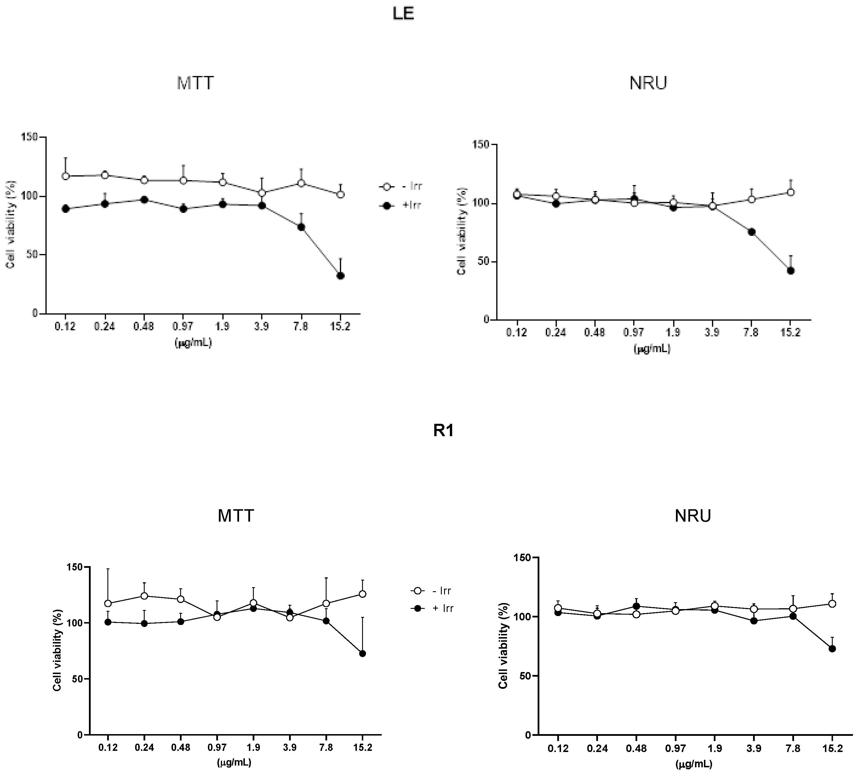
Figure 2.
Phototoxicity of CNF. Cell viability was measured by MTT and NRU assay after UVA irradiation at 4 J/cm2 (+Irr) or without UVA irradiation (-Irr). Results are expressed as the mean ± S.E.M of 3 independent experiments.
Figure 2.
Phototoxicity of CNF. Cell viability was measured by MTT and NRU assay after UVA irradiation at 4 J/cm2 (+Irr) or without UVA irradiation (-Irr). Results are expressed as the mean ± S.E.M of 3 independent experiments.
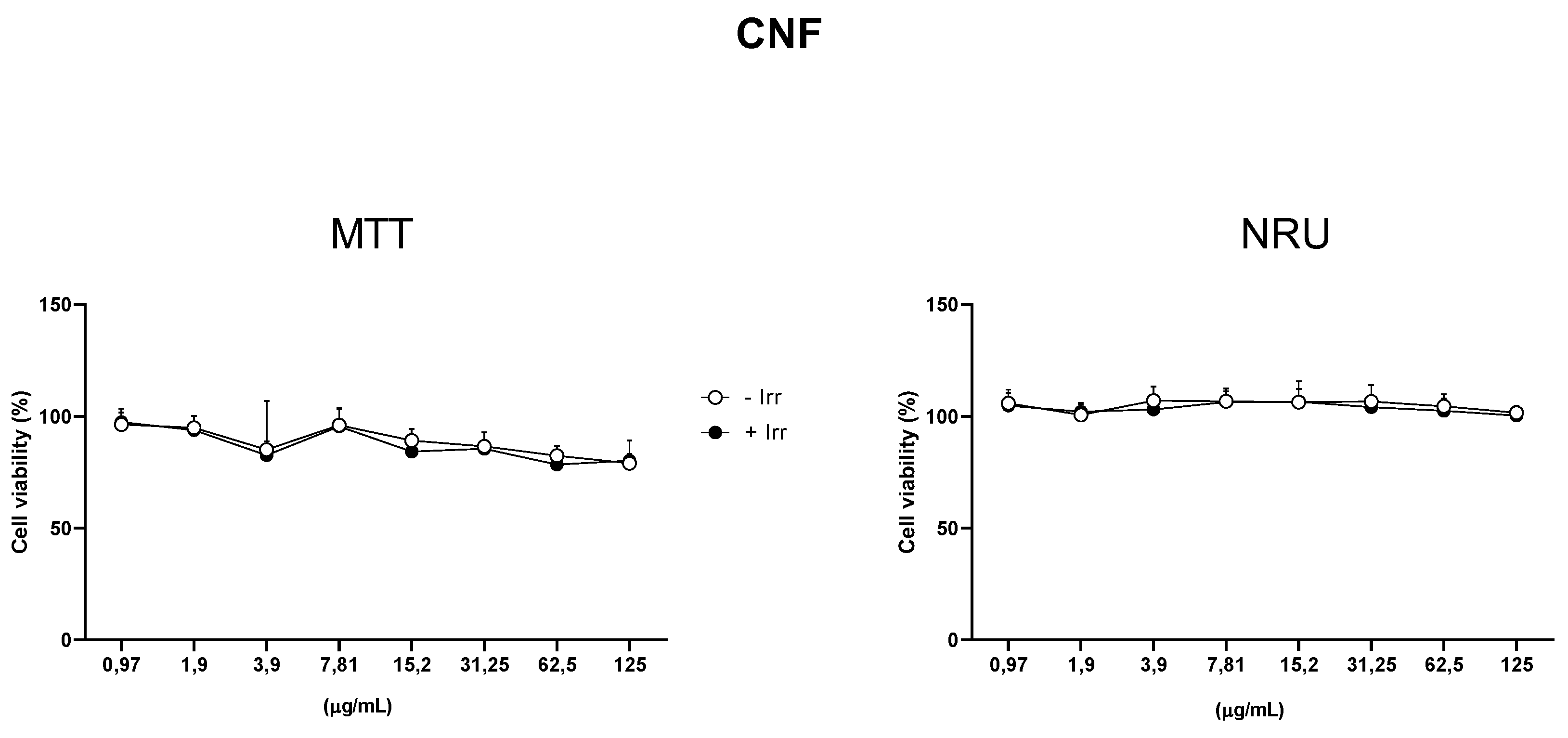
Figure 3.
Phototoxicity of LE and R1. Cell viability was measured by MTT and NRU assay after UVA irradiation at 4J (+Irr) or without UVA irradiation (-Irr). Results are expressed as n=3 data showed as the mean ± S.E.M of n = 3 independent experiments.
Figure 3.
Phototoxicity of LE and R1. Cell viability was measured by MTT and NRU assay after UVA irradiation at 4J (+Irr) or without UVA irradiation (-Irr). Results are expressed as n=3 data showed as the mean ± S.E.M of n = 3 independent experiments.
Figure 4.
Photoprotection effect of lignin measured by ROS production. The amount of ROS was determined by determining the fluorescence using DCF-DA probe. All the tested concentrations decreased the ROS produced by UV irradiation. Results are expressed as the mean ± S.E.M of 3 independent experiments. Statistical analyses were performed by two-way ANOVA, with Bonferroni post-hoc.
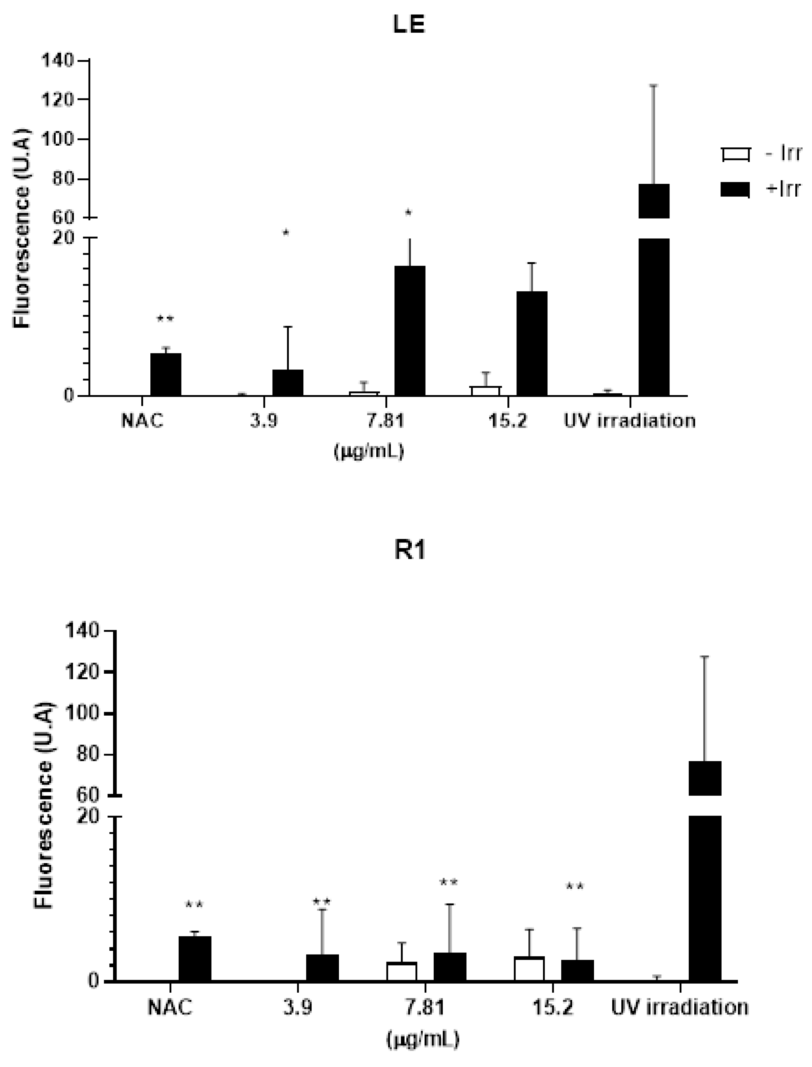
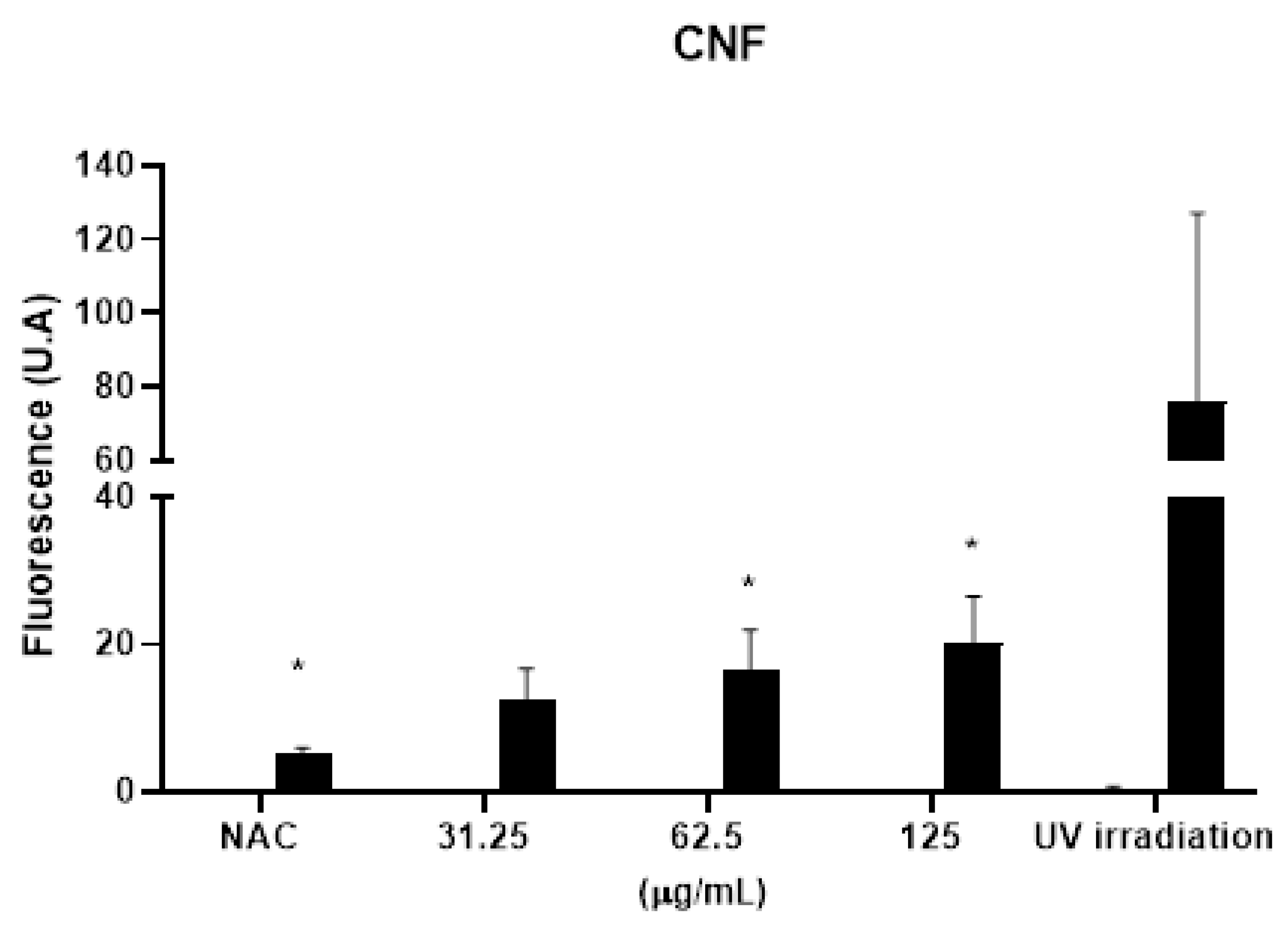

Table 1.
Photoirritation factor (PIF) determination of LE and R1 after MTT and NRU phototoxicity assay.
Table 1.
Photoirritation factor (PIF) determination of LE and R1 after MTT and NRU phototoxicity assay.
| MTT | NRU | |
|---|---|---|
| LE | 10.0 | 8.8 |
| R1 | 6.4 | 5.6 |
Disclaimer/Publisher’s Note: The statements, opinions and data contained in all publications are solely those of the individual author(s) and contributor(s) and not of MDPI and/or the editor(s). MDPI and/or the editor(s) disclaim responsibility for any injury to people or property resulting from any ideas, methods, instructions or products referred to in the content. |
© 2025 by the authors. Licensee MDPI, Basel, Switzerland. This article is an open access article distributed under the terms and conditions of the Creative Commons Attribution (CC BY) license (http://creativecommons.org/licenses/by/4.0/).
Copyright: This open access article is published under a Creative Commons CC BY 4.0 license, which permit the free download, distribution, and reuse, provided that the author and preprint are cited in any reuse.


