Preprint
Article

This version is not peer-reviewed.

Molecular and Genetic Analysis of the Increased Number of Genes for Trypanosoma cruzi Microtubule Associated Proteins in the Class Kinetoplastida

A peer-reviewed article of this preprint also exists.

Submitted:

24 February 2025

Posted:

25 February 2025

You are already at the latest version

Abstract

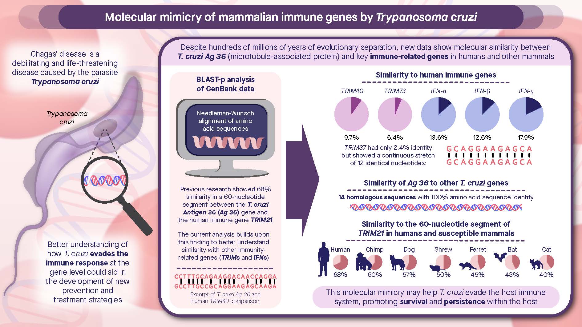
Trypanosoma cruzi GenBank® M21331 encodes for Antigen 36 (Ag 36), which is a tandemly repeated T. cruzi antigen. GenBank M21331 has a gene sequence similarity to human immune genes IFN-α, IFN-β, and IFN-γ, as well as to human TRIM genes. A BLAST-p search revealed that T. cruzi GenBank M21331 had seven gene sequences homologous to microtubule-associated protein (MAP) genes with a 100% amino acid sequence identity. There are 36 genes in the T. cruzi genome with >94% identity to GenBank M21331, and these genes encode proteins ranging in size from 38 to 2011 amino acids in length, the largest containing 20, 25, and 30 repeats of the Ag 36 thirty-eight amino acid sequence motif. The purpose of this study was to perform a genetic and molecular comparative analysis of T. cruzi GenBank M21331 to determine if this gene sequence is unique to the T. cruzi clade, present in the T. brucei clade, and/or exists in other trypanosomatids. There are seven homologous genes to GenBank M21331 in T. cruzi, but only one homologue found of this gene in T. brucei. The MAP genes in T. cruzi appear to have expanded at least eleven-fold in number compared to similar MAP genes in T. brucei. The DNA sequences and functions of these MAP genes in their respective species and clades will be discussed, and are a fascinating area for further scientific study.

Keywords: 
;  ;  ;  ;  ;  ;  

1. Introduction

Trypanosomatid parasites in the class Kinetoplastida include Trypanosoma cruzi, T. brucei ssp., and Leishmania spp., which are the causative human agents of Chagas disease, African sleeping sickness, and Leishmaniasis, respectively. Additionally, several other species of trypanosomes that cause infection in additional animal species in the T. brucei clade, such as T. b. gambiense, T. b. rhodesiense, T. congolense, T. theileri, T. equiperdum, and T. vivax. Trypanosoma rangeli and other species occur in the T. cruzi clade [1].
Trypanosoma cruzi was observed in 1909 when Dr. Carlos Chagas discovered the protozoan parasite in the blood of a Brazilian child and reported the clinical manifestations of the infection. The disease he described is named in his honor. Chagas disease is also called American trypanosomiasis. In some areas of Brazil, more than 13% of all deaths in individuals between the ages of 15 and 74 years are attributed to Chagas disease [2]. The disease is spread by the reduviid bug, which ranges from northern California to Maryland and to the southern regions of Argentina. When taking a bloodmeal from a human, the insect defecates, leaving behind an infective form of the parasite known as the metacyclic trypomastigote in the feces. These trypomastigotes enter the bloodstream, either by being scratched into the bite wound or by direct contact with the host’s mucosa. Although trypomastigotes are abundant in the blood in early infections, they do not reproduce until they have entered a cell and have transformed into the intracellular form of the parasite called the amastigote. The infection with this parasite causes a spectrum of disease in humans and susceptible animals ranging from acute, to latent, to a chronic stage. The parasite commonly invades the reticuloendothelial cells of the liver, spleen, and lymphatic system, as well as cardiac, smooth, and skeletal muscles. The nervous system, intestinal mucosa, skin, gonads, bone marrow, and placenta may also become infected. Today, an estimated 6 to 7 million human individuals and countless susceptible mammalian species are infected with this parasite. It has been estimated that an additional 75 million people may be at risk of infection [3,4] leading to approximately 12,000 deaths every year. It is unfortunate that these numbers have not decreased over several decades. In 2005-2006, a resurgence in neglected diseases arose [5,6,7,8,9,10], and Chagas disease has now been referenced as “the most neglected of the neglected diseases” [11]. This parasitic infection has the highest morbidity in the Americas.
Investigations into the biological mechanisms of T. cruzi are perplexing due to the multifaceted nature and unique characteristics of its genes. Trypanosoma cruzi is diploid, containing homologous chromosome pairs which are differentially sized [12]. Its genome has been sequenced and is estimated to be between 106.4-110.7 Mb in size (diploid) [13,14,15]. At least 50% of the genome contains repetitive sequences in simple tandem repeats and consists of a large family of surface proteins, gene retrotransposons, and subtelomeric repeats. These repetitive genes may also be found as secreted proteins which serve to evade the host immune responses that are expressed concurrently [16,17,18]. In addition, T. cruzi genes encode numerous families of surface proteins (e.g., mucins and mucin-associated surface proteins [19,20], and genes encoding trans-sialidases [17,18,21,22,23]. It is hypothesized that this strategy may assist the amastigote form of T. cruzi to survive and propagate in susceptible hosts by evading recognition by T-cells [24,25].
Trypanosoma cruzi exemplifies a genetically diverse intra-species [26,27,28]. Based on analysis of genetic and biochemical indicators, there may be up to seven lineages of TcI - TcVI [29,30,31,32,33,34,35] and TcBat [36]. These lineages may produce distinct pathological manifestations ranging from CCC (Chronic Chagas Cardiomyopathy) in endemic areas to enlargement of the esophagus and colon (known as mega syndromes). TcI is associated with human disease in endemic countries north of the Amazon basin [37,38,39,40] and is also found in the transmission cycle between insect vector and animals. TcII mainly occurs in the domestic settings in the southern countries of South America [37,38,40,[37,38,40,41] and causes tissue damage of the internal organs and digestive tract [39,40,43].
The African trypanosomes belonging to T. brucei spp. live extracellularly in the infected host, primarily in the bloodstream and cerebrospinal fluid. In the late 1990s, there were more than 35,000 cases reported annually in 1997 and 1998. However, the number of cases has been steadily declining over the past two decades. By the year 2000, it was estimated that there were greater than 25,000 cases of African sleeping sickness, caused by these organisms, and by the year 2020 there were less than 2000 yearly cases due to sustained control efforts [44]. While T. b. gambiense and T. b. rhodesiense affect the human host, T. b. brucei is known to cause infections in domestic and wild animals [44,45]. Transmission to the susceptible host is through the tsetse fly insect vector of the Glossina spp. when it takes a blood meal. Among the trypanosomatids, it is generally believed that T. brucei does not have intracellular stages in its life cycle and therefore, is exposed to constant attacks by antibody-mediated immune responses. Trypanosoma brucei spp. uses a mechanism termed antigenic variation as a defense against the mammalian antibody system [46]. This mechanism consists of a successive expression of a single or a multiple number of antigenic surface variants expressed within the mammalian host. Once the host immune response controls the foremost variant, the parasite switches to new variant, thus again evading host immune cells. This cycle continues until the host eventually succumbs. This one factor at a time (OFAT) method is henceforth an effective strategy when the parasite is continuously exposed to antibody-mediated immune modulated mechanisms. This is an example of a sophisticated genetic program by a trypanosome to overcome the immune system of the susceptible host.
Protozoan parasites of the genus Leishmania have a unique life cycle. The parasite undergoes a specific developmental transformation that permits it to infect a susceptible host. The insect vector (phlebotamine sandflies) harbors infective promastigote stages in its midgut, and eventually this stage move to the proboscis. When the insect takes a blood meal, it infects the host. The host phagocytic cells (e.g., macrophages) recognize these as foreign and engulf them, and there they transform to the amastigote stage [47]. The manifestations of disease produce a broad range of clinical and pathological symptoms (visceral and cutaneous leishmaniasis) depending on the parasite species causing the infection and the host immune response. In Africa, Asia, and Europe, L. major is considered the primary causative agent of cutaneous leishmaniasis, and L. donovani is mostly associated with visceral cases of the disease, known as kala azar. Species of New World Leishmania have a broad geographic range including South and Central America, the West Indies, and to a limited extent North America [48]. In the Americas, L. braziliensis, L. mexicana and L. infantum are considered the primary causative species of the visceral and cutaneous forms of the disease, respectively. It is estimated that 700,000 to one million new cases of Leishmaniasis occur annually [49]. An evolutionary historical prospective on the classification, evolution, and dispersion of Leishmania parasites and other trypanosomatids is described by Akhoundi [50] and Kaufer [51], respectively. In order to evade the immune system of the host, an outer lipophosphoglycan (LPG) surface coat engulfs the Leishmania parasite. Lipophosphoglycan can activate an innate immune signaling toll-like receptor 2 (TLR2), which forms heterodimers with TLR1 and TLR6. This receptor is involved in activating a cascading innate immune response. Considering that Leishmania parasites live and reproduce within macrophages, and that LPG inhibits the oxidative burst reaction of the complement system, which results in an inflammatory response, natural killer (NK) T-cells do not recognize that the macrophage is infected. Modifications of the glycan portion can occur and variations in the structure of LPG can be used as a biomarker for the promatigote and amastigote stages as well as for the species. Intracellular amastigotes in the L. mexicana complex also have an abundant developmentally-regulated cysteine proteinase activity which is associated with megasomes, also known as lysosomes [52,53]. In addition, a metalloenzymase (gp63) has been reported to have proteolytic activity at alkaline pH [54]. Proteinases appear to be essential to survival, parasite growth, and virulence of amastigotes within immune cells, as they serve as a liposome coating to protect them from phagolysosomal degradation. Therefore, it is hypothesized that these mechanisms for evasion could be exploited as targets for chemotherapeutic and immunological intervention.
The recognition of these impactful but neglected parasitic diseases have resulted in increased research in the areas of immunology, molecular biology, and genetics being used to study and characterize host-parasite relationships, and to identify antigenic molecules which are involved in mounting a protective immune response. However, at present, no vaccine has been approved for these diseases and available drugs are highly toxic with severe and frequent side effects and may only be effective in combating circulating forms of the parasite [55]. In addition, the occurrence of drug resistance is also a possibility. Therefore, there is an urgent necessity to identify gene, protein, and carbohydrate targets, and understand their mechanisms of action(s). These may result in unique and specific markers for vaccines, as well as in the development of therapeutics. There is also the need for the development of highly sensitive and specific analytical diagnostic assays in blood and tissue specimens to combat further spread of these diseases [3,56,57,58]. The genome sequences of the three trypanosomatids, T. cruzi, T. brucei, and L. major, are now available to help aid and identify possible targets in this regard [13,15,59,60].
In earlier investigations of diagnostic antigens, we identified one cloned gene from T. cruzi (Brazil strain) amastigotes from axenic culture [61] as a potential candidate. These axenic amastigotes have been shown to be comparable to amastigotes in cell culture (MRC-5 and Vero cells) [21]. This gene was sequenced and found to be identical to the repetitive antigen Clone 36, “Antigen 36” [16,62,63,64], and was also described as JL9 Antigen [65]. An initial search of the Wisconsin Package [66], with our DNA sequence disclosed similarity to human Ro52 with the translated sequence in the second reading frame of Ag 36. Direct comparison of the Ag 36 DNA sequence with Ro52 DNA sequence revealed a 70% identity in one sequence of 44 nucleotides between the Ag 36 DNA sequence and TRIM21, the gene for human Ro52 [62]. Once the function of TRIM21 was identified, we proposed that there may be a link between it and the gene for Ag 36 identified in CCC [62]. Ro52 is expressed in the immune system as a predominantly cytoplasmic protein that can be upregulated and translocated to the nucleus in a pro-inflammatory environment. A study was also conducted to compare TRIM21 region sequences among mammalian species to the human TRIM21 region, to evaluate any similarities in non-human genes. Results indicated that related sequences were present in 11 mammalian species [64]. Additionally, a BLAST-p search was conducted with GenBank® M21331 against the T. cruzi genome to determine the minimum number of genes coding for proteins closely related to Ag 36. The BLAST-p revealed seven unique GenBank accession entries which produced seven proteins 100% identical to Ag 36 of the 14 GenBank entries previously reported [64].
The purpose of our study primarily focuses on GenBank M21331. Previously we have shown that GenBank M21331 has a significant gene sequence identity to human immune genes (IFN-α, IFN-β, and IFN-γ) and to human TRIM genes, such as TRIM40 and TRIM21 [64]. Those results appeared to be the first description of molecular mimicry of immune genes in humans by a protozoan parasite [64]. The protein generated from this gene has also been used in the development and implementation of a diagnostic assay [67]. In this report, we further extend our molecular and genetic analysis of T. cruzi GenBank M21331 to organisms in the class Kinetoplastida.
A phylogenetic tree for these trypanosomatids was developed by Stevens [1], and indicated a T. cruzi clade, a T. brucei clade, and an aquatic clade. However, unfortunately no data is available in GenBank on the aquatic clade to perform a genetic comparison. Additional evolutionary history on T. cruzi is provided by Briones [68] and Rozas [69]. We focus on a genetic and molecular comparative analysis of T. cruzi GenBank M21331 to determine if this gene sequence is exclusive to members in the T. cruzi clade, or is present in the T. brucei clade, and/or other trypanosomatids. Microtubule associated proteins are present in the T. cruzi clade, as described above, and have also been described in trypanosomes present on the African continent, such as MARP-1 (a repetitive non-variable antigen) [70,71] that is localized on the microtubules of the parasite’s cell body and flagellum. MARP-1 is comprised of 50 repeats of a 38 amino acid motif described in T. b. brucei, and T. b. gambiense. In addition, MAPs are present in other African trypanosomes such as T. vivax, T. congolense, T. evansi, and T. equiperdum, as well as New World trypanosomes, such as T. rangeli and T. theileri, and in Leishmania spp.
The Kinetoplastid groups of parasites diverged approximately 500 million years ago in different habitats worldwide [1,50]. It is also speculated that T. cruzi arose over 150 million years ago, infecting animals throughout Laurasia and Gondwanaland, which are the regions that eventually formed North and South America, respectively [68]. It is theorized that the disease occurred in humans approximately 15,000-20,000 years ago in the late Pleistocene era when they were migrating into these areas. These trypanosomatids have thus genetically evolved over millennia to each develop unique molecular mechanisms to evade destruction from the innate immune system of the host. The MAP genes, the DNA sequences, and their functional role in trypanosomes and in their respective species and clades, are intriguing, and they will be further evaluated, investigated and analyzed.

2. Materials and Methods

2.1. Cloning of T. cruzi Amastigote Genes

We identified one cloned gene from T. cruzi (Brazil strain) amastigotes grown in axenic culture [61], which was sequenced by the Sanger method [72], and found to be identical to the repetitive antigen Clone 36, “Antigen 36” [16,62,63,64].

2.2. BLAST-p Search to determine number of Ag 36 Homologues

The GenBank M21331 gene was translated into its amino acid sequence using the translation tool at https://usegalaxy.org [73,74] and the sequence entered in the BLAST-p search box, to determine homologous genes in the T. cruzi genome. The BLAST-p search algorithm was selected at https://blast.ncbi.nlm.nih.gov/Blast.cgi. The searched database entered was Trypanosoma cruzi taxid 5693; and the first 100 most homologous genes (in order of their homology to GenBank M21331) and their amino acid sequences were downloaded and saved as a text file. The “e value” noted in Table 1 is the probability that this result happened by random chance.

2.3. BLAST-p Search of Trypanosoma brucei with Trypanosoma cruzi GenBank M21331

The GenBank M21331 gene was translated into its amino acid sequence using the translation tool at https://usegalaxy.org [73,74] and the sequence entered in the BLAST-p search box, to determine homologous genes in the T. brucei genome,. The BLAST-p search algorithm was selected at https://blast.ncbi.nlm.nih.gov/Blast.cgi. The searched database entered was Trypanosoma brucei taxid id 5691; and the first 100 most homologous genes (in order of their homology to GenBank M21331) and their amino acid sequences were downloaded and saved as a text file.

2.4. BLAST-p Search of Leishmania donovani with trypanosoma cruzi GenBank M21331

The GenBank M21331 gene was translated into its amino acid sequence using the translation tool at https://usegalaxy.org [73,74] and the sequence entered in the BLAST-p search box, to determine homologous genes in the L. donovani genome. The BLAST-p search algorithm was selected at https://blast.ncbi.nlm.nih.gov/Blast.cgi. The searched database entered was Leishmania donovani taxid id 5661; and the first 100 most homologous genes (in order of their homology to GenBank M21331) and their amino acid sequences were downloaded and saved as a text file.

3. Results

To determine the number of homologues of Ag 36 in the T. cruzi genome, a BLAST-p search of the https://blast.ncbi.nlm.nih.gov/Blast.cgi database with the Ag 36 protein sequence was conducted. It revealed seven unique GenBank accession entries which produced seven proteins 100% identical to Ag 36 of the 14 GenBank entries previously reported [64] and is shown in Table 1.
In addition, there were 36 genes with greater than 94% identity to GenBank M21331 (Table 2), with 35 proteins containing multiple copies of the Ag 36 sequence motif. These genes ranged from 38 to 2011 amino acids in length, as shown in Table 2. For each entry in Table 2, the number of repeats of the Ag 36 motif (identified as homologous domains by the BLAST-p search program) is shown in Table 3. There are repeats of 1-10, 11-12, 17, 19, 20, 25 copies, and two entries having a maximum of 30 repeats. In the 36 entries shown in Table 3, there are a total of 296 total copies of the Ag 36 protein sequence motif. The high number of genes with multiple copies of the Ag 36 sequence may be due to the internal sequence duplication in the gene for Ag 36, GenBank M21331. The gene sequence coding for Ag 36, M21331 was obtained from its GenBank entry in FASTA format to indicate internal duplication (Figure 1). The ten nucleotide sequence is shown in blue faced font and highlighted twice is an internal duplication found in this gene and its homologues. This internal sequence duplication may have increased the chance of unequal crossing over between two copies of the gene during meiosis, producing larger, duplicated genes [75]. It is of interest that meiosis has been reported in T. cruzi [76,77].
Figure 2 shows an example of a T. cruzi gene, GenBank PWU83737, with multiple (3) copies of the Ag 36 amino acid sequence.
This result allowed us to further explore the similarity of GenBank M21221 to genes in other species in the class Kinetoplastida MAPs. BLAST-p searches of Ag 36 amino acid sequence were also performed versus genomes of T. brucei (ssp.), T. congolense and L. donovani, T. theileri, and T. vivax. The resulting matches were ranked by percent identity to Ag 36 sequence and the highest percent identities are shown in Table 4. These highest percent identities are therefore, most directly related to the Ag 36 sequence.

4. Discussion

The purpose of this study was to compare, enumerate, and analyze DNA sequences between T. cruzi GenBank M21331, which codes for Ag 36, and similar genes found in the class Kinetoplastida. We focused on a on a genetic and molecular comparative analysis of T. cruzi GenBank M21331 (Ag 36) to determine if this gene sequence is exclusive to members in the T. cruzi clade, or present in the T. brucei clade, and/or other trypanosomatids. A BLAST-p search of Ag 36 protein versus the translated T. cruzi genome disclosed that there are 43 T. cruzi gene products (seven homologues and 36 genes containing multiple copies) homologous in protein sequence to Ag 36, implying that there are 43 genes that are very similar or identical to GenBank M21331 in T. cruzi. Thirty-six of the protein matches were greater in length than Ag 36 and contained one or more sequences homologous or partially homologous to Ag 36. A BLAST-p search of Ag 36 was performed on T. theileri, T. vivax, T. brucei, T. b. brucei, T. b. gambiense T. congolense and Leishmania spp. genomes. Trypanosoma theileri, and T. vivax showed 83 and 57% identities, in line with their close phylogenetic relationship to T. cruzi. Similarly, T. brucei, T. b. brucei, and T. b. gambiense showed proteins 69% homologous to Ag 36. Additionally, T. b. equiperdum and T. congolense had proteins 65% homologous to Ag 36, and L. donovani (L. tropica and L. mexicana - data not shown) had proteins 47% homologous to Ag 36 (Table 4). These results show that there is at least an eleven-fold greater number of copies of the MAP genes related to GenBank M21331 in T. cruzi compared to T. brucei spp. The specific and definitive roles of these T. cruzi MAP proteins may play, besides their association with microtubules, is yet to be defined. However, based on the similarity of GenBank M21331 to IFNs, TRIM21 and other TRIM (tripartite motif) genes, GenBank M21331 mRNAs may play a crucial part in suppressing host mRNA translation of these IFN and TRIM genes that are involved in the innate immune response to T. cruzi [63,64]. Trypanosoma theileri, a closely related species to T. cruzi in the Kinetoplast phylogeny [1], showed a gene 83% identical to GenBank M21331. The most identical gene in T. theileri did not show the internal ten nucleotide repeat found in GenBank M21331 (excluding the overall repeats due to it being a repetitive antigen). There were no additional larger homologues of the T. theileri gene as were observed in T. cruzi with GenBank M21331. This finding supports the theory that the internal repeat in GenBank M21331 may have given rise to the additional larger homologues. Trypanosoma theileri is a blood borne trypanosome of cattle commonly transmitted by biting flies. It does not typically invade and reside inside of cell as T. cruzi does, and the unique properties of GenBank M21331 may be relevant to the difference in parasitism. Trypanosoma vivax, another parasitic disease of cattle and wild mammals that survives extracellularly in its host, disclosed three genes (986, 1318, and 2957 nucleotides in length) with 57% identity which were much larger when compared to GenBank M21331. The results in this study indicate that there are at least an eleven-fold greater number of copies of MAP genes related to GenBank M21331 in T. cruzi compared to T. brucei spp. It is of interest that the Trypanosoma spp. that have multiple copies of GenBank M21331 are those that reside inside cells, such as T. cruzi, and those with fewer copies reside mainly in the blood or outside of cells (T. brucei clade).
The cytoskeletons of trpanosomatids are composed of MAPs which are cross-linked to form two defined structures. The first cross-linked structure is composed of subpellicular microtubules that surround the frame of the trypanosome throughout the morphological stages of its lifecycle. The second MAP arrays are the basal-body-axoneme complex in morphological stages that contain a flagellum. However, specific differences are outlined below depending on the species and clades of these Kinetoplastids. Trypanosoma cruzi epimastigote microtubules are attached to the inner membrane of the nuclear envelope at its poles. The flagellum arises from a basal body runs along the surface of the organism and becomes free at the anterior end. A row of four microtubules originating near the basal body appear to pass along the membrane of the flagellar pocket to the body surface. A paraxial rod of paracrystalline structure is present alongside the axoneme after the flagellum emerges from the pocket. A cytosome lined with microtubules opens near the base of the flagellar pocket [80]. Trypanosoma brucei trypomastigotes possess a flagellum which originates from the flagellar pocket at the posterior end of the cell and runs along its length. The flagellum has a 9 + 2 axoneme with a paraflagellar rod that is linked to the subfiber of microtubule doublet 7 [81]. Additionally, there appears to be strands beneath the surface membrane that form an array of subpellicular microtubules. A second attachment arises from the paraflagellar rod beneath the flagellar membrane. There are four microtubules on the flagellum which are associated with the endoplasmic reticulum and assist in flagellar movement. The flagellum of Leishmania promastigotes contains a paraxial rod originating at the axosome level within the flagellar pocket. The paraxial rod has the appearance of a cross hatched paracrystalline structure. Immediately beneath the plasma membrane lies a parallel array of microtubules oriented along the long axis of the organism from the anterior to the posterior ends. These subpellicular microtubules appear to end terminally at the posterior end. Occasional microtubules are seen along the membrane lining the flagellar pocket [82]. In contrast, the paraxial rod in the flagellum of T. cruzi epimastigotes originates at the opening of the flagellar pocket [80].
Microtubules are major structural components of the cytoskeleton that are involved in cell morphology, motility, division, and intracellular organization and transport. Meiosis has been reported in T. cruzi [76,77], with genetic exchange occurring between lineages [26,27]. There may be up to seven lineages of T. cruzi from analysis of genetic, molecular, and biochemical indicators [31,33,35,36]. Therefore, divergence can occur due to recombination between organisms once thought to reproduce asexually. However, due to biomarker and sequence analysis of conserved genes a common genetic ancestor may eventually be identified. The Kinetoplastid groups of parasites diverged approximately 500 million years ago in different parts of the world [1,50]. Trypanosoma cruzi arose over 150 million years ago, infecting animals in North and South America [68]. GenBank M21331 and related genes are conserved as MAPs between American and African trypanosomes and can be useful as genetic, immunological, and molecular biomarkers. The T. cruzi MAP genes partial identities to human immune genes (IFN-α, IFN-β, IFN-γ, and human TRIM genes), which are maintained over the hundreds of millions of years separating protozoa and humans, may be relevant to the parasite’s resistance to the mammalian innate immune system during infection. Further experiments can be designed [64] to test GenBank M21331 and related genes for their role in resistance to innate immunity.

Abbreviations/Definitions

The following abbreviations and definitions are used in this manuscript:
Antigen 36 (Ag 36) – (clone A2; clone 36; Tc36; JL9) – The tandemly repeated T. cruzi antigen reported by Ibañez [16], Levin [65], and Winkler [62,63,64], which is highly reactive with Chagasic sera [67].
GenBank M21331 - GenBank® is a database that contains publicly available nucleotide sequences for genus/species organisms. The library is obtained through submissions from laboratories and batch submissions from large-scale sequencing projects. T. cruzi GenBank M21331 encodes for Antigen 36 (Ag 36).
MAP (Microtubule Associated Proteins) – Microtubule associated proteins regulate assembly and stability of microtubules. Microtubules constitute a major part of the cytoskeleton and are important in cytoskeletal rearrangements during neuronal growth, axon guidance, and synapse formation. Ag 36 gene of T. cruzi has seven homologous genes sequences (MAP genes with 100% amino acid sequence identity) to GenBank M21331.
MARP-1 (Microtubule-Associated Repetitive Proteins) - The microtubular membrane framework of T. brucei containing two closely related, repetitive, high-molecular-weight MAPs, MARP-1 and MARP-2. The structure consists of a 38-amino-acid repeat over approximately its length of about 320 kDa which are tandemly arranged and conserved.

Author Contributions

All authors contributed equally to this work in the study, research, conception and design. In addition, both authors contributed correspondingly to material preparation, data collection, interpretation and analysis. Both authors wrote, read and approved the final manuscript. All authors agree to be accountable for all aspects of the work in ensuring that questions related to the accuracy or integrity of any part of the work are appropriately investigated and resolved. All authors have read and agreed to the published version of the manuscript

Funding

This research received no external funding. This research also did not receive any specific grant from funding agencies in the public, commercial, or not-for-profit sectors. The Galaxy server that was used for some calculations is in part funded by Collaborative Research Centre 992 Medical Epigenetics (DFG grant SFB 992/1 2012) and German Federal Ministry of Education and Research (BMBF grants 031 A538A/A538C RBC, 031L0101B/031L0101C de.NBI-epi, 031L0106 de.STAIR (de.NBI).

Institutional Review Board Statement

Not applicable.

Informed Consent Statement

Not applicable.

Data Availability Statement

The datasets generated during and/or analyzed during the current study are available in the online Galaxy repositories: https://usegalaxy.eu/u/martinawinklerphd/h/copy-of-copy-of-mammalian-trim-genes-compared-with-antigen-36 (accessed on 15 February 2025). https://usegalaxy.org/u/martinawinklerphd/h/ag36-and-mammalian-trim21-homologies (accessed on 15 February 2025). These bioinformatics workflows are available to all, but registration (which is free) is required to view the results. Web References: Genes from https://www.ncbi.nlm.nih.gov/gene (accessed on 15 February 2025). Species’ GenBank accession numbers at https://www.ncbi.nlm.nih.gov (accessed on 15 February 2025). Nucleotide sequence database from https://www.ncbi.nlm.nih.gov/nucleotide/ (accessed on 15 February 2025). BLAST-p search utility at https://blast.ncbi.nlm.nih.gov/Blast.cgi (accessed on 15 February 2025). NW algorithm alignment and comparison tool at https://usegalaxy.org (accessed on 15 February 2025) or https://usegalaxy.eu (accessed on 15 February 2025). Trypanosoma cruzi Clone 36 Antigen GenBank M21331

Acknowledgments

The authors would like to thank Abbott Laboratories and our colleagues where the foundational investigations were performed, and to Ms. Diana Rivera for her thorough review and helpful comments and suggestions.

Conflicts of Interest

The authors declare no conflicts of interest.

References

  1. Stevens, J.R.; Noyes, H.A.; Dover, G.A.; Gibson, W.C. The ancient and divergent origins of the human pathogenic trypanosomes, Trypanosoma brucei and T. cruzi. Parasitology 1999, 118, 107–116. [Google Scholar] [CrossRef]
  2. World Health Organization (WHO). Control of Chagas disease. Report of a WHO Expert Committee. World Health Organ. Tech. Rep. Ser 1991, 811, 1–95. [Google Scholar]
  3. Pan, A.A.; Winkler, M.A. The threat of Chagas’ disease in transfusion medicine. The presence of antibodies to Trypanosoma cruzi in the US blood supply. Lab. Med. 1995, 28, 269–274. [Google Scholar] [CrossRef]
  4. World Health Organization (WHO) Fact Sheet. Chagas disease (also known as American trypanosomiasis). 2024. Available online: https://www.who.int/news-room/fact-sheets/detail/chagas-disease- (accessed on 15 February 2025).
  5. Brady, M.A.; Hooper, P.J.; Ottesen, E.A. Projected benefits from integrating NTD programs in sub-Saharan Africa. Trends in Parasitology 2006, 22, 285–291. [Google Scholar] [CrossRef] [PubMed]
  6. Hotez, P.; Ottesen, E.; Fenwick, A.; Molyneux, D. Pollard, A.J., Finn, A., Eds.; The Neglected Tropical Diseases: The Ancient Afflictions of Stigma and Poverty and the Prospects for their Control and Elimination. In Hot Topics in Infection and Immunity in Children III; Springer: Boston, MA USA, 2006; Volume 582, pp. 23–33, Advances in Experimental Medicine and Biology. [Google Scholar]
  7. Hotez, P.J. A7 The Neglected Tropical Diseases and the Neglected Infections of Poverty: Overview of their Common Features, Global Disease Burden and Distribution, New Control Tools, and Prospects for Disease Elimination. In Institute of Medicine (US) Forum on Microbial Threats. The Causes and Impacts of Neglected Tropical and Zoonotic Diseases: Opportunities for Integrated Intervention Strategies; National Academies Press: Washington, DC USA, 2011. [Google Scholar]
  8. Lammie, P.J.; Fenwick, A.; Utzinger, J. A blueprint for success: Integration of neglected tropical disease control programmes. Trends Parasitol. 2006, 22, 313–321. [Google Scholar] [CrossRef]
  9. Molyneux, D.H.; Hotez, P.J.; Fenwick, A. “Rapid-impact interventions”: how a policy of integrated control for Africa’s neglected tropical diseases could benefit the poor. PLoS Med. 2005, 2, e336. [Google Scholar] [CrossRef]
  10. Utzinger, J.; de Savigny, D. Control of neglected tropical diseases: Integrated chemotherapy and beyond. PLoS Medicine 2006, 3, e112. [Google Scholar] [CrossRef]
  11. Winny, A. Chagas: The Most Neglected of Neglected Tropical Diseases. Johns Hopkins Bloomberg School of Public Health 2022. Available online: https://publichealth.jhu.edu/2022/chagas-the-most-neglected-of-neglected-tropical-diseases (accessed on 15 February 2025).
  12. Pedroso, A.; Cupolillo, E.; Zingales, B. Evaluation of Trypanosoma cruzi hybrid stocks based on chromosomal size variation. Mol. Biochem. Parasitol. 2003, 129, 79–90. [Google Scholar] [CrossRef]
  13. El-Sayed, N.M.; Myler, P.J.; Bartholomeu, D.C.; Nilsson, D.; Aggarwal, G.; Tran, A.N.; Ghedin, E.; Worthey, E.A.; Delcher, A.L.; Blandin, G.; et al. The genome sequence of Trypanosoma cruzi, etiologic agent of Chagas disease. Science 2005, 309, 409–415. [Google Scholar] [CrossRef]
  14. De Pablos, L.M.; Osuna, A. Multigene families in Trypanosoma cruzi and their role in infectivity. Infect. Immun. 2264, 80, 2258–2264. [Google Scholar] [CrossRef]
  15. DeCuir, J.; Tu, W.; Dumonteil, E.; Herrera, C. Sequence of Trypanosoma cruzi reference strain SC43 nuclear genome and kinetoplast maxicircle confirms a strong genetic structure among closely related parasite discrete typing units. Genome 2021, 64, 525–531. [Google Scholar] [CrossRef]
  16. Ibañez, C.F.; Affranchino, J.L.; Macina, R.A.; Reyes, M.B.; Leguizamon, S.; Camargo, M.E.; Åslund, L.; Pettersson, U.; Frasch, A.C.C. Multiple Trypanosoma cruzi antigens containing tandemly repeated amino acid sequence motifs. Mol. Biochem. Parasitol. 1988, 30, 27–34. [Google Scholar] [CrossRef]
  17. Kahn, S.; Colbert, T.G; Wallace, J.C.; Hoagland, N.A.; Eisen, H. The major 85-kDa surface antigen of the mammalian-stage forms of Trypanosoma cruzi is a family of sialidases. Proc. Natl. Acad. Sci. USA. 1991, 15, 4481–4485. [Google Scholar] [CrossRef] [PubMed]
  18. Kahn, S.J.; Nguyen, D.; Norsen, J.; Wleklinski, M.; Granston, T.; Kahn, M. Trypanosoma cruzi: monoclonal antibodies to the surface glycoprotein superfamily differentiate subsets of the 85-kDa surface glycoproteins and confirm simultaneous expression of variant 85-kDa surface glycoproteins. Exp. Parasitol. 1999, 92, 48–56. [Google Scholar] [CrossRef]
  19. Di Noia, J.M.; Sanchez, D.O.; Frasch, A.C.C. The protozoan Trypanosoma cruzi has a family of genes resembling the mucin genes of mammalian cells. J. Biol. Chem. 1995, 270, 24146–24149. [Google Scholar] [CrossRef] [PubMed]
  20. Herreros-Cabello, A.; Callejas-Hernández, F.; Gironès, N.; Fresno, M. Trypanosoma cruzi genome: organization, multi-gene families, transcription, and biological implications. Genes 2020, 11, 1196–1222. [Google Scholar] [CrossRef]
  21. Pan, A.A.; McMahon-Pratt, D. Amastigote and epimastigote stage-specific components of Trypanosoma cruzi characterized by using monoclonal antibodies. Purification and molecular characterization of an 83-kilodalton amastigote protein. J. Immunol. 1989, 143, 1001–1008. [Google Scholar] [CrossRef]
  22. Low, H.P.; Tarleton, R.L. Molecular cloning of the gene encoding the 83 kDa amastigote surface protein and its identification as a member of the Trypanosoma cruzi sialidase superfamily1. Mol. Biochem. Parasitol. 1997, 88, 137–149. [Google Scholar] [CrossRef]
  23. Herreros-Cabello, A.; Callejas-Hernández, F.; Gironès, N.; Fresno, M. Trypanosoma cruzi genome: organization, multi-gene families, transcription, and biological implications. Genes 2020, 11, 1196–1222. [Google Scholar] [CrossRef]
  24. Tarleton, R.L. The role of T cells in Trypanosoma cruzi infections. Parasitol. Today 1995, 11, 7–9. [Google Scholar] [CrossRef]
  25. Fernandes, M.C.; Andrews, N.W. Host cell invasion by Trypanosoma cruzi: a unique strategy that promotes persistence. FEMS Microbiol. Rev. 2012, 36, 734–747. [Google Scholar] [CrossRef] [PubMed]
  26. Machado, C.A.; Ayala, F.J. Nucleotide sequences provide evidence of genetic exchange among distantly related lineages of Trypanosoma cruzi. Proc. Natl. Acad. Sci. USA 2001, 98, 7396–7401. [Google Scholar] [CrossRef]
  27. Brisse, S.; Henriksson, J.; Barnabé, C.; Douzery, E.J.; Berkvens, D.; Serrano, M.; De Carvalho, M.R.; Buck, G.A.; Dujardin, J.C.; Tibayrenc, M. Evidence for genetic exchange and hybridization in Trypanosoma cruzi based on nucleotide sequences and molecular karyotype. Infect. Genet. Evol. 2003, 2, 173–183. [Google Scholar] [CrossRef]
  28. Messenger, L.A. Genetic diversity in Trypanosoma cruzi: marker development and applications; natural population structures, and genetic exchange mechanisms. Ph.D. Thesis, London School of Hygiene and Tropical Medicine, United Kingdom, September 2014. [Google Scholar]
  29. Barnabé, C.; Neubauer, K.; Solari, A.; Tibayrenc, M. Trypanosoma cruzi: presence of the two major phylogenetic lineages and of several lesser discrete typing units (DTUs) in Chile and Paraguay. Acta Trop. 2001, 78, 127–137. [Google Scholar] [CrossRef] [PubMed]
  30. Llewellyn, M.S.; Lewis, M.D.; Acosta, N.; Yeo, M.; Carrasco, H.J.; Segovia, M.; Vargas, J.; Torrico, F.; Miles, M.A.; Gaunt, M.W. Trypanosoma cruzi IIc: phylogenetic and phylogeographic insights from sequence and microsatellite analysis and potential impact on emergent Chagas disease. PLoS Negl Trop. Dis. 2009, 3, e510. [Google Scholar] [CrossRef]
  31. Pena, S.D.J.; Machado, C.R.; Macedo, A.M. Trypanosoma cruzi: ancestral genomes and population structure. Mem. Inst. Oswaldo Cruz 2009, 104, 108–114. [Google Scholar] [CrossRef] [PubMed]
  32. Subileau, M.; Barnabé, C.; Douzery, E.J.; Diosque, P.; Tibayrenc, M. Trypanosoma cruzi: new insights on ecophylogeny and hybridization by multigene sequencing of three nuclear and one maxicircle genes. Exp. Parasitol. 2009, 122, 328–337. [Google Scholar] [CrossRef]
  33. Zingales, B.; Andrade, S.G.; Briones, M.R.; Campbell, D.A.; Chiari, E.; Fernandes, O.; Guhl, F.; Lages-Silva, E.; Macedo, A.M.; Machado, C.R.; et al. A new consensus for Trypanosoma cruzi intraspecific nomenclature: second revision meeting recommends TcI to TcVI. Mem. Inst. Oswaldo Cruz. 2009, 104, 1051–1054. [Google Scholar] [CrossRef]
  34. Ferreira, R.C.; Briones, M.R. Phylogenetic evidence based on Trypanosoma cruzi nuclear gene sequences and information entropy suggest that inter-strain intragenic recombination is a basic mechanism underlying the allele diversity of hybrid strains. Infect. Genet. Evol. 2012, 12, 1064–1071. [Google Scholar] [CrossRef]
  35. Bhattacharyya, T.; Murphy, N.; Miles, M.A. Trypanosoma cruzi lineage-specific serology: new rapid tests for resolving clinical and ecological associations. Future Sci. OA 2019, 5, FSO422. [Google Scholar] [CrossRef] [PubMed]
  36. Lima, L.; Espinosa-Álvarez, O.; Ortiz, P.A.; Trejo-Varón, J.A.; Carranza, J.C.; Pinto, C.M.; Serrano, M.G.; Buck, G.A.; Camargo, E.P.; Teixeira, M.M. Genetic diversity of Trypanosoma cruzi in bats, and multilocus phylogenetic and phylogeographical analyses supporting Tcbat as an independent DTU (discrete typing unit). Acta Trop. 2015, 151, 166–177. [Google Scholar]
  37. Fernandes, O.; Souto, R.P.; Castro, J.A.; Pereira, J.B.; Fernandes, N.C.; Junqueira, A.C.; Naiff, R.D.; Barrett, T.V.; Degrave, W.; Zingales, B.; et al. Brazilian isolates of Trypanosoma cruzi from humans and triatomines classified into two lineages using mini-exon and ribosomal RNA sequences. Am. J. Trop. Med. Hyg. 1998, 58, 807–811. [Google Scholar]
  38. Zingales, B.; Souto, R.P.; Mangia, R.H.; Lisboa, C.V.; Campbell, D.A.; Coura, J.R.; Jansen, A.; Fernandes, O. Molecular epidemiology of American trypanosomiasis in Brazil based on dimorphisms of rRNA and mini-exon gene sequences. Int. J. Parasitol. 1998, 28, 105–112. [Google Scholar]
  39. Yeo, M.; Acosta, N.; Llewellyn, M.; Sánchez, H.; Adamson, S.; Miles, G.A.; López, E.; González, N.; Patterson, J.S.; Gaunt, M.W.; et al. Origins of Chagas disease: Didelphis species are natural hosts of Trypanosoma cruzi I and armadillos hosts of Trypanosoma cruzi II, including hybrids. Int. J. Parasitol. 2005, 35, 225–233. [Google Scholar] [PubMed]
  40. Rassi, A. Jr.; Rassi, A.; Marin-Neto, J.A. Chagas disease. Lancet 2010, 375, 1388–1402. [Google Scholar]
  41. Risso, M.G.; Sartor, P.A.; Burgos, J.M.; Briceño, L.; Rodríguez, E.M.; Guhl, F.; Chavez, O.T.; Espinoza, B.; Monteón, V.M.; Russomando, G.; et al. Immunological identification of Trypanosoma cruzi lineages in human infection along the endemic area. Am. J. Trop. Med. Hyg. 2011, 84, 78–84. [Google Scholar] [PubMed]
  42. Di Noia, J.M.; Buscaglia, C.A.; De Marchi, C.R.; Almeida, I.C.; Frasch, A.C. A Trypanosoma cruzi small surface molecule provides the first immunological evidence that Chagas’ disease is due to a single parasite lineage. J. Exp. Med. 2002, 195, 401–413. [Google Scholar]
  43. Freitas, J.M.; Lages-Silva, E.; Crema, E.; Pena, S.D.; Macedo, A.M. Real time PCR strategy for the identification of major lineages of Trypanosoma cruzi directly in chronically infected human tissues. Int. J. Parasitol. 2005, 35, 411–417. [Google Scholar]
  44. Franco, J.R.; Cecchi, G.; Paone, M.; Diarra, A.; Grout, L.; Ebeja, A.K.; Simarro, P.P.; Zhao, W.; Argaw, D. The elimination of human African trypanosomiasis: Achievements in relation to WHO road map targets for 2020. PLoS Negl. Trop. Dis. 2022, 16, e0010047. [Google Scholar]
  45. Franco, J.R.; Simarro, P.P.; Diarra, A.; Jannin, J.G. Epidemiology of human African trypanosomiasis. Clin. Epidemiol. 2014, 6, 257–275. [Google Scholar] [PubMed]
  46. Cross, G.A.M.; Kim, H-S.; Wickstead, B. Capturing the variant surface glycoprotein repertoire (the VSGnome) of Trypanosoma brucei Lister 427. Mol. Biochem. Parasitol. 2014, 195, 59–73. [Google Scholar] [CrossRef]
  47. Pan, A.A.; Honigberg, B.M. Leishmania mexicana pifanoi: in vivo and in vitro interactions between amastigotes and macrophages. Z. Parasitenkd. 1985, 71, 3–13. [Google Scholar] [PubMed]
  48. Jain, S.; Madjou, S.; Agua, J.F.V.; Maia-Elkhoury, A.N.; Valadas, S.; Warusavithana, S.; Osman, M.; Yajima, A.; Beshah, A.; Ruiz-Postigo, A. Global leishmaniasis surveillance updates 2023: 3 years of the NTD road map. Releve Épidémiologiqué Hebdomadaire 2024, 45, 653–669. Available online: https://iris.who.int/bitstream/handle/10665/379491/WER9945-653-669.pdf?sequence=1 (accessed on 15 February 2025).
  49. World Health Organization (WHO) Fact Sheet. Leishmaniasis. 2023. Available online: https://www.who.int/news-room/fact-sheets/detail/leishmaniasis (accessed on 15 February 2025).
  50. Akhoundi, M.; Kuhls, K.; Cannet, A.; Votýpka, J.; Marty, P.; Delaunay, P.; Sereno, D. A historical overview of the classification, evolution, and dispersion of Leishmania parasites and sandflies. PLoS Negl. Trop. Dis. 2016, 10, e0004349, Erratum in: PLoS Negl. Trop. Dis. 10: e0004770. [Google Scholar] [PubMed]
  51. Kaufer, A.; Ellis, J.; Stark, D.; Barratt, J. The evolution of trypanosomatid taxonomy. Parasit. Vectors. 2017, 10, 287. [Google Scholar] [CrossRef]
  52. Rainey, P.M.; Spithill, T.W.; McMahon-Pratt, D.; Pan, A.A. Biochemical and molecular characterization of Leishmania pifanoi amastigotes in continuous axenic culture. Mol. Biochem. Parasitol. 1991, 49, 111–118. [Google Scholar]
  53. Duboise, S.M.; Vannier-Santos, M.A.; Costa-Pinto, D.; Rivas, L.; Pan, A.A.; Traub-Cseko, Y.; De Souza, W.; McMahon-Pratt, D. The biosynthesis, processing, and immunolocalization of Leishmania pifanoi amastigote cysteine proteinases. Mol. Biochem. Parasitol. 1994, 68, 119–132. [Google Scholar]
  54. Chaudhuri, G.; Chaudhuri, M.; Pan, A. ; Chang, K-P. Surface acid proteinase (gp63) of Leishmania mexicana. A metalloenzyme capable of protecting liposome-encapsulated proteins from phagolysosomal degradation by macrophages. J. Biol. Chem. 1989, 264, 7483–7489. [Google Scholar] [CrossRef]
  55. Caldas, I.S.; Talvani, A.; Caldas, S.; Carneiro, C.M.; de Lana, M.; da Matta Guedes, P.M.; Bahia, M.T. Benznidazole therapy during acute phase of Chagas disease reduces parasite load but does not prevent chronic cardiac lesions. Parasitol. Res. 2008, 103, 413–421. [Google Scholar] [CrossRef]
  56. Pan, A.A.; Rosenberg, G.B.; Hurley, M.; Schock, G.J.H.; Chu, V.P.; Aiyappa, A.A. Clinical evaluation of an EIA for the sensitive and specific detection of serum antibody to Trypanosoma cruzi (Chagas’ disease). J. Infect. Dis. 1992, 165, 585–588. [Google Scholar] [CrossRef]
  57. Brashear, R.J.; Winkler, M.A.; Schur, J.D.; Lee, H.; Burczak, J.D.; Hall, H.J.; Pan, A.A. Detection of antibodies to Trypanosoma cruzi among blood donors in the southwestern and western United States. I. Evaluation of the sensitivity and specificity of an enzyme immunoassay for detecting antibodies to T. cruzi. Transfusion 1995, 35, 213–218. [Google Scholar] [CrossRef] [PubMed]
  58. Winkler, M.A.; Brashear, R.J.; Hall, H.J.; Schur, J.D.; Pan, A.A. Detection of antibodies to Trypanosoma cruzi among blood donors in the southwestern and western United States. II. Evaluation of a supplemental enzyme immunoassay and radioimmunoprecipitation assay for confirmation of seroreactivity. Transfusion 1995, 35, 219–225. [Google Scholar] [CrossRef]
  59. Berriman, M.; Ghedin, E.; Hertz-Fowler, C.; Blandin, G.; Renauld, H.; Bartholomeu, D.C.; Lennard, N.J.; Caler, E.; Hamlin, N.E.; Haas, B.; et al. The genome of the African trypanosome Trypanosoma brucei. Science 2005, 309, 416–422. [Google Scholar] [CrossRef] [PubMed]
  60. Ivens, A.C.; Peacock, C.S.; Worthey, E.A.; Murphy, L.; Aggarwal, G.; Berriman, M.; Sisk, E.; Rajandream, M.A.; Adlem, E.; Aert, R.; et al. The genome of the kinetoplastid parasite, Leishmania major. Science 2005, 309, 436–442. [Google Scholar] [CrossRef]
  61. Pan, S.C. Trypanosoma cruzi: intracellular stages grown in a cell-free medium at 37 C. Exp. Parasitol. 1978, 45, 215–224. [Google Scholar] [CrossRef]
  62. Winkler, M.A.; Rivera, D.M.; Pan, A.A.; Nowlan, S.F. Homology of Trypanosoma cruzi clone 36 repetitive DNA sequence to sequence encoding human Ro/SSA 52 kD autoantigen. Parasite 1998, 5, 94–95. [Google Scholar]
  63. Winkler, M.A.; Pan, A.A. Is there a link between the human TRIM21 and Trypanosoma cruzi clone 36 genes in Chagas’ disease? Mol. Immunol. 2010, 48, 365–367. [Google Scholar]
  64. Winkler, M.A.; Pan, A.A. Molecular similarities between the genes for Trypanosoma cruzi microtubule-associated proteins, mammalian interferons, and TRIMs. Parasitol. Res. 2024, 123, 319–330. [Google Scholar] [CrossRef]
  65. Levin, M.J.; Levitus, G.; Kerner, N.; Lafon, S.; Schijman, A.; Levy-Yeyati, P.; Finkielstein, C.; Chiale, P.; Schejtman, D.; Hontebeyrie-Joskowics, M. Autoantibodies in Chagas’ heart disease: Possible markers of severe Chagas’ heart complaint. Mem. Inst. Oswaldo Cruz 1990, 85, 539–543. [Google Scholar]
  66. Womble, D.D. GCG: The Wisconsin Package of sequence analysis programs. Meth. Mol. Biol. 2000, 132, 3–22. [Google Scholar]
  67. Majeau, A.; Dumonteil, E.; Herrera, C. Identification of highly conserved Trypanosoma cruzi antigens for the development of a universal serological diagnostic assay. Emerg. Microbes Infect. 2024, 13, 2315964. [Google Scholar] [CrossRef] [PubMed]
  68. Briones, M.R.; Souto, R.P.; Stolf, B.S.; Zingales, B. The evolution of two Trypanosoma cruzi subgroups inferred from rRNA genes can be correlated with the interchange of American mammalian faunas in the Cenozoic and has implications to pathogenicity and host specificity. Mol. Biochem. Parasitol. 1999, 104, 219–232. [Google Scholar] [CrossRef]
  69. Rozas, M.; De Doncker, S.; Coronado, X.; Barnabé, C.; Tibyarenc, M.; Solari, A.; Dujardin, J.-C. Evolutionary history of Trypanosoma cruzi according to antigen genes. Parasitology 2008, 135, 1157–1164. [Google Scholar] [CrossRef]
  70. Affolter, M.; Hemphill, A.; Roditi, I.; Müller, N.; Seebeck, T. The repetitive microtubule-associated proteins MARP-1 and MARP-2 of Trypanosoma brucei. J. Struct. Biol. 1994, 112, 241–251. [Google Scholar] [CrossRef]
  71. Müller, N.; Hemphill, A.; Imboden, M.; Duvallet, G.; Dwinger, R.H.; Seebeck, T. Identification and characterization of two repetitive non-variable antigens from African trypanosomes which are recognized early during infection. Parasitology 1992, 104, 111–120. [Google Scholar] [CrossRef]
  72. Sanger, F.; Nicklen, S.; Coulson, A.R. DNA sequencing with chain-terminating inhibitors. Proc. Natl. Acad. Sci. USA 1977, 74, 5463–5467. [Google Scholar] [CrossRef]
  73. Blankenberg, D.; Taylor, J.; Schenck, I.; He, J.; Zhang, Y.; Ghent, M.; Veeraaghavan, N.; Albert, I.; Miller, W.; Makova, K.D.; et al. A framework for collaborative analysis of ENCODE data: Making large-scale analyses biologist-friendly. Genome Res. 2007, 17, 960–964. [Google Scholar] [CrossRef]
  74. Afgan, E.; Baker, D.; Van den Beek, M.; Blankenberg, D.; Bouvier, D.; Cech, M.; Chilton, J.; Clements, D.; Coraor, N.; Eberhard, C.; et al. The Galaxy platform for accessible, reproducible and collaborative biomedical analyses. Nucleic Acids Res. 2016, 44, W3–W10. [Google Scholar] [CrossRef]
  75. Ohno, S. Evolution by Gene Duplication., 1st ed.; Springer: Berlin, Heidelberg, 1970; Volume XV, p. 160. [Google Scholar]
  76. Berry, A.S.F.; Salazar-Sánchez, R.; Castillo-Neyra, R.; Borrini-Mayorí, K.; Chipana-Ramos, C.; Vargas-Maquera, M.; Ancca-Juarez, J.; Náquira-Velarde, C.; Levy, M.Z.; Brisson, D. Sexual reproduction in a natural Trypanosoma cruzi population. PLoS Negl. Trop. Dis. 2019, 13, e0007392. [Google Scholar] [CrossRef]
  77. Schwabl, P.; Imamura, H.; Broeck, F.V.D.; Costales, J.A.; Maiguashca-Sánchez, J.; Miles, M.A.; Andersson, B.; Grijalva, M.J.; Llewellyn, M.S. Meiotic sex in Chagas disease parasite Trypanosoma cruzi. Nat. Commun. 2019, 10, 1–14. [Google Scholar] [CrossRef]
  78. Martins, C.; Reis-Cunha, J.L.; Silva, M.N.; Pereira, E.G.; Pappas, G.J. Jr.; Bartholomeu, D.C.; Zingales, B. Identification of genes encoding hypothetical proteins in open-reading frame expressed sequence tags from mammalian stages of Trypanosoma cruzi. Genet. Mol. Res. 2011, 10, 1589–1630. [Google Scholar] [CrossRef] [PubMed]
  79. Schneider, A.; Hemphill, A.; Wyler, T.; Seebeck, T. Large microtubule-associated protein of T. brucei has tandemly repeated, near-identical sequences. Science 1988, 22, 459–462. [Google Scholar] [CrossRef] [PubMed]
  80. Pan, S.C. Trypanosoma cruzi: Ultrastructure of morphogenesis in vitro and in vivo. Exp. Parasitol. 1978, 46, 92–107. [Google Scholar] [CrossRef] [PubMed]
  81. Robinson, D.; Beattie, P.; Sherwin, T.; Gull, K. [25] Microtubules, tubulin, and microtubule-associated proteins of trypanosomes. Methods in Enzymology 1991, 196, 285–299. [Google Scholar]
  82. Pan, A.A.; Pan, S.C. Leishmania mexicana: Comparative fine structure of amastigotes and promastigotes in vitro and in vivo. Exp. Parasitol. 1986, 62, 254–265. [Google Scholar] [CrossRef]
Figure 1. Trypanosoma cruzi antigen DNA (ENA|M21331) Ag 36 from GenBank. The internal ten nucleotide duplicated sequences are highlighted in blue and in blue face font.
Preprints 150404 g001
Figure 2. Example of an amino acid sequence comparison of Trypanosoma cruzi (GenBank PWU83737) with Ag 36. The Ag 36 amino acid sequences, repeated three times, are shown in bold face font and highlighted in blue, green and purple. Note there is one amino acid substitution (in the blue highlight, seventh nucleotide).
Preprints 150404 g002
Table 1. Output matrix resulting from the BLAST-p search of Ag 36 amino acid sequence versus the Trypanosoma cruzi genome performed at and retrieved from https://ncbi.nlm.nih.gov.
GenBank Accession Number % Identity Length (amino acids) Mismatches (nucleotides) Amino acid residues Start Amino acid residues End e value* % Positives
RNC30406 97.368 38 1 111 148 4.14E-17 100
PWU97874 97.368 38 1 107 144 8.82E-17 100
KAF8288323 97.222 36 3 33 68 2.35E-15 100
KAF8288323 97.368 38 1 145 182 1.89E-16 100
KAF8288323 96.667 30 9 1 30 4.22E-11 100
PWU84425 97.368 38 1 328 365 1.94E-16 100
PWV17283 97.368 38 1 182 219 3.99E-16 100
PWU83738 97.368 38 1 107 144 5.01E-16 100
PWU83738 97.368 38 1 335 372 5.01E-16 100
PWU83738 97.368 38 1 373 410 5.01E-16 100
PWU83738 97.368 38 1 411 448 5.01E-16 100
PWU83738 97.368 38 1 449 486 5.01E-16 100
PWU83738 97.368 38 1 487 524 5.01E-16 100
XP_809567 97.297 37 1 41 77 8.11E-15 100
* The “e value” is the probability that this result happened by random chance.
Table 2. Overall results of the BLAST-p search of Ag 36 amino acid sequence versus the Trypanosoma cruzi genome showing multiple copies of the Ag 36 protein sequence, performed at and retrieved from http://ncbi.nlm.nih.gov. Accession identifier links are to GenBank Gene.
Description e value * Percent Identity Amino Acid Length GenBankAccession ID
microtubule associated protein homolog 2.00E-20 94.59 38 AAB20531
microtubule-associated protein 5.00E-21 100 103 RNC30144
putative microtubule-associated protein 4.00E-21 100 116 KAF8291685
putative microtubule-associated protein 1.00E-20 97.37 121 KAF8288266
microtubule-associated protein 5.00E-20 97.37 142 RNF14378
putative microtubule-associated protein 8.00E-21 100 157 PWV17285
hypothetical protein TcYC6_0124180 3.00E-20 100 159 KAF8291204
hypothetical protein TcBrA4_0014660 6.00E-20 97.37 159 KAF8288041
microtubule-associated protein 3.00E-19 97.37 166 RNC29983
microtubule-associated protein 1.00E-19 97.37 170 RNC47282
putative microtubule-associated protein 8.00E-20 97.37 173 KAF8288373
putative microtubule-associated protein 1.00E-19 97.37 195 KAF8287749
microtubule-associated protein 5.00E-18 100 227 RNC30406
putative microtubule-associated protein 4.00E-20 100 233 PWV17284
putative microtubule-associated protein 4.00E-20 100 234 PWU83737
microtubule-associated protein-like 1.00E-19 100 235 KAF8291360
microtubule-associated protein 6.00E-14 97.06 240 RNC30522
MAP-TcD-TSSA-FRA-SAPA chimeric antigen 3.00E-13 100 266 UGO57631
microtubule-associated protein homolog 8.00E-19 97.37 299 AAD51095
hypothetical protein TcBrA4_0014630 3.00E-19 97.37 310 KAF8288323
microtubule-associated protein-like 3.00E-19 100 311 KAF8291458
microtubule-associated protein 3.00E-20 100 321 RNC47283
microtubule-associated protein-like 5.00E-19 100 363 KAF8291386
putative microtubule-associated protein 1.00E-10 100 385 PWV17283
microtubule-associated-like protein 4.00E-19 100 391 KAF8288016
hypothetical protein TcBrA4_0014640 2.00E-18 97.37 441 KAF8288063
microtubule-associated protein 6.00E-19 100 555 KAF8291499
putative microtubule-associated protein 6.00E-21 97.37 576 PWU83738
putative microtubule-associated protein 2.00E-20 100 644 PWU84425
putative microtubule-associated protein 6.00E-20 100 652 PWU83735
microtubule-associated protein, putative 6.00E-19 100 738 XP_803031
putative microtubule-associated protein 7.00E-19 100 990 KAF8291180
microtubule-associated protein, putative 4.00E-19 100 1091 XP_809567
putative microtubule-associated protein 3.00E-19 100 1122 PWV17287
putative microtubule-associated protein 6.00E-19 100 1180 PWU97875
putative microtubule-associated protein 4.00E-20 100 2011 PWU97874
Amino Acid Length = Total amino acids in the protein; HP = Hypothetical Protein [78]; MAP = Microtubule Associated Protein; ORF = Open Reading Frame; Percent Identity = Percent exact matches in sequence; pMAP = putative Microtubule Associated Protein. * The “e value” is the probability that this result happened by random chance.
Table 3. Copies of the Ag 36 amino acid sequence motif reported in the BLAST-p genes of Table 2.
Accession GenBank Ag 36 Motif Copies Accession GenBank Ag 36 Motif Copies
AAB20531 1 KAF8288323 6
RNC30144 3 KAF8291458 8
KAF8291685 3 RNC47283 17
KAF8288266 2 KAF8291386 4
RNF14378 1 PWV17283 30
PWV17285 5 KAF8288016 5
KAF8291204 4 KAF8288063 11
KAF8288041 2 KAF8291499 12
RNC29983 4 PWU83738 7
RNC47282 17 PWU84425 9
KAF8288373 2 PWU83735 1
KAF8287749 1 XP_803031 19
RNC30406 1 KAF8291180 25
PWV17284 1 XP_809567 20
PWU83737 3 PWV17287 20
KAF8291360 11 PWU97875 30
RNC30522 0 PWU97874 17
UGO57631 12 Copies Sum 296
AAD51095 1
GenBank Accessions are the genes listed from Table 2. Ag 36 motif copies are the number of copies of the Ag 36 amino acid sequences in the translated gene, as reported in the BLAST-p search of Ag 36 on the T. cruzi genome.
Table 4. Comparison in a BLAST-p search of Ag 36 in the Class Kinetoplastida.
Description Organism Percent Identity Accession LengthAmino Acid Residues
MAP Trypanosoma theileri 83 133
MAP [70] T. brucei 69 145
MAP [70] T. brucei 69 313
pMAP T. b. brucei TREU927 69 2105
HP Tb10.v4.0053 T. b. brucei TREU927 69 4119
MAP 2 T. b. brucei TREU927 69 4880
pMAP T. b. brucei TREU927 69 2257
pMAP T. b. gambiense DAL972 69 1687
pMAP, (fragment) T. b. gambiense DAL972 69 2245
HP, unlikely T. b. gambiense DAL972 65 416
MAP 2 T. b. equiperdum 65 679
UPP T. congolense IL3000 65 440
MAP MARP-1 [78] T. brucei 58 192
HP, unlikely T. b. gambiense DAL972 58 725
MAP Trypanosoma vivax 57 1318
MAP P320 T. b. brucei 48 290
HP Leishmania donovani * 47 1124
Accession Length = Total amino acids in the protein; HP = Hypothetical Protein [78]; MAP = Microtubule Associated Protein; MARP1 = Microtubule Associated Repetitive Protein; pMAP = putative Microtubule Associated Protein; Percent Identity = Percent exact matches in sequence; UPP = Unnamed Protein Product; BLAST-p search accomplished at http://ncbi.nlm.nih.gov. * BLAST-p search performed on L. tropica and L. mexicana were similar to L. donovani with proteins that were 47% identical.
Disclaimer/Publisher’s Note: The statements, opinions and data contained in all publications are solely those of the individual author(s) and contributor(s) and not of MDPI and/or the editor(s). MDPI and/or the editor(s) disclaim responsibility for any injury to people or property resulting from any ideas, methods, instructions or products referred to in the content.
Copyright: This open access article is published under a Creative Commons CC BY 4.0 license, which permit the free download, distribution, and reuse, provided that the author and preprint are cited in any reuse.
Prerpints.org logo

Preprints.org is a free preprint server supported by MDPI in Basel, Switzerland.

Subscribe

Disclaimer

Terms of Use

Privacy Policy

Privacy Settings

© 2025 MDPI (Basel, Switzerland) unless otherwise stated