Preprint
Article

This version is not peer-reviewed.

The impact of Preoperative Epithelium on Transepithelial Photorefractive Keratectomy for the Treatment of Hyperopia and Mixed Astigmatism

Submitted:

20 February 2025

Posted:

21 February 2025

You are already at the latest version

Abstract

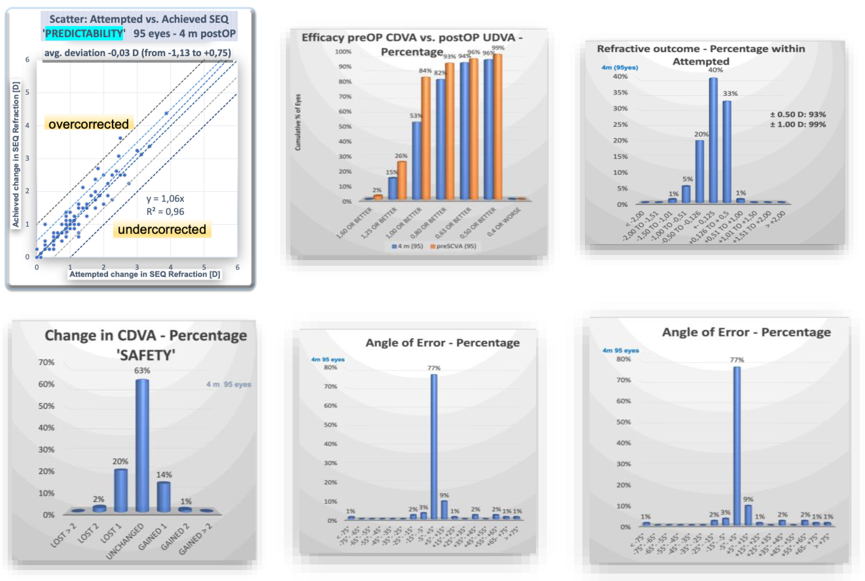
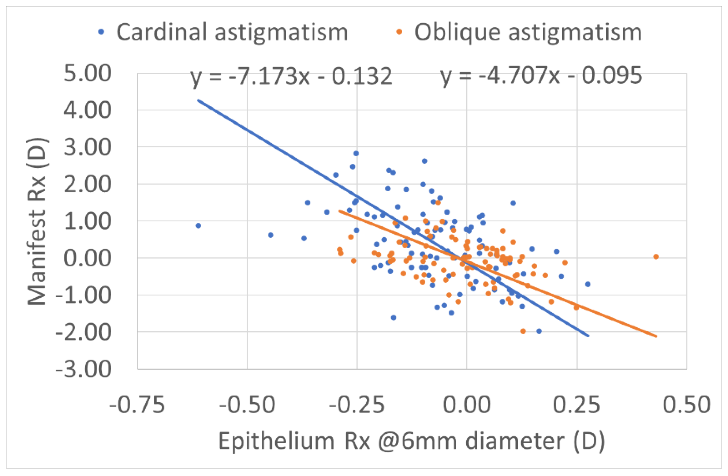
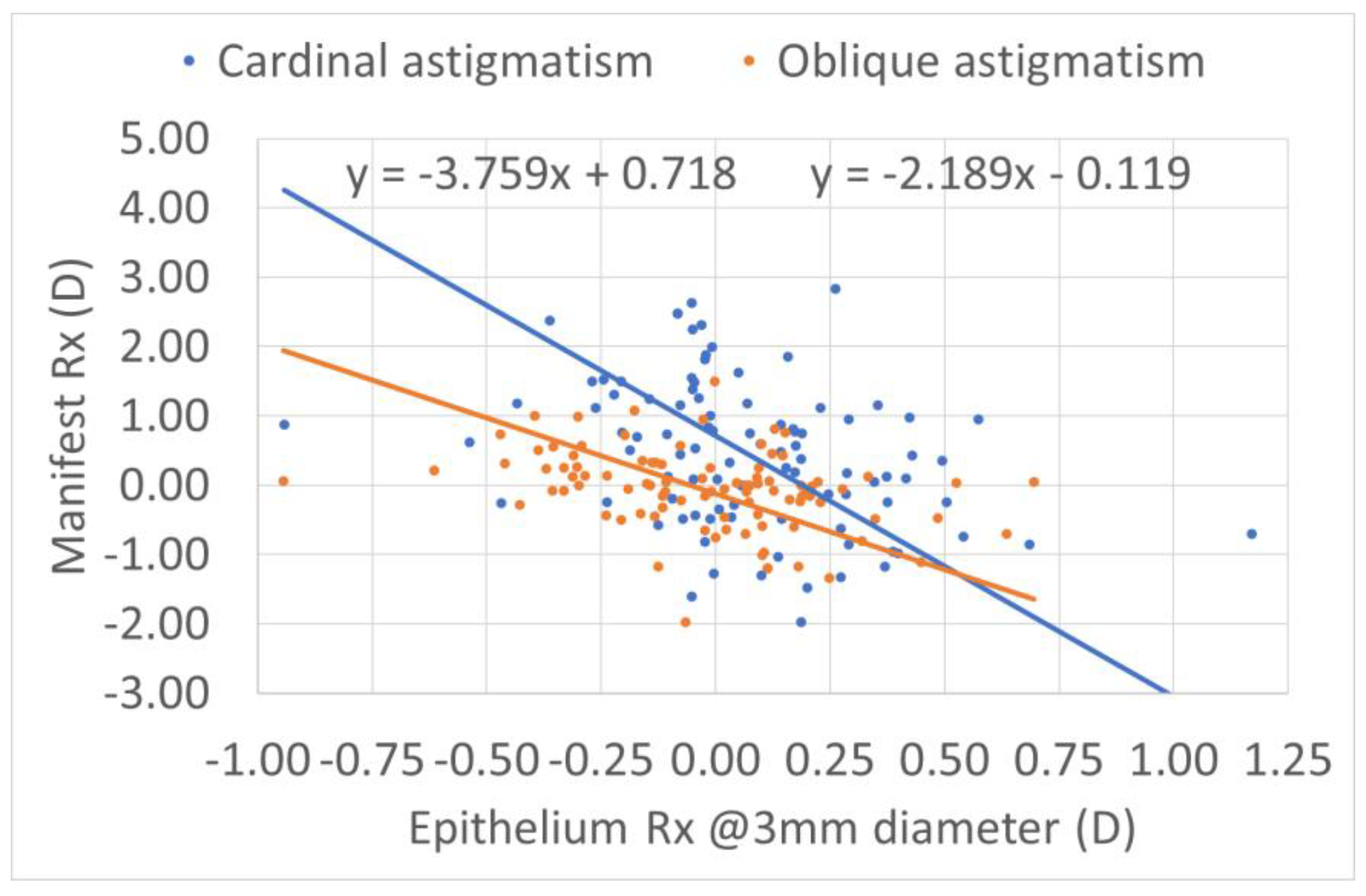
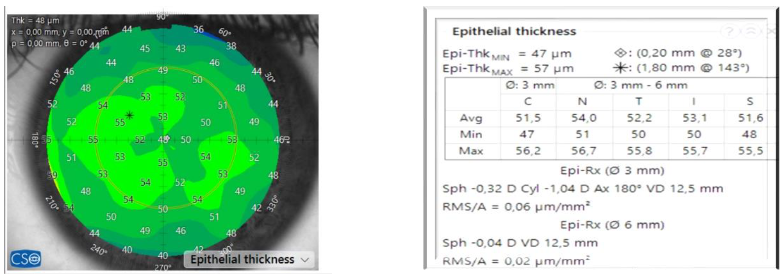
Purpose: To calculate the impact of the refractive power associated with the epithelial thickness in transepithelial refractive corrections Setting: Aurelios Augenzentrum Recklinghausen, Germany Methods: We analyzed the impact of the epithelium in a consecutive retrospective group of hiperopic eyes treated with TransPRK with the AMARIS 1050 RS laser platform (Schwind Eye-Tech Solutions, Kleinhostheim, Germany) with a minimal follow-up of 4 months. The group of 95 treated eyes had an average age of 36 years. The preoperative mean sphere was +1.6 dioptres (D) with a range till +5D, and the mean cylinder was -2.16 D with a range till -5.75D. For the epithelium measurements, we used the anterior segment optical coherence tomography MS-39 (CSO, Florence, Italy). Results: The refractive outcome showed a mean sphere of -0.03 D (range -0.5 to 1 D) and a mean cylinder of -0.33 (range -1.75 to 0 D). 93% and 98% of the cases had 0.5D, 1D, respectively, or less of spherical equivalent refractive error. The epithelium showed preoperative toricity, correlating to the preoperative manifest astigmatism. Preoperatively, at 6 mm, the epithelium showed compensational effect of ~15% of the refractive astigmatism (12% for cardinal and 18% for oblique astigmatism); whereas for 3mm the compensation accounted for ~25% of the refractive astigmatism (21% for cardinal and 31% for oblique astigmatism). No correlation was found postoperatively. Conclusions: The preoperative toricity of the epithelium is correlated with the amount of preoperative refractive astigmatism. No systematic correlation was found between preoperative epithelium and refractive deviations after hyperopia transepithelial treatments.

Keywords: 
;  ;  ;  ;  ;  ;  ;  

1. Introduction

With modern photorefractive keratectomy (PRK) in the form of transepithelial photorefractive keratectomy (TransPRK). With the SCHWIND AMARIS platform, we perform an advanced surface ablation [1] without break-through ablation in a reversal profile of the stroma and epithelium. Thus, we ablate the epithelium using the Excimer Laser.
The ablation rate varies between epithelium and stroma [2]. Ablation for the epithelium is approximately 15% greater than for the superficial stroma, resulting in potential inaccuracies if this discrepancy is not considered. The excimer laser experiences energy loss in the periphery, exacerbating the issue if not addressed [3].
The importance of epithelium thickness has been reported by several papers of Reinstein and Co [4]. Now, with the modern diagnostic systems with anterior optical coherence tomography, we get epithelial maps and can analyse the epithelium and stroma independently.
In this study, we calculated the impact of the optical power of the corneal epithelium in relation to the manifest refraction. We also calculated the toricity and wanted to know if there was an impact of the epithelium power on the manifest refraction.

2. Materials and Methods

The records of 95 eyes from 66 patients, who were consecutively treated with an aberration-neutral [5] TransPRK profile for the correction of hyperopia, with or without astigmatism and mixed astigmatism, have been examined. The demographic data are shown in Table 1.
The preoperatively refractive data, as well as the epithelium map, which was measured by the CSO MS-39 (Construzione Strumenti Oftlamici, Firenze, Italy), optical coherence tomography with placido rings for topography device, were included in the evaluation [6].
We analysed the refractive outcomes as proposed by the JRS [7]. For the preoperative epithelium map we used the data from the MS-39. With this device we get the epithelium power with sphere and cylinder at 3 and 6mm. After four months, we evaluate the refractive data.
All procedures were conducted by a sole surgeon, DdO, utilising the SCHWIND AMARIS 1050RS [8]. The surgical procedure has been described previously [9].
The AMARIS 1050RS uses a rotational symmetric ablation of the epithelium based on the parabolic fit of the individual epithelial thickness measured (provided) by the MS-39 OCT [10], so in certain manner it is customized based on the individual epithelium.
All eyes with a minimum follow-up of 4 months were included in the study; exclusion criteria were eyes with previous ophthalmic eye surgery or disease that could influence the healing of the cornea.
In addition, we calculated the epithelial refraction considering the toricity at the cardinal and oblique astigmatism to determine the relationship between this estimate and the manifest refraction.
Accession numbers. If the accession numbers have not yet been obtained at the time of submission, please state that they will be provided during review. They must be provided prior to publication.
Interventionary studies involving animals or humans, and other studies that require ethical approval, must list the authority that provided approval and the corresponding ethical approval code.

3. Results

The average preoperative treated sphere was +1.6 diopters, with a range of +0.25 to +6 diopters. The average cylinder measurement was -2.16 diopters; treatment extended to -5.75 diopters. Following TransPRK and a 4-month follow-up, the average manifest sphere was +0.13 diopters and the manifest cylinder was -0.33 diopters.
The refractive outcome is reported at Figure 1. The predictability with 93% achieving half of diopters or less of spherical equivalent and 99% under 1 diopter. The efficacy was approximately 90%, while the astigmatism showed an 82% under 0.5D and an 95% under 1D, respectively.
Preoperatively at 6mm, the cardinal astigmatism is seven times greater than the epithelium toricity. The cardinal astigmatism at 6mm equals manifest refraction 7 times more than the manifest epithelial refraction. Oblique astigmatism equals four times the epithelial refraction. The average for both axis is 5.8 astigmatism, which means that the epithelium compensates 17% of the refractive toricity at 6mm (Figure 2).
The oblique astigmatism in orange, the manifest Rx, is 4 times higher than the epithelium manifest Rx. In both cases, the epithelium has the toricity in a reversal sign.
At 3mm, the epithelium compensates by 35% on both axes (Figure 3).
There was no correlation between postoperative manifest astigmatism and preoperative epithelium toricity. The preoperative epithelium does not contribute to the manifest sphere.

4. Discussion

In this study, we calculated the impact of the corneal optical power of the corneal epithelium in relation to the preoperative manifest refraction. We also calculated the toricity and wanted to know if there was an impact of the epithelium power on the postoperative manifest refraction.
With the modern diagnostic devices, we get more information about the epithelium. In our case we demonstrated that preoperatively the epithelium exhibits toricity, with a reversal sign from the stroma. At 6 mm, the epithelium adjusts for the astigmatism, which is around 17% and at 3mm 35% mean for both diameter 26%.
We can estimate the spherical equivalent of the epithelium as the average of the axial power of all measured points. We can calculate the astigmatism component of the epithelium.
The difference is the average of the axial power along the horizontal and vertical meridians. The advanced anterior OCT devices automatically provide computed information by corneal measuring (Figure 4). This study employed this information to calculate the spherical component and astigmatism of the epithelium.
This section is not mandatory but can be added to the manuscript if the discussion is unusually long or complex.
We employ a rotationally symmetrical profile for the epithelium with the SCHWIND AMARIS. The system adjusts the epithelium at the center and the periphery. With the import of the data from the center, it calculates the epithelium thickness at the periphery.
In both instances the loss at the periphery of energy is considered, resulting in an increased number of ablation spots at the periphery to offset the energy loss.
In the presented cases the manifest astigmatism diminishes significantly postoperatively.
The transepithelial PRK technique in the ORK-CAM plans aspheric transepithelial ablation volumes (based on a predefined parabolic epithelial thickness profile) for the Schwind Amaris laser system for the combined removal of corneal epithelium and the correction of refractive volumes.
Single-step transepithelial PRK allows simultaneous ablation of the epithelium and the stroma to shorten the overall treatment time and minimize the risk for corneal dehydration. In addition to a shorter surgical time, epithelial tissue removal is optimized to avoid myopic-like corrections (approximately -0.75 D). This approach treats refractive errors by superimposing a defined epithelial thickness profile with a corneal aspheric ablation profile. In addition, the diameter of epithelial removal is calculated to match the total ablation zone, decreasing the wound surface and speeding the healing process.
Transepithelial approaches allow maximum correspondence between the corneal topography and the ablation profile. Although there is a slight difference in the photoablative rates of the stroma and the epithelial tissue (approximately 20% higher in the epithelium), the Amaris software compensates for this.
With current transepithelial PRK, a simplified customized epithelial ablation algorithm is used for every individual eye, based on the parabolic best-fit (rotational symmetric) of the actual topometry of the epithelial layer.
This leads for all eyes, to in some regions/locations more stroma than necessary might be ablated; whereas in other regions/locations, less epithelium than necessary might be ablated and some ablation corresponding to stroma would ablate the remaining epithelium.
It seems this has no influence on the outcomes, as our results show. This relative insensitivity from the simplified to the actual (detailed) epithelial profiles can be explained because our study is limited to initial treatments on normal corneas.
In any case, there is some minor wasted tissue when the actual corneal epithelial profile is thinner than the applied epithelial ablation profile, the stromal ablation is reduced when the actual corneal epithelial profile is thicker than the applied epithelial ablation profile.
If the epithelial profile from the center to the periphery is considered flat (equal thicknesses at all epithelial points, equivalent to a classic PTK ablation), a hyperopic shift of approximately 0.6 D can be expected.
The corneal epithelium plays an increasingly important role in corneal net power and, as a result, total ocular refraction. It is reported that epithelial refractive power is an average. of 1.03 D (range 0.55–1.85 D) over the central 2 mm diameter zone and 0.85 D (range 0.29–1.60 D) at the 3.6 mm diameter zone [11]. In our case we have also demonstrated that the epithelium has a corneal power but also a toricity that compensate the manifest refraction.
The advantage of treating with TransPRK the astigmatism with a rotational symmetric epithelium is that we treat the manifest astigmatism of the total cornea. If the epithelium is compensating about 26% of the total astigmatism that means that the stroma astigmatism is 26% higher than the manifest astigmatism and that explains why PRK or LASEK could have undercorrections as they treat after we withdraw the epithelium and therefore the toricity of the stroma is now different. Similar is with LASIK or intralamellar surgery where we treat the astigmatism without knowing the toricity of the epithelium, as the epithelium compensates the totricity theoretically some remodelling will occur after LASIK or intralamellar surgery.
The impact of the refractive power associated with the epithelial thickness in preoperative manifest refraction compensated for ~15% of the refractive astigmatism (12% for cardinal and 18% for oblique astigmatism); whereas for 3mm the compensation accounted for ~25% of the refractive astigmatism (21% for cardinal and 31% for oblique astigmatism). This means that the refractive astigmatism in the absence of epithelium (i.e. that of the bare stroma) would have been 18% and 36% larger at 6mm and 3mm respectively. Accepting that 3-4mm is closer to the pupil diameters for manifest refraction, it means that for each diopter of refractive astigmatism there is ~30% compensation by the epithelium, and the astigmatism at the bare stroma would be ~1.3D.
A better analysis could have been made based on corneal and stromal toricity, and not indirectly through the manifest refraction; but manifest refraction correction is the goal of all refractive therapies.
No correlation was found postoperatively. This appears to contradict the notion [12], that the toricity of the preoperative epithelium (acting as a masking agent as demonstrated here) should be reflected in refractive deviations postoperatively.
This can be explained by several factors. On the one hand we aimed to correlate preoperative epithelial toricity to postoperative manifest refraction (deviations). But the refraction, the corneal (and potentially stromal) toricities are modified (all reduced) by the transepithelial treatment, so that the correlation between them gets broken [13], and the epithelial remodeling over a less toric stromal surface would result in an even less toric postoperative epithelium. On the other hand, and contrary to common intuition, the application of a rotational symmetric epithelial profile may be an advantage (and not a limitation) of transepithelial ablations. This, if confirmed, would move away from the notion that a detailed epithelial map shall be used for transepithelial ablations.
Using a detailed epithelial map (assumed free of errors) would make transepithelial ablations the perfect laser PRK. But would suffer from the same underestimation of the refractive astigmatism at the stromal plane and would require the postoperative epithelium to again mask the toricity of the postoperative underlying stroma. Assuming the same level of postoperative compensation, an estimated 20% undercorrection in refractive astigmatism could be explained for PRK.
However, empirical nomograms may have accounted for that further reducing the differences between PRK and TransPRK as compared previously [14].
A rotationally symmetric epithelial model (as used here, and presented elsewhere [15]) effectively shifts the already compensated (i.e. less toric, and in general less aberrated) anterior epithelial surface to the anterior stroma, and then applies the correction (measured for the anterior epithelial surface). Further, the reverse transepithelial approach (as used here, and presented elsewhere [16], reshapes the correction (measured for the anterior epithelial surface) at the already compensated (i.e. less toric, and in general less aberrated) anterior epithelial surface, and the resulting (ideal) shape is then transferred to the anterior stroma by the rotationally symmetric epithelial model.
The error incurred is the mismatch between the detailed epithelial map (assumed free of errors) and rotationally symmetric epithelial model (as used here) multiplied by the mismatch between the ablation rates for epithelium and stroma. So that a 20% mismatch in epithelium combined by a 20% mismatch of the ablation rates, would be then again masked by the epithelium remodeling, resulting in merely <3% induced error.
Epithelial cardinal and oblique astigmatism (i.e. epithelial toricity) at both 3mm and 6mm analysis diameters are negatively correlated to manifest refractive astigmatism, suggesting a masking (partial) compensational effect; with a higher compensatory effect for the smaller diameter.
Consequently, the larger the manifest cylinder, the greater the epithelial toricity with reversal sign. For example, -2.25D manifest astigmatism has a compensation of the epithelium of +0.5 D. This is consistent with previous publications determined by direct imaging [17].
The preoperative epithelium does not contribute to the manifest sphere.
There was no correlation between postoperative manifest astigmatism and preoperative epithelium toricity, probably since postoperatively epithelium becomes more spherically shaped and has less toricity.
We found that the postoperative manifest refraction is near the epithelium refraction preoperatively. Thus, the preoperative epithelium might be one of the causes of undercorrection after hyperopic TransPRK, but the analysis showed a high variance with only a correlation of 0.32 (R2 <0.1, i.e. >90% of the variance unexplained by the correlation), that means that the epithelium is not the main cause of undercorrection in hyperopia after TransPRK.

5. Conclusions

The preoperative toricity of the epithelium is correlated with the amount of preoperative refractive astigmatism. No systematic correlation was found between preoperative epithelium and refractive deviations after hyperopia transepithelial treatments. Part of the success of transepithelial PRK (as used here) may be attributed to the use of a rotationally symmetric aspherical epithelial model.

Author Contributions

For Conceptualization, DO and SAM; methodology DO and SAM; data curation, DO.; writing—original draft preparation DO.; writing—review and editing DO and SAM; All authors have read and agreed to the published version of the manuscript.

Funding

“This research received no external funding”.

Institutional Review Board Statement

Institutional Review Board was given. The study was conducted according to the guidelines of the Declaration of Helsinki. The study was retrospective with anonymised data. Informed consent was obtained from all subjects involved in the study. According to the Medical College ÄKWL § 15 Paragraph 1, retrospective anonymised data do not require ethical approval. (https://www.medizin.uni-muenster.de/ek/ethik-kommission/antragsunterlagen/unterlagen-fuer-antraege/ausnahmen.html).

Informed Consent Statement

Informed consent was obtained from all subjects involved in the study.

Data Availability Statement

All the data were fully anonymised and are available upon request.

Conflicts of Interest

DO is Consultant for SCHWIND eye-tech solutions, SAM is employee for SCHWIND eye-tech solutions.

References

  1. Aslanides IM, Kymionis GD. Trans advanced surface laser ablation (TransPRK) outcomes using SmartPulseTechnology. Cont Lens Anterior Eye. 2017;40(1):42-46. [CrossRef]
  2. Seiler T, Kriegerowski M, Schnoy N, Bende T. Ablation rate of human corneal epithelium and Bowman’s layer with the excimer laser (193 nm). Refract Corneal Surg. 1990;6(2):99-102.
  3. Arba-Mosquera S, de Ortueta D. Geometrical analysis of the loss of ablation efficiency at non-normal incidence. Opt Express. 2008;16(6):3877-3895. [CrossRef]
  4. Reinstein DZ, Archer TJ, Gobbe M, Silverman RH, Coleman DJ. Epithelial thickness in the normal cornea: three-dimensional display with Artemis very high-frequency digital ultrasound. J Refract Surg. 2008;24(6):571-581. [CrossRef]
  5. de Ortueta D, Arba Mosquera S, Baatz H. Comparison of standard and aberration-neutral profiles for myopic LASIK with the SCHWIND ESIRIS platform. J Refract Surg. 2009;25(4):339-349. [CrossRef]
  6. Savini G, Schiano-Lomoriello D, Hoffer KJ. Repeatability of automatic measurements by a new anterior segment optical coherence tomographer combined with Placido topography and agreement with 2 Scheimpflug cameras. Journal of Cataract & Refractive Surgery. 2018;44(4). https://journals.lww.com/jcrs/Fulltext/2018/04000/Repeatability_of_automatic_measurements_by_a_new.10.aspx.
  7. Reinstein DZ, Archer TJ, Randleman JB. JRS standard for reporting astigmatism outcomes of refractive surgery. J Refract Surg. 2014;30(10):654-659. [CrossRef]
  8. de Ortueta D, von Rüden D, Verma S, Magnago T, Arba-Mosquera S. Transepithelial Photorefractive Keratectomy in Moderate to High Astigmatism With a Non-wavefront–Guided Aberration-Neutral Ablation Profile. J Refract Surg. 2018;34(7):466-474. [CrossRef]
  9. de Ortueta D. Transepithelial Photorefractive Keratektomy after a Clear Lens Exchange. Vision (Basel). 2021;5(1). [CrossRef]
  10. de Ortueta D, von Rüden D, Arba-Mosquera S. Customized versus Standard Epithelium Profiles in Transepithelial Photorefractive Keratectomy. Optics. 2021;2(4):266-275. [CrossRef]
  11. Simon G, Ren Q, Kervick GN, Parel JM. Optics of the corneal epithelium. Refract Corneal Surg. 1993;9(1):42-50.
  12. Gatinel D, Racine L, Hoang-Xuan T. Contribution of the corneal epithelium to anterior corneal topography in patients having myopic photorefractive keratectomy. Journal of Cataract and Refractive Surgery. 2007;33(11):1860-1865. [CrossRef]
  13. Benito A, Redondo M, Artal P. Laser in situ keratomileusis disrupts the aberration compensation mechanism of the human eye. Am J Ophthalmol. 2009;147(3):424-431.e1. [CrossRef]
  14. Aslanides IM, Padroni S, Arba Mosquera S, Ioannides A, Mukherjee A. Comparison of single-step reverse transepithelial all-surface laser ablation (ASLA) to alcohol-assisted photorefractive keratectomy. Clin Ophthalmol. 2012;6:973-980. [CrossRef]
  15. Arba Mosquera S, Awwad ST. Theoretical analyses of the refractive implications of transepithelial PRK ablations. Br J Ophthalmol. 2013;97(7):905-911. [CrossRef]
  16. Luger MHA, Ewering T, Arba-Mosquera S. Consecutive myopia correction with transepithelial versus alcohol-assisted photorefractive keratectomy in contralateral eyes: one-year results. J Cataract Refract Surg. 2012;38(8):1414-1423. [CrossRef]
  17. Salah-Mabed I, Saad A, Gatinel D. Topography of the corneal epithelium and Bowman layer in low to moderately myopic eyes. Journal of Cataract and Refractive Surgery. 2016;42(8):1190-1197. [CrossRef]
Figure 1. Left up histogram showing predictability and a regression coefficient near 1. (3 eyes needed a retreatment); Up on the middle graphic showing efficacy comparing preoperative best corrected visual acuity (BCVA) with postoperative uncorrected visual acuity; up right showing the percentage of eyes within the attempted 93% of the eyes had postoperatively a spherical equivalent (SEQ) of half of diopter or less refraction; at the bottom left safety no one eye loose 2 or more lines of BCVA; bottom middle showing the angle of error of less than 15° in 89% of the eyes; and bottom right showing the postoperative refractive astigmatism with 82% of half of diopter or les.
Preprints 150059 g001
Figure 2. At preoperative epithelium refraction (Rx) in relation to the preoperative refraction at 6 mm. In blue is the cardinal astigmatism. The manifest refraction is 7 times higher than the epithelium manifest Rx.
Preprints 150059 g002
Figure 3. Preoperative epithelium refraction (Rx) in relation to the preoperative refraction at 3nmm. In blue the cardinal Astigmatism. The manifest refraction is 3.8 times higher than the Epithelium Manifest Rx.The oblique astigmatism in orange, the manifest Rx is 2.2 times higher than the epithelium manifest Rx. In both cases the epithelium has the toricity in a reversal sign. For cardinal and oblique astigmatism, the mean is 2.9 is manifest Astigmatism higher. That means that the Epithelium compensates 35% of the refractive toricity at 3 mm.
Preprints 150059 g003
Figure 4. At the left an epithelium map from a preoperative eye. At the right epithelial data as thickness centrally and maximally. Epithelium data at different positions in relation to the vertex of the cornea. Epithelium refraction at 3 and 6mm.
Preprints 150059 g004
Table 1. Demographic data of the treated eyes. Preoperatively and postoperatively refractive outcome after 4months. SEQ (spherical equivalent) Cyl (Cylinder) all the data in mean with standard deviation.
n=95 eyes
Gender female 46.3% (44 eyes) male 53.7% (51) eyes)
eye left 51.6% (49 eyes) right 48.4% (46 eyes)
Preoperatively range Postop 4m range
SEQ +0.53 +/- 1.34 D (-1.75 to +3.88 D) -0.03 +/- 0.3 D (-1.13 D to +0.75 D)
Sphere +1.6 +/- 1.16 D (+0.12 to +6 D) +0.13 +/- 0.29 D (-0.5D to +1 D)
Cyl -2.16 +/- 1.32 D (-5.75 to -0 D) -0.33 +/- 0.38D (-1.75 D to 0 D)
Disclaimer/Publisher’s Note: The statements, opinions and data contained in all publications are solely those of the individual author(s) and contributor(s) and not of MDPI and/or the editor(s). MDPI and/or the editor(s) disclaim responsibility for any injury to people or property resulting from any ideas, methods, instructions or products referred to in the content.
Copyright: This open access article is published under a Creative Commons CC BY 4.0 license, which permit the free download, distribution, and reuse, provided that the author and preprint are cited in any reuse.
Prerpints.org logo

Preprints.org is a free preprint server supported by MDPI in Basel, Switzerland.

Subscribe

Disclaimer

Terms of Use

Privacy Policy

Privacy Settings

© 2025 MDPI (Basel, Switzerland) unless otherwise stated