Submitted:
13 February 2025
Posted:
14 February 2025
Read the latest preprint version here
Abstract
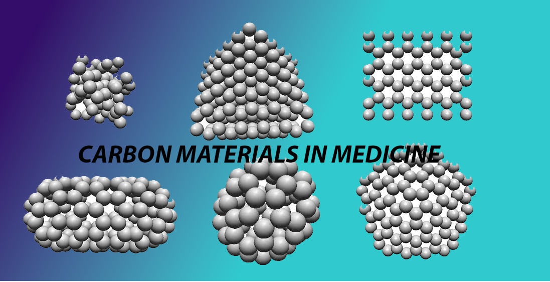
Carbon nanomaterials that include different forms such as graphene, carbon nanotubes, fullerenes, graphite, nanodiamonds, carbon nanocones, amorphous carbon, as well as porous carbon, are quite distinguished by their unique structural, electrical, and mechanical properties. This plays a major role in making them pivotal in various medical applications. The synthesis methods used for such nanomaterials, including techniques such as chemical vapor deposition (CVD), arc discharge, laser ablation, and plasma-enhanced chemical vapor deposition (PECVD) are able to offer very precise control over material purity, particle size, and scalability, enabling for nanomaterials catered for different specific applications. These materials have been explored in a range of different systems, which include drug delivery systems, biosensors, tissue engineering, as well as advanced imaging techniques such as MRI and fluorescence imaging. Recent advancements, including green synthesis strategies and novel innovative approaches like ultrasonic cavitation, have improved both the precision as well as the scalability of acrbon nanomaterial production. Despite challenges like biocompatibility and environmental concerns, these nanomaterials hold immense promise in revolutionizing personalized medicine, diagnostics, and regenerative therapies. Figures of the various carbon nanomaterials in this manuscript are presented by the conventional ball-and-stick model and the sphere-in-contact model that was perviously shown to be a better representation of the void space in carbon nanostructures. These sphere-in-contact models have a better description of the size of the electron density of carbon nanomaterials and therefore can help in the rational design of new structures for medical and biomedical appliactions.
Keywords:
graphene
; carbon nanotubes
; fullerene
; graphite
; nanodiamond
; carbon nanocones
; amorphous carbon
1.1. Background
The exact structure of carbon materials has been the topic of many research papers in this century. The structure of carbon materials is associated with the fact that it can have sp, sp2 and sp3 hybridized carbon atoms bound to each other in various cyclic and linear formations. This means that the carbon atoms form a single and triple covalent C-C bond, or two single and a double covalent C-C bond or four single carbon C-C bonds, respectively, in sp, sp2 and sp3 hybridised carbons.[1] Apart from that carbon can form stable 5 and 6 membered rings which when combined result in curved carbon surfaces such as in fullerenes and carbon nanocones. The sp2 hybridized hexagonal layer of graphite has very weak mechanical properties for bending which means that it can easily be rolled into a cylinder which is the case of carbon nanotube.[2] In the structure of diamond all carbons are sp3 hybridized resulting in a rigid structure with great mechanical strength. However the sp2 hybridised carbon atoms in graphite result in a pz orbital on each carbon atom that forms π-bonds, which are delocalised over the carbon rings. These π-bonds are very polarizable and cause the layers to bind through London dispersion forces (LDF) through induced-dipole/induced-dipole interactions.[3]
The polarizable nature of these π-clouds results in great electrical conductivity with the hexagonal layers and little conductivity through regions where the carbon-carbon atoms just interact through LDF. The fact that carbon materials also have a degree of unsaturation of the carbon atom due to double and triple bonds means that carbon materials can easily be functionalized or saturated with hydrogen. This causes local deformations of the carbon structure, which may lead to new properties of the nanomaterial and several applications in medicine and drug delivery.
1.2. Introduction
Carbon nanomaterials have a rich history which is rooted in both the exploration of carbon’s versatile chemistry and its unique physical properties. Starting from the initial discovery of fullerenes in the 1980s to the consequential isolation of graphene in 2004, these unique materials have revolutionized fields ranging from devices such as electronics to medicine. Their exceptional properties, such as high electrical conductivity, mechanical strength, and tunable chemical functionality, play a great role in setting them apart as indispensable tools for advancing biomedicine.
In terms of the biomedical field, carbon nanomaterials, including graphene, carbon nanotubes, fullerenes, and nanodiamonds, have shown a very remarkable potential. These materials are employed in drug delivery systems for targeted therapies, biosensors for disease diagnostics, as well as scaffolds for tissue engineering. Their biocompatibility and adaptability are crucial as they make them particularly suited for applications where precision and functionality are critical. However, competing materials, such as polymeric nanoparticles and metal-based systems, also play significant roles in biomedical research, with different advantages, as they play a factor in offering advantages in terms of biodegradability or enhanced imaging capabilities, respectively. This competition calls attention to the need to understand the unique advantages and challenges of carbon nanomaterials. This review focuses on the synthesis techniques that enable the production of carbon nanomaterials with their specific tailored properties, as well as their diverse applications in medicine.
2. Synthesis and Medical Applications of Carbon Nanomaterials
We present the various carbon materials in terms of their structure, history of invention, synthesis methods and applications in medicine and drug delivery. The carbon materials we focus on is amorphous carbon in Section 2.1, graphite and highly oriented pyrolytic graphite (HOPG) in Section 2.2, carbon nanocones in Section 2.3, Fullerene (C60) in Section Section 2.4, Graphene in Section 2.5, Reduced graphene oxide (RGO) in Section 2.6, single walled carbon nanotubes (SWCNT) in Section 2.7 and nanodiamond in Section 2.8. These are most of the carbon materials that have been applied in medical applications and drug delivery. These structures are described in terms of their geometry, their synthesis methods and their applications in these fields.
2.1. Amorphous Carbon
2.1.1. Structure of Amorphous Carbon
Amorphous carbon (a-C) is a non-crystalline allotrope of carbon, which lacks the ordered atomic structure of the more popular counterparts such as diamond or graphite. This structural irregularity in amorphous carbon results in a unique material composed of a mixture of sp2 (graphitic) and sp3 (diamond-like) carbon bonds, which offers it a broad spectrum of properties. Amorphous carbon is characterized, for the most part, by a tunable electrical conductivity, high hardness, and excellent chemical inertness, properties that are being harnessed more and more in various biomedical applications, ranging from simple medical devices to implants. Recent advances of the synthesis of amorphous carbon have further amplified the potential for use in medicine, catalysis, and nanotechnology.
2.1.2. Synthesis of Amorphous Carbon
The synthesis of amorphous carbon (see Figure 1) traditionally has depended on processes related to high-temperature such as chemical vapour deposition (CVD) and arc discharge. The two processes have slight variation with the process of CVD introducing volatile gases into a reaction chamber, allowing them to decompose at high temperatures to form carbon films. This specific technique allows for more precise control over the sp2/sp3 ratio, therefore having an influence on the material’s hardness and electrical properties, producing high-purity, uniform samples. On the other hand,Arc discharge utilizes a different method which passes a high-current electrical arc between carbon electrodes in an inert gas environment. This creates carbon soot which contains a mixture of carbon allotropes, including amorphous carbon. Even though arc discharge is efficient, typically it tends to create samples with less control over particle size and more impurities than CVD. The quality of amorphous carbon has several different factors which influence it. These factors include the sp2/sp3 carbon ratio, structural disorder, particle size, and purity. Furthermore, there are techniques such as Raman spectroscopy which are used to measure this ratio by analyzing the G-band (sp2 bonds) and D-band (disorder). Transmission electron microscopy (TEM) can also be used to help assess particle size and structural order, while chemical analysis techniques determine purity. There are key parameters such as deposition temperature, pressure, electric field (in arc discharge), and gas environment which effectively affect the quality of the final product. CVD has a tendency to produce higher quality amorphous carbon mainly because of its superior control over these parameters, which has made it more preferable for applications requiring uniformity and precise material properties.
However, innovative techniques have emerged, offering greater control over particle size, structure, and properties. One such advancement is the electric plasma discharge method in an ultrasonic cavitation field, described by Sergiienko and co-workers, which allows for the production of amorphous carbon nanoparticles in liquid benzene.[4]
In this process, a plasma discharge is generated in a cavitation field, where thousands of microbubbles collapse, producing localized regions of high temperature and pressure. This novel approach permits the synthesis of amorphous carbon nanoparticles smaller than 30 nm at relatively low power, representing a significant advancement over traditional dry synthesis methods. Dry synthesis methods are widely used for the production of carbon nanomaterials such as nanotubes, fullerenes, and carbon-encapsulated metal nanoparticles. These techniques avoid the use of liquids or solvents, relying instead on gas-phase or high-energy environments. One prominent method is arc discharge, where a high-temperature electric arc between carbon electrodes vaporizes carbon, which then condenses into nanostructures. Similarly, laser ablation uses a focused laser to vaporize carbon targets, providing fine control over particle size and enabling the formation of nanotubes and fullerenes. CVD decomposes carbon-containing gases at high temperatures on a substrate, allowing the controlled synthesis of carbon materials like graphene and nanotubes. Additionally, hydrocarbon flames can also generate carbon nanomaterials by combusting hydrocarbons, leading to the aggregation of carbon atoms into nanoparticles or tubes. These dry methods, operating in gas phases or vacuums, offer distinct advantages in precision and scalability over “wet” methods such as the electric plasma discharge process in ultrasonic cavitation, which uses liquid solvents to achieve similar outcomes.
In addition to this, plasma-enhanced chemical vapour deposition (PECVD) has been employed to fabricate amorphous carbon films, enabling precise control over the sp2/sp3 bond ratio. This allows for fine-tuning of the material’s properties, particularly its hardness and electrical conductivity.[1] Pulsed Laser Deposition (PLD) and Plasma-Enhanced Chemical Vapour Deposition (PECVD) differ primarily in their deposition methods and the physical processes involved. PLD, another recent technique, has proven effective for the deposition of amorphous carbon films on various substrates, further expanding the applications of this material in the medical field PLD uses a high-power laser to ablate material from a target, vaporising it and depositing it onto a substrate. It is a physical vapour deposition process that typically requires high or ultra-high vacuum chambers to maintain a clean environment. This makes PLD precise in controlling thin film composition but expensive due to the high-power laser system and sophisticated vacuum equipment required. PECVD, on the other hand, uses plasma to enhance chemical reactions of precursor gases, which deposit thin films on a substrate. It is a chemical vapour deposition process, where the plasma lowers the deposition temperature, allowing the deposition of materials that might not tolerate high temperatures. While PECVD also requires vacuum systems, they are generally less demanding than those used in PLD and do not always necessitate ultra-high vacuum conditions. However, PECVD systems still involve complex and costly plasma generation and control equipment. In terms of instrumentation, PLD typically requires more specialized and expensive setups, including ultra-high vacuum chambers, compared to PECVD, which operates at lower vacuum levels but still involves intricate plasma control systems.[5]
Despite the promising developments in the synthesis and application of amorphous carbon, challenges remain. One primary concern is the biocompatibility and potential toxicity of amorphous carbon nanoparticles, particularly when used in vivo. While amorphous carbon films such as DLC are generally biocompatible, the behavior of nanoparticles in biological systems is less understood, necessitating further toxicological studies.[4]
Additionally, ensuring the scalability of these advanced synthesis methods remains a challenge. Techniques such as plasma discharge in cavitation fields and PECVD are still largely confined to laboratory settings, and their industrial-scale implementation will require optimization.[4,5]
Amorphous carbon can be synthesized through several methods, each offering distinct advantages based on the desired application. Chemical vapour deposition (CVD) is one of the most widely used techniques, where hydrocarbon gases such as methane (CH4) or acetylene (HCCH) are decomposed at high temperatures in the presence of a metal substrate/catalyst. This method allows for fine-tuning of the carbon’s properties and is highly scalable, making it suitable for large-scale production. In contrast, the arc discharge process involves generating a high-temperature electric arc between two graphite electrodes in an inert gas atmosphere. While this method can produce a variety of carbon structures, including nanotubes and fullerenes, it often results in a mixture containing amorphous carbon, depending on the specific conditions used. Another approach, laser ablation, utilizes a high-power laser to vaporize a graphite target, leading to the condensation of carbon atoms into nanotubes, fullerenes, and amorphous carbon. Although laser ablation can produce high-purity carbon, it is less commonly employed for industrial-scale applications due to its high energy and cost requirements. Together, these methods provide versatile avenues for the controlled synthesis of amorphous carbon, each with its own trade-offs between scalability, purity, and cost.[6]
Another promising direction for future research lies in the development of green synthesis methods. These methods aim to minimize the environmental impact of amorphous carbon production, reducing the reliance on hazardous chemicals and high-energy processes. The production of carbon allotropes, particularly amorphous carbons and carbide-derived carbons, involves several processes that can have significant environmental impacts. During the chemical activation of carbon materials, harmful chemicals like zinc chloride (ZnCl₂), phosphoric acid (H₃PO₄), and sulfuric acid (H₂SO₄) are used, posing risks of water and soil contamination if not properly managed. In addition, the chlorination process used to synthesize carbide-derived carbons results in the release of chlorine gas (Cl₂) and hydrogen chloride (HCl), both of which are corrosive and harmful to the atmosphere and ecosystems. Moreover, pyrolysis and high-temperature synthesis can lead to the emission of carbon dioxide (CO₂) and volatile organic compounds (VOCs), contributing to global warming and air pollution. Combustion processes involved in carbon material production can also release carbon monoxide (CO), nitrogen oxides (NOₓ), and sulfur oxides (SOₓ), further exacerbating air quality issues and contributing to acid rain. These environmental hazards highlight the need for green synthesis methods and better emission control strategies to minimize the impact of carbon allotrope production on the environment.[5]
Graphite exfoliation involves several methods to separate its layers, each producing graphene with distinct properties. Mechanical exfoliation applies physical forces, such as ultrasonication or stirring in carefully chosen solvents, to peel off layers from graphite. This method often uses intercalating agents or surfactants to reduce re-aggregation of the exfoliated layers, though yields of monolayer graphene remain modest.[7] Thermal exfoliation, often performed on graphite oxide, involves rapid heating, which decomposes interlayer functional groups and generates gases, causing expansion and separation. High-temperature exfoliation (e.g., >1000°C) has been shown to produce single-layer graphene sheets effectively, though it can introduce defects into the graphene structure.[7] Electrochemical exfoliation uses voltage in a conductive solution to exfoliate graphite electrodes, simultaneously functionalizing the graphene sheets with solvent molecules. This method is advantageous for yielding functionalized graphene with fewer defects than oxidation methods like the Hummers method.[7] Finally, supercritical fluid exfoliation intercalates supercritical fluids between graphite layers, which expand to separate the layers upon pressure release. This approach can produce high-quality graphene sheets with a significant percentage of monolayers, especially when combined with surfactants.[7] Each of these methods provides graphene suited to different applications, balancing factors such as yield, defect levels, and scalability.[7]
2.1.3. Uses of Amorphous Carbon in Medicine
Amorphous carbon, particularly in the form of polyethylenimine, functionalized magnetic amorphous carbon (Fe3O4-PEI-ACTF NC), has shown significant potential in environmental and medical applications due to its high adsorption capacity and unique surface properties. Synthesised from agricultural waste like oil palm leaves, this material is characterised by its high surface area, biocompatibility, and chemical stability. The adsorption process follows the Langmuir isotherm model, indicating a monolayer adsorption on a uniform surface, and the kinetics align with a pseudo-second-order model, suggesting chemisorption through electron transfer and chemical bonding. Furthermore, the thermodynamic analysis reveals that the adsorption is both endothermic and spontaneous, making this material suitable for practical applications. [8]
Its ability to be deposited at room temperature without damaging sensitive substrates makes it a valuable material in diverse industries. In biomedical engineering, a-C is often employed as a protective coating for medical implants due to its biocompatibility, wear resistance, and antibacterial properties, enhancing both the durability and safety of medical devices. Additionally, its high adsorption capacity is utilized in advanced water purification systems, where it efficiently captures toxins and contaminants. Amorphous carbon’s chemical stability and electrical conductivity also make it ideal for developing sensors and biosensors used in real-time diagnostics. This breadth of applications solidifies amorphous carbon’s place as a critical material in advancing medical and technological innovations.[9]
Activated carbon (AC) has emerged as a highly versatile and promising material in the field of medicine, particularly as a carrier for amorphous drug delivery systems. Activated carbon is a highly porous form of carbon with a large surface area, produced through processes like chemical or steam activation, and is used for adsorption in applications such as filtration, purification, and drug delivery. In contrast, amorphous carbon lacks a crystalline structure and is chemically inert with mixed sp2 and sp3 bonds. The ability of AC to stabilize drugs in their amorphous form, which tends to have improved solubility and bioavailability compared to crystalline drugs, makes it particularly beneficial for oral drug delivery. For instance, the paper highlights that AC was used to carry drugs like paracetamol (PA) and ibuprofen (IBU), two model drugs with solubility challenges, and demonstrated enhanced drug release kinetics.[10]
The porous structure of AC prevents the crystallization of drugs within its matrix, helping to maintain the drugs in their amorphous and more soluble state. The study also demonstrated that drug release could be controlled based on the wettability of AC particles, which was essential for optimizing release rates for different pharmaceutical applications.
Furthermore, AC has proven to be an effective carrier for drugs in controlled release systems. When paired with temperature-responsive hydrogels, AC was shown to increase the drug loading capacity and mechanical strength of the hydrogel, making it a robust material for sustained drug release. In vitro studies revealed that more than 50% of the drug loaded onto AC was released within 10 minutes, a significant improvement over pure crystalline drug forms, and complete release was achieved with the addition of surfactants like sodium dodecyl sulphate (SDS). These features make AC particularly suitable for delivering drugs in a controlled and efficient manner, reducing side effects and enhancing therapeutic outcomes.[11]
In the study by Sergiienko et al., titanium carbide (TiC) and copper nanoparticles were encapsulated in multi-layered graphite cages. These composite particles are promising candidates for use in biomedical imaging, drug delivery, and catalysis.
Amorphous carbon nanoparticles and carbon-encapsulated metal nanoparticles, such as those synthesized via electric plasma discharge in an ultrasonic cavitation field, offer significant potential in biomedical imaging, drug delivery, and catalysis. In biomedical imaging, carbon-based nanomaterials, including amorphous carbon, are explored as contrast agents for techniques like MRI and fluorescence imaging, with metal-encapsulated nanoparticles enhancing image contrast through magnetic properties. In drug delivery, these nanoparticles’ high surface area and tunable release profiles make them ideal carriers for drug molecules, while the carbon-encapsulated metal cores enable magnetic targeting, ensuring controlled and precise delivery. Moreover, their biocompatibility makes them safe for use in biological systems. As catalysts, carbon-encapsulated metal nanoparticles offer enhanced stability and activity due to the protective carbon shells that prevent oxidation and agglomeration of the metal cores. The versatility of these nanoparticles thus spans across multiple domains, from advancing medical imaging and targeted drug therapies to driving innovations in catalytic processes. [4]
2.2. Graphite
2.2.1. Discovery of Graphite
Graphite’s structural discovery journey began with early mineralogists in the mid-1800s, who classified it based on macroscopic geometry and crystal system symmetry. Initially, H. Kenngott assigned graphite to the trigonal system due to its threefold symmetry axes, but H. Sjögren later reclassified it as hexagonal, drawing on morphology, thermal conductivity, and specific crystal characteristics such as twinning. This debate laid the groundwork for more refined analyses following Max von Laue’s 1912 discovery of X-ray diffraction, which enabled the study of internal crystal structures through diffracted X-rays. Shortly thereafter, P.P. Ewald confirmed the hexagonal structure by obtaining a Laue photograph of graphite along the c-axis. Early structural measurements by W.H. Bragg further refined graphite’s interlayer spacing to approximately 3.42 Å, close to the now-accepted value of 3.35 Å, affirming the hexagonal crystal system.[14]
However, A.W. Hull’s and V.P. Debye’s work in 1917 complicated the picture. Hull’s powder diffraction on natural and artificial graphite suggested a hexagonal unit cell, while Debye’s compression studies pointed to a rhombohedral structure. This inconsistency persisted until 1924, when J.D. Bernal’s precise re-evaluation of X-ray diffraction data firmly established hexagonal graphite’s ABAB stacking sequence and led to the dismissal of Debye’s rhombohedral interpretation. Further studies on graphite’s electron diffraction images by Finch and colleagues in the 1930s and by Lipson and Stokes in the 1940s revealed additional diffraction peaks, leading to the discovery of a minor rhombohedral phase (ABCA stacking) interspersed within hexagonal graphite.[14]
Later, in the 1950s, Lukesh and Pauling’s work on highly oriented pyrolytic graphite (HOPG) explored deviations from perfect hexagonal symmetry, proposing an orthorhombic structure with bond length alternation, known as the quinoid structure, which Linus Pauling argued would enhance interlayer attraction due to closer packing. Although controversial and later refuted by F. Laves and Y. Baskin’s studies showing hexagonal graphite as the more stable form, these investigations into structural alternation underscored graphite’s complexity. As a result, contemporary analyses incorporate both hexagonal and rhombohedral considerations in graphite research, setting a basis for advanced studies into graphite’s electronic and interlayer properties using high-resolution techniques like STM and computational methods such as DFT.[14]
Recently in a combined experimental and computational investigation of the STM image of HOPG Zeinalipour-Yazdi et al. provides a new interpretation of the image that is in accordance with Linus Pauling’s quinoid structure. In this interpretation what is observed in the STM image as bright elliptical spots are π orbitals that are located above double C=C bonds in the quinoid structure.[15]
2.2.2. Structure of Graphite
There are two crystallographic structures of graphite, hexagonal graphite and rhombohedral graphite. Hexagonal graphite has a stacking of the layers that is ABAB whereas rhombohedral graphite has a stacking of the layers that is ABCA. Hexagonal graphite is thermodynamically more stable than rhombohedral graphite and the second can be obtained by applying mechanical forces onto a hexagonal graphite crystal that cause the layers to shift from ABAB to ABCA. This causes the formation of some molecular channels in graphite that we have previously shown with the sphere-in-contact model that they can be utilised as X-ray filters.[16]
The primitive hexagonal unit cell of hexagonal graphite is shown in Figure 2. The unit cell displayed in the figure contains 4 carbon atoms, with 2 in each layer. In this stacked format where the layers are in an ABAB form, the carbon atoms in the layers are shifted by a C-C bond length. This essentially causes each other carbon atom to have a carbon atom in the layer directly above and below the layers that is under consideration. Due to the layered structure of hexagonal graphite the layers can slide due to the application of forces and this has found application in use of graphite particles as dry lubricants in sprays that help enlock key-locks.
We have used DFT calculations to calculate the band structure of hexagonal graphite and the density-of-states (DOS) plots using the utility available from the Materials Project. These results are shown in Figure 3 that shows clearly that graphite and HOPG are semimetals where the conduction band touches the valence band at a single point at the Fermi level.
Graphite can be found in nature or synthesized (e.g., Highly oriented pyrolytic graphite - HOPG). Natural Graphite forms when carbonaceous materials undergo compression and heating in the Earth’s crust and upper mantle. There are three types of natural graphite: a) amorphous b) flake c) vein. The surface structure of HOPG was one of the first surfaces to be imaged by Scanning Tunneling Microscopy by Rohrer and Binnig in 1987. In the image of graphite you can only see every other atom.[19]
Properties of natural graphite: used as a dry lubricant on key locks as it is used in fine particles in sprays that can introduce these HOPG particles into key locks.[14] Uses of graphite foundry moldings, automobile parts, batteries, pencils low cost solar panels and pebble bed nuclear reactors.[14] A way to enhance the quality of HOPG sample is via pyrolysis in which case the defects in the carbon structure heal and the degree of crystallinity increases.
2.2.3. Synthesis of Graphite
The production of graphite is primarily achieved through CVD of a hydrocarbon onto a planar substrate. The substrate is maintained at an elevated temperature throughout this process. The elevated temperature induces the hydrocarbon to adhere to the substrate and progressively decompose. During the decomposition the C-H bonds break and new C-C bonds are formed mainly in the form of hexagonal rings. The formation of the graphite crystal is spontaneous and results in a structure with various degrees of defects. The usual lateral size of HOPG samples is 1.2 cm in lateral size and 2 mm in thickness. Usually there are three different grades of HOPG (e.g., ZYA, ZYB, ZYC) which mainly have different mosaic spread in their crystal structure.
2.2.4. Uses of Graphite in Medicine
In the case of medical applications of graphite, there are quite limited publications, however, there are some reports about the use of graphitic carbon nitride (g-C₃N₄) that actually has a bandgap. g-C₃N₄ and its composites exhibit a range of properties that have capabilities of being driving innovations in medical applications. These two-dimensional nanomaterials have a graphite-like structure which allows the material to have stability, biocompatibility, and a moderate bandgap (2.7 eV). These different elements allow for use in photocatalytic and electrochemical applications. Specifically, in cancer therapy, g-C₃N₄ is employed in photodynamic therapy (PDT), where it it used as a photosensitizer in order to generate reactive oxygen species (ROS) under light exposure, thereby targeting cancer cells for destruction without harming surrounding tissues. There have been studies which have shown that g-C₃N₄’s photocatalytic activity can even be enhanced by coupling it with metals or metal oxides, thus showing improvement for ROS generation and extending its utility to photothermal therapy.[20]
In diabetes management, g-C₃N₄-based sensors are quite effective for glucose monitoring, especially as non-enzymatic glucose sensors. These sensors allow for a very stable, low-cost solution with quite a few renewable capabilities, which helps in overcoming limitations of enzyme-based sensors, such as temperature instability. There are further applications which are notable that include antimicrobial uses, where g-C₃N₄ composites such as Au/g-C₃N₄ enhance wound healing by producing ROS, which allows to eliminate bacteria when activated by light. This is quite beneficial and significant in preventing infections in medical implants.[20]
For neurodegenerative diseases such as Alzheimer’s, g-C₃N₄ nanosheets have shown the potential ability in inhibiting beta-amyloid (Aβ) aggregation, which is a key pathological feature. When combined specifically with light irradiation, g-C₃N₄ generates ROS that disrupt Aβ fiber formation, pointing to a promising approach for early intervention in Alzheimer’s treatment. Even more so, g-C₃N₄’s role in the detection of biomarkers for disease monitoring has been expanding and flourishing. This is particularly due to the composites which are being designed to detect both cancer markers as well as inflammatory biomarkers, enhancing diagnostic precision in clinical settings. [20]
2.3. Carbon Nanocones
2.3.1. Discovery of Carbon Nanocones
Carbon nanocones (CNCs) were first discovered on the surface of naturally occurring graphite in 1968, which had sparked interest due to their unique conical structure and remarkable physical properties. These nanostructures consist of graphene sheets rolled into cone-like geometries, resulting in apex angles that vary depending on the number of pentagonal defects in the carbon lattice. CNCs are very distinctive in that they exhibit exceptional mechanical strength, high surface area, and excellent electrical conductivity, making them a focal point of research for various nanotechnological applications. Their novel conical shape, more specifically seen in multi-walled carbon nanocones (MWCNCs) forms, provides clear-cut advantages over cylindrical structures such as carbon nanotubes (CNTs), specifically in applications like atomic force microscopy (AFM) tips and nanoscale sensors, where sharp tips and structural stability are critical. The MWCNCs are also ideal for use in medical devices and biosensors.
Their distinctive and unique geometry allows them to be implemented in very sensitive diagnostics, specifically, where their sharp tips allow for enhancing the precision of AFM probes. This allows for improvement of the imaging resolution necessary for detecting biomolecular interactions at nanoscale. Particularly due to this, they are fairly valuable in terms of detecting disease biomarkers, and even for understanding cellular processes. Furthermore, the ability of CNCs’ to be functionalized with various different chemical groups makes them considerably versatile in drug delivery systems. Their special conical shape increases their ability to penetrate tissues further, making it key and important for the targeted delivery of therapeutic agents to specific cells. This refines and enhances treatment efficacy while minimizing side effects, a key component that is considered. Even more so, the multi-layered structures of MWCNCs supply a stable platform for development of both durable implants and scaffolds in tissue engineering, considerably promoting cell attachment and growth. Because of this, CNCs are poised to have significant contributions and involvement in advancements of personalized medicine, drug delivery, and diagnostic technologies.[21]
2.3.2. Structure of Carbon Nanocones
Carbon nanocones are conical structures of carbon materials in which the carbon tip at the apex is a pentagon, a square and other arrangements where the carbon atoms form a pointy structure. Apart from the cone the carbon atoms are sp2 hybridised and there are various angles for the declination angle that form stable structures. In Figure 4(a) the ball-and-stick model of a C80 nanocone with a cone height of 5 Å and a disclination angle of 60° is presented. The carbon ring at the cone apex is a pentagon, which provides the possibility of the hexagonal rings to arrange in a conical arrangement. Therefore it is expected that at the tip apex the hybridisation of carbon is between sp2 and sp3. This suggests that these carbon atoms are very reactive for functionalization reactions. In Figure 4(b) we present a sphere-in-contact model of carbon nanocones which shows that the size of the atoms that diffuse through the cone are smaller in size than the atomic radius of the carbon atom. This may find applications in controlled release of atoms to the environment of cells which may find applications in drug delivery and certain biomedical sensor applications.
2.3.3. Synthesis of Carbon Nanocones
Carbon nanocones, primarily synthesized through pyrolysis of hydrocarbons in a plasma torch process, are also quite an intriguing carbon nanostructure. They have distinct wall structures which allow them to be considerably promising for medical applications. In terms of structure, these nanocones consist of a thin inner graphite-like layer which is also surrounded by an outer amorphous carbon envelope. The inner core provides the nanocones with long-range atomic ordering, while the amorphous carbon layer adds flexibility in functionalization. The wall thickness varies from 10 to 30 nm, and can occasionally reach up to 80 nm, depending on the cone’s size and the specific synthesis conditions. This combination of crystalline and amorphous features gives carbon nanocones unique mechanical and chemical properties, including the ability to be modified by heat treatment, which enhances the crystallinity of the outer layers by reducing the amorphous carbon content. Furthermore, the non-crystalline envelope of the cones can be tailored to have interactions with biological environments, thereby enhancing biocompatibility. This controlled synthesis as well as the structural adaptability seen from the carbon nanocones pave the way for implementation in tissue engineering. Especially where their mechanical properties can aid in supporting cell growth and differentiation, primarily in both nerve and bone regeneration. Heat treatment of these nanocones at temperatures up to 2700°C considerably raises the crystalline content of the outer layer, which then improves their durability and conductivity. Due to this, there is an even broader application potential in medical implants and biocompatible coatings for devices. This ability to manage and control the structural and chemical properties of carbon nanocones offers a very promising potential in advancing their use in biomedical fields.[22]
In the synthesis of multilayer graphene materials from carbon nanocones, the process begins with fluorination, where carbon nanocones are exposed to either pure fluorine gas or fluorinating agents like terbium tetrafluoride (TbF4). This fluorination introduces fluorine atoms into the carbon lattice, altering the nanocone structure. The method using TbF4, known as controlled fluorination, is preferred as it allows for a more homogeneous distribution of fluorine atoms, which minimizes structural defects such as CF2 and CF3 groups. The direct use of fluorine gas can lead to hyperfluorination, causing swelling and irregularities in the nanocone structure, particularly at the edges.
After fluorination, thermal defluorination at 600°C in air removes the fluorinated layers, which results in the formation of multilayer graphene-like structures. These materials, with a thickness of around 7–10 nm and a width of 400–500 nm, are highly organized with minimal defects, especially when synthesized via controlled fluorination. This synthesis method is advantageous because it ensures a better preservation of the carbon core and maintains the integrity of the nanocone structure during defluorination.
The resultant graphene layers have applications in medicine, particularly in drug delivery systems, due to their ability to penetrate cells with their sharp conical tips, facilitating the targeted release of therapeutic agents. Additionally, these materials can be used in biosensors because of their high surface area and electrical conductivity, which enhance the sensitivity and detection of biological markers. The mechanical strength and flexibility of the graphene layers also make them suitable for tissue engineering scaffolds, where they can support cell attachment and growth. The combination of these properties highlights the biomedical potential of carbon nanocones synthesized through this fluorination and defluorination process.[22]
2.3.4. Uses of Carbon Nanocones in Medicine
CNCs, as described in the research by Ge and Sattler, are nanometer-sized conical structures that display a unique combination of graphitic and fullerene-like characteristics. Their sharp apex, composed of a fullerene-type cap, and their atomically flat surfaces suggest potential for a wide range of biomedical applications. As mentioned above, one key area is targeted drug delivery. Due to their conical shape and hollow structure, carbon nanocones offer a high surface-to-volume ratio, which can be functionalized with therapeutic molecules for precise delivery to specific cells or tissues. The sharp, pointed apex of nanocones could also facilitate easier penetration of cell membranes, improving the efficacy of drug transport.
Moreover, the electronic properties of carbon nanocones, which transition from insulating at the apex to metallic at the base, provide opportunities in biosensing. Their structure allows for sensitive detection of biological markers or pathogens, making them suitable for use in diagnostic devices. The local helicity of the cone’s surface may contribute to the development of nanostructured scaffolds in tissue engineering, where precise control over cellular interactions is critical for promoting cell adhesion and growth. The high stability and durability of these structures, observed during their extended imaging over several days, further enhance their potential for long-term biomedical use.
The gradual transition in dimensionality from the 0D fullerene-like apex to the 2D graphitic base suggests that carbon nanocones could also be tailored for applications in nanomedical devices, where their conductivity and charge transport properties might be utilized in miniaturized bioelectronic systems. As research progresses, overcoming challenges such as biocompatibility and scalability will be crucial for realizing the full medical potential of carbon nanocones.[23]
CNCs possess unique structural and mechanical properties that make them highly suitable for various biomedical applications. Their high surface area and conical geometry are ideal for drug delivery systems, where targeted and efficient delivery of therapeutic molecules is crucial. Additionally, CNCs’ ability to be functionalized for selective binding allows them to be tailored for specific medical uses, such as biosensors that detect disease biomarkers with high sensitivity. The vibration behavior of double-walled carbon nanocones (DWCNCs), as explored in studies like the one on their resonance properties, highlights their potential as ultra-sensitive mass sensors. This property can be extended to detect small biological entities such as viruses or protein molecules, making CNCs promising candidates for diagnostic devices. Their mechanical strength also supports their use in tissue engineering as scaffolding materials for cell growth, owing to their robustness and adaptability in various environments. With further advancements in their synthesis and functionalization, CNCs are poised to revolutionize both therapeutic and diagnostic techniques in medicine.[24]
The electronic properties of carbon nanocones, as explored in the study by Charlier and Rignanese, highlight their potential for various medical applications due to their unique structural and electronic features. Carbon nanocones exhibit sharp resonant peaks near the Fermi energy, which arise from the topological defects at the cone’s apex, particularly the presence of pentagonal rings. This characteristic enables carbon nanocones to act as highly efficient electron emitters, a property that could be utilized in bioimaging and diagnostic devices. Their conical shape provides a stable and precise tip, making them ideal candidates for scanning probe microscopy, which can be employed for high-resolution medical imaging. Additionally, the localized electronic density around the pentagons suggests that these nanocones could serve as targeted delivery systems for drugs, offering high precision in biological environments. The versatility of carbon nanocones in manipulating electronic properties makes them promising tools in the development of advanced medical technologies, particularly in areas requiring nanoscale precision, such as biosensing and therapeutic delivery systems.[25]
The “pentagon model” discussed in the provided paper focuses on the surface reactivity of CNCs and boron nitride nanocones (BNNCs), which are characterized by their unique geometric structures and disclination angles. The model examines the effect of disclination angles (such as 60˚, 120˚, 180˚, 240˚, and 300˚) on the reactivity of the nanocones, determining that larger cone sizes and higher disclination angles increase surface reactivity. This enhanced reactivity is crucial in medical applications, particularly for drug delivery systems, where higher surface reactivity can allow for more effective attachment and release of therapeutic agents. Moreover, this model provides insight into how hydrogenation at different sites (such as apex and edge atoms) can further influence the reactivity, thereby improving CNCs’ potential for applications like hydrogen storage, which is also relevant for medical devices that may require sustainable energy solutions.[26]
Mahmoudinezhad et al. did a detailed analysis of the mechanical behavior of single-walled carbon nanocones (SWCNCs) using an innovative spring-mass finite element (FE) model.[27] In this approach, the covalent bonds between carbon atoms are represented as rotational and longitudinal springs, while the carbon atoms themselves are treated as mass elements. The model accurately simulates bond stretching, angle variation, and out-of-plane torsion, which are crucial for determining the mechanical properties of CNCs. Key parameters such as Young’s modulus and shear modulus are calculated based on apex angle and torsional loads. Notably, the study demonstrates that the apex angle plays a significant role in defining the mechanical properties of CNCs, with smaller apex angles leading to higher stiffness and stronger materials, as shown in the calculated Young’s modulus values. In contrast, other parameters like small radius and length have negligible effects on these properties, which is an important finding when considering CNCs for medical applications, where consistency and material strength are critical. In the context of medicine, this modeling approach enables the design of CNCs with tailored mechanical properties, ideal for applications requiring high precision and durability, such as drug delivery systems, biosensors, and scaffolding in tissue engineering. The high stiffness provided by certain apex angles makes CNCs suitable for load-bearing medical implants, while the ability to functionalize their surfaces enhances their potential in biosensing and targeted drug delivery. The study also validates the FE model through molecular dynamics (MD) simulations, ensuring that the proposed mechanical behavior predictions align with real-world nanoscale physics.[27]
2.4. Fullerene (C60)
2.4.1. Discovery of Fullerene
In 1985, Professors Harold W. Kroto, Richard Smalley, and Robert F. Curl made a groundbreaking discovery in carbon chemistry with the identification of fullerene (C60), also known as buckyballs. Their work involved the use of a time-of-flight mass spectrophotometer, an experimental setup that allowed them to vaporize graphite using a laser and observe the resulting carbon clusters. Through this innovative technique, they were able to detect a unique, highly stable molecule composed of 60 carbon atoms arranged in a spherical structure, resembling a soccer ball. This discovery not only revolutionized the field of carbon allotropes but also opened up new possibilities in medical applications. Fullerenes are a class of carbon nanomaterials which encompass quite a wide range of molecular structures, which include structures such as C60, C70, as well as higher fullerenes such as C84. Each variant possesses unique structural and electronic properties that make them valuable for a range of applications.
However, this section will focus primarily on C60 due to its foundational role in fullerene research and its well-characterized applications in medicine. As the first fullerene discovered and the most extensively studied, C60 serves as the benchmark for understanding fullerene behavior in drug delivery, antioxidant therapies, photodynamic therapy, and diagnostic imaging. This focus enables a detailed exploration of its synthesis, properties, and biomedical relevance, while laying the groundwork for future studies on other fullerene variants.
Fullerene’s structure, as mentioned above, provides a basis for potential use in drug delivery systems, photodynamic therapy, and antioxidant treatments, positioning it as a versatile molecule in medical research.[28] It is anticipated that fullerene containing compounds may act as delivery systems for small atoms that can diffuse through the structure of fullerenes once the fullerene is in a certain environment. These types of delivery systems can penetrate through organic membranes and release the atoms that are inside the fullerene once the fullerene is in an environment which due to a concentration difference causes the atoms to leak out due to diffusion from higher to lower concentration.
Richard E. Smalley’s Nobel Lecture delves into the diverse potential of fullerenes, particularly focusing on their applications in medicine. Fullerenes, with their unique spherical structure and stability, are exceptionally suitable for use in drug delivery systems, where they can encapsulate pharmaceuticals, allowing for controlled and targeted release within the body. Their ability to generate reactive oxygen species when exposed to light has paved the way for photodynamic therapy in oncology, a technique where these molecules are used to selectively kill cancerous cells upon light activation. Moreover, due to their distinct chemical properties, fullerenes can be functionalized for compatibility with biological systems, enabling them to serve as contrast agents in diagnostic imaging, which could improve the resolution and accuracy of detecting diseases. Smalley’s insights underscored the broader vision of integrating fullerenes into various therapeutic and diagnostic frameworks, illustrating how these nanostructures could significantly advance precision medicine.[29]
2.4.2. Structure of Fullerene
The ball-and-stick model of fullerene C60 and C70 is shown in Figure 5(a,b) and 5(c,d), respectively. It has a spherical or elliptical shape and it contains sp2 hybridised carbon atoms that form hexagonal and pentagonal rings. C60 resembles the shape of a football and has 12 pentagons and 20 hexagons. This combination of hexagons and pentagons is what offers spherical shape to this carbon allotrope. Some physical properties of fullerene are that it is diamagnetic, non-conductive, hydrophobic and an odourless solid. It can undergo Diels-Alder addition reactions (2+2) and it reacts as an electrophile. The surface carbon atoms of fullerene are very reactive and it can easily be functionalized. It has been shown to have a plethora of functionalization such as in photovoltaics, nonlinear optics, liquid crystals, hydrogen storage materials.
2.4.3. Synthesis of Fullerene
The first synthesis of fullerenes is attributed to the scientists that discovered it Professors Harold W. Kroto, Richard Smalley, and Robert F. Curl. In their method, which is called laser ablation, they used an intense laser beam to vaporize a sample of HOPG and then detected the fullerenes in a mass spectrometer. Other methods of forming fullerenes is the arc discharge method in which a high-current is passed between two carbon-containing electrodes inside an inert atmosphere such as helium or argon. Bulk production of fullerene was achieved by Krätschmer et al. through the production based upon evaporation and recondensation of graphite in 1990.[30] Other methods for producing fullerenes include the laser synthesis of fullerenes from benzene-oxygen mixtures [31] and the production of fullerenes in sooting flames.[32]
2.4.4. Uses of Fullerene in Medicine
Fullerenes, particularly C60, have demonstrated significant potential in various medicinal applications due to their unique structural and chemical properties. Their ability to act as radical scavengers makes them highly effective antioxidants, capable of protecting cells from oxidative stress. This property is especially relevant for diseases characterized by high oxidative damage, such as neurodegenerative disorders. Additionally, C60 fullerenes have been explored for antiviral therapies, notably in the inhibition of HIV protease, which is essential for viral replication. Fullerenes versatility also extends to drug and gene delivery systems, where their ability to cross cellular membranes and encapsulate therapeutic agents enhances targeted delivery. Moreover, in photodynamic therapy (PDT), C60 fullerenes act as photosensitizers, generating reactive oxygen species when exposed to light, which selectively destroy cancer cells. These multifaceted properties highlight the broad therapeutic potential of fullerenes in modern medicine.[33,34]
In addition, fullerenes are attracting increasing attention in the medical field due to their unique physical and chemical properties. One of their most significant medical applications is in drug delivery. The hydrophobic nature of the fullerene core allows for the encapsulation of therapeutic molecules, providing a stable environment for drug transport within the body. Additionally, surface functionalization of fullerenes enhances their solubility, making them suitable for biomedical applications. In antiviral treatments, fullerene derivatives have shown promise as potent inhibitors of HIV protease by effectively fitting into the enzyme’s hydrophobic active site, thus preventing the virus from replicating. Fullerenes are also used in antioxidant therapies, as they can neutralize reactive oxygen species (ROS) without being consumed in the process, which makes them effective in combating oxidative stress-related diseases like neurodegenerative disorders.
Another promising application of fullerenes is in photodynamic therapy (PDT) for cancer treatment. When exposed to light, fullerenes can generate singlet oxygen, a highly reactive species that can damage and destroy cancer cells. This property allows for targeted cancer treatment with minimal damage to surrounding healthy tissue. Furthermore, fullerenes are being explored as contrast agents in diagnostic imaging due to their ability to encapsulate metal atoms, making them potential candidates for utilization in MRI and X-ray imaging. Regardless of their low solubility in physiological media, breakthroughs in both surface modification and functionalization have continued grow and improve their biocompatibility, showing considerable promise for wider clinical applications.[35]
Fullerenes, including C70, have shown significant potential in medical applications due to their unique properties, such as their ability to quench reactive oxygen species (ROS) and their compatibility with organic solvents. The high purity which is achieved through advanced extraction and purification involve methods such as electric-arc synthesis and flash chromatography. This high purity allows for fullerenes to be used quite effectively in both drug delivery systems as well as antioxidant therapies. Fullerenes, with their hollow molecular structure, are also ideal candidates for being carriers for therapeutic agents. They also have antioxidant properties which further allow them to protect cells from oxidative stress, which is linked to aging and various diseases such as Alzheimer’s. Even more so, fullerenes are being explored and investigated for their role in photodynamic therapy, where they act as photosensitizers, generating reactive oxygen species under light exposure to target cancer cells. The continuous development of scalable, cost-effective synthesis methods, as highlighted by Grushko et al., is critical to further expanding the biomedical applications of these nanomaterials.[36]
Fullerene derivatives have also shown very considerable potential in different medical applications, for the most part, due to their distinctive structural, chemical, and electronic properties. Fullerene hybrids, particularly metallofullerenes, have been effectively used as catalysts in hydrogen transfer reactions. Reactions such as these, especially in terms of drug synthesis and activation, emphasize the role and importance of fullerenes in medicinal chemistry. Their ability to act both as homogeneous and heterogeneous catalysts gives a distinct edge with easy product separation and catalyst recyclability. In applications for medical capabilities, properties such as these are quite important as they could be leveraged for drug delivery systems. These systems typically require a controlled release mechanism or even targeted activation in specific biological environments. Additionally, these antioxidant properties that C60 possess give it the ability to scavenge free radicals, which are key components in developing these therapies related to oxidative stress-related conditions, including but not limited to neurodegenerative diseases and cancer. Photodynamic therapy (PDT) is another area where fullerenes, particularly C60 derivatives, show potential. They are able to generate reactive oxygen species (ROS) upon light activation, which is essential for killing cancer cells and pathogens without damaging surrounding healthy tissue. Furthermore, fullerenes have also demonstrated antimicrobial properties, with some derivatives inhibiting viral replication, including viruses like HIV, and displaying antibacterial activity against resistant bacterial strains. While the catalytic and reactive properties of fullerenes are exciting for medical applications, challenges such as their biocompatibility, solubility in biological fluids, and the potential long-term effects of their use in humans must be addressed before full adoption in clinical settings.[37]
These Fullerene derivatives, as mentioned above, play a crucial role in the advancement of medical applications but they also include prominent properties such as high electron affinity, antioxidative potential, and ability to form stable complexes with various drug molecules. Fullerenes are being explored further as carriers in areas such as gene therapy, facilitating the safe and efficient delivery of genetic material into cells. Despite these promising applications, challenges such as ensuring biocompatibility and improving the large-scale synthesis of fullerenes need to be addressed to fully leverage their potential in clinical settings.[38]
Fullerenes, with their conjugated double bonds, have the ability to efficiently and effectively neutralize free radicals, which are implicated in many diseases such as cancer and neurodegenerative disorders. Studies have shown that a single C60 molecule can react with up to 34 methyl radicals, marking it as one of the most efficient radical scavengers known.[39]
Fullerenes also demonstrate antiviral properties, particularly against HIV. Their molecular cage structure allows fullerene derivatives to inhibit HIV protease by forming stable complexes. Fulleropyrrolidines, a fullerene derivative, have shown activity against HIV-1 and HIV-2, illustrating the therapeutic potential of functionalized fullerenes in antiviral treatments.[39]
Furthermore, fullerenes have antibacterial properties when functionalized to become water-soluble. Fullerols and amino fullerene derivatives have been shown to exhibit photodynamic cytotoxicity, generating reactive oxygen species (ROS) such as singlet oxygen and superoxide through photosensitization, making them effective against multi-drug-resistant bacteria. [39]
In drug delivery systems, fullerenes offer biocompatibility and the ability to target specific cells or tissues. Functionalized C60 can cross cell membranes and localize in mitochondria, allowing for targeted and controlled drug release. This makes fullerenes ideal carriers for therapeutic agents in cellular delivery, offering enhanced delivery efficiency and reduced side effects compared to conventional delivery systems. DNA-functionalized fullerenes have also demonstrated superior effectiveness in comparison to lipid-based vectors used in gene therapy.[39]
Lastly, endohedral metallofullerenes (EMFs), which are fullerenes with metal ions trapped inside their cage, are gaining attention as next-generation MRI contrast agents. These EMFs can serve as isolation chambers for reactive atoms, protecting biological environments from potential damage. Gadolinium-encapsulated EMFs have been particularly noted for their potential in imaging technologies, and biodistribution studies suggest they can be selectively targeted to macrophage-rich tissues, making them valuable in the treatment of bone cancer and leukemia. [39]
These advancements highlight the versatility of fullerenes in medicine, from therapeutic agents and drug delivery systems to diagnostic tools like MRI contrast agents, positioning them as key materials in the development of future medical technologies.[39]
Buckminsterfullerene has garnered attention in the medical field due to its distinctive electronic and structural properties, as highlighted in studies like the UPS (ultraviolet photoelectron spectroscopy) investigation of carbon clusters. The paper reports that C60 possesses a large HOMO-LUMO gap (1.5–2.0 eV), which contributes to its chemical stability and potential as a closed-shell molecule. Its low electron affinity (2.6–2.8 eV) further enhances its desirability for biomedical applications, as stable, low-reactivity molecules are often required in therapeutic contexts. These characteristics make C60 particularly suited for applications in antioxidant therapies, where it can act as a radical scavenger to neutralize reactive oxygen species (ROS), reducing oxidative stress implicated in conditions such as neurodegenerative diseases, cancer, and cardiovascular disorders. Additionally, the study’s findings underscore the importance of functionalization for improving the solubility and biocompatibility of C60, which are key factors in drug delivery systems. By attaching functional groups, C60 can be engineered to target specific tissues or cells, enhancing its effectiveness in delivering drugs to desired locations while minimizing side effects. Moreover, its ability to be activated by light in photodynamic therapies (PDT) for cancer treatments allows for selective destruction of cancer cells, offering a less invasive treatment option. The unique electronic structure of C60, confirmed by the UPS data, ensures its effectiveness in such cutting-edge medical technologies.[40]
Fullerene (C60) and its derivatives present a broad range of applications in medicine due to their unique chemical and physical properties. According to the document, C60′s structure—a hollow sphere of carbon atoms—provides a robust platform for drug delivery systems. Water-soluble derivatives of C60 have demonstrated the ability to cross cell membranes, making them ideal candidates for targeting cells in drug delivery applications. For example, fullerene-based micelles have been developed for encapsulating drugs, thereby improving the bioavailability and targeting of therapeutic agents.[41] C60 role in photodynamic therapy (PDT) is also notable; it generates reactive oxygen species when exposed to light, which can induce cell death in cancer cells, positioning it as a powerful photosensitizer for treating tumors.[41]
Moreover, the antioxidative properties of C60 derivatives are promising in neuroprotection, where they can scavenge harmful free radicals and protect neurons from oxidative stress, a common factor in neurodegenerative diseases such as Alzheimer’s and Parkinson’s.[41]
In the field of imaging and diagnostics, C60 has been explored as an X-ray contrast agent due to its ability to enhance imaging resolution when conjugated with other compounds.[41] Additionally, C60 derivatives have shown efficacy as HIV-1 protease inhibitors, potentially contributing to antiviral therapies.[41]
These developments highlight C60 versatility and its growing significance in biomedical research, particularly in drug delivery, cancer treatment, neuroprotection, and diagnostic imaging. The simplicity and effectiveness of techniques like high-performance liquid chromatography (HPLC) in separating fullerenes, as discussed in the document, further facilitate their study and use in medical applications.[41]
Fullerenes, especially C60, represent a breakthrough in the field of nanomedicine due to their unique structural and chemical properties. Their closed, hollow spheroidal structure, resembling a geodesic dome, offers remarkable stability and the ability to carry and release molecules, making them ideal for drug delivery systems. Functionalization of C60 allows it to interact with biological systems, and its ability to act as a molecular cage provides a means to encapsulate therapeutic agents, protecting them from degradation until they reach their target site. This capability is particularly promising in cancer treatment, where C60 can deliver chemotherapeutic drugs directly to tumor cells, minimizing side effects on healthy tissues.
In addition to drug delivery, fullerenes are being investigated for their antioxidant properties, which arise from their ability to scavenge reactive oxygen species (ROS). This makes them useful in treating diseases related to oxidative stress, such as neurodegenerative disorders and cardiovascular diseases. Research has also demonstrated the use of fullerenes in photodynamic therapy (PDT), where they act as photosensitizers. Upon light activation, fullerenes generate ROS that can effectively target and destroy cancer cells while sparing surrounding healthy tissues.
Moreover, the electronic properties of fullerenes allow them to be used in diagnostic imaging and biosensors. Their ability to absorb and emit light at specific wavelengths enables them to enhance imaging techniques such as magnetic resonance imaging (MRI) and fluorescence imaging. Fullerenes also hold potential in tissue engineering, where they can be incorporated into scaffolds to promote cell growth and tissue regeneration due to their biocompatibility and structural strength.
Recent advancements in the synthesis of fullerenes, including laser vaporization and arc discharge methods, have enabled the production of fullerenes with greater precision and functionalization possibilities. These innovations allow for tailored fullerenes that can interact more effectively with biological systems, improving their performance in medical applications. However, challenges remain, particularly in ensuring the biocompatibility and safety of fullerenes, as some studies have raised concerns about their potential cytotoxicity. Future research aims to address these issues by refining synthesis methods and exploring functionalization strategies that mitigate toxicity while preserving the beneficial properties of fullerenes in medicine.[42]
2.5. Graphene
2.5.1. Discovery and Structure of Graphene
Discovery of Graphene was done by Professor Sir Andre Geim and Professor Sir Kostya Novoselov from the University of Manchester for which they received a Nobel Prize in Physics in 2010. The structure of a graphene nanoribbon is shown in Figure 6. It is a single layer of carbon rings bound by sp2 hybridised carbon atoms. In the ball-and-stick model shown in Figure 6(a) one can see clearly the connectivity of the atoms however the sphere-in-contact model shown in Figure 6(b) is a better representation of the electron density of the graphene nanoribbon. The latter model can give a better representation of the size of the atoms that can diffuse through the layer which finds applications in the use of graphene nanoribbons as molecular sieves. Also the adsorption of molecules to the graphene nanoribbon through van der Waals interaction can be used as a sensor material for ultra-high precision sensing.
2.5.2. Synthesis of Graphene
The discovery of monolayer graphene in 2004 by Geim and Novoselov through the process of micromechanical exfoliation marked a pivotal breakthrough, demonstrating the electronic and mechanical properties which are unique to it. This method, which was dubbed the “Scotch tape” technique, isolated graphene from graphite. Since then, techniques such as the chemical vapor deposition (CVD) and epitaxial growth on silicon carbide have given the ability for production of graphene on much larger scales, which has addressed a number of challenges in scalability and structural integrity. Today, synthesis of graphene has been quite diversified, with utilization of various methods such as liquid-phase exfoliation and chemical reduction, which have given the opportunity for varied applications across fields from electronics to biomedicine.[43]
In a research lab graphene is usually obtained by mechanical exfoliation of an HOPG sample. A similar method was used to obtain contaminant free substrates in Scanning Tunneling Microscopy (STM) where HOPG is used as a substrate by means of a Scotch tape. In HOPG mechanical exfoliation it is successively bound to a Scotch tape until a transparent sample is found on the Scotch tape, as graphene is transparent. There are also chemical methods of producing graphene at that is the derivation from graphite oxide and chemical vapour deposition can be used but it will form graphene that is deposited on another substrate. A more detailed review about the synthesis methods of graphene is given elsewhere.[44]
2.5.3. Uses of Graphene in Medicine
The paper by Lin Yuan et al. introduces a reliable mechanical exfoliation technique for producing large-scale, high-quality two-dimensional (2D) materials, including graphene and WSe₂.[45]
This specific development has had a number of implications for medical technology, mainly in terms of applications that need high-purity graphene as well as materials similar to such. Yuan’s team designed a modified exfoliation machine which was equipped with a velocity-controllable motor as well as adaptable stages. This gave the ability to have precise control over flake thickness and quality. This further ensures results reproducibility, which therefore reduces reliance on operator skill and achieving consistent material quality—a crucial factor in medical applications such as drug delivery systems, biosensors, and tissue engineering scaffolds.[45]
The study also demonstrates the innovative use of a nitrogen-filled glove box, which allows for exfoliation of very sensitive materials such as black phosphorus, preventing oxidation and allows for maintaining the material’s integrity.[45] Advancements such as these position Yuan’s method as a foundational technique in terms of utilizing 2D materials in medical implants, diagnostics, as well as regenerative therapies.[45]
On top of that, the insights from Bharech et al. allow for a comprehensive understanding of the impact graphene has had on medicine is displayed and can be understood by its unique physical and chemical properties, which allow for different types of advanced therapeutic, diagnostic, and regenerative applications. Graphene’s remarkable electrical conductivity and surface area are invaluable for developing sensitive biosensors capable of detecting biomarkers at low concentrations, a critical advancement for early disease detection. Its high mechanical strength and lightweight structure also allow graphene-based scaffolds to support cell proliferation and differentiation in tissue engineering, fostering new approaches for regenerating damaged tissues or organs. The modification of graphene’s surface with functional groups, particularly in forms like graphene oxide (GO) and reduced graphene oxide (rGO), enhances biocompatibility, expanding its utility in drug delivery systems. GO membranes, for example, permit water passage but block harmful particles and pathogens, proving promising for filtration and dialysis technologies. Moreover, graphene’s place in the future generation of battery development, including lithium-ion batteries with rapid recharging capabilities, could be adapted to power wearable and portable or implantable medical devices. This would create an intersection between both biomedical engineering and energy storage. These forms of application give rise to the potential for graphene to revolutionize medical technologies greatly, although there are current challenges in scalable production, and especially in long-term biocompatibility, which remain at the forefront of ongoing research.[46]
Castro Neto et al. investigates the different electronic properties of graphene, putting an emphasis on features which especially show great promise for applications in medicine. The unique behavior of Dirac fermions in graphene allows for facilitating exceptional charge mobility, making it very suitable for biosensors capable of detecting trace amounts of biomolecules—an essential function for early diagnosis of diseases. Furthermore, the two dimensional honeycomb lattice of graphene as well as it’s extensive surface area, allows for, once again, considerably efficient drug loading, while its tunable electronic properties aid in controlled drug release. These are quite ideal features for targeted therapeutic applications. The Klein paradox discussed in this paper, where Dirac electrons pass through potential barriers with simplicity, may even further enhance the utility of graphene in medical imaging. This is key as materials which are stable, high-contrast are crucial in this area.[47]
Moreover, the biocompatibility of graphene can also be amplified through surface modifications. This makes it an excellent choice for implantable devices that benefit from antibacterial, durable coatings. The paper’s investigation of stacked graphene structures also gives insight to the different potential for multi-layered devices, which allows for distinct layers to have specific electronic properties suitable for implants. The unique magnetic field interactions of graphene as well as the potential for encoding information through valleytronics could be significant in terms as advancements, as it may pave the way for precision medicine applications, specifically applications such as magnetically-guided drug delivery. With everything considered, these properties emphasizes much of graphene’s immense potential in advancing medical diagnostics, therapeutic devices, and implant technologies.[47]
According to Balandin et al., graphene’s very high thermal conductivity, which reaches up to 2000 W/mK, puts it in contention as a prime candidate for many applications in medicine, including but not limited to drug delivery, biosensing, and as a thermal interface material (TIM) for medical devices. This exceptional heat conduction allows for graphene to dissipate thermal energy quite efficiently, a key component for temperature-sensitive applications in biomedical implants. Its low thermal boundary resistance (RB) with many different substrates further amplifies its performance in TIM applications. This greatly improves the stability in devices such as sensors and pacemakers. Balandin also gives an emphasis on graphene composites, specifically the ones which are produced through liquid-phase exfoliation. These composites could stabilize heat in high-density medical environments, which would ensure durable thermal regulation. This combination of all these factors including thermal efficiency, biocompatibility, and versatility underpins graphene’s emerging role in advancing diagnostic and therapeutic technologies in medicine.[48]
Ferrari et al. discuss how Raman spectroscopy allows for precise characterization of both graphene’s structural and its electronic properties, which are crucial for its biomedical applications. This method, which is non-destructive, can reveal layer number, structural purity, as well as the presence of defects or chemical modifications. These are vital parameters for biocompatibility in terms of medical contexts. Specifically, the D and G peaks in Raman spectra give rise to insights regarding defect density and functional groups, allowing for controlled functionalization of graphene for applications like drug delivery and biosensing. Such defects further amplify graphene’s capability to bind with biomolecules, which gives potential to targeted delivery. Additionally, through the analysis and understanding of changes in the peak positions and intensities, including the ones which are caused by functionalization through hydrogenation or oxidation, researchers can make certain of graphene’s stability and functional adaptability. The responsiveness of Raman spectroscopy to both strain and doping effects gives the ability for the fine-tuning of graphene’s electronic properties even further. This especially facilitates and allows the design of advanced biosensors and responsive devices which can then leverage graphene’s unique conductivity.[49]
In their review, Homaeigohar and Elbahri highlight graphene’s atomic thinness, high surface area, and mechanical resilience, which render it highly suitable for medical applications. These special properties are quite evident in graphene oxide (GO), which have allowed for enabling controlled drug release in addition to targeting specific tissues. This greatly improves the drug delivery efficiency. Even more so, GO’s antimicrobial characteristics allow it to be an ideal coating material for medical implants, where it has the ability to reduce biofilm formation as well as infection risks, thereby enhancing implant longevity. In terms of biosensing, GO’s modifiable surface as well as its notable conductivity allow for interactions with biomolecules quite effectively. This further amplifies the sensitivity of biomarker detection which is extremely crucial for early diagnosis. Homaeigohar explores graphene’s potential even further in nanocomposites for tissue engineering, where its properties assist in creating scaffolds that are able to support cell proliferation and differentiation. This leads to facilitating tissue repair and regeneration. Through these different applications, graphene can be seen as substantial candidate as a basis for novel, effective diagnostic and therapeutic tools.[50]
Functionalized graphene has also emerged as a key groundbreaking material in the field of medicine, leveraging its exceptional properties for a range of biomedical applications. The ability for modification of graphene greatly enhances its dispersibility in aqueous solutions while also allowing for maintaining electrical conductivity and transparency. These factors are key and critical for applications including drug delivery and biosensing. For instance, chemically functionalized graphene shows quite remarkable electrochemical stability, which remains resilient even after 1000 cycles, a very notable feature, key for reliable biosensing devices. Recent studies by Kuila et al. have developed blood-compatible graphene/heparin conjugates through non-covalent interactions, which have showcased an effective method to improve biocompatibility while also allowing for easier processing compared to traditional covalent methods. This approach simplifies the design of graphene/biomolecule conjugates, which can be tailored for specific medical applications. Furthermore, innovative functionalization techniques using compounds like thionine have been utilized to exfoliate and stabilize chemically converted graphene in aqueous solutions, resulting in materials with significant electrical conductivity (10.1 S/m for thionine-coated graphene and 23.6 S/m for low-temperature exfoliated graphene). Such modifications enable enhanced performance in drug delivery systems, where functionalized graphene serves as an efficient nanocarrier for therapeutic agents, improving their solubility and controlled release profiles. Overall, the versatility and adaptability of functionalized graphene hold great promise for advancing medical technologies, particularly in developing next-generation biosensors and targeted drug delivery platforms.[51]
Georgakilas et al. highlight the vast medical potential of functionalized graphene due to its adaptable surface chemistry and intrinsic properties, including high surface area, conductivity, and mechanical strength. Through both covalent and non-covalent functionalization methods, graphene can be optimized for specific medical applications. For instance, covalent modifications, like Pegylation, significantly enhance graphene’s solubility and biocompatibility, making it an effective drug carrier. In targeted cancer therapies, Pegylated graphene oxide has been shown to carry hydrophobic drugs such as SN-38 (a camptothecin analog) with improved stability and bioavailability in physiological conditions, ensuring prolonged circulation for controlled drug release.
Non-covalent functionalization preserves graphene’s electronic properties, which is advantageous for biosensor and diagnostic applications. These methods, such as π-π stacking with aromatic molecules, enhance graphene’s sensitivity in detecting biomolecular interactions, making it ideal for developing biosensors that facilitate early disease diagnostics. Furthermore, graphene’s integration with nanoparticles and nanostructures expands its use in bioimaging applications, with its fluorescence quenching abilities employed in fluorescence resonance energy transfer (FRET)-based biosensors. These sensors allow for real-time cellular monitoring, underscoring functionalized graphene’s transformative role in diagnostics and therapeutics within medicine.[52]
Wei and colleagues highlight the significant potential of functionalized graphene in various medical applications, leveraging its large surface area, modification versatility, and unique physical properties. For drug delivery, Weili Wei explains that graphene oxide (GO) and reduced graphene oxide (rGO) offer substantial benefits in loading and targeting drug molecules due to their modifiable surface chemistry. GO’s hydrophilic functional groups allow it to disperse well in biological environments, while the enhanced conductivity of rGO makes it suitable for further biomolecular functionalization. In biosensing, Wei notes that graphene’s electrical properties, improved by functionalization with materials like polyelectrolytes and Nafion, enhance sensitivity in detecting biomolecules. This functionalization accelerates electron transfer between the analyte and electrode, enabling the detection of specific biomarkers with high precision. Additionally, Wei’s study emphasizes the photothermal effects of GO and rGO in cancer therapy: under near-infrared (NIR) irradiation, functionalized graphene generates localized heat to selectively ablate cancer cells, providing a minimally invasive treatment approach. These versatile applications underscore Wei et al.’s findings on functionalized graphene’s transformative potential in the medical field.
The paper recently progresses in application of functionalized graphene sheets by Lü Peng et al. elaborates on the distinct properties and functionalization strategies that make graphene suitable for medical applications. Functionalized graphene sheets (FGs) exhibit excellent mechanical strength, conductivity, and high surface area, which are crucial in biomedical applications where stability and reactivity are needed. By modifying graphene with covalent and non-covalent functionalization, researchers can enhance its solubility and biocompatibility, allowing graphene to disperse evenly in biological environments and interact safely with cells.[53]
In drug delivery, functionalized graphene can carry a high load of therapeutic molecules due to its large surface area, while specific functional groups enable targeted delivery to particular cells or tissues. This targeted approach minimizes side effects and enhances the therapeutic efficiency of drugs. Moreover, FGs’ ability to form stable dispersions in aqueous solutions increases their suitability for intravenous applications. The paper highlights examples where FGs modified with carboxyl and amine groups demonstrated efficient drug-loading capabilities, which are essential for controlled drug release.
FGs also hold promise in biosensing applications, especially for detecting biomolecules at low concentrations. Their high conductivity and surface area facilitate rapid electron transfer, which is essential for sensitive and fast biosensors. For instance, glucose oxidase-functionalized graphene sheets have been developed for glucose monitoring, showing improved electron transfer rates between the enzyme and electrode, thus increasing sensitivity.
In addition to therapeutic and diagnostic uses, FGs are explored in tissue engineering due to their biocompatibility and support for cell adhesion and growth. Functionalization with biocompatible molecules, such as poly-L-lysine, allows graphene to serve as a scaffold for cell proliferation, which is vital for tissue regeneration. This paper underscores that the antimicrobial properties of graphene further benefit wound healing applications, providing a protective barrier that can prevent infections and promote tissue repair.
The study concludes that the future of FGs in medicine depends heavily on advancing functionalization methods to achieve more precise control over biocompatibility and targeted functionality. These advances could revolutionize areas such as personalized medicine, regenerative medicine, and real-time health monitoring, where graphene’s multifunctional capabilities offer a significant advantage.[54]
Abdullaeva et al. explores the synthesis and biomedical applications of surface-modified graphene using sodium lauryl sulfate (SLS). Through a one-step solvothermal reaction, the SLS surfactant attaches functional groups, including ether, thiocarbonyl, and sulfoxide, to graphene’s surface, enhancing its chemical properties for medical use. High-resolution transmission electron microscopy (HRTEM) and Raman spectroscopy analyses confirm a multilayered structure with a stable honeycomb lattice, while atomic force microscopy (AFM) illustrates significant surface features induced by SLS modification. This functionalization not only increases the binding affinity of graphene with biocompatible polymers, such as ultra-high molecular weight polypropylene (UHMWPE), titanium, and hydroxyapatite, but also improves its cytocompatibility by reducing potential cytotoxicity in vitro and in vivo.
The modified graphene’s antibacterial efficacy, particularly against E. coli, was enhanced in direct relation to the quantity of SLS used, supporting its application in antimicrobial coatings on medical implants. The study demonstrates that graphene treated with 5 ml of SLS exhibits the highest antibacterial activity, effectively reducing bacterial colonies from 660 to 376 counts. This antimicrobial property is attributed to the inhibition of bacterial metabolic processes by functionalized graphene surfaces. The paper suggests that these features position SLS-modified graphene as a promising biocompatible coating material, offering mechanical reinforcement and infection prevention, particularly in medical implants and devices.[55]
Graphene-based materials, particularly graphene oxide (GO), offer substantial potential in medical applications due to their unique properties. Gadakh et al. note that GO’s hydrophilicity and broad surface area allow it to disperse well in polar solvents, making it ideal for biomedical uses, including tissue engineering and regenerative medicine. The functional groups on GO, such as carboxyl, hydroxyl, and carbonyl, provide versatile sites for conjugating biomolecules, enhancing cellular interactions crucial for tissue scaffolding.[56]
Furthermore, its high wear resistance and effective barrier properties against corrosion support its role in protective coatings for biomedical implants, increasing longevity and reducing patient complications. These attributes position graphene oxide as a promising material in advancing medical coatings and implant technologies.[56]
Atif and co-workers emphasize that graphene’s atomic structure, defined by its two-dimensional hexagonal lattice and covalent C-C bonds, contributes to its significant potential in medical applications. The sp2 hybridization within graphene provides extraordinary strength and flexibility, which are essential qualities for biomedical implants requiring resilience and biocompatibility. Furthermore, as noted by Atif, graphene’s topographical features—including ripples, wrinkles, and nanoscale defects—enhance its chemical reactivity, offering sites for targeted functionalization. These characteristics allow graphene to serve effectively in drug delivery systems, where its surface can be modified to bond with specific therapeutic agents, enabling controlled release directly at the target site. Additionally, these atomic defects increase graphene’s interaction with biological molecules, leading to improved sensitivity in biosensing applications by providing reactive sites that aid in the selective detection of disease markers. Atif’s work underscores that graphene’s stability under stress and deformation also makes it an excellent candidate for flexible and wearable medical sensors, advancing real-time patient monitoring technologies.[57]
Randviir et al. highlight graphene’s transformative potential in medical applications, emphasizing its role in diagnostics, drug delivery, and biosensing. With high electrical conductivity and large surface area, graphene excels in creating sensitive electrochemical sensors capable of detecting disease biomarkers with remarkable precision. For instance, Randviir describes how graphene electrodes, functionalized with DNA oligonucleotides, are designed to detect complementary DNA associated with diseases like Alzheimer’s. By blocking the electrode surface and enabling impedance changes, these sensors allow real-time disease detection at the point of care, potentially reducing patient wait times and accelerating intervention.
Graphene oxide (GO), synthesized through the Hummers’ process, also has unique properties suitable for drug delivery systems. Its hydrophilic nature and ability to carry therapeutic agents to specific targets make it ideal for advanced drug carriers. GO can be reduced to form reduced graphene oxide (rGO), which retains key conductive properties while being more scalable and cost-effective for large-scale production. Despite these advantages, Randviir et al. note the ongoing challenges in ensuring biocompatibility and high-quality, defect-free graphene necessary for medical-grade applications. Addressing these issues remains crucial to harnessing graphene’s full potential in healthcare.[58]
Avouris and co-workers have demonstrated that graphene’s distinctive electronic and mechanical properties make it highly valuable for biomedical applications, with synthesis techniques like chemical vapor deposition (CVD) enabling the production of medical-grade graphene. Due to its high surface area, graphene can be modified for efficient drug delivery, offering controlled release and targeted therapy, especially in cancer treatment. Its biocompatibility and conductivity enhance its function in biosensors, where graphene-based platforms improve sensitivity and response time for detecting biomarkers. Additionally, the mechanical strength of graphene as well as the flexibility provides significant structural support in tissue engineering, where it mimics extracellular matrices, facilitating cell adhesion and growth. Epitaxial graphene on insulating substrates, a CVD innovation detailed by Avouris, is particularly promising as it allows for integration into medical devices without compromising conductivity, essential for applications in bioelectronics and neural implants.[59]
Adetayo and colleagues emphasize the transformative role of both graphene and graphene oxide (GO) in applications related to medicine, leveraging their extraordinary structural, mechanical, and chemical properties. The large surface area of graphene, which has been noted by Adetayo, facilitates an impressively high drug-loading capacity, which, when combined with its biocompatibility, greatly amplifies its effectiveness in targeted drug delivery, especially for cancer therapies. GO’s functional groups also allow for specific targeting and controlled release of therapeutic agents, allowing for advanced drug delivery designs. In addition to this, graphene’s superior electrical conductivity supports innovations in biosensors for early disease diagnosis, which capitalizes on its sensitivity to detect biomarkers with high precision. Going further, the graphene-based scaffolds in tissue engineering show a great promise in cellular growth support due to their structural strength along with extracellular matrix-like properties. Adetayo and team also discuss recent synthesis techniques, such as CVD and chemical reduction, which are advancing the scalability and functionality of graphene for regenerative medicine and diagnostic applications.[60]
Cherian et al. discuss graphene’s vast biomedical applications, highlighting its unique properties in order to address multiple therapeutic and diagnostic challenges. In terms of drug delivery, Cherian describes how graphene’s high surface area along with its amphiphilic nature allow it to carry substantial drug payloads, with graphene oxide (GO) being capable of entering cells due to its functional groups like carboxyl and hydroxyl. Functionalized GO, conjugated with molecules such as folate or specific antibodies, allows for targeted delivery of chemotherapeutic agents, which further amplifies its effectiveness and precision in cancer treatment along with reducing damage to healthy cells. Additionally, GO’s affinity for single-stranded DNA and RNA through π-π interactions protects genetic material from enzymatic degradation, proving effective in gene delivery applications.
Cherian also puts an emphasis on graphene’s potential in phototherapy, where its near-infrared absorption allows for generation of heat as well as reactive oxygen species for cancer cell ablation. Pegylation of graphene nanosheets further enhances biocompatibility, which promotes safer accumulation at the tumor sites for both photothermal and photodynamic therapies. Beyond the therapeutic aspects, the mechanical strength of graphene along with its conductivity support applications in tissue engineering. This is seen particularly with scaffolds in neural and bone tissue repair, where they promote and encourage cell adhesion, proliferation, and differentiation.[61]
In medical applications, both graphene and graphene-based composites offer novel advancements for implantable and wearable medical devices by enhancing lithium-ion battery (LIB) performance, which was detailed in the paper by Xiaoyi Cai and colleagues. The study displays the unique structural properties of graphene, including its high specific surface area, exceptional electronic conductivity, and mechanical resilience. These factors make it quite an ideal material for LIB anodes and cathodes, especially in medical devices where compact and reliable power sources are necessary. Through forming these composites with materials like silicon or even metal oxides, graphene can not only mitigate volumetric expansion in high-capacity materials but also enhances cycling stability. This is a key factor in maintaining the battery life and reliability of medical devices.
The conductive network formed by graphene also supports efficient ion and electron transport, which effectively reduces internal resistance in LIBs. For instance, graphene-coated anodes can achieve greater lithium storage capacity, with the composite structure buffering against material degradation during charge-discharge cycles, which extends the battery’s operational life. This is a very essential requirement for long-term medical implants. The flexibility of graphene composites also allows for very adaptable, lightweight, and biocompatible battery designs, which guide the future for their use in the next generation of therapeutic devices together with diagnostic devices. Cai et al. also highlight the ability graphene’s has in terms of integration into LIBs as they could in fact be revolutionary to the medical device industry by providing stable, miniaturized, and durable energy solutions for a multitude of advanced medical technologies.[62]
Moving to diagnostics, graphene-based biosensors, primarily due to both their large surface area and excellent conductivity, allow for very high sensitivity for detection of biomolecules, which are, again, extremely crucial for early disease diagnosis. Graphene quantum dots (GQDs) provide photoluminescent properties for cellular imaging, which allow for non-invasive tracking in medical imaging applications. With current research and advancements coming forward regarding the area of reducing toxicity and improving functionalization altogether, Cherian et al. describe graphene as a foundational cornerstone material in precision medicine. This spans from drug delivery, gene therapy, phototherapy, tissue engineering, all the way to biosensing.[61]
Graphene’s exceptional properties and potential in advanced medical biosensing technologies position it at the forefront, as outlined by Pumera et al. Its single-atom thickness and sp2-hybridized honeycomb structure confer high mechanical strength, exceptional electron mobility, and tunable conductivity. These are, once again, very key and essential features for both sensitive and selective biosensing applications. In bio-field-effect transistors (bio-FETs), Pumera emphasizes that graphene serves as a highly sensitive transducer due to its zero-band gap and charge sensitivity, making it ideal for detecting biomolecular interactions such as DNA hybridization and protein binding. Modified graphene sheets, often functionalized with nanoparticles, enhance probe molecule immobilization, broadening response linearity and detection limits—benefits particularly valuable in genetic testing and cancer diagnostics. Furthermore, graphene’s role in electrochemical biosensors leverages its high surface area and defect sites, facilitating electron transfer essential for glucose monitoring in diabetes management. Additionally, Pumera highlights graphene oxide’s oxygen-containing groups, which allow for biomolecule functionalization, supporting its use in immunosensors for detecting antibodies and antigens related to various diseases. The fluorescence quenching ability of graphene enables high-sensitivity detection in fluorescence-based assays, making it possible to detect low concentrations of pathogens or cancer biomarkers through fluorescence resonance energy transfer (FRET) mechanisms. As research progresses, graphene’s role in medical diagnostics continues to expand, promising new biosensing devices that could significantly advance early disease detection, real-time monitoring, and personalized healthcare.[63]
As emphasized by Avouris, graphene’s properties make very powerful material for medical applications, specifically in diagnostics and even targeted drug delivery. Advances in synthesis techniques, such as CVD and thermal decomposition on silicon carbide (SiC), have allowed for the production of high-quality graphene suited for biomedical devices. These novel techniques enable graphene’s integration into highly sensitive biosensors, which can detect trace levels of disease biomarkers, thus enhancing early and precise diagnosis. Avouris’s work highlights the importance of scalable graphene production, facilitating the incorporation of graphene-based materials into broader medical applications.[64]
2.6. Reduced Graphene/Graphite Oxide (RGO)
2.6.1. Synthesis of Reduced Graphene/Graphite Oxides
The development of graphene synthesis has seen some significant evolution over time, appearing quite different from the foundational knowledge. In the early stages, Schafhaeutl’s work in the 1840s had created quite a solid foundation for the knowledge of graphite intercalation. This had involved inserting molecules such as acids between graphite layers. Further down the line, in 1859, Brodie advanced this by oxidizing graphite with potassium chlorate and nitric acid, producing graphite oxide. This new novel approach allowed for graphite layers to become chemically altered, leading to the possibility of creating different isolated layers. Continuing into the 20th century, Boehm and colleagues achieved a new chemical reduction of graphite oxide in the 1960s. This new reaction yielded in thin carbon layers, which was an early but great step toward modern graphene applications.[65]
2.6.2. Structure of Graphene Oxide
Graphene oxide is a sheet of a graphene nanoribbon that has been functionalized with various functional groups, such as -COOH, -OH and =O shown in Figure 7. These functional groups can alter the planar geometry of the graphene nanoribbon. They can also bind to drug molecules and ions. These may be released in different pH conditions or due to concentration difference inside human cells. Therefore graphene oxides have been described as effective drug delivery systems in medicine.
2.6.3. Synthesis of Reduced Graphite Oxide
In their study, Cote et al. present the Langmuir-Blodgett (LB) assembly as a refined synthesis route for producing high-quality monolayers of graphite oxide (GO), which can subsequently be reduced to conductive films of reduced graphene oxide (RGO). This synthesis method stands out for its ability to form stable, surfactant-free monolayers, taking advantage of electrostatic repulsion to prevent layer aggregation and collapse—a significant improvement over conventional methods. Through employing a LB assembly, the authors achieved a large-area GO monolayers which could then be densely packed or interlocked. This allowed for the creation of robust and fold-resistant sheets. These controlled structures are then reduced using hydrazine vapor to produce RGO with high transparency and conductivity, properties which are essential for medical applications. This synthesis route ensures that RGO films retain a consistent and reproducible architecture, which is critical for applications in biosensing, where stability and precision are paramount. Furthermore, the method enables scalable production, paving the way for RGO use in bioelectronic devices and drug delivery systems where the synthesis of uniformly conductive and biocompatible materials is necessary.[66]
In their research, Wu et al. emphasize the transformative impact of functionalized reduced graphene oxide (RGO) on creating high-performance materials for medical applications. Functionalizing RGO with amine groups (NH₂-RGO) enabled enhanced dispersibility and chemical compatibility within a waterborne polyurethane (WPU) matrix, critical for achieving uniformity and stability in nanocomposites. This functionalization step proved essential as it not only prevented the aggregation typically associated with untreated RGO but also established robust interfacial bonding with the polymer matrix, which is indispensable in medical materials requiring reliability under stress. The resulting NH₂-RGO/WPU nanocomposite demonstrated substantial increases in tensile strength, thermal stability (with a 40°C improvement), and thermal conductivity (up by 258%), suggesting its potential in medical devices and implants that benefit from high durability and biocompatibility. By addressing limitations in standard RGO, amine-functionalization opens up opportunities for NH₂-RGO composites to play a crucial role in medical fields where material performance is paramount.[67]
In the 2010 study by Marcano et al., an improved synthesis route for graphene oxide (GO) was developed, modifying the traditional Hummers’ method to enhance safety and yield. This method replaces sodium nitrate (NaNO3) with phosphoric acid (H3PO4) and increases the amount of potassium permanganate (KMnO4) in a sulfuric acid (H2SO4) and H3PO4 mixture, which enhances oxidation efficiency while minimizing the production of toxic gases like NO2 and N2O4. This modified synthesis route not only prevents harmful gas emissions but also yields a more uniformly oxidized GO with fewer defects, making it better suited for high-quality biomedical applications, such as drug delivery and biosensing.[68]
The 2018 correction emphasizes the critical order of KMnO4 addition to avoid potential explosiveness. In the corrected method, KMnO4 is added gradually in portions to a 9:1 H2SO4/H3PO4 mixture containing graphite, maintaining a temperature below 40°C. This controlled addition is crucial for safety in scaling the synthesis for clinical-grade applications. The resulting GO exhibits an organized structure with a hydrophilic surface, facilitating functionalization for medical use and ensuring biocompatibility across applications like tissue scaffolding and diagnostic platforms.[69]
Pei et al. describe various synthesis routes for reducing graphene oxide (GO) to produce reduced graphene oxide (RGO) with properties suited for medical applications. Chemical reduction methods, using reductants like hydrazine or ascorbic acid, allow for RGO production at lower temperatures; hydrazine achieves high conductivity, while ascorbic acid provides a biocompatible alternative. Thermal reduction involves heating GO in controlled atmospheres, driving off oxygen groups and restoring portions of the graphene lattice. This process yields RGO with excellent conductivity but may compromise some structural integrity. Electrochemical reduction, conducted through voltage application in aqueous solutions, offers an environmentally friendly option by avoiding toxic reagents and producing RGO with adjustable properties. These synthesis routes allow researchers to fine-tune RGO’s biocompatibility, conductivity, and surface chemistry, enhancing its effectiveness in medical applications like drug delivery, biosensors, and tissue engineering scaffolds.[70]
2.6.4. Uses of Graphite Oxide in Medicine
Dideikin et al. call particular attention to the transformative potential of reduced graphene oxide (rGO) in applications in medicine, which are largely due to its structurally versatile as well as the adaptable nature. This makes it quite a suitable material for drug delivery, biosensing, and cancer therapy. In terms of structure, rGO retains a partially restored sp2 carbon framework. This allows and amplifies electrical conductivity while also preserving oxygen-containing groups that allow and enable extensive functionalization. This structural duality provides rGO with both stability and adaptability, which allows for facilitation across various different biomedical applications. In terms of drug delivery, the unique structure allows for both efficient loading and controlled release of therapeutic agents, as the oxygen groups can be modified with biomolecules or polymers to enhance compatibility with biological tissues and increase targeting precision. In terms of biosensing, rGO’s conductive network and sensitive surface provide a resilient platform which allow for the detection of biomolecules with high specificity. Surface modifications can be tailored to interact specifically with targeted analytes, thereby increasing the accuracy and utility of these sensors. In cancer therapy, rGO’s structure supports conjugation with photothermal agents or chemotherapeutics, enabling precise targeting of tumor sites while minimizing side effects on healthy tissue. Additionally, the structural integrity of rGO aids its application in bioimaging, where its tunable photoluminescence offers high-contrast imaging capabilities. Overall, Dideikin et al. underscore how rGO’s distinct structural properties—balancing conductivity with functionalizability—make it a valuable tool in advancing medical diagnostics and treatment options.[71]
The atomic structure of reduced graphene oxide (RGO), as detailed by Gómez-Navarro et al., plays a pivotal role in its suitability for medical applications. This structure is characterized by defect-free graphene areas, typically 3-6 nm in size, interspersed with clustered topological defects, such as pentagons and heptagons, formed during the oxidation-reduction process. These defects, though disrupting perfect crystallinity, preserve an sp2 bonding framework, a feature crucial for biocompatibility and maintaining a high surface area. The arrangement of crystalline and quasi-amorphous regions in RGO creates a unique, varied surface ideal for biomedical applications, where functional groups at defect sites enhance its capacity for drug loading and targeted delivery. The structural complexity of RGO allows controlled interactions with biological systems, facilitating the attachment and gradual release of therapeutic agents, which is essential for precision in drug delivery. Additionally, the lattice distortions and increased surface functionalization support applications in biosensing and imaging by providing sites for molecular binding and enhanced signal sensitivity. Thus, RGO’s structured combination of defect-free and defect-rich regions highlights its versatile potential as a functional material in medicine, optimizing both therapeutic and diagnostic applications.[72]
In their study, Hidayah et al. highlight the critical role of synthesis routes in tailoring reduced graphene oxide (RGO) for medical applications. Starting with graphene oxide (GO) synthesized through the improved Hummer’s method, which incorporates oxygen-containing functional groups like hydroxyl, epoxy, and carboxyl, the process creates a hydrophilic material at the cost of reduced conductivity. To make RGO suitable for biomedical use, Hidayah et al. describe a chemical reduction process using hydrazine hydrate, which effectively removes many of these oxygen groups and restores the sp2 carbon domains essential for conductivity. This reduction yields RGO with a unique crumpled, layered morphology, enhancing its structural and electrical properties—attributes highly advantageous for applications such as drug delivery, biosensors, and tissue engineering. Through these synthesis strategies, Hidayah et al. demonstrate how RGO’s balance between conductivity and biocompatibility can be finely tuned, positioning it as a promising material in advanced medical technologies.[73]
Hua Yang and colleagues’ work on highly conductive, free-standing reduced graphene oxide (RGO) thin films underscores RGO’s promising role in medical photoelectric devices. These films, with exceptional conductivity (87,100 S/m) and swift photoresponse (around 100 ms), are well-suited for photoelectric medical applications such as wearable biosensors and implantable photodetectors. Yang’s RGO photodetector shows broad spectral sensitivity—from UV to terahertz wavelengths—allowing it to interact with light across various spectra. This characteristic is crucial for medical imaging and diagnostics, enabling the creation of non-invasive diagnostic tools and light-based therapeutic devices that require high-speed data capture and processing. The biocompatibility and flexible nature of RGO further support its integration into devices designed for continuous patient monitoring, highlighting its transformative potential in medical technology.[74]
Smith and colleagues highlight reduced graphene oxide’s (rGO) versatility in medical applications, especially in drug delivery, biosensing, and antibacterial coatings. With a high surface area and adaptable chemical properties, rGO allows efficient drug loading and controlled release, making it particularly valuable for targeted cancer therapies where focused delivery reduces adverse systemic effects. In diagnostics, rGO’s excellent electrical conductivity enhances biosensor sensitivity, enabling the rapid and precise detection of biomolecules, which is crucial for early disease monitoring. Additionally, Smith et al. emphasize rGO’s antibacterial properties, which make it an ideal coating for medical implants and devices, reducing bacterial adhesion and biofilm formation to lower infection risks. These diverse applications underscore rGO’s adaptability and growing importance in advancing both therapeutic and diagnostic medical technologies.[75]
2.7. Single-Walled Carbon Nanotubes (SWCNT) and Multi-Walled Carbon Nanotubes (MWCNTs)
2.7.1. Discovery of Carbon Nanotubes
The history of carbon nanotubes (CNTs) is marked by early discoveries that predate the popular recognition of the field following Sumio Iijima’s 1991 publication.[76] Although Iijima’s findings sparked widespread interest, hollow carbon filaments were first observed in 1952 by Radushkevich and Lukyanovich, who published electron microscope images revealing nanometer-sized, hollow carbon structures.[77] These early images are now considered some of the earliest evidence of multi-walled carbon nanotubes (MWCNTs). The 1970s and 1980s saw further developments, particularly through the work of scientists like Oberlin, Endo, and Koyama, who observed tubular carbon structures using methods like chemical vapor deposition (CVD). Carbon nanotubes with 2-7 sheets were discovered by Iijima at NEC corporation in 1991.[76] However, single-walled carbon nanotubes (SWCNTs) were not confirmed until 1993, when Iijima and Ichihashi from NEC corporation [78], alongside Bethune and his team at IBM, independently reported their synthesis.[79] Interestingly, these SWCNTs were formed unintentionally during experiments aimed at producing metal-filled MWCNTs, underscoring the serendipitous nature of their discovery. The scientific community’s delayed recognition of CNTs as significant nanostructures can be attributed to the lack of necessary imaging technology, as well as limited interest outside material science and chemistry circles until the 1990s, when the concept of “nano” materials gained traction. The impact of Iijima’s 1991 paper can also be understood as the culmination of scientific maturity and technological advancement, which allowed for the right combination of high-quality imaging, a receptive audience, and the context of nanotechnology’s rising appeal. This pivotal publication in Nature thus benefited from both prior research and an environment primed for a breakthrough, effectively cementing CNTs’ place in modern material science.[76,80]
2.7.2. Structure of Carbon Nanotubes
The cylindrical structure of carbon nanotubes is shown in Figure 8. The ball-and-stick model clearly shows hexagonal and pentagonal carbon rings. The pentagonal rings result in curved surfaces at the caps of the carbon nanotube which resembles half a fullerene. In Figure 8(b) we present the sphere-in-contact model of a carbon nanotube. It shows the connectivity of the atoms and that only atoms that have a radius smaller than carbon can diffuse into the inside space of the carbon nanotube. This finds applications in controlled release of substance in cells.
2.7.3. Synthesis of Carbon Nanotubes
Single-walled carbon nanotubes (SWCNTs) are synthesized using advanced techniques such as electric arc discharge, laser vaporization, hydrocarbon pyrolysis, and solar energy focusing. Each of these methods offers distinct advantages in producing SWCNTs suitable for various biomedical applications. The electric arc-discharge technique, for example, enables the formation of SWCNTs with relatively high purity and stability, which is crucial for applications where the nanotubes will directly interact with biological tissues. Laser vaporization allows for controlled diameter and length, essential for applications in targeted drug delivery systems. Hydrocarbon pyrolysis, particularly using catalysts like iron pentacarbonyl, is effective for bulk production and offers a cost-effective route for creating large quantities of SWCNTs functionalized for use as biosensors and bioimaging agents.
2.7.4. Uses of Carbon Nanotubes in Medicine
In medicine, these nanotubes serve as ideal carriers for drug molecules due to their high surface area, which facilitates the attachment of therapeutic agents. SWCNTs also show promise in tissue engineering applications, where their mechanical properties help form strong, flexible scaffolds that support cell growth. Moreover, their unique electrical properties enable SWCNTs to act as electrodes in biosensors, enhancing the sensitivity of diagnostic devices. For instance, SWCNTs’ conductivity and flexibility allow them to record electrical signals in neural tissues, offering potential in neural prosthetics. The synthesis process, therefore, plays a pivotal role in determining the nanotubes’ final properties and suitability for medical applications, as different synthesis methods affect purity, structural integrity, and functionalization capabilities critical for safe and effective biomedical usage.[81]
The paper by Karthikeyan et al. provides an in-depth analysis of carbon nanotube (CNT) synthesis, emphasizing the scalability, purity, and control over nanotube morphology—factors critical to their applications in medicine. The paper explores three primary synthesis methods: arc discharge, laser ablation, and chemical vapor deposition (CVD), each offering distinct benefits and limitations for biomedical applications.[82]
The arc discharge method involves creating high-temperature conditions to vaporize carbon electrodes, which results in multi-walled carbon nanotubes (MWNTs) with high crystallinity. This high degree of crystallinity lends itself well to applications that require robust and durable materials, such as biomedical implants and prosthetics. However, this method often leads to CNTs mixed with amorphous carbon and metallic impurities, necessitating extensive purification steps. For medical applications, such impurities must be minimized to avoid potential toxicity. Thus, while arc discharge produces structurally strong MWNTs suitable for load-bearing applications, the additional purification requirements are a consideration for large-scale biomedical use.[82]
The laser ablation technique employs a high-energy laser to vaporize a carbon target in the presence of metal catalysts, producing single-walled CNTs (SWCNTs) with excellent control over diameter and structural defects. This method is particularly valuable for applications in biosensing, where the electronic and optical properties of SWCNTs are utilized. For instance, the nearly defect-free SWCNTs produced by laser ablation provide high conductivity and stability, making them ideal for sensors used in real-time monitoring of biological systems. However, this technique has limitations in scalability and cost, which could restrict its use for large-scale biomedical manufacturing despite its high-quality output.[82]
Chemical vapor deposition (CVD) is highlighted in the paper as a versatile and scalable technique, capable of producing both SWCNTs and MWCNTs at relatively lower temperatures compared to the other methods. CVD enables precise control over the growth of CNTs on specific substrates, facilitating the production of aligned nanotubes, which are ideal for drug delivery systems and tissue engineering. The ability to grow CNTs in specific orientations is especially beneficial for applications that require structural organization, such as scaffolds for tissue repair, where CNT alignment can impact cell adhesion and proliferation. The adaptability and large-scale feasibility of CVD make it a preferred method for producing CNTs for various biomedical applications.[82]
In the study by Karthikeyan et al. the importance of purification and functionalization in enhancing CNT biocompatibility. Removing amorphous carbon and residual metals is essential to ensure CNTs are safe for medical use, as impurities can cause cytotoxicity. The functionalization of CNTs with specific chemical groups further improves their compatibility with biological systems, enabling their use in drug delivery, where conjugation with therapeutic agents can improve targeting and efficacy. These purification and functionalization steps underscore the adaptability of CNTs to different medical applications, making them promising candidates for future biomedical innovations.[82]
The paper by Sun et al. presents a sophisticated method for producing carbon nanomaterials, particularly focusing on a two-group arc discharge process that combines AC and DC plasma. This method optimizes synthesis parameters, including AC frequency, DC current, and propane-to-argon gas flow ratios, to create high-purity carbon nanotubes (CNTs) and carbon black with controlled morphologies. The synthesis leverages the catalytic effects of metal electrodes (iron or copper) to promote CNT growth, particularly when using iron electrodes in the AC arc discharge, as iron particles aid in the nucleation and elongation of CNT structures.[83]
The precise control over CNT morphology achieved through this plasma-based method enhances their suitability for roles such as drug delivery and biosensing. The high degree of graphitization and uniformity of CNTs reduce risks associated with impurities, which is critical for biocompatibility. Additionally, the synthesized CNTs display a large specific surface area, contributing to their effectiveness in adsorbing drug molecules or biomolecules, thereby increasing efficacy in targeted therapeutic applications. The scalability and environmental benefits of this plasma synthesis approach further position it as a promising method for producing CNTs that meet the high standards required for medical use, especially in fields like cancer therapy, regenerative medicine, and diagnostics.[83]
In the field of medicine, carbon nanotubes (CNTs) have been increasingly explored for their unique properties, particularly their application in drug delivery, imaging, and therapeutic treatments. Due to their high surface area, strength, and ability to penetrate cell membranes, CNTs serve as efficient carriers for targeted drug delivery, where therapeutic agents are attached to the nanotube’s surface and directed to specific cells, enhancing drug efficacy and minimizing side effects. Furthermore, CNTs are being developed for use in cancer therapies, both as carriers for chemotherapeutic agents and as vehicles for photothermal therapy, where they absorb near-infrared radiation to destroy cancer cells. Magnetic and fluorescent functionalization of CNTs has also made them useful in bioimaging, enabling the real-time tracking of therapeutic agents within the body and improving the precision of diagnostic tools. As a contrast agent in MRI and other imaging techniques, CNTs offer superior resolution and functionality. Despite these advancements, challenges remain regarding the biocompatibility and potential toxicity of CNTs, necessitating further research into their safe and effective use in medical applications.[84]
Zhu et al. explores the design and efficacy of a lysine-modified single-walled carbon nanotube (SWCNT) and thermo-sensitive liposome (TSL) system, targeting enhanced tumor treatment through precision drug delivery and photothermal therapy. In this system, SWCNTs are incorporated into liposomes containing the chemotherapeutic agent doxorubicin (DOX), with the nanotubes functionalized by lysine to improve solubility and biocompatibility. SWCNTs serve as photothermal agents due to their high near-infrared (NIR) absorption, allowing the system to harness laser irradiation at 808 nm to generate localized heat at the tumor site. This heat not only damages tumor cells directly but also triggers the liposome to release DOX in a controlled, temperature-dependent manner.[85]
In vitro and in vivo experiments demonstrated that the DOX-Lys/SWCNT-TSL system significantly enhanced drug uptake in human hepatic carcinoma cells and exhibited a higher anti-tumor efficacy compared to free DOX. The study also revealed that drug release was markedly faster at 42°C than at 37°C, underscoring the liposome’s thermo-sensitivity and allowing the system to be activated selectively at the tumor site. Additionally, in vivo studies with tumor-bearing mice showed that those treated with DOX-Lys/SWCNT-TSL combined with NIR laser exhibited reduced tumor volumes and a higher quality of life, with fewer toxicity-related symptoms compared to those receiving free DOX. This research highlights the potential of SWCNT-based nanocarriers as part of a multi-mechanism cancer therapy, combining hyperthermic treatment with controlled, site-specific drug release to maximize therapeutic efficacy while reducing systemic side effects.[85]
Carbon nanotubes (CNTs) have emerged as promising materials in the field of biomedicine, offering various biochemical applications due to their unique structural, mechanical, and electronic properties. This paper highlights their extensive use in drug delivery systems, where CNTs act as carriers that can encapsulate and deliver therapeutic molecules to specific sites, such as tumors, with improved control and reduced side effects. This specificity is achieved through functionalization, which enhances CNT biocompatibility and enables precise targeting capabilities, responding to stimuli like pH or temperature changes. In cancer therapy, multi-walled carbon nanotubes (MWCNTs) have been effectively used for photothermal treatment, exploiting their ability to absorb near-infrared radiation to generate localized heat, thereby killing cancer cells with minimal impact on surrounding healthy tissue.[86]
CNTs are also revolutionizing biosensor technology. Their high conductivity and chemical stability make them suitable for creating sensitive biosensors capable of detecting biomolecules, pathogens, and other medical indicators at very low concentrations. The electrochemical biosensors based on CNTs are especially noted for their applications in real-time glucose monitoring and dopamine detection, demonstrating remarkable precision and quick response times. Furthermore, the paper explores the antibacterial and antifungal properties of CNTs, which arise from their ability to disrupt bacterial membranes and induce oxidative stress, showcasing their potential in addressing microbial infections. The versatility of CNTs in biochemical and therapeutic applications positions them as a transformative material in advancing precision medicine and diagnostic technologies.[86]
Multiwalled carbon nanotubes (MWCNTs) have emerged as effective mediators for noninvasive cancer treatment, specifically in photothermal therapy, which utilizes near-infrared (NIR) radiation to heat and ablate tumors. The study demonstrated that MWCNTs, when exposed to a low-power NIR laser, can reach temperatures sufficient for tumor ablation—over 55°C within 30 seconds—allowing rapid and efficient destruction of kidney tumors. Notably, MWCNTs outperformed single-walled carbon nanotubes (SWCNTs) in heat generation, requiring lower concentrations and shorter exposure times, which minimizes potential off-target effects.[87]
In preclinical trials with mice, MWCNTs injected into tumors allowed for precise thermal ablation with minimal systemic toxicity and no evident damage to surrounding tissues or distant organs. This treatment significantly improved survival rates, with about 80% of treated mice remaining tumor-free for over three months after a single session. Additionally, MWCNTs exhibited advantages over other thermal treatments, such as radiofrequency ablation, by facilitating more uniform heat distribution across the tumor without the need for invasive probes, and enabling real-time monitoring with magnetic resonance imaging (MRI). This property allows MWCNTs to offer enhanced control over the ablation process, reducing recurrence risks associated with incomplete tumor heating.[87]
The study also highlights MWCNTs’ potential as multifunctional platforms for cancer therapy, as they can be combined with other therapeutic agents, such as chemotherapeutic drugs or imaging contrasts, further enhancing their therapeutic utility. This approach could enable integrated cancer treatment strategies that harness the MWCNTs’ capabilities for precise ablation, targeted drug delivery, and diagnostic imaging in a single platform, potentially revolutionizing noninvasive cancer treatment modalities.[87]
Carbon nanotubes (CNTs) have become instrumental in advancing medical applications due to their exceptional mechanical properties, high surface area, and versatility in chemical functionalization, which enable their use in drug delivery, bioimaging, and tissue engineering. In drug delivery, functionalized CNTs are engineered to carry and release therapeutic agents at targeted sites within the body, improving treatment precision and reducing side effects. Their capacity to penetrate cellular membranes enhances their effectiveness as carriers for small molecules, proteins, or even genetic material. CNTs’ unique optical properties also make them suitable for bioimaging; they serve as fluorescent markers that facilitate real-time visualization of cellular activities and tumor localization, offering critical insights in diagnostics and monitoring. Moreover, CNTs contribute significantly to tissue engineering, where CNT-based scaffolds support cell attachment and proliferation, fostering regeneration in damaged tissues like bone and cartilage. Electrical conductivity of CNTs is especially beneficial for engineering neural or cardiac tissues, which rely on electrical signals. Despite these benefits, challenges such as potential toxicity and the need for rigorous purification remain; however, recent advances in selective functionalization and biocompatibility modifications, such as coating CNTs with bio-friendly polymers, are addressing these issues. This ongoing research and improvement in CNT synthesis and integration are expanding their potential for safe, effective use in clinical applications, demonstrating CNTs’ versatility in medical innovation.[84]
Carbon nanotubes (CNTs) are increasingly valuable in medicine due to their versatile structural, chemical, and physical properties, particularly when functionalized to improve solubility and biocompatibility. One of their most promising applications is in targeted drug delivery for cancer therapy, where functionalized CNTs can carry anticancer drugs like doxorubicin and paclitaxel directly into tumor cells. This targeted delivery minimizes the drug’s impact on healthy tissues and overcomes drug resistance by bypassing cellular efflux mechanisms. Additionally, CNTs serve as carriers for gene therapy, delivering genetic material like DNA and RNA safely into cells while protecting these molecules from enzymatic degradation, thereby enhancing the efficacy of gene editing and gene expression treatments.[88]
In tissue engineering, CNTs act as robust scaffolds that support cell growth and differentiation, facilitating the regeneration of complex tissues such as bone and nerve tissues. When combined with polymers, CNTs provide mechanical reinforcement, which aids in cell attachment and growth, making them ideal for constructing implantable structures. Another significant area is CNTs’ use in biosensing applications, where their conductivity and high surface area enable the development of highly sensitive diagnostic tools. CNT-based biosensors can detect glucose, cancer biomarkers, and bacterial pathogens with precision, supporting real-time monitoring and early disease detection. CNTs have also shown potential as antioxidants, scavenging free radicals and thus offering protective effects in treatments against oxidative stress-related diseases. Furthermore, CNTs are explored for hyperthermia cancer therapy, where their strong absorbance in the near-infrared region allows localized heating to ablate tumor cells non-invasively. Although CNTs hold immense potential, addressing their pharmacokinetics, metabolism, and long-term toxicity remains essential for their safe use in clinical applications.[88]
2.8. Nanodiamond
2.8.1. Discovery of Nanodiamond
Both the discovery and development of nanodiamond synthesis was a unique achievement done within the Soviet scientific research community, which was also marked by multiple, independent breakthroughs. The initial discovery had happened in 1963 at the All-Union Research Institute of Technical Physics (VNIITF) in Snezhinsk. This was where scientists K.V. Volkov, V.V. Danilenko, and V.I. Elin produced nanodiamonds through shock compression of graphite. This was a result of exploring dynamic methods for superhard material synthesis from carbon based materials.[89]
After this initial discovery, a pause in active research followed until the rediscovery in 1982. After the short hiatus, this second wave of research, which was now led by scientists such as A.M. Staver and A.I. Lyamkin at the Institute of Hydrodynamics in Novosibirsk as well as G.I. Savvakin at the Institute of Problems of Materials Science in Kiev, had further advanced the understanding of both nanodiamond properties as well as optimized synthesis techniques. This resulted in higher yields in addition to the ability to create and produce ultradisperse diamonds (UDDs) more consistently. They had refined methodologies to attempt to enhance and amplify the effectiveness of explosive shock waves in the process of synthesis of nanodiamonds directly from carbon detonation products. Innovations including the specific explosive mixtures and cooling methods, had assisted greatly in preventing graphitization, which allowed for more formation of pure nanodiamond. [89]
The third phase, spanning from 1993 to 2003, saw a decline in large-scale production due to economic constraints and limited industrial applications, despite significant improvements in yield and purity. Within this time period, however, there were some Russian scientists who had continued to advocate for nanodiamond’s potential, even going as far as organizing the first International Symposium on Nanodiamonds in St. Petersburg. This had sparked renewed global interest. Even though there were several economic issues faced throughout the course of their work, it had created a strong foundational understanding of UDDs. This set the stage for the future applications in materials science, medicine, and nanotechnology as global interest in nanodiamonds continues to grow. [89]
2.8.2. Structure of Nanodiamond
In the structure of nanodiamond each carbon atom is tetrahedrally bound having sp3 hybridisation. Nanodiamond is a face-centered-cubic (FCC) crystal. The ball-and-stick model of nanodiamond is shown in Figure 9(a). It results in carbon rings that have a chair conformation. Due to the tetrahedral bonding of nanodiamond it is a material with high Young’s modulus and good mechanical properties. It is therefore used in cutting glass and other tools that make use of its hardness. The structure of nanodiamond using the sphere-in-contact model is shown in Figure 9(b) and it shows how compact the structure is due to the sp3 hydridisation of carbon atoms.
2.8.3. Synthesis of Nanodiamond
In addition to these sustainability-focused advancements, there have also been recent discoveries in the direct conversion of amorphous carbon into diamond. This has presented various exciting opportunities for both scientific and industrial applications. A study conducted by Narayan and Bhaumik has demonstrated that amorphous carbon can be converted directly into diamond at ambient temperatures and pressures using nanosecond laser pulses. The formation of these nanodiamonds and microdiamonds from amorphous carbon was achieved using an ArF excimer laser with a wavelength of 193 nm, pulse duration of 20 nanoseconds, and energy densities of 0.5 J/cm2 and 0.6 J/cm2, respectively, which created the undercooled state necessary for diamond nucleation. This process bypasses the traditional high-temperature and high-pressure conditions required for diamond synthesis, and instead utilizes a laser-induced melting system of amorphous carbon films, which allows for production in the state of highly undercooled carbon. From this undercooled state, nanodiamonds (with sizes less than 100 nm) are nucleated and act as seeds for the growth of microdiamonds (greater than 100 nm).[90]
Not only does this innovative breakthrough provide a more accessible method for nanodiamond production, but it also opens up a wide range of possibilities for applications across various fields. The ability to form nanodiamonds and microdiamonds from amorphous carbon under mild conditions has implications for industries ranging from abrasives and protective coatings to cutting-edge biomedical applications. The high hardness, chemical resistance, and biocompatibility of diamond make it a highly sought-after material for medical implants, biosensors, and drug delivery systems. Moreover, this laser-induced conversion method can be applied to produce diamond films on a variety of substrates, including heat-sensitive materials, thus expanding its potential use in electronics and advanced materials.
The general success of this method also allows for the understanding of the importance of controlling the sp2/sp3 bonding ratio within amorphous carbon. The study suggests that a critical level of sp3 bonding is in fact necessary to nucleate diamond from the undercooled carbon state, with the exact bonding characteristics playing quite a crucial role in the process of formation. This insight could pave the way and guide future efforts to optimize the structural properties of amorphous carbon for specific applications, particularly in fields that require materials with both high strength and biocompatibility. Further research could explore how different laser parameters, substrate choices, and bonding configurations influence the efficiency and scalability of this diamond conversion process.[4,5,90]
2.8.4. Uses of Nanodiamond in Medicine
Amorphous carbon also exists in specialized forms such as diamond-like carbon (DLC), which exhibits a high fraction of sp3 bonds (up to 85%). DLC’s properties, including extreme hardness and excellent wear resistance, have made it a popular choice for protective coatings, particularly in medical devices and implants.[5]
Amorphous carbon, especially in the form of diamond-like carbon (DLC), has found widespread use as a biocompatible coating for medical implants. DLC coatings improve the durability and wear resistance of devices such as joint prostheses and stents while preventing corrosion and minimizing inflammatory responses.[4,5]
Nanodiamonds (NDs) have shown considerable promise in various medical applications due to their unique properties, including high biocompatibility, excellent water solubility, and surface functionalization options. One of their primary uses is in drug delivery, where NDs enable enhanced retention of chemotherapeutic agents within cancer cells, reducing drug efflux and addressing chemoresistance. This attribute makes NDs especially valuable in treating resistant cancer types. Additionally, fluorescent nanodiamonds (FNDs) offer stability for bioimaging, as they avoid photobleaching, providing consistent contrast over time. FNDs modified with targeting molecules, such as mannose, have demonstrated efficacy in lymph node-specific imaging, enabling precise lymphatic targeting and reducing background interference. Furthermore, nanodiamonds have been incorporated into root canal therapies, where ND-modified gutta-percha (NDGP) shows potential in healing lesions, minimizing postoperative pain, and preventing reinfection. These diverse applications underscore the versatility of nanodiamonds, making them suitable for both diagnostic and therapeutic roles in biomedical settings.[91] In Table 1 we present the various synthesis methods of carbon nanomaterials and terms of their key features, their cost, scalability, purity, relevance to medical applications and challenges in their synthesis.
Challenges
Recent advances ofn the progress and outlook of carbon nanomaterials and their use in biomedical applications is given by Malode et al.. [92] Even with the various exceptional properties and broad applications of carbon nanomaterials, there have been several challenges which hinder their full integration into biomedical and technological fields. Broadly, these challenges cover a range of concerns which include toxicity concerns, long-term biocompatibility, scalability, and environmental impacts. All of these impacts will be benefited from additional and further investigation and innovation to address effectively.
One of the main issues that is faced is the toxicity of carbon nanomaterials. Although there are other materials like diamond-like carbon (DLC) films, which have demonstrated biocompatibility, nanoparticles such as amorphous carbon and carbon nanotubes pose various potential risks. In terms of these materials the toxicity depends on multiple different factors. These factors include particle size, surface area, and degree of functionalization. For example, the amorphous carbon nanoparticles, while they are promising for drug delivery and imaging, have been associated with oxidative stress as well as inflammatory responses in biological systems. In the same manner, the fibrous structure of carbon nanotubes may have an effect to induce adverse effects, mimicking toxicity which is deemed to be asbestos-like toxicity under certain conditions. Due to these various findings, the need for comprehensive toxicological studies to evaluate their safety profiles is crucial.
Another concern that is quite significant is the long-term biocompatibility of carbon nanomaterials. Although the initial studies have suggested that there are favorable interactions with biological tissues, the long-term effects have yet to be explored in-depth. Issues such as immune responses, bioaccumulation, as well as chronic inflammation could limit their clinical utility. For example, graphene oxide, which is widely used for its biocompatibility and it’s functional versatility, requires more investigation in order to determine its behavior in biological systems over extended periods. Uncertainties such as these highlight the necessity of longitudinal studies in order to establish the safety and efficacy of these materials in vivo.
Scalability also presents a very considerable barrier to the widespread adoption of carbon nanomaterials. Many of these synthesis techniques, including the plasma-enhanced chemical vapor deposition (PECVD) as well as the electric plasma discharge, are in large, confined to laboratory-scale production. For instance, while the PECVD does offer precise control over material properties, it is not yet quite optimized for such large-scale manufacturing. In a similar manner, there are innovative methods such as ultrasonic cavitation fields which provide high-quality outputs, however they lack the scalability which is necessary for industrial applications. Developing methods which are able to balance precision, consistency, and cost-effectiveness is critical for the advancement of the use of these materials in both commercial and clinical settings.
There are also environmental concerns which remain that further complicate the production of carbon nanomaterials. Conventional synthesis methods, such as chemical vapor deposition and arc discharge, are often involved in high energy consumption and thus, the release of harmful by-products. For example, the production of amorphous carbon and carbide-derived carbons generates hazardous substances such as hydrogen chloride (HCl) and chlorine gas (Cl₂). These pose a number of risks to ecosystems. Additionally, pyrolysis and high-temperature processes contribute to greenhouse gas emissions and play a role in air pollution. In order to mitigate these effects, researchers are exploring green synthesis approaches, such as utilizing agricultural waste or low-energy plasma methods. These techniques, at the moment, remain in early development and require further refinement in order to match the efficiency and scalability of traditional methods.
While carbon nanomaterials hold immense potential, addressing these challenges will be essential for their sustainable development. The efforts to improve biocompatibility, optimize large-scale production, and reduce environmental impacts as a whole, will be pivotal in realizing their applications in medicine and beyond.
Conclusions
Recent advancements in both the synthesis as well as the application of carbon nanomaterials have showcased the potential they possess across various diverse medical domains. There have been breakthroughs in synthesis methods, such as plasma discharge in ultrasonic cavitation fields, which have enabled the production of amorphous carbon nanoparticles with sizes under 30 nm at relatively low power, thereby offering scalable and energy-efficient alternatives. Techniques such as the plasma-enhanced chemical vapor deposition (PECVD) and pulsed laser deposition (PLD) have also further enhanced control over material properties, such as sp2/sp3 ratios, allowing for the customization of carbon nanomaterials for specific applications like drug delivery and biosensors. Additionally, the innovations in fullerenes for photodynamic therapy as well as graphene-based biosensors for ultra-sensitive disease detection highlight the crucial transformative impact of these materials in biomedicine.
Looking ahead, the field should prioritize the development of non-toxic functionalization methods as a means to improve both biocompatibility and enable clinical translation. Research efforts should also focus on targeted therapies, utilizing carbon nanomaterials for precision drug delivery in cancer treatment and neurodegenerative diseases, with a heavy emphasis placed on the progression into clinical trials. Researchers should explore ways to improve the solubility and biocompatibility of carbon nanomaterials through functionalisation. Also the possible toxicity of some carbon nanomaterials and the long term biocompatibility should be further studied. The effect of oxidative stress should be further adddresed and studied systematically to ensure the safe use of carbon nanomaterials in medicine By addressing these challenges and building on these recent breakthroughs, carbon nanomaterials are poised to revolutionize personalized medicine, diagnostics, and regenerative therapies.
Author Contributions
C.D.Z.-Y. was the supervisor of this research. He has written an outline of the manuscript and provided the figures for this manuscript. He read the final version of the manuscript and offered comments and corrections. N.E. has written and edited the draft of the manuscript. He has done some research of the various applications and synthesis methods included in the manuscript. All authors have read and agreed to the published version of the manuscript.
Funding
This research received no external or internal funding.
Data Availability Statement
Any data used in this manuscript is available from the corresponding author upon request.
Acknowledgments
The author acknowledges the journal Materials for offering to cover the APC charge of this manuscript to make it open access.
Conflicts of Interest
The authors declare no conflict of interest.
References
- Zeinalipour-Yazdi, C. D., and D. P. Pullman,. Journal of Physical Chemistry B 2008, 112, 7377-7386.). “Quantitative Structure - Property Relationships for Longitudinal, Transverse, and Molecular Static Polarizabilities in Polyynes.” Journal of Physical Chemistry B 112 (2008): 7377-86. [CrossRef]
- Zeinalipour-Yazdi, C. D., and C. Christofides. “Linear Correlation between Binding Energy and Young’s Modulus in Graphene Nanoribbons.” Journal of Applied Physics 106 (2009): 054318-23. [CrossRef]
- Zeinalipour-Yazdi, CD. “Electronic Structure and Interlayer Binding Energy of Graphite.” University of California, San Diego and San Diego State University, San Diego, Digital Dissertations (2006): 1-183.
- Akase, Z. Murakami, Y. Nakamura, T. Sergiienko, R. Shibata, E. Shindo, and H. D. Suwa. “Synthesis of Amorphous Carbon Nanoparticles and Carbon Encapsulated Metal Nanoparticles in Liquid Benzene by an Electric Plasma Discharge in Ultrasonic Cavitation Field.” Ultrasonics Sonochemistry 13, no. 1 (2006): 6-12. [CrossRef]
- Falcao, E., and F. Wudl. “Carbon Allotropes: Beyond Graphite and Diamond.” Chemical Technology and Biotechnology 82, no. 6 (2007): 524-41. [CrossRef]
- Rafique, Irum, Ayesha Kausar, Zanib Anwar, and Bakhtiar Muhammad. “Exploration of Epoxy Resins, Hardening Systems and Epoxy/Carbon Nanotube Composite Designed for High Performance Materials: A Review.” Polymer-Plastics Technology and Engineering 55, no. 3 (2019): 312-33. [CrossRef]
- Cai, M, D Thorpe, DH Adamson, and HC Schniepp. “Methods of Graphite Exfoliation.” Journal of Materials Chemistry 22 (2012): 24992-5000. [CrossRef]
- Eid, M. E-S. “Polyethylenimine-Functionalized Magnetic Amorphous Carbon Fabricated from Oil Palm Leaves as a Novel Adsorbent for Hg(Ii) from Aqueous Solutions.” Egyptian Journal of Petroleum 27, no. 4 (2018): 1051-60. [CrossRef]
- Clough, F, J., and S. Paul. “Use of Amorphous Carbon as a Gate Insulator for Gaas and Related Compounds.” Microelectric Engineering 70, no. 1 (2003): 78-82. [CrossRef]
- Zeinalipour-Yazdi, C.D. “A Dft Study of the Interaction of Aspirin, Paracetamol and Caffeine with One Water Molecule.” Journal of Molecular Modeling 28, no. 9 (2022): 285. [CrossRef]
- Miriyala, N, D Ouyang, Y Perrie, D Lowry, and DJ Kirby. “Activated Carbon as a Carrier for Amorphous Drug Delivery: Effect of Drug Characteristics and Carrier Wettability.” Eur J Pharm Biopharm. 115 (2017): 197-205. [CrossRef]
- Barlow, A.J., D.G. McCulloch, D.R. McKenzie, B.J. Murdoch, J.G. Partridge, T.J. Raeber, and Z. C. Zhao. “Light-Gated Amorphous Carbon Memristors with Indium-Free Transparent Electrodes.” Carbon 152 (2019): 59-65. [CrossRef]
- Zhang, Meng, Xuexhang Xiao, Jianfeng Mao, Zhenyun Lan, Xu Huang, Yunhao Lu, Bosang Luo, Meijia Liu, Man Chen, and Lixin Chen. “Synergistic Catalysis in Monodispersed Transition Metal Oxide Nanoparticles Anchored on Amorphous Carbon for Excellent Low-Temperature Dehydrogenation of Magnesium Hydride.” Materials Today Energy 12 (2019): 146-54. [CrossRef]
- Zeinalipour-Yazdi, Constantinos. Supervisor: David P. Pullman Electronic Structure and Interlayer Binding Energy of Graphite. Digital Dissertations, 2006. [CrossRef]
- CD Zeinalipour-Yazdi, DP Pullman. “A New Interpretation of the Scanning Tunneling Microscope Image of Graphite.” Chemical Physics 348, no. 1-3 (2008): 233-36. [CrossRef]
- Zeinalipour-Yazdi, C. D., and D. P. Pullman. “Study of Rhombohedral Graphite X-Ray Filter Using the Sphere-in-Contact Model.” Chem. Phys. Lett. 734 (2019): 136717. [CrossRef]
- “Persson, Kristin, Materials Data on C (Sg:194) by Materials Project, 7/2014, Https://Materialsproject.Org/Docs/Calculations. [CrossRef]
- Jain, A., S.P. Ong, G. Hautier, W. Chen, W.D. Richards, S. Dacek, S. Cholia, D. Gunter, D. Skinner, G. Ceder, and K.A. Persson. “The Materials Project: A Materials Genome Approach to Accelerating Materials Innovation.” APL Materials 1, no. 1 (2013): 011002. [CrossRef]
- Binnig, Gerd, and Heinrich Rohrer. “Scanning Tunneling Microscopy—from Birth to Adolescence.” Rev. Mod. Phys. 59 (1987): 615. [CrossRef]
- Zhang, J.-r., Y.-s. Kan, L.-l. Gu, C.-y. Wang, and Y. Zhang. “Graphite Carbon Nitride and Its Composites for Medicine and Health Applications.” Chem. Asian J. 16 (2021): 2003. [CrossRef]
- Narjabadifam, Ali, Farid Vakili-Tahami, and Mohammad Zehsaz. “Modal Analysis of Multi-Walled Carbon Nanocones Using Molecular Dynamics Simulation.” Computational Materials Science 137 (2017): 55-66. [CrossRef]
- Naess, Stine Nalum, Arnljot Elgsaeter, Geir Helgesen, and Kenneth D Knudsen. “Carbon Nanocones: Wall Structure and Morphology.” Science and Technology of Advanced Materials 10, no. 6 (2009): 065002. [CrossRef]
- Ge, Sattler. “Observation of Fullerene Cones.” Chemical Physics Letters 220 (1994): 192-96. [CrossRef]
- Ardeshana, Bhavik A., Umang B. Jani, Ajay M. Patel, and Anand Y. Joshi. “Characterizing the Vibration Behavior of Double Walled Carbon Nano Cones for Sensing Applications.” Materials Technology 33 (2018): 451-66. [CrossRef]
- Charlier, Jean-Christophe, and Gian-Marco Rignanese. “Electronic Structure of Carbon Nanocones.” Physical Review Letters 86 (2001): 5970-73. [CrossRef]
- El-Barbary, A.A., M.A. Kamel, M.A. Eid, H.O. Taha, R.A. Mohamed, and M.A. Al-Khateeb. “The Surface Reactivity of Pure and Monohydrogenated Nanocones Formed from Graphene Sheets.” Graphene 4 (2015): 75-83. [CrossRef]
- Ansari, Mahmoudinezhad. “Characterizing the Mechanical Properties of Carbon Nanocones Using an Accurate Spring-Mass Model.” Computational Materials Science 101 (2015): 260-66. [CrossRef]
- Kroto, H. W., J. R. Heath, S. C. O’Brien, R. F. Curl, and R. E. Smalley. “C60: Buckminsterfullerene.” Nature 318 (1985): 162-63.
- Smalley, R. E. “Discovering the Fullerenes, Nobel Lecture. Chemistry.” (1996): 89-103. [CrossRef]
- Krätschmer, W., Lowell D. Lamb, K. Fostiropoulos, and Donald R. Huffman. “Solid C60: A New Form of Carbon.” Nature 347 (1990): 354-58. [CrossRef]
- Voicu, I., X. Armand, M. Cauchetier, N. Herlin, and S. Bourcier. “Laser Synthesis of Fullerenes from Benzene-Oxygen Mixtures.” Chemical Physics Letters 256, no. 3 (1996): 261-68. [CrossRef]
- Howard, J.B., J.T. McKinnon, Y. Makarovsky, A.L. Lafleur, and M.E. Johnson. “Fullerenes C60 and C70 in Flames.” Nature 352, no. 6331 (1991): 139-41. [CrossRef]
- R, Bakry, Vallant RM, Najam-ul-Haq M, Rainer M, Szabo Z, Huck CW, and Bonn GK. “Medicinal Applications of Fullerenes.” Int J Nanomedicine 2, no. 4 (2007): 639-49.
- Chen, Z., Mao R., and Liu Y. “Fullerenes for Cancer Diagnosis and Therapy: Preparation, Biological and Clinical Perspectives.” Current Drug Metabolism 13, no. 8 (2012): 1035-45. [CrossRef]
- Somani, Rakesh, and Madhura Gokhale. “Fullerenes: Chemistry and It’s Applications.” Mini Reviews in Organic Chemistry 12 (2015): 1-12. [CrossRef]
- Grushko, Y. S., V. P. Sedov, and V. A. Shilin. “Technology for Manufacture of Pure Fullerenes C60, C70 and a Concentrate of Higher Fullerenes.” Russian Journal of Applied Chemistry (2007): 448-55. [CrossRef]
- Vidal, S., J. Marco-Martínez, S. Filippone, and N. Martín. “Fullerenes for Catalysis: Metallofullerenes in Hydrogen Transfer Reactions.” Chem. Commun. 53 (2017): 4842-44. [CrossRef]
- Lieber, C. M., and C.-C. Chen. “Preparation of Fullerenes and Fullerene-Based Materials.” Solid State Physics 48 (1994): 109-48. [CrossRef]
- Nimibofa, Ayawei, Ebelegi Augustus Newton, Abasi Yameso Cyprain, and Wankasi Donbebe. “Fullerenes: Synthesis and Applications.” Journal of Materials Science Research 7 (2018): 22-36. [CrossRef]
- Yang, S., C. Pettiette, J. Conceição, O. Cheshnovsky, and R. Smalley. “Ups of Buckminsterfullerene and Other Large Clusters of Carbon.” Chemical Physics Letters 589 (2013): 31-34. [CrossRef]
- Casadei, N., M. Mireille, Y. Guillaume, and C. André. “A Humic Acid Stationary Phase for the High Performance Liquid Chromatography Separation of Buckminsterfullerenes: Theoretical and Practical Aspects.” Analytica Chimica Acta 588 (2007): 268-73. [CrossRef]
- Kroto, W. H. “The Stability of the Fullerenes Cn, with N = 24, 28, 32, 36, 50, 60 and 70.” Nature 329 (1987): 529-31.
- Dreyer, D.R., R.S. Ruoff, and C.W. Bielawski. “From Conception to Realization: An Historial Account of Graphene and Some Perspectives for Its Future.” Angew. Chem. Int. Ed. 49 (2010): 9336-44. [CrossRef]
- Prekodravac, Jovana R., Dejan P. Kepić, Juan Carlos Colmenares, Dimitrios A Giannakoudakis, and Svetlana P. Jovanović. “A Comprehensive Review on Selected Graphene Synthesis Methods: From Electrochemical Exfoliation through Rapid Thermal Annealing Towards Biomass Pyrolysis.” J. Mater. Chem. C 9, no. 21 (2021): 6722-48. [CrossRef]
- Yuan, L., J. Ge, X. Peng, Q. Zhang, Z. Wu, Y. Jian, X. Xiong, H. Yin, and J. Han. “A Reliable Way of Mechanical Exfoliation of Large Scale Two Dimensional Materials with High Quality.” AIP advances 6 (2016): 125201. [CrossRef]
- Bharech, S., and R. Kumar. “A Review on the Properties and Applications of Graphene.” Journal of Material Science and Mechanical Engineering 2 (2015): 70-73.
- Neto, A. H. Castro, F. Guinea, N. M. R. Peres, K. S. Novoselov, and A. K. Geim. “The Electronic Properties of Graphene.” Review of modern physics 81 (2009): 109.
- Balandin, Alexander A. “Thermal Properties of Graphene and Nanostructured Carbon Materials.” Nature Materials 10 (2011): 569-81. [CrossRef]
- Ferrari, AC, and DM Basko. “Raman Spectroscopy as a Versatile Tool for Studying the Properties of Graphene.” Nature nanotechnology 8 (2013): 235-46. [CrossRef]
- Homaeigohar, S., and M. Elbahri. “Graphene Membranes for Water Desalination.” NPG Asia materials 9 (2017): e417. [CrossRef]
- Kuila, T, S Bose, AK Mishra, P Khanra, NH Kim, and JH Lee. “Chemical Functionalization of Graphene and Its Applications, Progress in Materials Science.” Progress in Materials Science 57 (2012): 1061-105. [CrossRef]
- Georgakilas, V, M Otyepka, AB Bourlinos, V Chandra, N Kim, KC Kemp, P Hobza, R Zboril, and KS Kim. “Functionalization of Graphene: Covalent and Non-Covalent Approaches, Derivatives and Applications.” Chemical Reviews 112 (2012): 6156-214. [CrossRef]
- Wei, W, and X Qu. “Extraordinary Physical Properties of Functionalized Graphene.” Small 8, no. 14 (2012): 2138-51. [CrossRef]
- Lü, P., Y. Feng, X. Zhang, Y. Li, and W. Feng. “Recent Progresses in Application of Functionalized Graphene Sheets.” Science China Technological Sciences 53, no. 9 (2010): 2311-19. [CrossRef]
- Abdullaeva, Z., Z. Kelgenbaeva, T. Masayuki, M. Hirano, S. Nagaoka, and T. Shirosaki. “Graphene Sheets with Modified Surface by Sodium Lauryl Sulfate Surfactant for Biomedical Applications.” Graphene 5, no. 4 (2016). [CrossRef]
- Gadakh, D., P. Dashora, and G. Wadhankar. “A Review Paper on Graphene Coated Fibres.” Graphene 8, no. 4 (2019): 53-74. [CrossRef]
- Atif, R., and F. Inam. “The Dissimilarities between Graphene and Frame-Like Structures.” Graphene 5 (2016): 55-72. [CrossRef]
- Randviir, E. P., D. A. C. Brownson, and C. E. Banks. “A Decade of Graphene Research: Production, Applications and Outlook.” Materials Today Energy 17, no. 9 (2014): 426-32.
- Dimitrakopoulos, C., and P. Avouris. “Graphene: Synthesis and Applications.” Materials Today Energy 15, no. 3 (2012): 86-97. [CrossRef]
- Adetayo, A., and D. Runsewe. “Synthesis and Fabrication of Graphene and Graphene Oxide: A Review.” Open Journal of Composite Materials, 9 (2019): 207-29. [CrossRef]
- Reshma, S., and P. Mohanan. “Graphene: A Multifaceted Nanomaterial for Cutting Edge Biomedical Application.” International Journal of Medical Nano Research, no. 1 (2014): 1-6.
- Cai, X., L. Lai, Z. Shen, and J. Lin. “Graphene and Graphene-Based Composites as Li-Ion Battery Electrode Materials and Their Application in Fuel Cells.” Journal of Materials Chemistry A 5 (2017): 15423-46. [CrossRef]
- Pumera, M. “Graphene in Biosensing.” Materials Today 14, no. 7-8 (2011): 308-15. [CrossRef]
- Avouris, P., and F. Xia. “Graphene Applications in Electronics and Photonics. Graphene Fundamentals and Functionality.” Cambridge University Press 37, no. 12 (2012): 1225-34. [CrossRef]
- Dreyer, D.R., R.S. Ruoff, and C.W. Bielawski. “From Conception to Realization: An Historial Account of Graphene and Some Perspectives for Its Future.” Angew. Chem. Int. Ed. 49 (2010): 9336-44. [CrossRef]
- Cote, L., F. Kim, and J. Huang. “Langmuir−Blodgett Assembly of Graphite Oxide Single Layers.” Journal of the American Chemical Society 131, no. 3 (2009): 1043-49.
- Wu, S., T. Shi, and L. Zhang. “Preparation and Properties of Amine-Functionalized Reduced Graphene Oxide/Waterborne Polyurethane Nanocomposites.” High Performance Polymers 28, no. 4 (2015): 453-65. [CrossRef]
- Marcano, D., D. Kosynkin, J. Berlin, A. Sinitskii, Z. Sun, A. Slesarev, L. Alemany, W. Lu, and J. Tour. “Improved Synthesis of Graphene Oxide.” ACS Nano 4, no. 8 (2010): 4806-14. [CrossRef]
- Marcano, Daniela C., Dmitry V. Kosynkin, Jacob M. Berlin, Alexander Sinitskii, Zhengzong Sun, Alexander S. Slesarev, Lawrence B. Alemany, Wei Lu, and James M. Tour. “Correction to Improved Synthesis of Graphene Oxide.” ACS Nano 12, no. 2 (2018): 2078-78. [CrossRef]
- Pei, S., and H. Cheng. “The Reduction of Graphene Oxide.” Carbon 50, no. 9 (2012): 3210-28.
- Dideikin, A., and A. Vul’. “Graphene Oxide and Derivatives: The Place in Graphene Family.” Frontiers in Physics 6 (2019): 1-13. [CrossRef]
- Gómez-Navarro, C., J. Meyer, R. Sundaram, A. Chuvilin, S. Kurasch, M. Burghard, K. Kern, and U. Kaiser. “Atomic Structure of Reduced Graphene Oxide.” Nano Letters 10, no. 4 (2010): 1144-48. [CrossRef]
- Hidayah, N., W. Liu, C. Lai, N. Noriman, C. Khe, U. Hashim, and H. Lee. “Comparison on Graphite, Graphene Oxide and Reduced Graphene Oxide: Synthesis and Characterization.” AIP Conf. Proc. 1892 (2017): 150002. [CrossRef]
- Yang, H., Y. Cao, J. He, Y. Zhang, B. Jin, J. Sun, Y. Wang, and Z. Zhao. “Highly Conductive Free-Standing Reduced Graphene Oxide Thin Films for Fast Photoelectric Devices.” Carbon 115 (2017): 561-70. [CrossRef]
- Smith, A., A. LaChance, S. Zeng, B. Liu, and L. Sun. “Synthesis, Properties, and Applications of Graphene Oxide/Reduced Graphene Oxide and Their Nanocomposites.” Nano Materials Science 1, no. 1 (2019): 31-47. [CrossRef]
- Iijima, S. “Helical Microtubules of Graphitic Carbon.” Nature 354 (1991): 56-58. [CrossRef]
- Radushkevich, L.V., and V.M. Lukyanovich. “The Structure of Carbon Forming in Thermal Decomposition of Carbon Monoxide on an Iron Catalyst.” Russian Journal of Physical Chemistry 26 (1952): 88-95.
- Iijima, S., and T. Ichihashi. “Single-Shell Carbon Nanotubes of 1-Nm Diameter.” Nature 363 (1993): 603-05. [CrossRef]
- Bethune, D. S., C. H. Kiang, M. S. de Vries, G. Gorman, R. Savoy, J. Vazquez, and R. Beyers. “Cobalt-Catalysed Growth of Carbon Nanotubes with Single-Atomic-Layer Walls.” Nature 363 (1993): 605-07. [CrossRef]
- Monthioux, M., and V. Kuznetsov. “Who Should Be Given the Credit for the Discovery of Carbon Nanotubes?” Carbon (2006): 1621-23.
- Deshpande, P., and A. Mahendru. “A Review of Single Wall Carbon Nanotube: Structure and Preparation.” International Journal of Scientific and Technology Research (2018): 132-34.
- S, Karthikeyan, Mahalingam P, and Karthik M. “Large Scale Synthesis of Carbon Nanotubes.” Journal of chemistry 6, no. 1 (2008): 1-12. [CrossRef]
- Sun, D. L., R. Y. Hong, J. Y. Liu, F. Wang, and Y. F. Wang. “Preparation of Carbon Nanomaterials Using Two-Group Arc Discharge Plasma.” Chemical Engineering Journal 303 (2016): 217–30. [CrossRef]
- Venkataraman, A., E. Victoria Amadi, Y. Chen, and C. Papadopoulos. “Carbon Nanotube Assembly and Integration for Applications.” Nanoscale Res. Lett. 14, no. 220 (2019): 1-47. [CrossRef]
- X, Zhu, Xie Y, Zhang Y, Huang H, Huang S, Hou L, Zhang H, Li Z, Shi J, and Zhang Z. “Thermo-Sensitive Liposomes Loaded with Doxorubicin and Lysine Modified Single-Walled Carbon Nanotubes as Tumor-Targeting Drug Delivery System.” J Biomater Appl. 29, no. 5 (2014): 769-79.
- Anzar, Nigar, Rahil Hasan, Manshi Tyagi, Neelam Yadav, and Jagriti Narang. “Carbon Nanotube - a Review on Synthesis, Properties and Plethora of Applications in the Field of Biomedical Science.” Sensors International 1 (2020). [CrossRef]
- A, Burke, Ding X, Singh R, Kraft RA, Levi-Polyachenko N, Rylander MN, Szot C, Buchanan C, Whitney J, Fisher J, Hatcher HC, D’Agostino R Jr, Kock ND, Ajayan PM, Carroll DL, Akman S, Torti FM, and Torti SV. “Long-Term Survival Following a Single Treatment of Kidney Tumors with Multiwalled Carbon Nanotubes and near-Infrared Radiation.” Proc Natl Acad Sci USA 106, no. 31 (2009): 12897-902. [CrossRef]
- H, He, Pham-Huy LA, Dramou P, Xiao D, Zuo P, and Pham-Huy C. “Carbon Nanotubes: Applications in Pharmacy and Medicine.” Biomed Res Int. 578290 (2013): 1-12.
- Danilenko, V.V. “On the History of the Discovery of Nanodiamond Synthesis.” Phys. Solid State 46 (2004): 595-99. [CrossRef]
- Narayan, J., and A. Bhaumik. “Research Update: Direct Conversion of Amorphous Carbon into Diamond at Ambient Pressures and Temperatures in Air.” APL Materials 3, no. 10 (2015): 100702. [CrossRef]
- Xu, Jingru, and Edward Kai-Hua Chow. “Biomedical Applications of Nanodiamonds: From Drug-Delivery to Diagnostics.” SLAS Technology 28, no. 4 (2023): 214-22. [CrossRef]
- Malode, S.J., Pandiaraj S., Alodhayb A., Shetti N.P., “Carbon Nanomaterials for Biomedical Applications: Progress and Outlook” ACS Applied Bio Materials 7, no. 2 (2024): 752-777. [CrossRef]
Figure 1.
Sphere-in-contact model of amorphous carbon.The spheres represent carbon atoms in a disordered, non-crystalline arrangement, highlighting the lack of long range order typical of amorphous carbon. Image produced in Nanotube Modeler Software.
Figure 1.
Sphere-in-contact model of amorphous carbon.The spheres represent carbon atoms in a disordered, non-crystalline arrangement, highlighting the lack of long range order typical of amorphous carbon. Image produced in Nanotube Modeler Software.
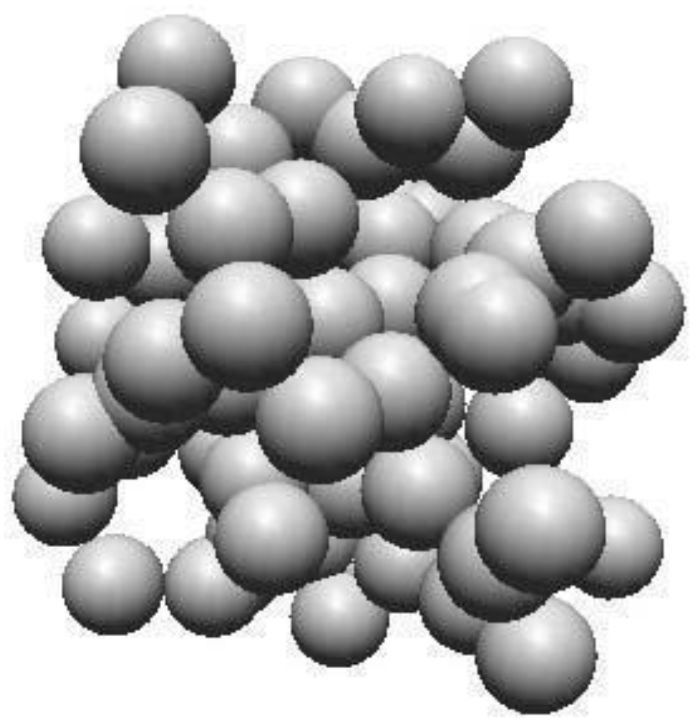
Figure 2.
Primitive unit cell of hexagonal graphite obtained by Materials Project.[17].
Figure 2.
Primitive unit cell of hexagonal graphite obtained by Materials Project.[17].
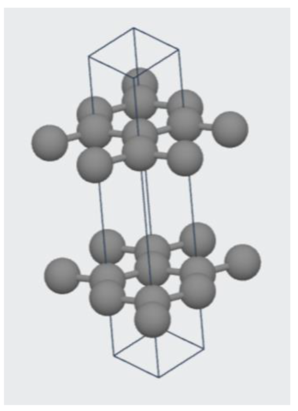
Figure 3.
Band structure and density-of-states plot of hexagonal graphite using DFT calculations done by the Materials Project.[17,18].
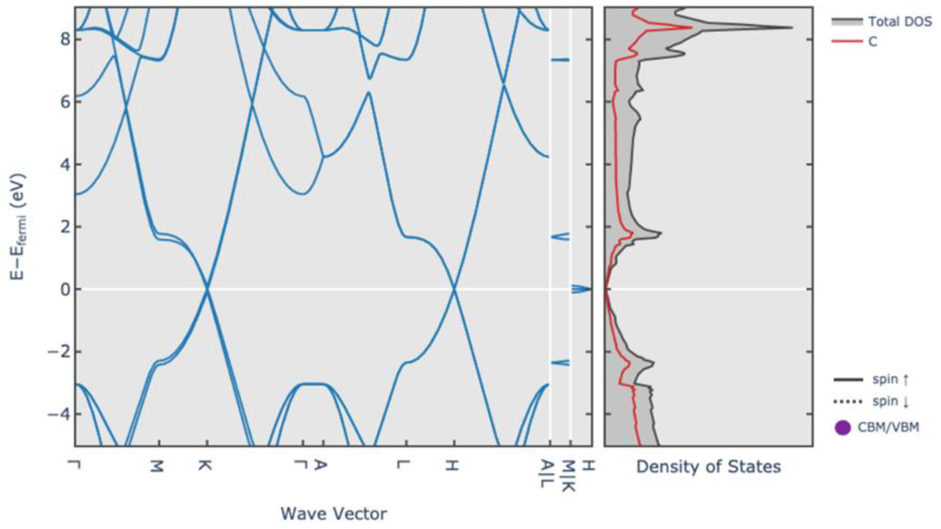
Figure 4.
(a) Ball-and-stick model and (b) Sphere-in-contact model of C80 carbon nanocone with a cone height of 5 Angstroms and a disclination angle of 60 degrees. Image produced in Nanotube Modeler Software.
Figure 4.
(a) Ball-and-stick model and (b) Sphere-in-contact model of C80 carbon nanocone with a cone height of 5 Angstroms and a disclination angle of 60 degrees. Image produced in Nanotube Modeler Software.
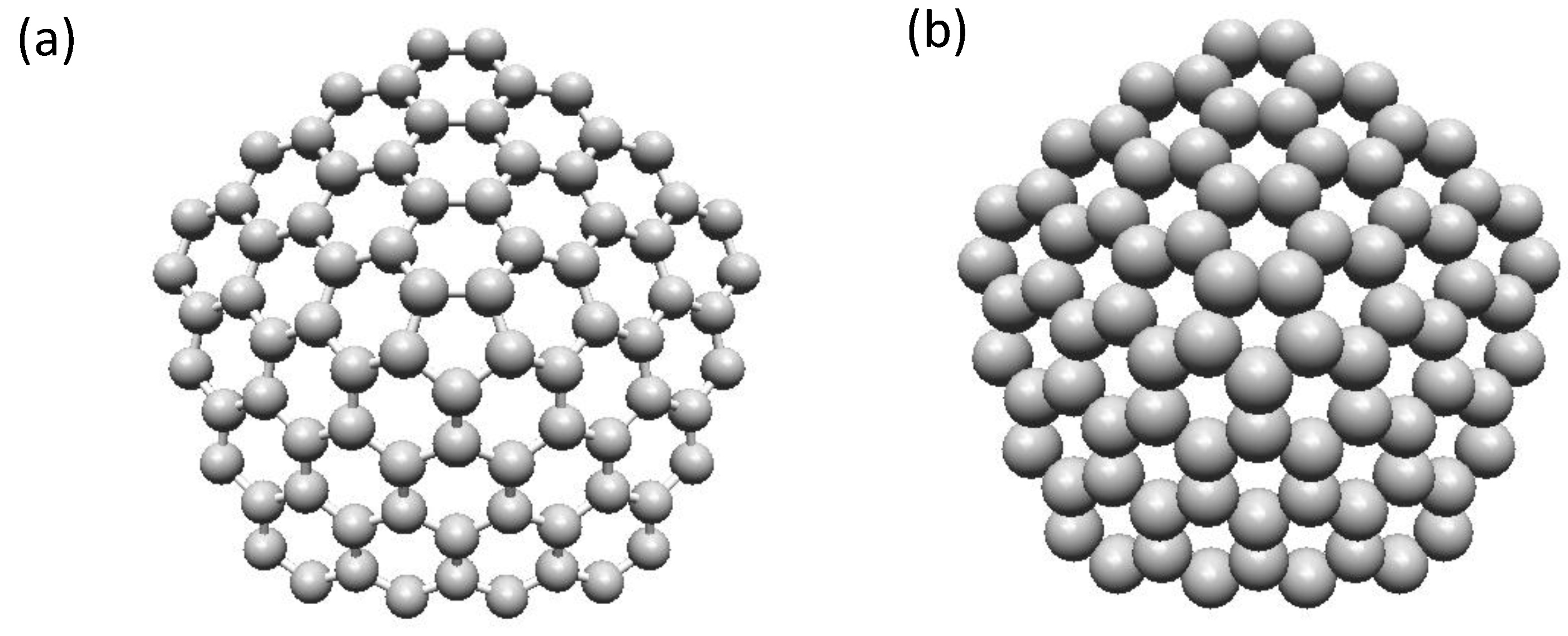
Figure 5.
(a) Ball-and-stick model of fullerene C60, (b) Sphere-in-contact model of fullerene C60, (c) Ball-and-stick model of fullerene C70 D5h and (d) Sphere-in-contact model of fullerene C70 D5h. Image produced in Nanotube Modeler Software.
Figure 5.
(a) Ball-and-stick model of fullerene C60, (b) Sphere-in-contact model of fullerene C60, (c) Ball-and-stick model of fullerene C70 D5h and (d) Sphere-in-contact model of fullerene C70 D5h. Image produced in Nanotube Modeler Software.
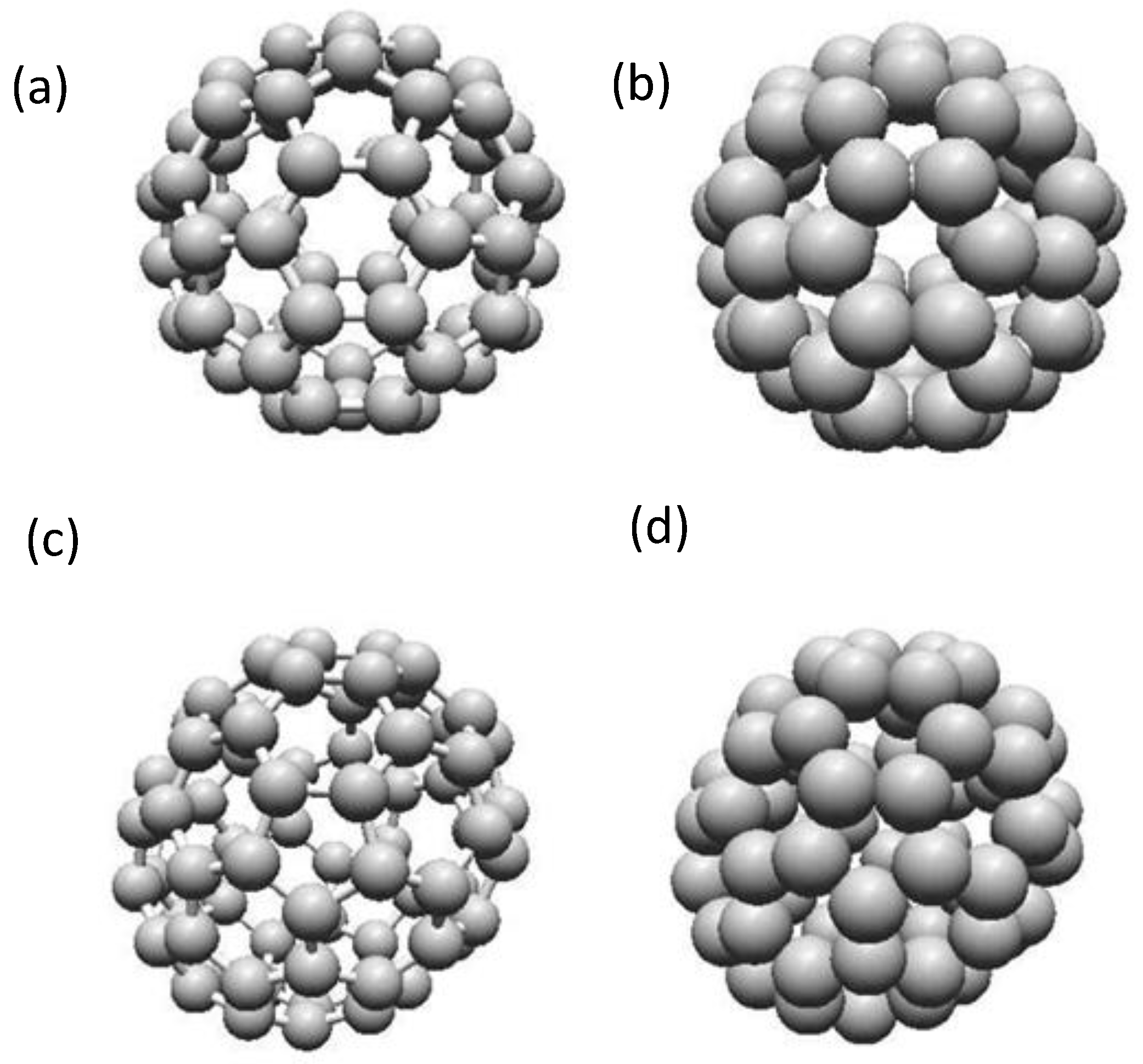
Figure 6.
(a) Ball-and-stick model and (b) Sphere-in-contact model of graphene nanoribbon. Image produced in Nanotube Modeler Software.
Figure 6.
(a) Ball-and-stick model and (b) Sphere-in-contact model of graphene nanoribbon. Image produced in Nanotube Modeler Software.
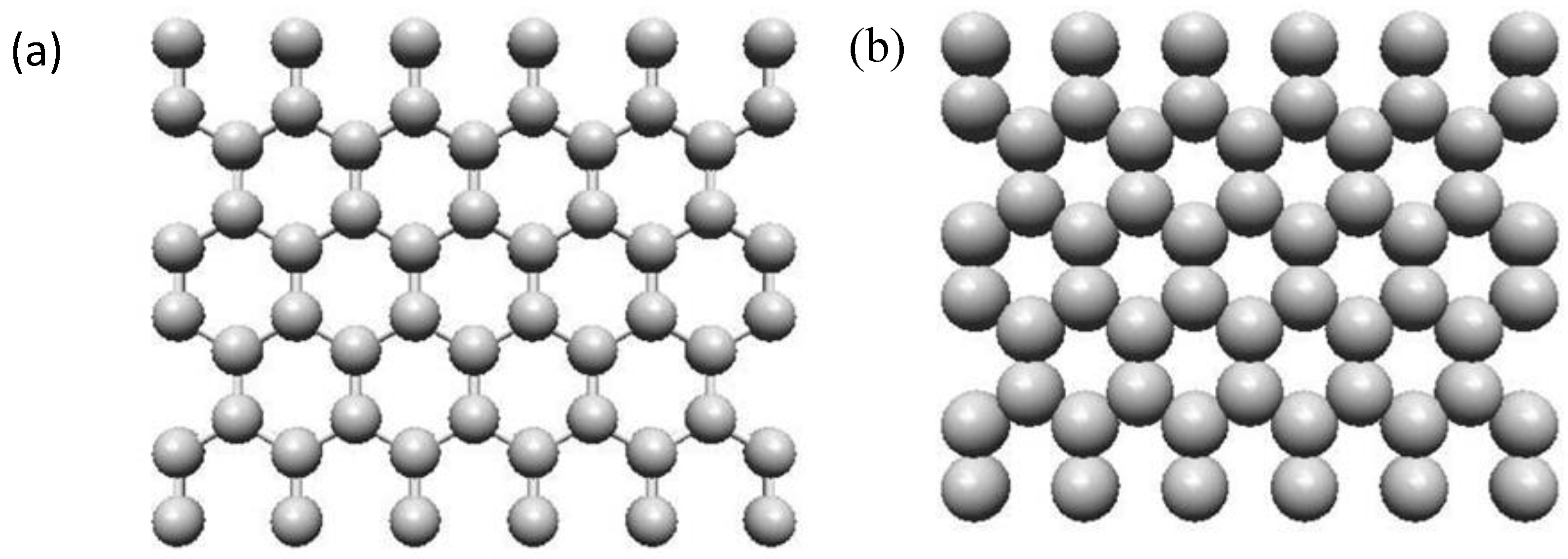
Figure 7.
Structure of graphene oxides showing the various functional groups COOH, C=O, -O-O-, OH, =O and -H at the periphery of a carbon sheet. Image drawn in Chemdraw.
Figure 7.
Structure of graphene oxides showing the various functional groups COOH, C=O, -O-O-, OH, =O and -H at the periphery of a carbon sheet. Image drawn in Chemdraw.
Figure 8.
(a) Ball-and-stick model of C100 SWCNT capped and (b) Sphere-in-contact model of C100 SWCNT capped. Image produced in Nanotube Modeler Software.
Figure 8.
(a) Ball-and-stick model of C100 SWCNT capped and (b) Sphere-in-contact model of C100 SWCNT capped. Image produced in Nanotube Modeler Software.
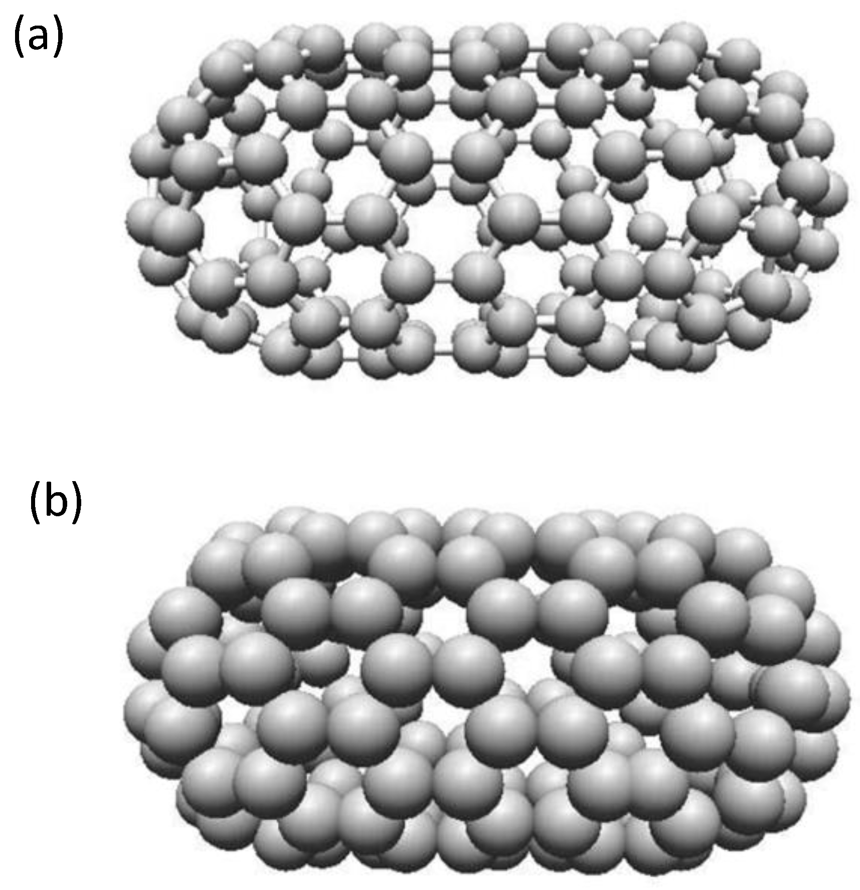
Figure 9.
(a) Ball-and-stick model and (b) Sphere-in-contact model of nanodiamond. Image produced in Nanotube Modeler Software.
Figure 9.
(a) Ball-and-stick model and (b) Sphere-in-contact model of nanodiamond. Image produced in Nanotube Modeler Software.
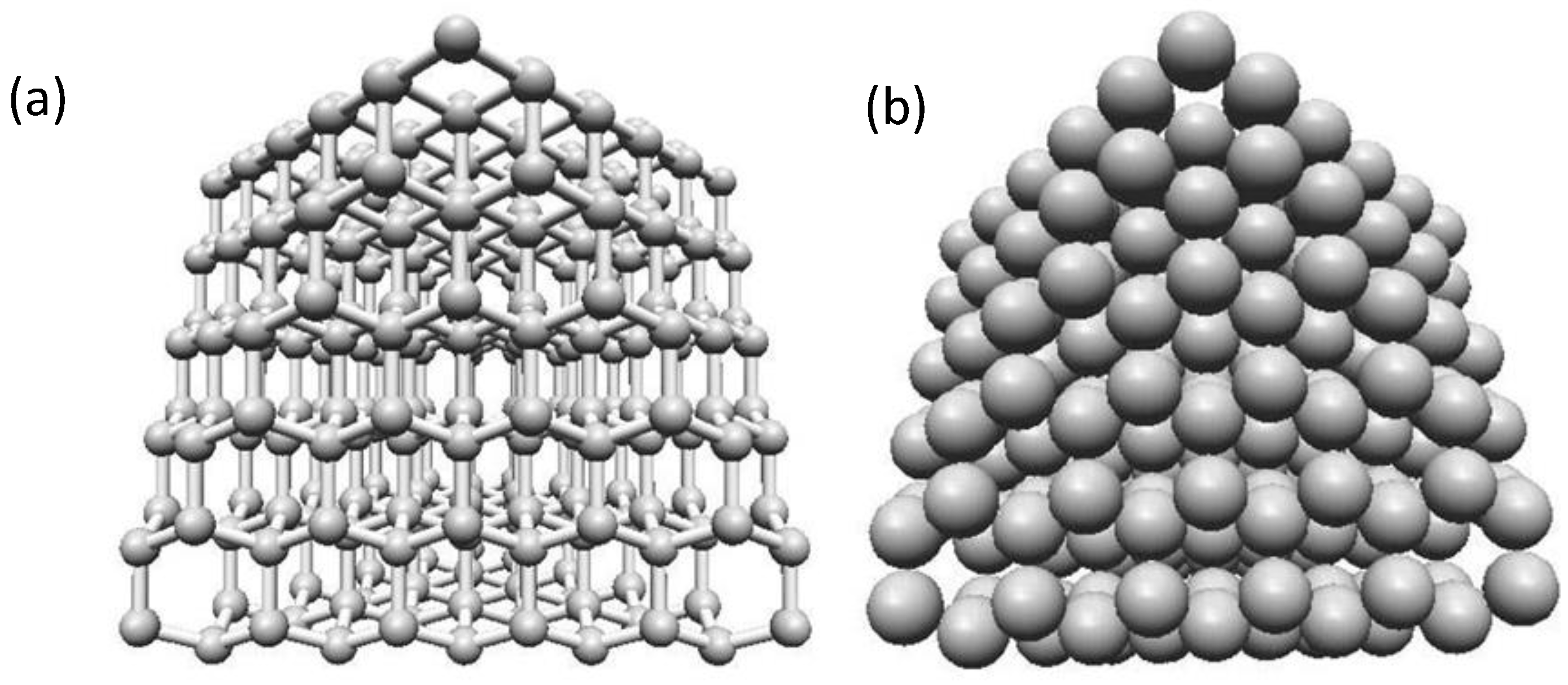

Table 1.
Presents various carbon nanomaterials and terms of their key features, their cost, scalability, purity, relevance to medical applications and challenges in their synthesis.
Table 1.
Presents various carbon nanomaterials and terms of their key features, their cost, scalability, purity, relevance to medical applications and challenges in their synthesis.
| Synthesis Method | Key Features | Cost | Scalability | Purity | Relevance to Medical Applications | Challenges |
|---|---|---|---|---|---|---|
| Chemical Vapor Deposition (CVD) | Produces high-quality carbon nanotubes (CNTs) and graphene with precise control over material properties. | High | Moderate | High | Ideal for biomedical applications requiring high purity, such as biosensors and advanced neural interfaces. | High cost and precise control required for optimal results. |
| Pyrolysis of Hydrocarbons | Thermal decomposition of hydrocarbons (e.g., methane) to produce amorphous carbon or graphene at scale. | Low | High | Moderate | Cost-effective approach for creating nanomaterials for coatings, drug delivery systems, and composite materials. | Impurities and limited control over structure. |
| Laser Ablation | Uses high-intensity laser pulses to vaporize graphite, yielding high-purity graphene or nanotubes. | Very High | Low | Very High | Suitable for advanced medical applications, such as prosthetics, imaging agents, and sensors requiring high purity. | High cost and low scalability limit industrial use. |
| Scotch Tape Technique | Mechanical exfoliation of graphite layers to produce graphene manually in small quantities. | Very Low | Very Low | Very High | Historically significant and used for research-level applications, such as early biosensing studies. | Labor-intensive and impractical for large-scale production. |
| Graphite Intercalation | Inserting molecules (e.g., acids, potassium chlorate) between graphite layers to create graphite oxide. | Moderate | Low to Moderate | Moderate to High | Early method for creating graphene oxide, paving the way for biomedical coatings, functional materials, and composites. | Chemical reactions can generate hazardous byproducts. |
| Electric Arc Discharge | High-energy discharge between graphite electrodes produces carbon nanotubes and fullerene structures. | Moderate | Low | High | Used for imaging and diagnostics due to high-quality nanotubes, but applications are still in research stages. | Limited scalability and energy-intensive process. |
| Laser Vaporization | High-powered lasers vaporize graphite targets to produce nanotubes or graphene nanomaterials. | Very High | Low | Very High | Suitable for specific medical devices requiring precise structural purity and uniformity, such as advanced implants. | Expensive and unsuitable for large-scale production. |
| Solar Energy Focusing | Concentrates solar energy to achieve high temperatures for producing carbon nanomaterials sustainably. | Low | Low to Moderate | Moderate | A promising eco-friendly method for producing materials for drug delivery systems and medical composites. | Limited control over structure and requires constant sunlight. |
| Laser-Induced Melting | Melts and restructures carbon materials using high-intensity lasers, producing novel nanostructures. | High | Low | High | Can create specialized nanomaterials for high-end biomedical applications, such as optical imaging tools. | Requires advanced equipment and expertise. |
Disclaimer/Publisher’s Note: The statements, opinions and data contained in all publications are solely those of the individual author(s) and contributor(s) and not of MDPI and/or the editor(s). MDPI and/or the editor(s) disclaim responsibility for any injury to people or property resulting from any ideas, methods, instructions or products referred to in the content. |
© 2025 by the authors. Licensee MDPI, Basel, Switzerland. This article is an open access article distributed under the terms and conditions of the Creative Commons Attribution (CC BY) license (http://creativecommons.org/licenses/by/4.0/).
Copyright: This open access article is published under a Creative Commons CC BY 4.0 license, which permit the free download, distribution, and reuse, provided that the author and preprint are cited in any reuse.


