Preprint
Article

This version is not peer-reviewed.

The Mitigation of Quercetin on Lead-induced Neuroinflammation in a Rat Model: Changes in Neuroinflammatory Markers, Hippocampal Neurons and Memory

Submitted:

11 February 2025

Posted:

12 February 2025

You are already at the latest version

Abstract

The present study investigated the mitigatory effects of quercetin on neuroinflammation, hippocampal degeneration, and memory deficits in lead (Pb)-exposed rats. Wistar rats were administered orally with quercetin and succimer (standard drug) for 21 days after Pb exposure of 21 days or in combination with Pb for 42 days. Working and reference memory was assessed using an eight-arm radial water maze (8-ARWM). The changes in brain Pb level, the neuronal nitric oxide synthase (nNOS) activity, and the level of neuroinflammatory markers like tumour necrosis factor-alpha (TNF-α) and interleukin 1 Beta (IL-1β) were determined. The number of neurons and astrocyte expression were all evaluated histologically and immunohistochemically, respectively. The brain level of Pb was increased significantly in Pb-exposed rats. In the hippocampus, the number of neurons decreased while the expression of astrocytes increased, and the levels of neuroinflammatory markers increased in Pb-exposed rats. Lead impaired reference and working memory. However, quercetin treatment effectively reduced neuronal loss, improved memory, and inhibited neuroinflammation. In conclusion, quercetin mitigates neuroinflammation, hippocampal degeneration, and memory deficits in Pb-exposed rats. Neuroinflammatory markers negatively correlated with memory function. Thus, quercetin may be a promising therapy in neuroinflammation and memory dysfunction in populations prone to Pb exposure.

Keywords: 
;  ;  ;  

1. Introduction

Neuroinflammation, a pathological state, is the cause of neurodegenerative illnesses like Huntington's, Parkinson's, and Alzheimer's diseases [1,2]. Reports showed that lead as heavy metal mediates neuroinflammatory injury [3,4] due to its ability to activate microglia, resulting in the simultaneous generation of cytokines that promote inflammation to cause the death of neurons [2]. The aggregation of amyloid beta (Aβ), as a result of lead, destroys the support of neurons provided by microglial cells by enhancing reacting oxygen species (ROS) and other toxins [5]. Microglial cells predominantly produce pro-inflammatory cytokines, including interferon-gamma (IFN-γ), interleukin-1 beta (IL-1β), and tumour necrosis factor-alpha (TNF-α) [6]. Furthermore, increased pro-inflammatory cytokines stimulated p38 mitogen-activated protein kinase (p38 MAPK) signaling [7]. Nuclear factor-kappa B (NF-κB), contributing to neuroinflammation, is activated by p38 MAPK [8]. The activated transcription factor NF-κB-induced neuroinflammatory response facilitates neurodegeneration. Thus, it supports the idea that neurodegenerative diseases are mediated by neuroinflammation [9]. Nuclear factor-kappa B negatively regulates nuclear factor erythroid 2-related factor 2 (Nrf2) [10,11,12]. There is evidence that the neuroinflammatory effects of NF-κB influence the activation of the Nrf2 protective mechanism [13,14].
A naturally occurring flavonoid, quercetin, is found in large quantities in vegetables, fruits, and foods, including ginkgo biloba, red wine, onions, and apples [15]. According to Kong et al. [16], quercetin strong anti-inflammatory, antioxidant, and radical-scavenger properties may be used to treat infections, cancer, diabetes, heart disease, and neurological diseases like Alzheimer’s and Parkinson’s [1]. Quercetin prevents the activation of microglia and neurons from inflammatory damage [17,18]. Through quercetin inhibition action on inducible nitric oxide synthase (iNOS) expression, nitric oxide (NO) synthesis is prevented [18,19,20], NF-κB and signal transducer and activator of transcription-1 (STAT1) are inactive, and heme oxygenase-1 or interleukin-10 expression is increased [18,19]. According to studies, quercetin stimulates the expression of Nrf2, controlling the synthesis of glutathione using glutamylcysteine synthase and restoring redox equilibrium [21,22,23]. The suppression of neuroinflammation and neuronal death by quercetin improves learning and memory functions [24,25,26,27]. According to a study conducted in a rat model, quercetin enhances learning and memory via activation of Cyclic-AMP Response Element Binding (CREB) and initiates neurogenesis, compensating for the death of neurons in the brains injected with Aβ1-42 [28]. Administration of 50 mg/kg of quercetin improves spatial memory deficit induced by repeated cerebral ischemia [29].
The neuroprotective role of inflammation from detrimental intrinsic and extrinsic factors has been reported. However, the mechanisms of quercetin on lead-induced neuroinflammation, neurodegeneration and memory dysfunctions, and their correlates needed to be added to the existing literature. Thus, this work examined quercetin’s mitigating roles on neurodegeneration and neuroinflammation in relation to memory performance in rats exposed to lead.

2. Materials and Methods

2.1. Obtainment of Equipment, Reagents, and Chemicals

Quercetin (Q4951-10G) from Sigma-Aldrich, USA, Succimer (D7881-5G) from Sigma-Aldrich, USA), and Lead (II) acetate trihydrate (32307-100G) from Honeywell, Germany, were purchased. The Enzyme-linked immune sorbent assay kits Tumour Necrosis Factor-α and Interleukin-1β were purchased from Fine Biological Technology (Wuhan, Hubei, China).

2.2. Experimental Rats

The study make use of thirty (30) male Wistar rats, reared in the animal house of the Department of Pharmacology at Ahmadu Bello University, Zaria-Nigeria. For two weeks, the rats were kept in clean, well-ventilated plastic cages with a natural light and dark cycle. They were fed a typical laboratory diet and given unlimited access to water.

2.3. Experimental Protocol

The rats received 37% (60 mg/kg) of the oral LD50 of quercetin, which was determined to be 161 mg/kg body weight [30]. The rats received 3.28% (125 mg/kg) of the oral LD50 of Lead (II) acetate trihydrate, determined by the current acute toxicity investigation to be 3808 mg/kg for Wistar rats. By Alan and Miller’s prescription, the rats were given a standard dosage of 10 mg/kg body of Succimer [31].

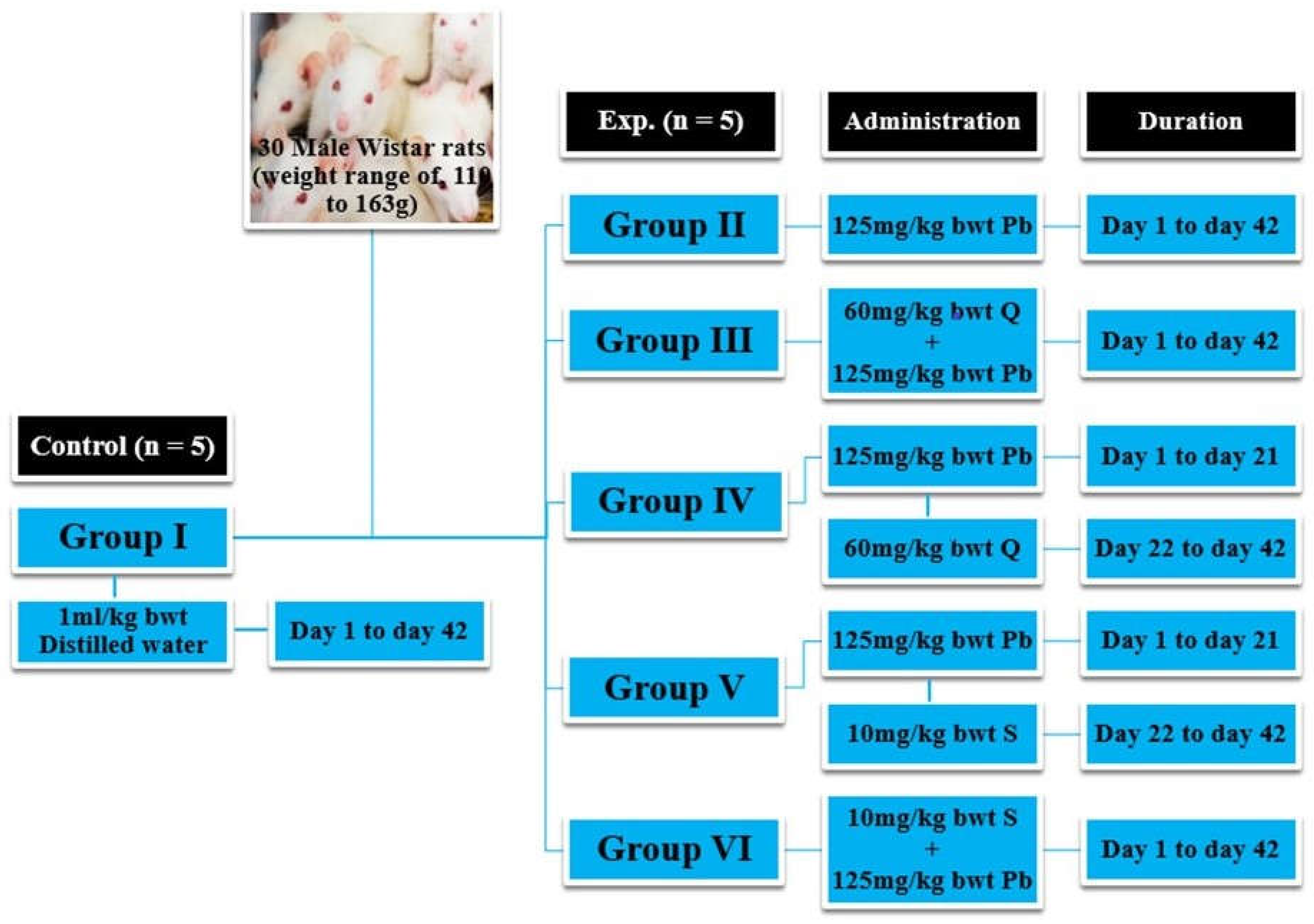
2.4. Experimental Design

A total of thirty (30) male Wistar rats weighing between 110 and 163 g were randomly assigned to six groups (each with n = 5). Group I (control) and Group II (Pb2+) were given distilled water (H2O: 1 ml/kg) and lead (Pb: 125 mg/kg), respectively for 42 days, while Group III (Q + Pb2+ COA) and Group VI (S + Pb2+ COA) received lead (Pb: 125 mg/kg) co-administered (COA) with quercetin (Q: 60 mg/kg) and Succimer (S: 10 mg/kg). Pb: 125 mg/kg was administered to Group IV (Pb2+ + Q TRT) and Group V (Pb2+ + S TRT) for 21 days. Then, Group IV and Group V received Q: 60 mg/kg and S: 10 mg/kg, respectively, for an additional 21 days. For 42 days, the administration was orally and once daily (Figure 1).

2.5. Behavioural Study

Following the previous protocol [32], reference and working memory were evaluated using a radial water maze with eight arms (8-ARWM). Four distinct arms assess reference memory; these arms were never equipped with an escape platform and were maintained constant during testing using additional maze cues. Rats entering these arms thus demonstrated shortfalls in long-term learning and poor reference memory ability. At the start of the day, escape platforms were located in the remaining four arms. But after the rat got onto an arm and escaped the water, each platform was removed [33,34]. In removing each platform throughout the four consecutive trials, the rat had to recall which arms it entered, which increased the amount of working memory required. Re-entries into reference memory arms or arms previously utilised for escape on the same testing day, evaluate working memory. As a result, three dependent variables were used to collect data: (i) Working memory correct errors: first and repeating entries into any arm that had a platform in the past. (ii) Reference memory errors: the number of initial entries into any arm never had a platform. (iii) Working memory incorrect errors: the number of repeated entries into any arm that has never had a platform.

2.6. Animals Sacrifice

The rats were euthanized, and the brains were harvested and split in half. Half of the brain was homogenized in phosphate buffer (pH 7.4), and the homogenate was centrifuged for ten minutes at 3000 rpm to extract aliquots of the supernatant for the estimation of brain-lead, TNF-α, and Il-1β levels. The other half was processed and used for histological and immunohistochemical studies.

2.7. Determination of Brain-Lead Concentration Using Atomic Absorption Spectrophotometry

The Tri-acid, namely, Perchloric acid (HClO4), Sulphuric acid (H2SO4), and Nitric acid (HNO3) were mixed in a ratio of 20 mls: 70 mls: 650 mls. The procedure was conducted in the Soil Science Department, Institution of Agricultural Research (IAR), Ahmadu Bello University, Zaria-Nigeria, according to Garcia-Arenas et al. [35]. The tissue homogenate (0.02 g) was weighed into a 100 mls digestion beaker, and 30 to 40 mls of Tri-acid was added immediately. The beaker was transferred into a hot digestion sand bath and digested for about 1 hour at 150 to 200 oC. The digestion was completed when the red NO2 and white dense fume of perchloric acid (HClO4) ceased to appear. The beaker was removed from the sand bath and allowed the digest to cool for about 15 minutes, and later added drops of distilled water. The digest was transferred into a 30 ml volumetric flask, rinsed into the beaker into the volumetric flask, made up to the required volume with distilled water, cork and shaken vigorously. The analysis was performed using Atomic Absorption Spectrophotometry (model number PG500) to determine the brain-lead level.
Calculation for Actual concentration:
A c t u a l   c o n c e n t r a t i o n   i n   p p m = A A S   r e a d i n g   x   D v W t (mg.kg-1) Where, Dv = final digestion volume, Wt = weight of sample used.

2.8. Measurement of Tumour Necrosis Factor Alpha Level

An enzyme-linked immune-sorbent assay (ELISA) for rat TNF-α was used to determine the level of TNF-α. As directed by the manufacturer, the procedure was carried out at the KAYOMEG Diagnostic Centre in Kaduna, Nigeria. Sandwich enzyme-linked immune-sorbent assay technology served as the basis for the principle. The biotin-conjugated anti-TNF-α antibody was employed as a detection antibody after the anti-TNF-α antibody was pre-coated onto plates. Before adding the standard, samples, and control (zero) wells, the plate was washed twice. Each well received 100 μL of the homogenates, and incubated at 37 °C for 90 minutes. After aspirating the solution, the plates were washed two times. Each well received around 100 μL of biotin-labeled antibody working solution, and then incubated at 37 °C for 60 minutes. After aspirating the solution, the plates underwent three rounds of washing. Each well received 100 μL of SABC Working Solution, which was then incubated at 37 °C for 30 minutes. After aspirating the solution, the plates underwent five rounds of washing. After adding roughly 90 μL of TMB Substrate, the mixture was incubated at 37 °C for 15 to 30 minutes. Stop solution of 50μL was added. The samples’ concentrations were determined using the standard curve after the O.D. absorbance at 450 nm was promptly measured in a microplate reader.

2.9. Measurement of Interleukin 1 Beta Level

The level of IL-1β was determined using an enzyme-linked immune-sorbent assay (ELISA) kit for rat Interleukin 1 Beta (IL-1β). As directed by the manufacturer, the procedure was carried out at the KAYOMEG Diagnostic Centre in Kaduna, Nigeria. Sandwich enzyme-linked immune-sorbent assay technology served as the basis for the principle. The biotin-conjugated anti-IL-1β antibody was employed as a detection antibody after the anti-IL-1β antibody was pre-coated onto plates. Before adding the standard, samples, and control (zero) wells, the plate was washed twice. Each well received 100 μL of the homogenates, which were then incubated at 37 °C for 90 minutes. After aspirating the solution, the plates were washed two times. Each well received around 100 μL of biotin-labeled antibody working solution, and then incubated at 37 °C for 60 minutes. After aspirating the solution, the plates underwent three rounds of washing. Each well received 100 μL of SABC Working Solution, which was then incubated at 37 °C for 30 minutes. After aspirating the solution, the plates underwent five rounds of washing. After adding roughly 90 μL of TMB Substrate, the mixture was incubated at 37 °C for 15 to 30 minutes. Stop solution of 50μL was added. The samples’ concentrations were determined using the standard curve after the O.D. absorbance at 450 nm was promptly measured in a microplate reader.

2.10. Histological Study and Histopathological Assessment

Following the procedures of Sheehan and Hrapchak [36], the hippocampus was processed and stained with Haematoxylin and Eosin, and the sections were viewed using a light microscope. The photomicrographs were captured with a digital Am scope (MD-900) microscope camera. By our previous procedures [37], pyramidal neurons in the CA1 and CA3 regions of the hippocampus were counted using Digimizer image analysis software.

2.11. Immunohistochemical Study

According to the method of Cesar et al. [38], the Glial fibrillary acidic protein (GFAP)-immunohistochemical staining was performed in Histopathology Lab, Ahmadu Bello University Teaching Hospital, Zaria-Nigeria. To inhibit endogenous peroxidase activity, each hippocampus’s free-floating sections was treated for ten minutes using a 0.3% hydrogen peroxide solution (Merck, Germany). They were then treated for 10 minutes with 1% Triton x-100 (Sigma) in PBS. Incubation with 5% bovine serum albumin (BSA; Amersham Biosciences, Buckinghamshire, UK) and 1% Triton X-100 in PBS (BSA-TXPBS) for two hours at room temperature inhibited non-specific binding sites. Rabbit polyclonal antibodies against GFAP (Biomeda, Foster City, CA, USA) were diluted 1:500 in BSA-TX-PBS and incubated on the floating sections for 48 hours at 4 oC. The sections were then treated with biotinylated anti-rabbit IgG (DAKO, Carpinteria, CA, USA) for one hour at room temperature. The sections were incubated with streptavidin-horseradish peroxidase (DAKO) at room temperature for an hour. The diaminobenzidine reaction (DAKO) was used to visualise immunoreactivity. In between phases, three 10-minute washes in 0.1 M PBS were conducted. After mounting the sections on slides coated with poly-L-lysine (Sigma), they were allowed to air dry, cleared with xylene, and covered with entellan (Merck). A microscope equipped with an MD900 Am scope was used to view the slides, and photomicrographs of each experimental group were obtained at a magnification of X 250.

2.12. Quantitative Assessment of Expression of Astrocytes

The percentage areas covered by astrocytes in relation to the background of GFAP immunohistochemical stained hippocampal sections were calculated using Image J as described by Small et al. [39]. The snapped micrograph (Mag x 250 and x 400) was uploaded into the image area of the software, converted to 8-bit type of image followed by setting of a threshold, and transformed into an auto threshold. From the final image, the area covered by astrocytes (black) against white background were measured as the expression of astrocytes (in %).

2.13. Data Analysis

The data was presented as mean plus or minus standard error of the mean (mean ± SEM). The mean differences were compared using a one-way analysis of variance (ANOVA), and the Tukey post-hoc test was performed, if required. A p-value less than 0.05 (p < 0.05) was considered to be significant. A correlation analysis was carried out. GraphPad Prism, Version 8.0.1 (244), was used to analyse the data and plot the graphs.

3. Results

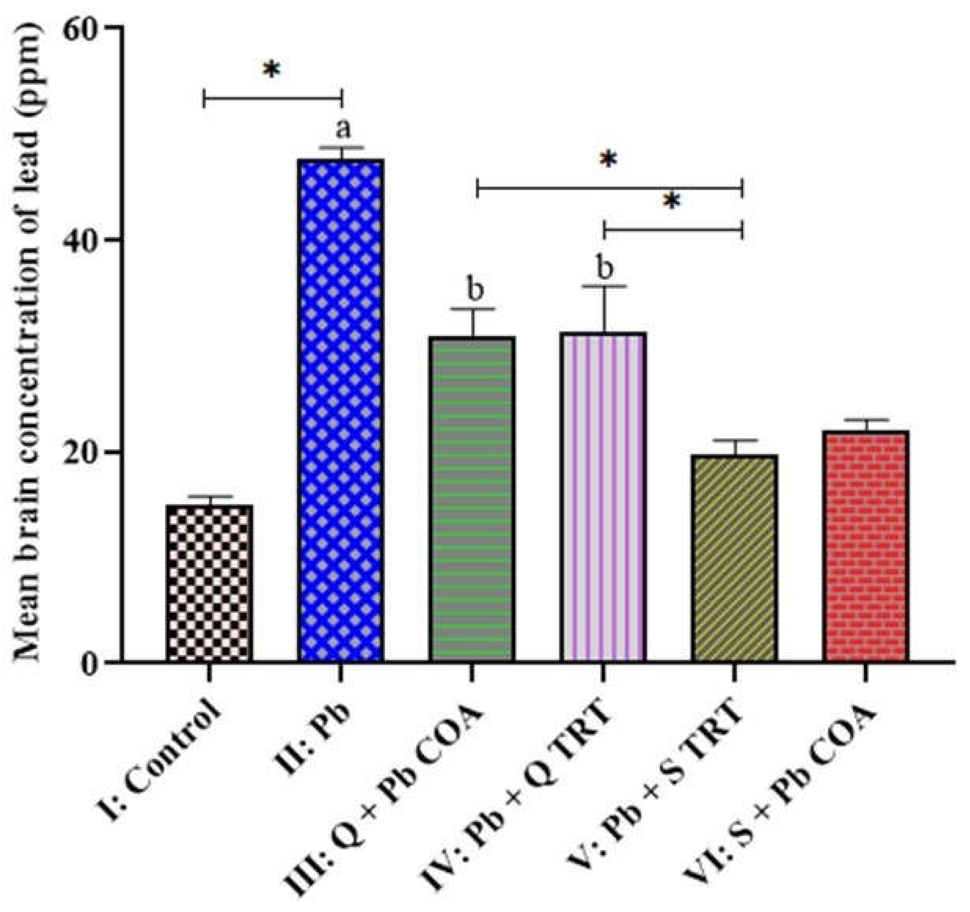
3.1. Brain-Lead Levels Across the Groups

The mean brain-lead concentration in lead-exposed group II (47.75 ± 0.99) was higher than the control group I (15.02 ± 0.77) at p < 0.05. The mean brain-lead concentrations in groups III (30.94 ± 2.56), IV (31.31 ± 4.33), V (19.68 ± 1.42) and VI (22.03 ± 1.01) were lower than group II (47.75 ± 0.99) at p < 0.05. The mean brain-lead concentrations in groups III (30.94 ± 2.56) and IV (31.31 ± 4.33) were higher than group V (19.68 ± 1.42) at p < 0.05 (Figure 2).

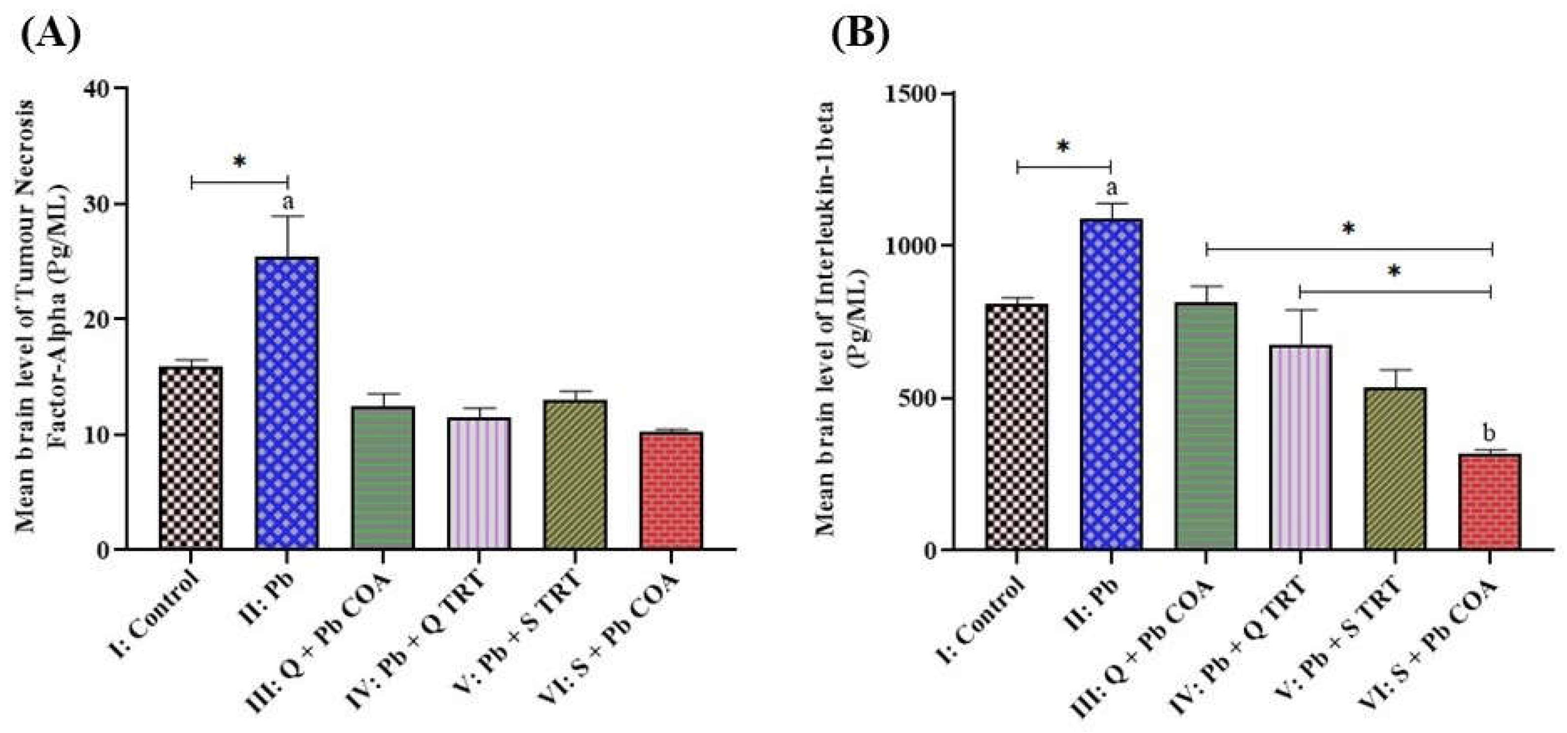
3.2. Quercetin Attenuated the Lead-Induced Increased Levels of TNF-α and IL-1β in the Brain of Wistar Rats

The study revealed that the mean brain level of tumour necrosis factor-alpha (TNF-α: Figure 3A) in group II (25.50 ± 3.44) was higher than group I (15.87 ± 0.63) at p < 0.05. The mean levels of the TNF-α in groups III (12.45 ± 1.13), IV (11.55 ± 0.79), V (12.98 ± 0.79) and V (10.25 ± 0.22) were lower than group II (Pb2+) at p < 0.05. Similarly, the mean brain level of Interleukin-1Beta (IL-1β: Figure 3B) in group II (1088.00 ± 50.33) was higher than group I (806.90 ± 22.25) at p < 0.05. The mean levels of IL-1β in groups III (816.30 ± 50.30), IV (677.60 ± 111.30), V (533.50 ± 58.79) and VI (320.30 ± 9.89) were lower than group II (1088.00 ± 50.33) at p < 0.05. The mean levels of IL-1β in group VI (320.30 ± 9.89) was lower than groups I (control), III (816.30 ± 50.30) and IV (677.60 ± 111.30) at p < 0.05.

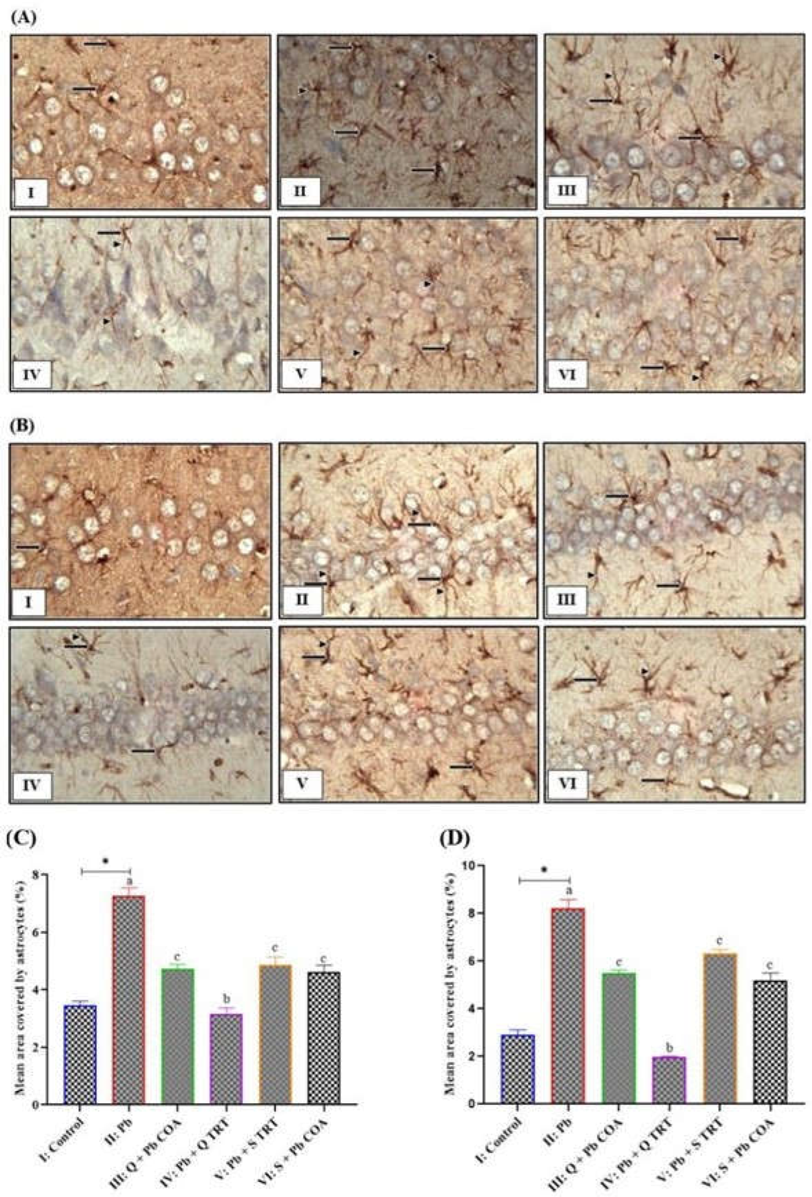
3.3. Quercetin Inhibited the Lead-Induced Increased Astrocyte Expression in CA3 and CA1 Regions of Hippocampus

Immunohistochemically, the anti-GFAP antibody revealed the protective effects of quercetin against lead-induced neurotoxicity in a rat model. Quercetin treatment inhibits GFAP-positive astrocytes in the hippocampal CA3 and CA1 regions (Figure 4A-D). The sections from Figures 4A (III-VI) and 4B (III-VI) showed few GFAP-positive astrocytes compared to 4A (II) and 4B (II), respectively, with abundant GFAP-positive astrocytes (arrows) with dark-brown cytoplasmic reaction with more branched which were longer and had thicker processes (arrowheads). The mean percentage area covered by astrocytes in the hippocampal CA3 region (Figure 4C) was higher in group II (7.28 ± 0.27) than group I (3.47 ± 0.15) at p < 0.05. The mean percentage area covered by astrocytes in groups III (4.74 ± 0.15), IV (3.17 ± 0.21), V (4.86 ± 0.29) and VI (4.62 ± 0.24) were lower than group II (7.28 ± 0.27) at p < 0.05. The mean percentage area covered by astrocytes in groups III (4.74 ± 0.15), V (4.86 ± 0.29) and VI (4.62 ± 0.24) were higher group I, at p < 0.05. The mean percentage area covered by astrocytes in group IV (3.17 ± 0.21) was lower than group V (4.86 ± 0.29) at p < 0.05. The mean percentage area covered by astrocytes in the hippocampal CA1 region (Figure 3D) in group II (8.22 ± 0.36) was higher than in group I (2.90 ± 0.22) at p < 0.05. The mean percentage area covered by astrocytes in groups III (5.49 ± 0.14), IV (1.97 ± 0.04), V (6.34 ± 0.15) and VI (5.20 ± 0.30) were lower than group II (8.22 ± 0.36) at p < 0.05. The mean percentage area covered by astrocytes in groups III (5.49 ± 0.14), V (6.34 ± 0.15) and VI (5.20 ± 0.30) were higher than group I at p < 0.05. The mean percentage area covered by astrocytes in group IV (1.97 ± 0.04) was lower than group V (6.34 ± 0.15) at p < 0.05.

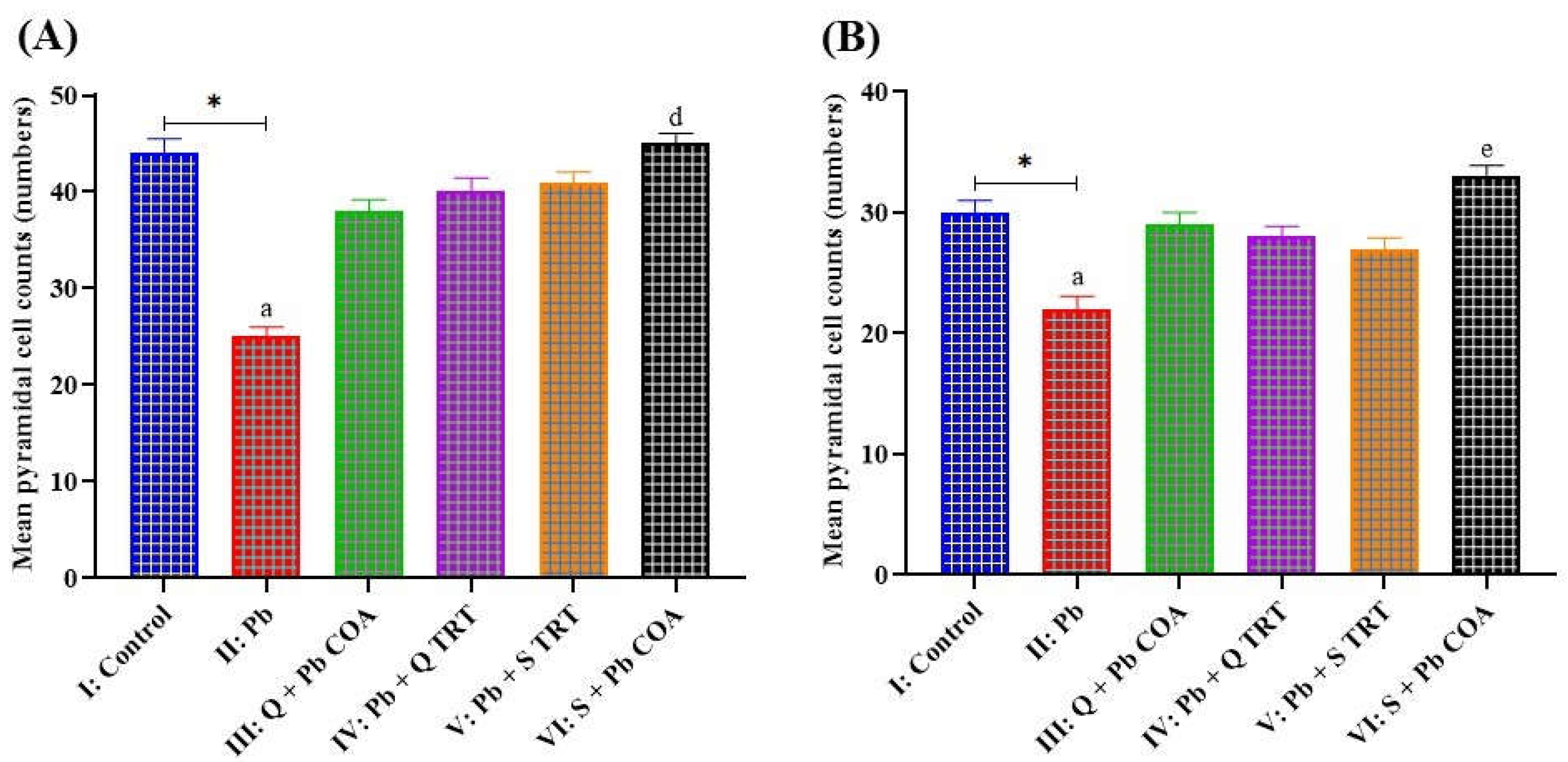
3.4. Lead-Induced Neuronal Degeneration Prevented by Quercetin in of the Hippocampal CA1 and CA3 Regions

The results (Figure 5A) demonstrated decrease in mean Pyramidal cell counts of CA1 region of the hippocampus in groups II (25.00 ± 1.00) and III (38.00 ± 1.18) when compared to group I (44.00 ± 1.52). There was increase in mean Pyramidal cell counts of CA1 in groups IV (40.00 ± 1.41), V (41.00 ± 1.05) and VI (45.00 ± 1.05) when compared to group II (25.00 ± 1.00) at p < 0.05. There was increase in mean Pyramidal cell counts of the CA1 region in group VI (45.00 ± 1.05) when compared to group III (38.00 ± 1.18) at p < 0.05. The results (Figure 5B) showed decrease in mean Pyramidal cell counts of the CA3 region of the hippocampus in group II (22.00 ± 1.10) when compared to group I (30.00 ± 1.00) at p < 0.05. There was increase in mean values of Pyramidal cell counts of the CA3 region in groups III (29.00 ± 1.00), IV (28.00 ± 0.89), V (27.00 ± 0.89) and VI (33.00 ± 0.89), when compared to group II (22.00 ± 1.10) at p < 0.05. There was increase in mean Pyramidal cell counts of CA3 in group VI (33.00 ± 0.89) when compared to groups IV (28.00 ± 0.89) and V (27.00 ± 0.89) at p < 0.05.

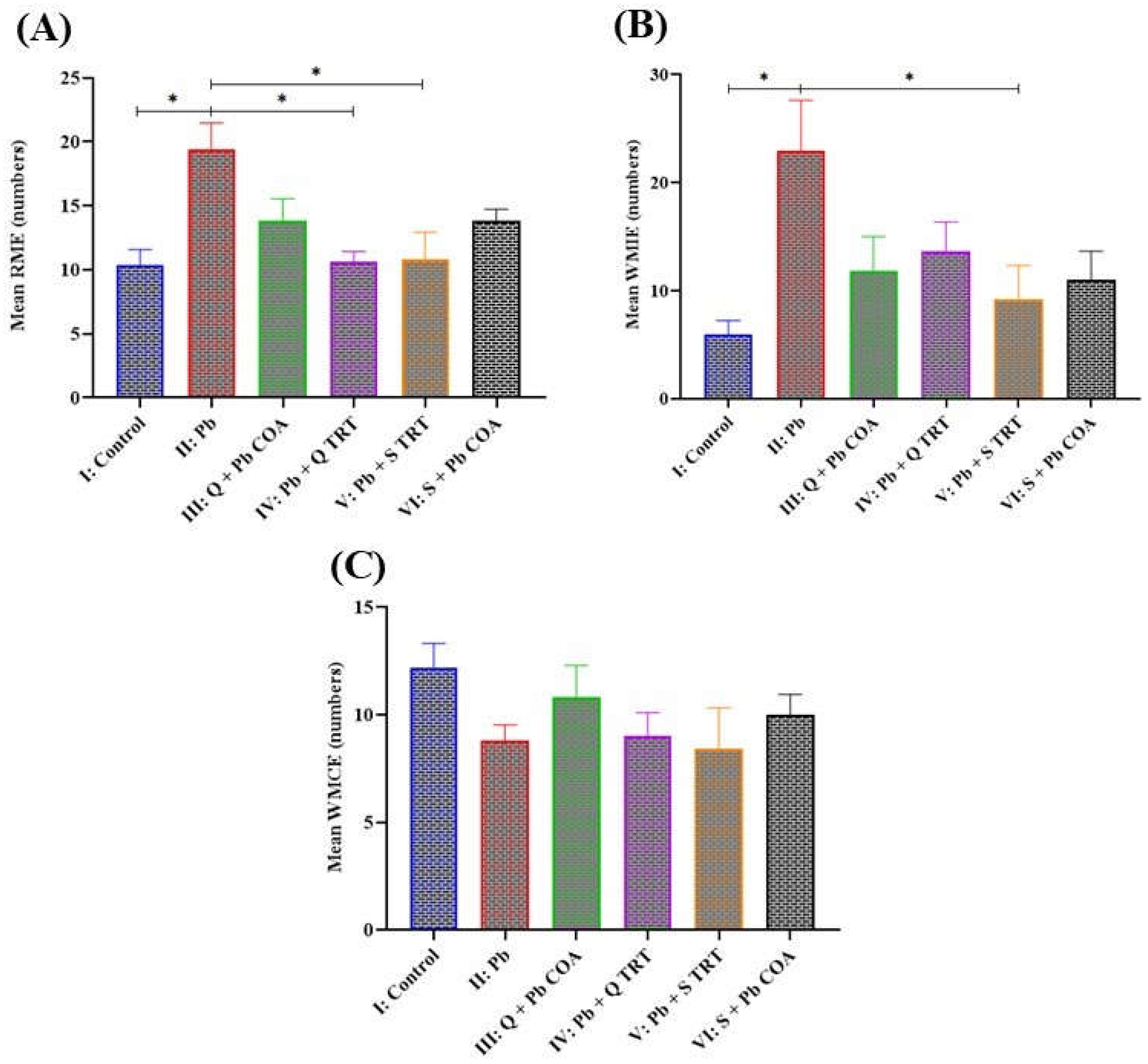
3.5. Quercetin Improved Reference and Working Memory in Lead-Induced Memory Deficits

The results (Figure 6A) showed increase in mean reference memory errors in group II (19.40 ± 2.06) when compared to groups I (10.40 ± 1.17), IV (10.60 ± 0.81) and V (10.80 ± 2.13) at p < 0.05. However, the results revealed no difference in the mean reference memory errors when comparing group II (19.40 ± 2.06) with groups III (13.80 ± 1.74) and VI (13.80 ± 0.92), p > 0.05. The results (Figure 6B) of mean working memory incorrect errors indicated higher errors in group II (23.00 ± 4.63) than groups I (6.00 ± 1.23) and V (9.20 ± 3.14) at p < 0.05. The mean working memory correct errors revealed no differences across the groups, p > 0.05 (Figure 6C).

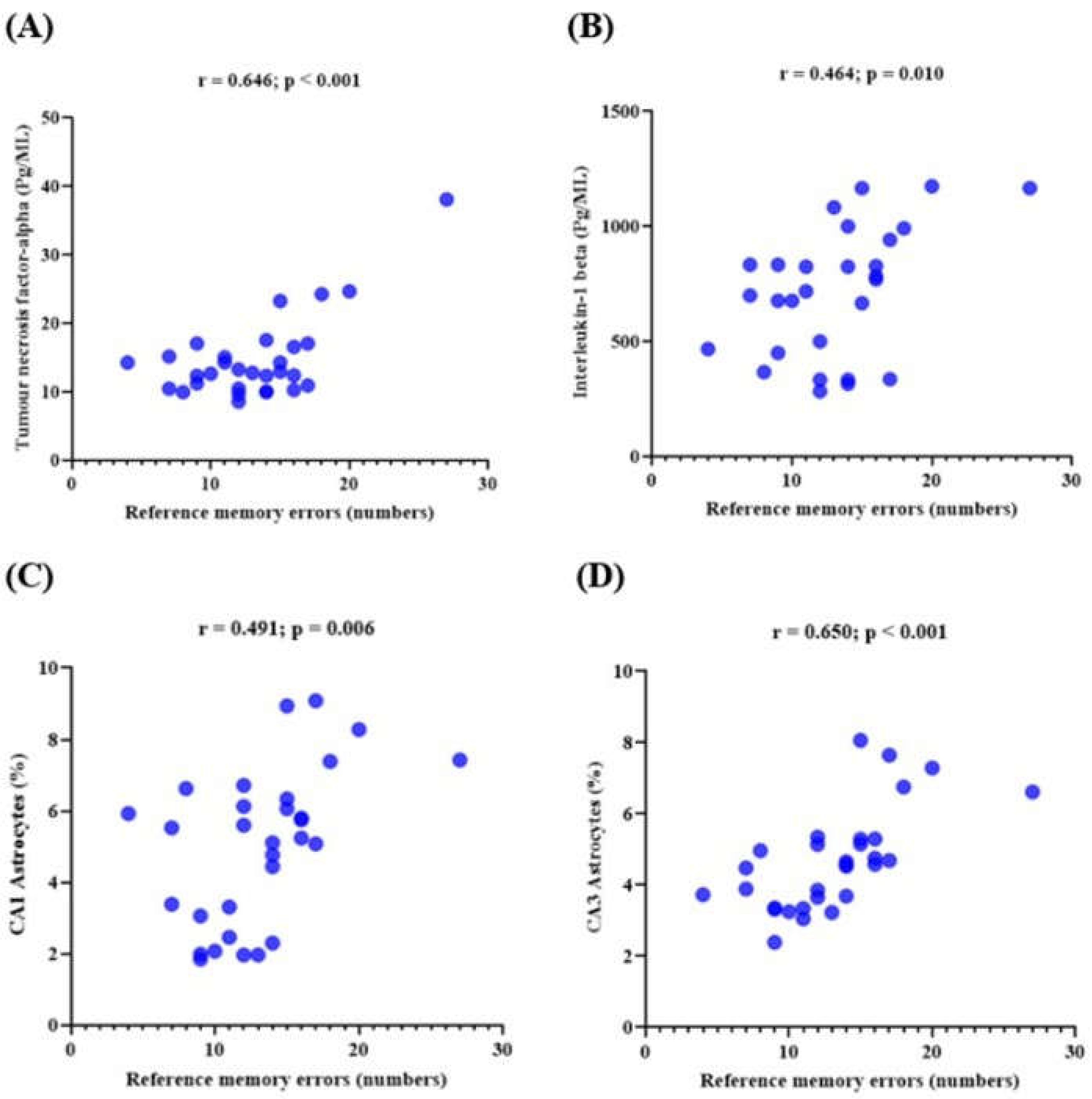
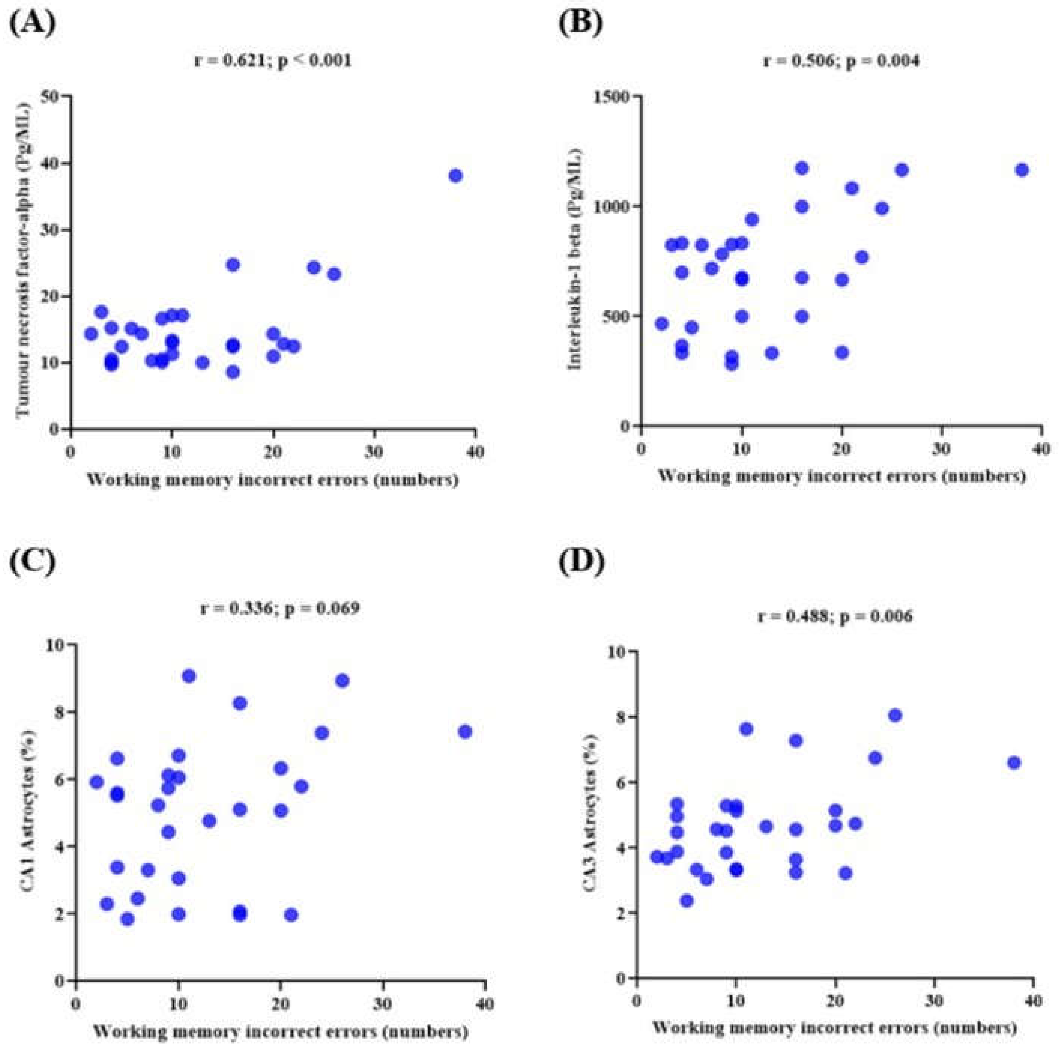
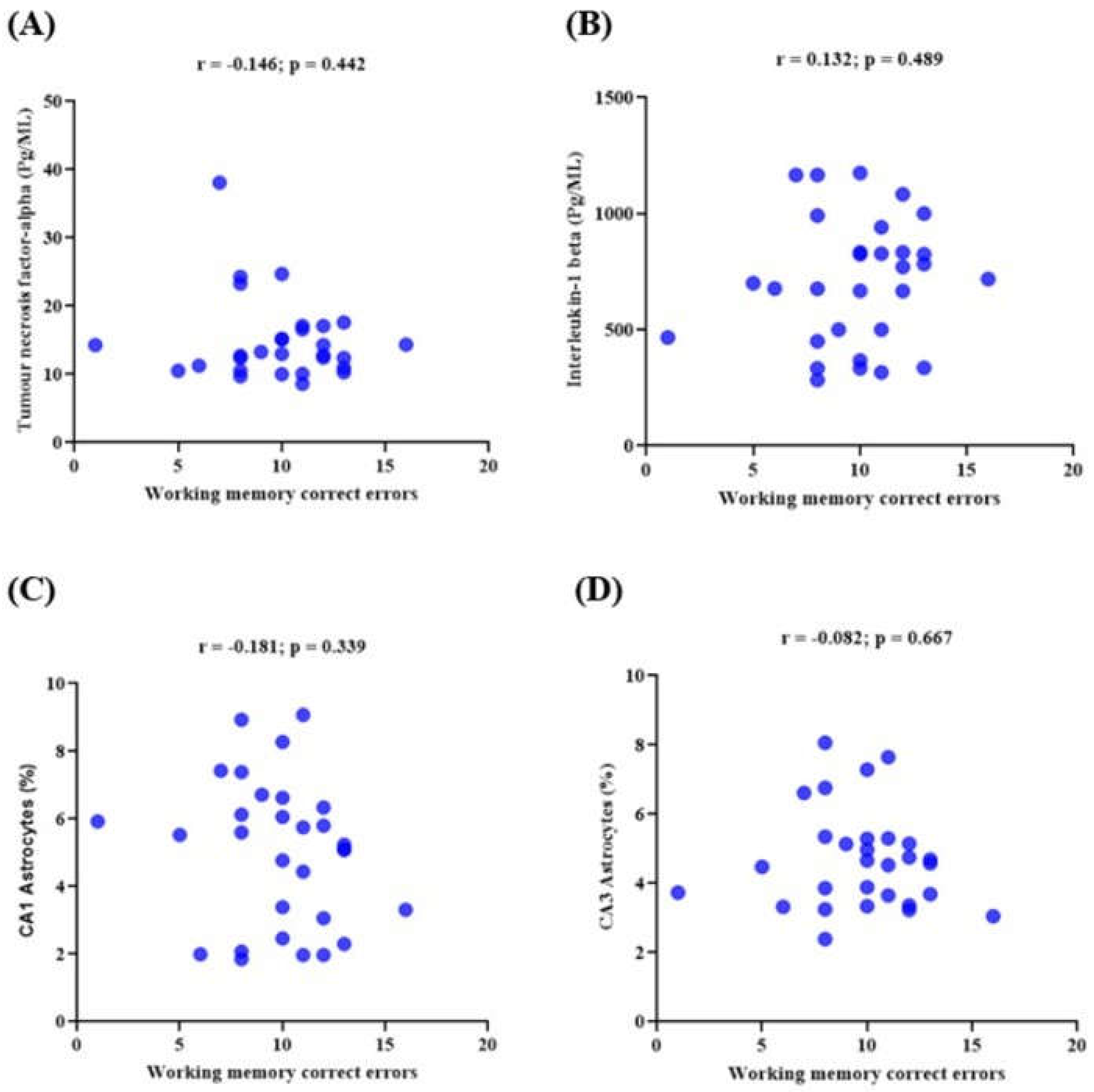
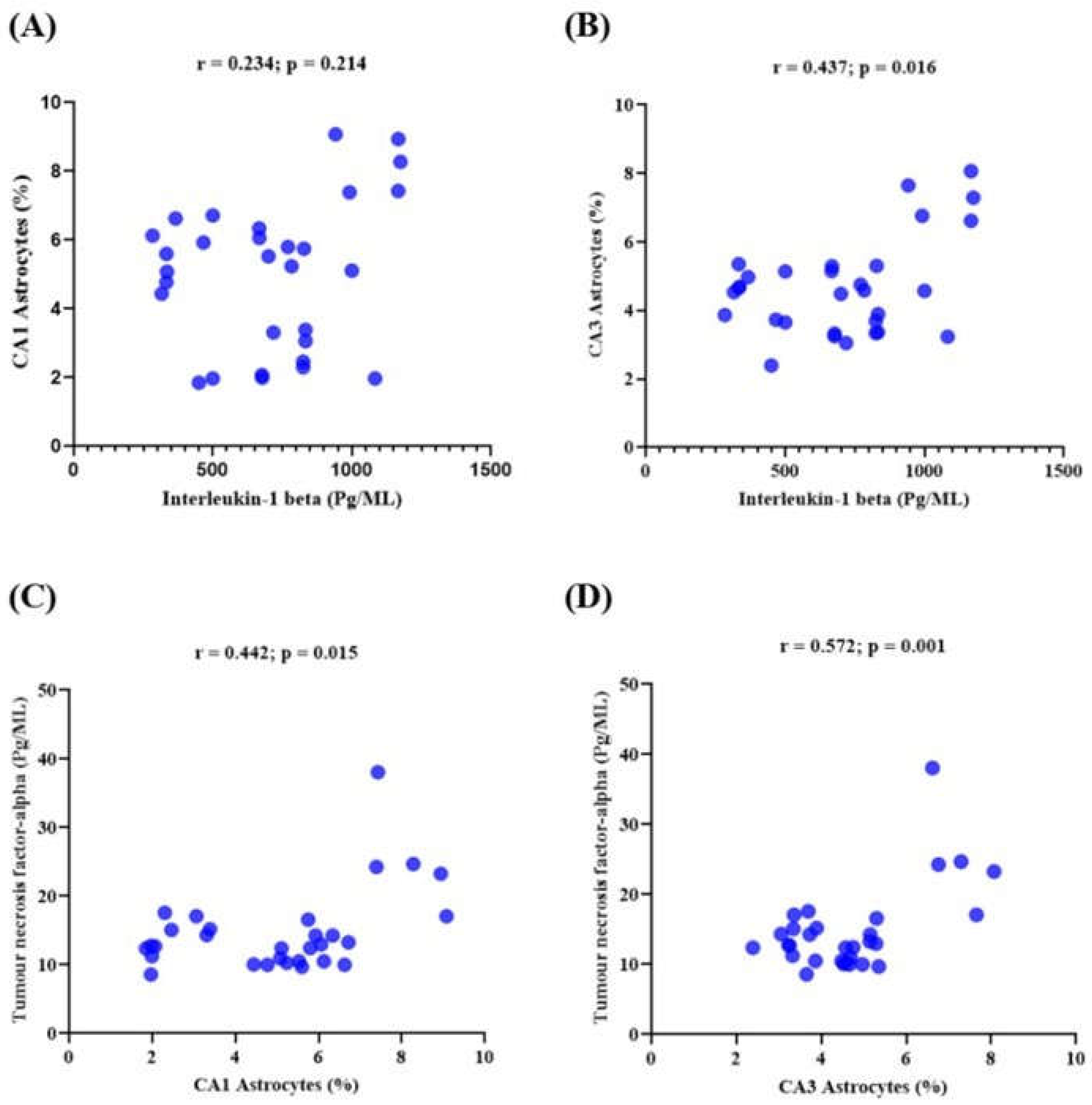
3.6. Memory Performance and Its Correlations with the Expression of Neuroinflammatory Markers in Lead-Induced Neuroinflammation

The results (Figure 7: A-D) showed Correlations of reference memory errors with proinflammatory cytokines and astrocyte expression. The reference memory errors positively correlated with tumour necrosis factor-alpha (r = 0.646; p < 0.001), interleukin-1 beta (r =0.464; p = 0.010) and astrocytes expression in hippocampal CA1 (r = 0.491; p = 0.006) and CA3 (r = 0.650; p < 0.001) regions. The results (Figure 8: A-D) revealed correlations between working memory incorrect errors with pro-inflammatory cytokines and astrocyte expression. The working memory incorrect errors correlated positively with tumour necrosis factor-alpha (r = 0.621; p < 0.001), interleukin-1 beta (r = 0.506; p = 0.004) and astrocytes expression in CA3 (r = 0.488; p < 0.006) region of the hippocampus. There is no significant correlation between incorrect working memory errors and astrocyte expression in hippocampal CA1 region. The results (Figure 9: A-D) showed correlations of working memory correct errors with pro-inflammatory cytokines and astrocyte expression. The working memory correct errors insignificantly correlated with tumour necrosis factor-alpha (r = -0.146; p = 0.442), interleukin-1 beta (r = 0.132; p = 0.489) and astrocytes expression in hippocampal CA1 (r = -0.181; p = 0.339) and CA3 (r = -0.082; p = 0.667) regions. The results (Figure 10: A-D) also showed the correlation of pro-inflammatory cytokines with astrocyte expression. The CA3 astrocyte expression correlated with interleukin-1 beta (r = 0.437; p = 0.016). The CA1 astrocyte expression insignificantly correlated with interleukin-1 beta (r = 0.234; p = 0.214). The CA3 and CA1 astrocyte expression were correlated (r = 0.572; p = 0.001) and (r = 0. 442; p = 0.015) respectively with tumour necrosis factor-alpha.

4. Discussion

Many fruits, vegetables, and foods, including red wine, onions, apples, and ginkgo biloba, contain quercetin, a naturally occurring flavonoid [15]. Due to its strong antioxidant, anti-inflammatory, and radical scavenging properties, quercetin has been used to treat cancer, infection, diabetes, heart disease, and neurological disorders [1,16]. In this work, the effects of quercetin on neuronal degeneration and neuroinflammation and their correlation with memory performance in lead-exposed rats were examined. The results demonstrated that rats exposed to lead had significantly higher brain-lead levels compared to the control group and groups treated with quercetin. This result is consistent with Kang et al. [40], who similarly noted increased lead levels in the brain tissues of rats given lead treatment, emphasising the brain-lead levels significantly correlated with the administered doses. Because lead can infiltrate the blood-brain barrier (BBB) and replace calcium ions (Ca2+) [41], it can readily enter developing neural tissue [42]. This property is shared by many of lead’s neurotoxic effects. Following the administration of quercetin [43], the primary active ingredient of Psidium guajava, acts as an agent to lower the amount of lead and its effects in rat brain tissues [44]. Quercetin treatment dramatically reduced the lead content in the current investigation. Previously, mice exposed to cadmium (Cd) showed comparable outcomes [45]. Quercetin may interact with reduced forms of transition metals, which produce free radicals [46]. Furthermore, during lead (Pb)-induced toxicity in rats, it was established that the quercetin structure’s numerous hydroxyl (OH) groups mediate its metal-chelating activities [47]. Quercetin’s chelating properties have the potential to lower metal toxicity and bioavailability [46].
Lead exposure has been associated with activated microglial and astroglial cells, followed by neuroinflammatory injury, mark morphological changes, and increased synthesis of cytokines [48,49,50], including interleukin-1 beta (IL-1β), tumour necrosis factor-alpha (TNF-α), and interferon-gamma (IFN-γ) [6]. In the current investigation, TNFα and IL-1β levels were considerably higher in rats exposed to lead. This result aligns with previous studies that demonstrated elevated levels of TNF-α and IL-β in the cerebral cortex and hippocampal regions following exposure to lead [51]. Lead-mediated Aβ aggregation in the brain may increase ROS and other toxins in activated microglial cells, which could affect the glial support of neurons [5]. Proinflammatory cytokines are released by activated microglia [52], which in turn activates the signalling of p38 mitogen-activated protein kinase (p38 MAPK) [7]. The activation of p38 MAPK induces nuclear factor-kappa B (NF-κB), contributing to neuroinflammation-mediated neurodegeneration [8,9]. Nuclear factor E2-related factor 2 (Nrf2) is negatively influenced by NF-κB [10,11,12]. Reports showed that iNOS, cyclooxygenase-2 (COX-2), TNFα, and IL-6 levels rise in addition to microglia activation in Nrf2-deficient rats [53]. Nonetheless, the current results showed that quercetin treatment reduced TNF-α and IL-1β levels, mitigating lead-induced neuroinflammation in rats. In a rat model of neonatal streptozotocin-mediated diabetic neuropathy, quercetin dramatically reduced the levels of TNF-α and IL-1β, according to a study by Kandhare et al. [54]. One of the various ways quercetin reduces inflammation is by crossing the blood-brain barrier [55,56]. Quercetin mitigates the NF-κB transcription factor, regulating the proinflammatory molecules’ expression, and inhibits the activity of cellular proteins implicated in the inflammatory response [57,58], all of which prevent neuronal death [59].
This study revealed that quercetin treatment significantly reduced lead-induced higher expression of astrocytes in lead-exposed rats. The increase in the proportion of astrocyte-covered area relative to the background of GFAP-immunohistochemically stained hippocampal slices using Image J analysis software is evidence of an increase in astrocyte expression. Astroglia collects and retains lead [60], creating a reservoir for continuous lead release and making the glia more hazardous [61]. Reactive gliosis is a pathological condition in which glial cells rapidly alter and express more GFAP [62]. Lead mediates astrocyte hypertrophy and proliferation [63], in addition to astrogliosis, increasing stained positive astrocytes and stronger staining [64]. Reactive astrocytes indicate neuronal degeneration [65,66]. Quantitative increases in GFAP due to neural trauma are associated with astrocyte proliferation and hypertrophy in the rat cortex [67]. According to Brock and O’Callaghan [63], lead-induced loss of hippocampus neurons promotes astrocyte proliferation, hypertrophy, increase in GFAP content. However, the anti-inflammatory action of quercetin suppresses activated astrocytosis and microgliosis [20,68,69]. Quercetin inhibits cytokine production by astrocytes [70] and GFAP expression in injured astrocytes [71]. Quercetin is also known to inhibit many kinases necessary for GFAP synthesis and the alteration of the phosphorylation of specific proteins, affecting the structure and/or function of intermediate filament proteins [71]. By decreasing the expression of NF-kB cascades, quercetin has been shown to protect against LPS-induced activated gliosis, according to Khan et al. [1].
The radial water maze with eight arms (8-ARWM) was employed to evaluate reference and working memory in this study. The results showed that quercetin enhanced memory function in rats with lead-induced memory loss. The rats exposed to lead had memory impairment, as evidenced by reference memory and working memory errors. According to a prior study, occupational lead exposure in adults impaired cognitive functions such as working and reference memory [72]. In previous study, rats exposed to lead for an extended period experienced hippocampus impairment and decreased learning capacity in the Morris water maze assessment [73]. N-methyl-D-aspartate (NMDA) receptors in the hippocampal regions are essential for memory and learning [74]. Exposure to lead alters NMDA receptor, causing myelin sheath degradation and damage to hippocampus neurons’ mitochondria, microfilaments, and microtubules [75,76]. In the dentate gyrus of the hippocampus, early embryonic lead exposure suppresses neurogenesis and alters the process of new cell differentiation [77]. Indeed, impairment of learning and memory caused by long-term lead exposure may be due to damage to the hippocampus and prefrontal cortex [78]. However, prior studies have shown that in the hippocampus of the adult mouse brain, quercetin can alleviate memory deficits caused by lipopolysaccharide-induced neuroinflammation and neurodegeneration [1]. Similarly, when rats with lipopolysaccharide treatment-induced cognitive impairment received quercetin intraperitoneally for seven days, memory was improved [79]. Numerous neuroprotective properties of quercetin include enhancing learning and memory processes, boosting neuronal survival, and suppressing neuroinflammation and neuronal death [24,25,27,80]. According to Spencer [81], quercetin can activate signaling pathways and create vascular effects, promoting the hippocampus’s development of new neurons for memory recovery following the production of brain injuries. In the rats’ brains injected with Aβ1-42, quercetin mediates neurogenesis and activation of Cyclic-AMP Response Element Binding (CREB) as compensation for the loss of neurons [28].
Additionally, this study revealed a negative correlation between memory performance and neuroinflammatory mediators such as TNF-α, IL-1β, and astrocyte expression. According to Tykhomyrov et al. [82], the GFAP expression in the hippocampal region may be responsible for the transmission signal from the cerebral regions and is involved in learning and memory processes. A rise in the intensity of GFAP expression and glial activation may influence the neuronal plasticity deficit in specific brain regions, leading to the advancement in cognitive deficiency [83].

5. Conclusions

The study established quercetin’s protective effects against neurodegeneration, neuroinflammation, and memory impairment in rats exposed to lead. The findings revealed that the levels of neuroinflammatory markers were negatively correlated with memory performance in lead-treated rats, indicating the role of quercetin in memory function enhancement and inhibition of neuroinflammation and neurodegeneration. Thus, quercetin may be a promising therapy in neuroinflammation, neurodegeneration and memory dysfunction in populations prone to lead toxicity.

Institutional Review Board Statement

The ABU Committee on Animal Use and Care (ABUCAUC), Ahmadu Bello University in Zaria, Nigeria, provided ethical clearance under approval number ABUCAUC/2018/087.

Informed Consent Statement

Not relevant

Data Availability Statement

The author will provide the data, supporting the results of this article upon request.

Acknowledgments

We gratefully acknowledge Abubakar Balaraba for providing technical support during entire administration. We are grateful to Dr. Akpulu Peter Stephen and Mr. A. Bamidele who assisted in tissue processing and histological slides. We extend our thanks to Mr. Z. Richard and Mr. Ahmad Is’haq for providing support in conducting neurobehavioural studies.

Conflicts of Interest

No conflicts of interest were disclosed by the authors.

Author Contributions

Conceptualization, Musa Iliyasu, Sunday Oladele, Sunday Musa and Ibrahim Iliya; Data curation, Musa Iliyasu; Formal analysis, Musa Iliyasu; Funding acquisition, Musa Iliyasu, Sunday Musa , Sunday Oladele, Ibrahim Iliya, Augustine Ibegbu, Nana-Hawau Nuhu, Abdullateef Yusuf, Abdulrazaq Animoku, James Sambo, Peter Akpulu and Abayomi Ajayi; Investigation, Musa Iliyasu, Nana-Hawau Nuhu, Abdullateef Yusuf, Abdulrazaq Animoku and Peter Akpulu; Methodology, Musa Iliyasu, Sunday Musa , Sunday Oladele, Ibrahim Iliya, Nana-Hawau Nuhu and Peter Akpulu; Project administration, Musa Iliyasu; Resources, Musa Iliyasu, Augustine Ibegbu, James Sambo and Abayomi Ajayi; Software, Musa Iliyasu; Supervision, Sunday Musa , Sunday Oladele and Ibrahim Iliya; Validation, Musa Iliyasu, Sunday Oladele, Sunday Musa and Ibrahim Iliya; Visualization, Musa Iliyasu; Writing – original draft, Musa Iliyasu; Writing – review & editing, Musa Iliyasu, Sunday Musa, Sunday Oladele, Ibrahim Iliya and Abdullateef Yusuf.

Funding

There was no external funding for this research.

References

  1. Khan, A.; Ali, T.; Rehman, S.U.; Khan, M.S.; Alam, S.I.; Ikram, M.; et al. Neuroprotective Effect of Quercetin Against the Detrimental Effects of LPS in the Adult Mouse Brain. Front Pharmacol. 2018, 9, 1–16. [Google Scholar] [CrossRef] [PubMed]
  2. Benameur, T.; Soleti, R.; Porro, C. The Potential Neuroprotective Role of Free and Encapsulated Quercetin Mediated by miRNA against Neurological Diseases. Nutrients. 2021, 13, 1–16. [Google Scholar] [CrossRef] [PubMed]
  3. Kasten-Jolly, J.; Heo, Y.; Lawrence, D.A. Central nervous system cytokine gene expression: Modulation by lead. J Biochem Mol Toxicol. 2011, 25, 41–54. [Google Scholar] [CrossRef]
  4. Kumawat, K.L.; Kaushik, D.K.; Goswami, P.; Basu, A. Acute exposure to lead acetate activates microglia and induces subsequent bystander neuronal death via caspase-3 activation. Neurotoxicology. 2014, 41, 143–153. [Google Scholar] [CrossRef] [PubMed]
  5. Nava-Ruiz, C.; Méndez-Armenta, M.; Ríos, C. Lead neurotoxicity: effects on brain nitric oxide synthase. J Mol Histol [Internet]. 2012, 43, 553–563. [Google Scholar] [CrossRef]
  6. Verstraeten, S.V.; Aimo, L.; Oteiza, P.I. Aluminium and lead: molecular mechanisms of brain toxicity. Arch Toxicol [Internet]. 2008, 82, 789–802. [Google Scholar] [CrossRef]
  7. Kim, E.K.; Choi, E.J. Compromised MAPK signaling in human diseases: an update. Arch Toxicol. 2015, 89, 867–882. [Google Scholar] [CrossRef]
  8. Kheiri, G.; Dolatshahi, M.; Rahmani, F.; Rezaei, N. Role of p38/MAPKs in Alzheimer’s disease: implications for amyloid beta toxicity targeted therapy. Rev Neurosci 2018, 30, 9–30. [Google Scholar] [CrossRef]
  9. Olajide, O.A.; Sarker, S.D. Alzheimer’s disease: natural products as inhibitors of neuroinflammation. Inflammopharmacology 2020, 28, 1439–1455. [Google Scholar] [CrossRef]
  10. Kim, J.E.; You, D.J.; Lee, C.; Ahn, C.; Seong, J.Y.; Hwang, J.I. Suppression of NF-κB signaling by KEAP1 regulation of IKKβ activity through autophagic degradation and inhibition of phosphorylation. Cell Signal. 2010, 22, 1645–1654. [Google Scholar] [CrossRef]
  11. Liu, G.H.; Qu, J.; Shen, X. NF-κB/p65 antagonizes Nrf2-ARE pathway by depriving CBP from Nrf2 and facilitating recruitment of HDAC3 to MafK. Biochim Biophys Acta 2008, 1783, 713–727. [Google Scholar] [CrossRef] [PubMed]
  12. Yu, M.; Li, H.; Liu, Q.; Liu, F.; Tang, L.; Li, C.; et al. Nuclear factor p65 interacts with Keap1 to repress the Nrf2-ARE pathway. Cell Signal. 2011, 23, 883–892. [Google Scholar] [CrossRef]
  13. Nair, S.; Doh, S.T.; Chan, J.Y.; Kong, A.N.; Cai, L. Regulatory potential for concerted modulation of Nrf2- and Nfkb1-mediated gene expression in inflammation and carcinogenesis. Br J Cancer 2008, 99, 2070. [Google Scholar] [CrossRef] [PubMed]
  14. Sandberg, M.; Patil, J.; D’angelo, B.; Weber, S.G.; Mallard, C. NRF2-regulation in brain health and disease: implication of cerebral inflammation. Neuropharmacology. 2013, 79, 298–306. [Google Scholar] [CrossRef]
  15. Palle, S.; Neerati, P. Quercetin nanoparticles attenuates scopolamine induced spatial memory deficits and pathological damages in rats. Bull Fac Pharmacy, Cairo Univ [Internet]. 2017, 55, 101–106. [Google Scholar] [CrossRef]
  16. Kong, Y.; Li, K.; Fu, T.; Wan, C.; Zhang, D.; Song, H.; et al. Quercetin ameliorates Aβ toxicity in Drosophila AD model by modulating cell cycle-related protein expression. Oncotarget. 2016, 7, 67716–67731. [Google Scholar] [CrossRef] [PubMed]
  17. Bureau, G.; Longpre, F.; Martinoli, M.G. Resveratrol and Quercetin, Two Natural Polyphenols, Reduce Apoptotic Neuronal Cell Death Induced by Neuroinflammation Genevieve. J Neurosci Res. 2007, 3253, 3244–3253. [Google Scholar]
  18. Chen, J.C.; Ho, F.M.; Chao, P.D.L.; Chen, C.P.; Jeng, K.C.G.; Hsu, H.B.; et al. Inhibition of iNOS gene expression by quercetin is mediated by the inhibition of IκB kinase, nuclear factor-kappa B and STAT1, and depends on heme oxygenase-1 induction in mouse BV-2 microglia. Eur J Pharmacol. 2005, 521, 9–20. [Google Scholar] [CrossRef]
  19. Comalada, M.; Ballester, I.; Bailón, E.; Sierra, S.; Xaus, J.; Gálvez, J.; et al. Inhibition of pro-inflammatory markers in primary bone marrow-derived mouse macrophages by naturally occurring flavonoids: Analysis of the structure-activity relationship. Biochem Pharmacol. 2006, 72, 1010–1021. [Google Scholar] [CrossRef]
  20. Kao T kuei, Ou Y chuan, Raung S ling, Lai C yi, Liao S lan, Chen C jung. Inhibition of nitric oxide production by quercetin in endotoxin / cytokine-stimulated microglia. Life Sci 2010, 86, 315–321. [Google Scholar] [CrossRef]
  21. Ishikawa, Y.; Sugiyama, H.; Stylianou, E.; Kitamura, M. Bioflavonoid quercetin inhibits interleukin-1-induced transcriptional expression of monocyte chemoattractant protein-1 in glomerular cells via suppression of nuclear factor-κB. J Am Soc Nephrol. 1999, 10, 2290–2296. [Google Scholar] [CrossRef]
  22. Acharya, A.; Das, I.; Chandhok, D.; Saha, T. Redox regulation in cancer: A double-edged sword with therapeutic potential. Oxid Med Cell Longev. 2010, 3, 23–34. [Google Scholar] [CrossRef]
  23. Arredondo, F.; Echeverry, C.; Abin-Carriquiry, J.A.; Blasina, F.; Antúnez, K.; Jones, D.P.; et al. After cellular internalization, quercetin causes Nrf2 nuclear translocation, increases glutathione levels, and prevents neuronal death against an oxidative insult. Free Radic Biol Med 2010, 49, 738–747. [Google Scholar] [CrossRef] [PubMed]
  24. Ahmad, A.; Ali, T.; Park, H.Y.; Badshah, H.; Rehman, S.U.; Kim, M.O. Neuroprotective Effect of Fisetin Against Amyloid-Beta-Induced Cognitive/Synaptic Dysfunction, Neuroinflammation, and Neurodegeneration in Adult Mice. Mol Neurobiol 2017, 54, 2269–2285. [Google Scholar] [CrossRef]
  25. Dey, A.; Bhattacharya, R.; Mukherjee, A.; Pandey, D.K. Natural products against Alzheimer’s disease: Pharmaco-therapeutics and biotechnological interventions. Biotechnol Adv. 2017, 35, 178–216. [Google Scholar] [PubMed]
  26. Ali, T.; Kim, T.; Rehman, S.U.; Khan, M.S.; Amin, F.U.; Khan, M.; et al. Natural Dietary Supplementation of Anthocyanins via PI3K/Akt/Nrf2/HO-1 Pathways Mitigate Oxidative Stress, Neurodegeneration, and Memory Impairment in a Mouse Model of Alzheimer’s Disease. Mol Neurobiol. 2018, 55, 6076–6093. [Google Scholar] [CrossRef] [PubMed]
  27. Shal, B.; Ding, W.; Ali, H.; Kim, S.Y.; Khan, S. Anti-neuroinflammatory Potential of Natural Products in Attenuation of Alzheimer’s Disease. Front Pharmacol 2018, 9, 548. [Google Scholar] [CrossRef]
  28. Karimipour, M.; Rahbarghazi, R.; Tayefi, H.; Shimia, M.; Ghanadian, M.; Mahmoudi, J.; et al. Quercetin promotes learning and memory performance concomitantly with neural stem/progenitor cell proliferation and neurogenesis in the adult rat dentate gyrus. Int J Dev Neurosci. 2019, 74, 18–26. [Google Scholar] [CrossRef]
  29. Schültke, E.; Griebel, R.W.; Juurlink, B. Quercetin attenuates inflammatory processes after spinal cord injury in an animal model. Spinal Cord 2010, 48, 857–861. [Google Scholar] [CrossRef]
  30. Sullivan, M.; Follis, R.H.; Hilgartner, M. Toxicology of Podophyllin. Proc Soc Exp Biol Med 1951, 77, 269–272. [Google Scholar] [CrossRef]
  31. Alan, L.; Miller, N. Dimercaptosuccinic acid (DMSA), a non-toxic, water-soluble treatment for heavy metal toxicity. Altern Med Rev. 1998, 3, 199–207. [Google Scholar]
  32. Penley, S.C.; Gaudet, C.M.; Threlkeld, S.W. Use of an eight-arm radial water maze to assess working and reference memory following neonatal brain injury. J Vis Exp. 2013, 82, 50940. [Google Scholar]
  33. Hyde, L.A.; Hoplight, B.J.; Denenberg, V.H. Water version of the radial-arm maze: Learning in three inbred strins of mice. Brain Res. 1998, 785, 236–244. [Google Scholar] [CrossRef] [PubMed]
  34. Bimonte, H.A.; Hyde, L.A.; Hoplight, B.J.; Denenberg, V.H. In two species, females exhibit superior working memory and inferior reference memory on the water radial-arm maze. Physiol Behav. 2000, 70, 311–317. [Google Scholar] [CrossRef]
  35. Garcia-Arenas, G.; Claudio, L.; Perez-Severiano, F.; Rios, C. Lead Acetate Exposure Inhibits Nitric Oxide Synthase Activity in Capillary and Synaptosomal Fractions of Mouse Brain. 1999. [Google Scholar]
  36. Sheehan, D.; Hrapchak, B. Theory and practice of histotechnology. 2nd Ed, Battelle Press Ohio. 1980, 235–237.
  37. Iliyasu, M.; Ibegbu, A.; Sambo, J.; Musa, S.; Akpulu, P. Histopathological Changes On The Hippocampus of Adult Wistar Rats Exposed To Lead Acetate And Aqueous Extract Of Psidium Guajava Leaves. African J Cell Pathol. 2015, 31, 26–31. [Google Scholar]
  38. Cesar, J.; Marı, R.; Rojas, P.; Cha, M. Alterations induced by chronic lead exposure on the cells of circadian pacemaker of developing rats. Internaional J Exp Pathol. 2011, 92, 243–250. [Google Scholar]
  39. Small, S.A.; Schobel, S.A.; Buxton, R.B.; Witter, M.P.; Barnes, C.A. A pathophysiological framework of hippocampal dysfunction in ageing and disease. Nat Rev Neurosci 2011, 12, 585. [Google Scholar] [CrossRef] [PubMed]
  40. Kang JK, Sul D, Kang JK, Nam S yoon, Kim H joon, Lee E. Effects of Lead Exposure on the Expression of Phospholipid Hydroperoxidase Glutathione Peroxidase mRNA in the Rat Brain. 2004, 236, 228–236.
  41. Lidsky, T.I.S.J. Lead neurotoxicity in children: Basic mechanisms and clinical correlates. Brain. 2003, 126, 5–19. [Google Scholar] [CrossRef]
  42. Bressler, J.P.; Goldstein, G.W. Mechanisms of lead neurotoxicity. Biochem Pharmacol. 1991, 41, 479–484. [Google Scholar] [CrossRef]
  43. Hu, P.; Wang, M.; Chen, W.H.; Liu, J.; Chen, L.; Yin, S.T.; et al. Quercetin relieves chronic lead exposure-induced impairment of synaptic plasticity in rat dentate gyrus in vivo. Naunyn Schmiedebergs Arch Pharmacol. 2008, 378, 43–51. [Google Scholar] [CrossRef] [PubMed]
  44. Lozoya, X.; Reyes-Morales, H.; Chávez-Soto, M.A.; Martínez-García, M.D.C.; Soto-González, Y.; Doubova, S.V. Intestinal anti-spasmodic effect of a phytodrug of Psidium guajava folia in the treatment of acute diarrheic disease. J Ethnopharmacol. 2002, 83, 19–24. [Google Scholar] [CrossRef] [PubMed]
  45. Bu, T.; Mi, Y.; Zeng, W.; Zhang, C. Protective Effect of Quercetin on Cadmium-Induced Oxidative Toxicity on Germ Cells in Male Mice. Anat Rec. 2011, 294, 520–526. [Google Scholar] [CrossRef] [PubMed]
  46. Malešev, D.; Kuntić, V. Investigation of metal-flavonoid chelates and the determination of flavonoids via metal-flavonoid complexing reactions. J Serbian Chem Soc. 2007, 72, 921–939. [Google Scholar] [CrossRef]
  47. Flora, G.; Gupta, D.; Tiwari, A. Toxicity of lead: A review with recent updates. Interdiscip Toxicol. 2012, 5, 47–58. [Google Scholar] [CrossRef]
  48. Zurich, M.G.; Eskes, C.; Honegger, P.; Bérode, M.; Monnet-Tschudi, F. Maturation-dependent neurotoxicity of lead acetate in vitro: implication of glial reactions. J Neurosci Res 2002, 70, 108–116. [Google Scholar] [CrossRef]
  49. Struzynskazynska, L.; Dabrowska-Bouta, B.; Koza, K.; Sulkowski, G. Inflammation-Like Glial Response in Lead-Exposed Immature Rat Brain. Toxicol Sci 2007, 95, 156–162. [Google Scholar] [CrossRef]
  50. Liu, M.C.; Liu, X.Q.; Wang, W.; Shen, X.F.; Che, H.L.; Guo, Y.Y.; et al. Involvement of Microglia Activation in the Lead Induced Long-Term Potentiation Impairment. PLoS One 2012, 7, e43924. [Google Scholar] [CrossRef]
  51. Li, Y.; Zhou, S.; Li, J.; Sun, Y.; Hasimu, H.; Liu, R.; et al. Quercetin protects human brain microvascular endothelial cells from fi brillar. Acta Pharm Sin B 2015, 5, 47–54. [Google Scholar] [CrossRef]
  52. Soltaninejad, K.; Kebriaeezadeh, A.; Minaiee, B.; Ostad, S.N.; Hosseini, R.; Azizi, E.; et al. Biochemical and ultrastructural evidences for toxicity of lead through free radicals in rat brain. Hum Exp Toxicol 2003, 22, 417–423. [Google Scholar]
  53. Rojo, A.I.; Innamorato, N.G.; Martín-Moreno, A.M.; De Ceballos, M.L.; Yamamoto, M.; Cuadrado, A. Nrf2 regulates microglial dynamics and neuroinflammation in experimental Parkinson’s disease. Glia 2010, 58, 588–598. [Google Scholar] [CrossRef] [PubMed]
  54. Kandhare, A.D.; Raygude, K.S.; Kumar, V.S.; Rajmane, A.R.; Visnagri, A.; Ghule, A.E.; et al. Ameliorative effects of quercetin against impaired motor nerve function, inflammatory mediators and apoptosis in neonatal streptozotocin-induced diabetic neuropathy in rats. Biomed Aging Pathol, Available from. [CrossRef]
  55. Faria, A.; Meireles, M.; Fernandes, I.; Santos-Buelga, C.; Gonzalez-Manzano, S.; Dueñas, M.; et al. Flavonoid metabolites transport across a human BBB model. Food Chem 2014, 149, 190–196. [Google Scholar] [CrossRef]
  56. Faria, A.; Pestana, D.; Teixeira, D.; Azevedo, J.; De Freitas, V.; Mateus, N.; et al. Flavonoid transport across RBE4 cells: A blood-brain barrier model. Cell Mol Biol Lett. 2010, 15, 234–241. [Google Scholar] [CrossRef] [PubMed]
  57. Federico, D. Life or death: neuroprotective and anticancer effects of quercetin. J Ethnopharmacol 2012, 143, 383–396. [Google Scholar]
  58. Federico, D.; Abin-Carriquiry, J.A.; Arredondo, F.; Blasina, F.; Echeverry, C.; Martínez, M.; et al. Quercetin in brain diseases: Potential and limits. Neurochem Int 2015, 89, 140–148. [Google Scholar]
  59. Bahar, E.; Kim, J.Y.; Yoon, H. Quercetin Attenuates Manganese-Induced Neuroinflammation by Alleviating Oxidative Stress through Regulation of Apoptosis, iNOS/NF-κB and HO-1/Nrf2 Pathways. Int J Mol Sci, /: Available from, 5618. [Google Scholar]
  60. Tiffany-Castiglioni, E.; Zmudzki, J.; Bratton, G.R. Cellular targets of lead neurotoxicity: in vitro models. Toxicology 1986, 42, 303–315. [Google Scholar] [CrossRef]
  61. Struzyńska, L. A glutamatergic component of lead toxicity in adult brain: the role of astrocytic glutamate transporters. Neurochem Int 2009, 55, 151–156. [Google Scholar] [CrossRef]
  62. Selvín-Testa, A.; Lopez-Costa, J.J.; Nessi De Avin̈n, A.C.; Pecci Saavedra, J. Astroglial alterations in rat hippocampus during chronic lead exposure. Glia 1991, 4, 384–392. [Google Scholar] [CrossRef]
  63. Brock, T.O.; O’Callaghan, J.P. Quantitative changes in the synaptic vesicle proteins synapsin I and p38 and the astrocyte-specific protein glial fibrillary acidic protein are associated with chemical-induced injury to the rat central nervous system. J Neurosci 1987, 7, 931–942. [Google Scholar] [CrossRef]
  64. Sofroniew, M.V.; Vinters, H.V. Astrocytes: biology and pathology. 2010, 119, 7–35. 119.
  65. Buffo, A.; Rolando, C.; Ceruti, S. Astrocytes in the damaged brain: Molecular and cellular insights into their reactive response and healing potential. Biochem Pharmacol. 2010, 79, 77–89. [Google Scholar] [CrossRef]
  66. Zhang, Q.; Chen, C.; Lü, J.; Xie, M.; Pan, D.; Luo, X.; et al. Cell cycle inhibition attenuates microglial proliferation and production of IL-1beta, MIP-1alpha, and NO after focal cerebral ischemia in the rat. Glia 2009, 57, 908–920. [Google Scholar] [CrossRef] [PubMed]
  67. Trautz, F.; Franke, H.; Bohnert, S.; Hammer, N.; Müller, W.; Stassart, R.; et al. Survival-time dependent increase in neuronal IL-6 and astroglial GFAP expression in fatally injured human brain tissue. Sci Rep. 2019, 9, 1–15. [Google Scholar] [CrossRef]
  68. Rinwa, P.; Kumar, A. Quercetin suppress microglial neuroinflammatory response and induce antidepressent-like effect in olfactory bulbectomized rats. Neuroscience 2013, 255, 86–98. [Google Scholar] [CrossRef]
  69. Yang, Y.; Liu, X.; Wu, T.; Zhang, W.; Shu, J.; He, Y.; et al. Quercetin attenuates AZT-induced neuroinflammation in the CNS. Sci Rep 2018, 8, 1–8. [Google Scholar] [CrossRef]
  70. Chinta, S.J.; Lieu, C.A.; Demaria, M.; Laberge, R.M.; Campisi, J.; Andersen, J.K. Environmental stress, ageing and glial cell senescence: a novel mechanistic link to Parkinson’s disease? J Intern Med 2013, 273, 429–436. [Google Scholar] [CrossRef] [PubMed]
  71. Wu, B.Y.; Yu, A.C.H. Quercetin inhibits c-fos, heat shock protein, and glial fibrillary acidic protein expression in injured astrocytes. J Neurosci Res. 2000, 62, 730–736. [Google Scholar] [CrossRef]
  72. Seo, J.; Lee, B.K.; Jin, S.U.; Park, J.W.; Kim, Y.T.; Ryeom, H.K.; et al. Lead-Induced Impairments in the Neural Processes Related to Working Memory Function. PLoS One, /: Available from, 4139. [Google Scholar]
  73. Iliyasu, M.O.; Ibegbu, A.O.; Sambo, J.S.; Musa, S.A.; Akpulu, P.S.; Animoku, A.A.; et al. The study of the behaviour, the hippocampus and cerebellar cortex of adult Wistar rats exposed to lead and treatment with Psidium guajava leaf extract. IBRO Reports. 2019, 6, S183–S184. [Google Scholar]
  74. McNamara, R.K.; Skelton, R.W. The neuropharmacological and neurochemical basis of place learning in the Morris water maze. Brain Res Brain Res Rev 1993, 18, 33–49. [Google Scholar] [CrossRef]
  75. Jett, D.; Guilarte, T. Developmental lead exposure alters N-methyl-D-aspartate and muscarinic cholinergic receptors in the rat hippocampus: an autoradiographic study. undefined. 1995.
  76. Xu, J.; Yan, H.C.; Yang, B.; Tong, L.S.; Zou, Y.X.; Tian, Y.; et al. Effects of lead exposure on hippocampal metabotropic glutamate receptor subtype 3 and 7 in developmental rats. 2009. Available from: http://www.jnrbm.
  77. Jaako-Movits, K.; Zharkovsky, T.; Romantchik, O.; Jurgenson, M.; Merisalu, E.; Heidmets, L.T.; et al. Developmental lead exposure impairs contextual fear conditioning and reduces adult hippocampal neurogenesis in the rat brain. Int J Dev Neurosci. 2005, 23, 627–635. [Google Scholar] [CrossRef]
  78. Mansouri, M.T.; Naghizadeh, B.; López-Larrubia, P.; Cauli, O. Behavioral deficits induced by lead exposure are accompanied by serotonergic and cholinergic alterations in the prefrontal cortex. Neurochem Int 2013, 62, 232–239. [Google Scholar] [CrossRef] [PubMed]
  79. Patil, C.S.; Singh, V.P.; Satyanarayan, P.S.V.; Jain, N.K.; Singh, A.; Kulkarni, S.K. Protective effect of flavonoids against aging- and lipopolysaccharide-induced cognitive impairment in mice. Pharmacology 2003, 69, 59–67. [Google Scholar] [CrossRef] [PubMed]
  80. Sanati, A.R.; Farkhondeh, T.; Samarghandian, S. Antidotal effects of thymoquinone against neurotoxic agents. Interdiscip Toxicol. 2018, 11, 122–128. [Google Scholar] [CrossRef] [PubMed]
  81. Spencer, J.P.E. Flavonoids and brain health: Multiple effects underpinned by common mechanisms. Genes Nutr. 2009, 4, 243–250. [Google Scholar] [CrossRef]
  82. Tykhomyrov, А.A.; Pavlova, A.S.; Nedzvetsky, V.S. Glial Fibrillary Acidic Protein ( GFAP ): on the 45th Anniversary of Its Discovery. 2016, 48, 54–71.
  83. Hwang, I.K.; Choi, J.H.; Li, H.; Yoo, K.Y.; Kim, D.W.; Lee, C.H.; et al. Changes in glial fibrillary acidic protein immunoreactivity in the dentate gyrus and hippocampus proper of adult and aged dogs. J Vet Med Sci 2008, 70, 965–969. [Google Scholar] [CrossRef]
Figure 1. Schematic of the Experimental Schedule, Rat Grouping and treatment. Pb: Lead, Q: Quercetin, S: Succimer.
Preprints 149022 g001
Figure 2. Mean brain-lead concentrations across the groups. Control: Distilled H2O, Pb, Lead; Q, Quercetin; S, Succimer; COA, Co-administration; TRT, Treatment; a, significantly higher than groups; III, IV, V and VI; b, significantly higher than group I (p < 0.05).
Preprints 149022 g002
Figure 3. Quercetin attenuated lead-induced increased levels of TNF-α and IL-1β. (A) Levels of Tumour necrosis factor-alpha (B) Levels of Interleukin-1 beta. *: significant (p < 0.05). ap < 0.05 vs groups III, IV, V and VI. bp < 0.05 vs group I. Pb, Lead; Q, Quercetin; S, Succimer; COA, Co-administration; TRT, Treatment.
Preprints 149022 g003
Figure 4. Quercetin inhibited the lead-induced increased astrocyte expression in the hippocampal CA3 and CA1 regions. A and B: The expression of astrocyte by immunohistochemistry methods at a magnification of x 400. C and D: Graphs display the quantitative analysis of GFAP positive astrocytes in the hippocampal CA3 (C) and CA1 (D) regions. *: significant (p<0.05). ap < 0.05 vs groups III, IV, V and VI. bp < 0.05 vs groups III, IV and VI. cp < 0.05 vs group I. Pb, Lead; Q, Quercetin; S, Succimer; COA, Coadministration; TRT, Treatment; I, control; II, Pb; III, Q + Pb COA; IV, Pb + Q TRT; V, Pb + S TRT; VI, S + Pb COA.
Preprints 149022 g004
Figure 5. Quercetin prevented lead-induced neuronal degeneration in CA1 and CA3 regions of the Hippocampus. (A) Mean pyramidal cell counts in CA1 region of the hippocampus. * = lower significantly compared to groups III, V and VI at p < 0.05; d = higher significantly compared to groups II and III at p < 0.05. (B) Mean pyramidal cell counts in CA3 region of the hippocampus. a = lower significantly compared to groups III, V and VI at p < 0.05, e = higher significantly compared to groups IV and V at p < 0.05. Control, Distilled H2O; Pb2+, Lead; Q, Quercetin; S, Succimer; COA, Co-administration; TRT, Treatment.
Preprints 149022 g005
Figure 6. Quercetin improved reference and working memory in lead-induced memory deficits in Wistar rats using the radial water maze with eight-arms (8-ARWM) Test. (A) The number of reference memory errors (RME). (B) The number of working memory incorrect errors (WMIE). (C) The number of working memory correct errors (WMCE). *: significant (p < 0.05). Pb, Lead; Q, Quercetin; S, Succimer; COA, Co-administration; TRT, Treatment.
Preprints 149022 g006
Figure 7. Correlations of reference memory errors with neuroinflammatory markers. Reference memory errors correlated with (A) tumour necrosis factoralpha, (B) interleukin-1 beta, (C) CA1 astrocyte expression, (D) CA3 astrocyte expression.
Preprints 149022 g007
Figure 8. Correlations of working memory incorrect errors with neuroinflammatory markers. Working memory incorrect errors correlated with (A) tumour necrosis factor-alpha, (B) interleukin-1 beta, (C) CA1 astrocyte expression, (D) CA3 astrocyte expression.
Preprints 149022 g008
Figure 9. Correlations of working memory correct errors with neuroinflammatory markers. Working memory correct errors correlated with (A) tumour necrosis factor-alpha, (B) interleukin-1 beta, (C) CA1 astrocyte expression, (D) CA3 astrocyte expression.
Preprints 149022 g009
Figure 10. Correlation of pro-inflammatory cytokines with astrocyte expression. Interleukin-1 beta correlated with (A) CA1 astrocyte expression and (B) CA3 astrocyte expression. Tumour necrosis factor-alpha correlated with (C) CA1 astrocyte expression and (D) CA3 astrocyte expression.
Preprints 149022 g010
Disclaimer/Publisher’s Note: The statements, opinions and data contained in all publications are solely those of the individual author(s) and contributor(s) and not of MDPI and/or the editor(s). MDPI and/or the editor(s) disclaim responsibility for any injury to people or property resulting from any ideas, methods, instructions or products referred to in the content.
Copyright: This open access article is published under a Creative Commons CC BY 4.0 license, which permit the free download, distribution, and reuse, provided that the author and preprint are cited in any reuse.
Prerpints.org logo

Preprints.org is a free preprint server supported by MDPI in Basel, Switzerland.

Subscribe

Disclaimer

Terms of Use

Privacy Policy

Privacy Settings

© 2025 MDPI (Basel, Switzerland) unless otherwise stated