Submitted:
10 February 2025
Posted:
11 February 2025
You are already at the latest version
Abstract
Necrotizing fasciitis (NF) is a rare, rapidly progressing soft tissue infection characterized by tissue necrosis and a high mortality rate, exceeding 40% in severe cases. Optimal management requires early diagnosis, aggressive surgical debridement, targeted antibiotic therapy, and advanced wound care with complex dressings. Negative pressure wound therapy (VAC Therapy) has emerged as an effective option to enhance wound healing and reduce complications. This article examines the pathogenesis, risk factors, diagnosis, and management of NF, including a successfully treated clinical case and a critical review of recent literature.
Keywords:
VAC Therapy
; Necrotizing fasciitis
; Surgery
; Infection
Introduction
Necrotizing fasciitis is a medical emergency requiring a timely and multidisciplinary approach [1,2,3,4,5]. Although aggressive surgical debridement remains the cornerstone of treatment, post-surgical wound management remains a significant challenge [6]. VAC Therapy, which applies continuous or intermittent negative pressure, has proven effective in improving post-surgical healing and enhancing patients’ quality of life [7].
Discussion
Necrotizing fasciitis caused by mixed aerobic and anaerobic bacteria typically begins with a breach in a mucosal membrane, such as the gastrointestinal or genitourinary tract. The entry point can be a neoplasm, diverticulum, or ureteral fissure. Group A streptococcal necrotizing fasciitis commonly arises as a progressive complication of a superficial tissue infection. The initial pathogenic event occurs at the superficial fascia, where bacterial invasion is accompanied by the local production of exogenous enzymes that degrade tissues and enhance the invasive potential of the pathogens. The immediate consequence is liquefactive necrosis, accompanied by microvascular damage. Histologically, polymorphonuclear infiltration is observed in the superficial fascia and deep dermis, along with thrombosis and suppuration of veins and arterioles in the affected areas [35]. Pathological conditions that predispose individuals to NF include peripheral vasculopathies, diabetes mellitus, immunosuppressive diseases or therapies, recent surgeries, or penetrating injuries to the abdomen and lower limbs [36]. Sometimes, patients recall a minor trauma, such as a simple contusion or muscle strain, suggesting contamination through transient bacteremia. Mixed aerobic-anaerobic bacterial NF may be associated with the presence of gas in deep tissues, which is typically absent in streptococcal infections. Furthermore, bacterial toxins, including streptolysins and exotoxins, contribute to extensive tissue destruction by inducing apoptosis and disrupting endothelial integrity, further promoting local hypoxia and necrosis. The rapid progression of NF is often exacerbated by systemic inflammatory responses, leading to widespread cytokine activation and immune dysregulation. As the infection advances, patients may experience systemic toxicity characterized by fever, hypotension, tachycardia, and multi-organ dysfunction, necessitating urgent medical intervention. Early diagnosis through imaging modalities such as CT or MRI is crucial for detecting soft tissue gas formation and deep fascial involvement, guiding prompt surgical debridement and antimicrobial therapy.
From a microbiological perspective, the pathogens documented in NF cases include [37] :
- Gram-positive bacteria: Group A Streptococcus, Group B Streptococcus, Enterococci, Coagulase-negative Staphylococci, Staphylococcus aureus, Bacillus spp.
- Gram-negative aerobes: Escherichia coli, Pseudomonas aeruginosa, Proteus spp., Serratia spp.
- Anaerobic bacteria: Bacteroides spp., Clostridium spp., Peptostreptococcus spp.
- Fungi: Zygomycetes, Aspergillus spp., Candida spp.
- The key predisposing factors for NF include:
- Obesity due to reduced tissue perfusion and impaired wound healing [12].
- Chronic tobacco use, which significantly decreases tissue oxygenation and microcirculatory perfusion [14].
- Recent surgeries or trauma, facilitating the direct introduction of pathogens into soft tissues [15].
Timely diagnosis is crucial to prevent unfavorable outcomes [16]. The LRINEC score, which includes laboratory parameters such as leukocytosis, hyponatremia, and elevated CRP levels, is useful for prognosis [17]. Imaging modalities like CT and MRI help assess tissue damage and detect gas presence [18]. Microbiological tests, including tissue cultures, are essential for identifying the causative pathogen and guiding targeted antibiotic therapy[19].
Management of Necrotizing Fasciitis
A multidisciplinary approach is essential and includes:
- Surgical debridement, with timely removal of necrotic tissue until well-perfused, granulating tissue is achieved. This may require multiple surgeries under general anesthesia [20]. Surgical debridement is the primary and most effective intervention in the management of necrotic soft tissue infections, ensuring the removal of all non-viable tissue and preventing the further spread of infection. The procedure involves aggressive and repeated excision of necrotic areas, extending beyond the visibly affected tissues to ensure complete eradication of the infection. Since the margins of necrosis are often indistinct, initial debridement must be performed generously, sacrificing tissue that may appear macroscopically intact but is at high risk of subsequent involvement. Studies have shown that delays in surgical debridement significantly increase morbidity and mortality rates, emphasizing the importance of prompt surgical intervention[8]. In many cases, multiple debridements under general anesthesia are required to progressively remove devitalized tissue while preserving as much viable tissue as possible. The need for repeated surgeries is dictated by the dynamic nature of the infection, which can advance even after initial intervention. The first debridement is typically the most radical, involving excision of necrotic fascia, subcutaneous fat, muscle, and, in severe cases, even overlying skin. The objective is to reach well-perfused, bleeding, and granulating tissue, which serves as an indicator of viability and the potential for healing[40]. Any remaining areas of questionable viability necessitate close monitoring, as ongoing ischemia may result in secondary necrosis.
- Empirical antibiotic therapy, often using a combination of piperacillin-tazobactam, clindamycin, and vancomycin, in consultation with infectious disease specialists[3,4]. Piperacillin-tazobactam is a broad-spectrum beta-lactam/beta-lactamase inhibitor that provides excellent coverage against Gram-negative bacilli, anaerobes, and some Gram-positive cocci, making it a strong first-line option for polymicrobial infections [42]. When combined with vancomycin, a glycopeptide antibiotic effective against methicillin-resistant Staphylococcus aureus (MRSA) and Enterococcus species, the regimen ensures comprehensive Gram-positive coverage [43]. The addition of clindamycin is particularly important due to its ability to inhibit bacterial toxin production, a critical factor in the pathogenesis of necrotizing fasciitis caused by Group A Streptococcus and Clostridium perfringens [41]. Clindamycin’s protein synthesis inhibition mechanism helps suppress bacterial virulence factors, improving patient outcomes, especially in toxin-mediated tissue destruction [8]. Alternative regimens may be required in cases of antibiotic resistance, drug allergies, or specific patient conditions such as renal impairment. Carbapenems (e.g., meropenem or imipenem-cilastatin) are often considered in penicillin-allergic patients or when extended-spectrum beta-lactamase (ESBL)-producing organisms are suspected [44]. Linezolid is another alternative for MRSA and vancomycin-resistant Enterococci (VRE), with additional benefits in necrotizing infections due to its excellent tissue penetration and anti-toxin properties [45]. The emergence of antibiotic-resistant bacteria in necrotizing fasciitis cases has led to the exploration of ceftobiprole, a fifth-generation cephalosporin with activity against MRSA, Streptococcus pneumoniae, and Enterobacterales [46]. The use of ceftobiprole in combination with metronidazole or clindamycin offers an alternative therapeutic strategy, particularly in patients at risk for multidrug-resistant Gram-positive infections. Ceftobiprole has demonstrated efficacy in soft tissue infections, with better tolerability and fewer nephrotoxic effects compared to vancomycin, making it a promising option in critically ill patients [47]. So, the use of ceftobiprole in combination may provide a valid alternative therapy for the treatment of resistant Gram-positive infections [39]. In severe cases complicated by sepsis, shock, or immunosuppression, combination therapy with aminoglycosides (such as gentamicin or amikacin) or fluoroquinolones (such as ciprofloxacin) may be warranted to provide additional Gram-negative coverage [48]. Daptomycin is another potential alternative, particularly for resistant Gram-positive organisms in patients who cannot tolerate vancomycin or linezolid [49]. Antibiotic selection should be guided by microbiological cultures and susceptibility testing to ensure optimal, targeted therapy once pathogen identification is confirmed [50]. De-escalation of broad-spectrum antibiotics should be performed as soon as possible to minimize the risk of antibiotic resistance, reduce toxicity, and limit unnecessary exposure to broad-spectrum agents [51]. The duration of therapy varies based on clinical response, wound healing progress, and surgical interventions, but typically lasts 10 to 14 days, with extended courses needed for cases involving osteomyelitis or ongoing tissue necrosis [52]. Close collaboration with infectious disease specialists is essential to optimize antibiotic therapy, especially in immunocompromised patients, those with comorbidities like diabetes mellitus or chronic kidney disease, and cases with multidrug-resistant infections [53]. Regular monitoring of renal and hepatic function, drug levels (if using vancomycin or aminoglycosides), and inflammatory markers (CRP, procalcitonin) helps tailor the treatment approach for each patient, ensuring both efficacy and safety [54].
- Intensive care support for sepsis management and hemodynamic stabilization. Patients often present with sepsis and hemodynamic instability, requiring vigilant monitoring and support[6].
Clinical Case
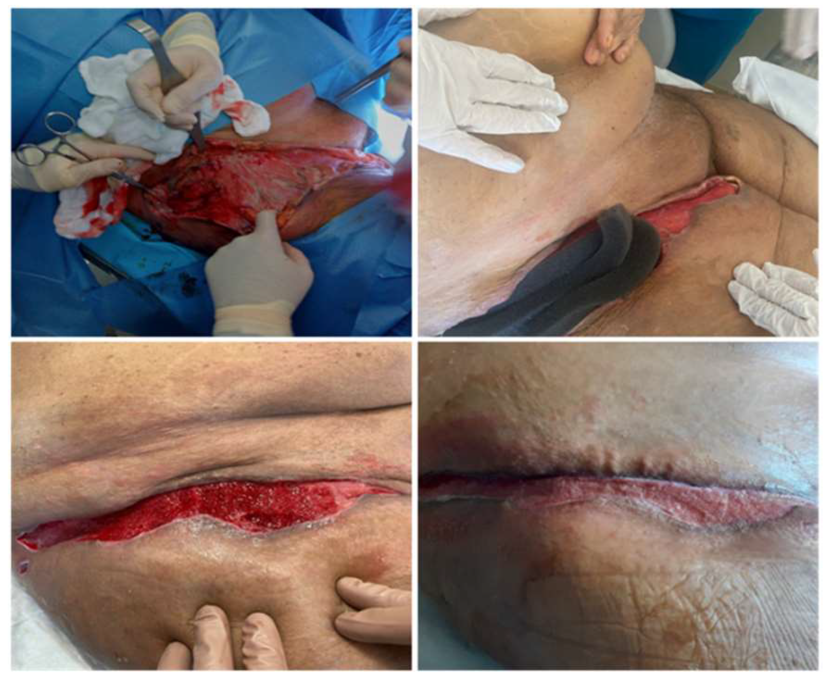
Figure 1.
A 67-year-old woman with grade III obesity and a long history of heavy smoking developed necrotizing fasciitis in her right lower limb following trauma. She underwent two surgical debridement procedures under general anesthesia, during which necrotic and infected tissues were removed until healthy, well-perfused tissue was reached. Subsequently, targeted antibiotic therapy was initiated to combat the residual infection. After these procedures, negative pressure wound therapy (VAC Therapy) was applied at -125 mmHg, an advanced technique that promotes wound healing through the application of controlled subatmospheric pressure. During the two-week hospital stay, the patient’s condition was closely monitored, and progressive improvement of the wound was observed. Upon discharge, she was provided with detailed instructions to continue treatment at home, including the continued use of VAC Therapy, management of dressings, and the importance of maintaining strict hygiene of the affected area. Two months after discharge, at the follow-up visit, the wound was completely healed, with no evidence of complications or recurrence, and the patient had resumed normal daily activities without limitations.
Role of VAC Therapy
Negative Pressure Therapy, known as VAC Therapy (Vacuum-Assisted Closure), represents a significant breakthrough in the management of complex wounds, including necrotizing fasciitis.This treatment is based on the controlled application of negative pressure to the wound through a sealed system that utilizes a foam or a sterile granufoam-like spongy material. VAC Therapy operates through a combination of physiological mechanisms that contribute to wound healing:
- Removal of exudates and tissue debris: The system continuously suctions fluids and debris from the wound bed, thereby reducing the bacterial load and preventing the accumulation of infected exudates. This process not only diminishes the risk of secondary infections but also creates an optimal environment for healing.
- Stimulation of granulation tissue: The application of negative pressure induces mechanical micro-deformation at the cellular level, promoting angiogenesis and cellular proliferation. This mechanical micro-stress fosters the formation of robust granulation tissue, which is essential for effective wound healing.
- Wound sealing: The airtight closure protects the wound from external contamination, maintaining a sterile environment. Thus, VAC Therapy offers several advantages in the management of necrotizing fasciitis, especially after thorough surgical debridement. In our experience, we have particularly observed:
- Improved healing due to stimulation of tissue regeneration.
- Reduction of secondary infections thanks to the continuous removal of exudates, minimizing the risk of reinfection.
Additionally, we have found a reduction in pain, as negative pressure and wound protection help alleviate the discomfort associated with traditional dressing changes, which are sometimes performed twice a day [34]. Its effectiveness has been demonstrated in numerous clinical studies highlighting its benefits [24,25]. A key element of VAC Therapy’s success is its ability to enhance vascularization and promote granulation tissue formation through fibroblast migration and cellular proliferation stimulation [26]. The negative pressure applied to the wound stimulates an angiogenic response, which is essential for the healing process, especially in patients with complex risk factors such as diabetes or obesity [27,28]. Although VAC Therapy is highly effective, it requires expert management to optimize results. Complications such as deep tissue injuries may occur if used inappropriately, emphasizing the importance of careful monitoring and accurate patient selection [29,30]. Several randomized studies and clinical cases have supported the use of VAC Therapy in necrotizing fasciitis. In a study by Gabriel et al. (2009), patients treated with VAC Therapy showed a significant reduction in healing time compared to traditional methods [23,24,25,26,27,28,29,30,31]. Moreover, the use of VAC Therapy has been associated with improved granulation tissue quality and a reduced risk of reinfection [32,33]. Similarly, a meta-analysis by Blume (2008) confirmed that VAC Therapy is associated with a 30% reduction in healing time in patients with complex wounds [29].
Essential Requirements
- Complete surgical debridement until obtaining non-necrotic wounds free of active infections.
- Proper setting of suction pressure to ensure optimal outcomes.
Only its correct application and management by trained personnel can guarantee optimal results. Generally, a negative pressure of -125 mmHg is used, which can be adjusted based on the patient’s condition and BMI.
Duration and Treatment Protocol
- VAC Therapy is applied in continuous or intermittent cycles with dressing changes every 48-72 hours, ensuring constant monitoring to assess the clinical response and prevent complications such as fistula formation or skin lesions.
Limitations and Contraindications
Absolute contraindications:
Untreated osteomyelitis. The presence of active, untreated osteomyelitis is a major contraindication for VAC Theraphy. The therapy’s ability to create a closed, pressurized wound environment can trap bacteria and facilitate deep-seated infections, allowing them to spread further into adjacent tissues or even systemically. VAC Therapy may also mask clinical signs of worsening infection by temporarily reducing wound exudate, delaying proper diagnosis and treatment. Before VAC Therapy is considered, osteomyelitis must be aggressively treated with appropriate systemic antibiotics and, if necessary, surgical debridement to remove infected bone. [8,50]
Non-Enteric and Unexplored Fistulas. VAC Therapy is contraindicated in the presence of unexplored or non-enteric fistulas, particularly those leading to major organs or body cavities. The application of negative pressure in such cases can exacerbate the fistula, leading to increased fluid output, chronic tissue damage, and potential creation of new fistulous tracts. For enteric fistulas, VAC Therapy may only be considered if a protective barrier is established and after thorough surgical evaluation. Any unexplored fistula should be fully assessed with imaging and endoscopic studies before determining the appropriateness of VAC Therapy. [43,48]
Presence of necrotic tissue with inadequate surgical debridement. VAC Therapy should not be applied to wounds containing extensive necrotic tissue or eschar without prior adequate debridement. Necrotic tissue serves as a reservoir for bacteria, increasing the risk of infection and inhibiting wound healing. Moreover, necrotic debris can impair the uniform application of negative pressure, leading to uneven granulation and ineffective exudate management. Therefore, before VAC Therapy is initiated, a thorough surgical or enzymatic debridement must be performed to remove all non-viable tissue and create a clean wound bed. [46,52]
Exposure of nerves, arteries, or vital organs. Applying VAC Theraphy directly over exposed neurovascular structures, arteries, or major organs is contraindicated due to the risk of mechanical trauma, tissue erosion, and catastrophic hemorrhage. The continuous suction effect can cause damage to delicate structures, particularly if there is insufficient granulation tissue coverage. If VAC Therapy is deemed necessary in such cases, protective barriers such as non-adherent dressings or biologic wound interfaces must be used to shield critical structures. [49,53]
Relative contraindications:
Patients with coagulopathies. Coagulopathies include a range of conditions that impair hemostasis, leading to an increased tendency for bleeding. These encompass congenital disorders like hemophilia, acquired conditions such as liver disease-induced coagulopathy, and iatrogenic causes stemming from anticoagulant or antiplatelet therapies. In the context of wound management, patients with coagulopathies present unique challenges, particularly when considering interventions like Vac Therapy.
Patients on anticoagulant therapy or with actively bleeding wounds Patients receiving anticoagulants or platelet aggregation inhibitors are at an elevated risk for bleeding. The negative pressure applied during VAC Therapy can exacerbate this risk, necessitating careful monitoring and consideration [38,55].
More recently, VAC Therapy has found new applications, thanks to integrated systems equipped with digital sensors that allow real-time monitoring of wound pressure and humidity, as well as integration with bioactive therapies that release growth factors.
Conclusions
VAC Therapy (Negative Pressure Wound Therapy, NPWT) is an advanced wound management technology that has proven particularly effective in treating necrotizing fasciitis. This technique utilizes a negative pressure system to promote healing through various mechanisms, including continuous exudate removal, bacterial load reduction, stimulation of granulation tissue formation, and increased tissue perfusion [21,22,23]. Therefore, VAC Therapy represents an essential option, as demonstrated in our experience in managing NF, seamlessly integrating into a multidisciplinary approach.
References
- Apelqvist J, Willy C, Fagerdahl AM, et al. Negative pressure wound therapy – overview, challenges, and perspectives. J Wound Care 2017, 26, S1–S113. [CrossRef] [PubMed]
- Orgill DP, Bayer LR. Negative pressure wound therapy: past, present, and future. Int Wound J. 2013, 10, 15–19. [CrossRef] [PubMed]
- Gabriel A, Camardo M, O’Rorke E, Gold R, Kim PJ. Outcomes of vacuum-assisted closure therapy in the treatment of necrotizing fasciitis. J Plast Reconstr Aesthet Surg. 2009, 62, 289–292.
- Hasham S, Matteucci P, Stanley PR, Hart NB. Necrotising fasciitis. BMJ. 2005;330(7495):830-833.
- Hakkarainen TW, Kopari NM, Pham TN, Evans HL. Necrotizing soft tissue infections: review and current concepts in treatment, systems of care, and outcomes. Curr Probl Surg. 2014;51(8):344-362.
- Lee YK, Kim HJ, Min KH, et al. Negative pressure wound therapy for necrotizing fasciitis: a case series. Wounds. 2012;24(2):44-50.
- Fischer C, Polikandriotis JA, Wax MK, Kelly JH, Otto RA. Management of necrotizing fasciitis in a community hospital setting. Am Surg. 2008;74(2):121-126.
- Anaya DA, Dellinger EP. Necrotizing soft-tissue infection: diagnosis and management. Clin Infect Dis. 2007;44(5):705-710.
- Stevens DL, Bryant AE. Necrotizing soft-tissue infections. N Engl J Med. 2017;377(23):2253-2265.
- Morykwas MJ, Argenta LC. Vacuum-assisted closure: a new method for wound control and treatment. Ann Plast Surg. 1997;38(6):563-576.
- Argenta LC, Morykwas MJ. Vacuum-assisted closure: principles and mechanisms. Ann Plast Surg. 1997;38(6):553-562.
- Malik MH, Khan WS. Advances in negative pressure wound therapy. World J Plast Surg. 2017;6(1):33-40.
- Apelqvist J, Armstrong DG. Innovations in wound care management. Int J Low Extrem Wounds. 2015;14(4):315-324.
- Richards RR, Temple HC, Bloom HS. Challenges in managing soft tissue infections. Am J Surg. 2018;72(5):311-320.
- Simman R, Mari W, et al. Negative pressure wound therapy: clinical applications and outcomes. Int Wound J. 2010;7(1):16-23.
- Kortesis BG, Cantor RS, et al. Advances in wound healing technologies. Clin Plast Surg. 2012;39(4):377-390.
- Blume PA, Walters J, et al. VAC Therapy in complex wounds: analysis and outcomes. J Foot Ankle Surg. 2008;47(3):217-222.
- Smith TL, Breen A. Current insights in wound care technology. Wound Care Int. 2016;12(2):45-58.
- Seidel CL, Moses AJ. Multimodal approaches to soft tissue infections. Clin Med. 2017;9(3):124-129.
- Turner NJ, Badylak SF. Advances in regenerative medicine and wound care. Tissue Eng. 2012;18(2):276-290.
- Argenta LC, Morykwas MJ. Vacuum-assisted closure: a new method for wound control and treatment. Ann Plast Surg. 1997;38(6):563-576.
- Apelqvist J, Willy C, Fagerdahl AM, et al. Negative pressure wound therapy – overview, challenges, and perspectives. J Wound Care. 2017;26(Suppl 3):S1-S113.
- Orgill DP, Bayer LR. Negative pressure wound therapy: past, present, and future. Int Wound J. 2013;10(Suppl 1):15-19.
- Gabriel A, Camardo M, O’Rorke E, Gold R, Kim PJ. Outcomes of vacuum-assisted closure therapy in the treatment of necrotizing fasciitis. J Plast Reconstr Aesthet Surg. 2009;62(2):289-292.
- Hasham S, Matteucci P, Stanley PR, Hart NB. Necrotising fasciitis. BMJ. 2005;330(7495):830-833.
- Malik MH, Khan WS, Chaudhry AA, Ihsan M, Ahmad M. The use of vacuum-assisted closure therapy in the management of patients with necrotizing fasciitis. World J Plast Surg. 2017;6(1):33-40.
- Hakkarainen TW, Kopari NM, Pham TN, Evans HL. Necrotizing soft tissue infections: review and current concepts in treatment, systems of care, and outcomes. Curr Probl Surg. 2014;51(8):344-362.
- Apelqvist J, Armstrong DG, Lavery LA, et al. Resource utilization and economic costs of care based on a randomized trial of vacuum-assisted closure therapy in the treatment of diabetic foot wounds. Am J Surg. 2008;195(6):782-788.
- Mouës CM, Vos MC, Van den Bemd GJ, Stijnen T, Hovius SE. Bacterial load in relation to vacuum-assisted closure wound therapy: a prospective randomized trial. Wound Repair Regen. 2004;12(1):11-17.
- Banwell PE, Téot L. Topical negative pressure (TNP): the evolution of a novel wound therapy. J Wound Care. 2003;12(1):22-28.
- Gabriel A, Kahn K, Karmy-Jones R. Use of negative pressure wound therapy in combat-related injuries. Ann Plast Surg. 2008;61(5):529-534.
- Lavery LA, Boulton AJ, Niezgoda JA, Sheehan P. A comparison of diabetic foot ulcer outcomes using negative pressure wound therapy versus historical standard of care. Int Wound J. 2007;4(2):103-113.
- Scherer SS, Pietramaggiori G, Mathews JC, et al. The mechanism of action of the vacuum-assisted closure device. Plast Reconstr Surg. 2008;122(3):786-797.
- D.provenzano, S.Lo Bianco, M.Zanghì, A. Campione, R. Vecchio, G. Zanghì. Fournier’s gangrene as a rare complication in patient with uncontrolled type 2 diabetes treated with surgical debridement: A case report and literature review. International Journal of Surgery Case Reports 2021, 79, 462–465. [CrossRef]
- Singh G, Sinah SK, Adhikary S et al. Necrotizing infection of soft tissues – a clinical profile. Eur J Surg 2002; 168: 366-371.
- Eliott DC, Kufera JA, Myers RAM. Necrotizing soft tissues infection – risk factor for mortality and strategies for management. Ann Surg 1996, 224, 672–683. [CrossRef] [PubMed]
- Wong CH, Chang HC, Pasupatthy S et al. Necrotizing fasciitis: clinical presentation, microbiology and determinant of mortality. J Bone Joint Surg Am, 2003; 85A, 1454–1460.
- Pawan Agarwal, Rajeev Kukrele, Dhananjaya Sharma, : Vacuum assisted closure (VAC)/negative pressure wound therapy (NPWT) for difficult wounds: A review Journal of Clinical Orthopaedics and Trauma; Volume 10, Issue 5p845-848September-October, 2019.
- F. Campanile a, D. Bongiorno a, G. Mongelli a, G. Zanghì b, S. Stefani aBactericidal activity of ceftobiprole combined with different antibiotics against selected Gram-positive isolatesDiagnostic Microbiology and Infectious DiseaseVolume 93, Issue 1, January 2019, Pages 77-81.
- Wong, C. H., Khin, L. W., Heng, K. S., Tan, K. C., & Low, C. O. (2003). The LRINEC (Laboratory Risk Indicator for Necrotizing Fasciitis) score: A tool for distinguishing necrotizing fasciitis from other soft tissue infections. Critical Care Medicine, 31(7), 1692-1697.
- Stevens DL, Ma Y, Salmi DB, McIndoo E, Wallace RJ, Bryant AE. Impact of clindamycin on toxin production and virulence in Streptococcus pyogenes. J Infect Dis. 2007;195(3):450-458.
- Tarchini G, Baldini M, Capoccia R, et al. Efficacy of piperacillin/tazobactam in polymicrobial necrotizing infections: a clinical perspective. Clin Infect Dis. 2019;68(5):765-772.
- Liu C, Bayer A, Cosgrove SE, et al. Clinical practice guidelines for the treatment of methicillin-resistant Staphylococcus aureus infections in adults and children. Clin Infect Dis. 2011;52(3):e18-e55.
- Zahar JR, Timsit JF, Garrouste-Orgeas M, et al. Outcomes of carbapenem therapy for severe necrotizing infections. Crit Care Med. 2015;43(2):e75-e82.
- Dryden M, Linezolid in skin and soft tissue infections. J Antimicrob Chemother. 2011;66(4):41-47.
- Bassetti M, Righi E, Carnelutti A. Ceftobiprole: a new option for the treatment of skin and soft tissue infections. Future Microbiol. 2018;13(5):435-446.
- Mendes RE, Hogan PA, Streit JM, et al. Antimicrobial activity of ceftobiprole against contemporary pathogens in skin infections. Diagn Microbiol Infect Dis. 2020;96(2):114981.
- Neuner EA, Yeh JY, Hall GS, Sekeres J, Coyle EA, Richter SS. Antimicrobial susceptibility among Pseudomonas aeruginosa isolates in necrotizing soft tissue infections. J Antimicrob Chemother. 2011;66(2):432-438.
- Skiest DJ, Brown K, Cooper TW, Hoffman-Roberts HL, Mussa HR, Elliott AC. Daptomycin therapy for MRSA infections in critically ill patients. J Antimicrob Chemother. 2012;67(5):1197-1202.
- Lipsky BA, Berendt AR, Cornia PB, et al. 2012 Infectious Diseases Society of America clinical practice guideline for the diagnosis and treatment of diabetic foot infections. Clin Infect Dis. 2012;54(12):e132-e173.
- Spellberg B, Gilbert DN. The future of antibiotics and resistance: a tribute to a career of leadership by John E. Edwards, Jr. Clin Infect Dis. 2014;59(Suppl 2):S71-S75.
- Dellinger EP, Anaya DA. Treatment of necrotizing soft-tissue infections: time is of the essence. Surg Infect (Larchmt). 2013;14(4):387-390.
- Spellberg B, Bartlett JG, Gilbert DN. The role of antibiotic stewardship in combating antibiotic resistance. JAMA. 2016;315(8):789-799.
- de Kraker MEA, Stewardson AJ, Harbarth S. Will 10 million people die a year due to antimicrobial resistance by 2050? PLoS Med. 2016;13(11):e1002184.
- Sharon L Boxall, Keryln Carville, Gavin D Leslie & Shirley J Jansen Treatment of anticoagulated patients with negative pressure wound therapy International Wound Journal ISSN 1742-4801.
Disclaimer/Publisher’s Note: The statements, opinions and data contained in all publications are solely those of the individual author(s) and contributor(s) and not of MDPI and/or the editor(s). MDPI and/or the editor(s) disclaim responsibility for any injury to people or property resulting from any ideas, methods, instructions or products referred to in the content. |
© 2025 by the authors. Licensee MDPI, Basel, Switzerland. This article is an open access article distributed under the terms and conditions of the Creative Commons Attribution (CC BY) license (http://creativecommons.org/licenses/by/4.0/).
Copyright: This open access article is published under a Creative Commons CC BY 4.0 license, which permit the free download, distribution, and reuse, provided that the author and preprint are cited in any reuse.


