Preprint
Review

This version is not peer-reviewed.

Protective Role of Physical Activity and Antioxidant Systems During Spermatogenesis

A peer-reviewed article of this preprint also exists.

Submitted:

07 February 2025

Posted:

10 February 2025

You are already at the latest version

Abstract

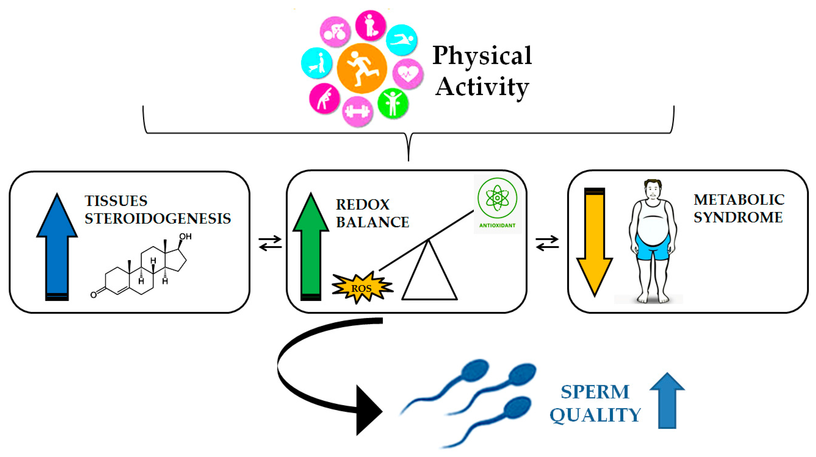
Oxidative stress represents a significant contributing factor to male infertility and sperm dysfunction. In this condition, an increase in ROS production exceeds the body's antioxidant defences, resulting in a decline in spermatozoa quality and fertilising capacity. Furthermore, excessive ROS production has been linked to the promotion of genomic damage, lipid peroxidation, inflammation, altered enzyme activity, and ultimately, irreversible alterations, cell death, and a decline in seminal parameters associated with male infertility. It is established that physical activity (PA), acting on inflammatory parameters and improving antioxidant defence, can alleviate the negative effects caused by free radicals, offering numerous health benefits and positively influencing sperm quality. The objective of this review is to highlight the mechanisms of ROS production, the physiological and pathophysiological roles of ROS in relation to the male reproductive system, and recent knowledge on the impact of some protocols of PA on these systems and the molecular mechanisms involved.

Keywords: 
;  ;  ;  ;  ;  

1. Introduction

Infertility is a significant global issue, affecting over 12% of couples worldwide, with male factors being the primary cause in most cases [1]. Male fertility relies heavily on spermatogenesis, the process that produces large quantities of sperm cells (spermatozoa) in the testis. Spermatogenesis is a meticulously orchestrated biological process that culminates in the production of mature, haploid spermatozoa capable of fertilizing an oocyte. This process involves three primary stages: proliferation of spermatogonia, the meiotic divisions and the spermiogenesis. One of the most significant aspects of the final stage is nuclear remodeling, where the compacting of DNA is achieved by replacing histones with protamines with the goal to safeguard the genetic material, ensuring it is protected during its journey through the female reproductive tract. Indeed, spermatozoa inherently generate large amounts of reactive oxygen species (ROS) that impairs sperm motility and damages DNA through the oxidation of membrane lipids and nucleic acids [2]. However, their role in spermatogenesis and sperm function is more nuanced, serving as both a potential disruptor and a key regulator of normal physiological processes. In spermatozoa, ROS mediate capacitation, hyperactivation and acrosome reaction, fundamental processes for fertilization, and they take part in intracellular signaling pathways essential for sperm development and function, such as the production of intracellular cAMP, which activates Protein Kinase A (PKA) [3]. Moreover, ROS contribute to cellular balance by regulating processes like vascular tone, oxygen sensing, immune response, and even adhesion properties.
This duality necessitates a robust but carefully regulated antioxidant system and spermatozoa possess several intracellular antioxidant enzymes to mitigate the effects of ROS and the importance of antioxidants extends beyond endogenous mechanisms [4]. As a matter of fact, exogenous supplementation with compounds such as coenzyme Q10, L-acetyl-Carnitine, vitamin C, and zinc has been widely studied for its potential to counteract oxidative stress in semen. These supplements have demonstrated benefits in improving sperm quality and fertility outcomes, but excessive antioxidant use can lead to a condition known as reductive stress, where the over-suppression of ROS disrupts sperm functionality [5].
Physical activity (PA) is widely recognized for its numerous health benefits, including reduced risks of obesity, diabetes, and cardiovascular disease, pathologies where oxidative stress-related inflammatory responses is considered a causative process [6]. Interestingly, it also appears to have a role in reducing oxidative stress in sperm, a key factor in maintaining male fertility. However, the relationship between PA and sperm quality is complex, with various levels and types of activity showing differing effects [7]. While moderate PA may provide protective benefits, strenuous exercise has been linked to reduced semen quality in some cases, particularly in activities such as long-distance running and cycling. Animal studies, for example, have shown that running can slow testicular aging by mitigating oxidative stress and it might be possible that regular exercise, improving the redox homeostasis, may lower the generation of ROS, thereby preventing damage to sperm DNA and cellular structures [8].
This review intends to offer an overview of the dual role of reactive oxygen species in spermatogenesis, focusing on the potential role of sport and physical activity in maintaining or impairing the redox homeostasis control during spermatogenesis or in sperm.

2. Spermatogenesis

Spermatogenesis is the mechanism by which spermatozoa are produced from primordial germ cells. The process transforms diploid spermatogonia into specialized haploid spermatozoa through mitosis, meiosis, and spermiogenesis [9,10]. These cells migrate to the gonadal ridge and, after a period of mitotic division to form spermatogonia, enter in meiosis. Meiotic divisions and cytological changes, transforms spermatogonia into spermatozoa, a highly specialised cell equipped with a tail that allows the cell to move. The head of the sperm contains the nucleus, which contains DNA and is located at the periphery, separated from the cell membrane and the acrosome, which contains various substances for penetrating the ovocyte. The central part of the sperm contains the mitochondria, organised in different helical arrays around the axoneme. Mitochondria in spermatozoa are crucial for energy production, redox balance, calcium regulation, and apoptotic pathways, all necessary for sperm motility, capacitation, and fertilization [11]. Mitochondrial dysfunction and quantity can lead to decreased sperm quality and infertility [11].
Spermatogenesis is a complex process involving several hormonal and cells interactions. The hypothalamic release of gonadotropin-releasing hormone (GnRH) stimulates the anterior pituitary gland to secrete follicle-stimulating hormone (FSH) and luteinizing hormone (LH). Sertoli and Leydig cells are two further testis cells that are pivotal for male reproduction [12]. These cells play critical roles in the process of spermatogenesis, ensuring proper development and maturation of sperm [13]. Sertoli cells, located within the seminiferous tubules of the testes, provide structural support and nourishment to developing germ cells [14]. They form the blood-testis barrier, regulate the environment of the seminiferous tubules, and secrete factors essential for the differentiation of spermatogonia into spermatozoa. Sertoli cells also produce inhibin, a hormone involved in the negative feedback regulation of FSH levels.
Leydig cells, located in the interstitial space between the seminiferous tubules, influenced by LH, are responsible for the production of testosterone, that exerts its effects on Sertoli cells, promoting the maturation of germ cells and the progression of spermatogenesis. Consequently, the coordinated actions of GnRH, FSH, LH, Sertoli cells, Leydig cells, and testosterone work in concert to establish a regulated environment that facilitates the sustained production of sperm throughout the male reproductive life.

3. Oxidative stress

The concept of "Oxidative Stress" refers to an imbalance between oxidants and antioxidants, favoring oxidants, which results in disrupted redox signaling, impaired control mechanisms, and/or molecular damage [15]. Originally introduced in 1985 [16], this concept has evolved to encompass advancements in our understanding of redox signaling [17]. Fundamentally, it describes a steady-state redox balance within an open metabolic system, maintained at a specific setpoint that ensures basal redox tone. Any deviation from this balance constitutes stress, triggering a stress response. The definition also acknowledges that (i) a shift to the opposite end of the balance constitutes "reductive stress," and (ii) deviations can be physiological ("oxidative eustress") or supraphysiological ("oxidative distress") [15] (Figure 1).
Oxidative eustress plays a crucial role in physiological redox signaling and control [15,16], aligning closely with the concept of redox homeostasis as the "golden mean" [18].
Metabolic regulation involves a wide array of chemical processes, including the orchestrated modification of proteins, lipids, carbohydrates, and nucleic acids to maintain structure and function. Redox reactions play a significant role in this regulation, with notable interactions between redox modifications and other regulatory mechanisms, such as the phosphorylation and dephosphorylation of proteins. Low-molecular-mass chemically reactive molecules, often referred to as “reactive species,” have been extensively studied for their regulatory roles. These include reactive oxygen species (ROS) [19], reactive nitrogen species (RNS) [20,21], reactive sulfur species (RSS) [22], reactive electrophile species (RES) [23], and reactive halogen species (RHS) [24].
The interplay among these reactive species forms a complex system of checks and balances essential for effective redox regulation [25]. On the opposite side of the redox equilibrium, the defense mechanisms against excessive oxidant levels involve a variety of antioxidant enzymes supported by their auxiliary systems, alongside low-molecular-weight antioxidants, collectively forming an integrated antioxidant network [26]. The expression of these antioxidant enzymes is regulated by key redox signaling pathways as part of the oxidative stress response [15].

4. Redox homeostasis and physio-pathological conditions

Maintaining homeostasis is a fundamental aspect of health [27], which is regarded as a dynamic and active biological process [28]. Achieving and sustaining physiological health depends on mechanisms that ensure homeostasis through resistance, tolerance, and resilience, reflecting a homodynamic nature [29,30]. The concept of eustress, as distinct from non-physiological distress, characterizes the body's continuous state of readiness to maintain homeostasis. This distinction, introduced by Selye [31], forms a foundational perspective in understanding stress and its responses [32].
Oxidation–reduction (redox) reactions are integral to life's processes. Research into elements such as oxygen, iron, copper, sulfur, selenium, and nitrogen, alongside studies on free radical oxidation and defense mechanisms, has uncovered the remarkable versatility and applications of redox reactions. These include roles in energy capture, mitigating oxygen toxicity, and producing biochemical defenses against harmful entities. Notably, redox reactions also play a pivotal role in signalling, as they enable fast and reversible processes essential for physiological regulation.
The complex integration of energetically demanding systems makes life in oxygen-rich environments possible. Similarly, this principle applies to the regulation of redox signalling, which governs numerous physiological processes. Ensuring redox homeostasis presents an ongoing challenge, requiring tight biochemical control [15]. For oxidation events that serve as physiological signals, counteractive mechanisms must deactivate the signals and restore the redox balance to maintain health [18].
For over three decades, an understanding of ill health has been associated with the oxidative stress concept in the form of ‘an imbalance between oxidants and antioxidants in favour of the oxidants’, which developed into the idea of a ‘disrupted Redox signalling’ [33]. The refinement of this concept has been hastened by the articulation of a ‘Redox Code’ in an influential paper describing a set of principles through which biological function is enabled and protected [34]. Many disease processes are attributed to enhanced production of reactive oxygen species (ROS) or ‘dysfunctional Redox regulation’. Still, not everything can be explained by excessive ROS production. Many pathophysiological conditions may in fact exemplify processes that can be accounted for by other bioactive entities such as reactive nitrogen or sulphur species (RNS, RSS), or other small signalling molecules such as hydrogen (H2), ammonia (NH3), and carbon monoxide (CO). Many of these entities can react with each other, with protein thiols or other biomolecules.
These varied interactions modulate the function of ion channels, enzymes, transcription factors, and other bio-logical targets, a scenario defined as the ‘Reactive Species Interactome’ [35]. This reactive species interactome concept provides a useful framework to explain the apparent complexity of adaptive signalling. There is no single marker or process that captures the complexity of these interactions adequately. Rather, it is likely that a combination of readouts from different levels of organization will be required to explain how and why mitochondrial function appears so intimately related to chronic disease, inflammation and metabolism.

5. The Role of Oxidative Stress in Male Infertility

Oxidative stress, defined as an imbalance between reactive oxygen species (ROS) and antioxidant defenses, has been identified as a significant contributing factor to male infertility [36,37].
While the presence of low levels of ROS is necessary for normal sperm function, excessive levels have been demonstrated to cause cellular damage, impaired sperm motility, and DNA fragmentation [38,39]. It appears that this delicate balance is of critical importance: low levels of oxidative stress have been shown to promote the appropriate activation of signaling pathways involved in spermatogenesis, while excessive ROS production has been linked to pathological changes, including the apoptosis of germ cells and a decline in sperm quality. These effects impact various aspects of male fertility, including testis cell structure and function, as well as sperm parameters (sperm count, motility, viability, and increased abnormal sperm morphology, sperm DNA fragmentation) [40].
It has been suggested that several conditions may possibly alter oxidant/antioxidant balance, which could in turn lead to oxidative stress. These conditions include endogenous factors such as deficiencies in antioxidants, immune system dysfunctions bacterial/viral infections, abnormal spermatozoa, leukocytospermia (LCS) [41,42], and exogenous factors such as smoke, alcohol, obesity, varicocele or sexual transmitted diseases [36,43] (Figure 2).
Testicular environment is particularly vulnerable to oxidative damage due to the high metabolic activity of spermatogenic cells [44] and poor vascularization, as well as the presence of a high content of polyunsaturated fatty acids (PUFA) in the plasma membrane in conjunction with the absence of cytoplasmic antioxidant enzymes [38,45,46,47,48]. ROS, indeed, can potentially damage sperm membranes organization, mitochondrial activity, and function of testicular cells [48], consequently affecting germ cells motility and ability to fuse with oocytes, testis steroid hormone synthesis and steroidogenic capacity [45,49,50]. Mitochondria are the energy powerhouses of the sperm cell, and any disruption in their function can lead to diminished motility, thus reducing the sperm's ability to fertilize an egg [51]. Studies have shown that men with lower sperm motility exhibit decreased glucose-6-phosphate dehydrogenase activity and increased levels of malondialdehyde, a marker of oxidative damage [44]. Furthermore, oxidative stress has been implicated in the disruption of the blood-testis barrier, the key structure that protects the developing sperm cells from harmful substances, including ROS [52]. However, all these factors are modifiable and reversible, and hence, by mere changing of lifestyle, many of these risk factors can be avoided.

5.1. Testicular Cells and Oxidative Stress

The testis is composed of several different cell types, each playing a vital role in the process of sperm production. Sertoli cells, Leydig cells, and germ cells are all integral components of this dynamic environment, and each is influenced by the redox status of the tissue. Sertoli cells, which provide nutritional and structural support to developing sperm cells, are particularly sensitive to changes in oxidative balance. Under conditions of chronic oxidative stress, Sertoli cell function is compromised, leading to impaired spermatogenesis and, consequently, a reduction in sperm count and quality [53]. The presence of ROS was associated with significant levels of apoptosis in Sertoli cells, a substantial decrease in connexin-43 (Cx43) expression, a key component of gap junctions, pivotal to regulate spermatogenesis, and a failure to maintain the viability of spermatogonial stem cells (SSCs) [54].
Research on Sertoli cells and oxidative stress suggests that various environmental toxins have the potential to induce oxidative damage and apoptosis. It has been suggested that exposure to 2,3,7,8-tetrachlorodibenzo-p-dioxin (TCDD) may possibly impair Sertoli cell function through oxidative stress, affecting mitochondrial activity and membrane potential [55]. Similarly, it appears that sodium fluoride exposure may contribute to a decline in cell viability and an increase in oxidative stress and apoptosis. Furthermore, acrylamide and its metabolite glycidamide have been observed to cause oxidative stress and apoptosis in both Leydig and Sertoli cells, affecting cell viability and gene expression of apoptotic markers [56].
Exposure to oxidative stress has been demonstrated to result in a decline in testosterone production, an escalation in apoptosis, and an impairment in steroidogenesis in Leydig cells [56,57]. In 2016, Duan et al. investigated the effects of hydrogen peroxide (H2O2) on primary rat Leydig cells and the role of peroxiredoxin 2 (Prdx2), an important antioxidant protein involved in oxidative stress response. The study revealed that H2O2 treatment led to a significant decline in cell viability, inducing apoptosis in a dose-dependent manner and altering Prdx2 protein expression [57]. Subsequently, in 2021, Anak and colleagues explored the impact of aging on testosterone production, conducting a comprehensive literature review that investigated the role of antioxidants in safeguarding Leydig cells against oxidative stress [58]. Two key defects in the steroidogenic pathway have been identified as contributing to age-related reductions in testosterone production: reduced LH-stimulated cAMP production, and impaired cholesterol transport to and within the mitochondria.
Increasing oxidative stress appears to play a crucial role in age-related testosterone reduction, and aging is associated with enhanced lipid peroxidation. Leydig cell membranes from older rats exhibit a two- to three-fold increase in basal thiobarbituric acid-reactive substances (TBARS) formation, indicating increased oxidative damage. They demonstrated that aging leads to a decrease in serum testosterone levels in both humans and rodents. This decline is not attributable to a loss of Leydig cells, but rather to their reduced capacity to produce testosterone in response to luteinizing hormone (LH) [59].
In spermatozoa, oxidative stress triggers lipid peroxidation of polyunsaturated fatty acids in sperm membranes [45,60], lead to protein aggregation in male germ cells, and potentially disrupting proteostasis [61]. Proteostasis in sperm cells is of pivotal significance for sustaining optimal functionality and fertility. It encompasses regulatory mechanisms that govern protein synthesis, folding, modification, and degradation [62]. Sperm cells are distinguished by their limited capacity for protein synthesis and repair, which renders proteostasis essential for their viability [62]. Disruptions in proteostasis can result in sperm dysfunction, impacting motility and fertilization potential, thus, the interplay between proteostasis and oxidative stress in sperm cells is pivotal to their functionality [61,62]. Proteostasis mechanisms have been shown to mitigate the harmful effects of oxidative stress [63]. Indeed, it has been demonstrated that the sustained production of misfolded proteins can exceed the capacity of the proteostasis network, resulting in its failure and subsequent cell death [64]. Morphological abnormalities in sperm are also frequently observed in men with high oxidative stress levels. These abnormalities, which may include defects in the acrosome, tail, or head, are often a result of lipid peroxidation and protein damage induced by ROS. These structural changes compromise the sperm's ability to penetrate the egg and perform its fertilization function.
Male infertility can be influenced by various endogenous sources of ROS in seminal plasma. The human semen sample contains a variety of cells, including immature and mature spermatozoa, round shaped cells of different phases of spermatogenesis, epithelial cells, and leukocytes [65]. A significant contributor is leukocytes, particularly polymorphonuclear leukocytes and macrophages, which originate from the seminal vesicles and prostate gland. In the presence of urogenital infections or inflammation, these leukocytes exhibit an enhanced immune response, generating up to 100 times more ROS, thereby leading to oxidative stress [66].
Human semen contains a variety of cells, including immature and mature spermatozoa, round-shaped cells from different phases of spermatogenesis, epithelial cells, and leukocytes [65]. Leukocytes, especially macrophages and neutrophils, are typically activated in response to infection or inflammation, while immature, morphologically abnormal spermatozoa also serve as primary sources of ROS [36]. However, the rate of ROS production is up to 1000 times higher in leukocytes (extrinsic source) compared to spermatozoa (intrinsic source) [67].

6. Spermatogenesis and Physical Activity: Role for Oxidative Stress Control.

Regular moderate-intensity physical exercise is associated with an increase in antioxidant defense systems and a reduction in systemic oxidative stress [68]. This is largely due to the adaptation of various enzymatic and non-enzymatic antioxidants, such as superoxide dismutase (SOD), catalase (CAT), and glutathione peroxidase (GPx), which play key roles in neutralizing ROS [69]. Furthermore, physical activity PA has been found to improve mitochondrial function, which may contribute to a more efficient regulation of oxidative metabolism and reduced ROS production [70]. Conversely, intense or prolonged PA, especially when performed in an unconditioned state, can result in an acute increase in oxidative stress. This phenomenon is primarily attributable to the enhanced production of ROS during high-intensity exercise, which can exceed the body's antioxidant defense mechanisms [71,72].
Consequently, oxidative damage may result, manifesting as muscle fatigue, inflammation, and potentially compromised sperm quality. It is important to note that the beneficial effects of physical activity on oxidative stress are dose-dependent, with moderate levels of exercise being most effective in improving antioxidant capacity and mitigating the negative effects of oxidative stress [68,73].

6.1. Impact of Physical Activity on Spermatogenesis

The aetiology of male infertility is multifactorial, with physiological factors such as age and lifestyle factors including physical activity playing a role. However, the specific impact of sporting activity on semen parameters, and consequently on male fertility, remains unclear. Recent studies have explored the impact of physical activity on male fertility, highlighting the complex relationship between exercise and reproductive health [74,75,76,77].
Moderate exercise has been shown to improve sperm quality, including count, motility, and morphology, while excessive or severe exercise may have detrimental effects [7,78,79]. Regular physical exercise has been shown to enhance sperm count, motility, and morphology in both human and rat subjects, with a concomitant improvement in testosterone, LH, and FSH levels in rats. It has been demonstrated that exercise can reduce the percentages of sperms with negative tubular differentiation (TDI) and spermiogenesis indices (SPI), DNA fragmentation, and also ameliorate the diabetes-induced apoptosis and improve sperm apoptosis index in animal models [80]. The positive effects of exercise on male fertility are thought to be mediated by reduced oxidative stress, enhanced antioxidant defense, and improved steroidogenesis [78,79,81]. Furthermore, the improvement of glucose metabolism that results from physical activity increase is often the primary factor that ameliorates sperm parameters in men, since the majority of studies performed engaged men affected by metabolic syndrome and type 2 diabetes [81,82,83,84,85,86] (Figure 3).
Steroidogenesis, the process of producing steroids such as testosterone, plays a crucial role in regulating various physiological functions, including muscle growth, sexual function, and overall health. In this context, PA has been identified as a potential modulator of steroidogenesis, influencing both testicular and muscular pathways. Acute bouts of intense exercise, such as heavy resistance training or endurance exercise, lead to transient increases in testosterone secretion [87]. This response is thought to be mediated by the hypothalamic-pituitary-gonadal axis, which stimulates the release of LH and, consequently, the production of testosterone by Leydig cells [87]. Research suggests that the impact of physical activity on male fertility depends on exercise intensity, duration as well as the profile of the participant [80]. Studies performed in recreational athletes, demonstrated positive effects of prolonged physical activity. In fact, 433 infertile men training at 70–85% of their maximal oxygen consumption revealed that high-intensity exercise may restrain inflammatory biomarkers, oxidative stress, and antioxidants, while concomitantly enhancing semen parameters and the pregnancy rate [80]. While moderate exercise may have beneficial effects on sperm quality, high-intensity or prolonged exercise can negatively affect semen parameters [88,89,90]. Intense physical activity has been associated with decreased sperm concentration, motility, and morphology, particularly in elite athletes [90]. It has been observed that endurance exercise, including activities such as long-distance running or extensive cycling, has the potential to adversely affect seminal parameters [75]. A 16-week low-to-intensive cycling training program has been shown to result in a decline in sperm quality parameters and an increase in seminal inflammatory markers, with some effects persisting even after a 30-day recovery period [91]. The same training regimen has also been shown to result in increased oxidative stress and decreased antioxidant capacity in semen [91]. A further study comparing endurance cyclists to sedentary controls found significantly lower proportions of morphologically normal sperm in cyclists [92]. At the same time, moderate running, has been shown to improve cardiovascular health, reduce oxidative stress, and enhance hormone regulation, all of which can potentially benefit male fertility. Moreover, the regulation of insulin sensitivity and the reduction of visceral fat, both outcomes of consistent running, are linked to improved hormonal profiles, including better testosterone levels. However, excessive endurance running or high-intensity training may have the opposite effect, leading to a decrease in sperm quality [90]. This decline in sperm quality is attributed to mechanical impact, gonadal overheating, wearing tight clothes, and increased oxidative stress [36,90]; however, these effects may be reversible with proper rest and recovery periods [75].
Ultimately, the relationship between exercise and male fertility remains complex, with some studies showing conflicting results and difficulties in quantifying physical activity [7]. Further evidences, showed the influence of physical exercise on the endocrine system, particularly the hypothalamo-pituitary-adrenal (HPA) axis and stress hormones production, influencing in turn testosterone production. The intensity and duration of exercise have been demonstrated to modulate the HPA axis response, resulting in increased cortisol secretion [93]. While endurance training does not lead to permanent hypercortisolism, it has been observed to result in decreased tissue sensitivity to glucocorticoids [93].
The relationship between cortisol and testosterone during exercise is a complex one, too. Post-exercise, a significant negative correlation between cortisol and total testosterone has been observed, while a positive correlation exists with free testosterone [94]. Prolonged imbalances in cortisol and growth hormone secretion can be detrimental to health [95]. In cases of overtraining, the sympathetic/parasympathetic imbalance and neuroendocrine dysfunction hypotheses have been proposed to explain performance decrements and recovery issues [96]. It is therefore crucial to ensure proper exercise planning with sufficient recovery in order to prevent overtraining and maintain hormonal balance.
Exercise-induced oxidative stress can be counteracted by the consumption of nutritional strategies [97] and antioxidant-rich foods [98], which can further support the maintenance of sperm quality. Antioxidant supplementation has shown potential in improving sperm quality and fertility outcomes in some individuals [98]. Specific antioxidants such as selenium, zinc, omega-3 fatty acids, CoQ10, and carnitines have been positively associated with sperm quality [99]. However, excessive antioxidant use may be detrimental to sperm function, resulting in a paradoxical decline in sperm quality [99] which is similar to the effect seen with oxidative stress.
However, an exact cut-off for "excessive" has not been well defined in the existing literature. A balanced diet rich in natural antioxidants from fruits, vegetables, whole grains, legumes, and seeds is recommended as a safe and effective approach to meet antioxidant requirements in physically active individuals and athletes [100].

7. Conclusion and Future Perspectives

This review underscores the significant role of exercise in modulating male reproductive health, primarily through its impact on oxidative stress and inflammatory pathways. The findings suggest that engaging in moderate-intensity exercise over a prolonged period can effectively suppress pro-inflammatory cytokine production, improve antioxidant defense mechanisms, and enhance sperm DNA integrity. Such effects are particularly beneficial for individuals experiencing infertility, as oxidative stress is known to impair sperm quality and overall testicular function. High-Intensity Interval Training (HIIT) has also demonstrated potential in improving sperm characteristics in men with fertility issues, further supporting the notion that structured physical activity can be an effective intervention for reproductive health.
The testicles are highly susceptible to oxidative stress due to their high rate of cell division, mitochondrial oxygen consumption, and abundance of unsaturated fatty acids. As a result, excessive production of reactive oxygen species (ROS) can disrupt spermatogenesis, impair steroidogenesis, and reduce sperm quality. Exercise exerts a dual effect on male reproductive function—while mild to moderate exercise improves testicular steroidogenesis, spermatogenesis, and sexual competence by increasing insulin sensitivity and regulating ROS production, excessive and prolonged exercise can have adverse effects by promoting oxidative stress and impairing testicular function. This highlights the importance of maintaining an optimal balance in exercise intensity and duration to support reproductive health.
Furthermore, research has demonstrated that exercise can improve fertility in men with lifestyle-induced conditions such as obesity and diabetes. By enhancing testicular antioxidant defense, reducing pro-inflammatory cytokine levels, and promoting steroidogenesis, exercise contributes to improved spermatogenesis and semen quality. However, the extent to which exercise benefits male fertility is influenced by multiple factors, including an individual’s overall health status, the type, intensity, and duration of exercise, and pre-existing metabolic or hormonal conditions. These factors should be carefully considered when prescribing exercise as a therapeutic strategy for male reproductive health.
Future research should focus on refining clinical guidelines that consider individual health status, exercise volume, intensity, and duration. Further clinical trials are required to validate the underlying mechanisms and establish exercise-based interventions for managing lifestyle-induced infertility. A more comprehensive understanding of the interaction between physical activity and male reproductive health will facilitate the development of evidence-based recommendations that optimize fertility outcomes.

Author Contributions

“Conceptualization, D.C.; C.A.; P.S.; I.D. and L.D.L.; writing—original draft preparation, C.A.; D.C.; L.S., I.D.; P.S.; writing—review and editing, D.C.; C.A.; P.S.; I.D. and L.D.L.;. All authors have read and agreed to the published version of the manuscript.”

Conflicts of Interest

“The authors declare no conflicts of interest.”

Abbreviations

The following abbreviations are used in this manuscript:
cAMP cyclic adenosine monophosphate
CAT catalase
CO carbon monoxide
Cx43 connexin-43
FSH follicle-stimulating hormone
GPx glutathione peroxidase
GnRH gonadotropin-releasing hormone
H2O2 hydrogen peroxide
H2 hydrogen
LCS leukocytospermia
LH luteinizing hormone
NH3 ammonia
PKA protein kinase A
PA physical activity
Prdx2 peroxiredoxin 2
PUFA polyunsaturated fatty acids
RES reactive electrophile species
RNS reactive nitrogen species
RHS reactive halogen species
REDOX oxidation–reduction
ROS reactive oxygen species
SSCs spermatogonial stem cells
SOD superoxide dismutase
SPI and spermiogenesis indices
TBARS thiobarbituric acid-reactive substances
TDI tubular differentiation

References

  1. Rivero, M.-J.; Kulkarni, N.; Thirumavalavan, N.; Ramasamy, R. Evaluation and Management of Male Genital Tract Infections in the Setting of Male Infertility: An Updated Review. Current Opinion in Urology 2023, 33, 180–186. [Google Scholar] [CrossRef] [PubMed]
  2. Agarwal, A.; Parekh, N.; Panner Selvam, M.K.; Henkel, R.; Shah, R.; Homa, S.T.; Ramasamy, R.; Ko, E.; Tremellen, K.; Esteves, S.; et al. Male Oxidative Stress Infertility (MOSI): Proposed Terminology and Clinical Practice Guidelines for Management of Idiopathic Male Infertility. World J Mens Health 2019, 37, 296. [Google Scholar] [CrossRef] [PubMed]
  3. Baker, M.A.; Hetherington, L.; Aitken, R.J. Identification of SRC as a Key PKA-Stimulated Tyrosine Kinase Involved in the Capacitation-Associated Hyperactivation of Murine Spermatozoa. Journal of Cell Science 2006, 119, 3182–3192. [Google Scholar] [CrossRef] [PubMed]
  4. Sharma, P.; Ghanghas, P.; Kaushal, N.; Kaur, J.; Kaur, P. Epigenetics and Oxidative Stress: A Twin-edged Sword in Spermatogenesis. Andrologia 2019, 51. [Google Scholar] [CrossRef] [PubMed]
  5. Kowalczyk, A. The Role of the Natural Antioxidant Mechanism in Sperm Cells. Reprod. Sci. 2022, 29, 1387–1394. [Google Scholar] [CrossRef]
  6. Andreadi, A.; Bellia, A.; Di Daniele, N.; Meloni, M.; Lauro, R.; Della-Morte, D.; Lauro, D. The Molecular Link between Oxidative Stress, Insulin Resistance, and Type 2 Diabetes: A Target for New Therapies against Cardiovascular Diseases. Current Opinion in Pharmacology 2022, 62, 85–96. [Google Scholar] [CrossRef] [PubMed]
  7. Adelowo, O.E.; Akindele, B.M.; Adegbola, C.A.; Oyedokun, P.A.; Akhigbe, T.M.; Akhigbe, R.E. Unraveling the Complexity of the Impact of Physical Exercise on Male Reproductive Functions: A Review of Both Sides of a Coin. Front. Physiol. 2024, 15, 1492771. [Google Scholar] [CrossRef]
  8. Ramírez-López, C.J.; Barros, E.; Vidigal, P.M.; Okano, D.S.; Gomes, L.L.; Carvalho, R.P.R.; De Castro, A.G.; Baracat-Pereira, M.C.; Guimarães, S.E.F.; Guimarães, J.D. Oxidative Stress Associated with Proteomic and Fatty Acid Profiles of Sperm from Nellore Bulls at Rest. Biology of Reproduction 2023, 109, 878–891. [Google Scholar] [CrossRef]
  9. Dunleavy, J.E.M.; O’Bryan, M.K.; Stanton, P.G.; O’Donnell, L. The Cytoskeleton in Spermatogenesis. Reproduction 2019, 157, R53–R72. [Google Scholar] [CrossRef]
  10. Sperry, A.O. The Dynamic Cytoskeleton of the Developing Male Germ Cell. Biology of the Cell 2012, 104, 297–305. [Google Scholar] [CrossRef]
  11. Boguenet, M.; Bouet, P.-E.; Spiers, A.; Reynier, P.; May-Panloup, P. Mitochondria: Their Role in Spermatozoa and in Male Infertility. Human Reproduction Update 2021, 27, 697–719. [Google Scholar] [CrossRef] [PubMed]
  12. Jones, R.E.; Lopez, K.H. Human Reproductive Biology, 4. ed.; Elsevier: Amsterdam Heidelberg, 2014; ISBN 978-0-12-382184-3. [Google Scholar]
  13. O’Shaughnessy, P.J.; Mitchell, R.T.; Monteiro, A.; O’Hara, L.; Cruickshanks, L.; Der Grinten, H.C.; Brown, P.; Abel, M.; Smith, L.B. Androgen Receptor Expression Is Required to Ensure Development of Adult Leydig Cells and to Prevent Development of Steroidogenic Cells with Adrenal Characteristics in the Mouse Testis. BMC Dev Biol 2019, 19, 8. [Google Scholar] [CrossRef]
  14. Wong, W.J.; Khan, Y.S. Histology, Sertoli Cell. In StatPearls; StatPearls Publishing: Treasure Island (FL), 2025. [Google Scholar]
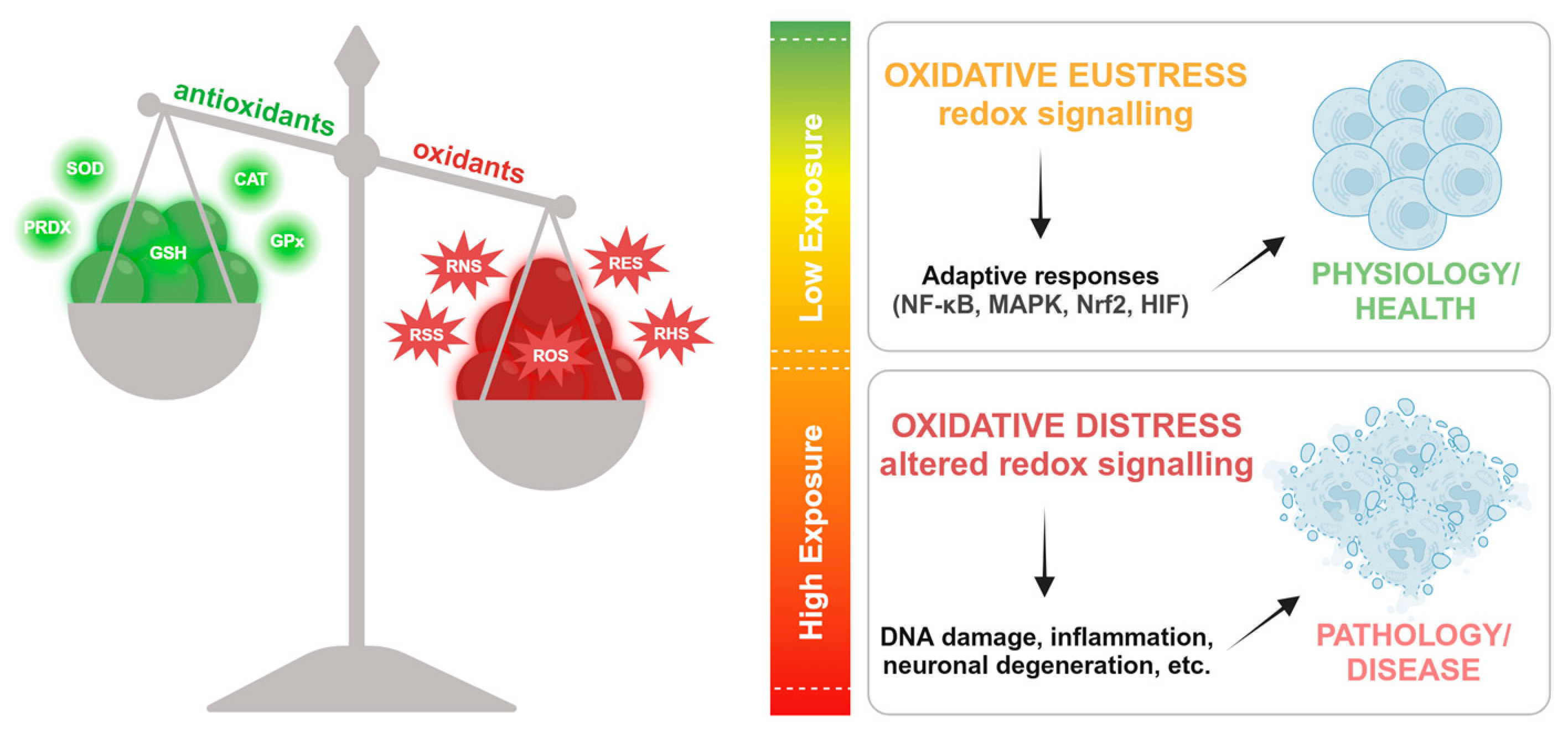
  15. Sies, H. Hydrogen Peroxide as a Central Redox Signaling Molecule in Physiological Oxidative Stress: Oxidative Eustress. Redox Biology 2017, 11, 613–619. [Google Scholar] [CrossRef]
  16. Sies, H. Oxidative Eustress and Oxidative Distress: Introductory Remarks. In Oxidative Stress; Elsevier, 2020; pp. 3–12 ISBN 978-0-12-818606-0.
  17. Sies, H. On the History of Oxidative Stress: Concept and Some Aspects of Current Development. Current Opinion in Toxicology 2018, 7, 122–126. [Google Scholar] [CrossRef]
  18. Ursini, F.; Maiorino, M.; Forman, H.J. Redox Homeostasis: The Golden Mean of Healthy Living. Redox Biology 2016, 8, 205–215. [Google Scholar] [CrossRef]
  19. Sies, H.; Jones, D.P. Reactive Oxygen Species (ROS) as Pleiotropic Physiological Signalling Agents. Nat Rev Mol Cell Biol 2020, 21, 363–383. [Google Scholar] [CrossRef] [PubMed]
  20. Möller, M.N.; Rios, N.; Trujillo, M.; Radi, R.; Denicola, A.; Alvarez, B. Detection and Quantification of Nitric Oxide–Derived Oxidants in Biological Systems. Journal of Biological Chemistry 2019, 294, 14776–14802. [Google Scholar] [CrossRef]
  21. Nakamura, T.; Lipton, S.A. Nitric Oxide-Dependent Protein Post-Translational Modifications Impair Mitochondrial Function and Metabolism to Contribute to Neurodegenerative Diseases. Antioxidants & Redox Signaling 2020, 32, 817–833. [Google Scholar] [CrossRef]
  22. Olson, K.R. Reactive Oxygen Species or Reactive Sulfur Species: Why We Should Consider the Latter. Journal of Experimental Biology 2020, 223, jeb196352. [Google Scholar] [CrossRef]
  23. Parvez, S.; Long, M.J.C.; Poganik, J.R.; Aye, Y. Redox Signaling by Reactive Electrophiles and Oxidants. Chem. Rev. 2018, 118, 8798–8888. [Google Scholar] [CrossRef]
  24. Davies, M.J.; Hawkins, C.L. The Role of Myeloperoxidase in Biomolecule Modification, Chronic Inflammation, and Disease. Antioxidants & Redox Signaling 2020, 32, 957–981. [Google Scholar] [CrossRef]
  25. Santolini, J.; Wootton, S.A.; Jackson, A.A.; Feelisch, M. The Redox Architecture of Physiological Function. Current Opinion in Physiology 2019, 9, 34–47. [Google Scholar] [CrossRef] [PubMed]
  26. Sies, H. Strategies of Antioxidant Defense. European Journal of Biochemistry 1993, 215, 213–219. [Google Scholar] [CrossRef]
  27. López-Otín, C.; Kroemer, G. Hallmarks of Health. Cell 2021, 184, 33–63. [Google Scholar] [CrossRef] [PubMed]
  28. Ayres, J.S. The Biology of Physiological Health. Cell 2020, 181, 250–269. [Google Scholar] [CrossRef]
  29. Demirovic, D.; Rattan, S.I.S. Establishing Cellular Stress Response Profiles as Biomarkers of Homeodynamics, Health and Hormesis. Experimental Gerontology 2013, 48, 94–98. [Google Scholar] [CrossRef]
  30. Lloyd, D.; Aon, M.A.; Cortassa, S. Why Homeodynamics, Not Homeostasis? The Scientific World JOURNAL 2001, 1, 133–145. [Google Scholar] [CrossRef] [PubMed]
  31. Selye, H. Stress and Distress. Compr Ther 1975, 1, 9–13. [Google Scholar]
  32. Lu, S.; Wei, F.; Li, G. The Evolution of the Concept of Stress and the Framework of the Stress System. CST 2021, 5, 76–85. [Google Scholar] [CrossRef]
  33. Sies, H.; Berndt, C.; Jones, D.P. Oxidative Stress. Annu. Rev. Biochem. 2017, 86, 715–748. [Google Scholar] [CrossRef]
  34. Jones, D.P.; Sies, H. The Redox Code. Antioxidants & Redox Signaling 2015, 23, 734–746. [Google Scholar] [CrossRef]
  35. Cortese-Krott, M.M.; Koning, A.; Kuhnle, G.G.C.; Nagy, P.; Bianco, C.L.; Pasch, A.; Wink, D.A.; Fukuto, J.M.; Jackson, A.A.; Van Goor, H.; et al. The Reactive Species Interactome: Evolutionary Emergence, Biological Significance, and Opportunities for Redox Metabolomics and Personalized Medicine. Antioxidants & Redox Signaling 2017, 27, 684–712. [Google Scholar] [CrossRef]
  36. Agarwal, A.; Virk, G.; Ong, C.; Du Plessis, S.S. Effect of Oxidative Stress on Male Reproduction. World J Mens Health 2014, 32, 1. [Google Scholar] [CrossRef] [PubMed]
  37. Kumar, N.; Singh, A.K. Reactive Oxygen Species in Seminal Plasma as a Cause of Male Infertility. Journal of Gynecology Obstetrics and Human Reproduction 2018, 47, 565–572. [Google Scholar] [CrossRef]
  38. Raj, C.J.; Aishwarya, C.V.S.; Mounika, K.V.S.S.N.; Mishra, B.; Sumithra, B.; Vishal, B.; Mandal, S.K. Deciphering the Nexus Between Oxidative Stress and Spermatogenesis: A Compendious Overview. In Oxidative Stress and Toxicity in Reproductive Biology and Medicine; Roychoudhury, S., Kesari, K.K., Eds.; Advances in Experimental Medicine and Biology; Springer International Publishing: Cham, 2022; Volume 1391, pp. 1–16. ISBN 978-3-031-12965-0. [Google Scholar]
  39. Tremellen, K. Oxidative Stress and Male Infertility—a Clinical Perspective. Human Reproduction Update 2008, 14, 243–258. [Google Scholar] [CrossRef]
  40. Kumar, N.; Singh, A.K. Impact of Environmental Factors on Human Semen Quality and Male Fertility: A Narrative Review. Environ Sci Eur 2022, 34, 6. [Google Scholar] [CrossRef]
  41. Agarwal, A.; Prabakaran, S.A. Mechanism, Measurement, and Prevention of Oxidative Stress in Male Reproductive Physiology. Indian J Exp Biol 2005, 43, 963–974. [Google Scholar]
  42. Tortolero, I.; Duarte Ojeda, J.M.; Pamplona Casamayor, M.; Alvarez González, E.; Arata-Bellabarba, G.; Regadera, J.; Leiva Galvis, O. [The effect of seminal leukocytes on semen quality in subfertile males with and without varicocele]. Arch Esp Urol 2004, 57, 921–928. [Google Scholar]
  43. Iommiello, V.M.; Albani, E.; Di Rosa, A.; Marras, A.; Menduni, F.; Morreale, G.; Levi, S.L.; Pisano, B.; Levi-Setti, P.E. Ejaculate Oxidative Stress Is Related with Sperm DNA Fragmentation and Round Cells. International Journal of Endocrinology 2015, 2015, 1–6. [Google Scholar] [CrossRef]
  44. Kurkowska, W.; Bogacz, A.; Janiszewska, M.; Gabryś, E.; Tiszler, M.; Bellanti, F.; Kasperczyk, S.; Machoń-Grecka, A.; Dobrakowski, M.; Kasperczyk, A. Oxidative Stress Is Associated with Reduced Sperm Motility in Normal Semen. Am J Mens Health 2020, 14, 1557988320939731. [Google Scholar] [CrossRef]
  45. Aitken, R.J.; Roman, S.D. Antioxidant Systems and Oxidative Stress in the Testes. In Molecular Mechanisms in Spermatogenesis; Cheng, C.Y., Ed.; Advances in Experimental Medicine and Biology; Springer New York: New York, NY, 2009; ISBN 978-0-387-79990-2. [Google Scholar]
  46. Asadi, N. The Impact of Oxidative Stress on Testicular Function and the Role of Antioxidants in Improving It: A Review. JCDR 2017. [Google Scholar] [CrossRef]
  47. Henkel, R.R. Leukocytes and Oxidative Stress: Dilemma for Sperm Function and Male Fertility. Asian J Androl 2011, 13, 43–52. [Google Scholar] [CrossRef] [PubMed]
  48. Vahedi Raad, M.; Firouzabadi, A.M.; Tofighi Niaki, M.; Henkel, R.; Fesahat, F. The Impact of Mitochondrial Impairments on Sperm Function and Male Fertility: A Systematic Review. Reprod Biol Endocrinol 2024, 22, 83. [Google Scholar] [CrossRef]
  49. Chandra, S.; Romero, M.; Shatanawi, A.; Alkilany, A.; Caldwell, R.; Caldwell, R. Oxidative Species Increase Arginase Activity in Endothelial Cells through the RhoA/Rho Kinase Pathway. British J Pharmacology 2012, 165, 506–519. [Google Scholar] [CrossRef] [PubMed]
  50. Dare, B. Role of Antioxidant in Testicular Integrity. ARRB 2014, 4, 998–1023. [Google Scholar] [CrossRef]
  51. Nowicka-Bauer, K.; Nixon, B. Molecular Changes Induced by Oxidative Stress That Impair Human Sperm Motility. Antioxidants 2020, 9, 134. [Google Scholar] [CrossRef]
  52. Hu, R.; Yang, X.; He, J.; Wu, S. Oxidative Stress and Autophagy: Unraveling the Hidden Threat to Boars’ Fertility. Antioxidants 2024, 14, 2. [Google Scholar] [CrossRef]
  53. Washburn, R.L.; Hibler, T.; Kaur, G.; Dufour, J.M. Sertoli Cell Immune Regulation: A Double-Edged Sword. Front. Immunol. 2022, 13, 913502. [Google Scholar] [CrossRef]
  54. Zhang, P.; Li, F.; Zhang, L.; Lei, P.; Zheng, Y.; Zeng, W. Stage-specific Embryonic Antigen 4 Is a Membrane Marker for Enrichment of Porcine Spermatogonial Stem Cells. Andrology 2020, 8, 1923–1934. [Google Scholar] [CrossRef]
  55. Aly, H.A.A.; Khafagy, R.M. 2,3,7,8-Tetrachlorodibenzo-p-Dioxin (TCDD)-Induced Cytotoxicity Accompanied by Oxidative Stress in Rat Sertoli Cells: Possible Role of Mitochondrial Fractions of Sertoli Cells. Toxicology and Applied Pharmacology 2011, 252, 273–280. [Google Scholar] [CrossRef]
  56. Yilmaz, B.; Yildizbayrak, N.; Aydin, Y.; Erkan, M. Evidence of Acrylamide- and Glycidamide-Induced Oxidative Stress and Apoptosis in Leydig and Sertoli Cells. Hum Exp Toxicol 2017, 36, 1225–1235. [Google Scholar] [CrossRef] [PubMed]
  57. Duan, P.; Quan, C.; Huang, W.-T.; Yang, K. [PI3K-Akt/LKB1-AMPK-mTOR-p70S6K/4EBP1 signaling pathways participate in the regulation of testis development and spermatogenesis: An update]. Zhonghua Nan Ke Xue 2016, 22, 1016–1020. [Google Scholar] [PubMed]
  58. Cinthya Riris, A.A.I.D.; I’tishom, R.; Khaerunnisa, S. Role of Antioxidant to Protect Leydig Cells Induced by Reactive Oxygen Species: A Literature Review. Qanun Medika 2021, 5, 49. [Google Scholar] [CrossRef]
  59. Wang, Y.; Chen, F.; Ye, L.; Zirkin, B.; Chen, H. Steroidogenesis in Leydig Cells: Effects of Aging and Environmental Factors. Reproduction 2017, 154, R111–R122. [Google Scholar] [CrossRef]
  60. Aitken, R.J.; Baker, M.A. Oxidative Stress, Sperm Survival and Fertility Control. Molecular and Cellular Endocrinology 2006, 250, 66–69. [Google Scholar] [CrossRef]
  61. Cafe, S.L.; Nixon, B.; Dun, M.D.; Roman, S.D.; Bernstein, I.R.; Bromfield, E.G. Oxidative Stress Dysregulates Protein Homeostasis Within the Male Germ Line. Antioxidants & Redox Signaling 2020, 32, 487–503. [Google Scholar] [CrossRef]
  62. Cafe, S.L.; Nixon, B.; Ecroyd, H.; Martin, J.H.; Skerrett-Byrne, D.A.; Bromfield, E.G. Proteostasis in the Male and Female Germline: A New Outlook on the Maintenance of Reproductive Health. Front Cell Dev Biol 2021, 9, 660626. [Google Scholar] [CrossRef]
  63. Duong, L.D.; West, J.D.; Morano, K.A. Redox Regulation of Proteostasis. Journal of Biological Chemistry 2024, 300, 107977. [Google Scholar] [CrossRef]
  64. Hipp, M.S.; Hartl, F.U. Interplay of Proteostasis Capacity and Protein Aggregation: Implications for Cellular Function and Disease. Journal of Molecular Biology 2024, 436, 168615. [Google Scholar] [CrossRef]
  65. Long, S.; Kenworthy, S. Round Cells in Diagnostic Semen Analysis: A Guide for Laboratories and Clinicians. Br J Biomed Sci 2022, 79, 10129. [Google Scholar] [CrossRef]
  66. Pavuluri, H.; Bakhtiary, Z.; Panner Selvam, M.K.; Hellstrom, W.J.G. Oxidative Stress-Associated Male Infertility: Current Diagnostic and Therapeutic Approaches. Medicina 2024, 60, 1008. [Google Scholar] [CrossRef] [PubMed]
  67. Alahmar, A.T. Role of Oxidative Stress in Male Infertility: An Updated Review. J Hum Reprod Sci 2019, 12, 4–18. [Google Scholar] [CrossRef]
  68. Simioni, C.; Zauli, G.; Martelli, A.M.; Vitale, M.; Sacchetti, G.; Gonelli, A.; Neri, L.M. Oxidative Stress: Role of Physical Exercise and Antioxidant Nutraceuticals in Adulthood and Aging. Oncotarget 2018, 9, 17181–17198. [Google Scholar] [CrossRef] [PubMed]
  69. Jena, A.B.; Samal, R.R.; Bhol, N.K.; Duttaroy, A.K. Cellular Red-Ox System in Health and Disease: The Latest Update. Biomedicine & Pharmacotherapy 2023, 162, 114606. [Google Scholar] [CrossRef]
  70. Bo, H.; Jiang, N.; Ji, L.L.; Zhang, Y. Mitochondrial Redox Metabolism in Aging: Effect of Exercise Interventions. Journal of Sport and Health Science 2013, 2, 67–74. [Google Scholar] [CrossRef]
  71. Meng, Q.; Su, C.-H. The Impact of Physical Exercise on Oxidative and Nitrosative Stress: Balancing the Benefits and Risks. Antioxidants 2024, 13, 573. [Google Scholar] [CrossRef]
  72. Powers, S.K.; Deminice, R.; Ozdemir, M.; Yoshihara, T.; Bomkamp, M.P.; Hyatt, H. Exercise-Induced Oxidative Stress: Friend or Foe? Journal of Sport and Health Science 2020, 9, 415–425. [Google Scholar] [CrossRef]
  73. Radak, Z.; Ishihara, K.; Tekus, E.; Varga, C.; Posa, A.; Balogh, L.; Boldogh, I.; Koltai, E. Exercise, Oxidants, and Antioxidants Change the Shape of the Bell-Shaped Hormesis Curve. Redox Biology 2017, 12, 285–290. [Google Scholar] [CrossRef]
  74. Assidi, M. Infertility in Men: Advances towards a Comprehensive and Integrative Strategy for Precision Theranostics. Cells 2022, 11, 1711. [Google Scholar] [CrossRef]
  75. Greco, F.; Guarascio, G.; Giannetta, E.; Oranges, F.P.; Quinzi, F.; Emerenziani, G.P.; Tarsitano, M.G. The Influence of an Intense Training Regime in Professional and Non-Professional Athletes on Semen Parameters: A Systematic Review. JCM 2025, 14, 201. [Google Scholar] [CrossRef]
  76. Sgrò, P.; Di Luigi, L. Sport and Male Sexuality. J Endocrinol Invest 2017, 40, 911–923. [Google Scholar] [CrossRef] [PubMed]
  77. Di Luigi, L.; Gentile, V.; Pigozzi, F.; Parisi, A.; Giannetti, D.; Romanelli, F. Physical Activity as a Possible Aggravating Factor for Athletes with Varicocele: Impact on the Semen Profile. Hum Reprod 2001, 16, 1180–1184. [Google Scholar] [CrossRef]
  78. Abedpoor, N.; Taghian, F.; Hajibabaie, F. Exploring the Dynamics of Exercise Intensity on Male Fertility and Reproductive Health: Advancements and Implications for Fertility Research. Front. Reprod. Health 2024, 6, 1423916. [Google Scholar] [CrossRef] [PubMed]
  79. Minas, A.; Fernandes, A.C.C.; Maciel Júnior, V.L.; Adami, L.; Intasqui, P.; Bertolla, R.P. Influence of Physical Activity on Male Fertility. Andrologia 2022, 54. [Google Scholar] [CrossRef]
  80. Lavín-Pérez, A.M.; Collado-Mateo, D.; Villafaina, S.; Calle-Guisado, V. The Role of Exercise to Reduce the Impact of Diabetes in the Seminal Quality: A Systematic Review. Medicina 2021, 57, 159. [Google Scholar] [CrossRef] [PubMed]
  81. Antinozzi, C.; Duranti, G.; Ceci, R.; Lista, M.; Sabatini, S.; Caporossi, D.; Di Luigi, L.; Sgrò, P.; Dimauro, I. Hydrogen Peroxide Stimulates Dihydrotestosterone Release in C2C12 Myotubes: A New Perspective for Exercise-Related Muscle Steroidogenesis? IJMS 2022, 23, 6566. [Google Scholar] [CrossRef]
  82. Lyons, H.E.; Gyawali, P.; Mathews, N.; Castleton, P.; Mutuku, S.M.; McPherson, N.O. The Influence of Lifestyle and Biological Factors on Semen Variability. J Assist Reprod Genet 2024, 41, 1097–1109. [Google Scholar] [CrossRef]
  83. Mehri, K.; Hamidian, G.; Babri, S.; Farajdokht, F.; Zavvari Oskuye, Z. Exercise and Insulin Glargine Administration in Mothers with Diabetes during Pregnancy Ameliorate Function of Testis in Offspring: Consequences on Apelin-13 and Its Receptor. Life Sciences 2024, 342, 122517. [Google Scholar] [CrossRef]
  84. Morano, S.; Gatti, A.; Mandosi, E.; Tiberti, C.; Fallarino, M.; Cipriani, R.; Buchetti, B.; Gandini, L.; Sgrò, P.; Jannini, E.A.; et al. Circulating Monocyte Oxidative Activity Is Increased in Patients With Type 2 Diabetes and Erectile Dysfunction. Journal of Urology 2007, 177, 655–659. [Google Scholar] [CrossRef]
  85. Zampieri, N. The Effect of Aerobic Training on Serum Levels of Adiponectin, Hypothalamic-Pituitary-Gonadal Axis and Sperm Quality in Diabetic Rats. Urology Journal 2019. [Google Scholar] [CrossRef]
  86. Antinozzi, C.; Lista, M.; Caponecchia, L.; Salacone, P.; Minganti, C.; Battaglia, F.A.; Di Luigi, L.; Sgrò, P. Exploratory Analysis in the Differences in Blood Serum and Seminal Plasma of Adipose-Tissue Related Peptides in Obese and Non-Obese Men and Their Correlations With Semen Parameters. Front. Endocrinol. 2021, 12, 681939. [Google Scholar] [CrossRef]
  87. Green, D.J.; Chasland, L.C.; Yeap, B.B.; Naylor, L.H. Comparing the Impacts of Testosterone and Exercise on Lean Body Mass, Strength and Aerobic Fitness in Aging Men. Sports Med Open 2024, 10, 30. [Google Scholar] [CrossRef] [PubMed]
  88. Belladelli, F.; Basran, S.; Eisenberg, M.L. Male Fertility and Physical Exercise. World J Mens Health 2023, 41, 482. [Google Scholar] [CrossRef]
  89. Di Luigi, L.; Sgrò, P.; Fierro, V.; Bianchini, S.; Battistini, G.; Magini, V.; Jannini, E.A.; Lenzi, A. Prevalence of Undiagnosed Testosterone Deficiency in Aging Athletes: Does Exercise Training Influence the Symptoms of Male Hypogonadism? The Journal of Sexual Medicine 2010, 7, 2591–2601. [Google Scholar] [CrossRef]
  90. Jóźków, P.; Rossato, M. The Impact of Intense Exercise on Semen Quality. Am J Mens Health 2017, 11, 654–662. [Google Scholar] [CrossRef]
  91. Hajizadeh Maleki, B.; Tartibian, B.; Chehrazi, M. The Effects of Three Different Exercise Modalities on Markers of Male Reproduction in Healthy Subjects: A Randomized Controlled Trial. Reproduction 2017, 153, 157–174. [Google Scholar] [CrossRef] [PubMed]
  92. Gebreegziabher, Y.; Marcos, E.; McKinon, W.; Rogers, G. Sperm Characteristics of Endurance Trained Cyclists. Int J Sports Med 2004, 25, 247–251. [Google Scholar] [CrossRef] [PubMed]
  93. Duclos, M.; Tabarin, A. Exercise and the Hypothalamo-Pituitary-Adrenal Axis. In Frontiers of Hormone Research; Lanfranco, F., Strasburger, C.J., Eds.; S. Karger AG, 2016; Vol. 47, pp. 12–26 ISBN 978-3-318-05868-0.
  94. Brownlee, K.K.; Moore, A.W.; Hackney, A.C. Relationship between Circulating Cortisol and Testosterone: Influence of Physical Exercise. J Sports Sci Med 2005, 4, 76–83. [Google Scholar]
  95. Duclos, M.; Guinot, M.; Le Bouc, Y. Cortisol and GH: Odd and Controversial Ideas. Appl. Physiol. Nutr. Metab. 2007, 32, 895–903. [Google Scholar] [CrossRef]
  96. Snyder, A.C.; Hackney, A.C. The Endocrine System in Overtraining. In Endocrinology of Physical Activity and Sport; Constantini, N., Hackney, A.C., Eds.; Humana Press: Totowa, NJ, 2013; pp. 523–534. ISBN 978-1-62703-313-8. [Google Scholar]
  97. Sansone, M.; Sansone, A.; Borrione, P.; Romanelli, F.; Di Luigi, L.; Sgrò, P. Effects of Ketone Bodies on Endurance Exercise. Curr Sports Med Rep 2018, 17, 444–453. [Google Scholar] [CrossRef]
  98. Walke, G.; Gaurkar, S.S.; Prasad, R.; Lohakare, T.; Wanjari, M. The Impact of Oxidative Stress on Male Reproductive Function: Exploring the Role of Antioxidant Supplementation. Cureus 2023. [Google Scholar] [CrossRef]
  99. Torres-Arce, E.; Vizmanos, B.; Babio, N.; Márquez-Sandoval, F.; Salas-Huetos, A. Dietary Antioxidants in the Treatment of Male Infertility: Counteracting Oxidative Stress. Biology 2021, 10, 241. [Google Scholar] [CrossRef]
  100. Yavari, A.; Javadi, M.; Mirmiran, P.; Bahadoran, Z. Exercise-Induced Oxidative Stress and Dietary Antioxidants. Asian J Sports Med 2015, 6, e24898. [Google Scholar] [CrossRef] [PubMed]
Figure 1. Redox balance and its physiological and pathological implications. The equilibrium between antioxidants (SOD, CAT, PRDX, GSH, GPx) and oxidants (RNS, RSS, ROS, RES, RHS) determines cellular redox responses. Low oxidant exposure induces oxidative eustress, promoting adaptive signaling (NF-κB, MAPK, Nrf2, HIF) and contributing to physiology and health. In contrast, high oxidant exposure leads to oxidative distress, disrupting redox signaling and causing pathological consequences such as DNA damage, inflammation, and neuronal degeneration. ROS, reactive oxygen species; RNS, reactive nitrogen species; RSS, reactive sulfur species; RES, reactive electrophile species; RHS, and reactive halogen species.
Preprints 148654 g001
Figure 2. Endogenous and exogenous factors that alter oxidant/antioxidant balance, and that lead to oxidative stress and sperm dysfunctions.
Preprints 148654 g002
Figure 3. Physical activity can improve sperm quality by acting on various aspects of an individual's health. Increases steroidogenesis (in muscle and testes), improves redox balance, enhances antioxidant and reduces metabolic dysfunction. In addition, these factors interact and influence each other to improve sperm quality.
Preprints 148654 g003
Disclaimer/Publisher’s Note: The statements, opinions and data contained in all publications are solely those of the individual author(s) and contributor(s) and not of MDPI and/or the editor(s). MDPI and/or the editor(s) disclaim responsibility for any injury to people or property resulting from any ideas, methods, instructions or products referred to in the content.
Copyright: This open access article is published under a Creative Commons CC BY 4.0 license, which permit the free download, distribution, and reuse, provided that the author and preprint are cited in any reuse.
Prerpints.org logo

Preprints.org is a free preprint server supported by MDPI in Basel, Switzerland.

Subscribe

Disclaimer

Terms of Use

Privacy Policy

Privacy Settings

© 2025 MDPI (Basel, Switzerland) unless otherwise stated