Preprint
Article

This version is not peer-reviewed.

Outbreak of Carbapenem Resistant High-Risk Clone ST244 Pseudomonas aeruginosa in Dogs and Cats in Algeria

A peer-reviewed article of this preprint also exists.

Submitted:

07 February 2025

Posted:

10 February 2025

You are already at the latest version

Abstract

Background/Objectives: Pseudomonas aeruginosa causes chronic infections in humans and animals, especially cats and dogs. This bacterium's ability to adapt and acquire antibiotic resistance traits may complicate and exacerbate antibacterial therapy. This study aimed to evaluate the antibiotic resistance patterns, virulence factors and ability to form biofilm of P. aeruginosa strains isolated from Algerian dogs and cats. Methods: Nineteen samples were collected from healthy and diseased dogs and cats. Isolates were studied for their antibiotic resistance patterns (disc diffusion method), biofilm formation (Microtiter assay) and were Whole genome sequenced (MinION). Results: Nineteen P. aeruginosa strains (15 from dogs and 4 from cats) were isolated. Antibiotic resistance phenotypes were observed against amoxicillin-clavulanic acid (100%), meanwhile resistance towards ticarcillin was 40% (dogs) and 25% (cats), ticarcillin-clavulanic acid was 13.33% and 25% for dogs and cats respectively and imipenem was 75% (cats) and 20% (dogs). 95% of strains were biofilm producers. Different antimicrobial resistance genes (ARGs) were found: beta-lactamase genes mainly PAO, OXA-494, OXA-50 and OXA-396, aminoglycosides gene (aph(3’)-IIb), fosA for fosfomycin and catB7 for phenicol. The main high risk STs were ST244, 2788, 388 and 1247. A large panel of virulence genes was detected: exoS, exoT, exoY, lasA, toxA, prpL, algD, rhIA and others. Conclusions: The genetic variety in antibiotic resistance genes of resistant and virulent P. aeruginosa strains in dogs makes public health protection difficult. Continuous monitoring and research in compliance with the One Health policy are needed to solve this problem.

Keywords: 
;  ;  ;  ;  ;  ;  ;  ;  ;  

1. Introduction

Pseudomonas aeruginosa is an important Gram-negative opportunistic pathogen of humans and animals [1]. In dogs, this bacterium can cause various infections, including ulcerative keratitis, otitis, pyoderma, urinary tract infections, skin infection, wound infections, and respiratory tract infections [2]. Infections caused by P. aeruginosa may be linked to immunosuppression in companion animals, including documented cases in dog’s post-kidney transplantation[3] and in connection with cancer treatments [4]. Moreover, it acts as a pathogen in cats, although it is less common than in dogs [5,6] and the reported infections include respiratory tract infections [3,7] as well as ulcerative keratitis and wound infections [8].
Due to its increased resistance to antibiotics, the World Health Organization (WHO) has classified P. aeruginosa carbapenem-resistant as a high-priority bacterium. It was designated as one of the ESKAPE pathogens (Enterococcus faecium, Staphylococcus aureus, Klebsiella pneumoniae, Acinetobacter baumannii, Pseudomonas aeruginosa and Enterobacter species) because of its capacity to escape killing by acquiring exogenous genes and developing resistance through a variety of internal pathways, which promotes the emergence of multidrug resistant strains [9,10]. The European Food Safety Authority's Panel on Animal Health and Welfare has classified P. aeruginosa as one of the most significant antimicrobial-resistant bacteria in the European Union (EU), with over 90% certainty [11].
P. aeruginosa infections are worrying, because their treatment poses a major global challenge due to the development of resistant strains in both humans and animals. Antibiotics have become less effective, or even ineffective, against this bacterium, due to its resistance mechanisms, which lead to therapeutic failure. The main resistance mechanisms are beta lactamase production, efflux pumps, induced mutations and biofilm production [12]. Some authors reported that P. aeruginosa strains showed a high rate of resistance to multiple antimicrobial agents [13]. Its infections in pets are currently generally treated with broad-spectrum antibiotics [14] and close interactions between pets and humans make significant opportunities for interspecies transmission of resistant bacteria and horizontal transfer of antibiotic resistance genes in both directions mainly through physical injuries, petting, or licking activities [15]. P. aeruginosa is also recognized for its ability to quickly acquire additional resistances, meaning that the combination of intrinsic and acquired resistance can result in therapeutic failures [16].
Biofilm formation is a crucial survival strategy used by P. aeruginosa to endure challenging conditions such as exposure to antibiotics and host immune defenses [17]. Moreover, it displays various virulence factors, including, exotoxins (toxA, toxR), elastases (lasB), proteases (plcH) and alginate (algD) all of which contribute to the development of severe diseases. A significant virulence factor is the Type 3 Secretion System (T3SS), which delivers four cytotoxins, including exoU [18].
There are few reports documenting the patterns of antimicrobial resistance and virulence factors of P. aeruginosa isolated from companion animals, and until now there are no studies focused on the epidemiology of this pathogen in Algeria. The aim of the study was to investigate the antimicrobial resistance profiles, and the associated resistance and virulence genes of Pseudomonas aeruginosa strains isolated from dogs and cats in some regions of Eastern Algeria. As the first study of its kind in Algeria, it aimed to provide valuable insights into the epidemiological characteristics and potential health risks posed by these strains in companion animals, highlighting their implications for veterinary and public health.

2. Results

2.1. General Population Information

Table 1 summarizes the basic information of each analyzed P. aeruginosa strain. Fifteen were isolated from dogs, mostly from nasal swabs (n=6), followed by rectum (n=5) and middle ear (n=4). Four cats were positive for P. aeruginosa from rectum. One isolate was collected per animal.

2.2. Antimicrobial Susceptibility Testing

Table 2 displays the prevalence of resistance to each antimicrobial agent. All the strains were resistant to AMC in both cats and dogs, 36.8% and 15.8% of the strains were resistant to TC and TCC from dogs (40%) and cats (25%), respectively. The resistance rate to Imipenem was 36.8%, with 20% in dogs and 75% in cats. No resistance towards aminoglycosides and fluoroquinolones was detected. None of the isolates were multidrug resistant.

2.3. Biofilm Formation

Diverse biofilm profiles were identified, 42% (n=8) classified as strong biofilm producers, 25% (n=5) as moderate producers, all isolated from dogs, and 27% (n=5) as weak producers, including four isolates from cats and one from a dog. One strain, a non-producer, was identified from a dog. All isolates identified as strong producers were derived from dogs; 37.5% were isolated from the rectum, with an equal amount from the nasal cavity. Additionally, 60% of the strains exhibiting moderate production in dogs were isolated from the nasal cavity. There is no relation between the sampling site and the ability of biofilm production (p= 0.430).

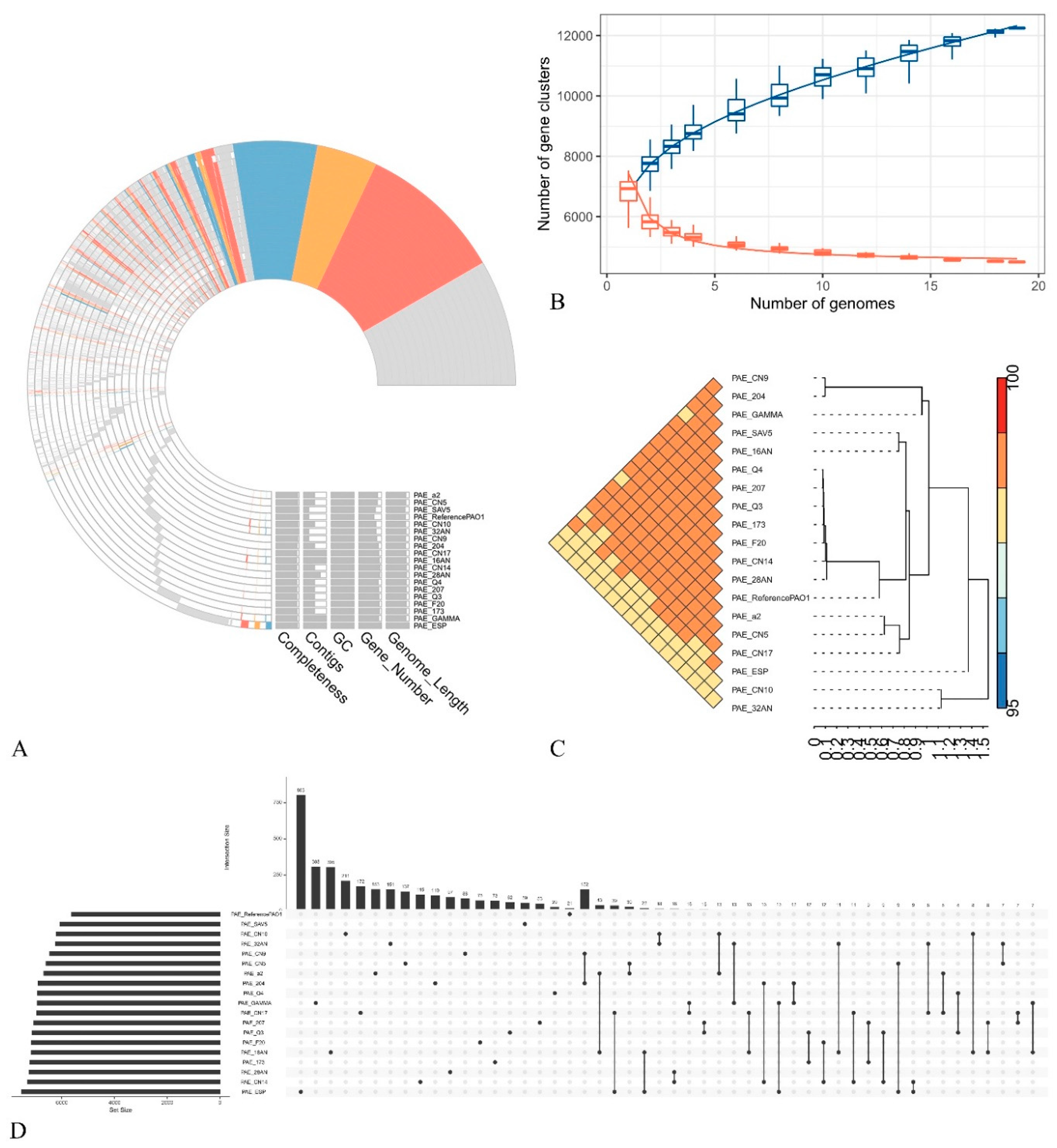
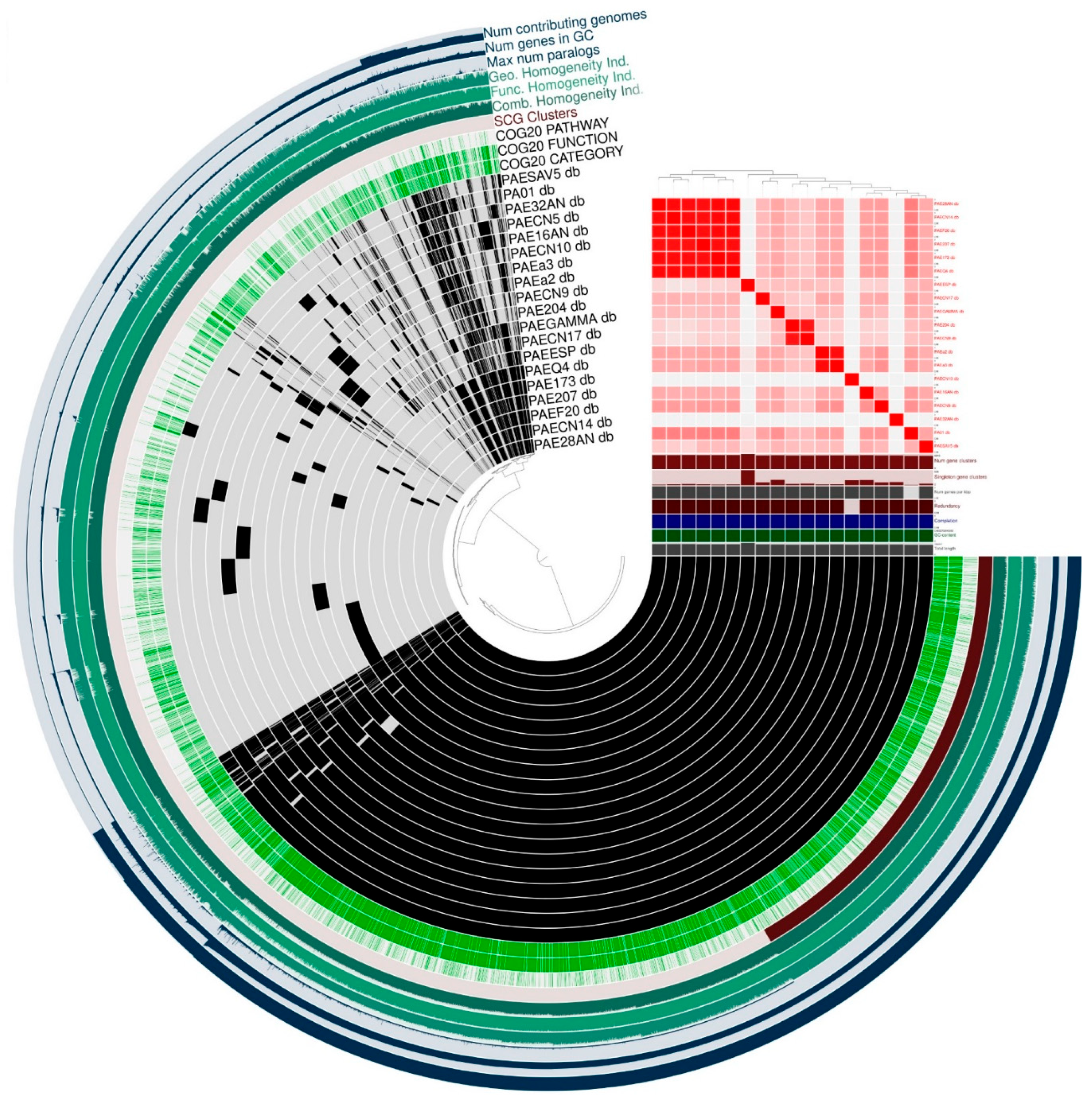
2.3. General Features of the Genomes

All the strains were uploaded to NCBI database under Bioproject PRJNA1153397. The sequencing data from the Nanopore Mk1C after genome assembly include a mean coverage of 120× with N50 of 6216739 bp and a genome size of 6560953.684 bp. Table 3 summarizes the main statistics of the genomes assembled and polished.
Whole-genome based phylogenetic tree (Figure 1) was built with the online tool Integrated Prokaryotes Genome and pan-genome Analysis service IPGA (v1.09) (accessed on June 13th 2024) including the reference strain (PAO1) used during the bioinformatic analyses [19].
The pan-genome profile of all the P. aeruginosa strains is reported in figure 2A. In red are represented metabolism genes, in orange those related to information storage and processing, while genes involved in cellular processes and signaling are in blue, finally the grey ones are poorly characterized or unannotated genes. Together these components constitute the core genes shared between the analyzed strains. As shown in Figure 2B, a total of 12,250 pan-gene clusters were identified. The Average Nucleotide Identity (ANI) analysis (Figure 2C) revealed a high (>98%) identity among all the strains. The upset plot (Figure 2D) indicated that the distinct gene clusters in each genome ranged from 15 to 803. The pan-genome profile derived from Clusters of Orthologous Genes (COG) annotation indicated that the core gene clusters comprised 2480 for metabolism, 1102 for information storage and processing, 1521 for cellular functions and signaling, and 718 were poorly characterized or unannotated. Figure 3 represent the pangenomes visualized using ANVI’O to address various aspects of interactive displays.

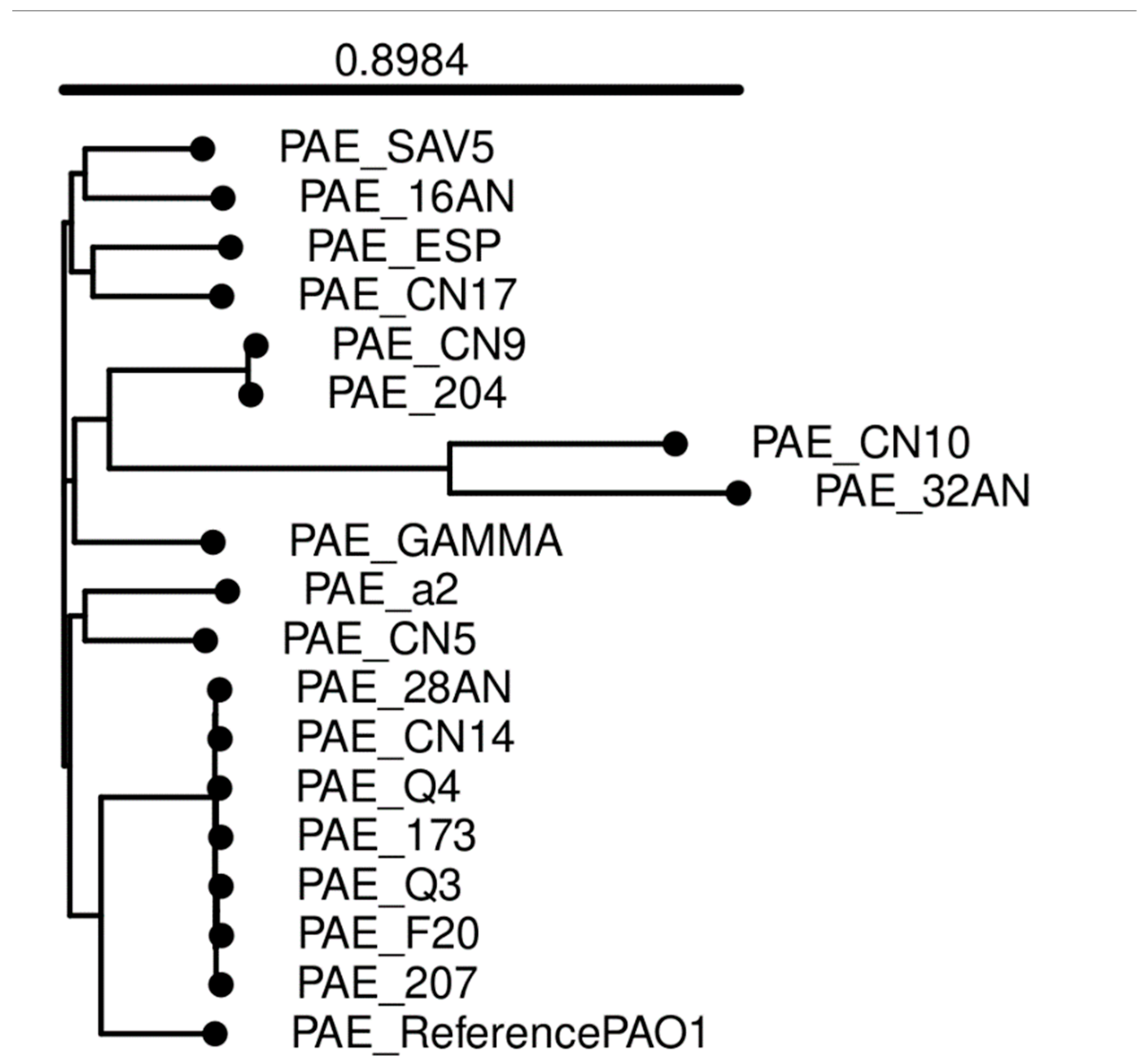
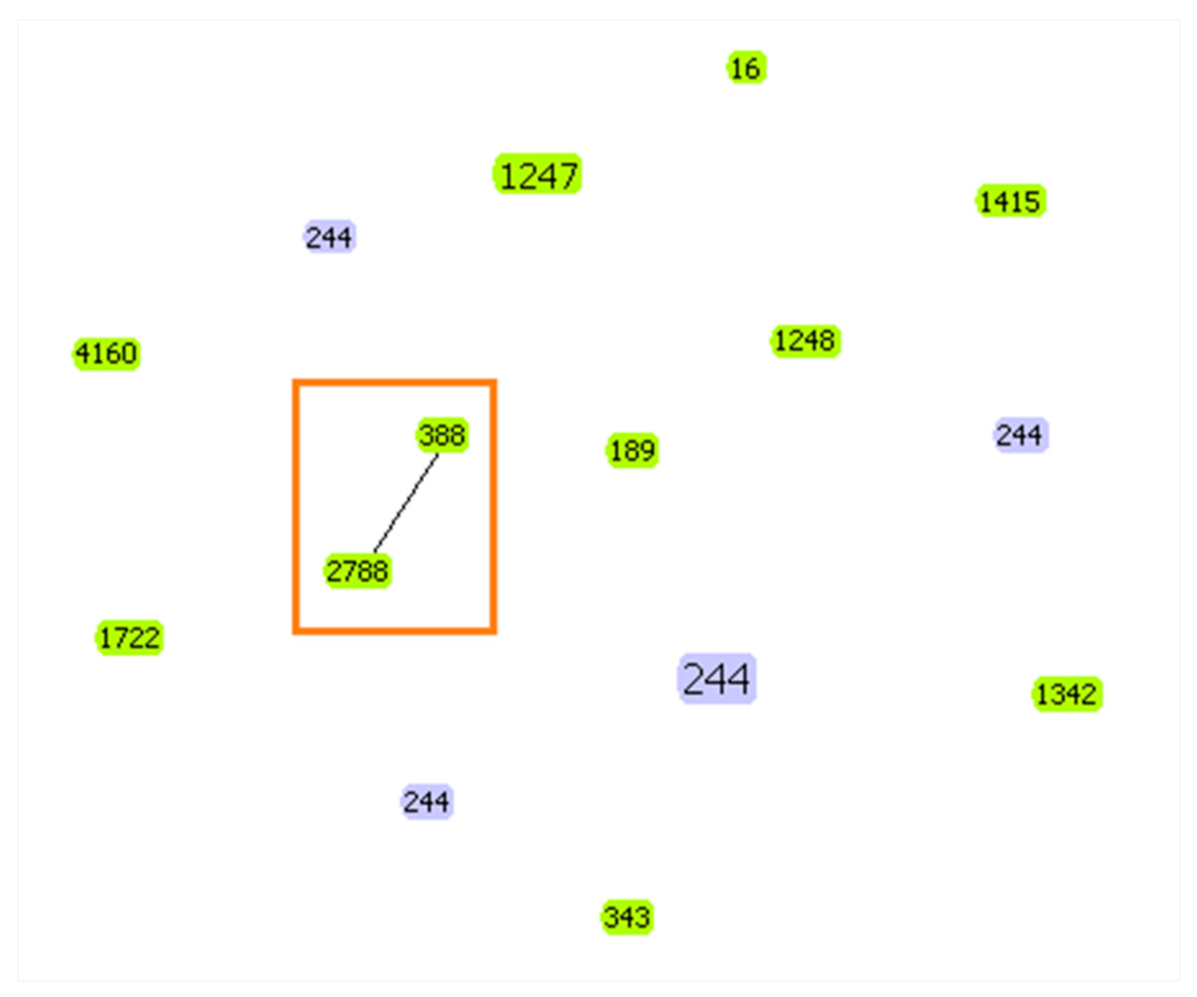
2.4. Multilocus Sequence Typing (MLST)

MLST analysis revealed 12 different sequence types (ST4160, ST1248, ST1247, ST2788, ST1722, ST189, ST343, ST16, ST1415, ST388, ST244, ST1342). The predominant ST was the high-risk clone ST244 found in 7 strains (2 from cats and 5 from dogs) firstly described in pets in Algeria. These strains were isolated from different sampling sites, rectum in cats and from the rectum, ear, nasal cavity in dogs. On the other hand, two other sequence types were isolated from cats (ST1247 and ST1342) and the others STs were recovered from dogs. One clonal complex (Figure 4) was displayed comprising two STs (2788 and 388) with a double loci variant, and the rest which did not share at least two out of the seven loci, were considered as singletons STs. The isolates from the same ST are clustered together according to the phylogenetic tree. Figure 4 was generated using the eBURST software [20].

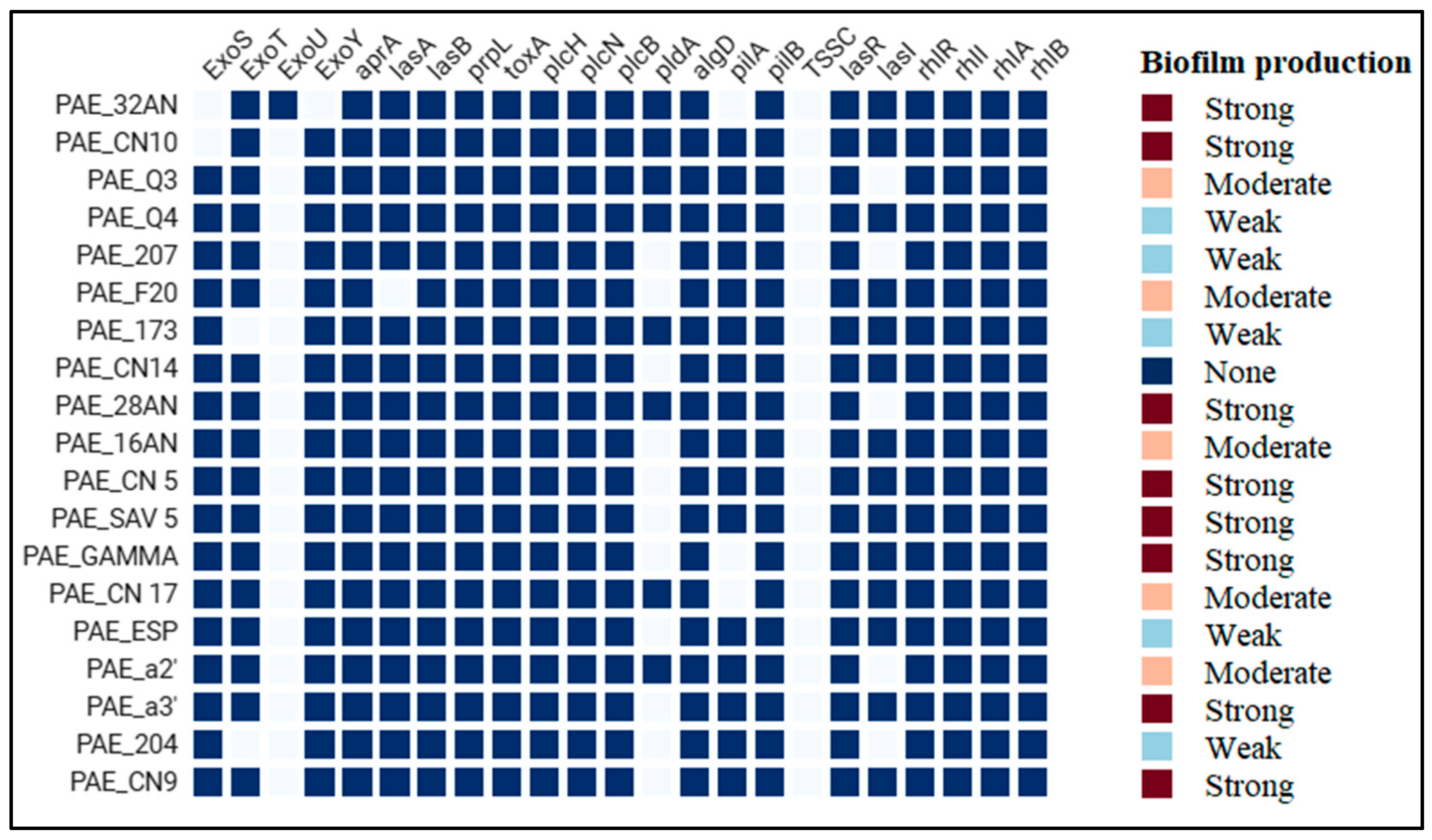
2.5. Antibiotic Resistance and Virulence Genes
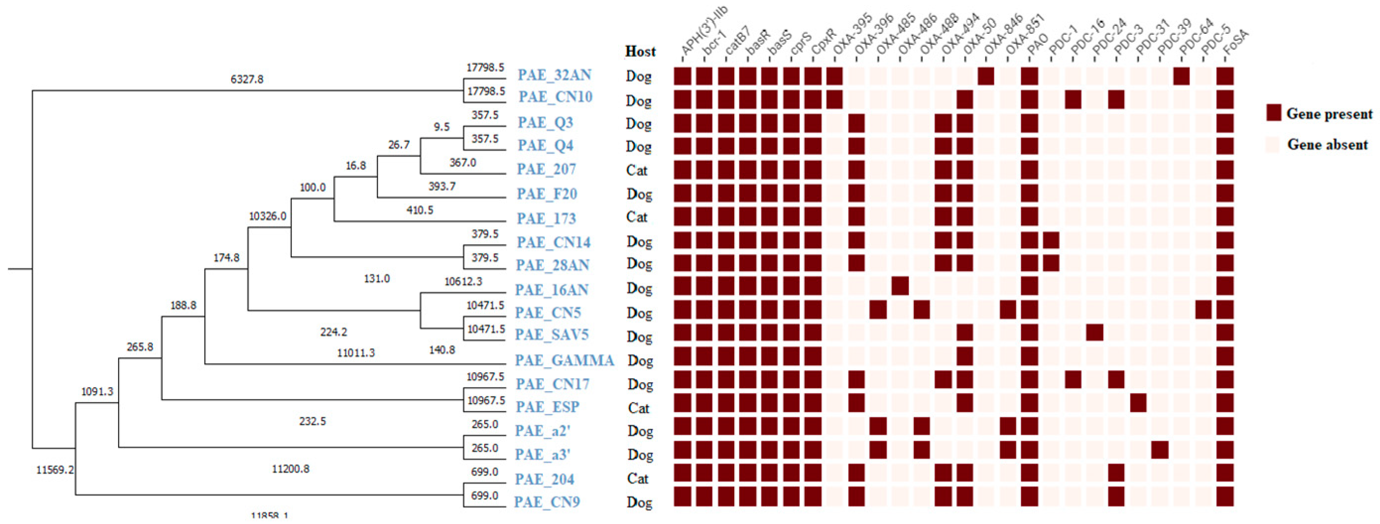
A total of 76 resistance genes were found in P. aeruginosa genomes (figure 5). Most of them were associated with the efflux pump systems. The genes aph(3’)IIb, bcr-1, catB7 and fosA, conferring resistance to aminoglycosides, bicyclomicin, phenicoles and fosfomycin respectively, were present in all isolates. Furthermore, the peptide resistance genes basR, basS, cprS and cpxR were also identified in all the isolates.
The results from WGS showed that all the strains harbored at least two genes responsible for betalactam resistance. The predominant gene was blaPAO found in all isolates, followed by blaOXA-50 and blaOXA-396 in 73.7% and 57.9% of the strains respectively.
All the strains resistant to imipenem harbored the following genes OXA396, OXA494, OXA50 and blaPAO. The combination between blaOXA and blaPDC was found in 57.9% of the strains. 84.2 % of the strains harbored more than three genes responsible for beta lactam resistance. Aminoglycoside resistance gene aph(3’)IIb was detected in all the isolates. At least one of the blaPDC variants was present in 11 strains (57.9%).
Figure 5. Phylogenetic tree and distribution of resistance genes among analyzed strains.
Preprints 148650 g005
All genomes were screened for virulence factors (Figure 6), resulting in the identification of those responsible for motility and adhesion, quorum sensing, biofilm production, type III secretion system, siderophores production, proteases, toxins, and enzymes.
A total of 80 genes associated with adherence and motility were detected, with variations observed in their distribution among the isolates. All of them contained the gene essential for lipopolysaccharide (LPS) production. Of the 46 genes involved in flagellar assembly, 35 were present across all strains, with fleS gene being the most frequently absent, missing in 63.2% of the genomes. Regarding the genes responsible for type IV pili biosynthesis, pilA and pilB were found in 84.2% and 100 % of the strains, respectively.
The frequency of exoU and exoS was 5.3% (1/19) and 89.5 % (17/19), respectively.
WGS analysis revealed that the predominant type three secretion system (T3SS) virulotypes were exoU - / exoS + found in 17 strains (89.5%), exoU + / exoS – and exoU - / exoS in one isolate each.
Proteases, toxins and enzymes are among the most effective virulence factors that contribute in the severity of Pseudomonas aeruginosa infection. All the isolates harbored the genes aprA, lasB and prpL coding for proteases. The toxA gene coding for the exotoxine A was also present in all the isolates. Regarding genes encoding enzymes plcH, plcN, plcB were present in all isolates, while pldA was found in only 8 isolates (42.1%)
Additionally, the isolated P. aeruginosa strains exhibited a 100% prevalence of the quorum sensing genes lasR, rhlR, and rhlI, while the lasI gene was identified in 73.7% of the strains.
Alginates and lipopolysaccharide are essential for the biofilm formation and the genes encoding their synthesis algD and LpS were found in all the strains.

3. Discussion

P. aeruginosa is a significant opportunistic and challenging to treat bacterium. This pathogen aligns with the concept of "One Health" [21] due to its unique characteristics, including extensive environmental diffusion [22], intrinsic resistance to several classes of antibiotics [23], high capacity to acquire new resistance mechanisms [24] and numerous virulence factors [25].
In the present study, P. aeruginosa was isolated at a low prevalence of 7.5% and 1.91%, respectively, from the middle ear, rectum, and nasal cavities of canines and cats.. These rates are lower than those documented in an earlier investigation in Thailand by [26] with 79.6% and 20.4%. In dogs, our results are similar to previous reports in Italy (8%) [24] and China (6.7%) [27]. However, prevalence is higher than earlier reports in India and California (3% each) [28,29]. Other studies conducted in Romania (40.84%) [30], South Korea (18.75%) [31] and Brazil (31.62%) [32] had reported high prevalence rates than our findings. In cats, we observed a prevalence of 1.9%, which is close to the results reported by Gentilini et al., (2018) in Italy with 1.41% [33].
With no MDR or XDR isolates, the antibiotic resistance profile of the isolates from this study was lower than that of academic research on P. aeruginosa isolated from dogs and cats. Research in Tunisia revealed that all isolates were susceptible to all antibiotics [34], despite other studies from humans and animals [35] reporting significant antibiotic resistance in P. aeruginosa isolates. According to Valero et al. (2019), the betalactams ceftazidime, cefepime, and piperacillin/tazobactam demonstrated susceptibilities greater than 85% [36]. In a similar study in Romania, strains isolated from dogs with superficial skin infections showed very high resistance rates to aminoglycosides (62.06% for CN, 55.17% for AK, and 91.37% for TOB) [30]. In contrast, our findings showed no resistance to this class of antibiotics. In our study, no resistance to ciprofloxacin was detected, which contrasts with the findings of Feßler et al. (2022), who reported a resistance rate of 16.1% in dogs' isolates, while no resistance was observed in cats, thus aligning with our findings [37]. Rubin et al. (2008) reported that ciprofloxacin was the most effective fluoroquinolone labeled for veterinary use with 16% of canine resistant P. aeruginosa [38].
Imipenem-resistant strains have been identified with a frequency of 20% in dogs and 75% in cats, indicating a significant public health risk. This class of antimicrobials, a key class in human medicine, is not used for treating infections in animals nor licensed for veterinary use [39] and its prescription is limited to treating urinary and respiratory tract infections in cats and dogs [40].Carbapenem resistance was not extensively studied, several reports have indicated a high resistance prevalence [11,41,42]. In Algeria this study is the first report underlying the presence of carbapenem resistant P. aeruginosa isolated from pets while other Algerian reports focused on carbapenem resistant enterobacterales in companion animals [43,44]. The emergence of such high levels of resistance could severely limit therapeutic options and complicate infection management in both animals and potentially humans. Notably, our results show significantly higher resistance rates than those reported by [45] in Japan, where only 3.4% of strains exhibited resistance to imipenem. Nevertheless, our results are approximately similar to those found by [46] at 30%, while [34,47] found one carbapenem resistant isolate among a collection of 181 P. aeruginosa strains in Portugal and 66 in Tunisia, respectively. This discrepancy in resistance may be attributed to the sampling site, the overall health condition of the animals and the impact of antibiotic use as a key contributing factor, particularly when antibiotics are used in an uncontrolled and indiscriminate way, all these factors may be involved. The isolation of carbapenem resistant strains in this context from non-human source is of a great risk to public health and their origin can possibly be the human-pet bond [40].
MLST is an excellent tool for long-term, worldwide epidemiological research. In our study, we identified 12 sequence types (STs) from dogs and cats; 12 STs were isolated, each from a different strain, while ST244 was found in eight strains and one clonal complex was detected (2788 and 388). The presence of a clonal complex indicates a strong resistance-related genetic association between the current isolates [45]. This can affect bacterial resistance, as closely related strains may harbor shared resistance genes or protective mechanisms against antibiotics.CC244 clonal complex was detected in P. aeruginosa in pediatric populations in China with ST244, ST8818, ST1701, and ST1103. Isolates belonging to CC244 demonstrated significantly higher resistance [48].
ST244 is significant for its role in the horizontal acquisition of antibiotic resistance genes through mobile genetic elements [49] and is recognized as the fourth among the top ten high-risk P. aeruginosa epidemic lineages worldwide [50] associated with MDR [51]. This epidemic clone is one the most extensively researched clones, as mentioned in 182 articles, highlighting its significance in the dissemination of antimicrobial resistance genes (ARGs) [49]. The global spread of high-risk P. aeruginosa clones poses a significant public health challenge since their microevolution in aggressive environmental conditions by acquiring new mutations in their genome leading to new antimicrobial resistances [52]. In addition, many difficult to treat infections resulted from high-risk clone strains with higher pathogenicity and virulence levels and increased capacity to colonize and persist within a host [53]. However, our strains did not show multidrug resistance phenotypes.
ST244, which is the second most prevalent Mediterranean P. aeruginosa clone, was frequently reported in the study [54] in hospitals in Annaba and Skikda in northeastern Algeria, as well as by [55] in Batna hospital in eastern Algeria. In Europe ST244 is one of the most prevalent epidemic high-risk genotypes [56]. Since ST244 was recovered from both dogs and cats, and since P. aeruginosa is considered as an important source of both community-and hospital-acquired infections [57] this suggests that these community acquired P. aeruginosa strains may be more prone to disseminate in the surrounding environment. Our study is the first conducted in Algeria on the sequencing of P. aeruginosa in companion animals, making impossible for us to compare our results with previous research.
In this study, we performed WGS focusing on the detection of resistance and virulence-associated genes. Fosfomycin, one of the earliest antimicrobials, has recently been reconsidered for its potential effectiveness against multidrug-resistant strains, including P. aeruginosa [58]. Further research may be required to understand the role of the fosA gene on P. aeruginosa susceptibility to fosfomycin. The detected ARGs aph(3′)-IIb, fosA, and catB7, are worldwide documented in P. aeruginosa and often located on the chromosome [59]. The blaPAO is a cephalosporinase encoded in the chromosome and in P. aeruginosa is prevalent among multidrug resistant strains [60].
Similarly to the ARGs detected in our strains conferring resistance to beta-lactams, fosfomycin, aminoglycosides and chloramphenicol, the complete genome sequence of P. aeruginosa strains from canine skin lesion showed the presence of aph(3’)-IIb, catB7, blaOXA-488, blaPAO and fosA as ARGs [1] and blaPAO, blaPDC-24, blaOXA-486, aph (3′)-IIb, fosA and catB7 in a carbapenem resistant P. aeruginosa isolate from red deer [47]. Also a multidrug-resistant P. aeruginosa isolate from a dairy cow with chronic mastitis carried blaOXA-485, blaOXA-488, aph(3')-IIb, blaPAO, fosA and catB7 [61]. blaPAO and blaOXA50 presented high prevalence in P. aeruginosa genome [62].
Whole genome sequencing of P. aeruginosa (Figure 2) identified many antibiotic resistance gene sequences, suggesting that this bacterium possesses the ability for spontaneous transformation as suggested from the literature [63].
Regarding the virulence genes, the transcription of many genes is controlled by a mechanism known as quorum sensing (QS). The lasR, rhlR and rhlI were identified in all the strains (100%), unlike the lasI which was found in 73.7% of the strains. So, 73.7% of the strains harbored all the genes of QS together.
The high prevalence of the exoS, exoT, and exoY genes in the present study (89.5%, 89.5%, and 94.7%, respectively) was consistent with the existing literature [46,64]. The most common virulotype identified in our study was exoU-/exoS+ (89.5%), while the least frequent was exoU+/exoS+ which was absent in all the strains. These findings are similar to those reported by Hayashi et al. (2021) with respective rates of 81.3% and 1.3% [64]. These type three secretion system effectors are crucial contributors to mortality [65]. Strains with both exoU and exoS cytotoxins have been found in other studies [66]. exoT is the most frequent effector in genomes of clinical and environmental P. aeruginosa [67,68]. ExoY gene was predominant in the genomes of both urinary and environmental strains [69].
In agreement with our results, the genes toxA, lasB, and plcH were identified in all strains confirming the findings reported in the literature [24,70]. Similarly, in line with our findings, the gene coding for alkaline protease aprA, was found in all strains, while the toxA gene was present in 91.7% confirming previously results [24].
The algD gene encodes for an enzyme that synthesizes the polysaccharide alginate, an important component of the biofilm produced by P. aeruginosa was identified in all the strains. Our finding showed that 95% of all isolates had the ability to form a biofilm. According to [21] biofilm-forming strains made up 90.6% of P. aeruginosa isolates from dogs and 86.4% from cats. Of these biofilm-forming bacteria, 26.3% had poor, 35.0% had intermediate, and 38.7% had strong biofilm formation. However, a study conducted in Portugal, in which the biofilm-forming ability of P. aeruginosa isolated from dogs was investigated, found that all the isolates appeared to be weak biofilm producers [46] and in another study conducted by Hattab et al. (2021) among canine isolates, five isolates (20.8%) were classified as strong biofilm producers, while 8 (33.3%) and 11 (45.8%) isolates were weak and intermediate biofilm producers respectively [24].

4. Materials and Methods

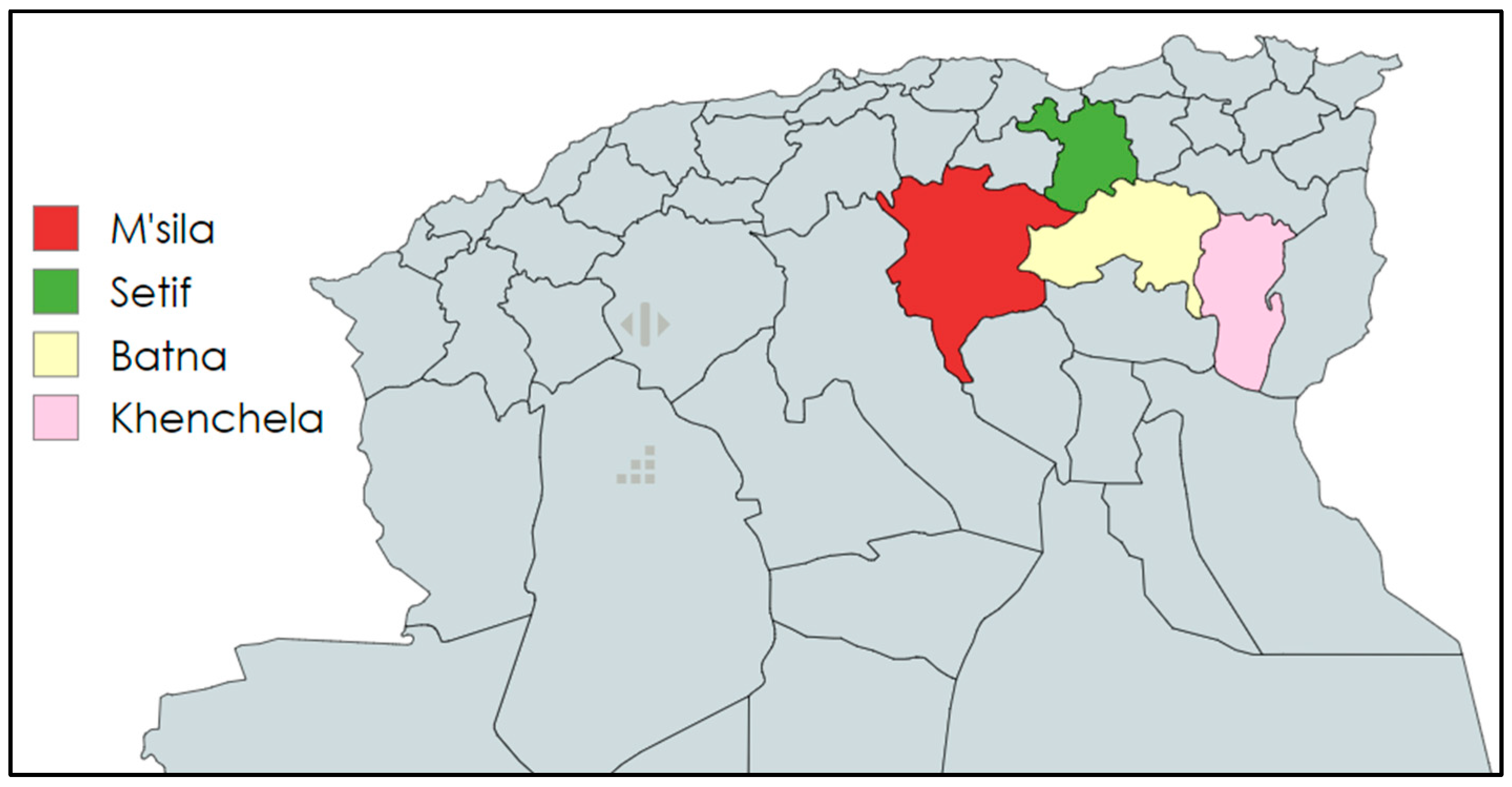
4.1. Population’s Study and Sample Collection

From February 2021 to December 2023, 409 samples were taken from healthy and clinically ill dogs and cats from different breeds in various regions of eastern Algeria; Batna (n=281), Khenchela (n=58), Setif (n=66) and M’sila (n=4) (figure 7). For cats, samples were collected from rectum (n=102), ear (n=30), abscesses (n=29), wound (n=17), uterus (n=12), nasal cavity (n=10), urine (n=1), buccal cavity(n=8) and 4 of other origin. In dogs, the sample sites included: ear (n=88), nasal cavity (n=70), rectum (n=25), wound (n=6), abscesses (n=4), eye (n=2) and vagina (n=3). After sampling the samples were refrigerated and shipped to the laboratory for bacterial investigation.
Figure 7. Map of northern Algeria highlighting the sampling areas (https://www.mapchart.net/).
Preprints 148650 g007
This study was conducted in accordance with the requirements of the Scientific committee of the Institute of Veterinary and Agricultural Sciences (University of Batna 1), under the certificate of Animal use Protocol No: 001/DV/ISVSA/UB1/2025.

4.2. Culture Conditions and Bacterial Identification

Upon arrival at the laboratory, swabs were incubated in BHIB (Brain Heart Infusion broth; HIMEDIA, Nashik, India) for 24 hours at 37°C, then cultivated aerobically on cetrimide agar (MERCK, Germany) at 42°C for 24-48 hours. A culture is considered positive when greenish colonies, oxidase and catalase positive, and Gram-negative rods (after Gram staining) are observed. Cultures were then purified on MacConkey agar (MERCK, Germany), and pure strains were identified using the API 20 NE system® (Biomérieux, France).

4.3. Antimicrobial Susceptibility Testing

Antimicrobial susceptibility testing was performed using the Kirby-Bauer disc diffusion method according to the EUCAST guidelines [71]. Bacterial suspensions were adjusted to the 0.5 McFarland turbidity standard. Bacterial isolates were tested to a panel of 13 antibiotics: amikacin (AK) 30µg, ceftazidime (CAZ) 30µg (Oxoid, Basingstoke, UK), gentamicin (CN) 15µg (Bioanalyse, Ankara, Turkey) netilmicin (NET) 30µg, tobramycin (TOB) 10µg, levofloxacin (LEV) 50µg, ciprofloxacin (CIP) 5µg, cefepime (FEP) 30µg, aztreonam (ATM) 30µg, ticarcillin (TC) 75µg, ticarcillin-clavulanic acid (TCC) 75/10µg, imipenem (IMP) 10µg and amoxicillin-clavulanic acid (AMC) 20/10µg (Biomaxima, Lublin, Poland). P. aeruginosa ATCC 27853 was used as a quality control strain.

4.4. In Vitro Biofilm Formation Assay

To study the biofilm formation ability, the microtiter assay was performed, using the protocol as previously described [72]. BHIB broth was used to induce biofilm formation, and P. aeruginosa ATCC 27853 was used as a positive biofilm producer.
Bacterial optical densities were measured using a microplate reader (M 960, Metertech), at 550 nm using as blank a 30 % (v/v) solution of acetic acid. Optical density for each isolate (A550) were obtained by averaging 8 wells, then compared with the cutoff value (0.102) which was determined arbitrarily by the mean of the negative control (0.042) plus three standard deviations (0.02). The level of biofilm production was classified in four categories: no production A550< 0.101, weak biofilm production 0.101 ≤ A550 < 0.202 (2 x negative control), moderate biofilm production 0.202 ≤ A550 < 0.408 (4 x negative control), and strong biofilm production 0.404 <A550 according to the literature [21].

4.5. Whole Genome Sequencing (WGS) and Bioinformatics Analysis

4.5.1. DNA Extraction

All P. aeruginosa strains were sequenced using a long-reads sequencing (LRS) approach based on MinION Mk1C platform (Oxford Nanopores Technologies, UK). Isolated colonies from a fresh culture of P. aeruginosa on blood agar were resuspended in 500 µL PBS (Phosphate Buffered Saline, Euroclone, Italy), centrifuged 10,000 ×g for 1 minute at room temperature and high molecular weight DNA was extracted with the Quick-DNATM HMW MagBead Kit (Zymo Research, Irvine, CA, USA) following the manufacturer's instructions. The DNA quantity and quality were assessed using a NanoReady Touch series Micro Volume (UV-Vis) (Aurogene, Italy), ensuring that the A260/A280 and A260/A230 ratios ranged between 1.8 and 2, respectively. Extracted DNA was also subjected to gel electrophoresis to check its integrity.

4.5.2. Whole Genome Sequencing

The sequencing libraries were prepared with 200 ng, as input DNA, which were subjected to transposase fragmentation with the Rapid Barcoding Sequencing kit (SQK-RBK114.24, Oxford Nanopore Technologies, UK). Then, 12 isolates were multiplexed on a single flow cell (FLO-MIN114, R10.4.1 version), and sequenced in a MinION Mk1C for 72h maximum.

4.5.3. Bioinformatic Analysis

Dorado (v0.8.2) was used to basecall (--dna_r10.4.1_e8.2_400bps_hac@v5.0.0), trim adapters and demultiplex .pod5 files (--config configuration.cfg --barcode_kits SQK-RBK114.24 --trim_barcodes; min_score threshold default 60). Summary statistics were obtained with NanoPlot (v1.44.0) (--verbose --tsv_stats –N50 --fastq) [73]. Reference guided filtration with a 1000 bp threshold was achieved using FiltLong (v0.2.1), and blasting each strain against Pseudomonas aeruginosa reference strains PAO1 (--assembly Ref_PAO1.fasta --trim --min_length 1000 --keep_percent 90) [74]. Genomes were de novo assembled using Flye (v2.8.1-b1676) (--nano-corr --genome-size 5m --asm-coverage 50 --plasmids --trestle) [75]. Assembled contigs were polished with Medaka (v2.0.1) (medaka_consensus -t 8 -m dna_r10.4.1_e8.2_400bps_hac@v4.1.0:variant) [76]. Genomic completeness and contamination were derived by CheckM2 (v1.0.2) (checkm2 predict --threads 30 –x fna) [77].
The NCBI Prokaryotic Genome Annotation Pipeline (PGAP) was used to annotate genomes and find out the total numbers of coding sequences, rRNA and tRNA [78].
Multilocus sequence types (MLSTs) were determined uploading the genomes on Pathogenwatch website (https://pathogen.watch/), while eBURST software was used to visualize clonal complexes [20]. Finally pan-genomes were visualized using both on online tool (IPGA) [19] and a command-line pipeline (ANVI’O) [79].

4.5.4. Bioinformatic Analysis of Antimicrobial Resistance Genes and Virulence Factors

To ascertain the presence of antibiotic resistance genes (ARGs), the Comprehensive Antibiotic Resistance Database (CARD), the National Centre for Biotechnology Information (NCBI) database, the ResFinder database, the Plasmidfinder and the virulence factor database database (all updated on 4 November 2023) were used in the analysis of genomes using Abricate (v1.0.1) (abricate /path/to/fna/*.fna --db card,vfdb,resfinder,ncbi,plasmidfinder --minid 95 --csv > /path/to/output/.xlsx) [80,81,82,83,84,85,86,87]. For all these analyses, a threshold identity ≥95% was set.

4.6. Statistical Analysis

To analyse associations in the distribution of P. aeruginosa strains across sample origins and antibiotic phenotypes comparison, a chi-squared test (Chi²) and Fisher's test were performed at a 95% confidence interval (α = 0.05) to assess if observed species distributions differed significantly across origins. All statistical analyses in this study were performed with SPSS.

5. Conclusions

This study investigated the resistance patterns of P. aeruginosa isolates from cats and dogs. In Algeria, this is the first identification of carbapenem resistant P. aeruginosa (31,56%) in pets with an arsenal of resistance genes mainly those related to aminoglycosides (Aph(3’)IIb), beta-lactams (blaOXA, blaPAO, and blaPDC), phenicols (catB7), fosfomycin (FosA) and bicyclomicin (bcr-1) resistance with the emergence of high-risk clone ST244. High capacity of biofilm production (42% strong producers) and a wide range of virulence genes was associated with third system secretion, quorum sensing and others. The research underscores the need of comprehending P. aeruginosa resistance patterns across diverse populations and areas, promoting judicious antibiotic utilisation and stringent infection control measures to avert the dissemination of resistant strains. Furthermore, the monitoring of antibiotic resistance in companion animals should be intensified to prevent potential transmission of the infections between animals and their owners.

Author Contributions

Conceptualization, NH.; M.M.; P.A.M. and A.S.; methodology, A.B.; G.M.; software, A.B.; A.S.; G.M..; validation, A.A.; resources, A.A; P.AM.; writing—original draft preparation, A.B.; G.M.; NH.; M.M.; writing—review and editing, G.M.; P.A.M., A.S.; NH.; M.M.; supervision, N.H.; M.M; A.S. P.A.M.; project administration, N.H. All authors have read and agreed to the published version of the manuscript

Funding

Please add: This research received no external funding.

Institutional Review Board Statement

This study was conducted in accordance with the requirements of the Scientific committee of the Institute of Veterinary and Agricultural Sciences (University of Batna 1 ), under the certificate of Animal use Protocol No : 001/DV/ISVSA/UB1/2025.

Informed Consent Statement

Not applicable.

Data Availability Statement

The strain descriptions and accession numbers are presented in Table 3; the genome assemblies, genomic data, and raw data are publicly available in GenBank under BioProject PRJNA1153397 and genome accession numbers CP169763, CP169759, CP169760, CP169762, JBHGZY000000000, CP169765, CP169758, JBHHAE000000000, JBHHAG000000000, JBHGZX000000000, JBHHAC000000000, CP169761, JBHHAF000000000, JBHHAD000000000, JBHHAB000000000, JBHHAA000000000, JBHGZW000000000, JBHGZZ000000000, CP169764.

Acknowledgments

Computational resources provided by INDACO Core facility, which is a project of High Performance Computing at the University of MILAN (https://www.indaco.unimi.it/)

Conflicts of Interest

The authors declare no conflicts of interest.

References

  1. Nakamura, K.; Fujiki, J.; Furusawa, T.; Nakamura, T.; Gondaira, S.; Sasaki, M.; Usui, M.; Higuchi, H.; Sawa, H.; Tamura, Y.; et al. Complete Genome Sequence of a Veterinary Pseudomonas Aeruginosa Isolate, Pa12. Microbiol Resour Announc 2021, 10, e0039821. [Google Scholar] [CrossRef]
  2. Hewitt, J.S.; Allbaugh, R.A.; Kenne, D.E.; Sebbag, L. Prevalence and Antibiotic Susceptibility of Bacterial Isolates From Dogs With Ulcerative Keratitis in Midwestern United States. Front Vet Sci 2020, 7, 583965. [Google Scholar] [CrossRef] [PubMed]
  3. Park, K.-M.; Nam, H.-S.; Woo, H.-M. Successful Management of Multidrug-Resistant Pseudomonas Aeruginosa Pneumonia after Kidney Transplantation in a Dog. J. Vet. Med. Sci. 2013, 75, 1529–1533. [Google Scholar] [CrossRef]
  4. Curran, K.; Leeper, H.; O’Reilly, K.; Jacob, J.; Bermudez, L.E. An Analysis of the Infections and Determination of Empiric Antibiotic Therapy in Cats and Dogs with Cancer-Associated Infections. Antibiotics (Basel) 2021, 10, 700. [Google Scholar] [CrossRef]
  5. Haenni, M.; Hocquet, D.; Ponsin, C.; Cholley, P.; Guyeux, C.; Madec, J.-Y.; Bertrand, X. Population Structure and Antimicrobial Susceptibility of Pseudomonas Aeruginosa from Animal Infections in France. BMC Veterinary Research 2015, 11, 9. [Google Scholar] [CrossRef] [PubMed]
  6. de Jong, A.; Youala, M.; El Garch, F.; Simjee, S.; Rose, M.; Morrissey, I.; Moyaert, H. Antimicrobial Susceptibility Monitoring of Canine and Feline Skin and Ear Pathogens Isolated from European Veterinary Clinics: Results of the ComPath Surveillance Programme. Vet Dermatol 2020, 31, 431-e114. [Google Scholar] [CrossRef] [PubMed]
  7. Mohan, K.; Fothergill, J.L.; Storrar, J.; Ledson, M.J.; Winstanley, C.; Walshaw, M.J. Transmission of Pseudomonas Aeruginosa Epidemic Strain from a Patient with Cystic Fibrosis to a Pet Cat. Thorax 2008, 63, 839–840. [Google Scholar] [CrossRef]
  8. Lin, C.-T.; Petersen-Jones, S.M. Antibiotic Susceptibility of Bacteria Isolated from Cats with Ulcerative Keratitis in Taiwan. J Small Anim Pract 2008, 49, 80–83. [Google Scholar] [CrossRef]
  9. Barlandas-Quintana, E.A.; Martinez-Ledesma, J.E. Detection of Carbapenems Resistant K-Mer Sequences in Bacteria of Critical Priority by the World Health Organization (Pseudomonas Aeruginosa and Acinetobacter Baumannii). In Proceedings of the 2020 7th International Conference on Internet of Things: Systems, Management and Security (IOTSMS); December 2020; pp. 1–8.
  10. Santaniello, A.; Sansone, M.; Fioretti, A.; Menna, L.F. Systematic Review and Meta-Analysis of the Occurrence of ESKAPE Bacteria Group in Dogs, and the Related Zoonotic Risk in Animal-Assisted Therapy, and in Animal-Assisted Activity in the Health Context. Int J Environ Res Public Health 2020, 17, 3278. [Google Scholar] [CrossRef]
  11. EFSA Panel on Animal Health and Welfare (AHAW); Nielsen, S.S.; Bicout, D.J.; Calistri, P.; Canali, E.; Drewe, J.A.; Garin-Bastuji, B.; Gonzales Rojas, J.L.; Gortázar, C.; Herskin, M.; et al. Assessment of Listing and Categorisation of Animal Diseases within the Framework of the Animal Health Law (Regulation (EU) No 2016/429): Antimicrobial-Resistant Pseudomonas Aeruginosa in Dogs and Cats. EFSA Journal 2022, 20, e07310. [Google Scholar] [CrossRef]
  12. Veetilvalappil, V.V.; Manuel, A.; Aranjani, J.M.; Tawale, R.; Koteshwara, A. Pathogenic Arsenal of Pseudomonas Aeruginosa: An Update on Virulence Factors. Future Microbiol 2022, 17, 465–481. [Google Scholar] [CrossRef] [PubMed]
  13. Poursina, S.; Ahmadi, M.; Fazeli, F.; Ariaii, P. Assessment of Virulence Factors and Antimicrobial Resistance among the Pseudomonas Aeruginosa Strains Isolated from Animal Meat and Carcass Samples. Vet Med Sci 2022, 9, 315–325. [Google Scholar] [CrossRef]
  14. Lenart-Boroń, A.; Stankiewicz, K.; Czernecka, N.; Ratajewicz, A.; Bulanda, K.; Heliasz, M.; Sosińska, D.; Dworak, K.; Ciesielska, D.; Siemińska, I.; et al. Wounds of Companion Animals as a Habitat of Antibiotic-Resistant Bacteria That Are Potentially Harmful to Humans-Phenotypic, Proteomic and Molecular Detection. Int J Mol Sci 2024, 25, 3121. [Google Scholar] [CrossRef]
  15. Guardabassi, L.; Schwarz, S.; Lloyd, D.H. Pet Animals as Reservoirs of Antimicrobial-Resistant Bacteria. J Antimicrob Chemother 2004, 54, 321–332. [Google Scholar] [CrossRef] [PubMed]
  16. Breidenstein, E.B.M.; de la Fuente-Núñez, C.; Hancock, R.E.W. Pseudomonas Aeruginosa: All Roads Lead to Resistance. Trends Microbiol 2011, 19, 419–426. [Google Scholar] [CrossRef]
  17. El-Ganiny, A.M.; Shaker, G.H.; Aboelazm, A.A.; El-Dash, H.A. Prevention of Bacterial Biofilm Formation on Soft Contact Lenses Using Natural Compounds. J Ophthalmic Inflamm Infect 2017, 7, 11. [Google Scholar] [CrossRef] [PubMed]
  18. Liao, C.; Huang, X.; Wang, Q.; Yao, D.; Lu, W. Virulence Factors of Pseudomonas Aeruginosa and Antivirulence Strategies to Combat Its Drug Resistance. Front. Cell. Infect. Microbiol. 2022, 12. [Google Scholar] [CrossRef]
  19. Liu, D.; Zhang, Y.; Fan, G.; Sun, D.; Zhang, X.; Yu, Z.; Wang, J.; Wu, L.; Shi, W.; Ma, J. IPGA: A Handy Integrated Prokaryotes Genome and Pan-Genome Analysis Web Service. Imeta 2022, 1, e55. [Google Scholar] [CrossRef]
  20. Francisco, A.P.; Bugalho, M.; Ramirez, M.; Carriço, J.A. Global Optimal eBURST Analysis of Multilocus Typing Data Using a Graphic Matroid Approach. BMC Bioinformatics 2009, 10, 152. [Google Scholar] [CrossRef]
  21. Płókarz, D.; Czopowicz, M.; Bierowiec, K.; Rypuła, K. Virulence Genes as Markers for Pseudomonas Aeruginosa Biofilm Formation in Dogs and Cats. Animals (Basel) 2022, 12, 422. [Google Scholar] [CrossRef]
  22. Laborda, P.; Sanz-García, F.; Hernando-Amado, S.; Martínez, J.L. Pseudomonas Aeruginosa: An Antibiotic Resilient Pathogen with Environmental Origin. Curr Opin Microbiol 2021, 64, 125–132. [Google Scholar] [CrossRef]
  23. Rizk, A.M.; Elsayed, M.M.; Abd El Tawab, A.A.; Elhofy, F.I.; Soliman, E.A.; Kozytska, T.; Brangsch, H.; Sprague, L.D.; Neubauer, H.; Wareth, G. Phenotypic and Genotypic Characterization of Resistance and Virulence in Pseudomonas Aeruginosa Isolated from Poultry Farms in Egypt Using Whole Genome Sequencing. Vet Microbiol 2024, 292, 110063. [Google Scholar] [CrossRef] [PubMed]
  24. Hattab, J.; Mosca, F.; Di Francesco, C.E.; Aste, G.; Marruchella, G.; Guardiani, P.; Tiscar, P.G. Occurrence, Antimicrobial Susceptibility, and Pathogenic Factors of Pseudomonas Aeruginosa in Canine Clinical Samples. Vet World 2021, 14, 978–985. [Google Scholar] [CrossRef]
  25. Raheem, S.A.; Abdalshheed, D.A. Detection of Virulence Factors of Pseudomonas Aeruginosa from Dog’s Wound Infections by Using Biochemical and Molecular Methods. Int. J. Vet. Sci. Anim. Husbandry 2023, 8, 01–07. [Google Scholar] [CrossRef]
  26. Jangsangthong, A.; Lugsomya, K.; Apiratwarrasakul, S.; Phumthanakorn, N. Distribution of Sequence Types and Antimicrobial Resistance of Clinical Pseudomonas Aeruginosa Isolates from Dogs and Cats Visiting a Veterinary Teaching Hospital in Thailand. BMC Veterinary Research 2024, 20, 234. [Google Scholar] [CrossRef]
  27. Lin, D.; Foley, S.L.; Qi, Y.; Han, J.; Ji, C.; Li, R.; Wu, C.; Shen, J.; Wang, Y. Characterization of Antimicrobial Resistance of Pseudomonas Aeruginosa Isolated from Canine Infections. J Appl Microbiol 2012, 113, 16–23. [Google Scholar] [CrossRef]
  28. Wong, C.; Epstein, S.E.; Westropp, J.L. Antimicrobial Susceptibility Patterns in Urinary Tract Infections in Dogs (2010-2013). J Vet Intern Med 2015, 29, 1045–1052. [Google Scholar] [CrossRef] [PubMed]
  29. Sankar, S.; Thresia; Bosewell, A.; Mini, M. Molecular Detection of Carbapenem Resistant Gram Negative Bacterial Isolates from Dogs. Indian Journal of Animal Research 2021.
  30. Dégi, J.; Moțco, O.-A.; Dégi, D.M.; Suici, T.; Mareș, M.; Imre, K.; Cristina, R.T. Antibiotic Susceptibility Profile of Pseudomonas Aeruginosa Canine Isolates from a Multicentric Study in Romania. Antibiotics 2021, 10, 846. [Google Scholar] [CrossRef]
  31. Park, Y.; Oh, J.; Park, S.; Sum, S.; Song, W.; Chae, J.; Park, H. Antimicrobial Resistance and Novel Mutations Detected in the gyrA and parC Genes of Pseudomonas Aeruginosa Strains Isolated from Companion Dogs. BMC Veterinary Research 2020, 16, 111. [Google Scholar] [CrossRef]
  32. Penna, B.; Thomé, S.; Martins, R.; Martins, G.; Lilenbaum, W. In Vitro Antimicrobial Resistance of Pseudomonas Aeruginosa Isolated from Canine Otitis Externa in Rio de Janeiro, Brazil. Braz J Microbiol 2011, 42, 1434–1436. [Google Scholar] [CrossRef]
  33. Gentilini, F.; Turba, M.E.; Pasquali, F.; Mion, D.; Romagnoli, N.; Zambon, E.; Terni, D.; Peirano, G.; Pitout, J.D.D.; Parisi, A.; et al. Hospitalized Pets as a Source of Carbapenem-Resistance. Front Microbiol 2018, 9, 2872. [Google Scholar] [CrossRef]
  34. Bel Hadj Ahmed, A.; Salah Abbassi, M.; Rojo-Bezares, B.; Ruiz-Roldán, L.; Dhahri, R.; Mehri, I.; Sáenz, Y.; Hassen, A. Characterization of Pseudomonas Aeruginosa Isolated from Various Environmental Niches: New STs and Occurrence of Antibiotic Susceptible “High-Risk Clones”. International Journal of Environmental Health Research 2020, 30, 643–652. [Google Scholar] [CrossRef] [PubMed]
  35. Badawy, B.; Moustafa, S.; Shata, R.; Sayed-Ahmed, M.Z.; Alqahtani, S.S.; Ali, M.S.; Alam, N.; Ahmad, S.; Kasem, N.; Elbaz, E.; et al. Prevalence of Multidrug-Resistant Pseudomonas Aeruginosa Isolated from Dairy Cattle, Milk, Environment, and Workers’ Hands. Microorganisms 2023, 11, 2775. [Google Scholar] [CrossRef] [PubMed]
  36. Valero, A.; Isla, A.; Rodríguez-Gascón, A.; Canut, A.; Ángeles Solinís, M. Susceptibility of Pseudomonas Aeruginosa and Antimicrobial Activity Using PK/PD Analysis: An 18-Year Surveillance Study. Enferm Infecc Microbiol Clin (Engl Ed) 2019, 37, 626–633. [Google Scholar] [CrossRef] [PubMed]
  37. Feßler, A.T.; Scholtzek, A.D.; Schug, A.R.; Kohn, B.; Weingart, C.; Hanke, D.; Schink, A.-K.; Bethe, A.; Lübke-Becker, A.; Schwarz, S. Antimicrobial and Biocide Resistance among Canine and Feline Enterococcus Faecalis, Enterococcus Faecium, Escherichia Coli, Pseudomonas Aeruginosa, and Acinetobacter Baumannii Isolates from Diagnostic Submissions. Antibiotics (Basel) 2022, 11, 152. [Google Scholar] [CrossRef]
  38. Rubin, J.; Walker, R.D.; Blickenstaff, K.; Bodeis-Jones, S.; Zhao, S. Antimicrobial Resistance and Genetic Characterization of Fluoroquinolone Resistance of Pseudomonas Aeruginosa Isolated from Canine Infections. Vet Microbiol 2008, 131, 164–172. [Google Scholar] [CrossRef]
  39. Elgenedy, E.M.; Abdelmoein, K.A.; Samir, A. Occurrence of Carbapenem-Resistant Organisms among Pet Animals Suffering from Respiratory Illness: A Possible Public Health Risk. Veterinary Medical Journal (Giza) 2024, 70, 18–25. [Google Scholar] [CrossRef]
  40. Sellera, F.P.; Da Silva, L.C.B.A.; Lincopan, N. Rapid Spread of Critical Priority Carbapenemase-Producing Pathogens in Companion Animals: A One Health Challenge for a Post-Pandemic World. J Antimicrob Chemother 2021, 76, 2225–2229. [Google Scholar] [CrossRef]
  41. Ramírez-Castillo, F.Y.; Guerrero-Barrera, A.L.; Avelar-González, F.J. An Overview of Carbapenem-Resistant Organisms from Food-Producing Animals, Seafood, Aquaculture, Companion Animals, and Wildlife. Front. Vet. Sci. 2023, 10. [Google Scholar] [CrossRef]
  42. Dossouvi, K.M.; Ametepe, A.S. Carbapenem Resistance in Animal-Environment-Food from Africa: A Systematic Review, Recommendations and Perspectives. Infect Drug Resist 2024, 17, 1699–1728. [Google Scholar] [CrossRef]
  43. Yousfi, M.; Touati, A.; Mairi, A.; Brasme, L.; Gharout-Sait, A.; Guillard, T.; De Champs, C. Emergence of Carbapenemase-Producing Escherichia Coli Isolated from Companion Animals in Algeria. Microb Drug Resist 2016, 22, 342–346. [Google Scholar] [CrossRef] [PubMed]
  44. Yousfi, M.; Touati, A.; Muggeo, A.; Mira, B.; Asma, B.; Brasme, L.; Guillard, T.; de Champs, C. Clonal Dissemination of OXA-48-Producing Enterobacter Cloacae Isolates from Companion Animals in Algeria. J Glob Antimicrob Resist 2018, 12, 187–191. [Google Scholar] [CrossRef] [PubMed]
  45. Elfadadny, A.; Uchiyama, J.; Goto, K.; Imanishi, I.; Ragab, R.F.; Nageeb, W.M.; Iyori, K.; Toyoda, Y.; Tsukui, T.; Ide, K.; et al. Antimicrobial Resistance and Genotyping of Pseudomonas Aeruginosa Isolated from the Ear Canals of Dogs in Japan. Front. Vet. Sci. 2023, 10. [Google Scholar] [CrossRef]
  46. de Sousa, T.; Garcês, A.; Silva, A.; Lopes, R.; Alegria, N.; Hébraud, M.; Igrejas, G.; Poeta, P. The Impact of the Virulence of Pseudomonas Aeruginosa Isolated from Dogs. Veterinary Sciences 2023, 10, 343. [Google Scholar] [CrossRef]
  47. Torres, R.T.; Cunha, M.V.; Ferreira, H.; Fonseca, C.; Palmeira, J.D. A High-Risk Carbapenem-Resistant Pseudomonas Aeruginosa Clone Detected in Red Deer (Cervus Elaphus) from Portugal. Sci Total Environ 2022, 829, 154699. [Google Scholar] [CrossRef] [PubMed]
  48. Yin, L.; Lu, L.; He, L.; Yan, G.; Lu, G.; Zhai, X.; Wang, C. Non-Carbapenem-Producing Carbapenem-Resistant Pseudomonas Aeruginosa in Children: Risk Factors, Molecular Epidemiology, and Resistance Mechanism. J Infect Public Health 2025, 18, 102634. [Google Scholar] [CrossRef]
  49. Lee, J.H.; Kim, N.-H.; Jang, K.-M.; Jin, H.; Shin, K.; Jeong, B.C.; Kim, D.-W.; Lee, S.H. Prioritization of Critical Factors for Surveillance of the Dissemination of Antibiotic Resistance in Pseudomonas Aeruginosa: A Systematic Review. International Journal of Molecular Sciences 2023, 24, 15209. [Google Scholar] [CrossRef]
  50. Del Barrio-Tofiño, E.; López-Causapé, C.; Oliver, A. Pseudomonas Aeruginosa Epidemic High-Risk Clones and Their Association with Horizontally-Acquired β-Lactamases: 2020 Update. Int J Antimicrob Agents 2020, 56, 106196. [Google Scholar] [CrossRef]
  51. Zurita, J.; Sevillano, G.; Solís, M.B.; Paz Y Miño, A.; Alves, B.R.; Changuan, J.; González, P. Pseudomonas Aeruginosa Epidemic High-Risk Clones and Their Association with Multidrug-Resistant. J Glob Antimicrob Resist 2024, 38, 332–338. [Google Scholar] [CrossRef]
  52. Cramer, N.; Klockgether, J.; Tümmler, B. Microevolution of Pseudomonas Aeruginosa in the Airways of People with Cystic Fibrosis. Curr Opin Immunol 2023, 83, 102328. [Google Scholar] [CrossRef]
  53. Oliver, A.; Mulet, X.; López-Causapé, C.; Juan, C. The Increasing Threat of Pseudomonas Aeruginosa High-Risk Clones. Drug Resist Updat 2015, 21–22, 41–59. [Google Scholar] [CrossRef] [PubMed]
  54. Mellouk, F.Z.; Bakour, S.; Meradji, S.; Al-Bayssari, C.; Bentakouk, M.C.; Zouyed, F.; Djahoudi, A.; Boutefnouchet, N.; Rolain, J.M. First Detection of VIM-4-Producing Pseudomonas Aeruginosa and OXA-48-Producing Klebsiella Pneumoniae in Northeastern (Annaba, Skikda) Algeria. Microb Drug Resist 2017, 23, 335–344. [Google Scholar] [CrossRef]
  55. Merradi, M.; Kassah-Laouar, A.; Ayachi, A.; Heleili, N.; Menasria, T.; Hocquet, D.; Cholley, P.; Sauget, M. Occurrence of VIM-4 Metallo-β-Lactamase-Producing Pseudomonas Aeruginosa in an Algerian Hospital. J Infect Dev Ctries 2019, 13, 284–290. [Google Scholar] [CrossRef]
  56. Oliver, A.; Rojo-Molinero, E.; Arca-Suarez, J.; Beşli, Y.; Bogaerts, P.; Cantón, R.; Cimen, C.; Croughs, P.D.; Denis, O.; Giske, C.G.; et al. Pseudomonas Aeruginosa Antimicrobial Susceptibility Profiles, Resistance Mechanisms and International Clonal Lineages: Update from ESGARS-ESCMID/ISARPAE Group. Clinical Microbiology and Infection 2024, 30, 469–480. [Google Scholar] [CrossRef]
  57. Lebreton, F.; Snesrud, E.; Hall, L.; Mills, E.; Galac, M.; Stam, J.; Ong, A.; Maybank, R.; Kwak, Y.I.; Johnson, S.; et al. A Panel of Diverse Pseudomonas Aeruginosa Clinical Isolates for Research and Development. JAC Antimicrob Resist 2021, 3, dlab179. [Google Scholar] [CrossRef] [PubMed]
  58. Lister, P.D.; Wolter, D.J.; Hanson, N.D. Antibacterial-Resistant Pseudomonas Aeruginosa: Clinical Impact and Complex Regulation of Chromosomally Encoded Resistance Mechanisms. Clin Microbiol Rev 2009, 22, 582–610. [Google Scholar] [CrossRef] [PubMed]
  59. Kocsis, B.; Gulyás, D.; Szabó, D. Diversity and Distribution of Resistance Markers in Pseudomonas Aeruginosa International High-Risk Clones. Microorganisms 2021, 9, 359. [Google Scholar] [CrossRef]
  60. Bonnin, R.A.; Bogaerts, P.; Girlich, D.; Huang, T.-D.; Dortet, L.; Glupczynski, Y.; Naas, T. Molecular Characterization of OXA-198 Carbapenemase-Producing Pseudomonas Aeruginosa Clinical Isolates. Antimicrob Agents Chemother 2018, 62, e02496-17. [Google Scholar] [CrossRef]
  61. Dassanayake, R.P.; Ma, H.; Casas, E.; Lippolis, J.D. Genome Sequence of a Multidrug-Resistant Pseudomonas Aeruginosa Strain Isolated from a Dairy Cow That Was Nonresponsive to Antibiotic Treatment. Microbiology Resource Announcements 2023, 12, e00289-23. [Google Scholar] [CrossRef]
  62. Madaha, E.L.; Mienie, C.; Gonsu, H.K.; Bughe, R.N.; Fonkoua, M.C.; Mbacham, W.F.; Alayande, K.A.; Bezuidenhout, C.C.; Ateba, C.N. Whole-Genome Sequence of Multi-Drug Resistant Pseudomonas Aeruginosa Strains UY1PSABAL and UY1PSABAL2 Isolated from Human Broncho-Alveolar Lavage, Yaoundé, Cameroon. PLoS One 2020, 15, e0238390. [Google Scholar] [CrossRef]
  63. Nolan, L.M.; Turnbull, L.; Katrib, M.; Osvath, S.R.; Losa, D.; Lazenby, J.J.; Whitchurch, C.B. Pseudomonas Aeruginosa Is Capable of Natural Transformation in Biofilms. Microbiology (Reading) 2020, 166, 995–1003. [Google Scholar] [CrossRef] [PubMed]
  64. Hayashi, W.; Izumi, K.; Yoshida, S.; Takizawa, S.; Sakaguchi, K.; Iyori, K.; Minoshima, K.-I.; Takano, S.; Kitagawa, M.; Nagano, Y.; et al. Antimicrobial Resistance and Type III Secretion System Virulotypes of Pseudomonas Aeruginosa Isolates from Dogs and Cats in Primary Veterinary Hospitals in Japan: Identification of the International High-Risk Clone Sequence Type 235. Microbiol Spectr 2021, 9, e0040821. [Google Scholar] [CrossRef]
  65. Juan, C.; Peña, C.; Oliver, A. Host and Pathogen Biomarkers for Severe Pseudomonas Aeruginosa Infections. J Infect Dis 2017, 215, S44–S51. [Google Scholar] [CrossRef]
  66. Hassuna, N.A.; Mandour, S.A.; Mohamed, E.S. Virulence Constitution of Multi-Drug-Resistant Pseudomonas Aeruginosa in Upper Egypt. Infect Drug Resist 2020, 13, 587–595. [Google Scholar] [CrossRef] [PubMed]
  67. Feltman, H.; Schulert, G.; Khan, S.; Jain, M.; Peterson, L.; Hauser, A.R. Prevalence of Type III Secretion Genes in Clinical and Environmental Isolates of Pseudomonas Aeruginosa. Microbiology 2001, 147, 2659–2669. [Google Scholar] [CrossRef]
  68. Jurado-Martín, I.; Sainz-Mejías, M.; McClean, S. Pseudomonas Aeruginosa: An Audacious Pathogen with an Adaptable Arsenal of Virulence Factors. Int J Mol Sci 2021, 22, 3128. [Google Scholar] [CrossRef]
  69. Gómez-Martínez, J.; Rocha-Gracia, R.D.C.; Bello-López, E.; Cevallos, M.A.; Castañeda-Lucio, M.; Sáenz, Y.; Jiménez-Flores, G.; Cortés-Cortés, G.; López-García, A.; Lozano-Zarain, P. Comparative Genomics of Pseudomonas Aeruginosa Strains Isolated from Different Ecological Niches. Antibiotics (Basel) 2023, 12, 866. [Google Scholar] [CrossRef] [PubMed]
  70. Morales-Espinosa, R.; Delgado, G.; Espinosa-Camacho, F.; Flores-Alanis, A.; Rodriguez, C.; Mendez, J.L.; Gonzalez-Pedraza, A.; Cravioto, A. Pseudomonas Aeruginosa Strains Isolated from Animal with High Virulence Genes Content and Highly Sensitive to Antimicrobials. J Glob Antimicrob Resist 2024, 37, 75–80. [Google Scholar] [CrossRef]
  71. Gaur, P.; Hada, V.; Rath, R.S.; Mohanty, A.; Singh, P.; Rukadikar, A. Interpretation of Antimicrobial Susceptibility Testing Using European Committee on Antimicrobial Susceptibility Testing (EUCAST) and Clinical and Laboratory Standards Institute (CLSI) Breakpoints: Analysis of Agreement. Cureus 2023, 15, e36977. [Google Scholar] [CrossRef]
  72. O’Toole, G.A. Microtiter Dish Biofilm Formation Assay. J Vis Exp 2011, 2437. [Google Scholar] [CrossRef]
  73. De Coster, W.; Rademakers, R. NanoPack2: Population-Scale Evaluation of Long-Read Sequencing Data. Bioinformatics 2023, 39, btad311. [Google Scholar] [CrossRef] [PubMed]
  74. Wick, R. Rrwick/Filtlong 2025.
  75. Kolmogorov, M.; Yuan, J.; Lin, Y.; Pevzner, P.A. Assembly of Long, Error-Prone Reads Using Repeat Graphs. Nat Biotechnol 2019, 37, 540–546. [Google Scholar] [CrossRef] [PubMed]
  76. Nanoporetech/Medaka 2025.
  77. Parks, D.H.; Imelfort, M.; Skennerton, C.T.; Hugenholtz, P.; Tyson, G.W. CheckM: Assessing the Quality of Microbial Genomes Recovered from Isolates, Single Cells, and Metagenomes. Genome Res. 2015, 25, 1043–1055. [Google Scholar] [CrossRef]
  78. Ncbi/Pgap 2025.
  79. Eren, A.M.; Kiefl, E.; Shaiber, A.; Veseli, I.; Miller, S.E.; Schechter, M.S.; Fink, I.; Pan, J.N.; Yousef, M.; Fogarty, E.C.; et al. Community-Led, Integrated, Reproducible Multi-Omics with Anvi’o. Nat Microbiol 2021, 6, 3–6. [Google Scholar] [CrossRef]
  80. Seemann, T. Tseemann/Abricate 2025.
  81. Feldgarden, M.; Brover, V.; Haft, D.H.; Prasad, A.B.; Slotta, D.J.; Tolstoy, I.; Tyson, G.H.; Zhao, S.; Hsu, C.-H.; McDermott, P.F.; et al. Validating the AMRFinder Tool and Resistance Gene Database by Using Antimicrobial Resistance Genotype-Phenotype Correlations in a Collection of Isolates. Antimicrob Agents Chemother 2019, 63, e00483-19. [Google Scholar] [CrossRef]
  82. Jia, B.; Raphenya, A.R.; Alcock, B.; Waglechner, N.; Guo, P.; Tsang, K.K.; Lago, B.A.; Dave, B.M.; Pereira, S.; Sharma, A.N.; et al. CARD 2017: Expansion and Model-Centric Curation of the Comprehensive Antibiotic Resistance Database. Nucleic Acids Res 2017, 45, D566–D573. [Google Scholar] [CrossRef]
  83. Zankari, E.; Hasman, H.; Cosentino, S.; Vestergaard, M.; Rasmussen, S.; Lund, O.; Aarestrup, F.M.; Larsen, M.V. Identification of Acquired Antimicrobial Resistance Genes. J Antimicrob Chemother 2012, 67, 2640–2644. [Google Scholar] [CrossRef] [PubMed]
  84. Gupta, S.K.; Padmanabhan, B.R.; Diene, S.M.; Lopez-Rojas, R.; Kempf, M.; Landraud, L.; Rolain, J.-M. ARG-ANNOT, a New Bioinformatic Tool to Discover Antibiotic Resistance Genes in Bacterial Genomes. Antimicrob Agents Chemother 2014, 58, 212–220. [Google Scholar] [CrossRef]
  85. Chen, L.; Zheng, D.; Liu, B.; Yang, J.; Jin, Q. VFDB 2016: Hierarchical and Refined Dataset for Big Data Analysis--10 Years On. Nucleic Acids Res 2016, 44, D694–D697. [Google Scholar] [CrossRef]
  86. Carattoli, A.; Zankari, E.; García-Fernández, A.; Voldby Larsen, M.; Lund, O.; Villa, L.; Møller Aarestrup, F.; Hasman, H. In Silico Detection and Typing of Plasmids Using PlasmidFinder and Plasmid Multilocus Sequence Typing. Antimicrob Agents Chemother 2014, 58, 3895–3903. [Google Scholar] [CrossRef]
  87. Doster, E.; Lakin, S.M.; Dean, C.J.; Wolfe, C.; Young, J.G.; Boucher, C.; Belk, K.E.; Noyes, N.R.; Morley, P.S. MEGARes 2.0: A Database for Classification of Antimicrobial Drug, Biocide and Metal Resistance Determinants in Metagenomic Sequence Data. Nucleic Acids Research 2020, 48, D561–D569. [Google Scholar] [CrossRef] [PubMed]
Figure 1. Whole-genome based phylogenetic tree built with IPGA.
Preprints 148650 g001
Figure 2. Pan-genome analysis of P. aeruginosa strains. (A) Pan-genome profile. (B) Number of pan-gene clusters (blue) and core gene clusters (orange) among the isolates. (C) Heatmap and hierarchical clustering based on pairwise average nucleotide identity (ANI) values. (D) Upset plot of comparisons among unique genes of strains.
Preprints 148650 g002
Figure 3. Pangenome visualization of P. aeruginosa analyzed strains. The pangenome analyses were visualised using ANVI’O. The central dendrogram grouping the samples is arranged according to the presence or absence of gene clusters. Order of items: Count of genomes for which each gene cluster contains matches. The phylogenetic tree displays the samples arranged according to ANI % identity. In the phylogenetic tree, each sample cluster is denoted by a red square, indicating ANI percentage identity values over 99%.
Preprints 148650 g003
Figure 4. Clonal Complex and singletons obtained.
Preprints 148650 g004
Figure 6. Distribution of the virulence genes and the biofilm production ability.
Preprints 148650 g006
Table 1. Distribution of the strains among the pets.
Isolate ID Animal Sex Breed Age (month) Sampling source
PAE 1 dog M German shepherd 3 nasal cavity
PAE 2 dog M na na rectum
PAE 3 dog M na na rectum
PAE 4 dog F German shepherd 4 middle ear
PAE 5 dog M German shepherd 6 nasal cavity
PAE 6 dog M na 12 rectum
PAE 7 dog M Malinois 12 nasal cavity
PAE 8 dog M German shepherd 48 middle ear
PAE 9 dog M Malinois 18 middle ear
PAE 10 dog M Malinois 18 nasal cavity
PAE 11 dog F na na rectum
PAE 12 dog M German shepherd 4 nasal cavity
PAE 13 dog M Poodle 8 middle ear
PAE 14 dog M na na rectum
PAE 15 dog F Crossbred 48 nasal cavity
PAE 16 cat na na na rectum
PAE 17 cat na na na rectum
PAE 18 cat F na na rectum
PAE 19 cat M na na rectum
na= not available.
Table 2. Prevalence (%) of phenotypic antimicrobial resistance.
Dogs Cats p-value
Antibiotic R S R S
Amoxicilin-clavulanic acid (AMC) 100 (15) 0 100 (4) 0 -
Ticarcillin (TC) 40 (6) 60 (9) 25 (1) 75 (3) 0.1
Ticarcillin-clavulanic acid (TCC) 13,33 (2) 87(13) 25 (1) 75 (3) 0.530
Cefepim (FEP) 0 100 (15) 0 100 (4) -
Ceftazidime (CAZ) 0 100 (15) 0 100 (4) -
Aztreonam (ATM) 6,6 (1) 60 0 75 (3) 1
Imipenem (IMP) 20 (3) 67 75 (3) 25(1) 0.303
Levofloxacin (LEV) 0 100 (15) 0 75 (3) 0.2
Ciprofloxacin (CIP) 0 100 (15) 0 100 (4) -
Netilmicin (NET) 0 100 (15) 0 100 (4) -
Tobramicin (TOB) 0 100 (15) 0 100 (4) -
Gentamicin (CN) 0 100 (15) 0 100 (4) -
Amikacin (AK) 0 100 (15) 0 75 (3) 0.211
Table 3. Prevalence (%) of phenotypic antimicrobial resistance.
ID BioSample Accession Genome Accession Comp Cont Cov Contig N50 (bp) Genome Size (bp) GC (%) Contigs Assembly level
PAE_16AN SAMN43392783 CP169763 98.28 1.84 186 6532852 6532852 66 1 Chromosome
PAE_173 SAMN43392795 CP169759 96.06 2.29 151 6605328 6605328 66 1 Chromosome
PAE_204 SAMN43392797 CP169760 90.89 4.27 116 6366466 6366466 66 1 Chromosome
PAE_207 SAMN43392796 CP169762 93.97 2.5 114 6602785 6602785 66 1 Chromosome
PAE_28AN SAMN43392793 JBHGZY000000000 96.02 1.64 161 6594872 6661963 66 2 Contig
PAE_32AN SAMN43392779 CP169765 99.94 0.46 63 6465512 6465512 66 1 Chromosome
PAE_a2 SAMN43392790 CP169758 94.75 2.82 86 6443410 6443410 66 1 Chromosome
PAE_a3 SAMN43392782 JBHHAE000000000 95.94 0.95 92 4124627 6542771 66 5 Contig
PAE_CN10 SAMN43392780 JBHHAG000000000 99.35 1.69 70 6314619 6410152 66 3 Contig
PAE_CN14 SAMN43392792 JBHGZX000000000 92.72 1.4 178 6593826 6691624 66 3 Contig
PAE_CN17 SAMN43392789 JBHHAC000000000 93.18 3.98 148 6510536 6570451 66 2 Contig
PAE_CN5 SAMN43392784 CP169761 93.96 1.27 91 6430499 6430499 66 1 Chromosome
PAE_CN9 SAMN43392781 JBHHAF000000000 90.65 3.41 38 6365670 6382583 66 2 Contig
PAE_ESP SAMN43392794 JBHHAD000000000 99.57 2.1 171 6755612 7332217 66 3 Contig
PAE_F20 SAMN43392791 JBHHAB000000000 95.06 2.26 136 6596684 6701622 66 2 Contig
PAE_GAMMA SAMN43392785 JBHHAA000000000 95.31 1.79 136 6358597 6483109 66 2 Contig
PAE_Q3 SAMN43392788 JBHGZW000000000 93.33 0.75 133 3680952 6608695 66 5 Contig
PAE_Q4 SAMN43392787 JBHGZZ000000000 94.82 1.78 137 6564132 6615011 66 2 Contig
PAE_SAV5 SAMN43392786 CP169764 95.11 1.52 78 6211070 6211070 66 1 Chromosome
Abbreviations: Comp=completeness; Cont=contamination; Cov=coverage.
Disclaimer/Publisher’s Note: The statements, opinions and data contained in all publications are solely those of the individual author(s) and contributor(s) and not of MDPI and/or the editor(s). MDPI and/or the editor(s) disclaim responsibility for any injury to people or property resulting from any ideas, methods, instructions or products referred to in the content.
Copyright: This open access article is published under a Creative Commons CC BY 4.0 license, which permit the free download, distribution, and reuse, provided that the author and preprint are cited in any reuse.
Prerpints.org logo

Preprints.org is a free preprint server supported by MDPI in Basel, Switzerland.

Subscribe

Disclaimer

Terms of Use

Privacy Policy

Privacy Settings

© 2025 MDPI (Basel, Switzerland) unless otherwise stated