Submitted:
07 February 2025
Posted:
08 February 2025
You are already at the latest version
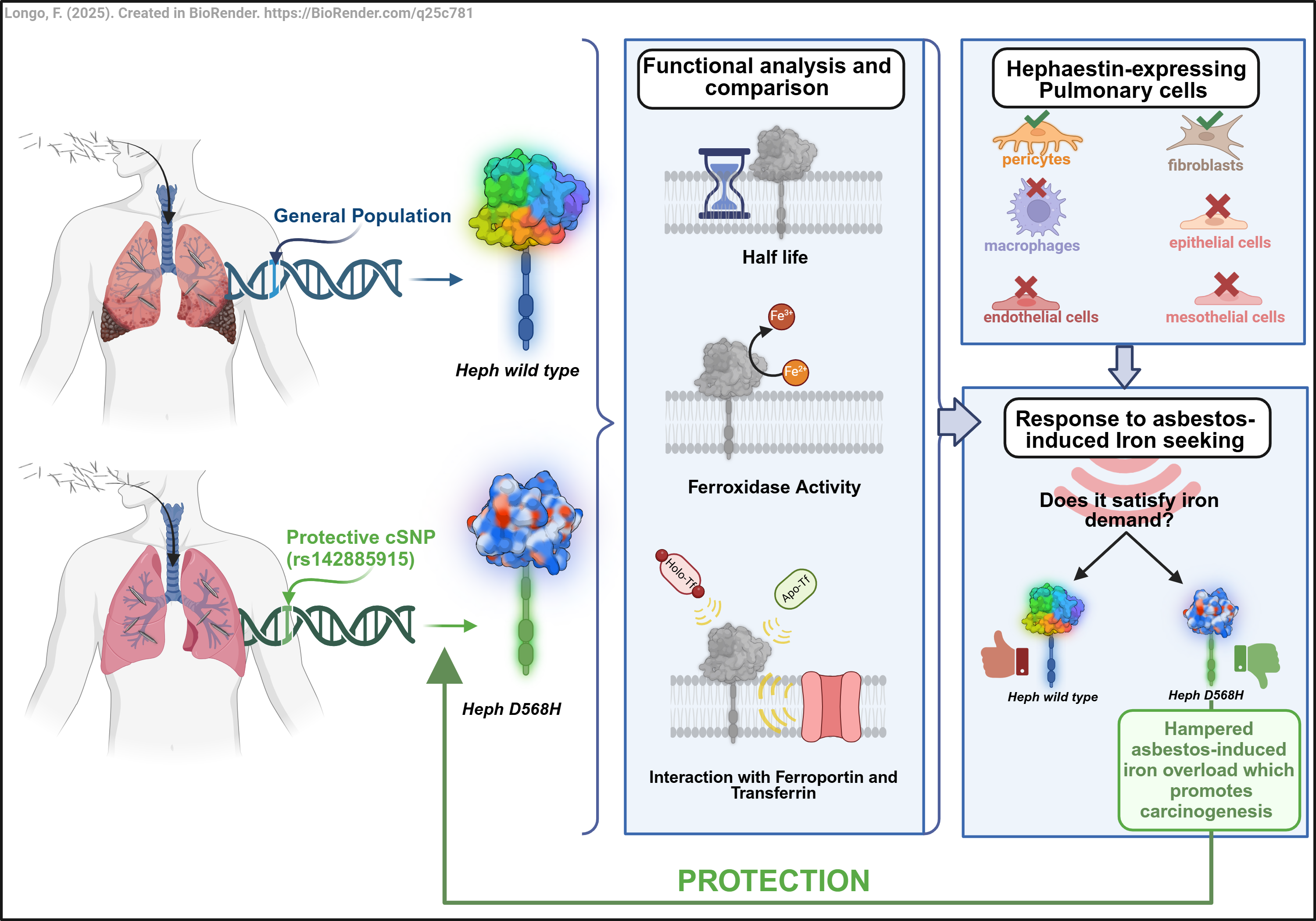
Abstract
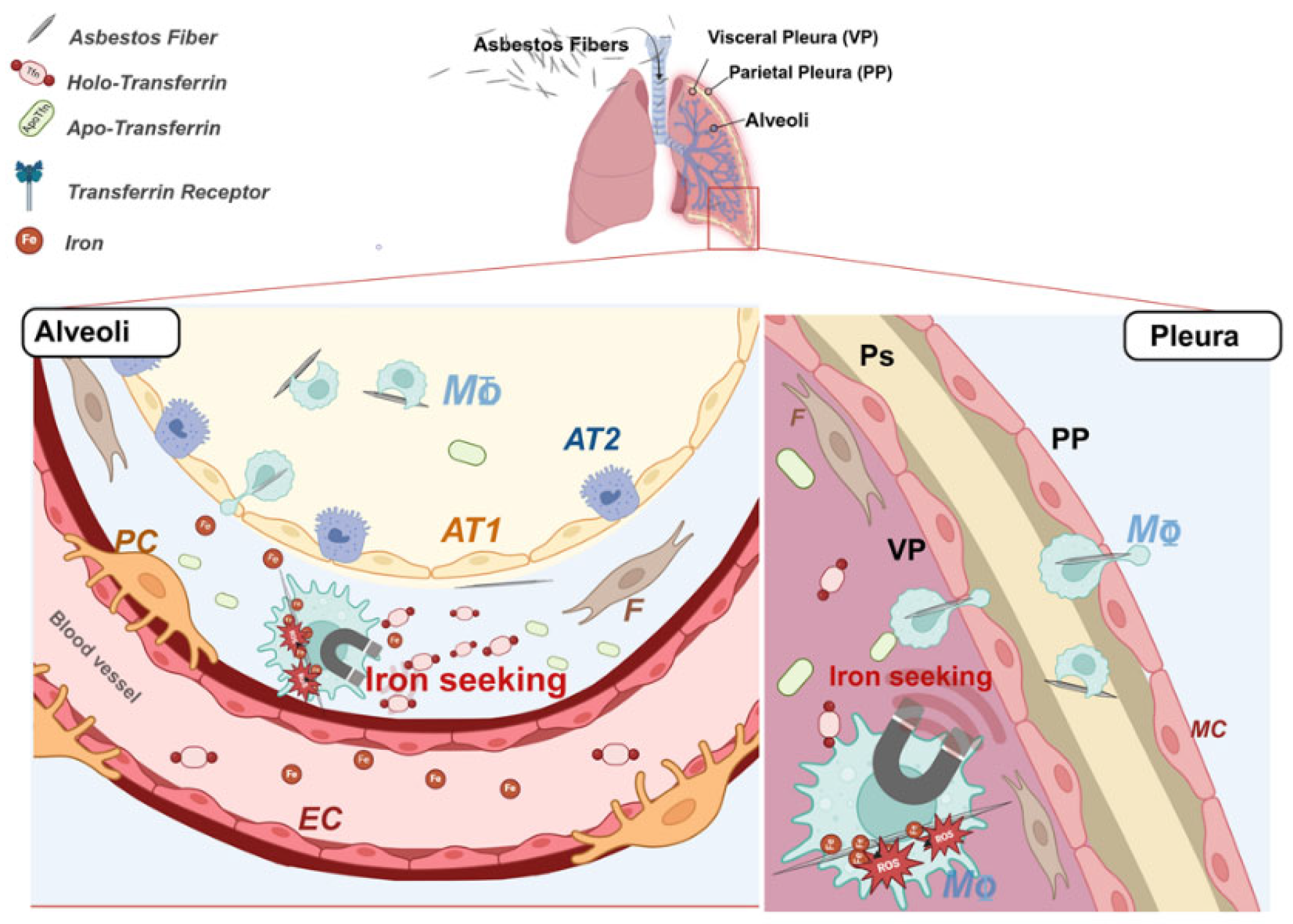
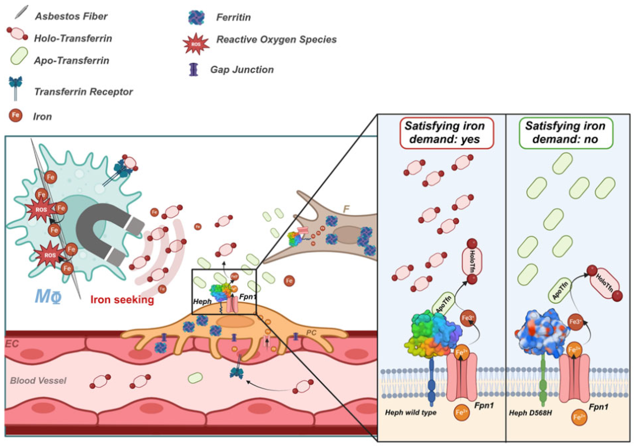
Local disruption of iron homeostasis leading to oxidative stress is considered one of the main mechanisms of asbestos-dependent cyto-/genotoxicity. Another aspect contributing to the risk of developing pathological consequences upon asbestos exposure is due to individual genetic fac-tors. In a previous study we identified a coding SNP in the hephaestin gene (HEPH) exerting a protective activity against the development of asbestos related thoracic cancers. Heph is a fer-roxidase which promotes iron export in concert with the permease ferroportin (Fpn1). Here, we performed an in-depth functional characterization of the HephD568H variant to gain insights on the molecular basis of its protective activity in asbestos-related carcinogenesis. We demonstrat-ed that HephD568H complexes with Fpn1 and possesses a full ferroxidase activity. Despite being more efficiently recruited at the plasma membrane, HephD568H is severely hampered in (iron-depleted) apo-Tfn binding, whose interaction with the WT ferroxidase emerged as a novel mechanism to perceive brain iron needs. Heph in human lung is expressed by pericytes and fi-broblasts and lung pericytes were shown to respond to iron request by up-regulating the iron exporter pair. These findings extend to the pulmonary vascular unit the paradigm of local iron regulation uncovered at the blood-brain barrier. Moreover, they mechanistically correlate alter-ations in iron sensing with the risk of developing asbestos-dependent malignancies.
Keywords:
1. Introduction
2. Results
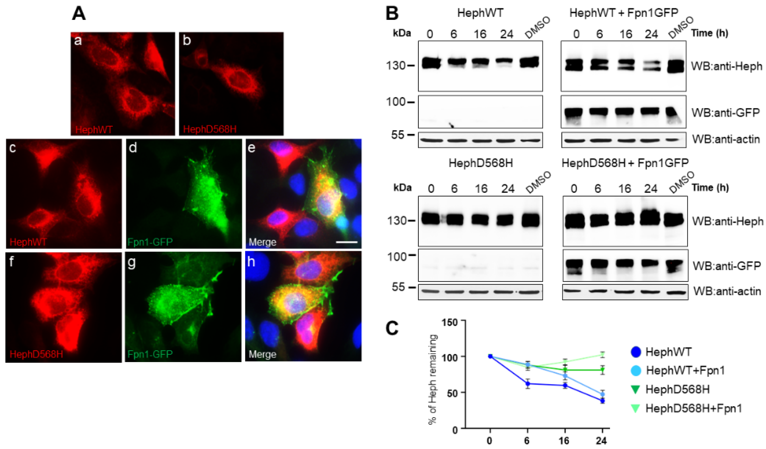
2.1. HephD568H Distributes as the HephWT upon Ectopic Expression in HEK293T Cells but Has an Extended Half Life
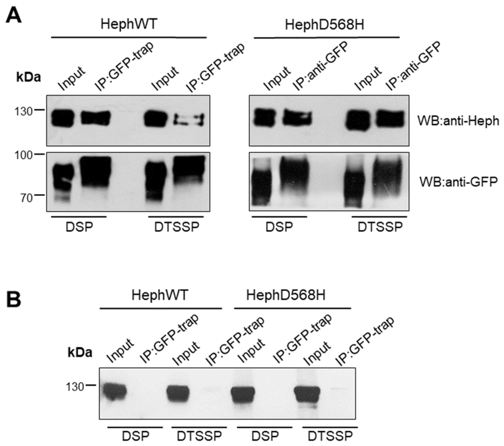
2.2. HephD568H Interacts with the Permease Fpn1 but the Complex Is More Enriched at the Cell Surface with Respect to HephWT
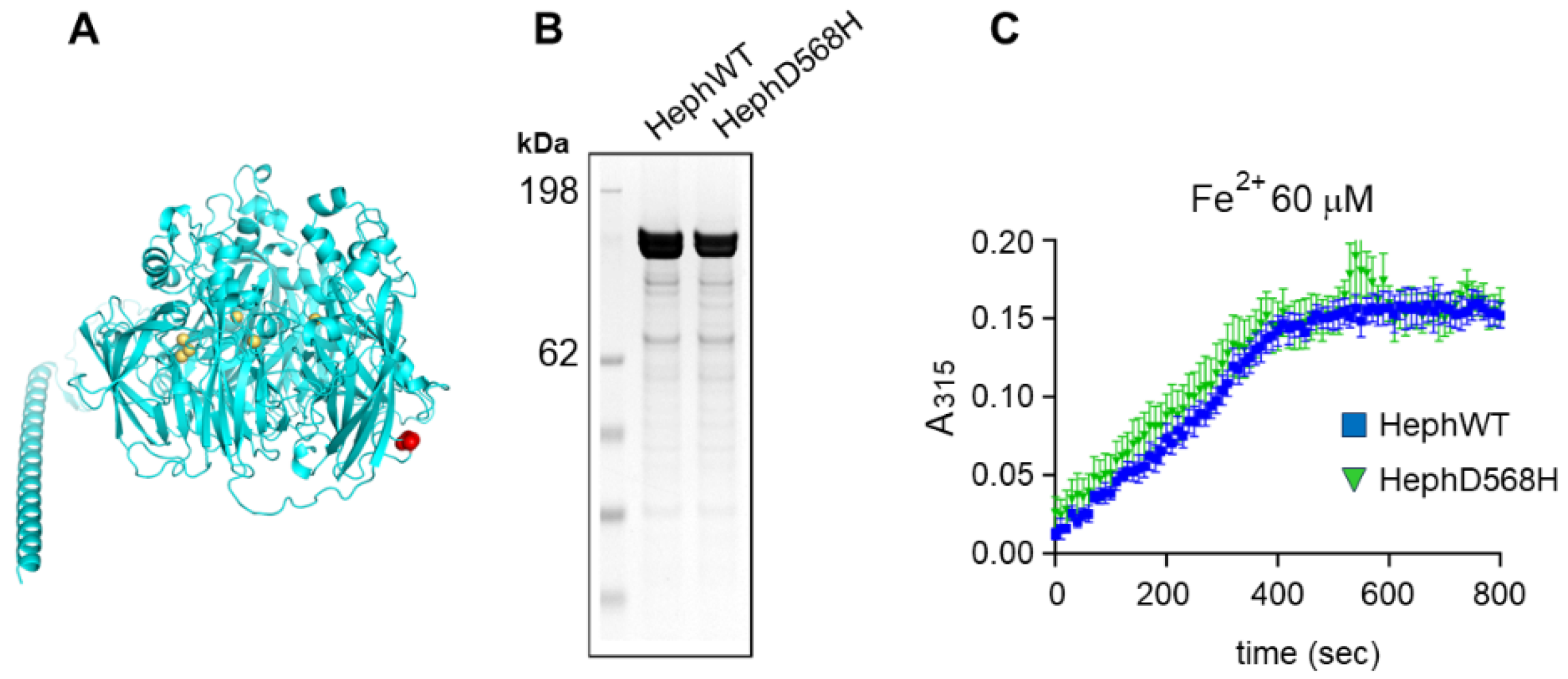
2.3. HephD568H Is Not Impaired in the Ferroxidase Activity
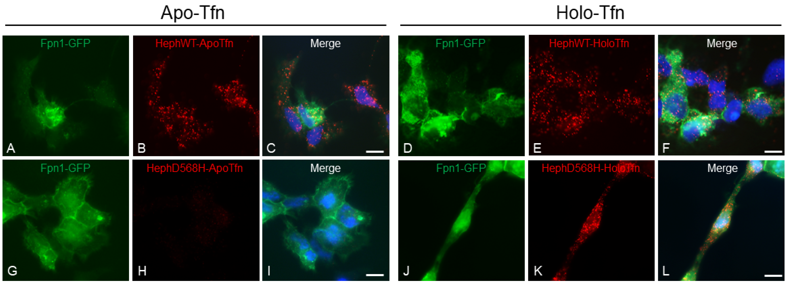
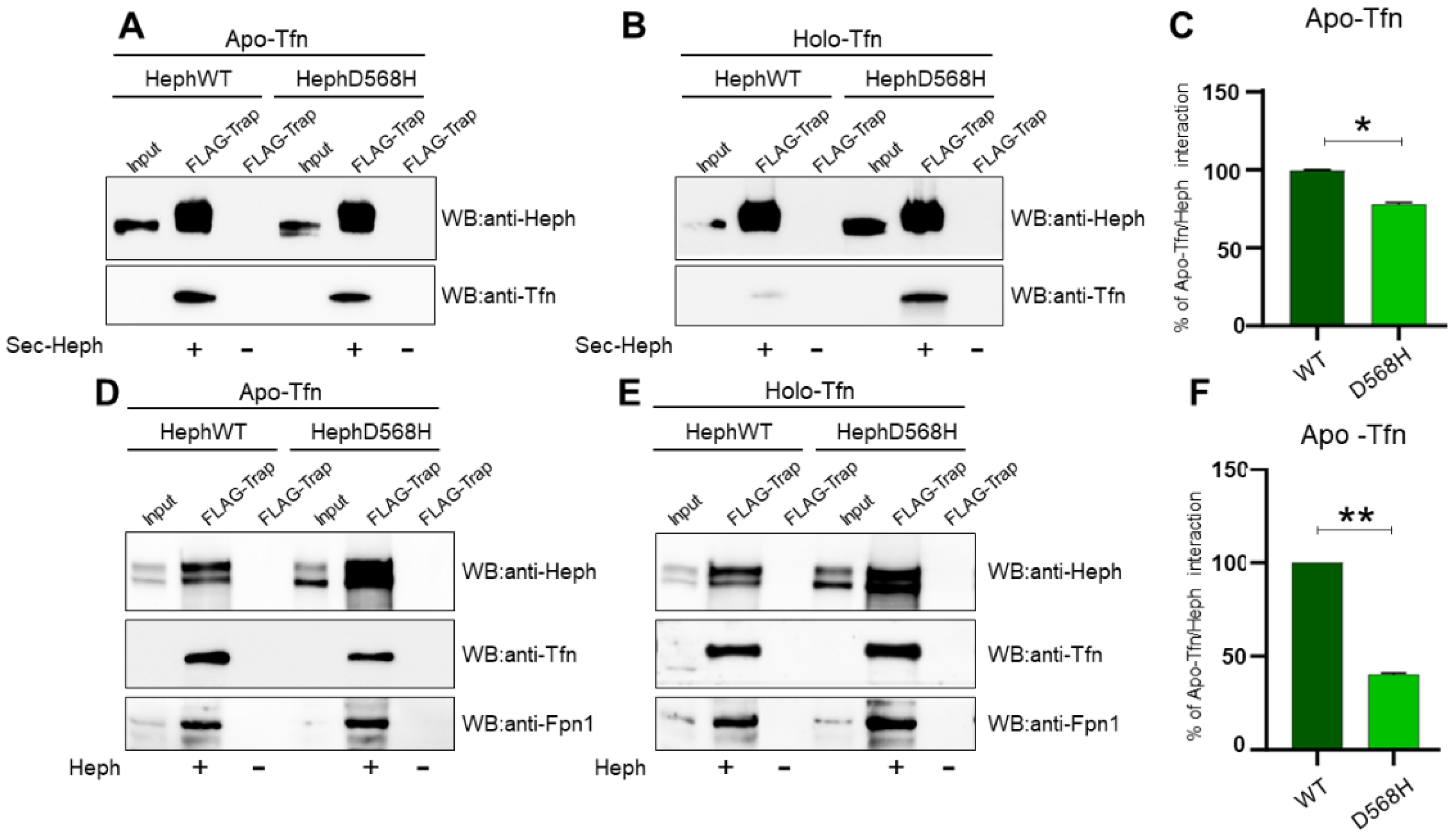
2.4. HephD568H Affects Iron Sensing
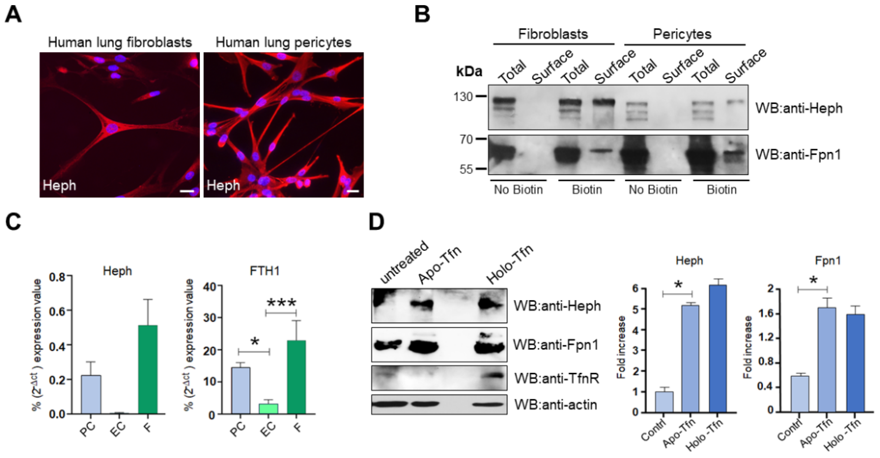
2.5. Heph Is Expressed by Human Lung Pericytes and Fibroblasts
3. Discussion
4. Materials and Methods
4.1. Plasmids and Mutagenesis
4.2. Chemicals and Antibodies
4.3. Cell Culture and Transfections
4.4. Recombinant Protein Expression and Purification
4.5. Ferroxidase Activity Assay
4.6. Immunocytochemistry and Proximity Ligation Assay (PLA)
4.7. Surface Biotinylation, Immunoprecipitation and Western Blot Analysis
4.8. Gene Expression Analysis
4.9. Statistics
Institutional Review Board Statement:. Not applicable.
Supplementary Materials
Author Contributions
Funding
Informed Consent Statement
Data Availability Statement
Acknowledgments
Conflicts of Interest
Abbreviations
| The following abbreviations are used in this manuscript | . |
| Apo-Tfn | Transferrin (protein), iron-depleted form |
| BBB | Blood Brain Barrier |
| CP | Ceruloplasmin |
| FBS | Fetal Bovine Serum |
| Fpn1 | Ferroportin |
| FTH1 | Ferritin Heavy Chain |
| GFP | Green Fluorescent Protein |
| GPI | Glycosylphosphatidylinositol |
| HEK293T | Human Embryonic Kidney cells |
| HEPH | Hephaestin (gene) |
| Heph | Hephaestin (protein) |
| HMC | Human Mesothelial primary cells |
| Holo-Tfn | Transferrin (protein), iron-loaded form |
| HPMEC | Human Pulmonary Microvascular Endothelial Cells |
| Huvec | Human Umbilical Vein Cells |
| LC | Lung Cancer |
| MeT-5A | Human Mesothelial Cells |
| MPM | Malignant Pleural Mesothelioma |
| PCR | Polymerase Chain Reaction |
| PLA | Proximity Ligation Assay |
| qRT-PCR | Quantitative Real-Time PCR |
| ROS | Reactive Oxygen Species |
| SNP | Single Nucleotide Polymorphism |
| TF | Transferrin (gene) |
| TfnR | Transferrin Receptor |
| TT1 | Immortal Alveolar Type 1-like cells |
References
- Jamrozik, E.; de Klerk, N.; Musk, A.W. Asbestos-related disease. Intern Med J. 2011, 41, 372–380. [Google Scholar] [CrossRef]
- Andujar, P.; Lacourt, A.; Brochard, P.; Pairon, J.-C.; Jaurand, M.-C.; Jean, D. Five years update on relationships between malignant pleural mesothelioma and exposure to asbestos and other elongated mineral particles. Journal of Toxicology and Environmental Health, Part B 2016, 19, 151–72. [Google Scholar] [CrossRef] [PubMed]
- Gilham, C.; Rake, C.; Burdett, G.; Nicholson, A.G.; Davison, L.; Franchini, A.; et al. Pleural mesothelioma and lung cancer risks in relation to occupational history and asbestos lung burden. Occup Environ Med. 2016, 73, 290–299. [Google Scholar] [CrossRef]
- Lemen, R.A. Mesothelioma from asbestos exposures: Epidemiologic patterns and impact in the United States. Journal of Toxicology and Environmental Health, Part B 2016, 19, 250–265. [Google Scholar] [CrossRef] [PubMed]
- Dostert, C.; Pétrilli, V.; Van Bruggen, R.; Steele, C.; Mossman, B.T.; Tschopp, J. Innate Immune Activation Through Nalp3 Inflammasome Sensing of Asbestos and Silica. Science (1979) 2008, 320, 674–677. [Google Scholar] [CrossRef] [PubMed]
- Ghio, A.J.; Hilborn, E.D.; Stonehuerner, J.G.; Dailey, L.A.; Carter, J.D.; Richards, J.H.; et al. Particulate Matter in Cigarette Smoke Alters Iron Homeostasis to Produce a Biological Effect. Am J Respir Crit Care Med. 2008, 178, 1130–1138. [Google Scholar] [CrossRef]
- Ghio, A.J.; Pavlisko, E.N.; Roggli, V.L. Iron and Iron-Related Proteins in Asbestosis. Journal of Environmental Pathology, Toxicology and Oncology 2015, 34, 277–285. [Google Scholar] [CrossRef]
- Jiang, L.; Akatsuka, S.; Nagai, H.; Chew, S.; Ohara, H.; Okazaki, Y.; et al. Iron overload signature in chrysotile-induced malignant mesothelioma. J Pathol. 2012, 228, 366–377. [Google Scholar] [CrossRef]
- Mossman, B.T. In vitro studies on the biologic effects of fibers: correlation with in vivo bioassays. Environ Health Perspect. 1990, 88, 319–322. [Google Scholar] [CrossRef]
- Mossman, B.t.; Churg, A. Mechanisms in the Pathogenesis of Asbestosis and Silicosis. Am J Respir Crit Care Med. 1998, 157, 1666–1680. [Google Scholar] [CrossRef]
- Mossman, B.T.; Shukla A, Heintz NH, Verschraegen CF. New Insights into Understanding the Mechanisms, Pathogenesis, and Management of Malignant Mesotheliomas. Am J Pathol. 2013, 182, 1065–1077. [Google Scholar] [CrossRef]
- Toyokuni, S. Iron overload as a major targetable pathogenesis of asbestos-induced mesothelial carcinogenesis. Redox Report 2014, 19, 1–7. [Google Scholar] [CrossRef]
- Shannahan, J.H.; Ghio, A.J.; Schladweiler, M.C.; McGee, J.K.; Richards, J.H.; Gavett, S.H.; et al. The role of iron in Libby amphibole-induced acute lung injury and inflammation. Inhal Toxicol. 2011, 23, 313–323. [Google Scholar] [CrossRef]
- Ghio, A.J.; Hilborn, E.D.; Stonehuerner, J.G.; Dailey, L.A.; Carter, J.D.; Richards, J.H.; et al. Particulate Matter in Cigarette Smoke Alters Iron Homeostasis to Produce a Biological Effect. Am J Respir Crit Care Med. 2008, 178, 1130–1138. [Google Scholar] [CrossRef]
- Chew, S.H.; Toyokuni, S. Malignant mesothelioma as an oxidative stress-induced cancer: An update. Free Radic Biol Med. 2015, 86, 166–178. [Google Scholar] [CrossRef] [PubMed]
- Aust, E.A.; Lund, L.G.; Chao, C.-C.; Park, S.-H.; Fang, R. Role of Iron in the Cellular Effects of Asbestos. Inhal Toxicol. 2000, 12, 75–80. [Google Scholar] [CrossRef] [PubMed]
- Aust, A.E.; Cook, P.M.; Dodson, R.F. Morphological and Chemical Mechanisms of Elongated Mineral Particle Toxicities. Journal of Toxicology and Environmental Health, Part B 2011, 14, 40–75. [Google Scholar] [CrossRef]
- Ather, J.L.; Martin, R.A.; Ckless, K.; Poynter, M.E. Inflammasome Activity in Non-Microbial Lung Inflammation. J Environ Immunol Toxicol. 2014, 1, 108–117. [Google Scholar] [CrossRef]
- Abbaspour, N.; Hurrell, R.; Kelishadi, R. Review on iron and its importance for human health. J Res Med Sci. 2014, 19, 164–174. [Google Scholar]
- Poprac, P.; Jomova, K.; Simunkova, M.; Kollar, V.; Rhodes, C.J.; Valko, M. Targeting Free Radicals in Oxidative Stress-Related Human Diseases. Trends Pharmacol Sci. 2017, 38, 592–607. [Google Scholar] [CrossRef]
- Ying, J.-F.; Lu, Z.-B.; Fu, L.-Q.; Tong, Y.; Wang, Z.; Li, W.-F.; et al. The role of iron homeostasis and iron-mediated ROS in cancer. Am J Cancer Res. 2021, 11, 1895–1912. [Google Scholar] [PubMed]
- Harington, J.S. Miller Klara, Macnab Gwen. Hemolysis by Asbestos. 1970. [Google Scholar]
- Lund, L.G.; Aust, A.E. Iron-catalyzed reactions may be responsible for the biochemical and biological effects of asbestos. Biofactors 1991, 3, 83–89. [Google Scholar]
- Hardy, J.A.; Aust, A.E. Iron in Asbestos Chemistry and Carcinogenicity. Chem Rev. 1995, 95, 97–118. [Google Scholar] [CrossRef]
- Ghio, A.J.; Stewart, M.; Sangani, R.G.; Pavlisko, E.N.; Roggli, V.L. Asbestos and Iron. Int J Mol Sci. 2023, 24, 12390. [Google Scholar] [CrossRef]
- Zangari, M.; Borelli, V.; Bernareggi, A.; Zabucchi, G. Asbestos fibers promote iron oxidation and compete with apoferritin enzymatic activity. J Toxicol Environ Health A 2023, 86, 69–73. [Google Scholar] [CrossRef]
- Mossman BT, Lippmann M, Hesterberg TW, Kelsey KT, Barchowsky A, Bonner JC. Pulmonary Endpoints (Lung Carcinomas and Asbestosis) Following Inhalation Exposure to Asbestos. Journal of Toxicology and Environmental Health, Part B. 2011;14:76–121.
- Neri M, Ugolini D, Dianzani I, Gemignani F, Landi S, Cesario A, et al. Genetic susceptibility to malignant pleural mesothelioma and other asbestos-associated diseases. Mutation Research/Reviews in Mutation Research. 2008;659:126–36.
- Carbone M, Yang H. Molecular Pathways: Targeting Mechanisms of Asbestos and Erionite Carcinogenesis in Mesothelioma. Clinical Cancer Research. 2012;18:598–604.
- Ascoli V, Cavone D, Merler E, Barbieri PG, Romeo L, Nardi F, et al. Mesothelioma in blood related subjects: Report of 11 clusters among 1954 Italy cases and review of the literature. Am J Ind Med. 2007;50:357–69.
- Ugolini D, Neri M, Ceppi M, Cesario A, Dianzani I, Filiberti R, et al. Genetic susceptibility to malignant mesothelioma and exposure to asbestos: The influence of the familial factor. Mutation Research/Reviews in Mutation Research. 2008;658:162–71.
- Testa JR, Cheung M, Pei J, Below JE, Tan Y, Sementino E, et al. Germline BAP1 mutations predispose to malignant mesothelioma. Nat Genet. 2011;43:1022–5.
- de Klerk N, Alfonso H, Olsen N, Reid A, Sleith J, Palmer L, et al. Familial aggregation of malignant mesothelioma in former workers and residents of Wittenoom, Western Australia. Int J Cancer. 2013;132:1423–8.
- Betti M, Casalone E, Ferrante D, Aspesi A, Morleo G, Biasi A, et al. Germline mutations in DNA repair genes predispose asbestos-exposed patients to malignant pleural mesothelioma. Cancer Lett. 2017;405:38–45.
- Betti M, Aspesi A, Ferrante D, Sculco M, Righi L, Mirabelli D, et al. Sensitivity to asbestos is increased in patients with mesothelioma and pathogenic germline variants in BAP1 or other DNA repair genes. Genes Chromosomes Cancer. 2018;57:573–83.
- Cadby G, Mukherjee S, Musk AW (Bill), Reid A, Garlepp M, Dick I, et al. A genome-wide association study for malignant mesothelioma risk. Lung Cancer. 2013;82:1–8.
- Bianchi C, Brollo A, Ramani L, Zuch C. Asbestos-related mesothelioma in monfalcone, Italy. Am J Ind Med. 1993;24:149–60.
- Ohar JA, Cheung M, Talarchek J, Howard SE, Howard TD, Hesdorffer M, et al. Germline BAP1 Mutational Landscape of Asbestos-Exposed Malignant Mesothelioma Patients with Family History of Cancer. Cancer Res. 2016;76:206–15.
- Liu C, Stücker I, Chen C, Goodman G, McHugh MK, D’Amelio AM, et al. Genome-wide Gene–Asbestos Exposure Interaction Association Study Identifies a Common Susceptibility Variant on 22q13.31 Associated with Lung Cancer Risk. Cancer Epidemiology, Biomarkers & Prevention. 2015;24:1564–73.
- Wei S, Wang L-E, McHugh MK, Han Y, Xiong M, Amos CI, et al. Genome-wide gene–environment interaction analysis for asbestos exposure in lung cancer susceptibility. Carcinogenesis. 2012;33:1531–7.
- Kettunen E, Hernandez-Vargas H, Cros M, Durand G, Le Calvez-Kelm F, Stuopelyte K, et al. Asbestos-associated genome-wide DNA methylation changes in lung cancer. Int J Cancer. 2017;141:2014–29.
- Crovella S, Bianco AM, Vuch J, Zupin L, Moura RR, Trevisan E, et al. Iron signature in asbestos-induced malignant pleural mesothelioma: A population-based autopsy study. J Toxicol Environ Health A [Internet]. 2016;79:129–41. Available from. [CrossRef]
- Celsi F, Crovella S, Moura RR, Schneider M, Vita F, Finotto L, et al. Pleural mesothelioma and lung cancer: the role of asbestos exposure and genetic variants in selected iron metabolism and inflammation genes. J Toxicol Environ Health A. 2019;82:1088–102.
- Donovan A, Brownlie A, Zhou Y, Shepard J, Pratt SJ, Moynihan J, et al. Positional cloning of zebrafish ferroportin1 identifies a conserved vertebrate iron exporter. Nature [Internet]. 2000;403:776–81. Available from. [CrossRef]
- McKie Andrew, T. , Marciani p., Andreas r., Brennan k., Wehr K, Barrrow, et al. A Novel Duodenal Iron-Regulated Transporter, IREG1, Implicated in the Basolateral Transfer of Iron to the Circulation. 2000.
- Helman SL, Zhou J, Fuqua BK, Lu Y, Collins JF, Chen H, et al. The biology of mammalian multi-copper ferroxidases. BioMetals. 2023;36:263–81.
- Vashchenko G, MacGillivray R. Multi-Copper Oxidases and Human Iron Metabolism. Nutrients. 2013;5:2289–313.
- Anderson GJ, Vulpe CD. Mammalian iron transport. Cellular and Molecular Life Sciences. 2009;66:3241–61.
- Vulpe CD, Kuo Y-M, Murphy TL, Cowley L, Askwith C, Libina N, et al. Hephaestin, a ceruloplasmin homologue implicated in intestinal iron transport, is defective in the sla mouse [Internet]. nature genetics •. 1999. Available from: http://www.informatics.jax.
- Anderson GJ, Frazer DM, McKie AT, Vulpe CD. The Ceruloplasmin Homolog Hephaestin and the Control of Intestinal Iron Absorption. Blood Cells Mol Dis. 2002;29:367–75.
- McCarthy RC, Kosman DJ. Ferroportin and Exocytoplasmic Ferroxidase Activity Are Required for Brain Microvascular Endothelial Cell Iron Efflux. Journal of Biological Chemistry. 2013;288:17932–40.
- McCarthy RC, Kosman DJ. Mechanisms and regulation of iron trafficking across the capillary endothelial cells of the blood-brain barrier. Front Mol Neurosci. 2015;8.
- Baringer SL, Palsa K, Simpson IA, Connor JR. Apo- and holo- transferrin differentially interact with ferroportin and hephaestin to regulate iron release at the blood-brain barrier. Available from, 2023. [CrossRef]
- Zacchi P, Belmonte B, Mangogna A, Morello G, Scola L, Martorana A, et al. The Ferroxidase Hephaestin in Lung Cancer: Pathological Significance and Prognostic Value. Front Oncol. 2021;11.
- Kemp SJ, Thorley AJ, Gorelik J, Seckl MJ, O’Hare MJ, Arcaro A, et al. Immortalization of Human Alveolar Epithelial Cells to Investigate Nanoparticle Uptake. Am J Respir Cell Mol Biol. 2008;39:591–7.
- Li P, Wu Y, Goodwin AJ, Halushka P V., Wilson CL, Schnapp LM, et al. Generation of a new immortalized human lung pericyte cell line: a promising tool for human lung pericyte studies. Laboratory Investigation. 2021;101:625–35.
- Schnell S, Mendoza C. Closed Form Solution for Time-dependent Enzyme Kinetics. J Theor Biol. 1997;187:207–12.
- Petrič B, Goličnik M, Bavec A. The Removal of Time–Concentration Data Points from Progress Curves Improves the Determination of Km: The Example of Paraoxonase 1. Molecules. 2022;27:1306.
- Vashchenko G, MacGillivray RTA. Functional role of the putative iron ligands in the ferroxidase activity of recombinant human hephaestin. JBIC Journal of Biological Inorganic Chemistry. 2012;17:1187–95.
- Burkhart A, Skjørringe T, Johnsen KB, Siupka P, Thomsen LB, Nielsen MS, et al. Expression of Iron-Related Proteins at the Neurovascular Unit Supports Reduction and Reoxidation of Iron for Transport Through the Blood-Brain Barrier. Mol Neurobiol. 2016;53:7237–53.
- Armulik A, Genové G, Betsholtz C. Pericytes: Developmental, Physiological, and Pathological Perspectives, Problems, and Promises. Dev Cell. 2011. p. 193–215.
- Abbott NJ, Rönnbäck L, Hansson E. Astrocyte–endothelial interactions at the blood–brain barrier. Nat Rev Neurosci. 2006;7:41–53.
- Neal EH, Marinelli NA, Shi Y, McClatchey PM, Balotin KM, Gullett DR, et al. A Simplified, Fully Defined Differentiation Scheme for Producing Blood-Brain Barrier Endothelial Cells from Human iPSCs. Stem Cell Reports. 2019;12:1380–8.
- Sasson E, Anzi S, Bell B, Yakovian O, Zorsky M, Deutsch U, et al. Nano-scale architecture of blood-brain barrier tight-junctions. Elife. 2021;10.
- Matsuoka RL, Buck LD, Vajrala KP, Quick RE, Card OA. Historical and current perspectives on blood endothelial cell heterogeneity in the brain. Cellular and Molecular Life Sciences. 2022;79:372.
- Chiou B, Neal EH, Bowman AB, Lippmann ES, Simpson IA, Connor JR. Endothelial cells are critical regulators of iron transport in a model of the human blood–brain barrier. Journal of Cerebral Blood Flow and Metabolism. 2019;39:2117–31.
- Brown LS, Foster CG, Courtney J-M, King NE, Howells DW, Sutherland BA. Pericytes and Neurovascular Function in the Healthy and Diseased Brain. Front Cell Neurosci. 2019;13.
- Benarroch, E. What Are the Roles of Pericytes in the Neurovascular Unit and Its Disorders? Neurology. 2023;100:970–7.
- Yuan K, Agarwal S, Chakraborty A, Condon DF, Patel H, Zhang S, et al. Lung Pericytes in Pulmonary Vascular Physiology and Pathophysiology. Compr Physiol. Wiley; 2021. p. 2227–47.
- Hare DJ, Double KL. Iron and dopamine: a toxic couple. Brain. 2016;139:1026–35.
- Santiago González DA, Cheli VT, Wan R, Paez PM. Iron Metabolism in the Peripheral Nervous System: The Role of DMT1, Ferritin, and Transferrin Receptor in Schwann Cell Maturation and Myelination. The Journal of Neuroscience. 2019;39:9940–53.
- Rogers LK, Cismowski MJ. Oxidative stress in the lung – The essential paradox. Curr Opin Toxicol. 2018;7:37–43.
- Khiroya H, Turner AM. The role of iron in pulmonary pathology. Multidiscip Respir Med. 2015;10:34.
- Neves J, Haider T, Gassmann M, Muckenthaler MU. Iron Homeostasis in the Lungs—A Balance between Health and Disease. Pharmaceuticals. 2019;12:5.
- Chen Y-T, Chang F-C, Wu C-F, Chou Y-H, Hsu H-L, Chiang W-C, et al. Platelet-derived growth factor receptor signaling activates pericyte–myofibroblast transition in obstructive and post-ischemic kidney fibrosis. Kidney Int. 2011;80:1170–81.
- Yao L, Hou J, Wu X, Lu Y, Jin Z, Yu Z, et al. Cancer-associated fibroblasts impair the cytotoxic function of NK cells in gastric cancer by inducing ferroptosis via iron regulation. Redox Biol. 2023;67:102923.
- Barron L, Gharib SA, Duffield JS. Lung Pericytes and Resident Fibroblasts. Am J Pathol. 2016;186:2519–31.
- Palsa K, Baringer SL, Shenoy G, Spiegelman VS, Simpson IA, Connor JR. Exosomes are involved in iron transport from human blood–brain barrier endothelial cells and are modified by endothelial cell iron status. Journal of Biological Chemistry. 2023;299:102868.
- Munson P, Lam Y, MacPherson M, Beuschel S, Shukla A. Mouse serum exosomal proteomic signature in response to asbestos exposure. J Cell Biochem. 2018;119:6266–73.

| Hephaestin | Km | SEM | Vmax (µM/min) | SEM |
|---|---|---|---|---|
| WT | 7.71 | 1.55 | 10.60 | 0.80 |
| D568H | 5.38 | 0.97 | 10.30 | 0.60 |
Disclaimer/Publisher’s Note: The statements, opinions and data contained in all publications are solely those of the individual author(s) and contributor(s) and not of MDPI and/or the editor(s). MDPI and/or the editor(s) disclaim responsibility for any injury to people or property resulting from any ideas, methods, instructions or products referred to in the content. |
© 2025 by the authors. Licensee MDPI, Basel, Switzerland. This article is an open access article distributed under the terms and conditions of the Creative Commons Attribution (CC BY) license (http://creativecommons.org/licenses/by/4.0/).


