Preprint
Article

This version is not peer-reviewed.

Biopriming of Pseudomonas aeruginosa Abates Fluoride Toxicity in Oryza sativa L. by Restricting Fluoride Accumulation, Enhancing Antioxidative System and, Boosting Activities of Rhizospheric Enzymes

A peer-reviewed article of this preprint also exists.

Submitted:

05 February 2025

Posted:

07 February 2025

You are already at the latest version

Abstract

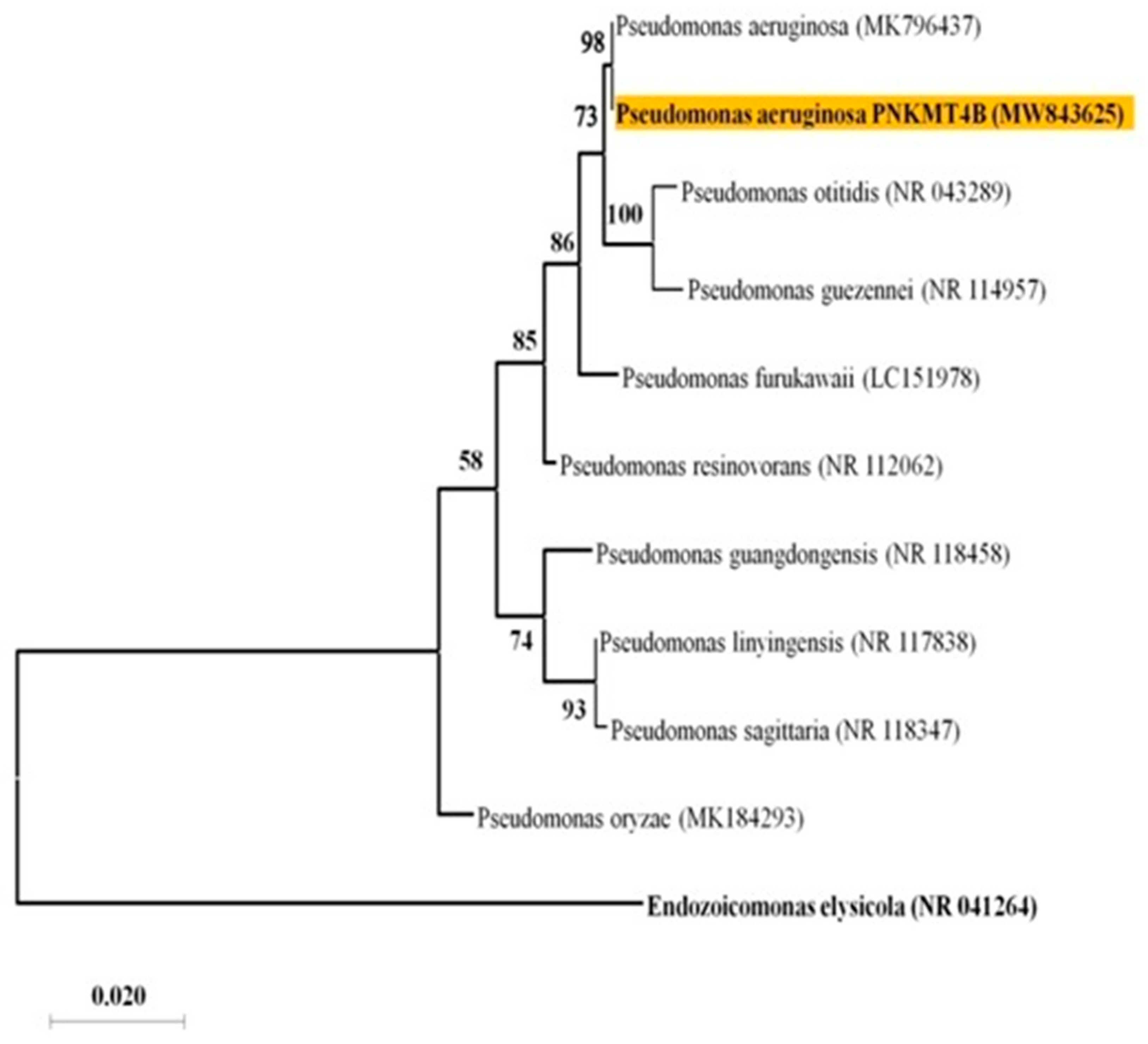
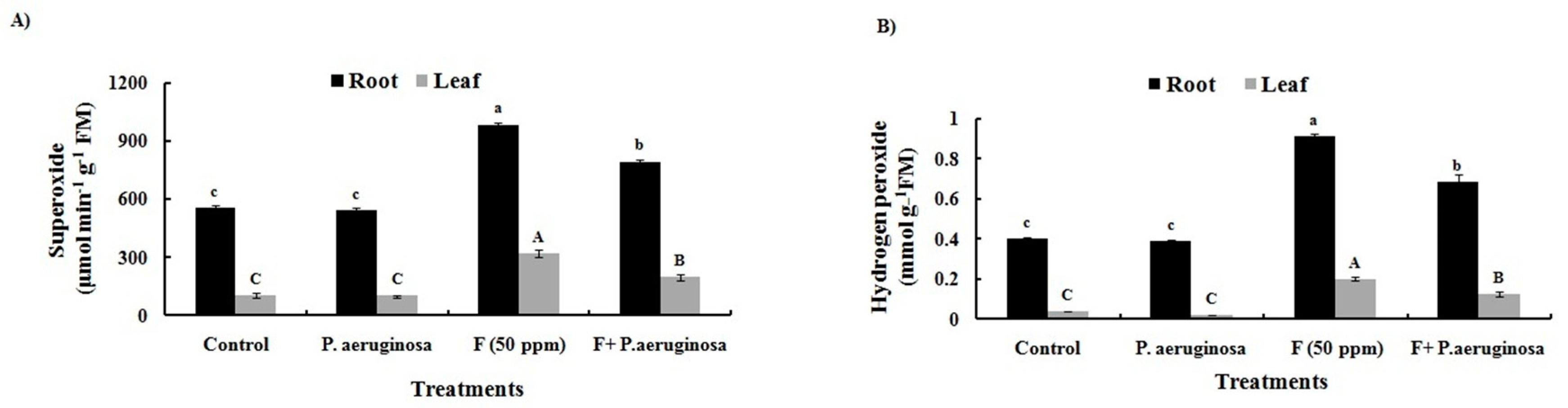
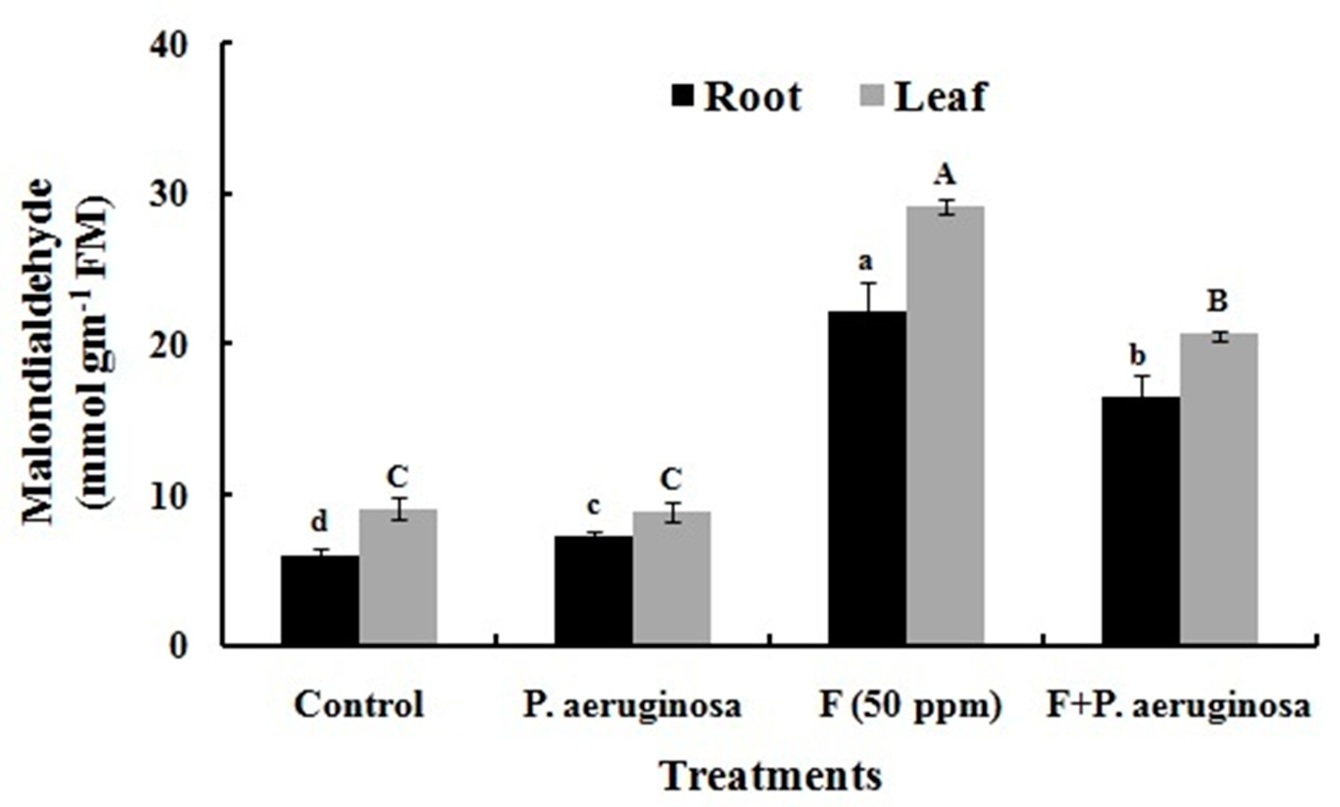
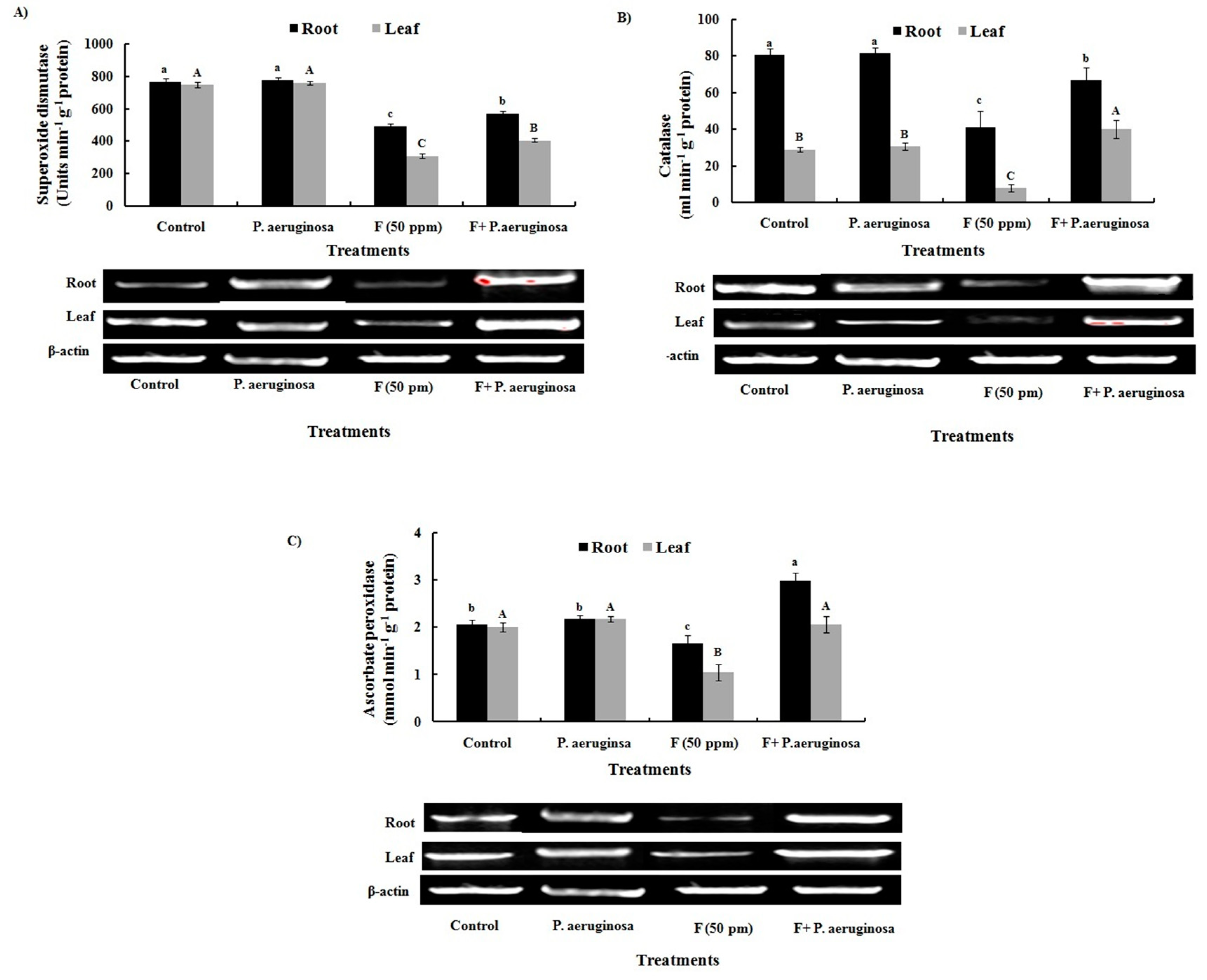
Plant-growth promoting bacteria (PGPB) are free-living microorganisms that actively reside in the rhizosphere and affects plants growth and development. These bacteria employ their own metabolic system to fix nitrogen, solubilize phosphate, and secrete hormones to directly impact metabolism of plants. Gaining a sustainable agricultural production under various environmental stresses requires a detailed understanding of mechanisms that bacteria use to promote plants growth. In the present study, Pseudomonas aeruginosa (MW843625), a PGP soil bacterium with a minimum inhibitory concentration (MIC) of 150 mM against fluoride (F) was isolated from agricultural fields of Chhattisgarh, India, and was assessed for remedial and PGP potential. This study concentrated on biomass accumulation, nutrient absorption, and oxidative stress tolerance in plants involving antioxidative enzymes. By determining MDA accumulation and ROS (O2.- and H2O2) in Oryza sativa L. under F (50 ppm) stress, oxidative stress tolerance was assessed. The results showed that inoculation with P. aeruginosa enhanced the ability of Oryza sativa L. seedlings to absorb nutrients, and increased the amounts of total chlorophyll (Chl), total soluble protein, and biomass. In contrast to plants cultivated under F-stress alone, those inoculated with P. aeruginosa along with F showed considerably reduced concentration of F in their roots, shoots, and grains. The alleviation of deleterious effects of F-stress on plants owing to P. aeruginosa inoculation has been associated with improved activity/ up-regulation of antioxidative genes (SOD, CAT, and APX) in comparison to only F subjected plants, which resulted in lower O2.-, H2O2, and MDA content. Additionally, it has also been reflected from our study that P. aeruginosa has the potential to increase the activities of soil enzymes such as urease, phosphatase, dehydrogenase, nitrate reductase and cellulase. Accordingly, the findings of the conducted study suggests that P. aeruginosa can be exploited not only as an ideal candidate for bioremediation but also enhancing soil fertility and promotion of growth and development of Oryza sativa L. under F contamination.

Keywords: 
;  ;  ;  ;  ;  

1. Introduction

Fluorine is 13th on the list of nature's most abundant elements and occurs mainly in the form of fluoride (F) compounds within the Earth’s crust [1]. Fluoride enters via both natural as well as geogenic means in the food chain and acts as a toxic xenobiotic causing serious threat to the biotic components of the ecosystem [2]. The widespread use of groundwater has caused the water table to decrease significantly leading to extraction of water from deep bored pipes which results in the release of water containing F contaminants that seep from the mineral-beds and contributes towards endemic fluorosis in many regions occupied for agricultural purposes [1,3]. The factors responsible for F leaching from mineral beds include weathering of rocks owing to variation in the climate and ion exchange reactions [4]. Extensive extraction of minerals rich in F such as fluorite, apatite and cryolite has significantly contaminated the groundwater which is the principal source of irrigating food crops and eventually leads to F accumulation, growth retardation and loss of productivity [1]. India, including other Asian countries like China, Bangladesh, Pakistan, etc., is the chief cultivator of Oryza sativa L., and has been currently recognized as the hotspot of F contamination [5]. The World Health Organization (WHO) has warned against using F beyond the threshold limit of 1.5 mg L-1. Ingestion of F above this limit has been widely revealed to cause severe fluorosis, including neurological diseases in living beings [6]. Considering plants, F toxicity impedes in water and minerals transportation which are essential requirements for their normal physiological and biochemical activities [7]. Fluoride is absorbed via vacuolar voltage gated chloride channels (CLC1 and CLC2) present in the roots and translocated by xylematic flow [1]. Prolong exposure of F to the plants results in several biochemical, physiological, and molecular alterations which could be acute or severe.
Fluoride negatively impacts metabolic activities, nutrients intake, germination potential, growth and development, photosynthesis, carbohydrate metabolism, respiration, nitrogen assimilation, protein synthesis, enzymes activities, and gene expression eventually leading to oxidative stress in plants [1]. Oxidative stress, following the overproduction of reactive oxygen species (ROS) viz.; hydrogen peroxide (H2O2), superoxide (O2.-), and hydroxyl radical (OH.), engenders membrane instability/ damage, accompanying release of several cytotoxic products [eg. malondialdehyde (MDA)] in the affected tissues. Stressed conditions in plants also accelerate protease activity which in turn causes protein inactivation and proteolysis [8]. Various enzymes associated with Krebs cycle such as succinate dehydrogenase, pyruvate dehydrogenase, and malate dehydrogenase; and enzymes involved in nitrogen acquisition; nitrate reductase, etc., are severely inhibited due to the F-stress in vulnerable varieties [9]. Plants possess strictly controlled antioxidative machinery to shield themselves from these extremely dangerous oxygen intermediates, regulating ROS generation and scavenging to prevent cellular damage [10]. The antioxidant defense system includes both enzymatic and non-enzymatic substances. Superoxide dismutase (SOD), catalase (CAT), ascorbate peroxidase (APX), and several other enzymatic components regulate equilibrium of ROS in living beings [11,12]. The non-enzymatic elements, which are usually made up of lipids, phenolic compounds, ascorbic acid, tocopherol, flavonoids, proline, carotenoids, and glutathion, efficiently reduce oxidative damage by either working with the enzymatic players to achieve effective antioxidant defense through the consumption of H2O2 or by acting independently of them [10].
Several methods have previously been suggested to reduce F toxicity which involves exogenous application of chemicals like spermidine, melatonin, etc. [7]. Utilization of melatonin limited F uptake in Oryza sativa L. seedlings and lowered the damage caused due to it by restoring the defense machinery, and altering homeostasis of hormones [1]. Calcium compounds such as calcium hydroxide [Ca(OH)2], calcium nitrate [Ca(NO3)2], and calcium chloride (CaCl2) have also been incorporated as seed priming agents for the mitigation of F toxicity thereby enhanced germination percentage, root-shoot length, biomass, impeded Chl degradation and electrolyte leakage; and minimized MDA concentration and ROS [3]. Sodium nitroprusside (SNP) has also been deemed to be effective for reducing F toxicity in Vigna radiata L. and Vigna mungo L. by narrowing F uptake, rescuing membrane damage, and antioxidant enzyme activity thereby alleviating oxidative stress along with increased plant growth [13]. Likewise, supplementation of biochar with soil (50 g/kg soil) has also been considered as the remedy for minimizing toxicity of F in Carthamus tinctorius L. [14]. These days, nanotechnology has become potential tool in agriculture because it permits sustainable cultivation [15]. Pulsing silicon nanoparticles exogenously stimulated growth and also assisted in alleviating molecular injuries and production loss in Oryza sativa L. which were resulted due to the F toxicity [3].
In addition to above mentioned ameliorative methods, much attention has been given to bioremediation that involves living organisms or enzymes to detoxicate environment from various contaminants [16]. Although, rather slow and time-consuming, it outperforms traditional chemical treatments and, most importantly, maintains soil fertility [17]. Contamination of F in the ecosystem generates selective pressure for the advent of tolerant resistant bacteria against it. These tolerant strains can be used to inhibit uptake of F by plants by altering reactivity and mobility of F [18]. Bacterial remediation of F from contaminated areas can be made possible by utilizing indigenous or genetically modified microorganisms for environmental protection [19,20]. Bacterial resistance against F can be transient due to the horizontal gene transfer among two cells or stable because of chromosomal aberrations. Fluoride interacts with proton and forms hydrogen fluoride (HF), which gets absorbed inside the bacterial cells through diffusion and dissociates into H+ and F ions [16]. These ions hinder activities of enzymes associated with glycolysis and F-ATPases, which in case of F-resistant bacteria are expected to be got mutated [21]. Furthermore, the cell wall of bacteria is composed of phosphate, sulfhydryl, and amine groups that can reduce F intake efficiently and thus help in its adherence to their surfaces [19,20,22]. Several other processes are also employed by bacteria like bioaccumulation, biosorption, and release of ligands like siderophores or biosurfactants to minimize F toxicity and its transport into the cells thereby affecting its availability in the environment. Plant growth promoting bacteria (PGPB) are essential for sustainable agriculture because they increase soil fertility, reduce pathogen growth and infection, boost biodiversity, and increase crop productivity [23]. Plant growth promoting bacteria can enhance the plant's rhizospheric environment by influencing soil enzyme activity. Fan et al. [24] highlighted the significant contribution of PGPB in sustaining the soil element cycle and crop yields. Soil microbial communities are essential for maintaining and supporting soil ecosystems. They serve as a vital connection between the aboveground and underground parts of plants in soil ecosystems [25]. Soil enzymatic activity is frequently utilized as a measure of overall soil microbial activity and fertility, as well as a potential indicator for assessing soil stress levels [26]. Several studies have reported the positive impact of PGPB on soil enzymatic activities. Based on their ability to increase plant output through rhizosphere management, numerous strains of beneficial soil bacteria have been identified and are currently being utilized in the biotechnology to enhance agricultural sustainability and food security [27]. Several bacterial strains viz.; Bacillus subtilis, B. amyloliquefaciens, B. licheniformis, B. megaterium, Paenibacillus mucilaginosus, Pseudomonas fluorescens, P. putida, P. fluorescens, P. striata, P. azotoformans, P. chlororaphis, P. fluorescens, etc., are employed as biofertilizer products in India, China, Vietnam, Cuba, Sweden, Sri Lanka, and many other countries [28]. Pseudomonas, a member of Proteobacteria, houses the most diverse group of bacteria having hundreds of different species [29]. They are the most researched bacterial species due to their presence in diverse environments including marine, freshwater, and terrestrial ecosystems, and closest association with higher life forms [30]. Prolific presence of Pseudomonas in the rhizosphere, and their remarkable growth promoting features like production of enzymes, metabolites, siderophores, phytohormones; better colonization over roots; solubilization of nutrients; and function as biocontrol agent makes them notable PGPB [31,32]. Thus, the current study aims at evaluation of plant growth promoting (PGP) properties of P. aeruginosa (MW843625), isolated from the F contaminated soil of agricultural fields, and its role in minimizing deleterious effects of F in Oryza sativa L. seedlings by decreasing the oxidative injuries and boosting activities of soil enzymes.

2. Results and Discussion

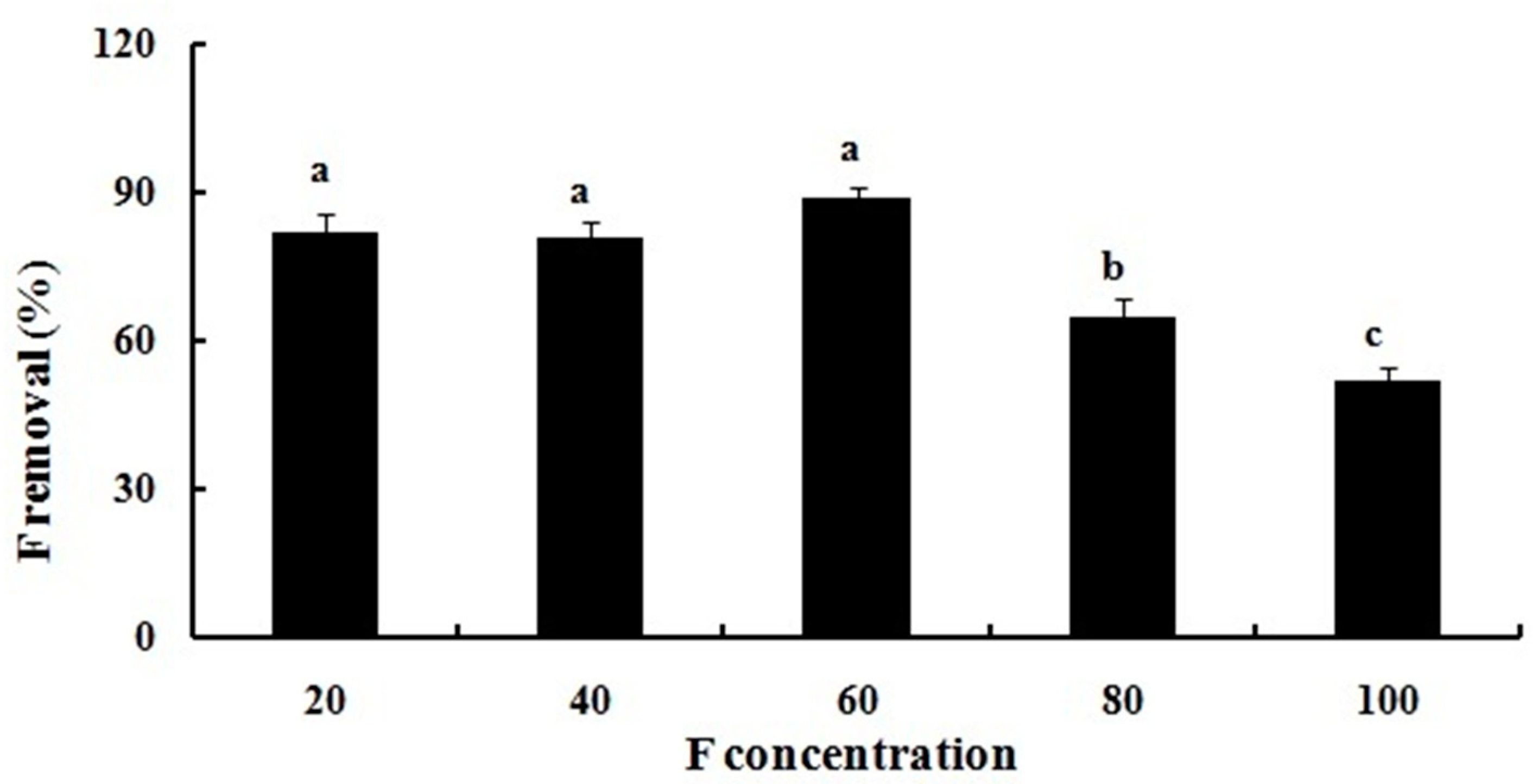
2.1. Fluoride Resistance and Removal

The isolated bacterium identified as P. aeruginosa (Figure 1) revealed resistance to relatively higher concentrations of F as MIC of it was seen to be 200 mM. The resistance of P. aeruginosa to F has also been shown by Chouhan et al. [62], and Edward Raja et al. [63,64]. This bacterium has lowered down F content of growth media to a significant level. Maximum (89.66%) removal of F was achieved with the concentration of 60 mM, and this efficiency gradually declined with increase in the concentration of F (Figure 2). The most plausible explanation for this is that at lower concentrations, capacity of the bacterium for absorption of F is not fully utilized. However, as the concentration increases, removal efficiency considerably decreases, probably as a result of saturation and less availability of sorption sites on the bacterial surface [19,20]. Within bacterial cells, there are various mechanisms that provide tolerance, such as efflux pumps, intracellular sequestration, and detoxifying enzymes. In addition, an increase in the DNA repair mechanisms, the presence of F riboswitches, ion antiporters/transporters, and genetic mutations also play a role towards improving F resistance in bacterial cells [65]. Other organisms like Staphylococcus lentus and Providencia vermicola (KX941098) have also been reported which responded similarly towards removal of F from the media amended with different concentrations of it [19,20].

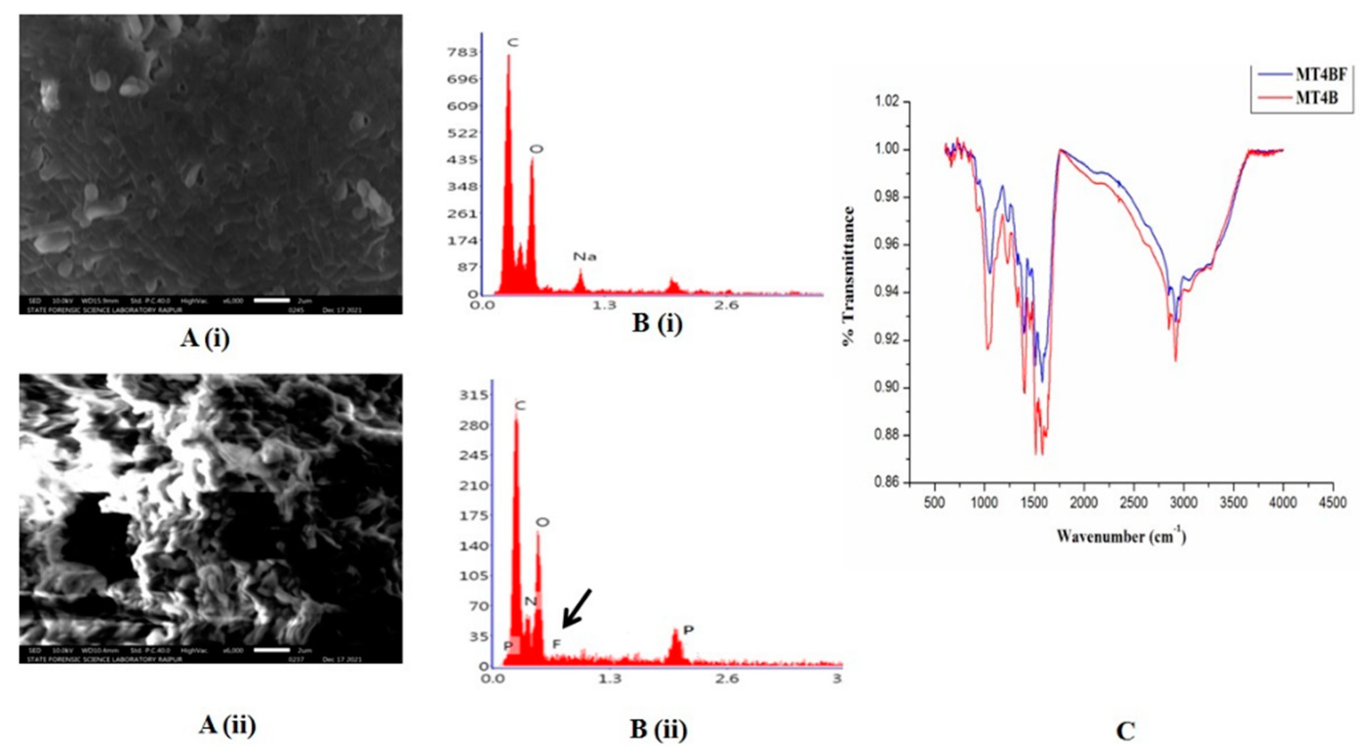
2.2. Fluoride Biosorption Potential of P. aeruginosa

Surface topography of treated and untreated P. aeruginosa biomass was depicted in the SEM micrographs (Figure 3A i and ii). Due to the uptake of F and its adhesion on the bacterial surface, significant morphological alterations and discrete aggregates were documented. Surface of the F treated P. aeruginosa appeared comparatively thicker than the untreated biomass which was found to be porous. Further, surface distortion and lump formation were clearer in the F treated bacterial cells. Mukherjee et al. Thesai et al. and Shanker et al. [19,66,67] showed similar changes in the surface topographies of Providencia vermicola (KX926492), Bacillus flexus (KX646392), and Acinetobacter sp. (GU566361), respectively after F sorption. The EDX analysis also supported presence of F in the treated cells, which is marked by the F peak in the graph (Figure 3B i and ii). The presence of positive ions like sodium, magnesium, and calcium in the bacterial cells could be the cause of this uptake/ sorption of F [19].
Fourier transform infrared spectroscopy has been used to portray structural changes in the bacterial cells through identification of functional groups responsible for binding of F to their membranes (Figure 3C). Maximum stretching of a peak around 3300 cm-1 represented the O–H bond. While, sharper peaks at 1100 cm-1, 1400 cm-1, 1600 cm-1, and 2900 cm-1 represented the primary amines (amine N-H stretch). Bands in the range of 1000-800 cm-1 related to the C-O-C and C-O-P stretching, suggested the presence of oligosaccharides and polysaccharides in the bacterial biomass [19]. It has been found that F was absorbed by the bacteria and gets accumulated once it enters inside the cell. Failure to synthesize any compound of metabolic pathways might be the possible explanation for insignificant shift in the transmittance of the raw and F loaded P. aeruginosa [19].

2.3. Plant Growth Promoting Attributes

The in vitro study of PGP traits of F-resistant P. aeruginosa displayed numerous characteristics of them, both with or without F (Table 1). Bacteria found in the soil may assist plants to grow better even under stress condition by increasing nutrient delivery, encouraging plants growth by synthesizing hormones like IAA, solubilizing inorganic P, accumulating or leaching metals, and reducing pathogen activity [68]. Usually, a bacterium has an immediate impact on growth and development of a plant by employing one or more of these processes [69]. One of these traits is the auxin (IAA), secreted by the PGPB which has a number of direct effects on growth and development of plants under stressful conditions [70,71]. The main effect of bacterially produced IAA is improvement in the development of lateral and adventitious roots, which leads to a rise in the absorption of minerals and nutrients [72,73]. Similar findings in concordance to the present results have been reported for different species of Pseudomonas that produced IAA and supported root growth in Triticum aestivum L. [74,75], Brassica napus L. [70], and Medicago truncatula L. [76] under stresses.
A sizeable amount of P available in the soil is inaccessible to plants because of its insoluble state. The action of soil microorganisms may help to improve this situation. Approximately, 1-50% soil dwelling bacteria are phosphate-solubilizing and can dissolve inorganic phosphates by producing organic acids, siderophores, and hydroxide ions [77,78]. According to Jiang et al. [79], phosphate solubilizing activity of P. aeruginosa boosted P availability in rooting media, which resulted in higher phosphate content in the inoculated Oryza sativa L. Furthermore, siderophores, which are chelating agents, enable bacteria to get beyond the nutritional iron (Fe) limitation in plants due to their high specificity and affinity for binding Fe3+ [80]. These bacterial siderophores foster plant growth by improving nutrition and preventing development of phytopathogens by sequestration of Fe from the environment [81,82,83].
It has also been demonstrated that the ammonia produced by the PGPB supplies N to their host plants, hence; encouraging root and shoot elongation and biomass production. Further, HCN acts as a biocontrol agent due to its ascribed toxicity against plant pathogen [84,85]. It has previously been shown that a wide variety of bacterial species, including Alcaligenes, Aeromonas, Bacillus, Pseudomonas, and Rhizobium, are capable of generating ammonia and HCN in sizable proportions [86].

2.4. Rhizosphere Colonization of P. aeruginosa

Fluoride tolerant P. aeruginosa was investigated for their potency to colonize in the Oryza sativa L. rhizosphere. Bacteria was found to be present (4.2 × 10⁴ CFU g-1) in the inoculated soil sample but not in the control and F added soil, demonstrating successful colonization in the rhizospheric region. Similar results have also been published by Islam et al. [75] regarding P. aeruginosa and Triticum aestivum L.

2.5. Soil Enzymes

Soil enzymes activities are very sensitive to any xenobiotic pollution which indirectly reflects the ability of soil microbial populations to restore soils health [87]. The differences of soil enzymes activities in each treatment were compared and are shown in Table 2. Activities of soil urease, nitrate reductase, phosphatase, cellulase and dehydrogenase were estimated before the transfer of plantlets in the earthen pots and were calculated as 96.42 ± 2.04 µg N-NH4+ g-1 dw h-1, 1.34 ± 0.06 µg NO2--N g-1 dw h-1, 716.2 ± 24.3 µg pNP g−1 dw h−1, 13.51 ± 1.39 µg D-Glu g−1 dw h−1, and 65.34 ± 2.06 µg TPF g-1 dw h-1 respectively. Activities of these enzymes were again calculated after the harvesting of crop, and was found that in the F treated pots activities of all these enzymes were got reduced (Table 2). Several studies have documented the impact of F, usually in the form of NaF, on soil microbial activity and community composition [88,89]. Wilke [90] demonstrated alterations in the chemical characteristics and microbiological activities in the F-adulterated humus soil and concluded that nitrite reductase, alkaline phosphatase, dehydrogenase, and arylsulfatase activities were suppressed after F addition in the soils. Reddy and Kaur [91] provided more evidence that F inhibits activities of ATPase and soil peroxidase. Similarly, Rao and Pal [92] measured detrimental effects of F on soil microorganisms, reporting that high concentrations (380-1803 mg g-1 soil) of F impeded microbial growth and enzyme activity as well as breakdown of organic materials. Fluoride concentrations of 200-2000 mg g-1 soil have been shown to decrease denitrification, whereas; F values below 200 mg g-1 have been shown to limit soil respiration and dehydrogenase activity [93].
Data gathered after P. aeruginosa treatment showed significant improvement in the soil enzymes in both with and without F stressed conditions (Table 2). Under F stressed and bacterium inoculated condition, urease activity increased by 36%, nitrate reductase activity by 57%, phosphatase activity by 14%, cellulase activity by 41.6%, and dehydrogenase activity by 50% over the only F treated soil. Bacterial assisted remediation stimulates plant growth while simultaneously improves soil quality [94]. Yu et al. [87] observed that inoculation of Pseudomonas sp. GHD-4 increased bacterial diversity, decreased lead (Pb) concentration, and enhanced soil enzymes activities in the soil. According to Hidri et al. [95], inoculation of Bacillus subtilis enhanced soil quality by increasing activities of soil enzymes such as urease, alkaline phosphatase, β-glucosidase, and dehydrogenase when compared to the uninoculated control. Additionally, Marques et al. [96] determined that as zinc and cadmium levels increased, diversity of bacterial community decreased. In contrast, Helianthus annuus, inoculated with PGPR strains in the rhizosphere maintained a greater diversity of bacteria throughout the experimental period.

2.6. Plant Growth and MSI

Root length, SL, FM, DM, and MSI were remarkably enhanced in the P. aeruginosa inoculated Oryza sativa L. than the positive control plants under non-F treated condition. Fluoride treatment markedly reduced the RL (47.82%), SL (62.56%), FM (root: 50.4%, shoot: 40.4%), DM (root: 43.8%, shoot: 38%), and MSI (31.18%) of Oryza sativa L. (Table 2). This might be because the developing tissues were absorbed inadequate amounts of nutrients. Membrane instability brought on by exposure of F affects lipid-protein interactions as well as functions of enzymes like H+-ATPase [97]. Many other plant species also exhibited similar decrease in growth metrics [97,98,99]. Furthermore, F exposure makes the plasma membrane prone to lipid peroxidation and cytoskeleton instability, lowering MSI, thereby increasing electrolyte leakage from cells [100,101,102]. In a study conducted by Pelc et al. [103], an increase in F concentration inhibited seedlings growth in all the winter wheat cultivars. Further, Chahine et al. [104] also observed a decline in growth of Jesca (an African-Tanzania bean variety) when was exposed to elevated levels of F. However, in P. aeruginosa inoculated plants, RL and SL were improved by 29% and 48.3%, respectively as compared to those noted with F stressed seedlings. Similarly, it also increased FM (root: 30%, shoot: 23%), DM (root: 28%, shoot: 10%), and MSI (23%) as compared to F alone treated plants (Table 2).
Under stress conditions, PGPR can improve plant nutrition generally by employing one or more mechanisms like associatively fixing nitrogen, solubilizing phosphate, or producing siderophores, and by producing hormones or participating in enzymatic processes, that can encourage growth of rhizobial or mycorrhizal symbiosis, which in turn can improve plant growth and development [65,75,105,106]. The hormones released by the PGPB have a wide range of direct impacts on plant growth under variety of abiotic stresses [71]. Several researchers have previously reported matching findings of plant growth in response to bacterial inoculation along with different metal stress [18,34,74,107,108].

2.7. Total Chlorophyll

Fluoride stress in Oryza sativa L. seedlings caused a significant reduction (50%) in the total Chl content, while P. aeruginosa inoculation in the unstressed seedlings increased it in comparison to the control. Content of total Chl in the leaves is directly related with photosynthetic activity which occurs to reduce under abiotic stresses [109]. Chlorosis, inhibition in Hill activity, and decreased accumulation of photosynthetic pigments were previously being seen in the seedlings irrigated with F rich water [110,111]. However, inoculation of P. aeruginosa enhanced the total Chl content by 29% compared to those estimated in the F alone administered ones (Table 2). The higher total Chl content in response to P. aeruginosa treatment might be due to the boosted uptake of mineral nutrition [78]. In line, Wang et al., Islam et al., Elekhtyar, Samaniego-Gámez et al., Zhang et al., and Abd El-Mageed et al. also noted increased Chl content in plants treated with PGPB as compared to untreated ones under diverse abiotic stresses [75,109,112,113,114,115].

2.8. Fluoride Accumulation in the Tissues

Fluoride treated seedlings accrued 30 ± 3.65, 22 ± 2.75, 9 ± 1.54, and 2.9 ± 0.12 ppm F g-1 DM in roots, shoot, leaves and grains, respectively (Table 2). Roots, being in direct contact with the soil accumulated significantly more amount of F followed by shoot, leaves, and grains. Such alarming hike of xenobiotics in the crops can be a menace to human health. Interestingly, inoculating P. aeruginosa with F stressed seedlings caused a substantial reduction in its accumulation by 33%, 35%, 47%, and 74% in roots, shoots, leaves, and grains respectively. The sorption of F by the bacterium might be the cause of lesser accumulation of it in the tissues. Presence of cells of P. aeruginosa; at 4.2 × 10⁴ CFU g-1 soil in the suspension of the inoculated Oryza sativa L. demonstrated that successful colonization of the bacteria probably caused reduced uptake of F in the tissues [116]. Similar decreases in the accumulation of toxic elements like As and Zn in Vigna radiata L., Oryza sativa L. and Zea mays L., respectively was reported by Pandey and Bhatt, Pandey et al. and Jain et al. [18,34,108].

2.9. Agronomical Attributes

Exposure of F inhibited process of reproduction, and development of panicles in the seedlings of Oryza sativa L. The PL, number of spikelet’s per panicle, FG per panicle, GL, GB, and HGW of grains were significantly decreased by 34%, 51%, 72%, 37%, 40%, and 23%, respectively, while EG per panicle was increased by 86% in response to F when compared to control.
Fluoride inhibited grain hardening owing to its accumulation in the spikelet’s [3]. On the other hand, when P. aeruginosa was added, all these agronomical parameters of the F stressed seedlings were nearly restored to control levels (Table 2). This could be due to the growth promotive effect of P. aeruginosa and also because of the reduced uptake of F in its presence. The results are in consistent with those of Khalid et al., Kumar et al., Amogou et al. , and Sedri et al. [117,118,119,120], who have discovered an upsurge in the grain production when various PGPRs were inoculated in the Triticum aestivum L. and Zea mays L.

2.10. Contents of Protein, Total Sugar, Zinc, and Iron in the Grains

The amounts of protein, total sugar, Fe, and Zn in the grains of F supplied seedlings were lowered by 1.8%, 30%, 22%, and 36% respectively (Table 2) than that determined in the grains of control plants. However, their contents were maintained up to the levels determined in the control tissues after being inoculated with P. aeruginosa. Plant growth promoting bacteria has been demonstrated to enhance exchange and uptake of mineral nutrients (both macro and micro). A change in the pH of the rhizospehric soil by organic acids, metal chelation by siderophores, and cycling of nutrients by the process of microbial mineralization probably boosts the availability of nutrients to plants [109,121].

2.11. ROS Generation

When Oryza sativa L. tissues (root/ leaf) were subjected to F stress, higher accumulations of O2•- and H2O2were observed (root: 43% and 55%; leaf: 67% and 80%) compared to the corresponding controls. On the other side, treatment of F stressed seedlings with P. aeruginosa resulted in a lesser accrual of O2•- and H2O2 (root: 19% and 33%; leaf: 38% and 50%) than that estimated in tissues treated with F alone (Figure 4A and B). Induction of ROS that occurs as a result of F poisoning increases the production of O2•-, as well as its subsequent by-products, such as H2O2 and OH.. Additionally, it has also been demonstrated that F can bind to sulfhydryl groups, which prevents proteins from being synthesized and incapacitates the activities of antioxidant enzymes [122]. As a result, an excessive accumulation of ROS is caused (2). Similarly, overproduction of ROS during F stress has also been noted in Camellia sinensis L. [123], Eriobotrya japonica L. [124], Olea europaea L. [99], and Triticum aestivum L. [125]. Normally, biological systems try to handle stress mediated over produced ROS involving a variety of ways under the guise of antioxidative defence mechanisms [126]. In addition, exogenous factors like PGPR, which can produce antioxidants in response to ROS, can also help to reduce negative effects of unfavorable environmental conditions. According to conducted studies, bacteria may be able to increase plants ability to withstand various stresses by inflicting physical and chemical changes in them, known as PGPR-induced systemic tolerance which was linked with increased levels of Chl a and b, as well as improved CAT, SOD, and other enzymes activities [127,128]. Further, O2•- and H2O2 productions in the Oryza sativa L. leaves were also determined under fluorescence microscope (Figure 5a and b). These observations clearly showed that exposure of F increased the intensity of ROS emission in leaves when compared to the untreated controls. Further, production of ROS was found to be declined noticeably in P. aeruginosa treated leaves to that of F alone subjected tissues, suggesting this bacterium successfully reduced F stress to a significant level. In parallel, Tak and Asthir [125], Banerjee et al. [3], Banerjee and Roychoudhury [129] and Singh et al. [65] also determined similar change in the ROS in F stressed leaves of Triticum aestivum L. Oryza sativa L. and Lycopersicon esculentum L., respectively.

2.12. Lipid Peroxidation

Lipid peroxidation was estimated as MDA level whose concentration was recorded to be increased by 45% in roots and 48% in leaves when seedlings were exposed to F alone. However, after addition of P. aeruginosa, amount of MDA in the roots and leaves reduced to 25% and 29% respectively, even in the presence of F (Figure 6). It is important to note that elevation in the ROS results in destruction of polyunsaturated fatty acids thereby increased concentration of MDA [130]. Similar change in MDA content has also been reported in Helianthus annus L., Salicornia brachiata L., Camellia sinensis L., Cajanus cajan L., Triticum aestivum L., and Oryza sativa L. [2,3,91,123,125,131].

2.13. Antioxidant Enzymes

Activities of SOD, CAT, and APX were considerably suppressed by prolonged exposure of F (leaf: 2.4, 4, and 2 folds, respectively; root: 1.5, 1.9, and 1.25 folds respectively) (Figure 7A, B and C). This might be as a result of binding of F ion with the functional groups of amino acids surrounding the active regions of the enzymes as well as their displacement or competition with magnesium, Fe, or copper ions of enzymes [133]. However, P. aeruginosa in close association with seedlings during F treatment significantly decreased the ROS production and overall oxidative damage by increasing the activities of SOD, CAT, and APX (Figure 7A, B and C). Several reports suggested that the plants having higher antioxidant content are more resistant to oxidative damage [134,135]. In agreement, Ma et al. [136] reported that the bacterial inoculated Ricinus communis L. and Helianthus annuus L. when grown in soils polluted with Ni displayed greater CAT and peroxidase (POX) activity. Similarly, Wang et al. [112] demonstrated that inoculation of an As-resistant Agrobacterium radiobacter with Populus deltoids L. (LH05-17) caused increased growth, Chl content, soluble sugar, and activities of SOD and CAT. Yet, another study discovered that the Pisum sativum L. infused with Ni and Zn tolerant Rhizobium sp. RP5 dramatically increased the antioxidants activities in the roots and nodules, and detoxified ROS up to an extent [137]. Further, genes of SOD, CAT, and APX were also analyzed following RT-PCR. Analysis revealed a down-regulation in the expression levels of their genes in F stressed tissues, while were up-regulated considerably in the presence of P. aeruginosa (Figure 7A, B and C). Expressions of these genes showed comparable patterns with their spectrophotometric data, demonstrating that the transcriptional regulation of genes played a major role in regulating activities of protective enzymes. Similar changes in the expressions of above genes have also been determined in the Vigna radiata L., and Zea mays L. under different abiotic stresses [18,34]. Khan et al. [138] demonstrated that inoculation of Bacillus pumilus enhanced growth of Oryza sativa L. under salinity and high boron conditions by upregulating expressions of antioxidants genes thereby cell protection.

3. Materials and Methods

3.1. Bacterial Strain Isolation and Identification

One of the F tolerant bacteria (FTB), namely MT4B was isolated from the F contaminated soil of agricultural fields of Rajnandgaon District (21o 30’ N, 82o 0’ E, 250-300 msl) of Chhattisgarh, India [18,33]. The isolated FTB was determined to be Gram negative, rod shaped, having green color colony and was identified as Pseudomonas aeruginosa based on the 16S rRNA gene sequencing. The sequence obtained was submitted to the NCBI GeneBank DNA database and bacterial accession number MW843625 was obtained [33]. Further, the phylogenetic analysis of the strain was executed by aligning the obtained sequences with related reference sequences retrieved from the GenBank database using Clustal Omega software. The neighbor joining method with MEGA X software was employed with bootstrap values generated from 100 replicates for distance analysis and phylogenetic tree construction.

3.2. Fluoride Resistance and Removal Assays

The minimum inhibitory concentration (MIC) of F for P. aeruginosa was determined by the agar dilution method. This strain was streaked on Luria Bertani (LB) agar plates amended with various concentrations (5, 10, 25, 50, 75, 100, 125, 150, 200, 250, 275 mM) of F and incubated at 35°C for 24 h. The least dose of F that inhibited growth of the isolate was considered as the MIC [18]. The ability of P. aeruginosa to remove F was ascertained by exposing it to a spectrum of F concentrations (20, 40, 60, 80, and 100 mM) selected on the basis of MIC value. The LB broths with varying concentrations of F were seeded with the bacterial suspension and incubated for 24 h at 35oC in shaking condition (100 rpm). Thereafter, bacterial cultures were centrifuged at 6000 rpm for 10 min at 4oC and the supernatants were collected. Now, concentration of F was determined using a F ion selective electrode (Orion 9609, Thermo Scientific, USA) by mixing the collected supernatants (10 mL) with 1 mL of TISAB (Total ionic strength adjustment buffer) solution (pH 5.5) and then percentage of F removed by the P. aeruginosa was calculated using the formula given below [19];
Percentage   Removal = [ C i C f C f ] × 100
Where, Ci and Cf is the initial and final concentration of F in the culture media

3.3. Determination of F Biosorption by P. aeruginosa

Scanning electron microscopy (SEM, JSM-IT300, JEOL, USA) was used to superficially characterize the biomass in order to ascertain how the shape and spatial orientation of cells have been changed after exposure to F [34]. Bacterial cells are not a natural conductor, therefore; before SEM analysis, the cells were coated with platinum to increase their surface conductivity. Energy dispersive X-ray (EDX, JEOL, USA) was carried out to determine presence of F within the bacterial biomass [19].
Fourier transform infrared (FTIR) spectroscopy was carried out following the methodology of Pandey and Bhatt [34] to demonstrate interaction between F and functional groups present in the untreated and F laden biomass of P. aeruginosa. For this, freshly grown culture of P. aeruginosa was centrifuged at 6000 rpm for 15 min; pellet was collected, and dried in an oven at 60oC. Now, dried biomass was grounded and then homogenized with potassium bromide in 1:1 (w/w) ratio. Lastly, spectral scanning of the sample was performed using FTIR spectrometer (Nicolet iS10, Thermo Scientific, USA) in the range 4000-400 cm-1.

3.4. Plant Growth Promoting Activities of P. aeruginosa Under F-Stress

Pseudomonas aeruginosa was screened for multiple PGP traits viz.; indole 3-acetic acid (IAA), ammonia, hydrogen cyanide (HCN), siderophore production, and solubilization of phosphorus (P), both in the absence and presence of F (60 mM; as maximum F removal was seen at this concentration). The quantitative analysis of IAA was performed by following the method of Gordon and Weber [35]. This strain was examined for ammonia production by adding 0.5 mL of Nesseler’s reagent to 10 mL of freshly grown cultures in peptone water, and absorbance was recorded at 425 nm using a UV-Vis spectrophotometer (Lambda-25, Perkin Elmer, USA) [36]. Production of HCN was determined in accordance with the methodology of Castric (1975) [37] by visualizing yellow to brown color change of Whatman filter paper No.1 drenched with 0.5% (v/v) picric acid solution, placed inside the Petri plate lids. Siderophore production was monitored following the chrome azurol-S (CAS) analytical method [38] by observing yellow to orange halos around the bacterial colonies. Likewise, phosphate solubilizing activity of the isolate was quantitatively assayed in the National Botanical Research Institute’s phosphate growth medium (NBRIP) containing tricalcium phosphate, following the method of Fiske and Subbarow [39].

3.5. Model Plant and Experimental Design

Soil utilized for sowing seeds of model species was collected from the School of Studies in Biotechnology, Pt. Ravishankar Shukla University, Raipur, Chhattisgarh, India. The collected soil was tested for the presence of F, if any. Further, the soil was autoclaved to exterminate the microorganisms present in it, so as to examine the effect of P. aeruginosa alone over growth of the plant. Pseudomonas aeruginosa cultured in the LB broth was used as a bioinoculant after its dilution with 0.9% (w/v) saline. The inoculant was mixed with soil at a cell count of 106 CFU g-1 soil [34]. Now, fresh healthy seeds of Oryza sativa L. (Var. MTU1010) procured from the Indra Gandhi Agricultural University, Raipur, Chhattisgarh, India, were sorted, and surface sterilized with 70% (v/v) ethanol and 0.1% (v/v) sodium hypochlorite solution for 3 min, separately, followed by multiple washing with sterile distilled water (DW). Sterilized seeds were soaked in DW for 24 h and sown in the conditioned soil. Four treatments were designed: (i) Seeds were sown in the soil lacking both F and P. aeruginosa: as control, (ii) Seeds were sown in the soil having P. aeruginosa as bioinoculant, (iii) Seeds were sown in the soil having 50 ppm F, and (iv) Seeds were sown in the soil having 50 ppm F and P. aeruginosa as bioinoculant.
For the present study, 50 ppm dose of F was chosen based on the results of pilot experiment performed following 10, 20, 30, 40, 50, and 60 ppm of F concentrations, which revealed 50% reduction in the germination percentage (GP) of Oryza sativa L. seeds treated with 50 ppm F, as compared to that recorded with the control. Initially, seeds were sown in sterilized plastic disposable glasses (with 30 g soil) each having 10 seeds for 25 consecutive days for germination and proper growth of seedlings by irrigating with sterile DW (10 mL) every day. Thereafter, seedlings were transplanted with utmost care into the bigger sized (5 kg soil containing capacity) earthen pots filled with exactly similar conditioned soil. Timely irrigation of the pots was done with sterilized DW till maturation of plant and ripening of grains. All the sets were maintained at normal photoperiod (10-12 h) and sunlight (5-6 h). After complete maturation and ripening of seeds, the roots, leaves and grains were harvested and stored at -80oC until completion of decided experiments.

3.6. Rhizosphere Colonization by P. aeruginosa

The colonization of P. aeruginosa in the rhizosphere was deduced by following the method of He et al. [40]. Rhizospheric soil (1g) was collected from the pots of all the four treatments, suspended separately in 10 mL sterile DW and kept in shaking condition for 30 min. The resulting suspensions were used as inoculums and were spread in the LB agar plates supplemented with F. Now, plates were incubated for 72 h at 35oC and bacterial growth was observed.

3.7. Determination of Soil Enzymes Activities

Soil was analyzed for activities of various enzymes (urease, nitrate reductase, phosphatase, cellulase, and dehydrogenase) before sowing the seed and after harvesting the crop. Soil urease (EC 3.5.1.5) activity was determined by Shcherbakova method [41] by using urea as a substrate The concentration of released N-NH4+ after adding Nessler’s reagent was measured by taking the absorbance at 400 nm and expressed as µg N-NH4+ g-1 dw h-1.
Soil nitrate reductase (EC 1.7.99.4) activity was determined using Kandeler method [42] by incubating soil with KNO3 as a substrate for 24 h. Activity was measured as released NO2- after adding coloring reagent (sulfanilamide and 0.1 g of N-(1-naphthyl) ethylenediamine dihydrochloride) by measuring the absorbance at 520 nm and expressed as µg N-NO2- g-1 dw h-1.
Soil cellulase (EC 3.2.1.4) activity was determined by the Pancholy and Rice [43] by adding sodium acetate buffer (50mM, pH 5.5) and carboxymethylcellulose in the soil. Afterwards, concentration of reducing sugars was measured by Somogyi-Nelson method [44] and by taking absorbance at 520 nm. Activity was expressed in the unit µg D-Glu g−1 dw h−1.
Soil phosphatase (EC 3.1.3.2) activity was determined by the procedure of Tabatabai and Bremner [45] using disodium p-nitophenyl phosphate as a substrate. Enzyme activity was measured spectrophotometrically at 400 nm by measuring the amount of p-nitophenyl phosphate liberated and expressed as µg pNP g−1 dw h−1.
Dehydrogenase (EC 1.1.1.x) activity was measured by the Thaimann method [46]. Soil was incubated with triphenyl tetrazolium chloride, as substrate. The content of triphenylformazan was measured with a spectrophotometer at 546 nm and activity was expressed as µg TPF g-1 dw h-1.

3.8. Assessment of Growth Attributes and Membrane Stability Index

Germination percentage and, root and shoot length (RL and SL) of mature plant was calculated from the mean of five seedlings and in three replicates. Fresh mass (FM) was estimated instantly after harvesting, while dry mass (DM) was recorded after drying the tissues in an oven at 70oC for 24 h [47]. By following the procedure of Rady [48], MSI of plant tissues was derived. Two sets of each treatment, in three replicates, were prepared by taking 1 g of plant tissues (leaves and roots) and imbibing them in test tubes with 10 mL of MilliQ water. The first set was kept in a water bath maintained at 40oC for 30 min, while the second set was kept at 100oC for 10 min. Thereafter, electrical conductance of each set was monitored. The data assisted with the first set was given the code C1, while the second set was coded as C2. At the end, below formula was followed to compute the MSI (%).
MSI   ( % ) = ( 1 C 1 C 2 ) × 100

3.9. Determination of Total Chlorophyll

The leaves (0.2 g) from three randomly selected plants of each treatment were acquired, homogenized using chilled acetone (80%, v/v) and centrifuged at 5000 rpm for 10 min to settle down all the debris [49]. Absorbance of the supernatant was recorded at 645 and 663 nm. Later, the total Chl content was calculated using below equation and expressed as mg g-1 FM;
Total   Chl = 20.2 × A 645 + 8.02 × A 663 Fresh   mass   of   leaves

3.10. Measurement of F Content in Plant Tissues

For the estimation of F concentration, 1 g of tissues (roots, shoots, leaves and grains) in triplicates were dehydrated in an oven at 70°C for 24 h, powdered, and digested with aquaregia [Conc HNO3: HCl (1:3, v/v)]. The digested samples were then centrifuged (5000 rpm, 15 min) and supernatant was collected. The supernatant acquired was incorporated with TISAB buffer (1:1, v/v) and were analyzed with F Ion Selective Electrode [2].

3.11. Determination of Agronomical Attributes

Agronomical parameters like panicle length (PL), spikelet count per panicle, number of filled and empty grains (FG and EG), hundred grain weight (HGW), and grain length/ breadth (GL/GB) were estimated. Panicle length was calculated by measuring five randomly selected panicles, in three replicates, for each treatment. To quantify FG and EG, grains were initially separated by hand threshing and winnowing, and then counted manually. To compute HGW, weight of 25 grains was first recorded, and then extrapolated to determine the HGW [3]. In order to compute length and breadth of a single grain, ten numbers of grains were randomly selected from each set of treatments and arranged horizontally and vertically. The length and breadth of the arranged grains were then measured using a scale manually, and registered data was then divided by ten to determine the measurement of individual grain [3].

3.12. Determination of Protein, Total Sugar, Zinc, and Iron in the Grains

Content of protein in the grains was estimated using the protocol of Bradford [50] and data was expressed in terms of µg ml-1. For the estimation of total sugar, methodology of Dubois et al. [51] was followed and the values were expressed as µg ml-1. To quantify zinc (Zn), and iron (Fe), methodologies of Malavolta et al. [52] and, Kalaimaghal and Geetha [53] respectively were followed. Samples were prepared and concentrations were estimated in ppm following an Atomic absorption spectroscopy [3].

3.13. Generation of ROS

Concentration of O2.-was spectrophotometrically estimated following the methodology of Sangeetha et al. [54] by taking absorbance at 540 nm. Content was calculated by its ability to diminish nitro blue tetrazolium (NBT), and its values were represented as µmol min-1 g-1 FM. Hydrogen peroxide was assayed following the protocol of Velikova et al. [55]. Using an extinction coefficient of 0.28 µM-1 cm-1 its amount was calculated and the results were expressed as µmol g-1 FM.

3.14. Fluorescence Microscopy

Production and intensities of both O2.-and H2O2 in the mature leaves of Oryza sativa L. were imaged using Confocal Fluorescence Microscope 2000 LED (Leica, Germany) after incubating them in the dihydroethidium (DHE), and 2,7- dichlorodihydrofluorescein diacetate (H2DCFDA), respectively [56].

3.15. Lipid Peroxidation

Methodology of Hodges et al. [57] was adopted to determine peroxidation of lipids by calculating amount of MDA. The 3 mL of 20% (w/v) trichloro acetic acid supplemented with 0.5% (w/v) thiobarbituric acid was used to homogenize 0.2 g of tissues (roots and leaves). The mixtures were heated for 30 min at 95°C in a water bath, and then chilled for 15 min in a freezer. The homogenate was then centrifuged for 15 min at 10000 rpm. By subtracting the non-specific absorbance at 600 nm, absorbance of the supernatant was calculated at 540 nm. The quantity of MDA was determined using an extinction coefficient of 155 mM-1 cm-1, and data was represented as mmol g-1 FM.

3.16. Enzyme Extraction

Using a chilled mortar and pestle, fresh roots and leaves (0.2 g) were individually homogenized in 2 mL of cold potassium phosphate buffer (50 mM, pH 7.0) amended with 1 mM EDTA. The supernatant obtained after centrifuging the homogenate at 12000 rpm for 15 min at 4oC was utilized for the assessment of antioxidative enzymes with the exception of APX activity, which involved grinding the tissues separately in a homogenizing solution that also contained 2 mM ascorbate along with the other components.

3.17. Enzyme Assays

The activity of SOD (EC 1.15.1.1) was assessed using the procedure of Marklund and Marklund [58] by calculating the percentage inhibition of pyrogallol auto-oxidation at 420 nm. In a test tube, 2.74 mL of Tris-HCl buffer (50 mM, pH 8.2) containing 1 mM of both diethylenetriaminepentaacetic acid and EDTA was taken, to which 0.2 mL of enzyme extract was added. The 60 µL aliquot of pyrogallol (0.2 mM in 10 mM HCl) was added to start the reaction, and after six min of incubation, a change in absorbance at 420 nm was noted and its activity was expressed as units of SOD min-1 g-1 protein.
The method proposed by Chance and Maehly [59] was used to measure the activity of CAT (EC 1.11.1.6). By observing decrease in absorbance at 240 nm, breakdown of H2O2 was estimated. The assay mixture included 60 µL of enzyme extract and 37.5 mM potassium phosphate buffer (pH 6.8). The H2O2 (200 µL of 60 mM) was added to start the enzymatic reaction, and change in absorbance was monitored for 5 min at intervals of 15 sec. The extinction coefficient of 0.039 M-1 cm-1 was used to quantify CAT activity, which was expressed as µmol min-1 g-1 protein.
According to Nakano and Asada [60], activity of APX (EC 1.11.1.11) was estimated by determining the rate of ascorbate oxidation at 290 nm. The reaction mixture contained 2.3 mL of 0.025 M potassium phosphate buffer (pH 7.0), 10 µL of enzyme extract, 190 µL of EDTA, and 500 µL of ascorbic acid. Initial absorbance was measured at 290 nm immediately after adding 10 µL of H2O2 (0.1 M) to the assay mixture. Final absorbance was recorded after 20 min of incubation. Activity was measured using an extinction coefficient of 0.0028 M-1 cm-1 and expressed as mmol min-1 g-1 protein.

3.18. Gene Expression Analysis

Extraction of RNA was carried out following hot phenol method of Verwoerd et al. [61]. After extraction, RNA quality and concentration were recorded using a Nanodrop Spectrophotometer (ND1000, Thermo Scientific, USA). Agarose gel electrophoresis (1.2%, w/v) was performed to validate RNA quality by observing two bands of rRNA (18s and 28s rRNA). Now, cDNA was synthesized by employing Hi-cDNA Synthesis Kit (HiMedia, India),
Exploiting set of gene-specific primers for each enzyme, a 20 µL reaction volume for reverse transcriptase-PCR was developed. By employing a house keeping gene β-actin templates were normalized. Distinct primers for SOD (forward 5’-CTGATCTAGAGGGAAGTCA-3’; reverse 5’-TGTATGGGAGCATGCTACT-3’), CAT (forward5′-CTATTGGAAGATTATCATCT-3'; reverse 5'-AGAATTCTTGATTTTTCTA-3') and APX (forward 5'- TGGCACTCTGGGTACTTT-3'; reverse 5'- GATTTGAGGGACCATGGACT-3') were created using software prim er3 (http://frodo.wi.mit.edu). Polymerase chain reaction was performed by setting the PCR program cycle as: initial denaturation at 95oC for 5 min; 35 cycles of denaturation at 95oC for 60 sec; annealing at 49-65oC for 45 sec; extension at 72oC for 60 sec; followed by final extension at 72oC for 5 min. The amplicons were electrophoresed for 40 min at a constant voltage of 50 V in 1.5% (w/v) agarose gel. By observing the fluorescence intensities of the bands under a Gel-Doc (BioRad, USA), degree of gene expression was determined.

3.19. Statistical Analysis

Data presented are mean values of three replicates ± SD and were analyzed using SPSS software (Version 20.0). One-way analysis of variance (ANOVA) was adopted followed by Duncan's multiple range tests to measure the significant difference between the means of all the treatments at P<0.05.

4. Conclusions

Pseudomonas aeruginosa (MW843625), isolated from the soil of agricultural fields of Chhattisgarh, India, having relatively higher concentration of F, was found to be a potent accumulator of F. Characterization of this isolate showed changes in surface morphology and elemental composition of its biomass in the presence of F ions which confirmed its uptake by the bacterium. Furthermore, P. aeruginosa displayed a number of PGP traits and was found to be significantly increase root-shoot length and biomass of F stressed Oryza sativa L. Bioinoculation of P. aeruginosa also resulted in significant improvements in soil enzyme activities, leading to an enhanced microecological environment. Additionally, it successfully decreased uptake and accumulation of F in mature tissues of Oryza sativa L., leading to appreciable level of F tolerance. The agronomical attributes and yield of stressed seedlings treated with bioinoculant showed significant improvements, and reduced F accumulation in the grains. Inoculation of P. aeruginosa reduced oxidative damage by increasing levels/ gene expressions of antioxidants (SOD, CAT and APX) and also facilitated the production of photosynthetic pigments. Based on the findings, P. aeruginosa can be utilized as a bio-tool for F remediation, and as microbial inoculant for improved agricultural practices in F contaminated soil. Additionally, focused field studies following the involvement of recombinant technology are necessary to determine the F bioremediation potential of this isolate and also to enhance the effectiveness of P. aeruginosa on various crops under F stress condition.

Acknowledgments

The authors would like to thank University Grants Commission, New Delhi, and Scientific and Engineering Research Board, New Delhi, for awarding fellowships to Priya Katiyar and Neha Pandey, respectively. Authors are also grateful to Department of Science& Technology, New Delhi, for financial support through DST-FIST scheme, Edisons Life Science, Hyderabad for bacterial identification, and National Center for Natural Resources [IR/SO/LU/0008/2011 (SERB)], Pt. Ravishankar Shukla University, Raipur, and Indira Gandhi Krishi Vishwavidyala, Raipur, Chhattisgarh for assistance in experimental part.

References

  1. Banerjee, A.; Roychoudhary, A. Melatonin application reduces fluoride uptake and toxicity in rice seedlings by altering abscisic acid, gibberellin, auxin and antioxidanr homeostatis. Plant Physiol. Biochem. 2019, 145, 164–173. [Google Scholar] [CrossRef] [PubMed]
  2. Yadu, B.; Chandrakar, V.; Korram, J.; Satnami, M.L.; Kumar, M.; Keshavkant, S. Silver nanoparticle modulates gene expressions, glyoxalase system and oxidative stress markers in fluoride tressed Cajanus cajan L. J. Hazard. Mater. 2018, 353, 44–52. [Google Scholar] [CrossRef] [PubMed]
  3. Banerjee, A.; Singh, A.; Sudarshan, M.; Roychoudhary, A. Silicon nanoparticle-pulsing mitigates fluoride stress in rice by fine tuning the ionomic and metabolomic balance and refining agronomical traits. Chemosphere. 2021, 262, 127826. [Google Scholar] [CrossRef] [PubMed]
  4. Mukherjee, I.; Singh, U.K.; Patra, P.K. Exploring a multi-exposure-pathway approach to assess human health risk associated with groundwater fluoride exposure in the semi-arid region of east India. Chemosphere. 2019, 233, 164–173. [Google Scholar] [CrossRef]
  5. Yadav, A.; Sahu, Y.K.; Rajhans, K.P.; Sahu, P.K.; Chakradhari, S.; Sahu, B.L.; Ramteke, S.; Patel, K.S. Fluoride contamination of groundwater and skeleton fluorosis in central India. J. Environ. Protec. 2016, 7, 784–792. [Google Scholar] [CrossRef]
  6. Choubisa, S.L. Fluoride toxicosis in immature herbivorous domestic animals living in low F water endemic areas of Rajasthan, India: an observational survey. Fluoride. 2013, 46, 19–24. [Google Scholar]
  7. Yadu, B.; Chandrakar, V.; Meena, R.K.; Poddar, A.; Keshavkant, S. Spermidine and melatonin attenuate fluoride toxicity by regulating gene expression of antioxidants in Cajanus cajan L. J. Plant Growth Regul. 2018, 37, 1113–1126. [Google Scholar] [CrossRef]
  8. Debska, K.; Bogatek, R.; Gniazdowska, A. Protein carbonylation and its role in physiological processes in plants. Postepy. Biochem. 2012, 58, 34–43. [Google Scholar]
  9. Che-Othman, M.H.; Millar, A.H.; Taylor, N.L. Connecting salt stress signalling pathways with salinity induced changes in mitochondrial metabolic processes in C3 plants. Plant Cell Environ. 2017, 40, 2875–2905. [Google Scholar] [CrossRef]
  10. Yin, X.M.; Huang, L.F.; Zhang, X.; Wang, M.L.; Xu, G.Y.; Xia, X.J. OsCML4 improves drought tolerance through scavenging of reactive oxygen species in rice. J. Plant Biol. 2015, 58, 68–73. [Google Scholar] [CrossRef]
  11. Kaur, R.; Chandra, J.; Keshavkant, S. Nanotechnology: an efficient approach for rejuvenation of aged seeds. Physiol. Mol. Biol. Plants. 2021, 27, 399–415. [Google Scholar] [CrossRef] [PubMed]
  12. Kaur, R.; Yadu, B.; Chauhan, N.S.; Keshavkant, S. Nano zinc oxide mediated resuscitation of aged Cajanus cajan via modulating aquaporin, cell cycle regulatory genes and hormonal response responses. Plant Cell Rep 2024, 43, 110. [Google Scholar] [CrossRef] [PubMed]
  13. Singh, A.; Banerjee, A.; Roychoudhury, A. Differential Responses of Vigna radiata and Vigna mungo to Fluoride-Induced Oxidative Stress and Amelioration via Exogenous Application of Sodium Nitroprusside. J. Plant Growth Regul. 2021, 40, 2342–2357. [Google Scholar] [CrossRef]
  14. Ghassemi, G.K.; Farhangi, A.S. Biochar alleviates fluoride toxicity and oxidative stress in safflower (Carthamus tinctorius L. ) seedlings. Chemosphere. 2019, 223, 406–415. [Google Scholar] [CrossRef]
  15. Prasad, R.; Bhattacharyya, A.; Nguyen, Q.D. Nanotechnology in Sustainable Agriculture: Recent Developments, Challenges, and Perspectives. Front. Microbiol. 2017, 8, 1014. [Google Scholar] [CrossRef]
  16. Katiyar, P.; Pandey, P.; Keshavkant, S. Biological approaches of fluoride remediation: potential for environmental clean-up. Environ. Sci. Pollut. Res. 2020, 27, 13044–13055. [Google Scholar] [CrossRef]
  17. Saranraj, P.; Stella, D. Bioremediation of sugar mill effluent by immobilized bacterial consortium. Int. J. Res. Pure App. Microbiol. 2012, 2, 43–48. [Google Scholar]
  18. Jain, D.; Kour, R.; Bhojiya, A.A.; Meena, R.M.; Singh, A.; Mohanty, S.R.; Rajpurohit, D.; Ameta, K.D. Zinc tolerant plant growth promoting bacteria alleviates phytotoxic effects of zinc on maize through zinc immobilization. Sci. Rep. 2020, 10, 13865. [Google Scholar] [CrossRef]
  19. Mukherjee, S.; Sahu, P.; Halder, G. Microbial remediation of fluoride contaminated water via a novel bacterium Providencia vermicola (KX926492). J. Env. Manage. 2017, 204, 413–423. [Google Scholar] [CrossRef]
  20. Mukherjee, S.; Sahu, P.; Halder, G. Comparative assessment of the fluoride removal capability of immobilized and dead cells of Staphylococcus lentus (KX941098) isolated from contaminated groundwater. Environ. Sci. Pollut. Res. 2017, 37, 1573–1586. [Google Scholar] [CrossRef]
  21. van Loveren, C.; Hoogenkamp, M.A.; Deng, D.M.; ten Cate, J.M. Effects of different kinds of fluorides on enolase and ATPase activity of Streptococcus mutans. Caries Research 2008, 42, 429–434. [Google Scholar] [CrossRef] [PubMed]
  22. Juwarkar, A.A. , Yadav, S.K. Bioaccumulation and biotransformation of heavymetals. In: Fulekar, M.H. (Ed.), Bioremediation Technology. Springer, Amsterdam, 2010; pp. 266-284.
  23. Vazquez, I.T.; Cruz, R.S.; Domínguez, M.A.; Ruan, V.L.; Reyes, A.S.; Chacon, D.P.; Garcia, R.A.B.; Mallol, J.L.F. Isolation and characterization of psychrophilic and psychrotolerant plant-growth promoting microorganisms from a high-altitude volcano crater in Mexico. Microbiol Res. 2020, 232, 126394. [Google Scholar]
  24. Fan, L.; Khan, M.W.; Zhang, Y.; Han, Y.; Rong, J.; Cai, X.; Chen, L.; Shi, C.; Zheng, Y. Patterns of soil microorganisms and enzymatic activities of various forest types in coastal sandy land, GECCO 2021, 28, e01625.
  25. Yang, Y.; Chen, Y.; Li, Z.; Zhang, Y.; Lu, L. Microbial community and soil enzyme activities driving microbial metabolic efficiency patterns in riparian soils of the Three Gorges Reservoir. Fron. Microbio. 2023, 14. [Google Scholar] [CrossRef] [PubMed]
  26. Ren, H.; Lv, C.; Fernández-García, V. ; Fernández-García, V. Biochar and PGPR amendments influence soil enzyme activities and nutrient concentrations in a eucalyptus seedling plantation. Biomass Conv. Bioref. 2021, 11, 1865–1874. [Google Scholar]
  27. Reed, M.; Glick, B.R. Applications of plant growth-promoting bac-teria for plant and soil systems. In: Gupta VK, Schmoll M, Maki M,Tuohy M, Mazutti MA, editors. Applications of microbial engineering. Enfield 809 (CT): Taylor and Francis: 2013; p.
  28. Ma, M.C. Risk analysis and management measure on micro-organism in microbial organic fertilizers. Quality and Safety of Agro-Products. 2019, 06, 57–61. [Google Scholar]
  29. Hesse, C.; Schulz, F.; Bull, C.T.; Shaffer, B.T.; Yan, Q.; Shapiro, N.; Hassan, K.A.; Varghese, N.; Elbourne, L.D.H.; Paulsen, I.T. Genome-based evolutionary history of Pseudomonas spp. Environ. Microbiol. 2018, 20, 2142–2159. [Google Scholar] [CrossRef]
  30. Selvakumar, GP. , Panneerselvam, GH., Bindu, AN., Ganeshamurthy. (2015). Pseudomonads: plant growthpromotion and beyond. Plant Microbes Symbiosis: Applied Facets. Springer India. 193-208.
  31. Podile, AR. , Kishore, GK. (2006). Plant growth-promoting rhizobacteria.In: Gnanamanickam SS, editor. Plant-associated bacteria.Netherlands: Springer. 195-230.
  32. Muleta, D.F.; Assefa, K.; Hjort, S.; Roos, G.U. Characterization of rhizobacteria isolated from wild Coffea arabica L. Eng. Life Sci. 2009, 9, 100–108. [Google Scholar] [CrossRef]
  33. Pandey, N.; Bhatt, R. Arsenic resistance and accumulation by two bacteria isolated from a natural arsenic contaminated site. J. Basic Microbiol. 2015, 55, 1275–1286. [Google Scholar] [CrossRef]
  34. Pandey, N.; Bhatt, R. Role of soil associated Exiguobacterium in reducing arsenic toxicity and promoting plant growth in Vigna radiata. Eur. J. Soil Biol. 2016, 75, 142–150. [Google Scholar] [CrossRef]
  35. Gordon, S.A.; Weber, R.P. Colorimetric estimation of indole acetic acid. Plant Physiol. 1951, 26, 192–195. [Google Scholar] [CrossRef]
  36. Dye, D.W. The inadequacy of the usual determinative tests for the identification of Xanthomonas sp. Nat. Sci. 1962, 5, 393–416. [Google Scholar]
  37. Castric, P.A. Hydrogen cyanide, a secondary metabolite of Pseudomonas aeruginosa. Can. J. Microbiol. 1975, 5, 613–618. [Google Scholar] [CrossRef] [PubMed]
  38. Alexander, D.B.; Zuberer, D.A. Use of chrome azurol S reagents to evaluate siderophore production by rhizosphere bacteria. Biol. Fert. Soils 1991, 12, 39–45. [Google Scholar] [CrossRef]
  39. Fiske, C.H.; Subbarow, Y. A colorimetric determination of phosphorus. J. Biol.Chem. 1925, 66, 375–400. [Google Scholar] [CrossRef]
  40. He, L.Y.; Chen, Z.J.; Ren, G.D.; Zhang, Y.F.; Qian, M.; Sheng, X.F. Increased cadmium and lead uptake of a cadmium hyperaccumulator tomato by cadmium-resistant bacteria. Ecotox. Environ. Safe. 2009, 72, 1343–1348. [Google Scholar] [CrossRef]
  41. Shcherbakova, T.A. (1983). Enzymatic activity of soils and transformation of soil organic matter. (Nauka i Tekhnika: Minsk, Belarus).
  42. Schinner, F.; Ohlinger, R.; Kandeler, E.; Margesin, R. (1996). Enzymes involved in nitrogen metabolism. In ‘Methods in Soil Biology’. (Eds F Schinner, R Öhlinger, E Kandeler, R Margesin. 162-184.
  43. Pancholy, S.K.; Rice, E.L. Soil enzymes in relation to old field succession: amylase, cellulase, invertase, dehydrogenase, and urease. Soil Sci. Society Am. J. 1973, 37, 47–50. [Google Scholar] [CrossRef]
  44. Deng, S.P.; Tabatabai, M.A. Colorimetric determination of reducing sugars in soils. Soil Bio. Biochem. 1994, 26, 473–477. [Google Scholar] [CrossRef]
  45. Tabatabai, M.A.; Bremner, J.M. Use of p-nitrophenyl phosphate for assay of soil phosphatase activity. Soil Biol. Biochem. 1969, 1, 301–307. [Google Scholar] [CrossRef]
  46. Alef, K. (1995). Estimation of microbial activities. In ‘Methods in applied soil microbiology and biochemistry’. (Eds K Alef, P Nannipieri) 193-270. L: (Academic Press.
  47. Xalxo, R.; Keshavkant, S. Growth and antioxidant responses of Trigonella foenum-graecum L. seedlings to lead and simulated acid rain exposure. Biologia. 2020, 75, 1115–1126. [Google Scholar]
  48. Rady, M.M. Effect of 24-epibrassinolide on growth, yield, antioxidant system and cadmium content of bean (Phaseolus vulgaris L. ) plants under salinity and cadmium stress. Sci. Hortic. 2011, 129, 232–237. [Google Scholar]
  49. Arnon, D. Copper enzymes isolated chloroplasts, polyphenoloxidase in Beta vulgaris. Plant Physiol. 1949, 24, 1–15. [Google Scholar] [CrossRef] [PubMed]
  50. Bradford, M.M. A rapid and sensitive method for the quantitation of microgram quantities of protein utilizing the principle of protein-dye binding. Anal Biochem 1976, 72, 248–54. [Google Scholar] [CrossRef] [PubMed]
  51. DuBois, M.K.; Gilles, A.; Hamilton, J.K.; Rebers, P.A.; Smith, F. Colorimetric method for determination of sugars and related substances. Anal Chem 1956, 28, 350–356. [Google Scholar] [CrossRef]
  52. Malavolta, E.; Vitti, G.C.; Oliveira, S.A. (1997). Avaliaçao do estado nutricional das plantas: princípiose aplicaçoes. 2. ed. Piracicaba: Potafos.
  53. Kalaimaghal, R.; Geetha, S.A. Simple and rapid method of estimation of iron in milled rice of early segregating generations. Electron J. Plant Breed. 2014, 5, 63–66. [Google Scholar]
  54. Sangeetha, P.; Das, V.N.; Koratkar, R.; Suryaprabha, P. Increase in free radical generations and lipid peroxidation following chemotherapy in patients with cancer. Free Radical Biology and Medicine 1990, 8, 15. [Google Scholar] [CrossRef] [PubMed]
  55. Velikova, V.; Yordanov, I.; Edreva, A. Oxidative stress and some antioxidant systems in acid rain- treated bean plants. Plant Sci. 2000, 151, 59–66. [Google Scholar] [CrossRef]
  56. Kovacik, J. , Babula, P. Fluorescence microscopy as a tool for visualization of metal-induced oxidative stress in plants. Acta Physiol. Plant. 2017, 39, 157–163. [Google Scholar]
  57. Hodges, D.M.; DeLong, J.M.; Forney, C.F.; Prange, R.K. Improving the thiobarbituric acid-reactive-substances assay for estimating lipid peroxidation in plant tissues containing anthocyanin and other interfering compounds. Planta 1999, 207, 604–611. [Google Scholar] [CrossRef]
  58. Marklund, S.; Marklund, G. Involvement of the superoxide anion radical in the autoxidation of pyrogallol a convenient assay for superoxide dismutase. Eur. J. Biochem. 1974, 47, 469–474. [Google Scholar] [CrossRef]
  59. Chance, M.; Maehly, A.C. Assay of catalases and peroxidases. Methods Enzymol. 1955, 2, 764–775. [Google Scholar]
  60. Nakano, Y.; Asada, K. Hydrogen Peroxide is scavenged by ascorbate-specific peroxidase in spinach chloroplasts, Plant Cell Physiol. 1981, 22, 867-880.
  61. Verwoerd, T.C.; Dekker, B.M.; Hoekema, A. A small-scale procedure for the rapid isolation of plant RNAs. Nucleic Acids Res 1989, 17, 2362. [Google Scholar] [CrossRef] [PubMed]
  62. Chouhan, S.; Tuteja, U.; Flora, S.J.S. Isolation, identification and characterization of fluoride resistant bacteria: possible role in bioremediation. Appl. Biochem. Microbiol. 2012, 48, 43–50. [Google Scholar] [CrossRef]
  63. Edward Raja, C.; Pandeeswari, R.; Ramesh, U. Isolation and identification of high fluoride resistant bacteria from water samples of Dindigul district, Tamil Nadu, South India. Curr. Res. Micro. Sci. 2021, 2, 100038. [Google Scholar]
  64. Edward Raja, C. , Pandeeswari, R. , Ramesh, U. Characterization of high fluoride resistant Pseudomonas aeruginosa species isolated from water samples. Environ. Res. Technol. 2022, 5, 325–339. [Google Scholar]
  65. Singh, A.; Patani, A.; Patel, M.; Vyas, S.; Verma, R.K.; Amari, A.; Osman, H.; Rathod, L.; Elboughdiri, N.; Yadav, V.K.; Sahoo, D.K.; Chundawat, R.S.; Patel, A. Tomato seed bio-priming with Pseudomonas aeruginosa strain PAR: a study on plant growth parameters under sodium fluoride stress. Front. Microbiol. 2024, 14, 1330071. [Google Scholar] [CrossRef]
  66. Thesai, S.; Rajakumar, S.; Ayyasamy, P.M. Removal of fluoride in aqueous medium under the optimum conditions through intracellular accumulation in Bacillus flexus (PN4). Environ. Technol. 2018, 41, 1–38. [Google Scholar]
  67. Shanker, A.; Dasaiah, S.; Pindi, P. A study on bioremediation of fluoride-contaminated water via a novel bacterium Acinetobacter sp. (GU566361) isolated from potable water. Results Chem. 2020, 2, 100070. [Google Scholar]
  68. Yang, R.; Luo, C.; Chen, Y.; Wang, G.; Xu, Y.; Shen, Z. Copper-resistant bacteria enhance plant growth and copper phytoextraction. Int. J. Phytoremed. 2013, 15, 573–584. [Google Scholar] [CrossRef]
  69. Olanrewaju, O.S.; Glick, B.R.; Babalola, O.O. Mechanisms of action of plant growth promoting bacteria. World J. Microbiol. Biotechnol. 2017, 33, 197. [Google Scholar] [CrossRef]
  70. Patten, C.L.; Glick, B.R. Role of Pseudomonas putida indoleacetic acid in development of the host plant root system. Appl. Environ. Microbiol. 2002, 68, 379 5–801. [Google Scholar] [CrossRef]
  71. Garcia de Salamone, I.E.; Hynes, R.K.; Nelson, L.M. Cytokinin production by plant growth promotin rhizobacteria selected mutants. Can. J. Microbiol. 2001, 47, 404–411. [Google Scholar] [CrossRef] [PubMed]
  72. Lambrecht, M.; Okon, Y.; Vande Broek, A.; Vanderleyden, J. Indole-3-acetic acid: a reciprocal signalling molecule in bacteria–plant interactions. Trends Microbiol. 2000, 8, 298–300. [Google Scholar] [CrossRef] [PubMed]
  73. Steenhoudt, O.; Vanderleyden, J. Azospirillum a free-living nitrogen-fixing bacterium closely associated with grasses: genetic, biochemical and ecological aspects. FEMS Microbiol. Rev. 2000, 24, 487–506. [Google Scholar] [CrossRef] [PubMed]
  74. Egamberdieva, D. Alleviation of Salt Stress by Plant Growth Regulators and IAA Producing Bacteria in Wheat. Acta Physiol. Plant. 2009, 31, 861–864. [Google Scholar] [CrossRef]
  75. Islam, F.; Yasmeen, T.; Ali, Q.; Ali, S.; Arif, M.S.; Hussain, S.; Rizvi, H. Influence of Pseudomonas aeruginosa as PGPR on oxidative stress tolerance in wheat under Zn stress, Ecotoxicol. Environ. Safety. 2014, 104, 285–293. [Google Scholar] [CrossRef]
  76. Lepinay, C.; Rigaud, T.; Salon, C.; Lemanceau, P.; Mougel, C. Interaction between Medicago truncatula and Pseudomonas fluorescens: evaluation of costs and benefits across an elevated atmospheric CO2. PLoS One. 201, 7, e45740.
  77. Khan, M.S.; Zaidi, A.; Wani, P.A. Role of phosphate solubilising microorganisms in sustainable agriculture - A review. Agron. Sustain. Dev. 2009, 27, 29–43. [Google Scholar] [CrossRef]
  78. Sharma, S.B.; Sayyed, R.Z.; Trivedi, M.H.; Gobi, T.A. Phosphate solubilizing microbes: Sustainable approach for managing phosphorus deficiency in agricultural soils. Springer plus 2013, 2, 1–14. [Google Scholar] [CrossRef]
  79. Jiang, H.; Qi, P.; Wang, T.; Wang, M.; Chen, M.; Chen, N.; Pan, L.; Chi, X. Isolation and characterization of halotolerant phosphate-solubilizing microorganisms from saline soils. 3 Biotech. 2019, 8, 461. [Google Scholar] [CrossRef]
  80. Krewulak, K.D.; Vogel, H.J. Structural biology of bacterial iron uptake. Biochem. Biophys. Acta. 2008, 1778, 1781–1804. [Google Scholar] [CrossRef]
  81. Burd, G.I.; Dixon, D.G.; Glick, B.R. (2000).
  82. Dimkpa, C.O.; Merten, D.; Svatos, A.; Buchel, G.; Kothe, E. Siderophores mediate reduced and increased uptake of cadmium by Streptomyces tendae F4 and sunflower (Helianthus annuus), respectively. J. App. Microbiol. 2009, 107, 1687–1696. [Google Scholar] [CrossRef]
  83. Glick, B.R. Plant growth-promoting bacteria: mechanisms and applications. Scientifica 2012, 2012, 963401. [Google Scholar] [CrossRef] [PubMed]
  84. Marques, A.; Moreira, H.; Franco, A.R.; Rangel, A.; Castro, P.M.L. Inoculating Helianthus annuus (sunflower) grown in zinc and cadmium contaminated soils with plant growth promoting bacteria effects on phytoremediation strategies. Chemosphere. 2013, 92, 74–83. [Google Scholar] [CrossRef] [PubMed]
  85. Bhattacharyya, C.; Banerjee, S.; Acharya, U.; Mitra, A.; Mallick, I.; Haldar, A. Evaluation of plant growth promotion properties and induction of antioxidative defence mechanism by tea rhizobacteria of Darjeeling, India. Sci. Rep. 2020, 10, 1–19. [Google Scholar] [CrossRef] [PubMed]
  86. Abd El-Rahman, A.F.; Shaheen, H.A.; Abd El-Aziz, R.M. Influence of hydrogen cyanide-producing rhizobacteria in controlling the crown gall and root-knot nematode, Meloidogyne incognita. Egypt J. Biol. Pest Control. 2019, 29, 41. [Google Scholar] [CrossRef]
  87. Yu, S.M. , Bai, X. , Liang, J.S., Wei, Y.N., Huang, S.Q., Li, Y., Dong, L.Y., Liu, X.S., Qu, J.J., Yan, L. Inoculation of Pseudomonas sp. GHD-4 and mushroom residue carrier increased the soil enzyme activities and microbial community diversity in Pb-contaminated soils. J. Soils Sediments 2019, 19, 1064–1076. [Google Scholar]
  88. Zorina, S.Y.; Pomazkina, L.V.; Lavrent’eva, A.S.; Zasukhina, T.V. Humus status of different soils affected by pollution with fluorides from aluminum production in the Baikal region. Contemp. Probl. Ecol. 2010, 3, 336–340. [Google Scholar] [CrossRef]
  89. Montagnolli, R.N.; Lopes, P.R.M.; Cruz, J.M.; Claro, E.L.T.; Quiterio, G.M.; Bidoia, E.D. The effects of fluoride based fire-fighting foams on soil microbiota activity and plant growth during natural attenuation of perfluorinated compounds. Environ. Toxicol. Phar. 2017, 50, 119–127. [Google Scholar] [CrossRef]
  90. Wilke, B.M. Fluoride-induced changes in chemical properties and microbial activity of mull, moder and mor soils. Biol. Fert. Soils. 1987, 5, 49–55. [Google Scholar] [CrossRef]
  91. Reddy, M.P.; Kaur, M. Sodium fluoride induced growth and metabolic changes in Salicornia brachiate Roxb. Water Air Soil Pol. 2008, 188, 171–179. [Google Scholar] [CrossRef]
  92. Rao, D.N.; Pal, D. Effect of fluoride pollution on the organic matter content of soil. Plant Soil. 1978, 49, 653–656. [Google Scholar] [CrossRef]
  93. Cronin, S.J.; Manoharan, V.; Hedley, M.J.; Loganathan, P. Fluoride: a review of its fate, bioavailability, and risks of fluorosis in grazed-pasture systems in New Zealand. New Zeal. J. Agr. Res. 2000, 43, 295–321. [Google Scholar] [CrossRef]
  94. Ju, W.; Jin, X.; Liu, L.; Shen, G.; Zhao, W.; Duan, C.; Fang, L. Rhizobacteria inoculation benefits nutrient availability for phytostabilization in copper contaminated soil: Drivers from bacterial community structures in rhizosphere. App. Soil Eco. 2019, 103450. [Google Scholar] [CrossRef]
  95. Hidri, R.; Barea, J.M.; Metoui-Ben Mahmoud, O.; Abdelly, C.; Azcon, R. Impact of microbial inoculation on biomass accumulation by Sulla carnosa provenances, and in regulating nutrition, physiological and antioxidant activities of this species under non-saline and saline conditions. J. Plant Physiol. 2016, 201, 28–41. [Google Scholar] [CrossRef] [PubMed]
  96. Marques, A.P.; Pires, C.; Moreira, H.; Rangel, A.O.; Castro, P.M. Assessment of the plant growth promotion abilities of six bacterial isolates using Zea mays as indicator plant. Soil Biol. Biochem. 2010, 42, 1229–1235. [Google Scholar] [CrossRef]
  97. Yadu, B.; Chandrakar, V.; Meena, R.K.; Keshavkant, S. Glycinebetaine reduces oxidative injury and enhances fluoride stress tolerance via improving antioxidant enzymes, proline and genomic template stability in Cajanus cajan L. South. Afr. J. Bot. 2017, 111, 68–75. [Google Scholar] [CrossRef]
  98. Yadu, B.; Chandrakar, V.; Tamboli, R.; Keshavkant, S. Dimethylthiourea antagonizes oxidative responses by upregulating expressions of pyrroline-5-carboxylate synthetase and antioxidant genes under arsenic stress. Int. J. Environ. Sci. Technol. 2019, 16, 8401–8410. [Google Scholar] [CrossRef]
  99. Zouari, M.; Elloumi, N.; Bellassoued, K.; Ahmed, C.B.; Krayem, M.; Delmail, D.; Elfeki, A.; Rouina, B.B.; Abdallah, F.B.; Labrousse, P. Enzymatic antioxidant responses and mineral status in roots and leaves of olive plants subjected to fluoride stress. South Afr. J. Bot. 2017, 111, 44–49. [Google Scholar] [CrossRef]
  100. Wang, A.G.; Xia, T.; Chu, Q.L. Effects of fluoride on lipid peroxidation, DNA damage and apoptosis in human embryo hepatocytes. Biomed. Environ. Sci. 2004, 17, 217–222. [Google Scholar]
  101. Liang, S.; Zhao, M.H.; Ock, S.A.; Kim, N.H.; Cui, X.S. Fluoride impairs oocyte maturation and subsequent embryonic development in mice. Environ. Toxicol. 2015, 31, 1486–1495. [Google Scholar] [CrossRef]
  102. Chen, L.; Ning, H.; Yin, Z. The effects of fluoride on neuronal function occur via cytoskeleton damage and decreased signal transmission. Chemosphere 2017, 185, 589–594. [Google Scholar] [CrossRef]
  103. Pelc, J.; Snioszek, M.; Wrobel, J.; Telesinski, A. Effect of fluoride on germination, early growth and antioxidant enzymes activity of three winter wheat (Triticum aestivum L. ) cultivars. Appl. Sci. 2020, 10, 6971. [Google Scholar] [CrossRef]
  104. Chahine, S.; Melito, S.; Giannini, V.; Seddai, G.; Roggero, P.P. Fluoride stress affects seed germination and seedling growth by altering the morpho-physiology of an African local bean variety. Plant Growth Regul. 2023, 1–12. [Google Scholar] [CrossRef]
  105. Richardson, A.E.; Barea, J.M.; McNeill, A.M.; Prigent-Combaret, C. Acquisition of phosphorus and nitrogen in the rhizosphere and plant growth promotion by microorganisms. Plant Soil. 2009, 321, 305–339. [Google Scholar] [CrossRef]
  106. Ghadamgahi, F.; Tarighi, S.; Taheri, P.; Saripella, G.V.; Anzalone, A.; Kalyandurg, P.B.; Catara, V.; Ortiz, R.; Vetukuri, R.R. Plant Growth-Promoting Activity of Pseudomonas aeruginosa FG106 and Its Ability to Act as a Biocontrol Agent against Potato, Tomato and Taro Pathogens. Biology (Basel) 2022, 14, 140. [Google Scholar] [CrossRef]
  107. Katooli, N.; Moghadam, E.; Taheri, A.; Nasrollahnejad, S. Management of root-knot nematode (Meloidogyne incognita) on cucumber with the extract and oil of nematicidal plants. Int. J. Agric. Res. 2010, 5, 582–586. [Google Scholar] [CrossRef]
  108. Pandey, N.; Manjunath, K.; Keshavkant, S. Screening of plant growth promoting attributes and arsenic remediation efficacy of bacteria isolated from agricultural soils of Chhattisgarh. Arch. Microbiol. 2019, 202, 567–578 791. [Google Scholar] [CrossRef]
  109. Abd El-Mageed, T.A.; Abd El-Mageed, S.A.; El-Saadony, M.T. Plant growth promoting rhizobacteria improve growth, morph-physiological responses, water productivity, and yield of rice plants under full and deficit drip irrigation. Rice. 2022, 15, 16. [Google Scholar] [CrossRef]
  110. Cao, T.; Xie, P.; Ni, L.; Wu, A.; Zhang, M.; Wu, S.; Smolders, A.J.P. The role of NH4 C toxicity in the decline of the submersed macrophyte Vallisneria natans in lakes of the Yangtze River basin, China. Mar. Freshwater Res. 2007, 58, 581–587. [Google Scholar] [CrossRef]
  111. Kumar, K.; Giri, A.; Vivek, P.; Kalaiyarasan, T.; Kumar, B. Effect of fluoride on respiration and photosynthesis in plants: An overview. Ann. Environ. Sci. Toxicol. 2017, 2, 043–047. [Google Scholar] [CrossRef]
  112. Wang, Q.; Xiong, D.; Zhao, P.; Yu, X.; Tu, B.; Wang, G. Effect of applying an arsenic-resistant and plant growth-promoting rhizobacterium to enhance soil arsenic phytoremediation by Populus deltoides LH0517. J. Appl. Microbiol. 2011, 111, 1065–1074. [Google Scholar] [CrossRef]
  113. Elekhtyar, N. Efficiency of Pseudomonas fluorescens as plant growth-promoting rhizobacteria (PGPR) for the enhancement of seedling vigor, nitrogen uptake, yield and its attributes of rice (Oryza sativa L. ) Int. J. Sci. Res. Agric. Sci. 2015, 2, 57–67. [Google Scholar]
  114. Samaniego-Gamez, B.Y. , Garruna, R., Tun-Suárez, J.M. Bacillus spp. Inoculation improves photosystem II efficiency and enhances photosynthesis in pepper plants. Chil. J. Agric. Res. 2016, 76, 409–416. [Google Scholar]
  115. Zhang, W.; Xie, Z.; Zhang, X. Growth-promoting bacteria alleviates drought stress of G. uralensis through improving photosynthesis characteristics and water status. J. Plant Interact. 2019, 14, 580–589. [Google Scholar]
  116. Walker, T. S, Bais, H.P., Déziel, E., Schweizer, H.P., Rahme, L.G., Fall, R., Vivanco, J.M. Pseudomonas aeruginosa-plant root interactions. Pathogenicity, biofilm formation, and root exudation. Plant Physiol 2004, 134, 320–31. [Google Scholar]
  117. Kumar S, Bauddh K, Barman SC, Singh RP Amendments of microbial bio fertilizers and organic substances reduces requirement of urea and DAP with enhanced nutrient availability and productivity of wheat (Triticum aestivum L. ). Ecol Eng 2014, 71, 432–437. [CrossRef]
  118. Khalid, A.; Arshad, M.; Zahirm, Z. ; A Screening plant growth-promoting rhizobacteria for improving growth and yield of wheat. J. Appl. Microbiol. 2004, 96, 473–80. [Google Scholar] [CrossRef]
  119. Amogou, O.; Agbodjato, N.; Dagbénonbakin, G.; Noumavo, P.; Sina, H.; Sylvestre, A.; Adoko, M.; Nounagnon, M.; Kakai, R.; Adjanohoun, A.; Baba-Moussa, L. Improved Maize Growth in Condition Controlled b PGPR Inoculation on Ferruginous Soil in Central Benin. Food and Nutrition Sciences. 2019, 10, 1433–1451. [Google Scholar] [CrossRef]
  120. Sedri, M.H.; Niedbała, G.; Roohi, E.; Niazian, M.; Szulc, P.; Rahmani, H.A.; Feiziasl, V. Comparative Analysis of Plant Growth-Promoting Rhizobacteria (PGPR) and Chemical Fertilizers on Quantitative and Qualitative Characteristics of Rainfed Wheat. Agronomy. 2022, 12, 1524. [Google Scholar] [CrossRef]
  121. Lugtenberg, B. , Malfanova, N., Kamilova, F., Berg, G. Plant growth promotion by microbes. In: de Bruijn FJ (ed) Molecular microbial ecology of the rhizosphere. Wiley-Blackwell, Hoboken. 2013; 561-573.
  122. Barbier, O.; Arreola-Mendoza, L.; Del Razo, L.M. Molecular mechanisms of fluoride toxicity. Chem. Biol. Interact. 2010, 188, 319–333. [Google Scholar] [CrossRef]
  123. Cai, H. , Peng, C. , Chen, J., Hou, R., Gao, H., Wan, X. X-ray photoelectron spectroscopy surface analysis of fluoride stress in tea (Camellia sinensis (L.) O. Kuntze) leaves. J. Fluorine Chem. 2014, 158, 11–15. [Google Scholar]
  124. Elloumi, N.; Zouari, M.; Mezghani, I. Adaptive biochemical and physiological responses of Eriobotry japonica to fluoride air pollution. Ecotoxicology 2017, 26, 991–1001. [Google Scholar] [CrossRef] [PubMed]
  125. Tak, Y.; Asthir, B. Fluoride-induced changes in the antioxidant defence system in two contrasting cultivars of Triticum aestivum L. Fluoride. 2017, 50, 324–333. [Google Scholar]
  126. Batool, T.; Ali, S.; Seleiman, M.F. Plant growth promoting rhizobacteria alleviates drought stress in potato in response to suppressive oxidative stress and antioxidant enzymes activities. Sci. Rep. 2020, 10, 16975. [Google Scholar] [CrossRef] [PubMed]
  127. Moreno-Galván, A.; Romero-Perdomo, F.A.; Estrada-Bonilla, G.; Meneses; CHSG; Bonilla, R. R. Dry Caribbean Bacillus spp. Strains Ameliorate Drought Stress in Maize by a Strain-Specific Antioxidant Response Modulation. Microorganisms. 2020, 8, 823. [Google Scholar] [CrossRef]
  128. Zandi, P.; Schnug, E. Reactive Oxygen Species, Antioxidant Responses and Implications from a Microbial Modulation Perspective. Biology 2022, 11, 155. [Google Scholar] [CrossRef]
  129. Banerjee, A.; Roychoudhury, A. Bio-priming with a Novel Plant Growth-Promoting Acinetobacter indicus Strain Alleviates Arsenic-Fluoride Co-toxicity in Rice by Modulating the Physiome and Micronutrient Homeostasis. App. Biochem. Biotechnol. 2023, 195, 6441–6464. [Google Scholar] [CrossRef]
  130. Mittler, R.; Vanderauwera, S.; Gollery, M.; Van Breusegem, F. Reactive oxygen gene network of plants. Trends Plant Sci. 2004, 9, 490–498. [Google Scholar] [CrossRef]
  131. Saleh, A.H.; Abdel-Kader, Z. Metabolic responses of two Hellianthus annuus cultivars to different fluoride concentrations during germination and seedling growth stages Egyptian J. Biol. 2003, 5, 43–54. [Google Scholar]
  132. Mondal, N.K. Effect of fluoride on photosynthesis, growth and accumulation of four widely cultivated rice (Oryza sativa L. ) varieties in India, Ecotoxicol. Environ. Saf. 2017, 144, 36–44. [Google Scholar] [CrossRef]
  133. Baunthiyal, M.; Ranghar, S. Accumulation of fluoride by plants: potential for phytoremediation. Clean Soil Air Water. 2014, 43, 127–132. [Google Scholar] [CrossRef]
  134. Boojar, M.M.; Goodarzi, F. The copper tolerance strategies and the role of antioxidative enzymes in three plant species grown on copper mine. Chemosphere. 2007, 67, 2138–2147. [Google Scholar] [CrossRef] [PubMed]
  135. Gill, S.S.; Tuteja, N. Reactive oxygen species and antioxidant machinery in abiotic stress tolerance in crop plants. Plant Physiol. Biochem. 2010, 48, 909–30. [Google Scholar] [CrossRef] [PubMed]
  136. Ma, Y.; Rajkumar, M.; Vicente, J.; Freitas, H. Inoculation of Ni-resistant plant growth promoting bacterium Psychrobacter sp. strain SRS8 for the improvement of nickel phytoextraction by energy crops. Int. J. Phytorem. 2011, 13, 126–39. [Google Scholar]
  137. Wani, P.A.; Khan, M.S.; Zaidi, A. Effects of heavy metal toxicity on growth, symbiosis, seed yield and metal uptake in pea grown in metal amended soil. Bull. Environ. Contam. Toxicol. 2008, 81, 152–158. [Google Scholar] [CrossRef]
  138. Khan, A. , Uddin, S., Zhao, X.Q., Javed, M.T., Khan, K., Bano, A., Shen, R.F., Masood, S. Bacillus pumilus enhances tolerance in rice (Oryza sativa L.) to combined stresses of NaCl and high boron due to limited 716 uptake of Na+. Environ. Exp. Bot. 2016, 124, 120–129. [Google Scholar]
Figure 1. Phylogenetic tree of P. aeruginosa (MW843625) based on 16S rDNA.
Preprints 148413 g001
Figure 2. Percentage removal of fluoride by P.aeruginosa from growth media supplemented with different concentrations of fluoride.
Preprints 148413 g002
Figure 3. (A) Scanning electron microscopic images of P. aeruginosa (i) the untreated cells having non-significant structural alterations, (ii) F treated cells showing severe structural distortion; (B) Energy dispersive X-ray analysis of P. aeruginosa (i) absence of F peak in the untreated cells, (ii) presence of F peak confirms its uptake by the treated cells; (C) Fourier transform infrared spectra of untreated and treated P. aeruginosa.
Preprints 148413 g003
Figure 4. Changes in the levels of superoxide (A) and hydrogen peroxide (B) in Oryza sativa L. tissues under different treatments. All the experiments were carried out in three replicates (±SD). Different alphabets on the bars indicate significant differences (P<0.05) in various treatments.
Preprints 148413 g004
Figure 5. Localization of superoxide radical (A) and hydrogen peroxide (B) in the leaves of Oryza sativa L. treated with fluoride (F) and P.aeruginosa either alone or in combination.
Preprints 148413 g005
Figure 6. Accumulation of malondialdehyde in the roots and leaves of Oryza sativa L. of various treatments. All the experiments were carried in three replicates (±SD). Different alphabets on the bars indicate significant differences (P<0.05) in various treatments.
Preprints 148413 g006
Figure 7. Spectrophotometric and gene expression analyses of (A) superoxide dismutase, (B) catalase, and (C) ascorbate peroxidase in the tissues of Oryza sativa L. under different treatments. Each bar represents mean ± SD of three independent observations. Data followed by different alphabets are statistically significant at P<0.05.
Preprints 148413 g007
Table 1. Plant growth promoting activities of P. aeruginosa both in absence and presence of fluoride. Data presented are means of 3 replicates ± SD.
PGPR traits Without F With F (60 mM)
Ammonia production (µg mL-1) 4.9a ± 0.9 4.1a ± 1.1
HCN production + +
Phosphate solubilisation (µg mL-1) 44.93a ± 1.3 44.53a ± 1.6
Siderophore production index 1.10a ± 0.1 1.23a ± 0.2
IAA production (µg mL-1) 18.60a ± 0.62 15.95b ± 0.52
Exopolysaccharide production (µg mL-1) 17b ± 1.04 24a ± 1.1
Table 2. Comparative analyses of basic physiological parameters, F accumulation in tissues, yield attributes, ROS production, and antioxidants in Oryza sativa L. seedlings grown in DW i.e. Control, P. aeruginosa treated soil, F added soil, and P. aeruginosa + F added soil. Data presented are means of 3 replicates ± SD.
Traits Parameters Control P. aeruginosa F (50ppm) F + P. aeruginosa
Soil enzyme activity (Post-harvest soil samples) Urease
(µg N-NH4+ g-1 dw h-1)
Nitrate reductase
(µg N-NO2- g-1 dw h-1)
Phophatase (µg pNPg−1 dw h−1)
Cellulase
(µg D-Glu g−1dw h−1)
Dehydrogense
(µg TPF g-1 dw h-1)

98.60c± 1.18
1.826c±0.063
721.4b±20.3
15.78b±1.09
68.45b±4.11

129.88a±1.38
2.401a±0.078
795.6a±19.5
20.87a±1.00
96.65a±3.26

67.55d±0.60
0.487d±0.06
621.6c±22.3
8.3c±0.66
26.76d±2.26

105.88b±0.99
1.139b±0.067
726.4b±21.6
14.23b±0.77
54.55c±2.65
Root length (cm) 23.4a ± 1.81 25.2a ± 1.01 12c ± 1.87 17.4b ± 2.07
Shoot length (cm) 82.8a ± 6.05 4.8a ± 4.03 31c ± 2.64 59.6b ± 3.28
Fresh weight (mg)

Basic Physiological parameters
-Root
-Shoot
Dry weight (mg)
-Root
90.23a ± 9.6
252.1a ±13.2
41.05a ± 2.6
92.23a ± 6.2
264.1a ± 10.2
43.05a ± 3.31
45.12c ± 4.7
151.23c ± 9.8
23.2c ± 1.7
65.23b ± 8.9
198.87b ± 12.4
32.01b ± 4.7
-Shoot 95.49a ± 5.2 99.49a ± 1.02 58.21c ± 3.8 65.23b ± 8.9
Membrane stability index (%)
72.66a ± 2.5

75.06a ± 2.5

50c ± 4.0

65b ± 1.0
Total Chlorophyll
(mg g-1FM)

55.4a ± 3.35

57.5a ± 2.05

27.2c ± 2.41

38.78b ± 0.34
Root (ppm g-1 DM) Not done Not done 30a ± 3.65 20b ± 3.71
F accumulation Shoot (ppm g-1 DM) Not done Not done 22a ± 2.75 14.24b ± 3.02
Leaves (ppm g-1 DM) Not done Not done 9a ± 1.54 4.71b ± 1.13
Grain (ppm g-1 DM) Not done Not done 2.9a±0.12 0.74b±0.05
Panicle length (cm) 21.8a ± 1.09 22.4a ± 0.06 14.2b ± 1.4 21.4a ± 0.89
Number of spikelets per panicle 17b ± 1.6 20a ± 1.0 8b ± 1.14 15b ± 1.3
Number of filled grain per panicle 95b ± 1.8 107a ± 1.2 26d ± 3.6 79c ± 6.9
Yield attributes Number of empty grains per panicle 5c ± 1.3 3c ± 0.3 41a ± 2.3 18b ± 1.6
Grain Length (cm) 0.8a ± 0.04 0.8a ± 0.02 0.5b ± 0.05 0.7a ± 0.05
Grain Breadth (cm) 0.26a ± 0.01 0.26a ± 0.0 0.15c ± 0.01 0.2b ± 0.0
1000 grain weight (g) 26.8b ± 1.3 28.9a ± 1.1 20.4c ± 1.14 26.8b ± 1.4
Protein (µg mL-1) 80.03a ± 0.73 81.04a ± 0.81 78.5a ± 1.8 79.88a ± 2.78
Nutrient Total Sugar (µg mL-1) 836.6a ± 3.2 840.6a ± 2.7 581.4c ± 6.7 690.9b ± 3.7
Contents Iron (ppm) 43.4a ± 1.5 45.1a ± 1.03 33.6c ± 1.2 37.2b ± 1.6
Zinc (ppm) 38a ± 3.78 40a ± 2.08 24b ± 4.58 34a ± 4.04
Disclaimer/Publisher’s Note: The statements, opinions and data contained in all publications are solely those of the individual author(s) and contributor(s) and not of MDPI and/or the editor(s). MDPI and/or the editor(s) disclaim responsibility for any injury to people or property resulting from any ideas, methods, instructions or products referred to in the content.
Copyright: This open access article is published under a Creative Commons CC BY 4.0 license, which permit the free download, distribution, and reuse, provided that the author and preprint are cited in any reuse.
Prerpints.org logo

Preprints.org is a free preprint server supported by MDPI in Basel, Switzerland.

Subscribe

Disclaimer

Terms of Use

Privacy Policy

Privacy Settings

© 2025 MDPI (Basel, Switzerland) unless otherwise stated