Preprint
Review

This version is not peer-reviewed.

Comparative Evaluation of Urolithin A and Spermidine: A Duel for Autophagic and Mitophagic Dominance in Dietary Supplements

Submitted:

04 February 2025

Posted:

05 February 2025

You are already at the latest version

Abstract

The increasing focus on longevity and cellular health has brought into the spotlight two key compounds, urolithin A (UroA) and spermidine, for their promising roles in autophagy and mitophagy. UroA, a natural metabolite derived from ellagitannins, stimulates mitophagy through pathways such as PINK1/PRKN, leading to improved mitochondrial health and enhanced muscle function. On the other hand, spermidine, a polyamine found in various food sources, induces autophagy by regulating key signaling pathways such as AMPK and SIRT1, thus mitigating age-related cellular decline and promoting cardiovascular and cognitive health. While both UroA and spermidine target cellular maintenance, they affect overlapping as well as distinct signaling pathways. Thus, they do not have completely identical effects, although they overlap in many ways, and offer varying benefits in terms of metabolic function, oxidative stress reduction, and longevity. This review article aims to describe the mechanisms of action of UroA and spermidine not only on the maintenance of cellular health, which is mediated by the induction and maintenance of autophagy and mitophagy, but also on their potential clinical relevance. The analysis presented here suggests that although both compounds are safe and offer substantial health benefits and are involved in both autophagy and mitophagy, the role of UroA in mitophagy places it as a targeted intervention for mitochondrial health, whereas the broader influence of spermidine on autophagy and metabolic regulation may provide more comprehensive anti-aging effects.

Keywords: 
;  ;  ;  ;  

Introduction

Autophagy and mitophagy are fundamental processes that play a crucial role in cellular maintenance, stress response, and aging [1,2]. Both processes involve the degradation and recycling of cellular components, with autophagy targeting damaged or redundant cell organelles, and mitophagy focusing specifically on dysfunctional mitochondria. In recent years, attention has turned to two promising compounds, urolithin A (UroA) and spermidine, which have been shown to enhance these pathways and offer potential benefits for human health. UroA is a metabolite derived from ellagitannins found in foods such as pomegranates; spermidine is a naturally occurring polyamine found in various plant-based foods such as wheat germ and soybeans. There have been several in vitro studies with both compounds that have traced their involvement in autophagy and mitophagy, as well as in vivo studies confirming the positive health effects of supplementation with UroA and spermidine [3,4].
UroA and spermidine have garnered interest as potential anti-aging supplements [5,6]. However, the critical question remains: which of these compounds offers the most substantial benefits for human health, particularly as a dietary supplement? The objective of this article is to critically assess the comparative advantages of UroA and spermidine, with focus on their respective roles in cellular health, longevity, and clinical application.

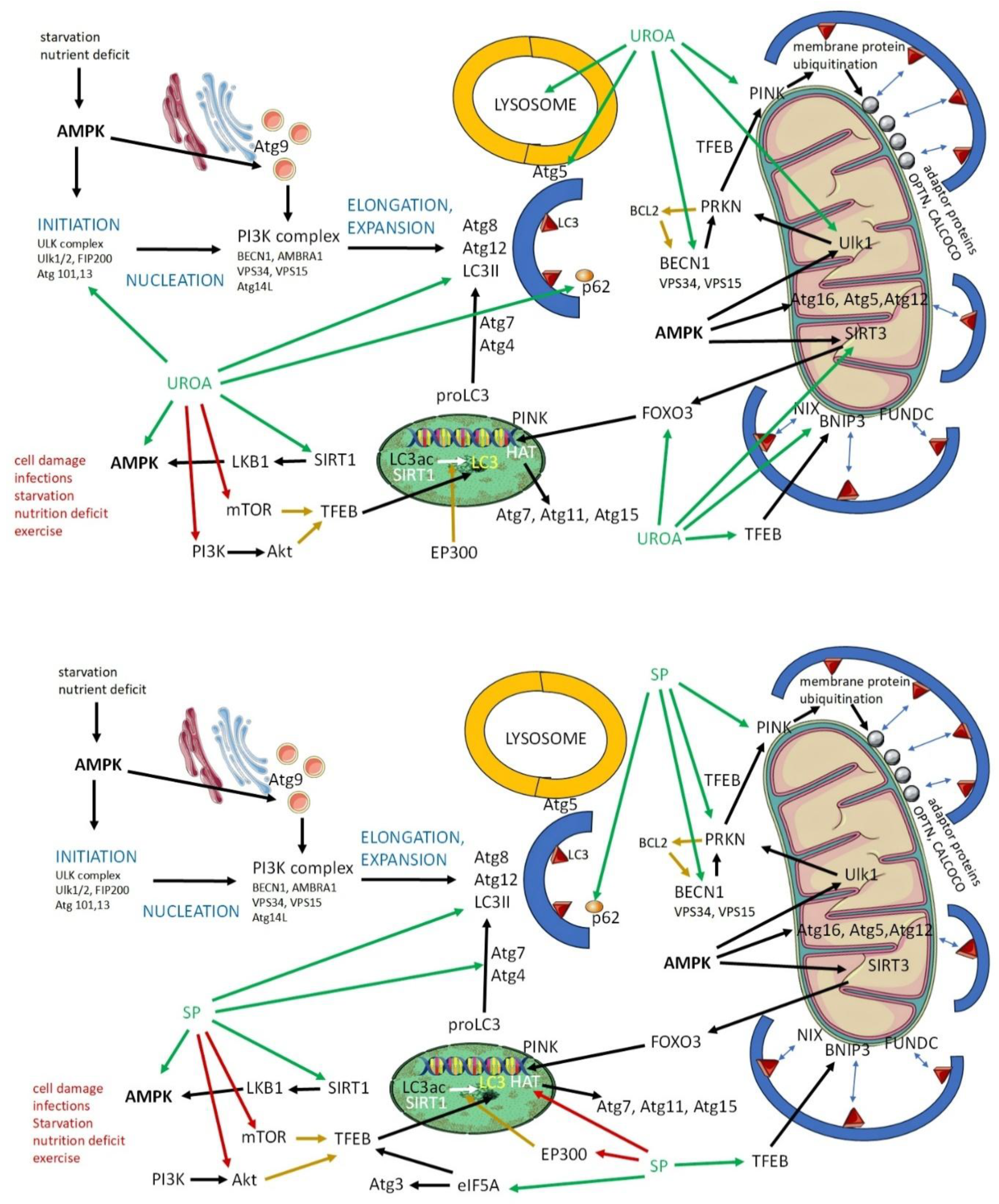
Urolithin A: Sources, Dosage, and Mechanism of Action

UroA is a metabolite produced by the gut microbiota through the transformation of ellagitannins, a class of polyphenols commonly found in fruits such as pomegranates, raspberries, strawberries, and nuts, especially walnuts. The metabolic conversion occurs through microbial fermentation in the gastrointestinal tract, primarily involving bacterial species such as Proteobacteria, Clostridium, and Bifidobacterium [7]. However, not all individuals produce UroA efficiently due to variations in gut microbiome composition, which has led to the development of direct supplementation strategies to achieve consistent levels across different populations [8].
The typical supplemental dosage of UroA ranges between 500 mg to 1000 mg per day, which has demonstrated effective bioavailability in humans. This dosage results in a dose-dependent increase in plasma concentrations of UroA and its glucuronide and sulfate conjugates, supporting its physiological activity in target tissues, particularly muscles and their mitochondria [9]. UroA's bioavailability is higher when consumed in encapsulated or liposomal forms, which enhances its absorption compared to dietary sources alone [9].
The primary action of UroA centers around its ability to induce mitophagy and autophagy. Research suggests that UroA is more potent in inducing mitophagy than autophagy (see below). Mitophagy is a process by which damaged mitochondria are selectively degraded and recycled to maintain mitochondrial quality. This mechanism is critical for energy metabolism, particularly in tissues with high energy demands such as skeletal muscle. UroA activates the PINK1 (PTEN induced kinase 1) / PRKN (RBR E3 ubiquitin protein ligase) pathway, which tags dysfunctional mitochondria for degradation, while also stimulating mitochondrial biogenesis via the SIRT1 (sirtuin 1) / PGC-1α (peroxisome proliferator-activated receptor gamma coactivator-1α) signaling axis [10]. These combined effects result in improved mitochondrial function, increased ATP production, and enhanced muscle endurance, making UroA particularly effective in combating age-related mitochondrial dysfunction [11].
By contrast, autophagy is a process that involves complex molecular interactions and performs key functions in cellular homeostasis, adaptation, and cell survival. The detailed mechanisms of autophagy are further discussed in relation to spermidine and in the following text.
In addition to its role in mitophagy and autophagy, UroA exhibits anti-inflammatory and antioxidant properties. Research demonstrates that UroA modulates the inflammatory response by reducing the expression of pro-inflammatory cytokines such as TNF-α (tumor necrosis factor α) and increasing the production of anti-inflammatory markers such as IL-10. Furthermore, it enhances the activity of antioxidant enzymes such as superoxide dismutase (SOD) and glutathione peroxidase, thereby reducing reactive oxygen species (ROS) and protecting cells from oxidative damage [11]. These properties not only support mitochondrial health but also contribute to UroA’s potential to reduce the risk of chronic inflammatory diseases and support overall cellular longevity. Recent in vivo studies have further demonstrated UroA’s efficacy in extending lifespan and improving muscle function in animal models. For instance, administration of UroA to C. elegans led to a significant extension of lifespan, while rodent studies showed enhanced muscle endurance and reduced signs of muscle aging [10]. Human clinical trials have also shown promising results, with older adults experiencing improved muscle strength and endurance following supplementation, highlighting UroA’s potential as a therapeutic agent for age-related muscle decline [12].
In summary, UroA offers a potent mechanism for targeting mitochondrial health through mitophagy, which not only supports muscle function but also plays a critical role in cellular health, inflammation control, and oxidative stress reduction. These characteristics position UroA as a promising candidate for dietary supplementation, particularly for individuals aiming to enhance mitochondrial function and mitigate the effects of aging.

Spermidine: Sources, Dosage, and Mechanism of Action

Spermidine is a naturally occurring polyamine that is ubiquitously found in various organisms, including humans. It is abundant in plant-based foods such as wheat germ, soybeans, mushrooms, and aged cheese. Spermidine plays a crucial role in cellular processes such as DNA stabilization, regulation of gene expression, and modulation of cell growth and proliferation. However, its levels decline with age, sparking interest in spermidine supplementation as a mean of supporting healthy aging [13]. Spermidine supplementation is typically administered through extracts from polyamine-rich plant sources, with dosages varying depending on the formulation. Clinical trials commonly use doses of 750 mg of spermidine-rich plant extract, corresponding to approximately 1.2 mg of pure spermidine per day [14]. Higher doses, such as 15 mg/day, have also been tested in short-term studies, though these did not significantly increase plasma spermidine levels due to pre-systemic conversion into other polyamines, such as spermine [15].
Spermidine’s primary health benefit arises from its ability to induce autophagy. Autophagy is a conserved fundamental mechanism used by cells to maintain homeostasis, remove damaged or unnecessary organelles, proteins, and other cellular debris, pathogens, and toxins, and respond to stress conditions such as nutrient deprivation. By doing so, autophagy plays a critical role in maintaining cellular homeostasis and has been implicated in the regulation of aging and longevity. Spermidine enhances autophagy by modulating several key signaling pathways, including the AMP-activated protein kinase (AMPK) pathway and the SIRT1 pathway, both of which are central regulators of energy metabolism, stress responses, and cellular longevity [16]. Moreover, spermidine inhibits EP300 (histone acetyltransferase p300), a protein that suppresses autophagy, thereby enabling the activation of autophagy-related genes (Atg) [13].
Beyond its autophagic role, spermidine also influences other cellular processes, including mitophagy, the selective removal of damaged mitochondria. Studies have shown that spermidine activates the PINK1/PRKN mitophagy pathway, similar to UroA, but spermidine’s broader effects on autophagy extend its impact to other organelles and cellular components [16]. These effects contribute to its ability to protect against oxidative stress, reduce inflammation, and improve overall cellular health. Additionally, spermidine has been shown to reduce the accumulation of ROS and enhance the activity of antioxidant enzymes such as SOD and catalase, thereby mitigating oxidative damage at the cellular level [17].
The benefits of spermidine extend to various systems of the body, with particular influence on neuroprotective effects. Animal studies indicate that spermidine supplementation improves cognitive function, delays neurodegeneration, and protects neurons from apoptosis by enhancing autophagy and reducing inflammation in the brain. These effects are mediated through the activation of autophagy-related proteins, such as LC3 (microtubule-associated protein 1A/1B-light chain 3) and Beclin-1, and the reduction of pro-inflammatory markers such as IL-1β and TNF-α [18]. Furthermore, spermidine’s impact on neurogenesis and synaptic plasticity suggests its potential to mitigate age-related cognitive decline and improve memory function in both animals and humans [14].
In summary, spermidine is a potent inducer of autophagy and mitophagy with broad cellular benefits, ranging from neuroprotection to anti-inflammatory and antioxidant effects. Its role in maintaining cellular homeostasis, promoting cognitive health, and reducing the risk of age-related diseases makes it a valuable candidate for dietary supplementation. Spermidine’s ability to modulate key signaling pathways involved in cellular longevity, combined with its accessibility through dietary sources and supplementation, supports its growing popularity as an anti-aging nutraceutical.

Urolithin A and Spermidin in Autophagy and Mitophagy: Studies In Vitro

The mechanism of autophagy involves the formation of double-membraned vesicles, called autophagosomes, that sequester damaged organelles, proteins, etc. The autophagosome fuses with lysosomes and its contents are degraded by enzymes. Breakdown products (lipids, saccharides, and amino acids) can be recycled and reused as energy sources or for building new cellular components [19].
Within autophagy, several distinct types can be distinguished: macroautophagy, microautophagy (including selective autophagy such as mitophagy), and chaperone-mediated autophagy.
Autophagy can be induced by nutrient deprivation, which activates AMPK (a sensor of energy depletion) and inhibits mTOR (mammalian target of rapamycin, an autophagy suppressor), as well as oxidative stress, hypoxia, exercise, drugs (rapamycin, metformin), and hormones (glucagon). Natural substances can also induce autophagy. Besides UroA and spermidine, resveratrol can also be mentioned as an example [20].
Numerous studies have confirmed that compromised autophagy and mitophagy are associated with aging, leading to shortened lifespan in a wide range of aging models, whereas restoration and enhancement of autophagy prolong life and health in various animal species [19,21].
Both autophagy and mitophagy proceed through multiple steps, during which the autophagosome is formed and enlarged. The autophagosome is subsequently fused with the lysosome to allow degradation of the sequestered material [22] (Figure 1).
The first step of autophagy is initiation, which depends on AMPK activation. AMK activates the ULK complex (ULK1/2, FIP200, Atg 101, and Atg3; unc-51 like kinase 1/2, focal adhesion kinase-interacting protein of 200 kDa, adapter proteins autophagy-related 3). AMPK is activated by nutrient deficiency and starvation. Other factors that can induce AMPK include SIRT1, which induces LBK1 (liver kinase B1), in turn activating AMPK. It is necessary to mention that SIRT1 influences more than one step of autophagy.
UroA can induce autophagy as it is a positive regulator of the activity of AMPK, ULK 1,2 and SIRT 1 [23,24,25]. Similarly to UroA, spermidine supports the activity of AMPK and SIRT 1. Both substances are recognized as inducers of autophagy [26,27].
The second step, nucleation, is more complex, involving the Atg9 system and PI3K (phosphoinositide 3-kinase) complex. The PI3K complex consists of BECN1 (beclin 1), AMBRA1 (autophagy and beclin 1 regulator 1), VPS34 and VPS15 (components of phosphatidylinositol 3-kinases), and Atg14L. Atg9 vesicles are produced by the endoplasmic reticulum and the Golgi apparatus, and their production is regulated by AMPK, whose activity, as we know, is enhanced by UroA and spermidine [28].
UroA also increases the expression and activity of BECN1, which also plays an important role in mitophagy. Spermidine also influences the amount of BECN1. This is due to its ability to target caspase 3, which reduces BECN1 levels. Caspase 3 cleaves BECN1, and inhibition of caspase 3 activity leads to an increase in BECN1 levels [23,29].
The third step of autophagy is elongation and expansion. Key roles in these processes are played by the LC3, Atg8, Atg12 conjugation system. We first focused on the genetic and epigenetic aspects of the expression of the molecules mentioned.
LC3 expression depends, among others, on the activity of SIRT1, TFEB (transcription factor EB), HAT (histone acetyltransferase), and EP300. SIRT1 is a deacetylase that epigenetically influences the expression of a wide variety of genes, including the gene for LC3. HAT and EP300 are acetyltransferases that prevent deacetylation and gene activation. TFEB acts as a transcription factor that regulates LC3 expression and its function can be blocked by mTOR Akt (protein kinase B). It is worth mentioning that the production of TFEB, as well as Atg3, is enhanced by eIF5A (eukaryotic translation initiation factor 5A-1) which increases the translation of mRNA for these substances on ribosomes [30,31].
Both UroA and spermidine increase the activity of SIRT1, and thus the expression and function of LC3, while inhibiting mTOR activity. Spermidine also blocks Akt activity, while UroA inhibits PI3K activity, which can activate Akt [32,33,34,35].
Spermidine supports the activity of eIF5A and thus enhances the synthesis of TFEB and Atg3. Spermidine reduces EP300 and HAT levels, which results in an increase in the production of LC3, Atg7, Atg11, and Atg15. Atg3 and Atg7 are important in the processing of pro-ILC3, which must be transformed into the active product [26,36,37,38,39] .
To form the definitive autophagosomes, the p62 protein is crucial. It is a sequestosome that delivers the cargo into developing autophagosomes and reflects autophagic flux. p62 is degraded through autophagic processes, along with the cargo it delivers to the autophagosome. p62 is important for the fusion of autophagosome with lysosome, which leads to the formation of autophagolysosomes [40]. p62 is also regulated by UroA and spermidine, which improve lysosome functions [41,42,43].
Atg5, whose activity is enhanced by UroA, plays a role in the final step of autophagy, specifically in the fusion of the autophagosome and the lysosome. Thus, UroA is also involved in the final step of autophagy [44].
Mitophagy is closely related to autophagy; an autophagosome must be formed, enclosing the mitochondria, which then fuses with the lysosome. Mitophagy is induced when mitochondrial stress occurs, including mitochondrial damage, ROS production, depolarization, or mtDNA (mitochondrial DNA) mutations. Factors involved in induction and progression of autophagy, especially AMPK, TFEB, LC3 and BECN1, play crucial roles in mitophagy [45].
As we mentioned previously, UroA and spermidine enhance AMPK activity. AMPK is involved in the recruitment of UKL1, SIRT3 (sirtuin 3), and Atg16L (Atg16, Atg5, Atg12). Atg16L binds LC3 on the membrane of the developing autophagosome. UroA also increases the activity of SIRT3 which mobilizes FOXO3 (forkhead box O3), responsible for the expression of PINK. PINK activity is also enhanced by UroA and spermidine. The interaction between PRKN and PINK is modulated by TFEB [46,47,48].
There are two main pathways driving mitophagy: PINK/PRKN dependent and PINK/PRKN non-dependent, both of which are regulated by UroA and spermidine. In the PINK/PARKIN-dependent pathway, PRKN activates PINK, leading to the ubiquitination of membrane proteins on the mitochondrial membrane. These proteins recruit adaptor proteins OPNT (optineurin), CALCOCO (calcium binding and coiled-coil domain), etc., that subsequently bind LC3.
The activity of PRKN is increased by UroA, ULK1, and BECN1, all of which are induced by UroA and spermidine. BECN1 is blocked by BCL2 (B-cell lymphoma 2, apoptosis regulator), which, in turn, is inhibited by PRKN. UroA also stabilizes the interaction between PRKN and PINK [9,48,49,50].
The PINK/PARKIN non-dependent pathway consists of membrane receptors that can bind to LC3. These include NIX (Nip3-like protein X), BNIP3 (BCL2 interacting protein 3), FUNDC (FUN14 domain-containing protein). UroA regulates the activity of NIX and BNIP3 [9,51].
The fusion of the autophagosome or automitophagosome is regulated by TECRP1 (tectonin beta-propeller repeat containing 1), which is located in the lysosome, and Atg5. The efficacy of degradation is mediated by lysosomal function, which can be supported by UroA [44].

In Vivo Studies on Urolithin A and Spermidine

Even though both UroA and spermidine can affect autophagy and mitophagy, in vivo studies with UroA predominantly focus on mitophagy, while those with spermidine focus more on autophagy.

Urolithin A: In Vivo Studies

UroA has emerged as a compound with extensive potential for improving health across multiple biological systems. Numerous in vivo studies have demonstrated its remarkable efficacy in enhancing mitochondrial function, promoting muscle health, and potentially extending longevity (Table 1).
One of the most prominent studies conducted on Caenorhabditis elegans showed that Uro A can significantly prolong lifespan by inducing mitophagy. The study revealed that treated C. elegans exhibited enhanced mitochondrial clearance and reduced levels of oxidative stress, both of which contributed to the observed lifespan extension [10]. Similar results have been observed in rodent models, where UroA supplementation led to improvements in muscle function and endurance. In a study involving aged mice, UroA enhanced mitochondrial biogenesis and increased muscle strength, effectively reversing some of the age-related declines in muscle performance [10]. Activation of the SIRT1/PGC-1α pathway was identified as a critical factor in this process, supporting the hypothesis that UroA’s effects on mitophagy play a key role in its ability to improve muscle health [9].
UroA has also been used in models of Duchenne's muscular dystrophy and amyotrophic lateral sclerosis. C. elegans and mice were used in the experiments. In both models, UroA supplementation led to restoration of impaired mitophagy, reduced muscular atrophy and fibrosis, and improved muscle function [52,53]. The induction of mitophagy may also serve as a prevention against age-related hearing loss, as Sung et al. described. Their study showed that treatment with UroA improves mitochondrial DNA integrity and ATP production in the inner ear and auditory cortex. The health of mitochondria was preserved through the maintenance of mitophagy [54].
Studies show that UroA does not only promote muscle health, but also cardiovascular health. It can mitigate the effects of an unhealthy lifestyle on the myocardium and also reduce the effects of blood vessel damage leading to ischemia on the affected tissues. One study indicated that UroA may help with obesity-induced cardiomyopathy. The authors of the study found that decreased autophagy and mitophagy in this condition can be fully restored with UroA administration, resulting in improved cardiac function [55].
Proper functioning of the heart and other organs requires a solid vascular supply. Studies demonstrate that UroA may play a critical protective role in cases of ischemia-reperfusion injury. Ischemic vascular diseases are very common, and even if reperfusion occurs, tissue damage can be significant, with severely impaired function in affected tissues. The most serious conditions tend to occur after blood vessel occlusion in the brain or heart. In mouse models of cerebral or renal artery occlusion, pretreatment with UroA appeared to mitigate the effects of ischemia, reducing the extent of tissue damage and restoring impaired autophagy [56,57]. Su et al. showed that the positive effect of UroA on ischemia-reperfusion injury is mediated by attenuation of oxidative stress and ferroptosis as well as Nrf2 pathway activation [58].
With the aging of the population, the incidence of not only cardiovascular but also neurodegenerative diseases is increasing. Supplementation with UroA in mouse models of Alzheimer's disease leads to the removal of amyloid β and a slowing of decline in brain function through restoration of autophagy and mitophagy [59,60]. The positive effect of UroA on the central nervous system is also seen in brain injuries. Two studies focused on traumatic brain injury showed that supplementation with UroA in mice alleviates its consequences (neurological dysfunctions, neural pain, brain edema, and disruption of blood-brain barrier functions) by restoring autophagy. It increases LC3 and decreases the activity of Akt and mTOR [61,62].
However, the positive effect of UroA on health does not end there; it can also aid in managing infections, inflammation, and metabolic diseases. In a mouse model of pediatric pneumonia, supplementation with Uro A resulted in a decrease in lung inflammation and damage [63].
The effect of UroA on the immune system and inflammation was evaluated in a mouse model of inflammatory bowel disease. Researchers developed nanoparticles loaded with UroA, and these formulations alleviated inflammation [64]. Guo et al. also confirmed that UroA has potential to limit inflammation and ameliorate the intestinal damage caused by hexavalent chromium. It significantly improved tissue repair and intestinal barrier function [65].
The regenerative effect of UroA was also described in a study by Guo et al., which focused on healing damaged corneal epithelium. They found that eyedrops containing UroA accelerated the healing of corneal epithelium, reduced cell senescence, and inhibited ferroptosis [66].
The impact of UroA on metabolic health was verified in a mouse model of streptozotocin-induced type II diabetes. Mice with this condition were supplemented with UroA, which led to decreased fasting blood glucose, glycated hemoglobin levels, plasma C-peptide, malondialdehyde (MDA), and interleukin-1β levels. It also increased reduced glutathione (GSH), interleukin-10, and glucose tolerance [63,67].

Spermidine: In Vivo Studies

Spermidine has a wide range of effects that have been tested in animal models (Table 2).
In vivo studies on spermidine have provided compelling evidence of its role in promoting longevity and improving cognitive function. Lifespan extension has been observed in various animal models, including yeast, flies, and rodents. A study on bees showed that spermidine supplementation supports longevity by improving autophagy, which depends on the epigenetic changes that are induced by the presence of spermidine. It enhances the expression of genes coding for Atg proteins [39].
In a study on C57BL/6J mice, lifelong supplementation with spermidine led to a significant extension of lifespan, with improvements in mitochondrial function, reduced oxidative stress, and enhanced autophagic activity [16].
The aforementioned oxidative stress is a key factor that accelerates biological aging. In a study using a rat model of D-galactose-induced senescence and aging, the authors focused on cellular redox status and ionic homeostasis. Spermidine consumption prevented the onset of age-induced increase in production of ROS, thus reducing lipid peroxidation, protein oxidation, etc. Spermidine also increased antioxidant levels, which were associated with reduced cell damage and inflammation, and delayed biological aging [68]. Of note, these results are consistent across multiple species, highlighting the conserved nature of spermidine’s effects on cellular health and aging.
Oxidative stress, inflammation, and accelerated aging are accompanied by damage to the central nervous system, including neurodegeneration. Spermidine’s ability to protect against neurodegeneration has been particularly well-documented. In a study involving SAMP8 mice, a model for aging and cognitive decline, spermidine supplementation resulted in significant improvements in memory function and reduced neuroinflammation. Spermidine enhanced autophagic flux in neurons, facilitating the clearance of damaged proteins and improving mitochondrial function. These findings were accompanied by an increase in neurotrophic factors, such as BDNF (brain-derived neurotrophic factor) and NGF (nerve growth factor), which are critical for synaptic plasticity and neuronal survival [18].
The protection of the nervous system was also confirmed in mouse models of Alzheimer’s disease, as well as in mouse and zebrafish models of ataxia (Machado-Joseph disease)). Supplementation with spermidine preserved brain functions, increased amyloid β degradation, and enhanced coordination and stability in mice. These improvements were dependent on the restoration of autophagy, confirmed by detection of increased levels of LC3 and components that enhance lysophagy [42,69,70].
Another highly prevalent neurodegenerative disease among elderly people is Parkinson disease (PD). Spermidine has also been utilized in PD models. In a Drosophila melanogaster model of PD, spermidine reduced brain function decline and decreased accumulated α-synuclein by restoring autophagy [71].
Some studies confirmed that in neurodegenerative diseases, spermidine also activates mitophagy by enhancing PINK expression, which prolongs lifespan, improves health span, protects against memory loss, and increases locomotor activity [72,73].
Cardiovascular benefits have also been observed in vivo, particularly in models of age-related cardiac decline. A study involving aged mice found that spermidine supplementation reduced cardiac hypertrophy and improved left ventricular function. This was attributed to spermidine’s ability to activate mitophagy and autophagy, helping maintain mitochondrial quality and preventing the accumulation of damaged mitochondria in heart tissue [74].
In the Wistar rat model of acute myocardial infarction (AMI), supplementation before AMI induction reduced cardiac dysfunction and cardiomyocyte damage. Spermidine increased levels of LC3-II, TFEP, and p62, which are important components of autophagy [75].
In the context of myocardial infarction, it is important to mention that spermidine administration before liver ischemia activates autophagy, reduces liver damage, and decreases the number of apoptotic cells, suggesting a protective effect comparable to that observed in myocardial infarction [76]. Similarly to heart muscle, the improvement in its functionality despite AMI suggests that spermidine has a similar effect on skeletal muscle. Spermidine was used in a mouse model of collagen type VI-related myopathies. Importantly, spermidine induces both autophagy and mitophagy (LC3-II, p62, BNIP3 and OPTN) and restores muscle strength [77].
Spermidine protects the cardiovascular system, even when mice are exposed to a high-salt diet. Such diet leads to increased blood pressure and damage to blood vessels and the myocardium. These negative effects can be alleviated by spermidine. It reduces blood pressure and myocardial hypertrophy by increasing autophagic flux [74]. Spermidine supplementation may also suppress aortic aneurysm formation and inflammation in a mouse model. In addition, it may reverse arterial aging [78,79]. The effect of spermidine on the vascular system was further confirmed by Duan et al., who supplemented sows with spermidine during gestation, which improved placental quality and enhanced angiogenesis, and thus increased the number of healthy offsprings [80]. Systemic inflammatory diseases are accompanied by endothelial damage. In a study using a mouse model of systemic lupus erythematosus, endothelial dysfunction was confirmed. This damage was ameliorated by spermidine supplementation, which reduced inflammation, decreased antibody production, and increased vascular relaxation and mitophagy [81].
Joints are very often affected by inflammation and damage; this can result from autoimmune inflammation or degenerative processes caused by joint abrasion. In a study of induced osteoarthritis in rats, the authors demonstrated that spermidine supplementation reduced the pyroptosis and proinflammatory cytokine production, and limited pathological processes in the joints [82].
The anti-inflammatory effect of spermidine was also demonstrated in a mouse model of methicillin resistant Staphylococcus aureus (MRSA) infection. In mice infected with MRSA, which is an important and dangerous human pathogen, treatment with spermidine reduced the MRSA burden in the bloodstream and limited systemic inflammation by polarizing macrophages toward the anti-inflammatory M2 phenotype. Thus, the results of the studies show that spermidine has the potential to prolong life, protecting both the nervous and cardiovascular systems [83].
Table 1. In vivo studies on urolithin A.
Animal model Intervention Dose Effect Mechanism
C57BL/6 mice High fat diet (20 weeks); mitophagy and mitochondrial function 50 mg/kg/day, 4 weeks; p.o. ↑ Mitophagy
↑ Cardiac diastolic function
↓ Cardiac remodeling
↑ PINK1/PRKN-dependent mitophagy
↓ Mitochondrial defects
[55]
C. elegans Lifespan
Mitochondria
50 µM ad libitum, 10 days; p.o. ↑ Mitophagy
↑ Mobility
↑ Pharyngeal pumping rate
↑ Lifespan
↑ LC3II/LC3I
↑ PINK1
↓ p62 [10]
Sprague-Dawley rats Muscle
Mitochondria
25 mg/kg/day, 7 days; p.o. ↑ Endurance
↑ Grip strength
↑ Mitophagy
↑ Muscle function
↑ LC3II/LC3I
↑ PINK1
↓ p62 [10]
mdx mice mdx + mdx/Utr−/− (DKO), muscular dystrophy 50 mg/kg/day, 10 weeks; p.o. ↑ Grip strength
↑ Tetanic force
↑ Running activity
↑ Cardiac diastolic function
↑ PINK1/PRKN-dependent mitophagy
↓ Mitochondrial defects
[52]
SOD1G93A transgenic mice Motor dysfunction after administration of copper 50 mg/kg/day, 6 weeks after a 7-week Cu exposure; p.o. ↓ Muscle atrophy and fibrosis
↑ Motor neurons, astrocytes, and microglia
↓ p62
↑ PRKN
↑ PINK1
↑ LAMP1 [53]
C57BL/6J mice Age-related hearing loss

0.5 g/kg UroA mixed with diet; p.o. Prevented mitochondrial function decline and age-related hearing loss ↑ Mitophagy-related RNAs (Pink1, Parkin, Bnip3, Ambra1, Nix, Bcl2, Atg3, Atg5, Atg7, Atg12, and Atg13) and proteins (PINK1, PRKN, BNIP, LC3B), in the cochleae
↑ OXPHOS complex I, II, III, IV, in the cochleae [54]
C57BL/6J mice Traumatic brain injury, neural pain
2.5, 5, or 10 mg/kg UroA; groups: sham, sham + vehicle, TBI + vehicle, TBI + UroA; i.p. ↓ Blood-brain barrier permeability
↓ Neuronal apoptosis in injured cortex
↑ Neurological function
↑ p62
↑ LC3
↓ Akt
↓ mTOR [61]
C57BL/6J mice CCI injury/traumatic brain injury
2.5 mg/kg; groups: sham, TBI + vehicle, TBI + UroA; administered immediately after CCI injury and every 24 h for 3 days; i.p. ↓ NSS score
↓ Brain edema
↓ TUNEL+/NeuN+ cells
↓ Caspase-3
↑ BCL2
↑ LC3II/LC3I
↓ p62
↓ p-Akt/Akt
↓ p-mTOR/mTOR
↓ p-IKKα/IKKα
↓ p-NFκB/NFκB,
in the hippocampus [62]
10-week-old C57BL/6 mice STZ-induced model of type 2 diabetes

50 mg/kg/day, 8 weeks; oral gavage ↓ Fasting blood glucose
↓ Glycated hemoglobin
↓ Plasma C-peptide
↑ Glucose tolerance
↓ Malondialdehyde
↓ IL-1β
↑ IL-10
↑ Reduced glutathione
↓ Inflammation
↑ Metabolic health
↑ LC3II/LC3I
↑ Beclin1
↑ ATG5
↓ Mitochondrial swelling
↓ p62
↑ p-Akt
↑ p-mTOR [67]
C57BL/6 mice Obesity induced cardiomyopathy, high fat diet (20 weeks)
50 mg/kg/day, 4 weeks; gavage ↑ Cardiac diastolic function ↓ Cardiac remodeling ↑ PINK1/PRKN-dependent mitophagy [55]
C57BL mice Cardiac I/R model 1 mg/kg UroA; groups: sham, sham + UroA, cardiac I/R, cardiac I/R + UroA; administered 24 h prior to surgery; i.p. Cardiac tissue protection ↓ Oxidative stress
↑ Nrf2 pathway
↓ Tissue damage [58]
C57BL/6 mice Middle cerebral artery occlusion 2.5 or 5.0 mg/kg UroA, 24 h and 1 h prior to surgery; i.p. Neuroprotection ↑ LC3
↓ p62 [57]
C57BL/6 male mice I/R injury, kidney 50 mg/kg, 3 days prior and
30 min before surgery; i.p.
Pretreatment attenuated kidney dysfunction in IRI ↑ TFEB
↑ LAMP1
↑ Atp6ap1
↑ Beclin1 [56]
1-week-old C57BL/6 Pediatric pneumonia 2.5 and 5 mg/kg, 1 h before LPS; i.p. Alleviated lung inflammation ↑ LC3II
↑ Beclin
↓ p62 [63]
C57BL/6 mice IBD model (colitis induced by 2.5% DSS) 4 mg/kg UroA, orally administered on day 4, 6, and 8; groups: inflammation-targeting nanoparticles (ITNP), ITNP-UroA, free UroA; p.o. ↓ Inflammation
↑ Barrier functions
↓ Shortening of colon
↓ Myeloperoxidase (neutrophil marker)
↓ Inflammatory cytokines (e.g., TNF-α) [64]
pp2r1aflox/flox, Vill-Cre mice, and C57BL/6-Rosa26/tdTomato mice
Intestinal damage, hexavalent chromium 20 mg/kg; p.o. ↓ Epithelial damage
↑ Barrier functions
↑ Reparation
↑ Phosphorylation of YAP1 ↑ Proliferation/repair defects in intestinal epithelium [65]
APP/PS1 mouse
3xTgAD and 3xTgAD/Polβ+/−
Alzheimer disease model 200 mg/kg/day, 5 months;
gavage
↑ Brain function
↑ Learning and memory
↓ Inflammation
↓ Aβ and tau protein deposition
↑ Lysosomal functions
↓ IL-β
↑ Sirtuin
↑ Mitophagy
↓ DNA damage [44]
APP/PS1 mouse, 3×TgAD mouse Alzheimer disease model 200 mg/kg/day; p.o. ↓ Brain function decline
↓ Aβ plaque
↑ PINK1 [60]
3xTg-AD mice, female B6129SF2/J (#101,045), and male C57BL/6NJ Alzheimer disease model 25 mg/kg, alternate weeks (1 week on, 1 week off), 9-10 months; p.o. ↑ Removing Aβ
Prevents the onset of cognitive deficits
↑ LC3
↓ p62 [59]
C57BL/6 mice Hyperosmotic stress and corneal epithelial injury UroA eye drops, 10 drops/day, 7 days ↓ Accumulation of senescent cells in corneal epithelial wounds
↑ Wound healing
↓ Oxidative stress
↓ Lipid peroxidation
↓ Malondialdehyde
↓ Ferroptosis [66]
Table 2. In vivo studies on Spermidine.
Animal model Intervention Dose Effect Mechanism
Bees Longevity 0.1 and 1 mM, 17 days; p.o. ↑ Longevity ↑ Autophagy-related genes (Atg3, Atg5, Atg9, Atg13)
↑ Genes associated with epigenetic changes (HDAC1, HDAC3, SIRT1, KAT2A, KAT6B, P300, DNMT1A, DNMT1B) [39]
C. elegans Longevity 1–20 mM; ad libitum ↑ Lifespan
↑ Healthspan
↓ Memory loss
↓ Loss of locomotor capacity
↑ PINK-PDR-1 pathway [73]
C57BL/6 mice Longevity 0.3 and 3 mM, 200 days; p.o. ↑ Longevity ↑ Mitochondrial function
↓ Acetylation of histone H3
↓ HAT activity
↑ Atg7 [16]
Mouse-8 (SAMP8) Senescence accelerated 0.78 mg/kg/day, spermidine, spermine, rapamycin, or saline, 8 weeks; i.g. ↑ Memory retention
↑ Synaptic plasticity
↑ Neurotrophic factors
↓ Oxidative stress
↑ AMPK
↑ LC3
↑ Beclin1
↑ p62 [18]
Sprague-Dawley rats Aging 25 mg/kg/day, from 18 months old until death; p.o. ↓ Inflammation
↓ Neurodegeneration
↑ MAP1B-LC3a
↑ LAMP1 [72]
Young rats (4 months) D-galactose induced senescence
10 mg/kg, 6 weeks;
p.o
↓ Aging
↓ Oxidative stress
↓ ROS production
↓ Lipid and protein oxidation
↑ Antioxidants
↑ Plasma membrane redox system in erythrocyte membrane [68]
Zebrafish embryos Machado-Joseph disease, also known as spinocerebellar ataxia type 3 Single administration of spermidine (62.5 µM, 125 µM and 250 µM, solubilized in E3 medium) ↓ Neurological deficit ↑ LC3
↑ p62 [42]
CMVMJD135 mouse model Machado-Joseph disease, also known as spinocerebellar ataxia type 3 3 mM; p.o. ↑ Neurological score
↑ Balance
↑ Coordination
↑ p-ULK1
↑ LC3
↑ p62 [42]
Mice Collagen type VI-related myopathies 30 mM, 60 or 100 days; p.o. Rescue muscle strength (↑ Absolute and normalized contractile force, 100 days administration; ↑ Hanging performance, 60 and 100 days administration) ↑ LC3II
↑ p62
↑ BNIP3
↑ OPTN [77]
APPPS1+/− mice Alzheimer disease model 3 mM, 90 or 260 days; p.o. ↑ Brain function ↑ SIRT3
↑ Aβ degradation
↑ Autophagy [70]
C57BL/6*GFP-LC3 transgenic mice Alzheimer disease model 0.3 mM + PQ and 3 mM + PQ, every 3 days for 3 weeks; i.p. ↓ Neuronal toxicity of paraquat ↑ Autophagic flux
↑ LC3 [69]
Drosophila melanogaster Parkinson disease model 5 mM spermidine; p.o. ↓ Brain function ↓ α-synuclein
↑ Atg8 [71]
Wistar rats Acute myocardial infarction (AMI) model induced by isoproterenol injections Before AMI induction, 2.5 mg/kg/day, 7 days; i.p. ↓ Dysfunction and cardiac enzymes ↑ LC3II
↑ TFEB
↑ p62 [75]
C57BL/6J mice High-salt diet
myocardial injury
3 mM, 4 weeks; p.o. ↓ Systemic blood pressure ↓ Cardiac hypertrophy
↓ Decline in diastolic function
↑ Titin phosphorylation
↑ Cardiomyocyte autophagic flux
↑ LC3II [74]
C57BL/6 mice Ischemia-reperfusion injury 3mM, 4 weeks before IR; gavage ↓ I/R-induced apoptosis in the liver
↓ Loss of liver function
↑ Beclin1
↑ LC3II/LC3I (AMPK-mTOR-ULK1 pathway) [76]
C57B/L6 mice Arterial aging (cardiovascular disease) 3 mM, 4 weeks; p.o. Normalized aortic pulse wave velocity
↓ Nitrotyrosine
↓ Superoxide
↓ AGEs
↓ Collagen in arterial wall
↓ Acetylation of histone H3
↑ Atg3
↑ LC3II
↓ p62 in old mice [79]
C57BL/6J mice Abdominal aortic aneurysm 3 mmol/L, on the day of or 3 days after PPE infusion, continued until euthanasia; p.o. Prevented aneurysm development
↓ Inflammation in aorta and systemic
↑ Autophagy
↑ LC3II/LC3I
↑ Beclin1
↓ mTOR
↓ p62 [78]
Lupus-prone (MRL/lpr) mice Model of systemic lupus erythematosus 1 M aqueous stock solution, every 3–4 days, 8 weeks;
p.o.
Prevented endothelial dysfunction ↓ Inflammation
↓ Autoantibodies
↑ Mitophagy
↑ Vasorelaxation [81]
BALB/c mice Model of infections
(MRSA)
3 mmol in drinking water, 2 weeks, with antibiotic treatment and fecal microbiota transplantation ↓ Bacterial load ↑ PTPN2 expression
↑ M2 macrophages [83]
Sows Placentogenesis, gravidity 10 or 20 mg/kg, at day 60 of gestation ↑ Placental functions
↑ Healthy offsprings
↓ Mummified litter
↓ Weak offsprings
↑ Expression PECAM-1
↑ Density of placental stromal vessels
↑ Amino acid transporters,
glucose transporters [80]
Sprague-Dawley rats Osteoarthritis model 2.5 mg/kg and 5.0 mg/kg;
i.p.
Counteract the disease progression ↓ Inflammation
↓ Pyroptosis
↓ IL-1β
↑ AhR-spermidine binding
↓ AhR/NF-κB and NLRP3/caspase-1/GSDMD signaling pathways [82]
Abbreviations: Aβ, amyloid beta; AGEs, advanced glycation end-products; AhR, aryl hydrocarbon receptor; Akt, protein kinase B; AMBRA1, activating molecule in Beclin1-regulated autophagy protein 1; AMI, acute myocardial infarction; AMPK, AMP-activated protein kinase; Atg, autophagy-related genes; Atp6ap1, ATPase H+ Transporting Accessory Protein 1; BCL2, B-cell lymphoma 2; BNIP3, BCL2 interacting protein 3; CCI, controlled cortical impact; DNMT1, DNA (cytosine-5)-methyltransferase 1; DSS, dextran sodium sulphate; GSDMD, gasdermin D; HAT, histone acetyltransferase; HDAC, histone deacetylase; IBD, inflammatory bowel disease; IKKα, IκB kinase α; IL, interleukin; I/R, ischemia/reperfusion; IRI, ischemia-reperfusion injury; ITNP, inflammation-targeting nanoparticles; KAT, lysine acetyltransferase; LAMP1, lysosome-associated membrane glycoprotein 1; LC3, microtubule-associated protein 1 light chain 3; LPS, lipopolysaccharide; MAP1B, microtubule-associated protein 1B; MRSA, methicillin-resistant Staphylococcus aureus; mTOR, mammalian target of rapamycin; NeuN, neuronal nuclei; NF-κB, nuclear factor kappa-light-chain-enhancer of activated B cells; NIX, NIP3-like protein X; NLRP3, NOD-like receptor family pyrin domain containing 3; Nrf2, nuclear factor erythroid 2–related factor 2; NSS, neurological severity score; OPTN, optineurin; OXPHOS, oxidative phosphorylation; PRKN, parkin RBR E3 ubiquitin protein ligase; PDR-1, Parkin homolog in Caenorhabditis elegans; PECAM-1, platelet endothelial cell adhesion molecule 1; PINK1, PTEN induced kinase 1; p.o., per os; PQ, paraquat; PTPN2, protein tyrosine phosphatase non-receptor type 2; ROS, reactive oxygen species; SIRT, sirtuin; STZ, streptozotocin; TBI, traumatic brain injury; TFEB, transcription factor EB; TNF-α, tumor necrosis factor-alpha; TUNEL, terminal deoxynucleotidyl transferase dUTP nick-end labeling; ULK1, unc-51 like kinase 1; UroA, urolithin A; YAP1, Yes-associated protein 1.

Clinical Studies on Urolithin A and Spermidine

Urolithin A: Clinical Studies

Due to the promising results of in vivo studies with UroA, several clinical studies with volunteers have also been conducted (Table 3).
Clinical trials on UroA have demonstrated its potential in improving muscle function and mitochondrial health, particularly in older adults. One notable study was a randomized, double-blind, placebo-controlled trial to evaluate the effects of UroA supplementation in elderly individuals. Participants were given daily doses of UroA over a period of four months. The results showed significant improvements in ATP production, muscle strength, 6-minute walk distance and endurance in those who received the supplement compared to the placebo group. In addition, the study observed increased mitochondrial activity in muscle biopsies, confirming UroA’s role in enhancing mitochondrial function [12]. Indeed, the increase in muscle strength and endurance is closely linked to mitochondrial function. As was already mentioned, homeostasis is crucial, including the biogenesis of new mitochondria and the removal of damaged and dysfunctional ones. [12].
Studies show that in people over 50, supplementation with UroA in the form of pomegranate juice for at least one month led to improvements in brain function, as well as in visual and verbal memory [84,85]. Similarly, consumption of pomegranate juice by trained cyclists resulted in increased time to exhaustion and time to reach the ventilatory threshold [86]. Considering the fact that pomegranate juice is an important source of UroA, such beneficial effects can be indirectly ascribed to this compound.
Zhao et al. conducted an 8-week study on male athletes undergoing resistance training to evaluate the effect of UroA on muscle endurance, strength, inflammation, oxidative stress, and protein metabolism. Participants consumed 1 g of UroA daily. The UroA supplementation increased voluntary isometric contraction and repetitions to failure performance. The levels of CRP and superoxide dismutase were lower compared to the control group. UroA improved physical condition and reduced oxidative stress and inflammation [87].
In another clinical trial, UroA supplementation was tested for its impact on mitochondrial biogenesis and metabolic health. Participants, including both healthy middle-aged and older adults, received UroA supplements for 28 days. The study reported an increase in the expression of genes related to mitochondrial biogenesis, as well as improvements in markers of mitochondrial function and reduction in fatigue. These findings suggest that UroA may have broad applications for improving energy metabolism, especially in populations experiencing age-related declines in mitochondrial efficiency [9].
It can be summarized that UroA´s positive effects are due to its ability to inhibit inflammation and oxidative stress while inducing and enhancing mitophagy and autophagy. In vitro and in vivo experiments confirmed that UroA is able to protect nerve cells and alleviate neurodegenerative processes such as those seen in Alzheimer's disease, where autophagy and mitophagy play a significant role in the pathogenesis of such neurodegenerative diseases. It is therefore possible to assume that in clinical trials in which neurodegenerative diseases were alleviated, mitophagy and autophagy were also enhanced.
It has been also reported that polyphenols, including UroA, can also protect the brain in the event of a cerebral infarction, shorten hospital stay after a stroke, and improve neuropsychiatric and motor function [88]. Finally, the effect of UroA on human health is also mediated by its effect on the gut microbiota. The influence of the gut microbiota on UroA levels has been described, as it is responsible for producing UroA from the polyphenol ellagic acid. However, the consumption of certain foods, in this case walnuts, has also been shown to influence the composition of the gut microbiome. In a study with healthy volunteers, the gut microbiome composition differed before and 21 days after walnut consumption. Alpha and beta diversity were significantly altered. Roseburia, Rothia, Parasutterella, Lachnospiraceae UCG-004, Butyricicoccus, Bilophila, Eubacterium eligens, Lachnospiraceae UCG-001, Gordonibacter, Paraprevotella, Lachnospira, Ruminococcus torques, and Sutterella were identified as the 13 genera most enriched following daily walnut intake [89].
Importantly, safety and tolerability studies have been conducted to ensure that UroA is safe for human consumption. One study assessed the safety of UroA in humans, finding no adverse effects at dosages ranging from 250 mg to 1000 mg per day administered over a 4-week period. Participants exhibited no significant changes in vital signs, blood chemistry, or electrocardiograms, confirming the compound’s safety profile for long-term use [9].

Spermidine: Clinical Studies

Spermidine has also been studied extensively in human populations, with a focus on its ability to improve cognitive function, promote longevity, and reduce the risk of age-related diseases (Table 4). One pivotal randomized, placebo-controlled trial tested the effects of spermidine supplementation in older adults with subjective cognitive decline. Over a period of 12 months, participants received daily doses of spermidine-rich plant extract. The study reported significant improvements in memory performance and cognitive function, particularly in those with mild cognitive impairment. These findings suggest that spermidine supplementation may help slow cognitive decline and enhance brain function in aging individuals [90]. Another study found that spermidine supplementation improves brain functions in elderly people aged 60 to 96 years. Participants showed improvements in standardized tests of mental function, including Mini Mental State Examination and phonematic fluidity [91]. It has also been shown that a diet rich in spermidine can alter brain morphology in the elderly. Interestingly, subjects with higher dietary spermidine intake had greater hippocampal volume, mean cortical thickness, and cortical thickness in AD-vulnerable brain regions [14]. Another large-scale epidemiological study examined the relationship between dietary spermidine intake and all-cause mortality. This study followed a cohort of more than 800 participants over a 20-year period and found that individuals with higher dietary spermidine intake had a significantly lower risk of death from all causes, including cardiovascular disease and cancer. These findings align with spermidine’s role in promoting autophagy, reducing oxidative stress, and enhancing cellular longevity [92].
In terms of metabolic health, clinical trials have shown that spermidine supplementation can improve glucose metabolism and reduce insulin resistance in obese individuals. In one study, participants who consumed a spermidine-rich diet for three months exhibited improved insulin sensitivity and lower fasting glucose levels, suggesting spermidine’s potential as a therapeutic agent for managing metabolic disorders [93]. The anti-aging, immune-stimulating, and antioxidation properties of spermidine were tested in a study by Felix et al., who supplemented participants with capsules containing AM3, spermidine, and hesperidin. The supplementation reduced biological age and oxidative stress while increasing immune system function [94]. Finally, the anti-inflammatory, antioxidant, and cardioprotective effects of spermidine were evaluated and confirmed in a study by Rhodes et al. [95].
There was also a cardioprotective effect of spermidine as demonstrated by Wang et al. In this study, they determined levels of spermidine in the blood of participants. They found that higher spermidine levels are associated with a lower risk of hypertension, lower levels of blood glucose and LDL, and higher levels of HDL. This data suggests that increased spermidine intake can also improve cardiovascular and metabolic health [96].
A bidirectional Mendelian randomization study was conducted by He et al., who, based on a genome-wide associated study of 8299 European individuals, found that the spermidine to (N(1) + N(8))-acetylspermidine ratio negatively correlated with gastric cancer [97]. A study performed by Iorio-Siciliano et al. used a product with spermidine to treat peri-implant mucositis. Spermidine was incorporated into a gel that was applied locally to the gums. Although no significant differences were initially observed between the groups after three months, the use of the gel resulted in an 85% disease resolution rate, compared to a 70% resolution rate without adjunctive application of the gel [98].
Finally, spermidine’s safety profile has been extensively tested. In a 12-month randomized clinical trial, spermidine supplementation was found to be safe, with no significant adverse effects reported. Participants receiving up to 1.2 mg of spermidine daily exhibited no significant changes in blood parameters or organ function, confirming the safety of long-term supplementation [90]. The safety and tolerability of spermidine were also tested by Keohane et al., who supplemented older men with a dose as high as 40 mg/day for seven and 28 days. The dose was well tolerated, and no adverse events were reported. Spermidine did not alter lipid levels, blood chemistry, or hematological parameters[99].

Conclusion

Urolithin A and spermidine are two promising compounds that have gained significant attention for their potential to enhance cellular health, promote longevity, and mitigate the effects of aging through the activation of autophagic and mitophagic pathways.
UroA stimulates mitophagy, a process that selectively clears damaged mitochondria. Its ability to target mitochondrial dysfunction makes it particularly effective in improving muscle function and energy metabolism, as demonstrated in both preclinical and clinical studies. Moreover, UroA has shown significant potential in addressing age-related muscle decline, metabolic disorders, and enhancing mitochondrial biogenesis, particularly in older adults. Its safety profile is well-established, making it a viable option for long-term supplementation, particularly for individuals seeking to improve mitochondrial health.
By inducing autophagy, spermidine supports the degradation and recycling of a wider range of cellular components, including damaged proteins and organelles. This comprehensive impact on cellular homeostasis enables spermidine to provide additional benefits, including neuroprotection, cardiovascular health, and cognitive enhancement. Spermidine’s ability to reduce inflammation and oxidative stress, and support cognitive function in aging individuals makes it a versatile anti-aging supplement with far-reaching effects beyond muscle health.
When comparing the two candidates based on available clinical studies, UroA is more focused on mitochondrial health and may be particularly beneficial for individuals facing age-related muscle decline and metabolic dysfunctions. Spermidine, in contrast, offers broader systemic benefits, particularly in terms of neuroprotection and cognitive health, while also contributing to overall longevity through its modulation of autophagy.
Based on the current evidence, spermidine may be the superior supplement for individuals seeking comprehensive anti-aging benefits that extend beyond mitochondrial health, particularly for those concerned with cognitive decline and cardiovascular health. However, UroA may be more appropriate for those specifically targeting muscle endurance, metabolic health, and mitochondrial function.
In conclusion, there are no reports that highlight any specific toxicity of either compound. Only non-specific side effects such as allergies, overuse, and potential interactions with drugs which target mitochondrial or metabolic function should be considered. Thus, both compounds are safe and hold great potential as dietary supplements, but the choice between UroA and spermidine ultimately depends on the individual’s specific health goals. For a more targeted intervention aimed at mitochondrial function, UroA stands out, while for broader anti-aging effects, including cognitive and cardiovascular health, spermidine is the more versatile option.

Author Contributions

PB was responsible for conceptualization, conducted the literature search, and wrote the original draft. DH, OS, TP, and MH assisted in the literature search. DH, OS, TP, ZF, and MH performed manuscript editing. LB and OS provided funding acquisition.

Funding

The review was supported by The Biomedical Indicators for Personalized Medicine project (BIPOLE), project ID CZ.02.01.01/00/23_021/0008439, co-funded by the European Union. The review was supported by Charles University, Faculty of Medicine in Hradec Kralove, the Czech Republic, by project SVV-2023-260656.

Acknowledgments

We would like to acknowledge using pictures from Servier Medical Art, by Servier (http://smart.servier.com).

Conflicts of Interest

The authors do not report any conflicts of interests.

Consent for publication

Not applicable.

Declaration of Generative AI and AI-assisted technologies in the writing process

During the preparation of this work the authors used Chatgpt, verison o1 in order to rewrite parts of the manuscript. After using this service, the authors reviewed and edited the content as needed and take full responsibility for the content of the publication.

List of Abbreviations

Akt protein kinase B
AMI acute myocardial infarction
AMPK AMP-activated protein kinase
Atg autophagy-related genes
BECN1 beclin 1
BCL2 B-cell lymphoma 2
BNIP3 BCL2 interacting protein 3
eIF5A eukaryotic translation initiation factor 5A-1
EP300 histone acetyltransferase p300
HAT histone acetyltransferase
MRSA methicillin resistant Staphylococcus aureus
LC3 microtubule-associated protein 1 light chain 3
mTOR mammalian target of rapamycin
NIX NIP3-like protein X
OPTN optineurin
PGC-1α peroxisome proliferator-activated receptor gamma coactivator-1α
PRKN parkin RBR E3 ubiquitin protein ligase
PINK1 PTEN induced kinase 1
ROS reactive oxygen species
SOD superoxide dismutase
SIRT sirtuin
ULK1 unc-51 like kinase 1
UroA urolithin A
TFEB transcription factor EB
TNF-α tumor necrosis factor α

References

  1. Palmer JE, Wilson N, Son SM, Obrocki P, Wrobel L, Rob M, et al. Autophagy, aging, and age-related neurodegeneration. Neuron. 2025;113(1):29-48.
  2. Picca A, Faitg J, Auwerx J, Ferrucci L, D’Amico D. Mitophagy in human health, ageing and disease. Nature Metabolism. 2023;5(12):2047-61. [CrossRef]
  3. Kuerec AH, Lim XK, Khoo AL, Sandalova E, Guan L, Feng L, et al. Targeting aging with urolithin A in humans: A systematic review. Ageing Res Rev. 2024;100:102406. [CrossRef]
  4. Arthur R, Jamwal S, Kumar P. A review on polyamines as promising next-generation neuroprotective and anti-aging therapy. Eur J Pharmacol. 2024;978:176804. [CrossRef]
  5. Kothe B, Klein S, Petrosky SN. Urolithin A as a Potential Agent for Prevention of Age-Related Disease: A Scoping Review. Cureus. 2023;15(7):e42550. [CrossRef]
  6. Hofer SJ, Daskalaki I, Bergmann M, Friščić J, Zimmermann A, Mueller MI, et al. Spermidine is essential for fasting-mediated autophagy and longevity. Nat Cell Biol. 2024;26(9):1571-84. [CrossRef]
  7. Hua Z, Wu Q, Yang Y, Liu S, Jennifer TG, Zhao D, et al. Essential roles of ellagic acid-to-urolithin converting bacteria in human health and health food industry: An updated review. Trends in Food Science & Technology. 2024;151:104622. [CrossRef]
  8. Singh A, D’Amico D, Andreux PA, Dunngalvin G, Kern T, Blanco-Bose W, et al. Direct supplementation with Urolithin A overcomes limitations of dietary exposure and gut microbiome variability in healthy adults to achieve consistent levels across the population. European Journal of Clinical Nutrition. 2022;76(2):297-308. [CrossRef]
  9. Andreux PA, Blanco-Bose W, Ryu D, Burdet F, Ibberson M, Aebischer P, et al. The mitophagy activator urolithin A is safe and induces a molecular signature of improved mitochondrial and cellular health in humans. Nature Metabolism. 2019;1(6):595-603. [CrossRef]
  10. Ryu D, Mouchiroud L, Andreux PA, Katsyuba E, Moullan N, Nicolet-dit-Félix AA, et al. Urolithin A induces mitophagy and prolongs lifespan in C. elegans and increases muscle function in rodents. Nature Medicine. 2016;22(8):879-88. [CrossRef]
  11. Liu S, D'Amico D, Shankland E, Bhayana S, Garcia JM, Aebischer P, et al. Effect of Urolithin A Supplementation on Muscle Endurance and Mitochondrial Health in Older Adults: A Randomized Clinical Trial. JAMA Netw Open. 2022;5(1):e2144279.
  12. Singh A, D'Amico D, Andreux PA, Fouassier AM, Blanco-Bose W, Evans M, et al. Urolithin A improves muscle strength, exercise performance, and biomarkers of mitochondrial health in a randomized trial in middle-aged adults. Cell Rep Med. 2022;3(5):100633. [CrossRef]
  13. Madeo F, Carmona-Gutierrez D, Kepp O, Kroemer G. Spermidine delays aging in humans. Aging (Albany NY). 2018;10(8):2209-11. [CrossRef]
  14. Schwarz C, Horn N, Benson G, Wrachtrup Calzado I, Wurdack K, Pechlaner R, et al. Spermidine intake is associated with cortical thickness and hippocampal volume in older adults. Neuroimage. 2020;221:117132. [CrossRef]
  15. Senekowitsch S, Wietkamp E, Grimm M, Schmelter F, Schick P, Kordowski A, et al. High-Dose Spermidine Supplementation Does Not Increase Spermidine Levels in Blood Plasma and Saliva of Healthy Adults: A Randomized Placebo-Controlled Pharmacokinetic and Metabolomic Study. Nutrients. 2023;15(8). [CrossRef]
  16. Eisenberg T, Knauer H, Schauer A, Büttner S, Ruckenstuhl C, Carmona-Gutierrez D, et al. Induction of autophagy by spermidine promotes longevity. Nature Cell Biology. 2009;11(11):1305-14. [CrossRef]
  17. Jeong JW, Cha HJ, Han MH, Hwang SJ, Lee DS, Yoo JS, et al. Spermidine Protects against Oxidative Stress in Inflammation Models Using Macrophages and Zebrafish. Biomol Ther (Seoul). 2018;26(2):146-56. [CrossRef]
  18. Xu TT, Li H, Dai Z, Lau GK, Li BY, Zhu WL, et al. Spermidine and spermine delay brain aging by inducing autophagy in SAMP8 mice. Aging (Albany NY). 2020;12(7):6401-14. [CrossRef]
  19. Aman Y, Schmauck-Medina T, Hansen M, Morimoto RI, Simon AK, Bjedov I, et al. Autophagy in healthy aging and disease. Nature Aging. 2021;1(8):634-50. [CrossRef]
  20. Yu L, Chen Y, Tooze SA. Autophagy pathway: Cellular and molecular mechanisms. Autophagy. 2018;14(2):207-15. [CrossRef]
  21. Zimmermann A, Madeo F, Diwan A, Sadoshima J, Sedej S, Kroemer G, et al. Metabolic control of mitophagy. Eur J Clin Invest. 2024;54(4):e14138. [CrossRef]
  22. Lamark T, Johansen T. Mechanisms of Selective Autophagy. Annu Rev Cell Dev Biol. 2021;37:143-69. [CrossRef]
  23. Wang S, Long H, Hou L, Feng B, Ma Z, Wu Y, et al. The mitophagy pathway and its implications in human diseases. Signal Transduction and Targeted Therapy. 2023;8(1):304. [CrossRef]
  24. Velagapudi R, Lepiarz I, El-Bakoush A, Katola FO, Bhatia H, Fiebich BL, et al. Induction of Autophagy and Activation of SIRT-1 Deacetylation Mechanisms Mediate Neuroprotection by the Pomegranate Metabolite Urolithin A in BV2 Microglia and Differentiated 3D Human Neural Progenitor Cells. Mol Nutr Food Res. 2019;63(10):e1801237. [CrossRef]
  25. Lin J, Zhuge J, Zheng X, Wu Y, Zhang Z, Xu T, et al. Urolithin A-induced mitophagy suppresses apoptosis and attenuates intervertebral disc degeneration via the AMPK signaling pathway. Free Radical Biology and Medicine. 2020;150:109-19. [CrossRef]
  26. Hofer SJ, Daskalaki I, Bergmann M, Friščić J, Zimmermann A, Mueller MI, et al. Spermidine is essential for fasting-mediated autophagy and longevity. Nature Cell Biology. 2024;26(9):1571-84. [CrossRef]
  27. Kriebs A. Spermidine controls autophagy during fasting. Nature Aging. 2024;4(9):1172-. [CrossRef]
  28. Holzer E, Martens S, Tulli S. The Role of ATG9 Vesicles in Autophagosome Biogenesis. J Mol Biol. 2024;436(15):168489. [CrossRef]
  29. Yang Y, Chen S, Zhang Y, Lin X, Song Y, Xue Z, et al. Induction of autophagy by spermidine is neuroprotective via inhibition of caspase 3-mediated Beclin 1 cleavage. Cell Death Dis. 2017;8(4):e2738. [CrossRef]
  30. Lubas M, Harder LM, Kumsta C, Tiessen I, Hansen M, Andersen JS, et al. eIF5A is required for autophagy by mediating ATG3 translation. EMBO Rep. 2018;19(6). [CrossRef]
  31. Prasher P, Sharma M, Singh SK, Gulati M, Chellappan DK, Rajput R, et al. Spermidine as a promising anticancer agent: Recent advances and newer insights on its molecular mechanisms. Front Chem. 2023;11:1164477. [CrossRef]
  32. Niu C, Jiang D, Guo Y, Wang Z, Sun Q, Wang X, et al. Spermidine suppresses oxidative stress and ferroptosis by Nrf2/HO-1/GPX4 and Akt/FHC/ACSL4 pathway to alleviate ovarian damage. Life Sci. 2023;332:122109.
  33. Zhang Y, Jiang L, Su P, Yu T, Ma Z, Liu Y, et al. Urolithin A suppresses tumor progression and induces autophagy in gastric cancer via the PI3K/Akt/mTOR pathway. Drug Dev Res. 2023;84(2):172-84. [CrossRef]
  34. Sahashi H, Kato A, Yoshida M, Hayashi K, Naitoh I, Hori Y, et al. Urolithin A targets the AKT/WNK1 axis to induce autophagy and exert anti-tumor effects in cholangiocarcinoma. Front Oncol. 2022;12:963314. [CrossRef]
  35. Hofer SJ, Simon AK, Bergmann M, Eisenberg T, Kroemer G, Madeo F. Mechanisms of spermidine-induced autophagy and geroprotection. Nature Aging. 2022;2(12):1112-29. [CrossRef]
  36. Zhou J, Pang J, Tripathi M, Ho JP, Widjaja AA, Shekeran SG, et al. Spermidine-mediated hypusination of translation factor EIF5A improves mitochondrial fatty acid oxidation and prevents non-alcoholic steatohepatitis progression. Nature Communications. 2022;13(1):5202. [CrossRef]
  37. Zhao W, Shi F, Guo Z, Zhao J, Song X, Yang H. Metabolite of ellagitannins, urolithin A induces autophagy and inhibits metastasis in human sw620 colorectal cancer cells. Mol Carcinog. 2018;57(2):193-200. [CrossRef]
  38. Satarker S, Wilson J, Kolathur KK, Mudgal J, Lewis SA, Arora D, et al. Spermidine as an epigenetic regulator of autophagy in neurodegenerative disorders. Eur J Pharmacol. 2024;979:176823. [CrossRef]
  39. Kojić D, Spremo J, Đorđievski S, Čelić T, Vukašinović E, Pihler I, et al. Spermidine supplementation in honey bees: Autophagy and epigenetic modifications. PLoS One. 2024;19(7):e0306430. [CrossRef]
  40. Liu WJ, Ye L, Huang WF, Guo LJ, Xu ZG, Wu HL, et al. p62 links the autophagy pathway and the ubiqutin–proteasome system upon ubiquitinated protein degradation. Cellular & Molecular Biology Letters. 2016;21(1):29. [CrossRef]
  41. Jiménez-Loygorri JI, Viedma-Poyatos Á, Gómez-Sintes R, Boya P. Urolithin A promotes p62-dependent lysophagy to prevent acute retinal neurodegeneration. Molecular Neurodegeneration. 2024;19(1):49. [CrossRef]
  42. Watchon M, Wright AL, Ahel HI, Robinson KJ, Plenderleith SK, Kuriakose A, et al. Spermidine treatment: induction of autophagy but also apoptosis? Mol Brain. 2024;17(1):15.
  43. de Wet S, Du Toit A, Loos B. Spermidine and Rapamycin Reveal Distinct Autophagy Flux Response and Cargo Receptor Clearance Profile. Cells. 2021;10(1).
  44. Hou Y, Chu X, Park JH, Zhu Q, Hussain M, Li Z, et al. Urolithin A improves Alzheimer's disease cognition and restores mitophagy and lysosomal functions. Alzheimers Dement. 2024;20(6):4212-33.
  45. Wang S, Chen Y, Li X, Zhang W, Liu Z, Wu M, et al. Emerging role of transcription factor EB in mitochondrial quality control. Biomed Pharmacother. 2020;128:110272. [CrossRef]
  46. Faitg J, D'Amico D, Rinsch C, Singh A. Mitophagy Activation by Urolithin A to Target Muscle Aging. Calcif Tissue Int. 2024;114(1):53-9. [CrossRef]
  47. Zheng B, Wang Y, Zhou B, Qian F, Liu D, Ye D, et al. Urolithin A inhibits breast cancer progression via activating TFEB-mediated mitophagy in tumor macrophages. Journal of Advanced Research. 2024. [CrossRef]
  48. Fairley LH, Lejri I, Grimm A, Eckert A. Spermidine Rescues Bioenergetic and Mitophagy Deficits Induced by Disease-Associated Tau Protein. Int J Mol Sci. 2023;24(6). [CrossRef]
  49. Jayatunga DPW, Hone E, Khaira H, Lunelli T, Singh H, Guillemin GJ, et al. Therapeutic Potential of Mitophagy-Inducing Microflora Metabolite, Urolithin A for Alzheimer's Disease. Nutrients. 2021;13(11). [CrossRef]
  50. Jang JS, Hong SJ, Mo S, Kim MK, Kim YG, Lee Y, et al. PINK1 restrains periodontitis-induced bone loss by preventing osteoclast mitophagy impairment. Redox Biol. 2024;69:103023. [CrossRef]
  51. Cho SI, Jo E-R, Song H. Urolithin A attenuates auditory cell senescence by activating mitophagy. Scientific Reports. 2022;12(1):7704. [CrossRef]
  52. Luan P, D’Amico D, Andreux PA, Laurila P-P, Wohlwend M, Li H, et al. Urolithin A improves muscle function by inducing mitophagy in muscular dystrophy. Science Translational Medicine. 2021;13(588):eabb0319. [CrossRef]
  53. Zhang H, Gao C, Yang D, Nie L, He K, Chen C, et al. Urolithin a Improves Motor Dysfunction Induced by Copper Exposure in SOD1(G93A) Transgenic Mice Via Activation of Mitophagy. Mol Neurobiol. 2024.
  54. Cho SI, Jo ER, Jang HS. Urolithin A prevents age-related hearing loss in C57BL/6J mice likely by inducing mitophagy. Exp Gerontol. 2024;197:112589. [CrossRef]
  55. Huang JR, Zhang MH, Chen YJ, Sun YL, Gao ZM, Li ZJ, et al. Urolithin A ameliorates obesity-induced metabolic cardiomyopathy in mice via mitophagy activation. Acta Pharmacol Sin. 2023;44(2):321-31.
  56. Wang Y, Huang H, Jin Y, Shen K, Chen X, Xu Z, et al. Role of TFEB in autophagic modulation of ischemia reperfusion injury in mice kidney and protection by urolithin A. Food and Chemical Toxicology. 2019;131:110591. [CrossRef]
  57. Ahsan A, Zheng YR, Wu XL, Tang WD, Liu MR, Ma SJ, et al. Urolithin A-activated autophagy but not mitophagy protects against ischemic neuronal injury by inhibiting ER stress in vitro and in vivo. CNS Neurosci Ther. 2019;25(9):976-86. [CrossRef]
  58. Su Z, Li P, Ding W, Gao Y. Urolithin A improves myocardial ischemia-reperfusion injury by attenuating oxidative stress and ferroptosis through Nrf2 pathway. Int Immunopharmacol. 2024;143(Pt 2):113394. [CrossRef]
  59. Ballesteros-Álvarez J, Nguyen W, Sivapatham R, Rane A, Andersen JK. Urolithin A reduces amyloid-beta load and improves cognitive deficits uncorrelated with plaque burden in a mouse model of Alzheimer's disease. Geroscience. 2023;45(2):1095-113. [CrossRef]
  60. Fang EF, Hou Y, Palikaras K, Adriaanse BA, Kerr JS, Yang B, et al. Mitophagy inhibits amyloid-β and tau pathology and reverses cognitive deficits in models of Alzheimer’s disease. Nature Neuroscience. 2019;22(3):401-12. [CrossRef]
  61. Wang C, Wang Z, Xue S, Zhu Y, Jin J, Ren Q, et al. Urolithin A alleviates neuropathic pain and activates mitophagy. Mol Pain. 2023;19:17448069231190815. [CrossRef]
  62. Gong QY, Cai L, Jing Y, Wang W, Yang DX, Chen SW, et al. Urolithin A alleviates blood-brain barrier disruption and attenuates neuronal apoptosis following traumatic brain injury in mice. Neural Regen Res. 2022;17(9):2007-13. [CrossRef]
  63. Cao X, Wan H, Wan H. Urolithin A induces protective autophagy to alleviate inflammation, oxidative stress, and endoplasmic reticulum stress in pediatric pneumonia. Allergol Immunopathol (Madr). 2022;50(6):147-53.
  64. Ghosh S, Singh R, Goap TJ, Sunnapu O, Vanwinkle ZM, Li H, et al. Inflammation-targeted delivery of Urolithin A mitigates chemical- and immune checkpoint inhibitor-induced colitis. J Nanobiotechnology. 2024;22(1):701. [CrossRef]
  65. Guo P, Yang R, Zhong S, Ding Y, Wu J, Wang Z, et al. Urolithin A attenuates hexavalent chromium-induced small intestinal injury by modulating PP2A/Hippo/YAP1 pathway. J Biol Chem. 2024;300(9):107669. [CrossRef]
  66. Guo XX, Chang XJ, Pu Q, Li AL, Li J, Li XY. Urolithin A alleviates cell senescence by inhibiting ferroptosis and enhances corneal epithelial wound healing. Front Med (Lausanne). 2024;11:1441196.
  67. Tuohetaerbaike B, Zhang Y, Tian Y, Zhang NN, Kang J, Mao X, et al. Pancreas protective effects of Urolithin A on type 2 diabetic mice induced by high fat and streptozotocin via regulating autophagy and AKT/mTOR signaling pathway. J Ethnopharmacol. 2020;250:112479. [CrossRef]
  68. Singh S, Verma AK, Garg G, Singh AK, Rizvi SI. Spermidine protects cellular redox status and ionic homeostasis in D-galactose induced senescence and natural aging rat models. Z Naturforsch C J Biosci. 2024. [CrossRef]
  69. Lumkwana D, Peddie C, Kriel J, Michie LL, Heathcote N, Collinson L, et al. Investigating the Role of Spermidine in a Model System of Alzheimer's Disease Using Correlative Microscopy and Super-resolution Techniques. Front Cell Dev Biol. 2022;10:819571. [CrossRef]
  70. Freitag K, Sterczyk N, Wendlinger S, Obermayer B, Schulz J, Farztdinov V, et al. Spermidine reduces neuroinflammation and soluble amyloid beta in an Alzheimer’s disease mouse model. Journal of Neuroinflammation. 2022;19(1):172. [CrossRef]
  71. Büttner S, Broeskamp F, Sommer C, Markaki M, Habernig L, Alavian-Ghavanini A, et al. Spermidine protects against α-synuclein neurotoxicity. Cell Cycle. 2014;13(24):3903-8. [CrossRef]
  72. Filfan M, Olaru A, Udristoiu I, Margaritescu C, Petcu E, Hermann DM, et al. Long-term treatment with spermidine increases health span of middle-aged Sprague-Dawley male rats. Geroscience. 2020;42(3):937-49. [CrossRef]
  73. Yang X, Zhang M, Dai Y, Sun Y, Aman Y, Xu Y, et al. Spermidine inhibits neurodegeneration and delays aging via the PINK1-PDR1-dependent mitophagy pathway in C. elegans. Aging (Albany NY). 2020;12(17):16852-66. [CrossRef]
  74. Eisenberg T, Abdellatif M, Schroeder S, Primessnig U, Stekovic S, Pendl T, et al. Cardioprotection and lifespan extension by the natural polyamine spermidine. Nature Medicine. 2016;22(12):1428-38. [CrossRef]
  75. Omar EM, Omar RS, Shoela MS, El Sayed NS. A study of the cardioprotective effect of spermidine: A novel inducer of autophagy. Chin J Physiol. 2021;64(6):281-8.
  76. Liu H, Dong J, Song S, Zhao Y, Wang J, Fu Z, et al. Spermidine ameliorates liver ischaemia-reperfusion injury through the regulation of autophagy by the AMPK-mTOR-ULK1 signalling pathway. Biochem Biophys Res Commun. 2019;519(2):227-33.
  77. Gambarotto L, Metti S, Corpetti M, Baraldo M, Sabatelli P, Castagnaro S, et al. Sustained oral spermidine supplementation rescues functional and structural defects in COL6-deficient myopathic mice. Autophagy. 2023;19(12):3221-9. [CrossRef]
  78. Liu S, Huang T, Liu R, Cai H, Pan B, Liao M, et al. Spermidine Suppresses Development of Experimental Abdominal Aortic Aneurysms. J Am Heart Assoc. 2020;9(8):e014757. [CrossRef]
  79. LaRocca TJ, Gioscia-Ryan RA, Hearon CM, Jr., Seals DR. The autophagy enhancer spermidine reverses arterial aging. Mech Ageing Dev. 2013;134(7-8):314-20. [CrossRef]
  80. Duan B, Ran S, Wu L, Dai T, Peng J, Zhou Y. Maternal supplementation spermidine during gestation improves placental angiogenesis and reproductive performance of high prolific sows. J Nutr Biochem. 2024;136:109792. [CrossRef]
  81. Kim H, Massett MP. Effect of Spermidine on Endothelial Function in Systemic Lupus Erythematosus Mice. Int J Mol Sci. 2024;25(18). [CrossRef]
  82. Guo X, Feng X, Yang Y, Zhang H, Bai L. Spermidine attenuates chondrocyte inflammation and cellular pyroptosis through the AhR/NF-κB axis and the NLRP3/caspase-1/GSDMD pathway. Front Immunol. 2024;15:1462777.
  83. Li Q, Tian P, Guo M, Liu X, Su T, Tang M, et al. Spermidine Associated with Gut Microbiota Protects Against MRSA Bloodstream Infection by Promoting Macrophage M2 Polarization. ACS Infect Dis. 2024;10(11):3751-64. [CrossRef]
  84. Bookheimer SY, Renner BA, Ekstrom A, Li Z, Henning SM, Brown JA, et al. Pomegranate juice augments memory and FMRI activity in middle-aged and older adults with mild memory complaints. Evid Based Complement Alternat Med. 2013;2013:946298. [CrossRef]
  85. Siddarth P, Li Z, Miller KJ, Ercoli LM, Merril DA, Henning SM, et al. Randomized placebo-controlled study of the memory effects of pomegranate juice in middle-aged and older adults. Am J Clin Nutr. 2020;111(1):170-7. [CrossRef]
  86. Torregrosa-García A, Ávila-Gandía V, Luque-Rubia AJ, Abellán-Ruiz MS, Querol-Calderón M, López-Román FJ. Pomegranate Extract Improves Maximal Performance of Trained Cyclists after an Exhausting Endurance Trial: A Randomised Controlled Trial. Nutrients. 2019;11(4). [CrossRef]
  87. Zhao H, Zhu H, Yun H, Liu J, Song G, Teng J, et al. Assessment of Urolithin A effects on muscle endurance, strength, inflammation, oxidative stress, and protein metabolism in male athletes with resistance training: an 8-week randomized, double-blind, placebo-controlled study. J Int Soc Sports Nutr. 2024;21(1):2419388. [CrossRef]
  88. Bellone JA, Murray JR, Jorge P, Fogel TG, Kim M, Wallace DR, et al. Pomegranate supplementation improves cognitive and functional recovery following ischemic stroke: A randomized trial. Nutr Neurosci. 2019;22(10):738-43.
  89. Liu H, Birk JW, Provatas AA, Vaziri H, Fan N, Rosenberg DW, et al. Correlation between intestinal microbiota and urolithin metabolism in a human walnut dietary intervention. BMC Microbiol. 2024;24(1):476. [CrossRef]
  90. Schwarz C, Benson GS, Horn N, Wurdack K, Grittner U, Schilling R, et al. Effects of Spermidine Supplementation on Cognition and Biomarkers in Older Adults With Subjective Cognitive Decline: A Randomized Clinical Trial. JAMA Netw Open. 2022;5(5):e2213875.
  91. Pekar T, Bruckner K, Pauschenwein-Frantsich S, Gschaider A, Oppliger M, Willesberger J, et al. The positive effect of spermidine in older adults suffering from dementia : First results of a 3-month trial. Wien Klin Wochenschr. 2021;133(9-10):484-91.
  92. Kiechl S, Pechlaner R, Willeit P, Notdurfter M, Paulweber B, Willeit K, et al. Higher spermidine intake is linked to lower mortality: a prospective population-based study. Am J Clin Nutr. 2018;108(2):371-80. [CrossRef]
  93. Wirth M, Benson G, Schwarz C, Köbe T, Grittner U, Schmitz D, et al. The effect of spermidine on memory performance in older adults at risk for dementia: A randomized controlled trial. Cortex. 2018;109:181-8. [CrossRef]
  94. Félix J, Díaz-Del Cerro E, Baca A, López-Ballesteros A, Gómez-Sánchez MJ, De la Fuente M. Human Supplementation with AM3, Spermidine, and Hesperidin Enhances Immune Function, Decreases Biological Age, and Improves Oxidative-Inflammatory State: A Randomized Controlled Trial. Antioxidants (Basel). 2024;13(11).
  95. Rhodes CH, Hong BV, Tang X, Weng CY, Kang JW, Agus JK, et al. Absorption, anti-inflammatory, antioxidant, and cardioprotective impacts of a novel fasting mimetic containing spermidine, nicotinamide, palmitoylethanolamide, and oleoylethanolamide: A pilot dose-escalation study in healthy young adult men. Nutr Res. 2024;132:125-35. [CrossRef]
  96. Wang T, Li N, Zeng Y. Protective effects of spermidine levels against cardiovascular risk factors: An exploration of causality based on a bi-directional Mendelian randomization analysis. Nutrition. 2024;127:112549. [CrossRef]
  97. He Y, Cai P, Hu A, Li J, Li X, Dang Y. The role of 1400 plasma metabolites in gastric cancer: A bidirectional Mendelian randomization study and metabolic pathway analysis. Medicine (Baltimore). 2024;103(48):e40612. [CrossRef]
  98. Iorio-Siciliano V, Marasca D, Mauriello L, Vaia E, Stratul SI, Ramaglia L. Treatment of peri-implant mucositis using spermidine and calcium chloride as local adjunctive delivery to non-surgical mechanical debridement: a double-blind randomized controlled clinical trial. Clin Oral Investig. 2024;28(10):537. [CrossRef]
  99. Keohane P, Everett JR, Pereira R, Cook CM, Blonquist TM, Mah E. Supplementation of spermidine at 40 mg/day has minimal effects on circulating polyamines: An exploratory double-blind randomized controlled trial in older men. Nutr Res. 2024;132:1-14.
Figure 1. Involvement of urolithin A and spermidine in autophagy and mitophagy. Legend: UroA/spermidine (SP): green arrows, positive influence (induction/activation/increase activity); red arrows, negative influence (block function, block synthesis). Other factors: black arrows, positive influence; brown arrows, negative influence. Abbreviations: Akt, protein kinase B; AMBRA1, activating molecule in Beclin-1-regulated autophagy; AMPK, adenosine monophosphate-activated protein kinase; Atg, autophagy-related genes; BCL2, B-cell lymphoma 2; BECN1, Beclin-1; BNIP3, BCL2 interacting protein 3; CALCOCO, calcium-binding and coiled-coil domain-containing protein; EIF5A, eukaryotic translation initiation factor 5A; EP300, E1A binding protein p300; FIP200, focal adhesion kinase family interacting protein of 200 kD; FOXO3, forkhead box O3; FUNDC, FUN14 domain-containing protein; HAT, histone acetyltransferase; LC3, microtubule-associated protein 1 light chain 3; LKB1, liver kinase B1; mTOR, mammalian target of rapamycin; NIX, NIP3-like protein X; OPTN, optineurin; PI3K, phosphoinositide 3-kinase; PINK, PTEN-induced kinase; PRKN, parkin RBR E3 ubiquitin protein ligase; SIRT, sirtuin; SP, spermidine; TFEB, transcription factor EB; ULK1, unc-51 like kinase 1; UROA, urolithin A; VPS, vacuolar protein sorting. Image was created using pictures from Servier Medical Art, by Servier (http://smart.servier.com) .
Preprints 148266 g001
Table 3. Clinical studies on urolithin A.
Patients Trial Treatment Effect Finding
N = 32 (N = 28 completers)
(54–72 y), self-reported age-related memory complaints
Randomized, placebo-controlled, double-blind 240 mL/day of pomegranate juice (N = 15) or placebo drink (N = 13), 4 weeks ↓ Anti-age-related memory decline ↑ fMRI activity during verbal and visual memory tasks
↑ Memory ability
↑ Plasma antioxidant status (84)
N = 261 (50–75 y), age-related memory
decline
Randomized, placebo-controlled, double-blind 236.5 mL/day of pomegranate juice (N = 98) or placebo drink
(N = 102), 12 months
↓ Anti-age-related memory decline ↑ Visual memory
↑ Visual learning and recall
↑ Verbal memory, words recall (85)
N = 60 (single ascending dose N = 24, multiple ascending dose N= 36) (60-80 y) Randomized, placebo-controlled, double-blind 250 mg, 1000 mg, or 2000 mg of UroA, 28 days ↑ Mitochondria health 1,000 mg modulated plasma acylcarnitines and skeletal
muscle mitochondrial gene expression (9)
N = 88 (40-65 y), overweight Randomized, placebo-controlled, double-blind 500 mg (N = 29), 1,000 mg (N = 30) of UroA or placebo, 4 months ↓ Inflammation
↑ Condition
↑ Muscle strength, endurance, physical performance
↓ Acylcarnitines, CRP, IL-1β, TNF-α, IFNγ in plasma (12)
N = 66 (65- 90 y), healthy Randomized, placebo-controlled 1000 mg/day of UroA, assessments at 2 and 4 months ↓ Inflammation
↑ Condition
↑ Muscle endurance, physical performance
↓ Acylcarnitines, ceramides, and CRP in plasma (11)
N = 16, patients with stroke Parallel, block-randomized 1 g of polyphenols derived from whole pomegranate/twice a day, 7 days Neuroprotection ↑ Neuropsychological and functional improvement
↓ Time in the hospital (88)
N = 26 (34.9 ± 10.0 y), trained cyclists Randomized, placebo-controlled, double-blinded, balanced, cross-over trial with two arms 750 mg/day of POMANOX® P30 (225 mg punicalagins α + β), 15 days ↑ Condition ↑ Total time to exhaustion
↓ Time to reach ventilatory threshold (86)
N = 39, healthy adults Dietary intervention study 2 oz of walnuts/day, 21 days Changes in microbiota ↑ UroA metabolites (89)
Table 4. Clinical studies on Spermidine.
Patients Trial Treatment Effect Finding
N = 85 (60-96 y) Randomized, two-group, double-blind, multicentric and longitudinal 3.3 mg/day of spermidine (roll A) or 1.9 mg/day of spermidine (roll B), 6 times/week, 3 months ↑ Brain function ↑ Test performance in the mini mental state examination
↑ Phonematic fluidity (91)
N = 30 (60-80 y) Randomized, placebo-controlled, double-blind phase IIa 1.2 mg/day of spermidine (750 mg spermidine-rich plant extract + 510 mg cellulose), 3 months ↑ Brain function ↑ Memory performance
↑ Mnemonic discrimination ability (93)
N = 100 (60-90 y) Randomized, double-masked, placebo-controlled phase IIb 0.9 mg/day of spermidine (750 mg plant extract including 0.5 mg spermine, 0.2 mg putrescine, <0.004 mg cadaverine, and 0.12 mg L-ornithine), 12 months ↑ Brain function ↑ Mnemonic discrimination performance (90)
N = 137 (60–90 y) Cross-sectional study Dietary spermidine intake assessed via self-reported FFQ ↑ Brain morphology ↑ Hippocampal volume
↑ Mean cortical thickness (14)
N = 829 (45–84 y) Prospective community-based cohort study, follow-up of 20 y Dietary spermidine intake estimated from 20-year cumulative FFQ data ↓ All causes mortality ↓ Vascular, cancer, non-vascular, non-cancer mortality (92)
N = 41 (30-60 y) Randomized, placebo-controlled, double-blind Total daily dose: 1.2 mg spermidine, 300 mg AM3 (20%), 100 mg hesperidin, 598.14 mg calcium phosphate dihydrate, 16.66 mg zinc sulfate, 50 mg talcum powder, 2 months ↑ Immune functions that constitute the Immunity Clock
↓ Biological age by 11 years
↓ Oxidative-inflammatory state
↓ Biological age
↑ Phagocytic index and efficacy
↑ Neutrophil and lymphocyte chemotaxis
↑ Lymphoproliferation in response to phytohemagglutinin and lipopolysaccharide
↑ Antioxidant activities of glutathione reductase and peroxidase
↓ Concentration of oxidized glutathione
↓ Oxidative lipid damage (TBARs) (94)
N = 5 (20-40 y), healthy men Pilot dose-escalation study
Low dose: 5 mg spermidine, 250 mg nicotinamide, 300 mg PEA, 200 mg OEA; medium dose: 2×, high dose: 3×; 1 week per dose arm, 1-week washouts, total 4 weeks. ↓ Inflammation
↓ Oxidative stress
↓ TNF-α concentration
↓ ROS (95)
N = 40 (52.9 ± 7.8 y), peri-implant mucositis
Superiority, parallel arm, double-blinded, randomized controlled trial Single application of gel A (spermidine, sodium alginate, sodium hyaluronate) followed by gel B (calcium chloride), assessments at 1 and 3 months Non-significant results in healing of implant mucositis 85% with spermidine and 70% of control implants resulted in disease resolution (98)
Samples (over 500,000 participants from UK Biobank datasets) Bi-directional Mendelian randomization analysis
No treatment; spermidine blood levels measured ↓ Risk of cardiovascular and metabolic diseases ↓ Risk of hypertension
↓ Risk of elevated blood glucose
↓ LDL-C, HDL-C (96)
8299 European individuals Bi-directional Mendelian randomization analysis
No treatment; spermidine blood levels measured ↓ Risk of gastric cancer (97)
Abbreviations: AM3, Immunoferon®; CRP, C-reactive protein; FFQ, food frequency questionnaire; fMRI, functional magnetic resonance imaging; HDL-C, high-density lipoprotein cholesterol; IFNγ, interferon gamma; IL-1β, interleukin 1 beta; LDL-C, low-density lipoprotein cholesterol; OEA, oleoylethanolamide; PEA, palmitoylethanolamide; ROS, reactive oxygen species; TBARs, thiobarbituric acid-reactive substances; TNF-α, tumor necrosis factor alpha; UroA, urolithin A.
Disclaimer/Publisher’s Note: The statements, opinions and data contained in all publications are solely those of the individual author(s) and contributor(s) and not of MDPI and/or the editor(s). MDPI and/or the editor(s) disclaim responsibility for any injury to people or property resulting from any ideas, methods, instructions or products referred to in the content.
Copyright: This open access article is published under a Creative Commons CC BY 4.0 license, which permit the free download, distribution, and reuse, provided that the author and preprint are cited in any reuse.
Prerpints.org logo

Preprints.org is a free preprint server supported by MDPI in Basel, Switzerland.

Subscribe

Disclaimer

Terms of Use

Privacy Policy

Privacy Settings

© 2025 MDPI (Basel, Switzerland) unless otherwise stated