Preprint
Article

This version is not peer-reviewed.

Ticks and Pathogen Infection Rates in Ixodes scapularis at Two Residential Sites in Baltimore County, Maryland, USA

Submitted:

29 January 2025

Posted:

30 January 2025

You are already at the latest version

Abstract

Reports of tick-borne diseases are on the rise globally, with Lyme disease as the most prevalent vector-borne disease in the United States. Tick and tick-borne pathogen distributions have been expanding, increasing the populations at risk for tick-vectored pathogens. In endemic regions, such as Maryland in the eastern United States, individuals and homeowners are concerned about the personal risk of exposure to these vectors and pathogens. In response, we carried out a pilot study at two residential properties in Baltimore County, Maryland. Tick drag collections were carried out March-December 2023 and resulted in the capture of 139 ticks. Collections were comprised of 114 Ixodes scapularis, 7 Dermacentor variabilis, and 18 invasive Haemaphysalis longicornis. Pathogen screening of the Ix. scapularis using qPCR and PCR revealed 42 (36.8%) infected with Borrelia burgdorferi and 3 infected with Anaplasma phagocytophilum. One tick was co-infected with both pathogens. Phylogenetic analysis of OspA genetic variability of B. burgdorferi revealed little variation both between samples and with reference sequences. The overall B. burgdorferi infection rate was higher than has been previously reported in Maryland. Whether this reflects an increase, represents variability between regional studies or is biased by relatively small sample sizes is unknown. The findings of this pilot study highlight the need for more robust tick and tick-borne pathogen surveillance in the mid-Atlantic region.

Keywords: 
;  ;  ;  ;  ;  

1. Introduction

Of the 642,602 vector-borne disease cases reported in the United States and Territories between 2004 and 2016 to the National Notifiable Disease Surveillance System (NNDSS), tick-borne diseases accounted for 77% of the total reports.[1] Lyme disease specifically accounted for 63% of those reports, solidifying it as the most commonly reported vector-borne disease in the United States, with over 62,000 cases reported in 2022.[2] Concerns about the prevalence of Lyme disease in the United States are further compounded by the fact that while the number of annually reported cases has hovered around 35,000 for much of the last 15 years, studies based on insurance claims data estimate the actual number of annual cases to be 476,000.[3,4] These cases alone have been estimated to represent an economic burden of ~786M USD per annum for just the United States.[5,6]
The etiologic agent of Lyme disease in North America is the bacterial pathogen Borrelia burgdorferi sensu stricto (s.s.).[7] This spirochete is exclusively vectored by ticks in the Ixodes ricinus complex; in North America the primary vectors to humans are Ix. scapularis in eastern North America and Ix. pacificus along the western coast of the continent.[8,9,10] In endemic regions of North America which have expanded dramatically in the last 50 years,[9] high tick infection rates coupled with robust exposure rates to humans result in significant risk.[11,12] Additionally, there are considerable diagnostic challenges and nonspecific symptoms associated with early infection and disease, regardless of tick-borne pathogen.[13,14,15] Prevention largely relies on personal protection; appropriate clothing, use of repellents and thorough tick checks, but the nymphal Ixodes ticks most associated with transmission are extremely small and easy to miss.[16,17] Therefore, alternate methods of tackling Lyme disease continue to be explored, with a special emphasis on preventative vaccine development. Currently, there is no available Lyme disease vaccine for humans, with the only commercially manufactured vaccine ‘LYMERix’ discontinued in 2004.[18] A new Lyme disease vaccine named ‘VLA15’, in development by Valneva and Pfizer, is currently in Phase III clinical trials and boasts a wider protective range than its predecessor, conferring protection against both American and European genotypes of Borrelia spirochetes that cause Lyme disease.[19,20]
Both LYMERix® and VLA15 are transmission-blocking vaccines, relying on a high titre of circulating antibodies against the B. burgdorferi membrane outer surface protein A (OspA).[21] The ospA gene itself is an 822 bp gene on the 49 kbp linear plasmid lp54 and encodes for a 31 kDa protein.[22,23] While not as polymorphic as other Borrelia outer surface proteins such as ospC, ospA can be used as a marker of both interspecies and intraspecies genetic diversity in Borrelia. Although the VLA15 vaccine has incorporated genetic variability of ospA across Borrelia species associated with Lyme disease globally,[19,21] there could be value in knowing what ospA variants are currently in circulation in ticks. This was last examined in eastern Maryland in the mid-2000s, when new variants known as mobility classes were reported.[24,25] Therefore, a contemporary examination of ospA genetic variation could be insightful.
Despite a potential new tool to reduce the incidence of Lyme disease, the rise in all tick-borne diseases in North America and world-wide is of tremendous concern.[26] In eastern North America, Ix. scapularis is not only the vector of B. burgdorferi (Lyme disease) but also has the potential to vector at least seven microorganisms including the bacterium Anaplasma phagocytophilum (anaplasmosis), the protozoan parasite Babesia microti (babesiosis), and the virus Powassan encephalitis virus (encephalitis), all pathogens capable of infecting and causing severe disease in humans.[27] In addition to Ix. scapularis, inhabitants of the state of Maryland could also encounter other tick species including Dermacentor variabilis, Amblyomma americanum, Am. maculatum, Rhipicephalus sanguineus, and Haemaphysalis species.[28,29,30,31] These Dermacentor and Amblyomma species are well-known vectors of disease-causing agents including Rickettsia, Ehrlichia, Francisella, tick-borne viruses and bites from Amblyomma are most associated with Alpha-gal syndrome.[26] In this context, residents and homeowners in Maryland have expressed concern to know what their risk of tick bite and pathogen exposure may be on their properties. To address this concern, we carried out a small pilot project at two private properties in 2023, to investigate presence of ticks and the pathogens they carry.

2. Materials and Methods

2.1. Study Sites and Collections

Collection sites were located in central (Hunt Valley) and northern (Parkton) Baltimore County, Maryland. These sites were geolocated using ArcGIS (Esri, California, USA) at Hunt Valley (76.6778435°W 39.4864086°N) and Parkton (76.6811641°W 39.6458404°N). Collection trips were planned at approximately 2-week intervals from March-November 2023 and ticks were collected using tick drags on defined transects (total transects distance 188m for Parkton and 254m for Hunt Valley) determined by grade and accessibility of the properties. Any ticks encountered were immediately preserved in 70% ethanol to be returned to the laboratory where they were identified using a morphological reference key and stored individually at -80°C until molecular processing.

2.2. Nucleic Acid Extraction

Genomic DNA was extracted following adapted protocols to use the Qiagen DNeasy Blood & Tissue Kit (Qiagen, Hilden, Germany) (Henning et al., 2014).[32] Briefly, specimens were homogenized using a TissueLyser II (Qiagen, Hilden, Germany) at 30 Hz (frequency) for 3 minutes. Manufacturer’s guidelines for DNA extraction were followed using the tick homogenate, extracted DNA was allowed to re-hydrate at 4°C overnight, and DNA concentration was determined by Qubit (Thermo Fisher Scientific, Waltham, Massachusetts, USA).

2.3. Pathogen Detection

Ixodes scapularis samples were screened for the presence of B. burgdorferi s.s. and B. microti following the M2 assay with minor modifications to the probes and quenchers used.[33] Each well contained 7.5 μL of TaqMan fast advanced master mix, 5.0 μL of sample DNA/control, 300 nM of primer, 200 nM of probe and enough dH20 to make up the total volume to 15.0 μL. The reaction was run on a StepOne Real-Time PCR System (Thermo Fisher Scientific, Waltham, Massachusetts, USA) at 95℃ for 3 minutes for one cycle followed by 95℃ for 10 seconds and 60℃ for one minute repeated for a total of 40 cycles. These same tick extractions were separately screened for the presence of A, phagocytophilum using PCR as previously described.[34,35] Briefly, each PCR reaction was comprised of 1.0 μL sample DNA, 5.0 μL 10X, 2.0 μL of 2.5 mM dNTPs, 50 pmol primers, 2 U Taq polymerase and enough dH2O to make up the reaction volume to 50 μL. PCR products were visualized on a 2% agarose gel with Thermo Fisher Scientific 6X TriTrack DNA Loading Dye (Thermo Fisher Scientific, Waltham, Massachusetts, USA) and GeneRuler 100bp ladder (Thermo Fisher Scientific, Waltham, Massachusetts, USA). The gel was viewed on an Azure 200 gel imager (Azure Biosystems, Dublin, California, United States) using ethidium bromide staining. Amplicons indicating a positive tick sample for A. phagocytophilum were processed for Sanger sequencing.

2.4. Amplification and Gel Visualization of ospA

Samples positive for B. burgdorferi s.s. were identified for amplification of a targeted OspA product via a nested PCR protocol adapted from.[24,25,36] All reactions were run on a MultiGene OptiMax thermal cycler (Labnet International, Inc., Edison, New Jersey, USA). For both the inner and outer reaction, each PCR reaction had 1.0 μL template, 5.0 μL 10X, 1.0 μL of 2.5 mM dNTPs, 0.5 μL of 100 μM primers, 1 U Taq polymerase and 41.5 μL dH2O to make up the reaction volume to 50 μL. PCR products were viewed on a 2% agarose gel. Amplicons indicating a positive tick sample for B. burgdorferi OspA were processed for Sanger sequencing and analysis.

2.5. Sequence Analysis

PCR products were purified for sequencing using the QIAquick PCR Purification Kit (Qiagen, Hilden, Germany) following manufacturer’s instructions and sent to the Johns Hopkins Medical Institutions (JHMI) Synthesis and Sequencing Facility for Sanger sequencing. Using Geneious Prime version 2025.03 (Biomatters, Auckland, New Zealand), forward and reverse sequence reads were trimmed and aligned to generate a consensus sequence for each isolate. The consensus sequences were compared using BLASTN in the National Centre for Biotechnology Information (NCBI) GenBank repository. The nucleotide sequences generated for B. burgdorferi OspA, reference OspA sequences available on GenBank and the vaccine VLA15 sequence were aligned using Clustal W in the MEGAX 10.0.5 software.[37] jModelTest version 2.1.10 [38] was used to determine the best substitution model from the aligned sequences under the Akaike Information Criterion and the Bayesian Information Criterion (BIC) for phylogenetic tree analysis in the MEGA X 10.0.5 software [37] set at 1000 bootstrap replicates.

3. Results

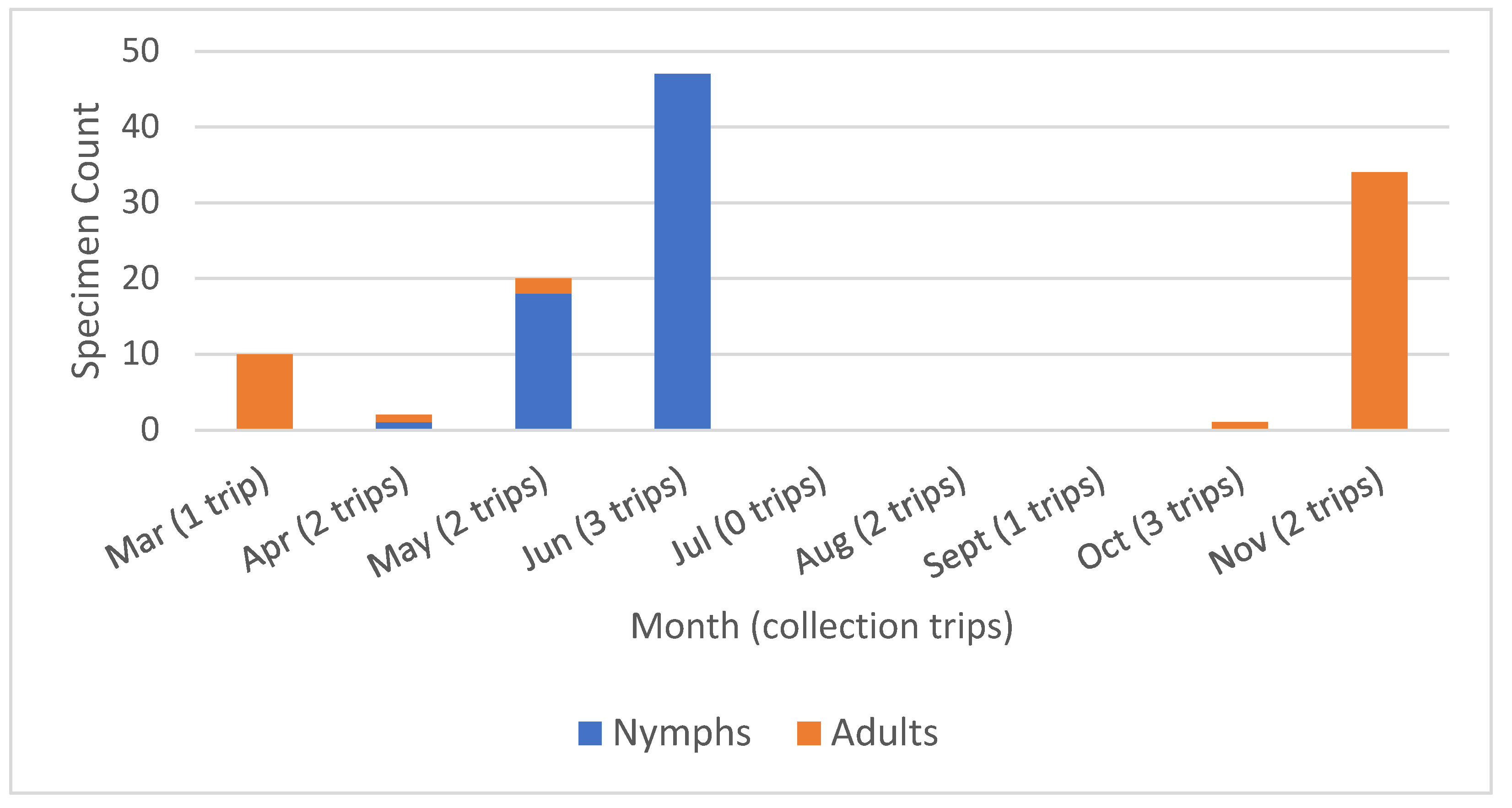
3.1. Specimen Collection

A total of 139 ticks were captured in 16 collection trips between the months of March and November 2023. Some planned collection trips were shifted or cancelled due to weather, including an extremely dry summer with excessive temperatures that would have made collections unsafe. Collected ticks included 114 Ix scapularis (48 adults and 66 nymphs, detailed in Figure 1), 7 adult D. variabilis (3 at Hunt Valley and 4 at Parkton) and 18 nymphal Haemaphysalis longicornis. All H. longicornis were collected at the Hunt Valley site over two consecutive collection visits in late May and early June 2023. No other life stages were collected for the Asian longhorned tick.

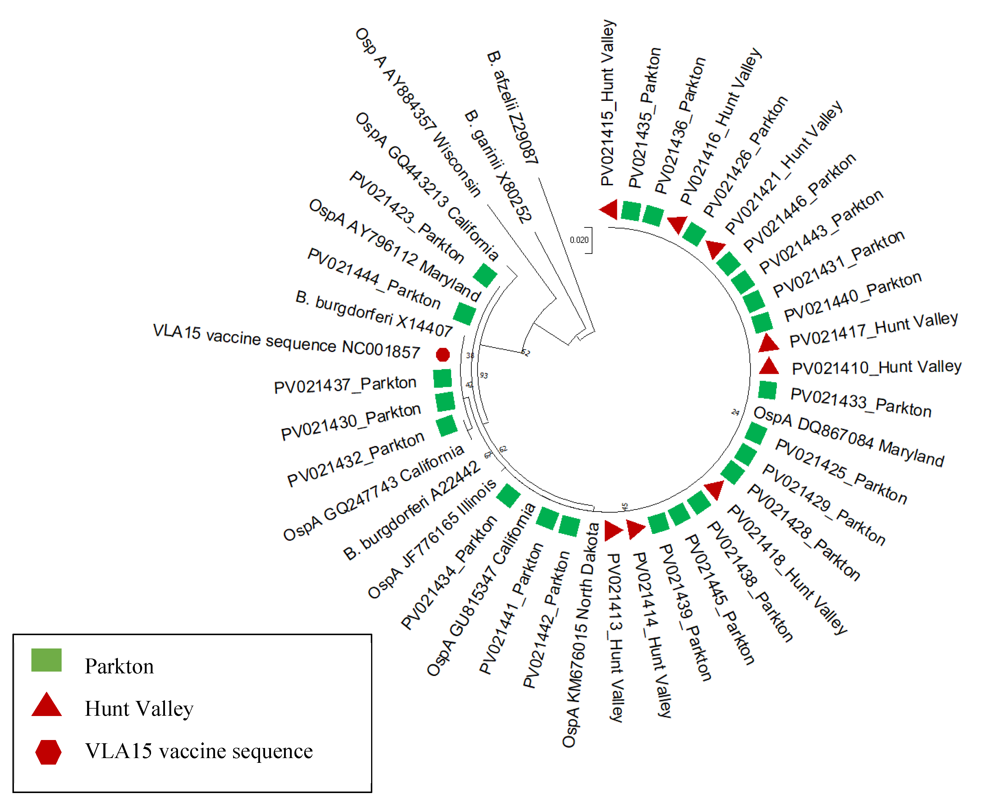
3.2. Pathogen Detection, Sequencing and Phylogenetic Analysis

Only Ix. scapularis were of sufficient sample size to screen for pathogen. qPCR pathogen detection indicated 42 (36.8%) ticks infected with B. burgdorferi s.s. and zero ticks infected with B. microti. The summary of B. burgdorferi-infected ticks by collection site and life stage is provided in Table 1. A. phagocytophilum screening yielded 3 positive ticks (2.6%).
The three Anaplasma amplicons (881-906 bp) aligned with 99.23-99.61% nucleotide identity to A. phagocytophilum as determined by BLASTN analysis. Of the Anaplasma-positive Ix. scapularis, one was a nymph collected at the Hunt Valley site in June 2023. The other two positives were males collected at the Parkton site in October and November. The tick collected in November was co-infected with B. burgdorferi.
OspA PCR was successful for 41 samples and subsequent sequencing was successful for 40 samples. Alignment and comparison of these resulting sequences to the reference ospA sequence of B. burgdorferi B31 (GenBank Accession AY030279) revealed multiple silent point mutations. The resulting maximum likelihood tree demonstrated that the Hunt Valley and Parkton samples cluster together in a strongly supported clade with the VLA15 vaccine sequence (GenBank Accession NC001857) and most other North American sequences (Figure 2).

4. Discussion

A total of 114 Ix. scapularis, 7 D. variabilis and 18 H. longicornis ticks were collected over an overall collection period of nine months from March until November 2023. Ixodes scapularis was the most numerous species encountered and mirrors regional collections from Maryland in 2011-12 [31] and more recent collections conducted in Howard County, Maryland approximately 50 km south.[30] The 18 H. longicornis samples, all collected from the Hunt Valley site, are of note as they have never been encountered before at this collection site. The first confirmed H. longicornis detected in Maryland was in July 2018.[39] While the USDA’s APHIS ‘Asian Longhorned Ticks’ webpage confirms established and reported populations in Maryland, it does not [30,31,35currently include the site in Baltimore County where our collections were made.[40] This highlights the need for improved H. longicornis surveillance in Maryland.
In total, 42 Ix. scapularis tested positive for B. burgdorferi s.s. by qPCR. Despite similar numbers collected at both sites, the B. burgdorferi infection rate in nymphal Ix. scapularis was over 2-fold higher at the Hunt Valley site. However, this may be an artifact of the overall small sample sizes. The overall infection rate of 36.8% is consistent with prior infection rates of ~30% for Ix. scapularis collected in Baltimore County (Norris, unpublished), but is considerably higher than that reported for Ix. scapularis on the Eastern Shore of Maryland in 2003, across multiple counties in central Maryland in 2011-12, or from Howard County in 2017.[30,31,35] It is unknown whether this represents a true increase in prevalence, or whether this is representative of regional/ecological enhancement. This and prior studies have demonstrated significant differences in tick densities and infection rates between even geographically clustered study sites.[30,31,35,36] In the United States in 2022, Maryland ranked 13th of all states in incidence (33/1000) with 2,035 cases (10th highest case count in the U.S.), the most cases reported in the state since 2008.[2] Mayland has historically fallen in the top 10 states in annual case counts for Lyme disease.
Of the ospA sequences recovered from 40 individual Ix. scapularis, 19 were 100% identical, with the remaining 21 sequences revealing point mutations across the length of sequence. All mutations were silent mutations and would have not resulted in any translational effect of the resulting outer surface protein. Overall, ospA within these samples is highly conserved and consistent with other North American sequences included in the phylogenetic analysis.
PCR and subsequent sequencing confirmed 3 Ix. scapularis positive for A. phagocytophilum,1 adult male was co-infected with B. burgdorferi. Considering the small overall sample size (n = 114), it was surprising to find 3 A. phagocytophilum-positive ticks across 3 different collections and two locations. The overall 2.6% infection rate is much higher than reported in prior studies in Maryland.[30,31,35] This might suggest that A. phagocytophilum is circulating at higher rates in Maryland than has been seen in historic surveillance.
This small pilot study demonstrates the need for more extensive contemporary surveillance of ticks and tick-borne pathogens in the mid-Atlantic region. Although Babesia was not detected in the 114 ticks screened, clinical infections have been reported from Maryland and human babesiosis cases have been increasing in the United States.[41] The discovery of invasive Asian longhorned tick nymphs in Baltimore County is also of note, and broader collections throughout the region are needed to determine the distribution of this potential vector of human and livestock pathogens. Due to the very small sample sizes, the non-Ixodes ticks were not screened for pathogen in this study. However, these other species should not be overlooked, as they have the potential to vector a considerable number of human pathogens.

Supplementary Materials

The following supporting information can be downloaded at the website of this paper posted on Preprints.org.

Author Contributions

Conceptualization, A.A. and D.E.N.; methodology, A.A., R.L.M.N.A. and D.E.N.; software, R.L.M.N.A.; formal analysis, A.A., R.L.M.N.A. and D.E.N.; investigation, A.A. and D.E.N.; resources, D.E.N.; data curation, R.L.M.N.A.; writing—original draft preparation, A.A. and D.E.N.; writing—review and editing, A.A., R.L.M.N.A. and D.E.N.; visualization, A.A., R.L.M.N.A. and D.E.N.; supervision, R.L.M.N.A. and D.E.N.; project administration, D.E.N.; funding acquisition, D.E.N. All authors have read and agreed to the published version of the manuscript.

Funding

This research received no external funding.

Institutional Review Board Statement

Not applicable.

Data Availability Statement

The data for sequences generated in this study are available in NCBI’s GenBank database (https://www.ncbi.nlm.nih.gov/genbank/) under accession numbers PV021407 - PV021446 for Borrelia burgdorferi OspA and PV022143 - PV022145 for Anaplasma phagocytophilum.

Acknowledgments

We would like to thank the individuals who allowed us use of their property for sampling. We would also like to thank the Johns Hopkins Lyme and Tickborne Diseases Research and Education Institute, the Sullivan Lab at the Johns Hopkins Bloomberg School of Public Health and the Nicholson Lab at the Centers for Disease Control and Prevention for their guidance and resources provided.

Conflicts of Interest

The authors declare no conflicts of interest.

References

  1. Rosenberg, R.; Lindsey, N.P.; Fischer, M.; Gregory, C.J.; Hinckley, A.F.; Mead, P.S.; et al. Vital Signs: Trends in reported vectorborne disease cases - United States and Territories, 2004-2016. MMWR Morb Mortal Wkly Rep. 2018, 2018. 67, 496–501. [Google Scholar] [CrossRef]
  2. Centers for Disease Control and Prevention. Lyme Disease Surveillance Data 2024 [updated December 17, 2024; cited 2025 January 26, 2025]. Available from: https://www.cdc.gov/lyme/data-research/facts-stats/surveillance-data-1.html#cdc_data_surveillance_section_2-dashboard-data-files.
  3. Kugeler, K.J.; Schwartz, A.M.; Delorey, M.J.; Mead, P.S.; Hinckley, A.F. Estimating the frequency of Lyme disease diagnoses, United States, 2010-2018. Emerg Infect Dis. 2021, 27, 616–619. [Google Scholar] [CrossRef] [PubMed]
  4. Schwartz, A.M.; Kugeler, K.J.; Nelson, C.A.; Marx, G.E.; Hinckley, A.F. Use of commercial claims data for evaluating trends in Lyme disease diagnoses, United States, 2010-2018. Emerg Infect Dis. 2021, 27, 499–507. [Google Scholar] [CrossRef] [PubMed]
  5. Adrion, E.R.; Aucott, J.; Lemke, K.W.; Weiner, J.P. Health care costs, utilization and patterns of care following Lyme disease. PLoS One. 2015, 10, e0116767. [Google Scholar] [CrossRef] [PubMed]
  6. Mac, S.; da Silva, S.R.; Sander, B. The economic burden of Lyme disease and the cost-effectiveness of Lyme disease interventions: A scoping review. PLoS One. 2019, 14, e0210280. [Google Scholar] [CrossRef]
  7. Hayes, S.F.; Burgdorfer, W.; Barbour, A.G. Bacteriophage in the Ixodes dammini spirochete, etiological agent of Lyme disease. J Bacteriol. 1983, 154, 1436–1439. [Google Scholar] [CrossRef]
  8. Halsey, S.J.; Allan, B.F.; Miller, J.R. The role of Ixodes scapularis, Borrelia burgdorferi and wildlife hosts in Lyme disease prevalence: A quantitative review. Ticks Tick Borne Dis. 2018, 9, 1103–1114. [Google Scholar] [CrossRef]
  9. Eisen, L.; Eisen, R.J. Changes in the geographic distribution of the blacklegged tick, Ixodes scapularis, in the United States. Ticks Tick Borne Dis. 2023, 14, 102233. [Google Scholar] [CrossRef]
  10. Burgdorfer, W.; Lane, R.S.; Barbour, A.G.; Gresbrink, R.A.; Anderson, J.R. The western black-legged tick, Ixodes pacificus: a vector of Borrelia burgdorferi. Am J Trop Med Hyg. 1985, 34, 925–930. [Google Scholar] [CrossRef]
  11. Davidson, A.; Kelly, P.H.; Davis, J.; Major, M.; Moisi, J.C.; Stark, J.H. Historical summary of tick and animal surveillance studies for Lyme disease in Canada, 1975-2023: A Scoping Review. Zoonoses Public Health. 2025, 72, 9–22. [Google Scholar] [CrossRef]
  12. Price, L.E.; Winter, J.M.; Cantoni, J.L.; Cozens, D.W.; Linske, M.A.; Williams, S.C.; et al. Spatial and temporal distribution of Ixodes scapularis and tick-borne pathogens across the northeastern United States. Parasit Vectors. 2024, 17, 481. [Google Scholar] [CrossRef] [PubMed]
  13. Eilbert, W.; Matella, A. Tick-borne diseases. Emerg Med Clin North Am. 2024, 42, 287–302. [Google Scholar] [CrossRef] [PubMed]
  14. Bahadori, A.; Ritz, N.; Zimmermann, P. Diagnosis and treatment of Lyme disease in children. Arch Dis Child Educ Pract Ed. 2023, 108, 422–428. [Google Scholar] [CrossRef] [PubMed]
  15. Sanchez-Vicente, S.; Tokarz, R. Tick-borne co-infections: Challenges in molecular and serologic diagnoses. Pathogens. 2023, 12. [Google Scholar] [CrossRef]
  16. Hansen, A.P.; Wilkinson, M.M.; Niesobecki, S.; Rutz, H.; Meek, J.I.; Niccolai, L.; et al. Barriers to the uptake of tickborne disease prevention measures: Connecticut, Maryland 2016-2017. J Public Health Manag Pract. 2025, 31, E52–E60. [Google Scholar] [CrossRef]
  17. Lewandowski, D.; Sulik, A.; Raciborski, F.; Krasnodebska, M.; Gebarowska, J.; Stalewska, A.; et al. Barriers and predictors of Lyme disease vaccine acceptance: A cross-sectional study in Poland. Vaccines (Basel). 2025, 13. [Google Scholar] [CrossRef]
  18. Nigrovic, L.E.; Thompson, K.M. The Lyme vaccine: a cautionary tale. Epidemiol Infect. 2007, 135, 1–8. [Google Scholar] [CrossRef]
  19. Comstedt, P.; Schuler, W.; Meinke, A.; Lundberg, U. The novel Lyme borreliosis vaccine VLA15 shows broad protection against Borrelia species expressing six different OspA serotypes. PLoS One. 2017, 12, e0184357. [Google Scholar] [CrossRef]
  20. Ghadge, S.K.; Schneider, M.; Dubischar, K.; Wagner, L.; Kadlecek, V.; Obersriebnig, M.; et al. Immunogenicity and safety of an 18-month booster dose of the VLA15 Lyme borreliosis vaccine candidate after primary immunisation in healthy adults in the USA: results of the booster phase of a randomised, controlled, phase 2 trial. Lancet Infect Dis. 2024, 24, 1275–1286. [Google Scholar] [CrossRef]
  21. Comstedt, P.; Hanner, M.; Schuler, W.; Meinke, A.; Lundberg, U. Design and development of a novel vaccine for protection against Lyme borreliosis. PLoS One. 2014, 9, e113294. [Google Scholar] [CrossRef]
  22. Howe, T.R.; LaQuier, F.W.; Barbour, A.G. Organization of genes encoding two outer membrane proteins of the Lyme disease agent Borrelia burgdorferi within a single transcriptional unit. Infect Immun. 1986, 54, 207–212. [Google Scholar] [CrossRef] [PubMed]
  23. Howe, T.R.; Mayer, L.W.; Barbour, A.G. A single recombinant plasmid expressing two major outer surface proteins of the Lyme disease spirochete. Science. 1985, 227, 645–646. [Google Scholar] [CrossRef] [PubMed]
  24. Guttman, D.S.; Wang, P.W.; Wang, I.N.; Bosler, E.M.; Luft, B.J.; Dykhuizen, D.E. Multiple infections of Ixodes scapularis ticks by Borrelia burgdorferi as revealed by single-strand conformation polymorphism analysis. J Clin Microbiol. 1996, 34, 652–656. [Google Scholar] [CrossRef] [PubMed]
  25. Anderson, J.M.; Norris, D.E. Genetic diversity of Borrelia burgdorferi sensu stricto in Peromyscus leucopus, the primary reservoir of Lyme disease in a region of endemicity in southern Maryland. Appl Environ Microbiol. 2006, 72, 5331–5341. [Google Scholar] [CrossRef]
  26. Rochlin, I.; Toledo, A. Emerging tick-borne pathogens of public health importance: a mini-review. J Med Microbiol. 2020, 69, 781–791. [Google Scholar] [CrossRef]
  27. Eisen, R.J.; Eisen, L. The blacklegged tick, Ixodes scapularis: An increasing public health concern. Trends Parasitol. 2018, 34, 295–309. [Google Scholar] [CrossRef]
  28. Eisen, L. Tick species infesting humans in the United States. Ticks Tick Borne Dis. 2022, 13, 102025. [Google Scholar] [CrossRef]
  29. Merten, H.A.; Durden, L.A. A state-by-state survey of ticks recorded from humans in the United States. J Vector Ecol. 2000, 25, 102–113. [Google Scholar]
  30. Milholland, M.T.; Eisen, L.; Nadolny, R.M.; Hojgaard, A.; Machtinger, E.T.; Mullinax, J.M.; et al. Surveillance of ticks and tick-borne pathogens in suburban natural habitats of central Maryland. J Med Entomol. 2021, 58, 1352–1362. [Google Scholar] [CrossRef]
  31. Feldman, K.A.; Connally, N.P.; Hojgaard, A.; Jones, E.H.; White, J.L.; Hinckley, A.F. Abundance and infection rates of Ixodes scapularis nymphs collected from residential properties in Lyme disease-endemic areas of Connecticut, Maryland, and New York. J Vector Ecol. 2015, 40, 198–201. [Google Scholar] [CrossRef]
  32. Henning, T.C.; Orr, J.M.; Smith, J.D.; Arias, J.R.; Norris, D.E. Spotted fever group rickettsiae in multiple hard tick species from Fairfax County, Virginia. Vector-borne Zoon Dis. 2014, 14, 482-485. [CrossRef]
  33. Hojgaard, A.; Lukacik, G.; Piesman, J. Detection of Borrelia burgdorferi, Anaplasma phagocytophilum and Babesia microti, with two different multiplex PCR assays. Ticks Tick Borne Dis. 2014, 5, 349–351. [Google Scholar] [CrossRef] [PubMed]
  34. Massung, R.F.; Slater, K.; Owens, J.H.; Nicholson, W.L.; Mather, T.N.; Solberg, V.B.; et al. Nested PCR assay for detection of granulocytic ehrlichiae. J Clin Microbiol. 1998, 36, 1090–1095. [Google Scholar] [CrossRef] [PubMed]
  35. Swanson, K.I.; Norris, D.E. Co-circulating microorganisms in questing Ixodes scapularis nymphs in Maryland. J Vector Ecol. 2007, 32, 243–251. [Google Scholar] [CrossRef] [PubMed]
  36. Anderson, J.M.; Swanson, K.I.; Schwartz, T.R.; Glass, G.E.; Norris, D.E. Mammal diversity and infection prevalence in the maintenance of enzootic Borrelia burgdorferi along the western Coastal Plains of Maryland. Vector-borne Zoon Dis. 2006, 6, 411-422. [CrossRef]
  37. Kumar, S.; Stecher, G.; Li, M.; Knyaz, C.; Tamura, K. MEGA X: Molecular Evolutionary Genetics Analysis across Computing Platforms. Mol Biol Evol. 2018, 35, 1547–1549. [Google Scholar] [CrossRef] [PubMed]
  38. Posada, D. jModelTest: phylogenetic model averaging. Mol Biol Evol. 2008, 25, 1253–1256. [Google Scholar] [CrossRef]
  39. Maryland Division of Natural Resources. First Confirmed Longhorned Tick Found in Maryland 2018 [updated August 7, 2018; cited 2025 January 26, 2025]. Available from: https://news.maryland.gov/dnr/2018/08/07/first-confirmed-longhorned-tick-found-in-maryland/.
  40. United States Department of Agriculture. Asian Longhorned Ticks 2024 [updated September 11, 2024; cited 2025 January 26, 2025]. Available from: https://www.aphis.usda.gov/livestock-poultry-disease/cattle/ticks/asian-longhorned.
  41. Centers for Disease Control and Prevention (CDC). Surveillance for babesiosis—United States, 2019 annual summary. Atlanta, Georgia: U.S. Department of Health and Human Services, CDC; 2021.
Figure 1. Ixodes scapularis specimen distribution by collection month and life stage. Also shown are the number of collection trips undertaken each month.
Preprints 147727 g001
Figure 2. Maximum Likelihood tree of OspA sequences generated in this study with VLA15 vaccine sequence and reference Borrelia sequences from the GenBank database.
Preprints 147727 g002
Table 1. Counts and number of B. burgdorferi-positive Ix. scapularis by collection site.
Hunt Valley No. Collected No. Positive at Hunt Valley Parkton No. Collected No. Positive at Parkton Total Tick Counts Total Positive Ticks
Nymphs 32 15 (46.9%) 33 7 (21.2%) 65 22 (33.8%)
Adults 2 1 (50.0%) 47 19 (40.4%) 49 20 (40.8%)
Total 34 16 (47.1%) 80 26 (32.5%) 117 42 (36.8%)
Disclaimer/Publisher’s Note: The statements, opinions and data contained in all publications are solely those of the individual author(s) and contributor(s) and not of MDPI and/or the editor(s). MDPI and/or the editor(s) disclaim responsibility for any injury to people or property resulting from any ideas, methods, instructions or products referred to in the content.
Copyright: This open access article is published under a Creative Commons CC BY 4.0 license, which permit the free download, distribution, and reuse, provided that the author and preprint are cited in any reuse.
Prerpints.org logo

Preprints.org is a free preprint server supported by MDPI in Basel, Switzerland.

Subscribe

Disclaimer

Terms of Use

Privacy Policy

Privacy Settings

© 2025 MDPI (Basel, Switzerland) unless otherwise stated