Preprint
Interesting Images

This version is not peer-reviewed.

Endoscopic Features of Early Autoimmune Gastritis: Insights From a Case Study in a Young Male Patient

Submitted:

26 January 2025

Posted:

27 January 2025

You are already at the latest version

Abstract

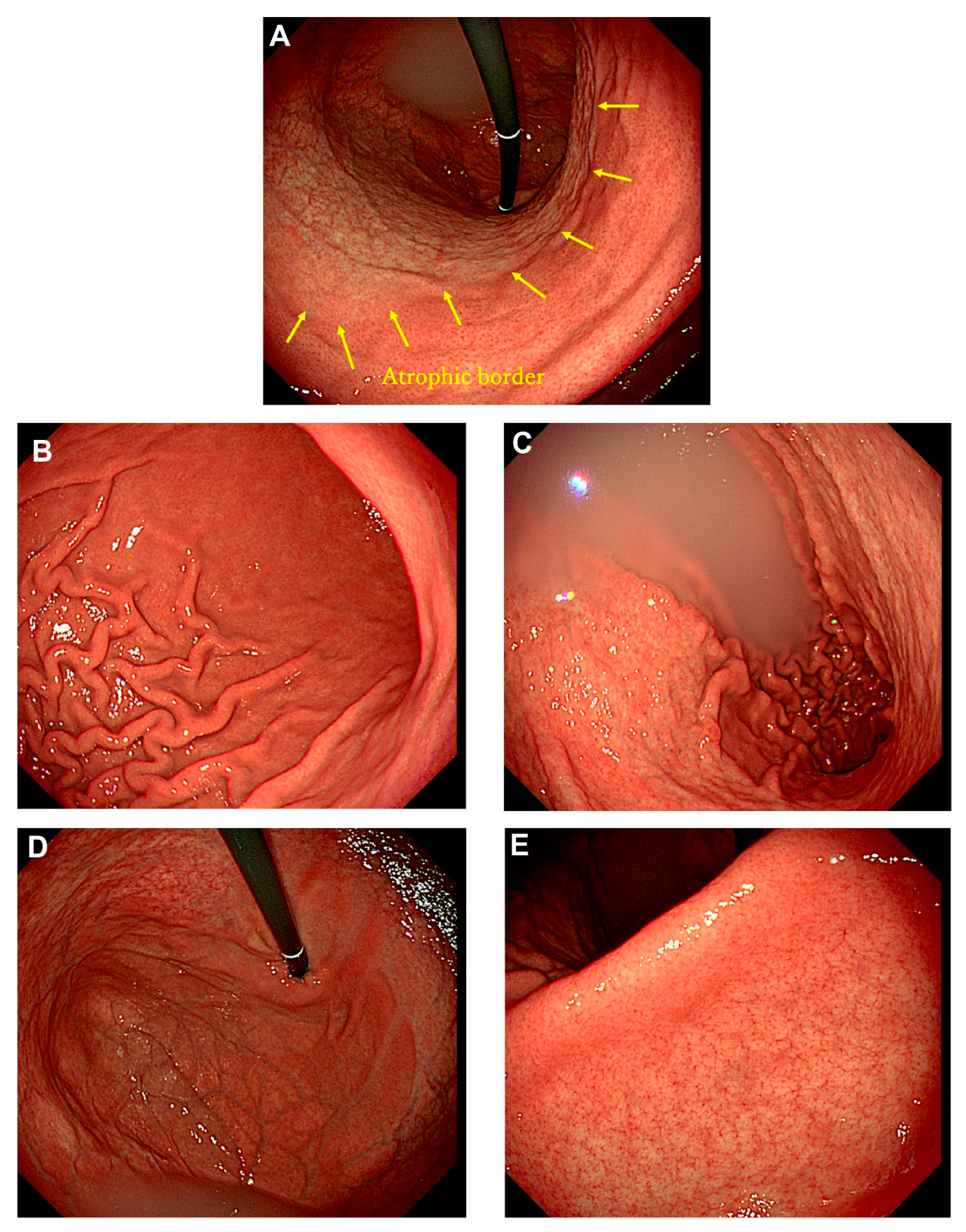
Autoimmune gastritis, traditionally called type A gastritis, is characterized by cor-pus-predominant atrophic gastritis caused by autoimmune mechanisms. Most cases are diagnosed in middle-aged or elderly individuals, as complications such as pernicious anemia and impaired absorption of iron and vitamin B12 typically manifest in advanced stages. Additionally, patients with autoimmune gastritis are often asymptomatic, making reports of early-stage endoscopic findings exceedingly rare. A 22-year-old male presented to our hospital with complaints of epigastric pain and lower back pain. He had undergone eradication therapy for Helicobacter pylori infection at another hospital three months prior to presentation. A urea breath test confirmed the successful eradication of H. pylori. Endoscopic examination revealed extensive, sharply demarcated mucosal atrophy extending orally from the middle of the gastric body, while the gastric antrum showed no evidence of atrophy or intestinal metaplasia. Laboratory tests revealed a mild elevation of anti-parietal cell antibody levels by a factor of 10 (reference range: 0–9), whereas serum gastrin and vitamin B12 levels remained within normal limits. Iron metabolism parameters were also normal. This report presents a rare case of early-stage autoimmune gastritis with distinctive endoscopic findings in a young male.

Keywords: 
;  ;  ;  
Figure 1. Endoscopic findings of early autoimmune gastritis. Early Endoscopic Findings of Autoimmune Gastritis. Autoimmune gastritis, historically termed type A gastritis by Strickland and Mackey [1], is an autoimmune disorder targeting the parietal cells of the fundic glands, resulting in inflammation and atrophy predominantly in the corpus and fundus. Traditionally, patients with autoimmune gastritis present with neurological symptoms secondary to vitamin B12 or iron deficiency, often leading to diagnoses such as pernicious anemia or iron deficiency anemia in advanced stages [2]. However, the majority of cases are asymptomatic and frequently discovered incidentally. Diagnosis relies on a combination of clinical, endoscopic, histological, and serological findings, though standardized diagnostic criteria for autoimmune gastritis have yet to be universally established. In 2023, the “Study Group on Establishment of Diagnostic Criteria for Type A Gastritis,” an adjunctive group of the Japanese Society of Gastrointestinal Endoscopy (JSGE), proposed new diagnostic criteria [3]. According to these criteria, autoimmune gastritis is definitively diagnosed when characteristic endoscopic or histological findings are accompanied by positive gastric autoantibodies (anti-parietal cell or anti-intrinsic factor antibodies). Despite increasing global interest in the early-stage endoscopic features of autoimmune gastritis, reports in young adults remain scarce [3,4,5,6]. A 22-year-old male presented with complaints of epigastric pain and lower back pain. He had received two COVID-19 vaccinations and had undergone Helicobacter pylori eradication therapy at another hospital three months prior to his visit. Laboratory investigations revealed no evidence of anemia (red blood cell count: 5.20 × 10⁶/μL; hemoglobin: 17.4 g/dL; hematocrit: 49.8%; mean corpuscular volume: 95.9 fL), normal iron metabolism (serum iron: 161 μg/dL [40-188]; total iron-binding capacity: 336 μg/dL [253–366]; ferritin: 58.2 ng/mL [31-325]), and normal serum levels of gastrin (170 pg/mL [0-200]) and vitamin B12 (497 pg/mL [180-914]). Anti-parietal cell antibody levels were slightly elevated (10-fold; reference range: 0–9). An upper gastrointestinal endoscopy was performed using the Olympus GIF-290 endoscope. An upward view of the lesser curvature of the gastric body revealed a regular arrangement of collecting venules (RAC) [7], a hallmark endoscopic feature indicative of H. pylori-negative status, in the lower body. A sharply demarcated atrophic border was observed between the lower and middle corpus. A downward view of the greater curvature of the body revealed no atrophic changes in the lower segment but extensive atrophy in the middle and upper segments. Scattered diffuse redness and atrophy were visible in the upper body and fundus. The antral mucosa showed transvascular findings without severe atrophy or intestinal metaplasia. The natural history of autoimmune gastritis is thought to progress through four stages. In the first stage, serum anti-parietal cell and anti-intrinsic factor antibodies become positive, representing a condition referred to as potential autoimmune gastritis, which lacks histological evidence of corpus-predominant atrophic gastritis [8]. In the second stage, histological abnormalities develop in the corpus mucosa, but no endoscopic atrophic changes are observed. The third stage is characterized by evident corpus atrophy on endoscopy, while most patients remain asymptomatic. In the final stage, clinical manifestations such as vitamin B12 deficiency and anemia emerge. Notably, childhood autoimmune gastritis may present with symptoms and histological or serological abnormalities despite the absence of endoscopic atrophic changes [9]. Reports on the early-stage endoscopic findings of autoimmune gastritis are limited. Childhood cases are often discovered incidentally due to anemia or comorbidities such as chronic thyroiditis or type 1 diabetes mellitus [9,10]. The low prevalence and asymptomatic nature of most cases have limited the routine use of endoscopy for diagnosis. In Japan, where gastric cancer screening is prevalent, most cases of atrophic gastritis are attributed to H. pylori infection, often associated with pangastritis caused by the cytotoxin-associated gene A (CagA) virulence factor [11,12,13]. Eradication therapy for H. pylori has uncovered cases of atrophic gastritis unrelated to infection, with corpus-predominant atrophy increasingly recognized as autoimmune gastritis [14,15]. Studies have reported that autoimmune gastritis is more frequently observed in patients with multiple failed H. pylori eradication attempts, potentially due to achlorhydria allowing the survival of urease-positive bacteria other than H. pylori [14,15,16,17,18]. This phenomenon may lead to false-positive urea breath test results [16]. As interest in autoimmune gastritis grows, the adjunctive study group of the JSGE has proposed diagnostic criteria that incorporate endoscopic findings [3]. However, early-stage endoscopic features remain poorly defined due to limited case accumulation. Kotera et al. recently described characteristic endoscopic findings, including longitudinal pseudo-polyps and erythematous swelling, in 12 patients with early-stage autoimmune gastritis (mean age: 56.2 years; range: 41–71 years) [6]. Notably, these findings have not been reported in younger patients. In this case, we report the endoscopic findings of ear-ly-stage autoimmune gastritis in a 22-year-old male. Although the endoscopic appearance did not exhibit bamboo-like or salmon-like patterns described by Kotera et al., a distinct atrophic border demarcating the pyloric and fundic glands was observed. Despite the absence of histological examination and only a slightly elevated anti-parietal cell antibody titer, this case highlights unique endoscopic features of early-stage autoimmune gastritis in a young adult.
Preprints 147382 g001

Author Contributions

Patient endoscopic study: I.I. and S.O. Data collection and formal analysis: T.K. and I.I. Writing—original draft preparation: I.I. and S.O. Writing—review and editing: T.Y., E.C.G., and N.H. Interpretation of data and intellectual contribution: E.C.G., T.Y., N.H., T.K., and K.K. All authors have read and agreed to the published version of the manuscript.

Funding

This research received no external funding.

Institutional Review Board Statement

No ethical approval was required due to the study's retrospective nature, as indicated by our institutional review board.

Informed Consent Statement

Written informed consent was obtained from the patient.

Data Availability Statement

The authors declare that all the data described in this article and available upon reasonable request.

Conflicts of Interest

The authors declare no conflict of interest.

References

  1. Strickland, R. G.; Mackay, I. R., A reappraisal of the nature and significance of chronic atrophic gastritis. Am J Dig Dis 1973; 18(5): 426-40. [CrossRef]
  2. Hall, S. N.; Appelman, H. D., Autoimmune Gastritis. Arch Pathol Lab Med 2019; 143(11): 1327-1331. [CrossRef]
  3. Kamada, T.; Watanabe, H.; Furuta, T.; Terao, S.; Maruyama, Y.; Kawachi, H.; Kushima, R.; Chiba, T.; Haruma, K., Diagnostic criteria and endoscopic and histological findings of autoimmune gastritis in Japan. J Gastroenterol 2023; 58(3): 185-195. [CrossRef]
  4. Ayaki, M.; Aoki, R.; Matsunaga, T.; Manabe, N.; Fujita, M.; Kamada, T.; Kobara, H.; Masaki, T.; Haruma, K., Endoscopic and Upper Gastrointestinal Barium X-ray Radiography Images of Early-stage Autoimmune Gastritis: A Report of Two Cases. Intern Med 2021; 60(11): 1691-1696. [CrossRef]
  5. Kishino, M.; Nonaka, K., Endoscopic Features of Autoimmune Gastritis: Focus on Typical Images and Early Images. J Clin Med 2022; 11(12). [CrossRef]
  6. Kotera, T.; Ayaki, M.; Sumi, N.; Aoki, R.; Mabe, K.; Inoue, K.; Manabe, N.; Kamada, T.; Kushima, R.; Haruma, K., Characteristic endoscopic findings in early-stage autoimmune gastritis. Endosc Int Open 2024; 12(3): E332-E338. [CrossRef]
  7. Yagi, K.; Aruga, Y.; Nakamura, A.; Sekine, A., Regular arrangement of collecting venules (RAC): a characteristic endoscopic feature of Helicobacter pylori-negative normal stomach and its relationship with esophago-gastric adenocarcinoma. J Gastroenterol 2005; 40(5): 443-52. [CrossRef]
  8. Lenti, M. V.; Miceli, E.; Vanoli, A.; Klersy, C.; Corazza, G. R.; Di Sabatino, A., Time course and risk factors of evolution from potential to overt autoimmune gastritis. Dig Liver Dis 2022; 54(5): 642-644. [CrossRef]
  9. Krzewska, A.; Ben-Skowronek, I., Effect of Associated Autoimmune Diseases on Type 1 Diabetes Mellitus Incidence and Metabolic Control in Children and Adolescents. Biomed Res Int 2016; 2016: 6219730. [CrossRef]
  10. Mitsinikos, T.; Shillingford, N.; Cynamon, H.; Bhardwaj, V., Autoimmune Gastritis in Pediatrics: A Review of 3 Cases. J Pediatr Gastroenterol Nutr 2020; 70(2): 252-257. [CrossRef]
  11. Tanaka, C.; Otani, K.; Tamoto, M.; Yoshida, H.; Nadatani, Y.; Ominami, M.; Fukunaga, S.; Hosomi, S.; Kamata, N.; Tanaka, F., et al., Efficacy evaluation of upper gastrointestinal endoscopy screening for secondary prevention of gastric cancer using the standardized detection ratio during a medical check-up in Japan. J Clin Biochem Nutr 2024; 74(3): 253-260. [CrossRef]
  12. Park, J. Y.; Forman, D.; Waskito, L. A.; Yamaoka, Y.; Crabtree, J. E., Epidemiology of Helicobacter pylori and CagA-Positive Infections and Global Variations in Gastric Cancer. Toxins (Basel) 2018; 10(4). [CrossRef]
  13. Kotera, T.; Yoshioka, U.; Takemoto, T.; Kushima, R.; Haruma, K., Evolving Autoimmune Gastritis Initially Hidden by Active Helicobacter pylori Gastritis. Case Rep Gastroenterol 2022; 16(1): 103-109. [CrossRef]
  14. Iwata, E.; Sugimoto, M.; Akimoto, Y.; Hamada, M.; Niikura, R.; Nagata, N.; Yanagisawa, K.; Itoi, T.; Kawai, T., Long-term endoscopic gastric mucosal changes up to 20 years after Helicobacter pylori eradication therapy. Sci Rep 2024; 14(1): 13003. [CrossRef]
  15. Ihara, T.; Ihara, N.; Kushima, R.; Haruma, K., Rapid Progression of Autoimmune Gastritis after Helicobacter pylori Eradication Therapy. Intern Med 2023; 62(11): 1603-1609. [CrossRef]
  16. Furuta, T.; Baba, S.; Yamade, M.; Uotani, T.; Kagami, T.; Suzuki, T.; Tani, S.; Hamaya, Y.; Iwaizumi, M.; Osawa, S., et al., High incidence of autoimmune gastritis in patients misdiagnosed with two or more failures of H. pylori eradication. Aliment Pharmacol Ther 2018; 48(3): 370-377. [CrossRef]
  17. Ihara, T.; Kushima, R.; Haruma, K., Enhanced activity of autoimmune gastritis following Helicobacter pylori eradication therapy. Clin J Gastroenterol 2025. [CrossRef]
  18. Sumi, N.; Haruma, K.; Inoue, K.; Hisamoto, N.; Mabe, K.; Sasai, T.; Ichiba, T.; Ayaki, M.; Manabe, N.; Takao, T., A case of nodular gastritis progression to autoimmune gastritis after 10 years of Helicobacter pylori eradication. Clin J Gastroenterol 2024; 17(2): 216-221. [CrossRef]
Disclaimer/Publisher’s Note: The statements, opinions and data contained in all publications are solely those of the individual author(s) and contributor(s) and not of MDPI and/or the editor(s). MDPI and/or the editor(s) disclaim responsibility for any injury to people or property resulting from any ideas, methods, instructions or products referred to in the content.
Copyright: This open access article is published under a Creative Commons CC BY 4.0 license, which permit the free download, distribution, and reuse, provided that the author and preprint are cited in any reuse.
Prerpints.org logo

Preprints.org is a free preprint server supported by MDPI in Basel, Switzerland.

Subscribe

Disclaimer

Terms of Use

Privacy Policy

Privacy Settings

© 2025 MDPI (Basel, Switzerland) unless otherwise stated