Preprint
Review

This version is not peer-reviewed.

LncRNAs Regulate Vasculogenic Mimicry in Human Cancers

A peer-reviewed article of this preprint also exists.

Submitted:

21 January 2025

Posted:

22 January 2025

You are already at the latest version

Abstract

Vasculogenic mimicry (VM) has been recently discovered as an alternative mechanism to nourish cancer cells in vivo. During VM, tumor cells align and organize in three-dimensional (3D) channel-like structures to transport nutrients and oxygen to the internal layers of tumors. This mechanism occurs mainly in aggressive solid tumors and has been associated with poor prognosis in oncologic patients. Long non-coding RNAs (lncRNAs) are essential regulators of protein-encoding genes involved in cancer development and progression. These single-stranded RNA molecules regulate critical cellular functions in cancer cells, including cell proliferation, apoptosis, angiogenesis, VM, therapy response, migration, invasion, and metastasis. Recently, high-throughput RNA sequencing technologies have identified thousands of lncRNAs, but only a tiny percentage have been functionally characterized in human cancers. The vast amount of data about its genomic expression in tumors can allow us to dissect their functions in cancer biology and make them suitable biomarkers for cancer diagnosis and prognosis. Here, we have reviewed the current knowledge about the role of lncRNAs in VM in human cancers, with a special emphasis on their potential to be used as novel therapeutic targets in RNA-based molecular interventions.

Keywords: 
;  ;  ;  ;  

1. Introduction

An efficient distribution of nutrients and oxygen supplies is required for cancer cells’ nourishment and tumor growth. Recent studies indicate that tumor cells grow in three-dimensional (3D) architectures, where gradients for nutrients and oxygen uptake are formed [1]. Indeed, cancer cells located at the external layers of tumors showed high proliferation rates, whereas the internal core is represented mainly by necrotic cells. Therefore, to maintain a concentration gradient of O2 and nutrients and provide novel routes for metabolic waste, the cancer cells may alternatively align and reorganize to form 3D channel-like structures in vivo, a phenomenon termed vasculogenic mimicry (VM) [2]. The 3D channels representative of VM are functional as they transport red blood cells and nutrients, providing an alternative fuel source to support the nourishment of tumors. VM has been detected in almost all solid human tumors, conferring aggressive behavior, higher metastatic potential, and resistance to therapy, resulting in a poor prognosis for oncologic patients [3]. The formation of new blood vessels by endothelial cells from preexisting vasculature (angiogenesis) and VM can function in a coordinated fashion, resulting in mosaic vessels featuring a mixture of connected VM channels and blood vessels [4]. Mechanistically, VM is activated by proteins involved in the response to hypoxia orchestrated by the hypoxia-induced factor 1 alpha (HIF1-A) transcription factor as well as the VEGF-A, Wnt, EphA2, FAK, PI3K, AKT, Notch, and TGF-SMAD signaling pathways [5]. Although it has been reported that non-coding RNAs, such as microRNAs, may regulate VM in diverse types of cancer [5], the role of long non-coding RNAs (lncRNAs) in modulating VM remains poorly understood.
Long non-coding RNAs (lncRNAs) are a relatively novel class of single-strand RNAs defined as transcripts of about 200 nucleotides in their mature form, transcribed from thousands of genetic loci dispersed across the human genome [6]. Two early concepts about its evolutionary origin and functions have been now demystified: i) while widely accepted as RNAs lacking protein-coding potential, recent studies showed that lncRNAs might contain small open reading frames (less than 100 codons in length) which are pervasively translated into mini peptides [7], and ii) lncRNAs were considered mainly as “junk” genetic material, an old concept in genomics [8], but now has been widely accepted as non-coding RNAs which are transcribed in a regulated manner modulating gene expression and phenotypes in human cancers. Nevertheless, lncRNAs are predominantly non-coding RNAs that exhibit critical roles in regulating gene expression in a physiological context. LncRNAs are mainly classified according to the physical location in the genome. Therefore, they are dubbed natural antisense transcripts, overlapping transcripts, and intronic or exonic transcripts, depending on the genomic arrangement of the lncRNA for their closer protein-encoding gene locus [9]. Functions of lncRNAs are diverse, but they can be summarized as follows: i) microRNAs sponges acting as competing endogenous RNAs (ceRNAs), ii) molecular scaffolds for proteins, for instance, organizing the chromatin-modifying enzymes, iii) sequestering proteins facilitating or inhibiting long-range chromatin interactions or transcription factors [9]. LncRNAs are essential regulators of genes involved in cancer development and progression. These small molecules regulate critical cellular functions in cancer cells, including cell proliferation, apoptosis, angiogenesis, vasculogenic mimicry, therapy response, migration, invasion, and metastasis [10]. Recently, high-throughput RNA sequencing technologies have identified many lncRNAs, but only a tiny percentage have been functionally characterized in human cancers. The vast amount of data about its genomic expression in tumors allows us to dissect their functions in cancer biology and makes them suitable biomarkers for cancer diagnosis and prognosis. However, the functions of lncRNAs in regulating the genes involved in VM activation in the different types of human cancers remain poorly characterized.

2. Materials and Methods

Considering that the role of lncRNAs and VM in diverse types of human cancers remains poorly understood, we performed PubMed searches to identify the previously reported lncRNAs associated with VM in cancer. The search was based on the keywords “lncRNA” AND “vasculogenic mimicry” from 2017 to 2024. The query strategy and the number of identified lncRNAs allowed us to retrieve 43 articles on lncRNAs associated with VM. The published manuscripts were classified and described according to the specific type of cancer.

3. Results

VM is a unique process where tumor cells mimic endothelial cells to form vessel-like structures, providing an alternative route for blood supply and facilitating tumor growth and metastasis [2,5]. This process is independent of angiogenesis, forming new blood vessels from pre-existing ones, and poses a significant challenge to conventional anti-angiogenic therapies. The presence of VM has been linked to increased tumor grade, invasiveness, and resistance to treatment, ultimately contributing to poor patient outcomes [5]. In this regard, dysregulation of lncRNAs has been implicated in various cancers, contributing to tumorigenesis, progression, and treatment resistance [3]. Understanding the molecular mechanisms underlying VM, particularly the involvement of lncRNAs, is crucial for developing novel therapeutic strategies to target this process and improve oncologic patient outcomes (Figure 1).

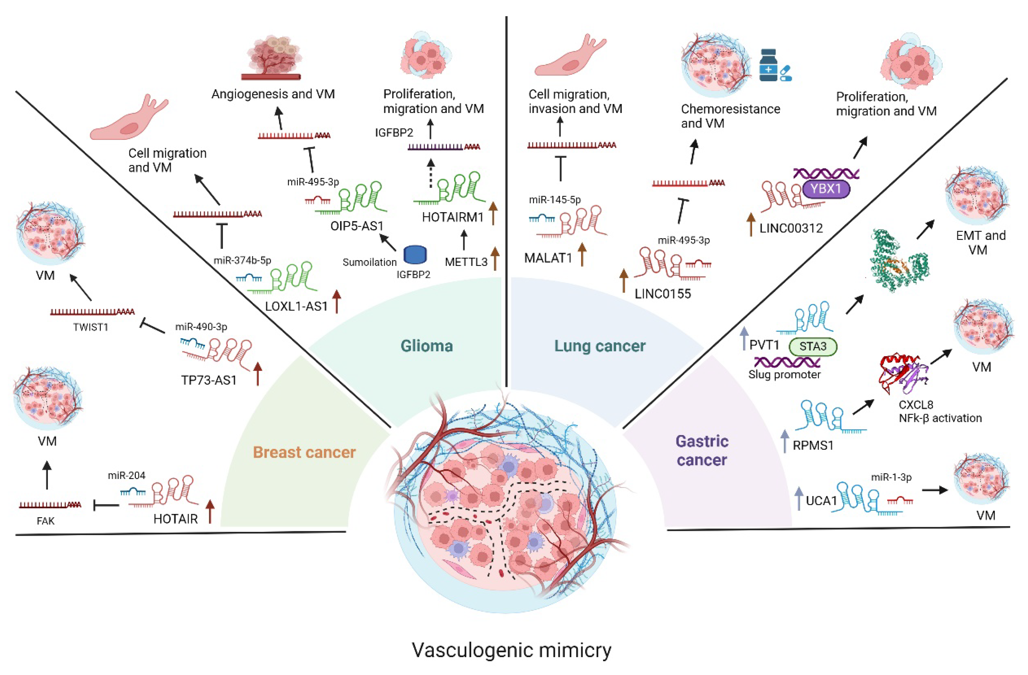
3.1. LncRNAs Functions in Glioma

Glioma is the most common type of primary brain tumor, originating in the glial cells that support and protect neurons in the brain [11]. These tumors are highly aggressive and can be challenging to treat, with a poor prognosis for patients, particularly those with high-grade gliomas. Gliomas account for approximately 30% of all brain and central nervous system tumors and 80% of all malignant brain tumors. Despite advancements in treatment modalities, the average lifespan of newly diagnosed patients remains less than 2.5 years, with a 5-year survival rate below 10%.
VM is particularly important in glioma due to the highly vascularized nature of these tumors. Several reports highlight the role of lncRNAs in regulating VM in tumors. For instance, LINC00339 plays a crucial role in promoting VM formation in glioma by regulating the miR-539-5p/TWIST1/MMPs axis in U87 and U251 cells. LINC00339 functions as a competing endogenous RNA (ceRNA) for miR-539-5p, preventing it from binding to its target mRNAs, including TWIST1 [11]. When LINC00339 levels are low, miR-539-5p is free to bind to the mRNA of TWIST1, thus decreasing its protein levels. On the contrary, by suppressing miR-539-5p, LINC00339 indirectly increases TWIST1 levels, leading to increased MMP-2 and MMP-14 expression and ultimately promoting VM formation [11].
HOTAIRM1 is a lncRNA shown to play an oncogenic role in various cancers, including glioma, by promoting VM. Research suggests that HOTAIRM1 promotes VM in glioma through its overexpression, contributes to the aggressive behavior of glioma cells, and increases glioma cell proliferation, migration, invasion, and promotion of tumor growth and VM formation in vivo. While the exact mechanisms by which HOTAIRM1 promotes VM are still being investigated, the evidence suggests that this lncRNA plays a crucial role in this process through METTL3 [12]. METTL3, increases the stability of HOTAIRM1 through a process called N6-methyladenosine (m6A) modification, essentially attaching a chemical tag to HOTAIRM1, making it more resistant to degradation and thus increasing its lifespan. This process increases the expression of IGFBP2, the final player in this cascade, and directly contributes to VM formation [12]. It promotes the expression of proteins like CD144 and MMP2, which are involved in cell adhesion, migration, and the breakdown of the extracellular matrix - all crucial steps in forming those channel-like structures that characterize VM [13].
There is also a report that SUMOylation of IGF2BP2 increases its stability. This modification protects IGF2BP2 from degradation, leading to its accumulation in the cell. IGF2BP2, now stabilized, interacts with and stabilizes OIP5-AS1, a long non-coding RNA, increasing its levels within the glioma cell and acting as a sponge for miR-495-3p preventing it from binding to and degrading its target mRNAs, effectively augmenting their expression [14]. miR-495-3p, when not sequestered by OIP5-AS1, targets the mRNAs of HIF1A and MMP14, two proteins crucial for VM formation [14]. HIF1A promotes angiogenesis and VM, while MMP14 helps break down the extracellular matrix, facilitating cell migration and VM channel formation. The lncRNA, OIP5-AS1, plays a central role in this process; acting as a sponge for miR-495-3p shifts the balance in favor of VM formation [14].
The interplay between LOXL1-AS1, TIAR, miR-374b-5p, and MMP14 forms a complex regulatory network that influences VM in glioma. TIAR, an RNA-binding protein, directly interacts with LOXL1-AS1, a long non-coding RNA, destabilizing it [15]. LOXL1-AS1 functions as a ceRNA, binding to and sequestering microRNAs, preventing them from binding to their target messenger RNAs (mRNAs) [15]. In this case, LOXL1-AS1 acts as a sponge for miR-374b-5p, effectively reducing its availability to regulate its target genes. miR-374b-5p, when not sequestered by LOXL1-AS1, can bind to the mRNA of MMP14, a protein involved in the breakdown of the extracellular matrix, a crucial process for VM formation, leading to the degradation of MMP14 mRNA, reducing its expression (15). The interactions between these molecules ultimately impact VM formation since high TIAR levels lead to low LOXL1-AS1, which allows miR-374b-5p to suppress MMP14, hindering VM. Conversely, low TIAR levels allow LOXL1-AS1 to sponge miR-374b-5p, increasing MMP14 expression and promoting VM [15].
LncRNA HULC has been identified as a key player in the development of VM in glioblastoma, contributing to the tumor’s aggressiveness and growth. HULC stimulates the epithelial-mesenchymal transition process in glioblastoma cells; this transition is crucial for VM as it allows glioma cells to detach, move freely, and form the vessel-like structures characteristic of VM, showing a direct link between HULC expression and increased VM formation in glioblastoma [16]. This is important since there is a positive correlation between the number of VM structures and disease progression in glioblastoma patients. Higher HULC expression was associated with shorter progression-free survival periods in glioblastoma patients [16]. This makes HULC a potential target for therapeutic interventions to disrupt VM and hinder glioblastoma progression.
The lncRNA linc00707 plays a pivotal role as a key regulator of VM through an intricated process that begins with the function of HNRNPD, an RNA-binding protein, that interacts with the mRNA of ZHX2, a transcription factor. This interaction enhances the stability of ZHX2 mRNA, leading to increased ZHX2 protein levels [17]. ZHX2, acting as a transcription factor, binds to the promoter region of linc00707, reducing its expression. linc00707 functions as a competing endogenous RNA (ceRNA) for miR-651-3p that, when not bound by linc00707, targets the mRNA of SP2, a transcription factor involved in various cellular processes, including VM formation. This binding leads to the degradation of SP2 mRNA and a decrease in SP2 protein levels [17]. SP2 directly promotes VM formation by binding to the promoter regions of genes involved in VM, such as MMP2, MMP9, and VE-cadherin. These genes contribute to the breakdown of the extracellular matrix and the formation of vessel-like structures, characteristic of VM [17]. Thus, HNRNPD, by stabilizing ZHX2, indirectly suppresses linc00707. This suppression allows miR-651-3p to inhibit SP2, a promoter of VM. Therefore, high HNRNPD levels ultimately impede VM formation. Conversely, low HNRNPD levels lead to increased linc00707, which sponges miR-651-3p, allowing SP2 to promote VM.
Another important lncRNA is HCG15, which participates in the PABPC5/HCG15/ZNF331 feedback loop regulating VM in glioma by influencing the expression of key proteins involved in this process [18]. PABPC5, a protein involved in mRNA stability, binds to HCG15, increasing its levels within the glioma cells. HCG15 influences the degradation of ZNF331 mRNA through the STAU1-mediated mRNA decay pathway. ZNF331, a transcription factor, binds to the promoter region of the PABPC5 gene, inhibiting its transcription. This inhibition reduces PABPC5 protein levels, completing the feedback loop. Thus, high levels of PABPC5 and HCG15, which lead to low ZNF331 levels, would promote VM [18]. On the contrary, low PABPC5 and HCG15 levels would lead to higher ZNF331 levels and suppress VM.
The ZRANB2/SNHG20/FOXK1 axis also plays a crucial role in regulating VM formation in U87 and U251 glioma cell lines. This regulatory mechanism involves the lncRNA SNHG20, a key intermediary. ZRANB2, an RNA-binding protein, directly interacts with SNHG20, enhancing its stability and increasing its levels within glioma cells [19]. SNHG20 influences the degradation of FOXK1 mRNA through the STAU1-mediated mRNA decay pathway. However, SNHG20 acts as a decoy, competitively binding to STAU1 and preventing it from targeting FOXK1 mRNA for degradation (8). This protective effect leads to an increased expression of FOXK1, a transcription factor that directly inhibits the transcription of genes crucial for VM formation, including MMP1, MMP9, and VE-cadherin [19]. The ZRANB2/SNHG20/FOXK1 axis, with SNHG20 acting as a key regulator, is crucial in suppressing VM formation in specific glioma cell lines [19]. This finding highlights the potential of targeting this axis, particularly SNHG20, as a novel therapeutic strategy for treating glioma by disrupting its ability to form VM and sustain its growth.

3.2. LncRNAs Functions in Lung Cancer

Lung cancer remains a leading cause of cancer-related mortality globally [20]. It arises from uncontrolled cell growth in the tissues of the lung, often leading to the formation of tumors [21]. This complex disease encompasses various subtypes with distinct characteristics, influencing treatment strategies and patient outcomes. Lung cancer is broadly classified into two major types: Small Cell Lung Cancer (SCLC), this aggressive subtype, accounting for approximately 15% of lung cancer cases, is characterized by rapid growth and early metastasis and is strongly associated with smoking history and often presents at an advanced stage, leading to a poor prognosis [22]. Non-Small Cell Lung Cancer (NSCLC) encompasses several subtypes, including adenocarcinoma, squamous cell carcinoma, and large cell carcinoma. NSCLC is more common than SCLC, representing most lung cancer diagnoses [22]. Lung cancer incidence is significantly influenced by smoking, with tobacco smoke identified as a significant risk factor [20,21]. However, other factors, including air pollution, exposure to environmental hazards (radon, asbestos), and genetic predisposition, also contribute to the disease burden. Notably, lung cancer in never-smokers is an emerging concern, particularly in women and Asian populations [21].
LncRNAs can function as both oncogenes and tumor suppressors in lung cancer. For instance, some lncRNAs promote tumor growth, metastasis, and chemoresistance [16,23]. Conversely, other lncRNAs exhibit tumor-suppressive effects by inhibiting cancer cell proliferation and metastasis [24]. Emerging evidence suggests that lncRNAs play a crucial role in regulating VM in lung cancer (Figure 1).
LncRNA-MALAT1 (metastasis-associated lung adenocarcinoma transcript 1) is often found to be upregulated in various cancers, including lung cancer; it acts as a ceRNA by sponging miR-145-5p, a microRNA with tumor-suppressive properties, this sponging action suppresses miR-145-5p’s ability to target and downregulate NEDD9, a protein involved in cell adhesion, migration, and invasion [25]. Elevated levels of NEDD9, facilitated by the MALAT1/miR-145-5p interaction, contribute to the EMT process, allowing cancer cells to acquire migratory and invasive characteristics and further promoting VM formation. ERβ can indirectly promote VM by modulating this axis. By increasing MALAT1 expression, ERβ ultimately leads to higher NEDD9 levels, which, in turn, fosters VM formation [25].
LINC00312 plays a crucial role in inducing VM in lung adenocarcinoma by directly interacting with YBX1, a transcription factor involved in various cellular processes, including angiogenesis and cell proliferation (this binding occurs within the 0-2410 nucleotide region at the 5’ end of LINC00312) [23]. The direct interaction between LINC00312 and YBX1 ultimately leads to increased migration, invasion, and VM formation in lung adenocarcinoma A549 cells [23].
On the other hand, Linc01555 promotes chemoresistance in small-cell lung cancer by modulating the miR-122-5p/CLIC1/Amot-p130 axis, ultimately influencing VM formation [23]. Linc01555 acts as a ceRNA for miR-122-5p, effectively sponging it and preventing its interaction with its target mRNA, CLIC1. Consequently, linc01555 upregulation leads to increased CLIC1 expression. CLIC1, in turn, negatively regulates Angiomotin-p130. Therefore, increased CLIC1 levels, resulting from linc01555 upregulation, lead to decreased Amot-p130 expression and activity. Amot-p130, a known suppressor of VM, plays a crucial role in chemotherapy sensitivity [23]. Its downregulation, mediated by the linc01555/miR-122-5p/CLIC1 axis, promotes VM formation. This process contributes to chemoresistance by providing SCLC cells with an alternative nutrient and oxygen supply pathway, enabling them to evade the cytotoxic effects of chemotherapy drugs [23].
Likewise, LINC00987 has a tumor-suppressive role in lung adenocarcinoma. LINC00987 expression is downregulated in lung adenocarcinoma due to promoter hypermethylation, which correlates with poor prognosis. Mechanistically, LINC00987 directly binds to SND1, promoting its phosphorylation and subsequent degradation [26]. This interaction inhibits lung adenocarcinoma cell proliferation, migration, invasion, and VM formation.

3.3. LncRNAs Functions in Gastric Cancer

Gastric cancer is the 5th most common cancer in terms of incidence and mortality worldwide, with an estimated 968,000 new cases and 660,000 deaths in 2022. Gastric cancer can be divided into two epidemiologically distinct entities: cardia (upper stomach) and non-cardia (lower stomach). H. pylori infection is the leading cause of non-cardia gastric cancer. However, only a proportion of these patients will develop cancer, which is explained by differences in bacterial genetics, host genetics, age of acquisition of infection, and environmental factors, including the consumption of alcohol, tobacco, and some foods [27]. Early diagnosis of gastric cancer is currently a challenge due to the absence of specific symptoms, causing most patients to be diagnosed in the advanced stages of the disease; therefore, the prognosis is adverse, with survival rates less than 30% at 5 years [28].
The formation of 3D channel-like structures originating from aggressive cancer cells has already been reported in gastric cancer [29,30], (Figure 1). Recent studies have shown that some lncRNAs have an essential role in VM formation in gastric cancer, such as the lncRNA metastasis-associated lung adenocarcinoma transcript 1 (MALAT1), whose oncogenic function is widely known in digestive tract neoplasia. The role of MALAT1 in VM formation and angiogenesis in gastric cancer has been recently proposed. High levels of MALAT1 expression are associated with poor survival in patients with gastric cancer. Knockdown of MALAT1 in BGC823 and SGC901 cells reduces migration, invasion, VM formation, and angiogenesis and markedly increases vascular permeability [31]. MALAT1 promotes VM formation and angiogenesis via VE-cadherin/β-catenin complex, ERK/MMP, and FAK/paxillin signaling. The mechanism involves activation of the expression of VM and angiogenesis markers, such as β-catenin, VE-cadherin, MT1-MMP, MMPs 2 and 9, p-ERK, p-paxillin and p-FAK [31].
Plasmacytoma variant translocation 1 (PVT1) is another lncRNA involved in VM formation in gastric cancer. The overexpression of PVT1 promotes VM formation and high VE-cadherin expression in vivo. EMT is regulated by transcription factors, including SLUG [32]. Overexpression of PVT1 was found to increase the expression levels of EMT- and VM-related genes such as Slug, VE-cadherin, N-cadherin, Vimentin, and MMP2 while decreasing the expression of E-cadherin. PVT1 interacts with and recruits STAT3 to the SLUG promoter, increasing the transcriptional activity of Slug to promote EMT and VM formation. In turn, STAT3 can also induce PVT1 expression, thus creating a positive feedback loop. In summary, PVT1 plays a pivotal role by promoting VM formation in gastric cancer through the PVT1/STAT3/Slug axis [32].
RPMS1 is an EBV-induced lncRNA highly overexpressed in gastric cancer cell lines. CXCL8 is also overexpressed in this cell model, activating the NFKβ signaling cascade to induce VM formation. RPMS1 silencing reduces VM formation, proliferation, and migration in EBV-infected cells, which can be reversed by CXCL8 treatment [33]. RPMS1 increases CXCL8 expression to induce VM formation through the NFK-β signaling pathway. NFK-β signaling promotes CCL8-induced VM formation through overexpression of MMP1, MMP9, and TWIST, and the use of NFκβ signaling pathway inhibitors represses this effect. RPMS1 Knockdown decreases H3k27me mark expression, indicating that RPMS1 might regulate CXCL8 expression through an epigenetic mechanism [33].
The oncogenic function of lncRNA UCA1 has been widely documented in GC, involving the activation of signaling pathways such as PI3K/AKT and AKT/GSK3-β, involved in proliferation and metastasis. Furthermore, the lncRNA UCA1 may also act as a miRNA sponge by regulating downstream target proteins. In this context, a study by Lu Y, et al., 2024 revealed high levels of expression of the lncRNA UCA1 in GC and its association with a poor prognosis [34]. Furthermore, high levels of UCA1 expression are associated with tumor size, depth of invasion, metastasis to lymphoid nodules, and TNM stage. The lncRNA UCA1 acts as a sponge for miR-1-3p, a miRNA with a tumor suppressor function that significantly inhibits the growth and formation of VMs. Ectopic expression of MiR-1-3P produces apoptosis in human GC cell lines (SGC7901 and AGS) by increasing the expression levels of BAX, BAD, and P53 proteins. The lncRNA UCA1 induces VM formation, while overexpression of miR-1-3p induces the reverse effect [34]. These findings highlight the role of lncRNA UCA1 and its downstream target miR-1-3P in GC growth and progression and their potential role as potential therapeutic targets in anti-angiogenic therapy.

3.4. LncRNAs Functions in Breast Cancer

Breast cancer is the most frequently diagnosed cancer and the leading cause of death, with an estimated 2.3 million new cases and 66,000 deaths, according to figures provided by Globocan 2022 [35]. Breast cancer is a heterogeneous disease and is usually considered a group of diseases with a distinct genetic profile and clinical behavior. Histologically, breast cancer is divided into two main subtypes: invasive ductal carcinoma (the most common) and invasive lobular carcinoma. Immunohistochemical classification divides breast cancers into five molecular subtypes according to the expression of their two hormonal receptors: ER and PR (estrogen receptor progesterone receptor), HER2 (human epidermal growth factor receptor), and Ki-67. Thus, we have luminal A (ER+, PR+, HER-2 negative, Ki-67 < 14%); luminal B HER2 –( ER, PR+, HER-2 negative, Ki-67 ≥ 14%); luminal B HER2+ (ER+, PR+, HER-2+, any Ki-67); HER2 enriched(ER-, PR-, HER2+); and triple-negative [TNBC (triple negative), ER-, PR-, HER2-)] [36]. Of these, TNBC is the most aggressive and has the worst prognosis. Risk factors for breast cancer include both exogenous and endogenous factors, including age, personal and family history, use of hormonal therapy, early menarche, late menopause and breast density, and genetic risk factors such as the presence of mutations [27,35].
Recent studies have reported VM-associated lncRNAs that are differentially expressed in breast cancer (Figure 1); for example, HOTAIR is highly expressed in breast cancer and shows a significant increase in breast cancer cell lines under hypoxic conditions [37]. Hypoxia is a process that induces VM formation. In this context, silencing of HOTAIR in MDA-MB-231 and HS578t cell lines leads to inhibition of 3D capillary network formation and cell migration. HOTAIR acts as a sponge of miR-204, a microRNA with tumor suppressor activity in breast cancer development and progression. MiR-204 performs its function by targeting FAK, a protein tyrosine kinase involved in cell migration, a crucial process in VM formation [37].
Another lncRNA implicated in VM formation in triple-negative breast cancer is the long non-coding RNA TP73 antisense RNA 1 (TP73-AS1). TP73-AS1 is highly expressed in VM-positive TN breast cancer cell lines. Knockdown of TP73-AS1 inhibits VM formation in the MDA-MB-231 cell line. TP73-AS1 acts as a sponge for miR-490-3p, preventing binding to its target genes. According to this, in triple-negative breast cancer, there is low expression of miR-490-3p, indicating that it has tumor suppressor functions. Overexpression of miR-490-3p in the MDA-MB-231 cell line suppresses VM formation by targeting TWIST1. TWIST1 is a transcription factor that regulates the expression of MMPs and VE-cadherin [38]. Furthermore, TWIST1 induces VM formation by increasing the population of stem cells in the MDA-MB-231 cell line [38]. TP73-AS1 induces VM formation by releasing TWIST from miR-490-3p-induced transcriptional repression [39].

3.5. LncRNAs Functions in Renal Cell Carcinoma

Renal cell carcinoma (RCC) has slowly increased in recent decades. CRC represents 90% of all renal cancers, with 5-year survival rates of around 75% [40]. Tobacco use and obesity are the main associated risk factors. However, most of them occur sporadically. CRC represents a heterogeneous disease that can be subclassified considering histopathological and molecular characteristics. Clear cell CRC represents the most prevalent subtype of the various subtypes, with 70-80% of cases characterized by its high aggressiveness and low survival rates [40].
Many lncRNAs have critical roles in RCC therapy response (Figure 1). In RCC, treatment with sunitinib, an angiogenesis inhibitor, induces VM formation both in vivo and in vitro. The mechanism activates the lncRNA-ECVSR/ERβ/Hif2-α axis [41]. The lncRNA-ECVSR induced by sunitinib treatment increases ERβ mRNA stability by directly binding to its 3’UTR region. Interestingly, lncRNA-ECVSR possesses EREs in its promoter region, allowing its regulation by ERβ, thus creating a positive feedback loop. Erβ induces VM formation at least partially through induction of the hypoxia-inducible transcription factor, Hif2-α, which regulates the expression of CSCS markers such as SOX2, Oct4, and Nanog in A498 cells. Using an ERβ-specific inhibitor via shRNA or an Erβ antagonist combined with sunitinib treatment allows inhibition of VM formation in an in vivo model of RCC [41].
The lncRNA SERB is highly expressed in renal cell carcinoma and is associated with a poor prognosis. The ectopic expression of the lncRNA SERB increases VM formation in the renal cell carcinoma cell line A498. The lncRNA SERB can bind to the 3’UTR region of the promoter of ERβ, a nuclear transcription factor activated by estrogen [42]. The binding of lncRNA SERB to ERβ promotes the expression of ERβ, and the knockdown of ERβ reverses the formation of VM induced by lncRNA SERB. ZEB1 possesses EREs in its promoter region, which allows its regulation at the messenger level by ERβ [42]. In summary, lncRNA SERB acts as an oncogenic lncRNA inducing VM formation in RCC through the lncRNA SERB/ERβ/ZEB1 axis, which could be helpful in the development of a new therapy in RCC.
Angiogenesis and VM are processes closely associated with metastatic progression, contributing to greater aggressiveness and poor prognosis. AR inhibitors may improve the response to sunitinib [43]. Treatment of human RCC cell lines 7860 and SW839 with an AR agonist and antagonist increases and suppresses VM formation, respectively. A recent study reported that AR induces VM formation in RCC through a mechanism involving transcriptional regulation of the lncRNA TANAR. The lncRNA TANAR has androgen response elements (ERAs) in its promoter, which allows AR to increase its expression directly at the transcriptional level. Overexpression of lncRNA TANAR increases VM formation, even after knocking down AR, while silencing of lncRNA TANAR inhibits VM formation. RA-induced VM activation indirectly involves TWIST1, a gene that promotes epithelial-mesenchymal transition and VM. TANAR regulates the expression of TWIST1, inhibiting the UPF1-TWIST1 mRNA interaction, which prevents the nonsense-mediated decay of TWIST1 mRNA, preventing its degradation through direct binding to the 5’UTR of TWIST1. Furthermore, TWIST1 transcriptionally regulates VE-cadherin expression, a cell adhesion protein responsible for VM formation [43].

3.6. LncRNAs Functions in Osteosarcoma

Osteosarcoma is the most common type of malignant bone tumor originating from mesenchymal cells. Osteosarcoma is responsible for many deaths in children and adolescents, with an incidence of 3 individuals per 1,000,000 [44]. Osteosarcoma most frequently occurs in the long bones, and treatment options considerably limit daily life. Five-year survival rates in patients with localized tumors can exceed 70%; in contrast, when there is metastasis at the time of diagnosis, survival can fall to 20%. Survival rates have not improved in recent years because there has been no improvement in treatment options since the introduction of chemotherapy in the late 1970s. 30-40% of patients with localized disease will relapse, with the lung being the most frequent site of metastasis [45]. Therefore, developing new treatment options for osteosarcoma is extremely necessary, especially those targeting metastatic stages.
The role of lncRNAs in VM formation in osteosarcoma has been poorly explored (Figure 1). The lncRNA LINC00265/ miR-382-5p/ SAT1-VAV3 axis is pivotal in VM regulation in osteosarcoma. The mechanism involves lncRNA LINC00265 functioning as a sponge for miR-382-5p by indirectly upregulating SAT1 and VAV3 expression [46]. Overexpression of lncRNA LINC00265 contributes to the aggressive behavior of osteosarcoma by increasing VM formation, proliferation, invasion, and migration. Inhibition of LINC00265 downregulates SAT1 and VAV3 expression while upregulating miR-382-5p expression. miR-382-5p functions as a tumor suppressor by inhibiting VM formation, proliferation, migration, and invasion. SAT1 promotes proliferation, migration, and invasion, while VAV3 induces angiogenesis by regulating Rho/Rac GTPases [46]. Therefore, the LINC00265/miR-382-5p-SAT1/VAV3 axis plays a fundamental role in VM formation to induce metastasis in osteosarcoma.

3.7. LncRNAs Functions in Colorectal Cancer (CRC)

Colorectal cancer is the third most prevalent type of cancer, as well as the second cause of cancer-related death worldwide. The overall 5-year survival rate of CRC is approximately 65%, significantly influenced by the stage at diagnosis [47]. This cancer originates in the colon or rectum. It begins with the development of adenomatous polyps that grow slowly, giving a window of opportunity to detect this cancer at an early stage. The histological classification of CRC includes three main subtypes: adenocarcinoma, mucinous adenocarcinoma, and signet ring cell carcinoma. The first one is the most common. CRC is a complex and genetically heterogeneous disease, drastically influencing the patient’s treatment. Several recent investigations have shown that lncRNAs play an essential role in CRC initiation, progression, and metastasis [47,48], (Figure 1).
Glycolysis plays a pivotal role in SOX2-induced VM induction. The mechanism involves upregulating lncRNA AC005392.2 expression [49]. SOX2 binds to the AC005392.2 promoter and reduces the H3K27me3 transcriptional repression mark on the AC005392.2 promoter, activating AC005392.2 expression in CRC cells. Overexpression of AC005392.2 increases the stability of the glucose transporter GLUT1 by increasing its sumoylation, leading to decreased ubiquitination and preventing proteasome-mediated degradation. Blockade of GLUT1 inhibits the expression of VM-associated molecules (VE-cadherin and EPH2A). AC005392.2, GLUT1, and EPH2A expression levels are associated with poor prognosis [49]. Therefore, blocking glycolysis could be an effective therapeutic strategy to inhibit VM formation in CRC
The lncRNA NORAD plays a crucial role in CRC progression and chemoresistance by modulating processes such as VM and EMT. Exposure to hypoxic conditions in CRC cell lines HCT116 and SW480 increases the formation of tube-like structures as well as cell viability in cells exposed to 5-FU while increasing the expression of VE-cadherin, a marker of VM [50]. Suppression of lncRNA NORAD expression leads to increased apoptosis and E-cadherin expression levels while decreasing the expression levels of HIF-1α. This transcription factor regulates gene expression in cellular adaptation to hypoxia. NORAD functions as a sponge for miR-495-3p, preventing binding to its target mRNA HIF-1α. In summary, the lncRNA NORAD/ miR-495-3p/ and HIF-1α axis plays a fundamental role in the induction of VM in CRC so that lncRNA NORAD could function as a new therapeutic target in CRC [50].

3.8. LncRNAs Functions in Hepatocellular Carcinoma

Hepatocellular carcinoma (HCC) is the most common histological subtype and accounts for approximately 80% of all liver cancers, ranking 6th in incidence and the 3rd cause of cancer-related death worldwide in both sexes, according to GLOBOCÁN 2022 statistics [36]. The main risk factor for the development of HCC is liver cirrhosis caused by infection with Hepatitis B Virus (HBV) and C Virus (HCV) due to the expression of oncogenic viral proteins that induce cell growth and transformation [51].
LncRNAs play important roles in contributing to HCC progression (Figure 1). The processes they participate in include cell proliferation, differentiation, apoptosis, migration, and invasion. lncRNAs may function indirectly by increasing the expression of oncogenes or inhibiting the expression of GST. For example, lncRNA n339260 shows increased expression in HCC tissues, and its high expression correlates with the overexpression of CSC markers such as CD133, SOX2, Nanog, c-Myc, and the VM marker, VE-cadherin [52]. Furthermore, ectopic expression of n339260 in HepG2 cells promotes VM formation, while its overexpression in patient samples correlates with metastasis and poor prognosis. CD33 and VE-cadherin protein levels are increased following n339260 upregulation, whereas, conversely, n339260 knockdown in HCC cells inhibits VM and CSC formation. The ectopic expression of n339260 in the HCCLM3 cell line decreases miR-30e-5p expression while increasing migration, invasion, and VM formation. In HCC patient tissues, n339260 and miR-30e-5p show a negative correlation in their expression. n339260 acts as a sponge for miR-30e-5p, binding to its 3UTR region. Ectopic expression of n339260 or inhibition of miR-30e-5p expression decreases E-cadherin expression while increasing the expression of EMT markers such as vimentin, N-cadherin, MMP2, MMP9, VE-cadherin and Snail, a transcription factor downstream of TP53INP1. TP53INP1-WT is considered a tumor suppressor, and inhibition of miR-30e-5p expression inhibits the expression of TP53INP1-WT, indicating that TP53INP1 is a downstream target gene of miR-30e-5p [52]. Knockdown of TP53INP1 in HCC cells increases MMP2, MMP9, and VE-cadherin expression, migration, invasion, and VM, indicating that TP53INP1 acts as an ETM inhibitor in HCC. Finally, overexpression of n339260 in MHCC97-H cells in a xenograft model induces VM formation and increased Snail and Vimentin expression [52]. In summary, the n339260/ miR-30e-5p/ TP53INP1 axis plays an important role in HCC progression by regulating HCC in vivo and in vitro VM formation.
Figure 2. LncRNAs overexpressed in colorectal cancer, osteosarcoma, renal carcinoma, hepatocellular carcinoma, and sinonasal carcinoma associated with vasculogenic mimicry. The lncRNAs and sponging microRNAs, as well as the mRNAs targets of microRNAs, are indicated. The cancer hallmarks regulated by the lncRNA/microRNA/mRNA axes are denoted. Created in BioRender.
Preprints 146852 g002

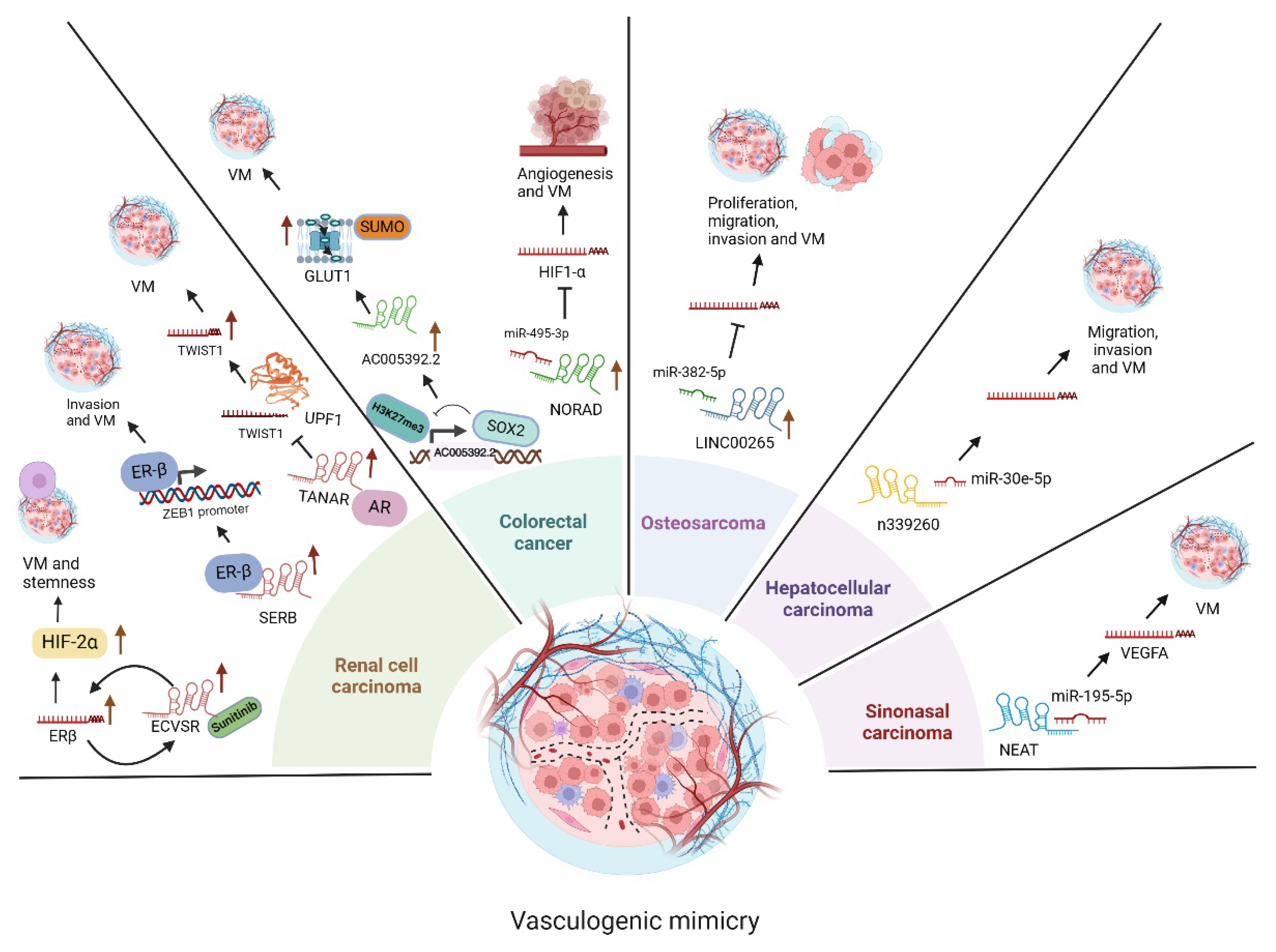
3.9. LncRNA Functions in Sinonasal Cancer

SNSCC is the most common sinonasal cancer, accounting for <5% of head and neck cancers with an incidence of 0.5-1.0 individuals per 100,000. It is a disease whose pathogenesis is poorly understood; some associated risk factors are exposure to various industrial compounds such as wood dust, glue, chromium, leather dust, nickel, formaldehyde, arsenic, various textile industry compounds, tobacco smoke, and VPH infection, which so far has no prognostic significance. Studies on genetic abnormalities in SNSCC are scarce and include EGFR overexpression, TP53 and KRAS mutations, and EGFR and SOX2 amplifications [53]. Some studies have implicated lncRNAs in the development of sinonasal cancers. However, only one study has evaluated the involvement of lncRNAs in the VM in this type of cancer (Figure 1). The lncRNA NEAT1 is overexpressed in SNSCC tissues, and its expression negatively correlates with miR-195 [54]. Downregulation of NEAT1 inhibits cell viability and the formation of three-dimensional channel-like structures while significantly increasing apoptosis. The expression of VEGF, p-PI3K, and p-AKT is decreased after NEAT1 silencing in SNCC cells, whereas overexpression of miR-195-5p produces the opposite effect. NEAT1 acts as a sponge for miR-195-5p to upregulate VEGFA expression, inducing the activation of the PI3K/AKT signaling pathway to promote SNSCC progression. In summary, lncRNA NEAT1 can activate VM in SNSCC cells via the NEAR/miR-195-2p/VEGFA axis in SNSCC [54].

4. Conclusions

Here, we have reviewed the functions of lncRNAs regulating VM in cancer. Data highlights the essential role of these non-coding RNAs in modulating genes that activate VM in diverse types of neoplasia. Remarkably, lncRNA signatures from cancer patients can be used as novel molecular biomarkers of progression, prognosis, and VM. However, exhaustive functional validation of the lncRNAs as oncogenes or tumor suppressors to targeting with inhibitors or precursors using in vivo disease models is still needed.

Author Contributions

Conceptualization, E.I.-S., M.B., C.E.V.-M C.P.P., and C.L.-C.; writing—original draft preparation, E.I.-S., M.B., C.E.V.-M C.P.P., and C.L.-C.; writing—review and editing, C.L.-C.; supervision, C.L.-C.; funding acquisition, C.L.-C. All authors have read and agreed to the published version of the manuscript.

Funding

This research was funded by Consejo Nacional de Humanidades Ciencia y Tecnologia (CONAHCYT), Mexico, Grant FOSSSIS A3-S-33674.

Institutional Review Board Statement

Not applicable.

Informed Consent Statement

Not applicable.

Data Availability Statement

Not applicable.

Acknowledgments

We acknowledge Universidad Autónoma de la Ciudad de México and CONAHCYT for their support.

Conflicts of Interest

The authors declare no conflict of interest.

References

  1. Tahara, S.; Rentsch, S.; De Faria, F. C. C.; Sarchet, P.; Karna, R.; Calore, F.; Pollock, R. E. Three-dimensional models. Oncol Res. 2024, 33, 1–13. [Google Scholar] [CrossRef] [PubMed]
  2. Hendrix, M. J.; Seftor, E. A.; Hess, A. R.; Seftor, R. E. Vasculogenic mimicry and tumour-cell plasticity: lessons from melanoma. Nat Rev Cancer. Nature reviews. 2003, 411–421. [Google Scholar] [CrossRef]
  3. Pezzella, F.; Ribatti, D. Vascular co-option and vasculogenic mimicry mediate resistance to antiangiogenic strategies. Cancer reports 2022, 5, e1318. [Google Scholar] [CrossRef]
  4. van der Schaft, D. W.; Seftor, R. E.; Seftor, E. A.; Hess, A. R.; Gruman, L. M.; Kirschmann, D. A.; Yokoyama, Y.; Griffioen, A. W.; Hendrix, M. J. Effects of angiogenesis inhibitors on vascular network formation by human endothelial and melanoma cells. Journal of the National Cancer Institute. 2004, 96, 1473–1477. [Google Scholar] [CrossRef] [PubMed]
  5. García-Hernández, A. P.; Sánchez-Sánchez, G.; Carlos-Reyes, A.; López-Camarillo, C. Functional roles of microRNAs in vasculogenic mimicry and resistance to therapy in human cancers: an update. Expert review of clinical immunology. 2024, 20, 913–926. [Google Scholar] [CrossRef]
  6. Ulitsky, I.; Bartel, D. P. lincRNAs: genomics, evolution, and mechanisms. Cell. 2013, 154, 26–46. [Google Scholar] [CrossRef]
  7. Chekulaeva, M.; Rajewsky, N. Roles of Long Noncoding RNAs and Circular RNAs in Translation. Cold Spring Harbor perspectives in biology. 2019, 11, a032680. [Google Scholar] [CrossRef]
  8. Kuska, B. Should scientists scrap the notion of junk DNA? J Natl Cancer Inst. 1998, 90, 1032–3. [Google Scholar] [CrossRef]
  9. Kopp, F.; Mendell, J. T. Functional Classification and Experimental Dissection of Long Noncoding RNAs. Cell. 2018, 172, 393–407. [Google Scholar] [CrossRef]
  10. Schmitt, A. M.; Chang, H. Y. Long Noncoding RNAs in Cancer Pathways. Cancer cell. 2016, 29, 452–463. [Google Scholar] [CrossRef]
  11. Guo, J.; Cai, H.; Liu, X.; Zheng, J.; Liu, Y.; Gong, W.; Chen, J.; Xi, Z.; Xue, Y. Long Non-coding RNA LINC00339 Stimulates Glioma Vasculogenic Mimicry Formation by Regulating the miR-539-5p/TWIST1/MMPs Axis. Molecular therapy. Nucleic acids. 2018, 10, 170–186. [Google Scholar] [CrossRef]
  12. Wu, Z.; Wei, N. METTL3-mediated HOTAIRM1 promotes vasculogenic mimicry icontributionsn glioma via regulating IGFBP2 expression. Journal of translational medicine. 2023, 21, 855. [Google Scholar] [CrossRef]
  13. Liu, Y.; Li, F.; Yang, Y. T.; Xu, X. D.; Chen, J. S.; Chen, T. L.; Chen, H. J.; Zhu, Y. B.; Lin, J. Y.; Li, Y.; et al. IGFBP2 promotes vasculogenic mimicry formation via regulating CD144 and MMP2 expression in glioma. Oncogene. 2019, 38, 1815–1831. [Google Scholar] [CrossRef] [PubMed]
  14. Li, H.; Wang, D.; Yi, B.; Cai, H.; Wang, Y.; Lou, X.; Xi, Z.; Li, Z. SUMOylation of IGF2BP2 promotes vasculogenic mimicry of glioma via regulating OIP5-AS1/miR-495-3p axis. International journal of biological sciences. 2021, 17, 2912–2930. [Google Scholar] [CrossRef]
  15. Yi, B.; Li, H.; Cai, H.; Lou, X.; Yu, M.; Li, Z. LOXL1-AS1 communicating with TIAR modulates vasculogenic mimicry in glioma via regulation of the miR-374b-5p/MMP14 axis. Journal of cellular and molecular medicine. 2022, 26, 475–490. [Google Scholar] [CrossRef] [PubMed]
  16. Yin, T.; Wu, J.; Hu, Y.; Zhang, M.; He, J. Long non-coding RNA HULC stimulates the epithelial-mesenchymal transition process and vasculogenic mimicry in human glioblastoma. Cancer medicine. 2021, 10, 5270–5282. [Google Scholar] [CrossRef]
  17. Yu, S.; Ruan, X.; Liu, X.; Zhang, F.; Wang, D.; Liu, Y.; Yang, C.; Shao, L.; Liu, Q.; Zhu, L.; et al. HNRNPD interacts with ZHX2 regulating the vasculogenic mimicry formation of glioma cells via linc00707/miR-651-3p/SP2 axis. Cell death & disease 2021, 12, 153. [Google Scholar]
  18. Jing, F.; Ruan, X.; Liu, X.; Yang, C.; Wang, D.; Zheng, J.; Xue, Y.; Shen, S.; Shao, L.; Yang, Y.; et al. The PABPC5/HCG15/ZNF331 Feedback Loop Regulates Vasculogenic Mimicry of Glioma via STAU1-Mediated mRNA Decay. Molecular therapy oncolytics. 2020, 17, 216–231. [Google Scholar] [CrossRef]
  19. Li, X.; Xue, Y.; Liu, X.; Zheng, J.; Shen, S.; Yang, C.; Chen, J.; Li, Z.; Liu, L.; Ma, J.; et al. ZRANB2/SNHG20/FOXK1 Axis regulates Vasculogenic mimicry formation in glioma. Journal of experimental & clinical cancer research 2019, 38, 68. [Google Scholar]
  20. Akbari Kordkheyli, V.; Rashidi, M.; Shokri, Y.; Fallahpour, S.; Variji, A.; Nabipour Ghara, E.; Hosseini, S. M. CRISPER/CAS System, a Novel Tool of Targeted Therapy of Drug-Resistant Lung Cancer. Advanced pharmaceutical bulletin. 2022, 12, 262–273. [Google Scholar] [CrossRef]
  21. Marshall, H. M., Fong, K. M. Lung cancer screening - Time for an update? Lung cancer (Amsterdam, Netherlands). 2024, 196, 107956.
  22. Li, D.; Shen, Y.; Ren, H.; Wang, L.; Yang, J.; Wang, Y. Repression of linc01555 up-regulates angiomotin-p130 via the microRNA-122-5p/clic1 axis to impact vasculogenic mimicry-mediated chemotherapy resistance in small cell lung cancer. Cell cycle. 2023, 22, 255–268. [Google Scholar] [CrossRef]
  23. Peng, Z.; Wang, J.; Shan, B.; Li, B.; Peng, W.; Dong, Y.; Shi, W.; Zhao, W.; He, D.; Duan, M.; et al. The long noncoding RNA LINC00312 induces lung adenocarcinoma migration and vasculogenic mimicry through directly binding YBX1. Molecular cancer 2018, 17, 167. [Google Scholar] [CrossRef] [PubMed]
  24. Zhang, Y.; Yuan, J.; Guo, M.; Xiang, R.; Xie, T.; Zhuang, X.; Dai, W.; Li, Q.; Lai, Q. Upregulation of long intergenic non-coding RNA LINC00326 inhibits non-small cell lung carcinoma progression by blocking Wnt/β-catenin pathway through modulating the miR-657/dickkopf WNT signaling pathway inhibitor 2 axis. Biology direct. 2023, 18, 3. [Google Scholar] [CrossRef] [PubMed]
  25. Yu, W.; Ding, J.; He, M.; Chen, Y.; Wang, R.; Han, Z.; Xing, E. Z.; Zhang, C.; Yeh, S. Estrogen receptor β promotes the vasculogenic mimicry (VM) and cell invasion via altering the lncRNA-MALAT1/miR-145-5p/NEDD9 signals in lung cancer. Oncogene. 2019, 38, 1225–1238. [Google Scholar] [CrossRef]
  26. Lai, Q.; Wan, Y.; Zhang, Y.; Huang, Y.; Tang, Q.; Chen, M.; Li, Q.; Ma, K.; Xiao, P.; Luo, C. Hypomethylation-associated LINC00987 downregulation induced lung adenocarcinoma progression by inhibiting the phosphorylation-mediated degradation of SND1. Molecular carcinogenesis. 2024, 63, 1260–1274. [Google Scholar] [CrossRef] [PubMed]
  27. Blancas-Zugarazo, S. S.; Langley, E.; Hidalgo-Miranda, A. Exosomal lncRNAs as regulators of breast cancer chemoresistance and metastasis and their potential use as biomarkers. Frontiers in oncology. 2024, 14, 1419808. [Google Scholar] [CrossRef]
  28. Cui, Y.; Pu, M.; Gong, Y.; Li, R.; Wang, X.; Ye, J.; Huang, H.; Liao, D.; Yang, Y.; Yin, A. METTL3-driven m6A modification of lncRNA FAM230B suppresses ferroptosis by modulating miR-27a-5p/BTF3 axis in gastric cancer. Biochimica et biophysica acta. General subjects 2024, 1868, 130714. [Google Scholar] [CrossRef]
  29. Chen, J.; Wang, Y.; Wu, M.; Yu, K.; Liu, J.; Chang, J. Vasculogenic mimicry triggers early recidivation and resistance to adjuvant therapy in esophageal cancer. BMC cancer, 2024, 24, 1132.
  30. Wang, H.; Ding, Q.; Zhou, H.; Huang, C.; Liu, G.; Zhao, X.; Cheng, Z.; You, X. Dihydroartemisinin inhibited vasculogenic mimicry in gastric cancer through the FGF2/FGFR1 signaling pathway. Phytomedicine : international journal of phytotherapy and phytopharmacology. 2024, 134, 155962.
  31. Li, Y.; Wu, Z.; Yuan, J.; Sun, L.; Lin, L.; Huang, N.; Bin, J.; Liao, Y.; Liao, W. Long non-coding RNA MALAT1 promotes gastric cancer tumorigenicity and metastasis by regulating vasculogenic mimicry and angiogenesis. Cancer letters, 2017, 395, 31–44.
  32. Zhao, J.; Wu, J.; Qin, Y.; Zhang, W.; Huang, G.; Qin, LLncRNA PVT1 induces aggressive vasculogenic mimicry formation through activating the STAT3/Slug axis and epithelial-to-mesenchymal transition in gastric cancer. Cellular oncology, 2020, 43, 863–876.
  33. Zhang, J. Y.; Du, Y.; Gong, L. P.; Shao, Y. T.; Wen, J. Y.; Sun, L. P.; He, D.; Guo, J. R.; Chen, J. N.; Shao, C. K. EBV-Induced CXCL8 Upregulation Promotes Vasculogenic Mimicry in Gastric Carcinoma via NF-κB Signaling. Frontiers in cellular and infection microbiology. 2022, 12, 780416. [Google Scholar] [CrossRef] [PubMed]
  34. Lu, Y.; Yang, B.; Shen, A.; Yu, K.; Ma, M.; Li, Y.; Wang, H. LncRNA UCA1 promotes vasculogenic mimicry by targeting miR-1-3p in gastric cancer. Carcinogenesis. 2024, 45, 658–672. [Google Scholar] [CrossRef]
  35. Bray, F.; Laversanne, M.; Sung, H.; Ferlay, J.; Siegel, R. L.; Soerjomataram, I.; Jemal, A. Global cancer statistics 2022: GLOBOCAN estimates of incidence and mortality worldwide for 36 cancers in 185 countries. CA: a cancer journal for clinicians. 2024, 74, 229–263.
  36. Kondov, B.; Milenkovikj, Z.; Kondov, G.; Petrushevska, G.; Basheska, N.; Bogdanovska-Todorovska, M.; Tolevska, N.; Ivkovski, L. Presentation of the Molecular Subtypes of Breast Cancer Detected By Immunohistochemistry in Surgically Treated Patients. Open access Macedonian journal of medical sciences. 2018, 6, 961–967. [Google Scholar] [CrossRef] [PubMed]
  37. Lozano-Romero, A.; Astudillo-de la Vega, H.; Terrones-Gurrola, M. C. D. R.; Marchat, L. A.; Hernández-Sotelo, D.; Salinas-Vera, Y. M.; Ramos-Payan, R.; Silva-Cázares, M. B.; Nuñez-Olvera, S. I.; Hernández-de la Cruz, O. N. HOX Transcript Antisense RNA HOTAIR Abrogates Vasculogenic Mimicry by Targeting the AngiomiR-204/FAK Axis in Triple Negative Breast Cancer Cells. Non-coding RNA, 2020 6(2), 19.
  38. Tao, W.; Sun, W.; Zhu, H.; Zhang, J. Knockdown of long non-coding RNA TP73-AS1 suppresses triple negative breast cancer cell vasculogenic mimicry by targeting miR-490-3p/TWIST1 axis. Biochemical and biophysical research communications. 2028, 504, 629–634. [Google Scholar] [CrossRef] [PubMed]
  39. Zhang, D.; Sun, B.; Zhao, X.; Ma, Y.; Ji, R.; Gu, Q.; Dong, X.; Li, J.; Liu, F.; Jia, X.; et al. Twist1 expression induced by sunitinib accelerates tumor cell vasculogenic mimicry by increasing the population of CD133+ cells in triple-negative breast cancer. Molecular cancer. 2014, 13, 207. [Google Scholar] [CrossRef] [PubMed]
  40. Khandpur, U.; Haile, B.; Makary, M. S. Early-Stage Renal Cell Carcinoma Locoregional Therapies: Current Approaches and Future Directions. Clinical Medicine Insights. Oncology. 2024, 18, 11795549241285390. [Google Scholar] [CrossRef]
  41. He, M.; Yang, H.; Shi, H.; Hu, Y.; Chang, C.; Liu, S.; Yeh, S. Sunitinib increases the cancer stem cells and vasculogenic mimicry formation via modulating the lncRNA-ECVSR/ERβ/Hif2-α signaling. Cancer letters. 2022, 524, 15–28. [Google Scholar] [CrossRef] [PubMed]
  42. Tang, S.; Chen, F.; Zhang, J.; Chang, F.; Lv, Z.; Li, K.; Li, S.; Hu, Y.; Yeh, S. LncRNA-SERB promotes vasculogenic mimicry (VM) formation and tumor metastasis in renal cell carcinoma. The Journal of biological chemistry. 2024, 300, 107297. [Google Scholar] [CrossRef] [PubMed]
  43. You, B.; Sun, Y.; Luo, J.; Wang, K.; Liu, Q.; Fang, R.; Liu, B.; Chou, F.; Wang, R.; Meng, J. Androgen receptor promotes renal cell carcinoma (RCC) vasculogenic mimicry (VM) via altering TWIST1 nonsense-mediated decay through lncRNA-TANAR. Oncogene. 2021, 40, 1674–1689. [Google Scholar] [CrossRef]
  44. Hu, S.; Han, X.; Liu, G.; Wang, S. LncRNAs as potential prognosis/diagnosis markers and factors driving drug resistance of osteosarcoma, a review. Frontiers in endocrinology. 2024, 15, 1415722. [Google Scholar] [CrossRef]
  45. Frankenbach-Désor, T.; Niesner, I.; Ahmed, P.; Dürr, H. R.; Klein, A.; Knösel, T.; Gospos, J.; McGovern, J. A.; Hutmacher, D. W.; Holzapfel, B. M. Tissue-engineered patient-derived osteosarcoma models dissecting tumour-bone interactions. Cancer metastasis reviews. 2024, 44, 8. [Google Scholar] [CrossRef]
  46. Xiao, Y.; Li, C.; Wang, H.; Liu, Y. LINC00265 targets miR-382-5p to regulate SAT1, VAV3 and angiogenesis in osteosarcoma. Aging. 2020, 12, 20212–20225. [Google Scholar] [CrossRef]
  47. Lin, Y.; Zhao, W.; Lv, Z.; Xie, H.; Li, Y.; Zhang, Z. The functions and mechanisms of long non-coding RNA in colorectal cancer. Frontiers in oncology. 2024, 14. [Google Scholar] [CrossRef] [PubMed]
  48. Saha, S.; Ghosh, S.; Ghosh, S.; Nandi, S.; Nayak, A. Unraveling the complexities of colorectal cancer and its promising therapies - An updated review. International immunopharmacology. 2024, 143(Pt 1) Pt 1, 113325. [Google Scholar] [CrossRef]
  49. Huang, S.; Wang, X.; Zhu, Y.; Wang, Y.; Chen, J.; Zheng, H. SOX2 promotes vasculogenic mimicry by accelerating glycolysis via the lncRNA AC005392.2-GLUT1 axis in colorectal cancer. Cell death & disease. 2023, 14, 791.
  50. Zhang, L.; Wu, H.; Zhang, Y.; Xiao, X.; Chu, F., Zhang, L. Induction of lncRNA NORAD accounts for hypoxia-induced chemoresistance and vasculogenic mimicry in colorectal cancer by sponging the miR-495-3p/ hypoxia-inducible factor-1α (HIF-1α). Bioengineered. 2022, 13, 950–962.
  51. Zhang, X., Wang, H., Yuan, Y., Zhang, J., Yang, J., Zhang, L., & He, J. PPM1G and its diagnostic, prognostic and therapeutic potential in HCC (Review). International journal of oncology. 2024, 65, 109.
  52. Liu, T., Liao, S., Mo, J., Bai, X., Li, Y., Zhang, Y., Zhang, D., Cheng, R., Zhao, N., Che, N., Guo, Y., Dong, X., & Zhao, X. LncRNA n339260 functions in hepatocellular carcinoma progression via regulation of miRNA30e-5p/TP53INP1 expression. Journal of gastroenterology. 2022, 57, 784–797.
  53. Thawani, R.; Kim, M. S.; Arastu, A.; Feng, Z.; West, M. T.; Taflin, N. F.; Thein, K. Z.; Li, R.; Geltzeiler, M.; Lee, N. The contemporary management of cancers of the sinonasal tract in adults. CA: a cancer journal for clinicians. 2023, 73, 72–112.
  54. Lu, H.; Kang, F. Down-regulating NEAT1 inhibited the viability and vasculogenic mimicry formation of sinonasal squamous cell carcinoma cells via miR-195-5p/VEGFA axis. Bioscience reports. 2020, 40, BSR20201373. [Google Scholar] [CrossRef]
Figure 1. LncRNAs overexpressed in glioma, breast, lung, and gastric cancers associated with vasculogenic mimicry. The lncRNAs and sponging microRNAs, as well as the mRNAs targets of microRNAs, are indicated. Also, the cancer hallmarks regulated by the lncRNA/microRNA/mRNA axes are denoted. Created in BioRender.
Preprints 146852 g001
Disclaimer/Publisher’s Note: The statements, opinions and data contained in all publications are solely those of the individual author(s) and contributor(s) and not of MDPI and/or the editor(s). MDPI and/or the editor(s) disclaim responsibility for any injury to people or property resulting from any ideas, methods, instructions or products referred to in the content.
Copyright: This open access article is published under a Creative Commons CC BY 4.0 license, which permit the free download, distribution, and reuse, provided that the author and preprint are cited in any reuse.
Prerpints.org logo

Preprints.org is a free preprint server supported by MDPI in Basel, Switzerland.

Subscribe

Disclaimer

Terms of Use

Privacy Policy

Privacy Settings

© 2025 MDPI (Basel, Switzerland) unless otherwise stated