Submitted:
21 January 2025
Posted:
22 January 2025
Read the latest preprint version here
Abstract
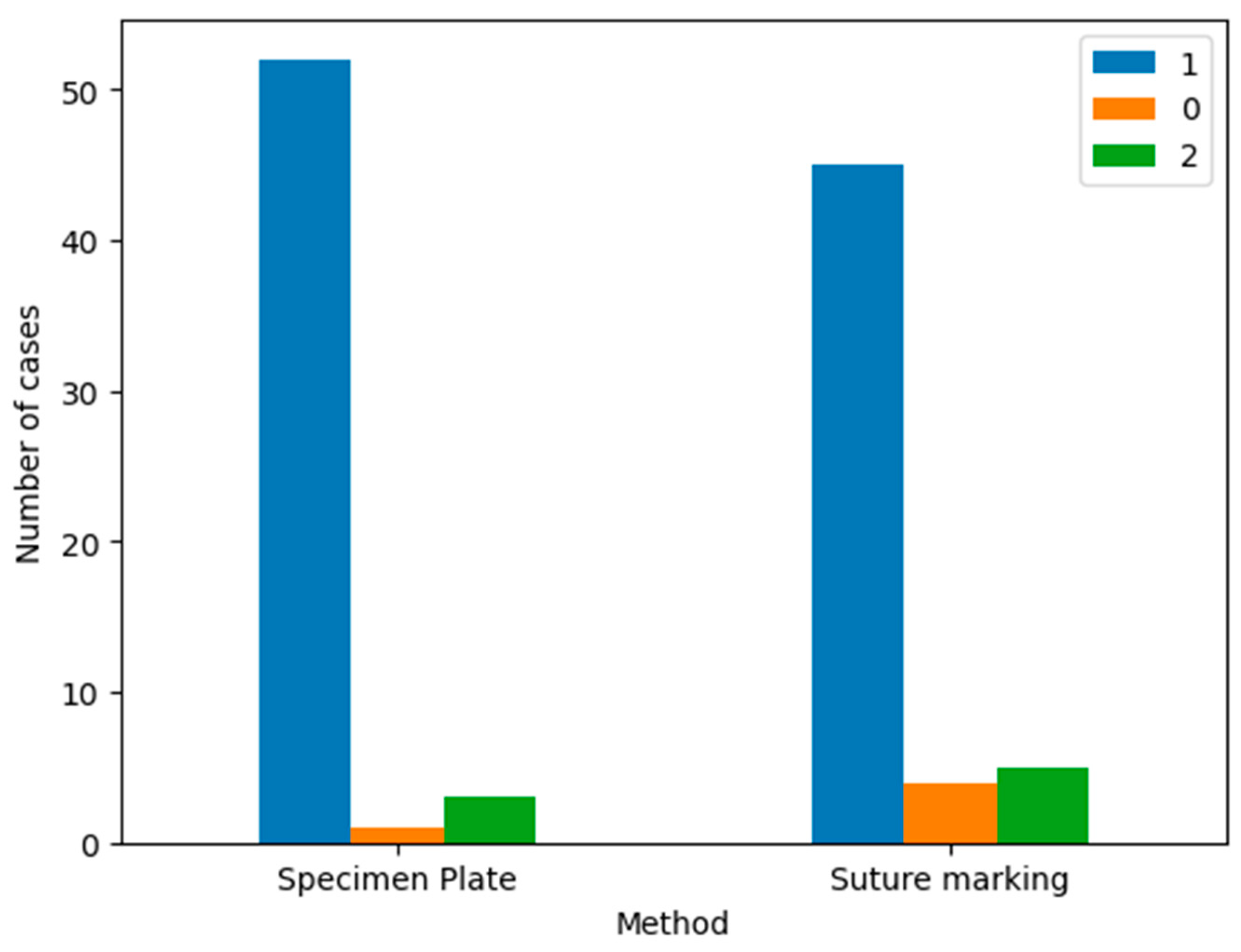
Background: Accurate orientation of resected breast specimens is crucial for proper pathological evaluation and reliable margin assessment. Misorientation can compromise margin analysis, potentially leading to imprecise re-excisions and increasing the risk of local recurrence. This study aims to evaluate a new specimen plate designed to maintain consistent tissue orientation and compare its effectiveness with traditional suture marking techniques. Methods: A single-center, prospective, randomized two-arm trial compared the efficacy of the novel specimen plate (n=56) with conventional suture marking (n=54) during pathological examinations. The study assessed the impact on specimen handling, intraoperative imaging interpretation, and pathological outcomes, focusing on orientation clarity, margin assessment, and ease of tissue evaluation. Results: The specimen plate demonstrated superior performance in maintaining clear orientation during intraoperative imaging and facilitated accurate margin assessment, significantly lowering (p<0.01) misorientation rate compared to suture marking. Pathologists reported improved ease at identifying direction and extent of tumor-free zones and enhanced diagnostic accuracy. Number of non-R0 resections requiring re-excisions was significantly lower in the specimen plate group in comparation with the suture markings (8,9% vs. 22,2%) indicating the former’s potential to improve surgical outcomes. Conclusions: The novel specimen plate offers a reliable solution for improving specimen orientation in breast cancer surgery. By ensuring consistent orientation and enhancing diagnostic clarity, it can contribute to better surgical accuracy, reduce re-excisions, and ultimately improve patient safety. These findings support the specimen plate as an effective tool for enhancing outcomes in breast cancer treatment.
Keywords:
1. Introduction
2. Patients and Methods
2.1. Study Design
2.2. Patient Selection and Randomization
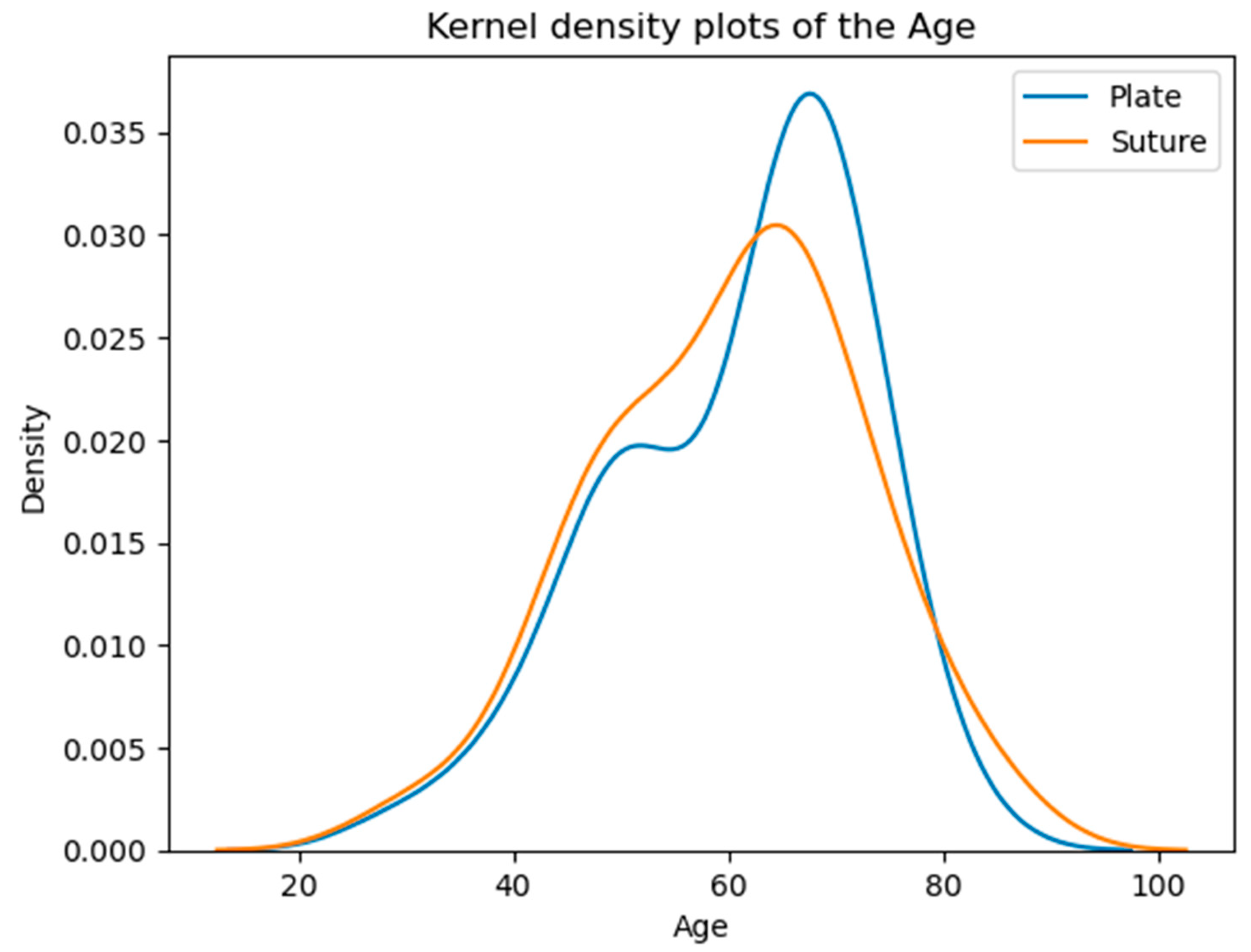
2.3. Ages of patients in the examined groups at the time of the operation.
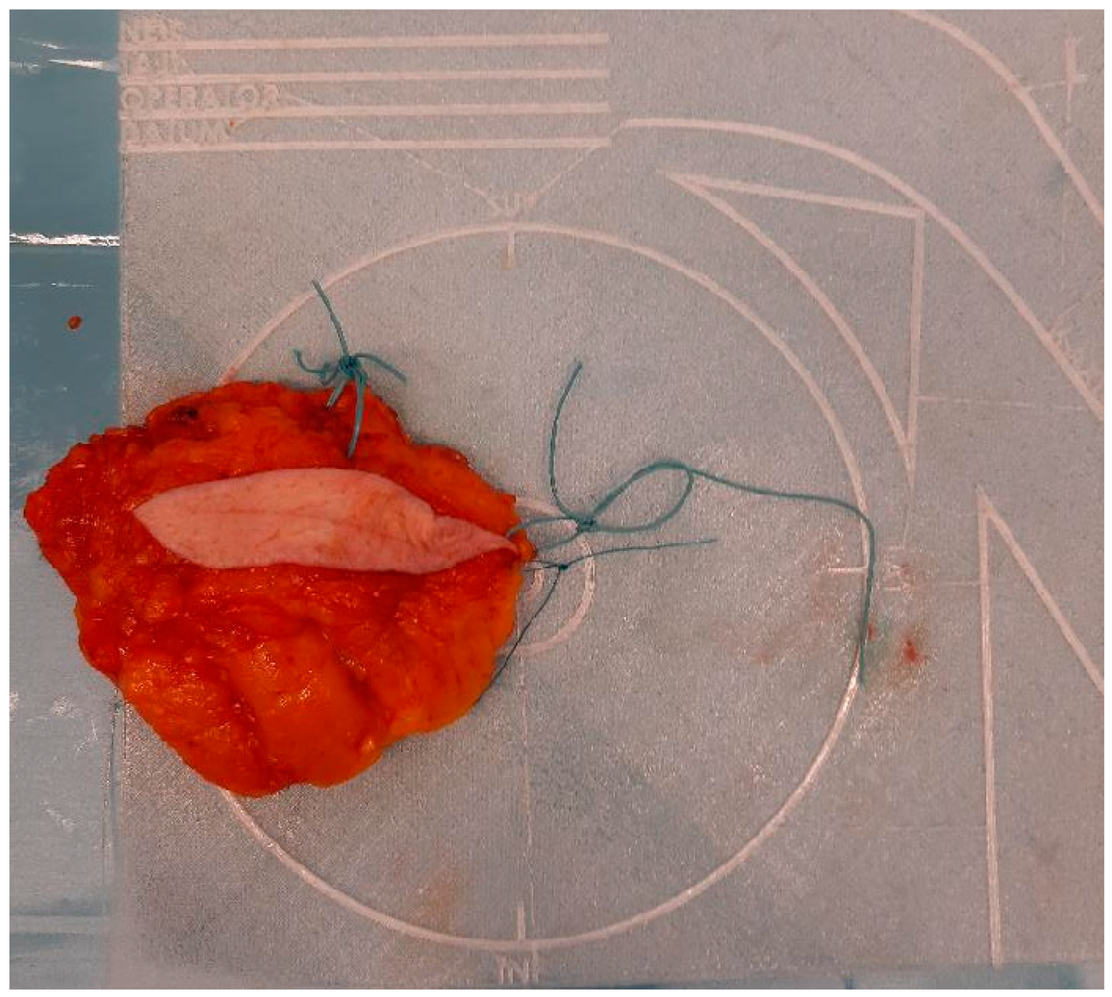
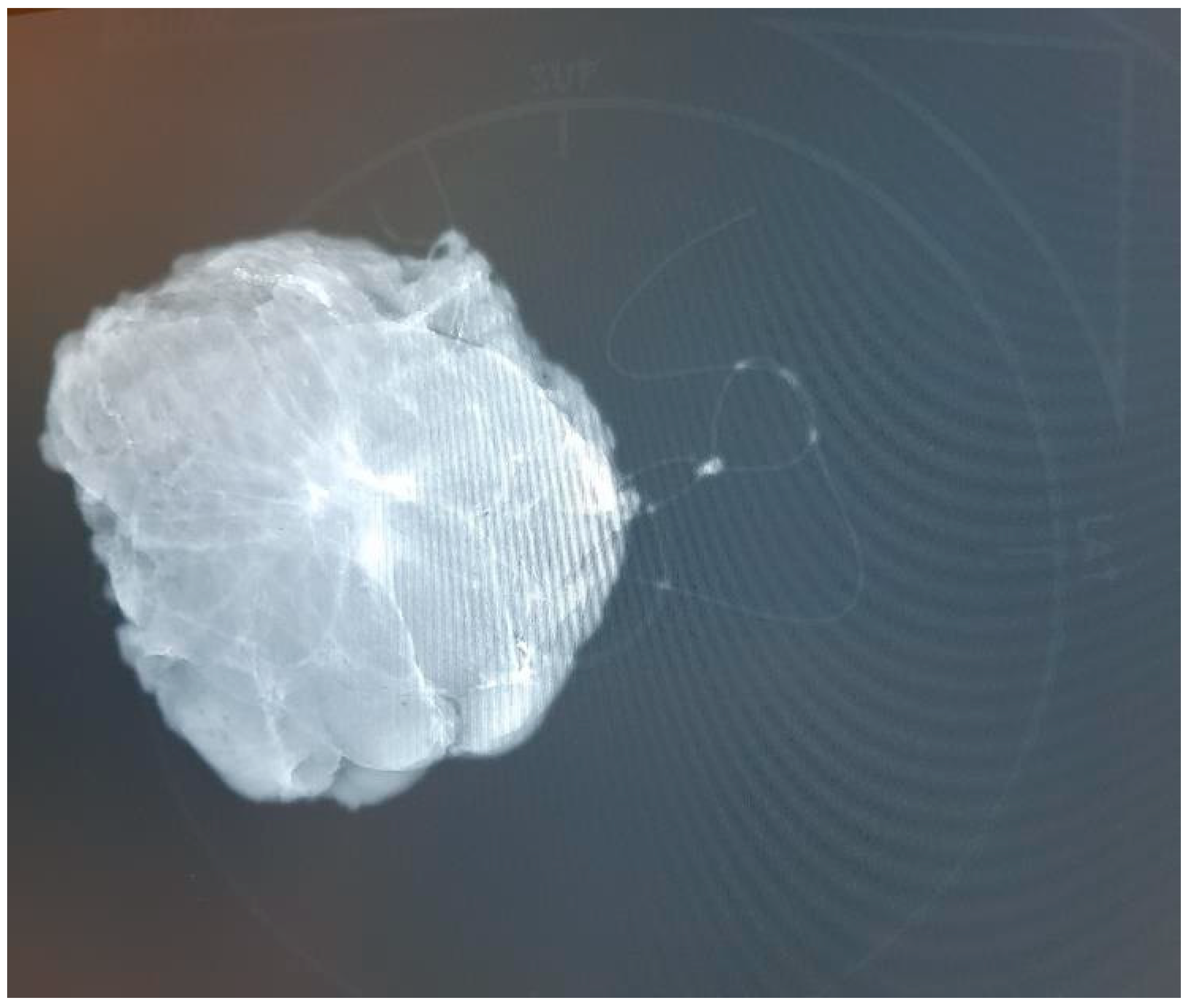
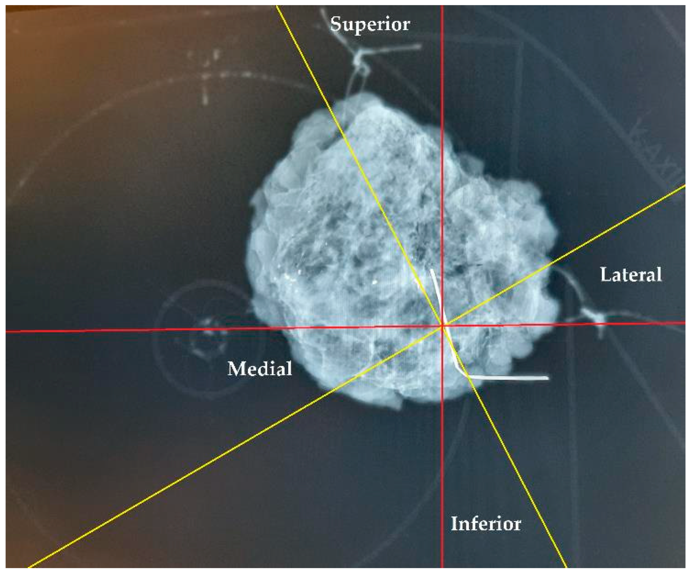
2.4. Specimen Orientation Assessment
2.5. Statistical analyses
2.5. Criteria for Assessing Specimen Orientation
3. Results
3.1. Specimen Orientation and Tumor Localization
3.2. Orientation Accuracy and Reliability – Comparison of Specimen Plate and Suture Marking Methods
4. Discussion
5. Conclusions
6. Patents
Author Contributions
Funding
Institutional Review Board Statement
Informed Consent Statement
Data Availability Statement
Acknowledgments
Conflicts of Interest
References
- Burstein, H.J.; Curigliano, G.; Loibl, S.; Dubsky, P.; Gnant, M.; Poortmans, P.; Colleoni, M.; Denkert, C.; Piccart-Gebhart, M.; Regan, M.; et al. Estimating the benefits of therapy for early-stage breast cancer: The St. Gallen International Consensus Guidelines for the primary therapy of early breast cancer 2019. Ann. Oncol. 2019, 30, 1541–1557. [Google Scholar] [CrossRef] [PubMed]
- Volleamere, A.J.; Kirwan, C.C. National survey of breast cancer specimen orientation marking systems. Eur. J. Surg. Oncol. 2013, 39, 255–259. [Google Scholar] [CrossRef]
- Drozgyik, A.; Szabó, T.; Kovács, G.; Kollár, D.; Molnár, T.F. A New Approach to Breast Specimen Orientation: Avoiding Pitfalls with the Specimen Plate Concept. Curr. Oncol. 2024, 31, 4589–4598. [Google Scholar] [CrossRef] [PubMed]
- Benjamin, M.A.; Sinnott, C.; Bawa, S.; Kaufman, D.I.; Guarino, K.; Addona, T. Re-excision rate after partial mastectomy in oncoplastic breast-conserving surgery: A single-institutional experience and review of the literature. Ann. Plast. Surg. 2019, 82, S170–S172. [Google Scholar] [CrossRef] [PubMed]
- McCahill, L.E.; Single, R.M.; Aiello Bowles, E.J.; Feigelson, H.S.; James, T.A.; Barney, T.; Engel, J.M.; Onitilo, A.A. Variability in reexcision following breast conservation surgery. JAMA 2012, 307, 467–475. [Google Scholar] [CrossRef] [PubMed]
- Tang, S.S.; Kaptanis, S.; Haddow, J.B.; Mondani, G.; Elsberger, B.; Tasoulis, M.K.; Obondo, C.; Johns, N.; Ismail, W.; Syed, A.; et al. Current margin practice and effect on re-excision rates following the publication of the SSO-ASTRO consensus and ABS consensus guidelines: A national prospective study of 2858 women undergoing breast-conserving therapy in the UK and Ireland. Eur. J. Cancer 2017, 84, 315–324. [Google Scholar] [CrossRef] [PubMed]
- Ahmed, G.A.; Baron, D.H.; Agrawal, A. Oncologic and cosmetic outcomes of oncoplastic breast-conserving surgery after neoadjuvant systemic therapy: Systematic review and meta-analysis. Breast Cancer Res. Treat. 2024. [Google Scholar] [CrossRef] [PubMed]
- Benjamin, M.A.; Sinnott, C.; Bawa, S.; Kaufman, D.I.; Guarino, K.; Addona, T. Re-excision rate after partial mastectomy in oncoplastic breast-conserving surgery: A single-institutional experience and review of the literature. Ann. Plast. Surg. 2019, 82, S170–S172. [Google Scholar] [CrossRef] [PubMed]
- Fregatti, P.; Gipponi, M.; Atzori, G.; De Rosa, R.; Diaz, R.; Cornacchia, C.; Sparavigna, M.; Garlaschi, A.; Belgioia, L.; Fozza, A.; et al. The margins’ challenge: Risk factors of residual disease after breast-conserving surgery in early-stage breast cancer. In Vivo 2022, 36, 814–820. [Google Scholar] [CrossRef] [PubMed]
- Schulman, A.M.; Mirrielees, J.A.; Leverson, G.; Landercasper, J.; Greenberg, C.; Wilke, L.G. Reexcision surgery for breast cancer: An analysis of the American Society of Breast Surgeons (ASBrS) MasterySM database following the SSO-ASTRO "no ink on tumor" guidelines. Ann. Surg. Oncol. 2017, 24, 52–58. [Google Scholar] [CrossRef] [PubMed]
- Banys-Paluchowski, M.; Kühn, T.; Masannat, Y.; Rubio, I.; de Boniface, J.; Ditsch, N.; Karadeniz Cakmak, G.; Karakatsanis, A.; Dave, R.; Hahn, M.; et al. Localization techniques for non-palpable breast lesions: Current status, knowledge gaps, and rationale for the MELODY study (EUBREAST-4/iBRA-NET, NCT 05559411). Cancers 2023, 15, 1173. [Google Scholar] [CrossRef]
- Zacharioudakis, K.; Down, S.; Bholah, Z.; Lee, S.; Khan, T.; Maxwell, A.J.; Douek, M. Is the future magnetic?—Magseed localization for non-palpable breast cancer: A multicenter nonrandomized control study. Eur. J. Surg. Oncol. 2019, 45, 2016–2021. [Google Scholar] [CrossRef]
- Di Leone, A.; Franceschini, G.; Mason, E.J.; D’Archi, S.; D’Angelo, A.; Scardina, L.; Sanchez, A.M.; Conti, M.; Trombadori, C.; Terribile, D.A.; et al. Image-guided localization techniques for surgical excision of non-palpable breast lesions: An overview of current literature and our experience with preoperative skin tattoo. J. Pers. Med. 2021, 11, 99. [Google Scholar] [CrossRef] [PubMed]
- Cheung, B.H.H.; Co, M.; Lui, T.T.N.; Kwong, A. Evolution of localization methods for non-palpable breast lesions: A literature review from a translational medicine perspective. Transl. Breast Cancer Res. 2024, 5, 12. [Google Scholar] [CrossRef] [PubMed]

| Group | Mean Age (years) | Median Age (years) |
| Specimen Plate Group | 61.1 | 64.0 |
| Suture Marking Group | 60.2 | 62.5 |
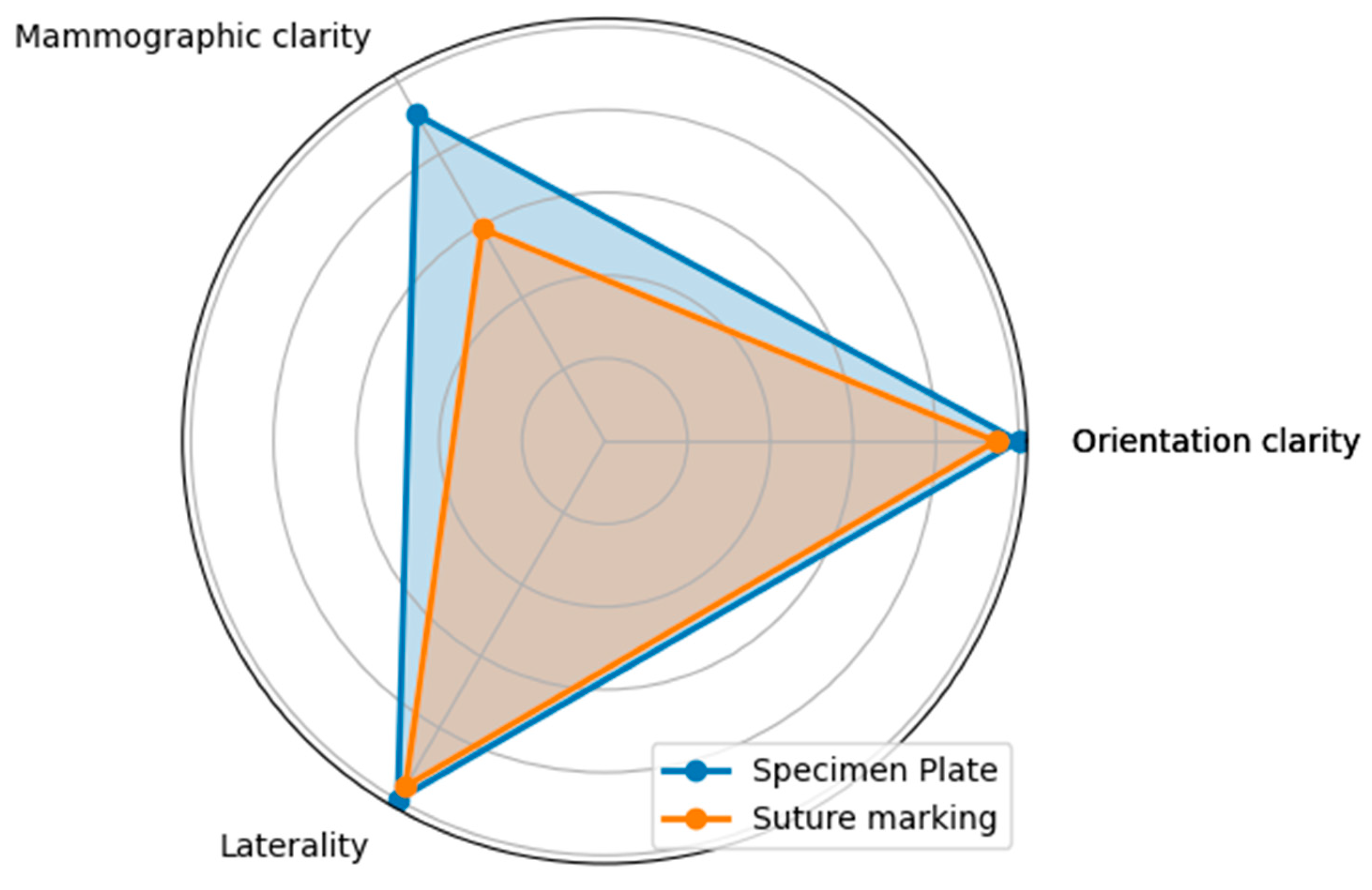
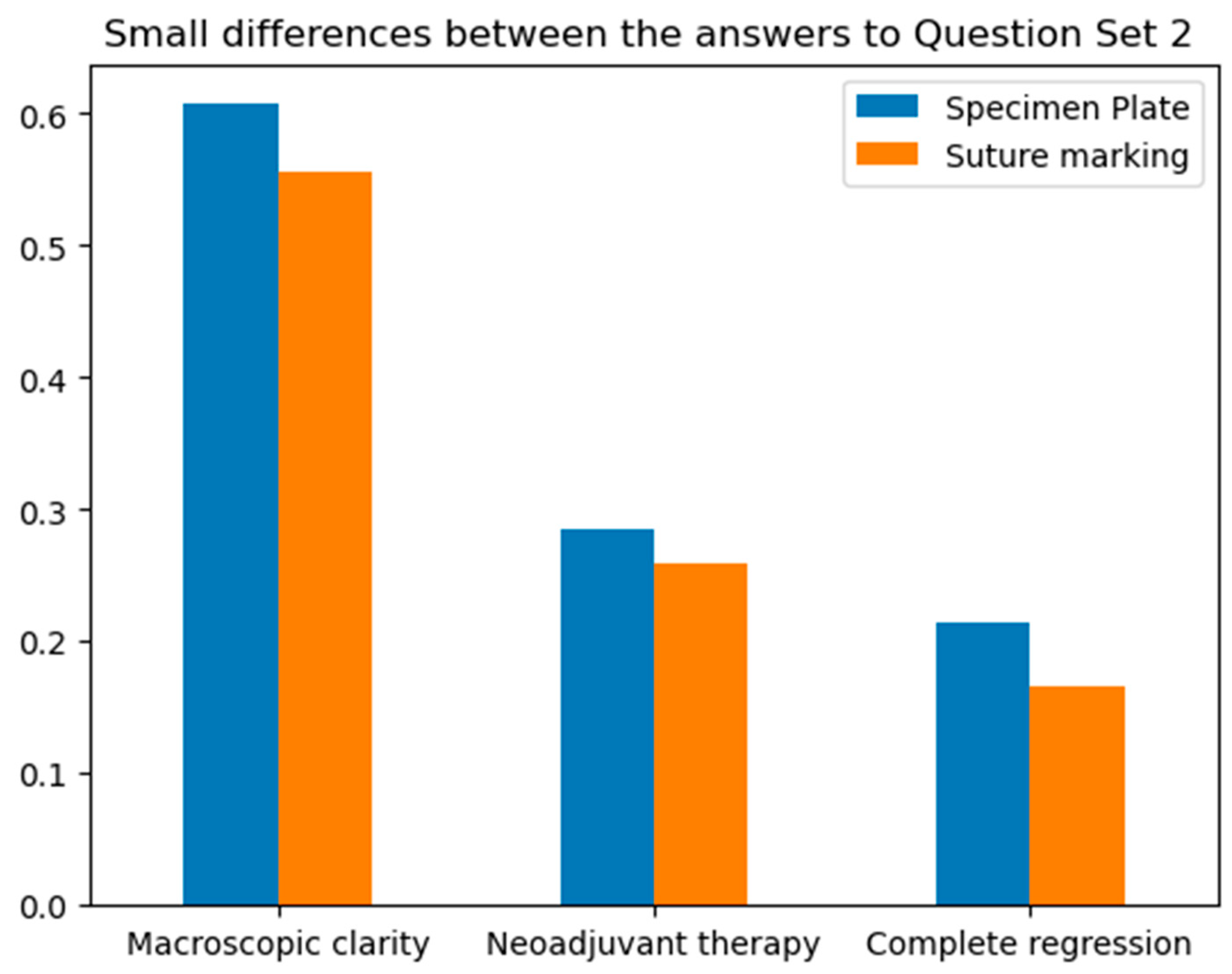
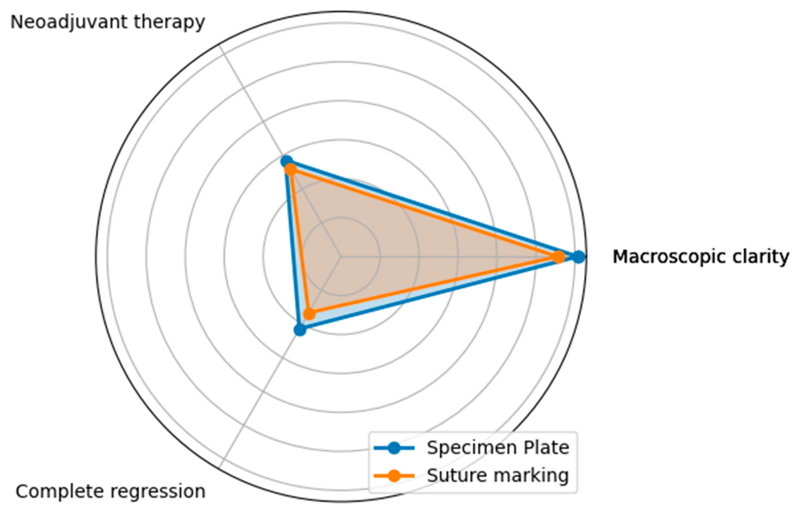
| Criteria | Specimen Plate Group (n=56) | Suture Marking Group (n=54) |
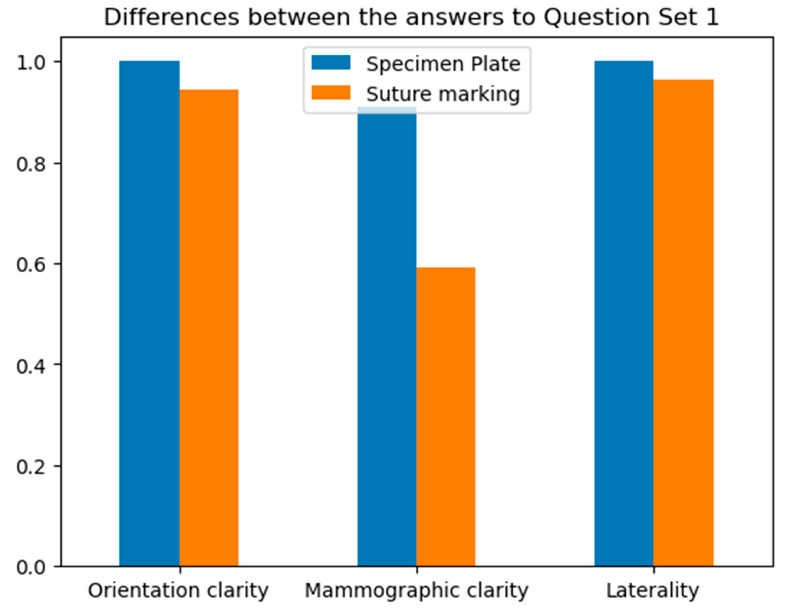
| Clear orientation upon arrival to Pathology | 100% | 96.3% (52 cases) |
| Clear tumor localization (macroscopic) | 60.71% (34 cases) | 44.44% (24 cases) |
| Clear laterality | 100% | 96.3%(52 cases) |
| Possibility of 180-degree rotation | 0 | 0 |
| Clear mammographic orientation | 80.4% (45 cases) | 13% (7 cases) |
| Successful R0 resection | 91.1% (51 cases) | 77.8% (42 cases) |
Disclaimer/Publisher’s Note: The statements, opinions and data contained in all publications are solely those of the individual author(s) and contributor(s) and not of MDPI and/or the editor(s). MDPI and/or the editor(s) disclaim responsibility for any injury to people or property resulting from any ideas, methods, instructions or products referred to in the content. |
© 2025 by the authors. Licensee MDPI, Basel, Switzerland. This article is an open access article distributed under the terms and conditions of the Creative Commons Attribution (CC BY) license (http://creativecommons.org/licenses/by/4.0/).


