
Submitted:
16 January 2025
Posted:
20 January 2025
You are already at the latest version
Abstract
The use of various nanoformulations for cancer radiotherapy has great potential due to their increased effectiveness and safety. At the stage of preclinical evaluation of such nanoformulations in vitro, they are tested on monolayers of tumor cell cultures, which is an unrealistic representation of tumors in vivo, which leads to an inaccurate selection of suitable drugs. To solve this problem, 3D cellular spheroids are becoming an additional tool for confirming the effectiveness of new therapeutic nanoformulations. Here we show the possibility of using citrate-stabilized cerium oxide nanoparticles (nanoceria, CeO2 NPs) as a nanoradiosensitizer in an experimental 3D model of tumor spheroids formed from 4T1 human breast cancer cells. It has been shown that ultra-small size (about 1-2 nm) CeO2 NPs have pH-sensitive radiocatalytic activity, increasing the generation of ROS under irradiation conditions. It has been shown that preloading of CeO2 NPs into a cellular spheroid at a micromolar concentrations does not adversely affect the rate of spheroid formation and its final size. However, such preloading has a pronounced inhibitory effect on the rate of cell migration from the spheroid after X-ray irradiation and also reduces their clonogenic activity. Thus, CeO2 NPs can be considered as a promising nanoradiosensitizer for radiotherapy of tumors.
Keywords:
nanoparticles
; 3D tumor spheroid
; cerium oxide
; irradiation
; redox activity
1. Introduction
Functional nanomaterials, due to their physico-chemical properties and unique biological activity, are widely used in various biomedical fields, opening up new therapeutic and diagnostic possibilities for cancer therapy [1]. There is a wide range of metal-containing nanoformulations that have already been introduced into clinical practice, providing both visualization and selective therapy of tumors [2]. For example, hafnium oxide (HfO2) nanoparticles are used as a radiosensitizer in the framework of radiation therapy of tumors [3], and iron oxide nanoparticles [4] are used to effectively contrast tumor nodes in the brain in combination with their therapy by remote heating of these nanoparticles in a magnetic field. Gadolinium-containing nanoformulations (AGuIX®) are used for imaging and subsequent radiotherapy of brain tumors [5]. These nanoparticles are a 5 nanometer macromolecular polysiloxane complexes containing 15 gadolinium atoms, which provides unique relaxation characteristics (8.9 mM-1s−1 per Gd3+ ion). AGuIX® are at the 3rd stage of clinical trials and show high efficacy in combination therapy with glioblastoma and gliosarcoma [6]. Thus, we can confidently say that biomedical nanotechnology is being actively introduced into clinical practice in the field of tumor radiotherapy.
Inorganic nanozymes belong to a new promising class of functional nanomaterials for biomedical purposes [7]. Despite the fact that natural enzymes have unprecedented advantages in neutralization of reactive oxygen species (ROS), such as high efficiency, specificity and selectivity, at the same time they also have low stability and inability to recover their activity, which, in turn, limits their use. Inorganic nanozymes are considered as the most promising substances for the effective correction of critical pathological conditions associated with oxidative stress [8]. The class of inorganic nanozymes includes nanomaterials based on manganese [9], vanadium [10], gold [11], platinum [12], iron [13], cerium [14] etc. Such compounds are able to mimic the activity of a wide range of endogenous enzymes, such as superoxide dismutase, catalase, peroxidase, phosphatase, etc. [15]. The most prominent representative of the class of inorganic nanozymes is nanocrystalline cerium oxide [14,16,17]. Ce3+ and Ce4+ ions coexist on the surface of the crystal lattice of cerium oxide nanoparticles, forming oxygen vacancies for the implementation of redox reactions [18]. CeO2 NPs are able to mimic the activity of enzymes such as superoxide dismutase (SOD) [19], catalase [20], phosphatase [21], peroxidase [22] etc. The non-stoichiometric ratio of Ce3+/Ce4+ on the surface determines the catalytic characteristics of cerium oxide nanoparticles and strongly depends on the synthesis methods [23], ion microenvironment [24] and рН [25]. CeO2 NPs can easily change and adjust its electronic configuration by reacting directly to the microenvironment, which allows it to exhibit antioxidant or prooxidant activity. CeO2 NPs is considered as a promising nanomaterial for use in oncotherapy of various types of tumors [26]. The use of CeO2 NPs in cancer treatment can be considered from the side of 4 main concepts: (1) Selective protection of normal cells from oxidative stress at physiological pH (antioxidant activity) or induction of oxidative stress in cancer cells by generation of ROS in a slightly acidic microenvironment of tumor tissue (prooxidant activity) [27]. (2) Generation of molecular oxygen to enhance oxygenation of hypoxified tumor sites and sensitization of tumor cells for photodynamic, photothermal and radiation therapy [28,29]. (3) Increased destruction of cancer cells through synergistic combination with other anti-cancer agents thar are already used in the clinic [30,31]. (4) Using as a nanocarrier to improve the delivery of anti-cancer drugs [32,33]. Thus, cerium-containing nanoparticles can be considered as promising antitumor agents and radiosensitizers. It is worth noting that cerium oxide nanoparticles can be easily additionally functionalized in order to give them multimodality. In particular, we have previously shown that the doping of CeO2 NPs with gadolinium ions gives them the properties of MRI contrast. Such nanoparticles have selective cytotoxic activity against MCF-7 tumor cells [34]. Controlled absorbtion of calcein onto the surface of a CeO2 NPs ensures the creation of a ROS-sensitive theranostic sensor, which allows for accurate visualization of the inactivation zone of intracellular ROS [35]. Cerium-containing nanoparticles are also used as luminescent nanosensors for analytical chemistry [36]. Given the unique biological activity and high degree of biocompatibility of cerium-containing nanomaterials, it is safe to talk about the prospects for their clinical implementation in the near future.
Cellular spheroids are a unique experimental model that allows to perform primary screening of potential antitumor agents and new promising compounds, including nanoformulations. The structure of the cellular spheroid makes it possible to simulate the conditions of the tumor cellular microenvironment as accurately as possible [37]. The spheroid has a structure close to the structure of the tumor node in vivo, modeling all zones: the proliferation zone, the hypoxia zone and the necrosis zone [38]. Spheroids are considered as a relevant model for testing the cytotoxicity [39] and biological activity of nanoparticles [40]. This experimental model makes it possible to conduct accelerated screening of potential radiosensitizers, successfully screening out ineffective and toxic compounds, which further ensures more effective preparation and conduct of preclinical studies on animal tumor models.
Here we demonstrate the radiosensitizing properties of citrate-stabilized CeO2 nanoparticles in a 3D tumor spheroid model under X-ray irradiation, discussing possible molecular mechanisms of radiation-induced cytotoxic action of cerium-containing nanoparticles.
2. Materials and Methods
2.1. Synthesis and Characterization of CeO2 NPs
CeO2 NPs were obtained by hydrothermal method. 5.88 g of Sodium citrate tribasic dihydrate was dissolved in 50 ml of water (solution 1). Separately, 3.73 g of cerium heptahydrate chloride was dissolved in 50 ml of water (solution 2). Solution 1 and solution 2 were mixed with active stirring on a magnetic stirrer. Then, without stopping stirring, 1.5 ml of 25% ammonia solution was added to the resulting solution. Further, the mixing was continued for 24 hours. After obtaining a dark brown sol of nanoparticles, it was dried at 80 ° C in a dry oven. The resulting powder formed a colloidal sol well when dissolved in water. The resulting СеО2 sol was analyzed by a wide range of methods. СеО2 NPs size and shape were determined using transmission electron microscopy on a Zeiss Leo912 AB Omega electron microscope at an accelerating voltage of 100 kV. A diffraction pattern reflecting the diffraction of electrons on nanoparticles in the sample was also obtained. The concentration of the СеО2 NPs sols was determined gravimetrically. A DS-11FX spectrophotometer (Denovix, USA) was used to measure СеО2 NPs sol absorbance in the UV-visible range. Measurements were carried out in the wavelength range from 220 to 600 nm in 0.1 nm increments. The size distribution of СеО2 NPs in deionized water, Hank’s Balanced Salt Solution (HBSS) (PanEco, Russia), cell culture medium DMEM/F-12 (1:1) (PanEco, Russia), containing 2 mM of L-glutamine, 100 U/mL of penicillin and 100 µg/mL of streptomycin (PanEco, Russia) with and without 10% of Fetal Bovine Serum (FBS) (HyClone, USA) were measured by Dynamic Light Scattering (DLS) at 25 °C using a BeNano 90 Zeta particle analyzer (BetterSize, China).
2.2. Detection of ROS Generation after X-Ray Irradiation of CeO2 NPs
2′,7′-Dichlorodihydrofluorescein diacetate (H2DCF-DA) dye was used as the probe for ROS level detection under X-ray irradiation. For this aim, 2 mM solution of H2DCF-DA in PBS was deacetylated to form H2DCF in 10 mM of NaOH water solution for 30 min at 4 °С. The H2DCF solution was stored on the ice and in the dark for further use. Oxidation of this dye by ROS leads to formation of fluorescent compound, 2′,7′-dichlorofluorescein (DCF). CeO2 NPs at 0-100 µg/mL concentration were mixed with cold solution of 8 µM H2DCF in 0.1 M Tris-HCl. Then, samples were placed on the ice and irradiated by X-ray irradtiation for 1 min at doses of 2 and 4 Gy. Then, CeO2 NPs were eliminated by centrifugation at 20 000 g for 20 min at 4 °C using D1524R microcentrifuge with refrigerator (DLab Scientific, China). Fluorescence intensity of the solutions was measured on a INNO S plate reader (LTEK, Korea) using FITC filter (Ex=467-498 nm, Em=513-556 nm). Then, four experimental groups were analyzed to measure the production of ROS: (1) non-treated H2DCF solution, (2) non-irradiated H2DCF solution treated with CeO2 NPs NPs, (3) H2DCF solution irradiated under X-ray, and (4) H2DCF solution both treated with CeO2 NPs NPs and irradiated under X-ray. ROS generation effectivity was determined as:
where Sx – mean fluorescence signal from the corresponding DCF solutions.
2.3. Cell Culture
4Т1 human breast cancer cell line was obtained from European Collection of Authenticated Cell Cultures (ECACC). 4Т1 cells were cultivated in DMEM/F-12 medium (1:1) (PanEco, Russia), containing 2 mM of L-glutamine, 100 U/mL of penicillin and 100 µg/mL of streptomycin (PanEco, Russia) and 10% of Fetal Bovine Serum (FBS) (HyClone, USA). The cells were cultivated in incubator at 37 °C in humidified atmosphere containing 5% CO2. As the cells grew and reached subconfluent state, they were treated with a 0.25% trypsin-EDTA (PanEco, Russia) solution and passed into new T25 or T75 cell culture flasks (SPL Life Sciences, Korea) at a ratio of 1:4.
2.4. Formation of Spheroids
3D tumor spheroids were formed from 4T1 cells by the hanging drop method for 72 hours. The seeding density of cells was 1000 cells per 1 spheroid. The diameter of the formed spheroids was about 350-370 µm.
2.5. Irradiation of Spheroids
X-ray irradiation of spheroids was carried out at the RUT-15 device (ITEB RAS, Pushchino) with a dose rate of 1 Gy/min. The spheroids were placed in a 2 ml tube in HBSS for the period of irradiation.
2.6. Analysis of Cell Migration from Irradiated Spheroid
After X-ray irradiation, the spheroids were transferred (1 piece per well) into the wells of a 24-well plate (SPL Life Sciences, Korea), photographed on a Clone Select Imager (Molecular devices, USA) at x6 magnification. The migration of cells from the spheroid was observed for 3 days. The analysis of the area of the cell migration zone from the spheroid was carried out using the ImageJ software.
2.7. Colony Assay
After X-ray irradiation, the spheroids were broken down into suspension of individual cells by incubation in a solution of 0.25% trypsin-EDTA for 1 hour. Then cells were seeded in 6-well plates (SPL Life Sciences, Korea) at concentration of 1000 cells per well in DMEM/F12 with 10% FBS culture medium and cultured in incubator at 37°C in humidified atmosphere containing 5% CO2. Colony formation was monitored daily using a CloneSelect Imager plate reader (Molecular Devices, USA). After the completion of colony formation in the control group (9 days), the cells were washed 3 times with phosphate buffered saline and fixed in 4% paraformaldehyde solution (Sigma Aldrich, USA), and then stained with 0.1% crystal violet (PanEko, Russia). Cell aggregations of more than 50 cells were considered as 1 colony. Colonies were counted manually using a magnifying glass.
2.8. Statistical Analysis
The experimental data are presented as the Mean ± Standard Deviation (SD). The statistical significance of differences between the values in experimental groups was determined using the Welch’s t-test in GraphPad Prism. Differences were considered statistically significant at p < 0.05 (*), p < 0.01 (**), p < 0.005 (***), p < 0.001 (****).
3. Results
Physico-Chemical Properties of CeO2 NPs
CeO2 NPs were synthesized in a one-step manner as shown in Figure 1a. The analysis of transmission electron microscopy data confirms the ultra-small size of nanoparticles about 1-2 nm, which are partially aggregated into larger nanoparticles (apparently, this is due to sample preparation) (Figure 1b). The proposed synthesis method makes it possible to obtain CeO2 NPs stable coloid sol by dissolution of dried powder in water without the formation of aggregates or sedimentation as shown in Figure 1c. The UV spectrum corresponds to the composition of cerium oxide nanoparticles (Figure 1d). It is shown that the hydrodynamic size distribution of CeO2 NPs in deionized water was about 1-2 nm (Figure 1e). However, hydrodynamic size distributions of CeO2 NPs in various physiological media increases almost tenfold: size=110.6±21 nm and PDI=0.211 in HBSS and size=83.14±14.28 nm and PDI=0.296 in DMEM/F-12 culture medium without FBS. At the same time, the use of a DMEM/F-12 containing 10% FBS ensures maintaining CeO2 NPs size at the level of 4.85 ±1.25 nm (PDI: 0.580), which indicates the effective opsonization of CeO2 nanoparticles and the important role of the protein corona in their stabilization in physiological environment.
It is well known that cerium-containing nanoparticles exhibit pH-dependent redox activity [41]. CeO2 NPs are characterized by a combination of fully oxidized Ce4+ and fully reduced Ce3+. The ratio of Ce3+/Ce4+ plays an important role in determining the catalytic properties of these nanoparticles and determines their action in a pro- or antioxidant ways. To identify the radiocatalytic activity of CeO2 NPs, we analyzed the generation of ROS in solutions containing different concentrations of nanoparticles, which were irradiated under X-ray at doses of 2 and 4 Gy (Figure 2). It has been shown that at a pH of 6.5, CeO2 NPs increase the level of ROS in solution, thereby confirming a significant level of their prooxidant activity. On the contrary, under neutral conditions (pH 7.4), CeO2 NPs do not induce an increase in ROS levels. Thus, it can be concluded that the synthesized nanoparticles have pH-dependent radiation-induced redox activity.
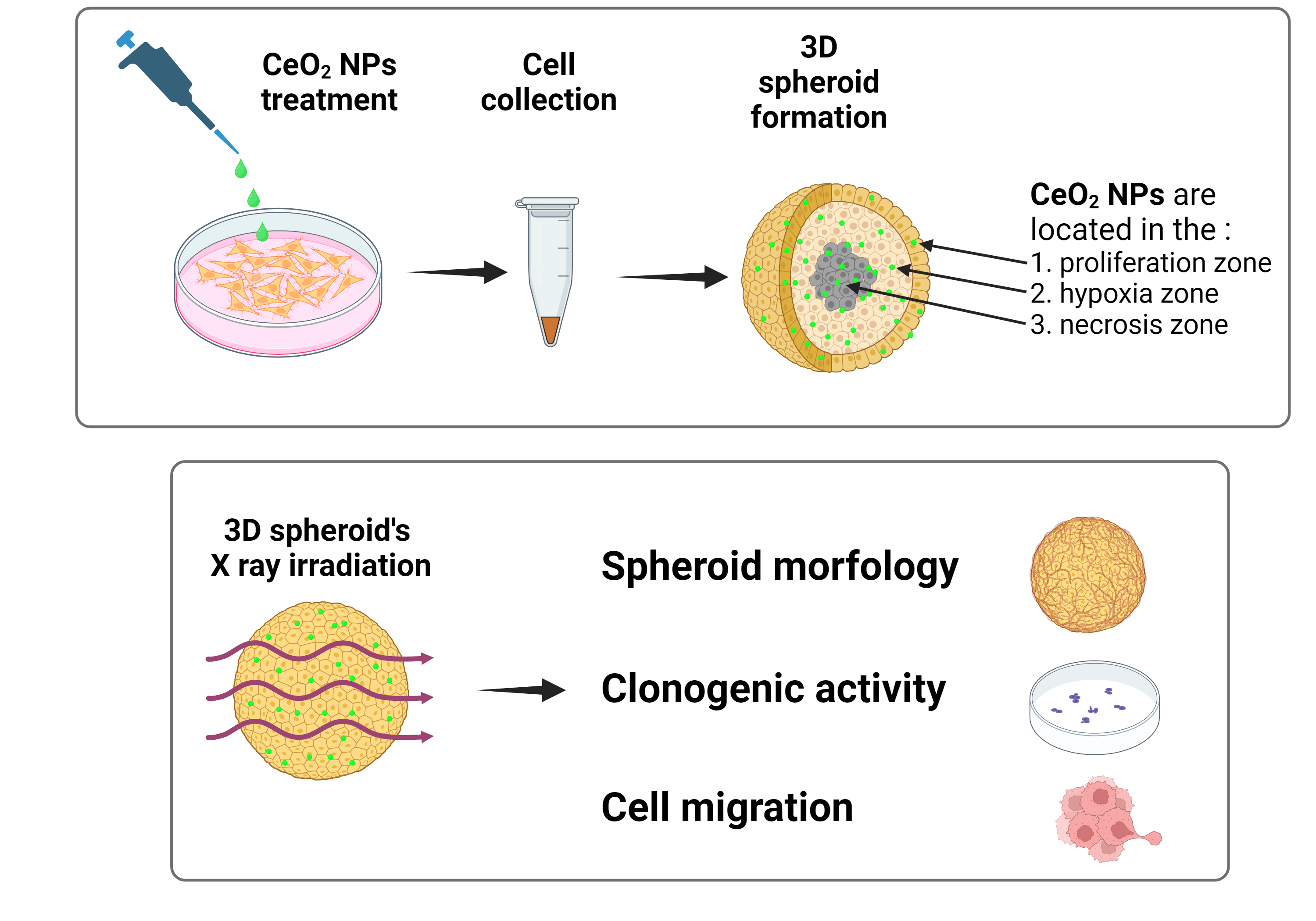
Testing of cytotoxicity and biological activity of various nanoformulations on 3D cellular spheroids is carried out by coincubation of the spheroid at various time intervals with nanoparticles at different concentrations. Fluorescent labels are often used to visualize the penetration of nanoparticles into various layers to analyze their accumulation and localization [42] At the same time, the diffusion of nanoparticles deep into the structure of the spheroid can be severely limited and largely depends on the size, shape, charge and stabilizer of the nanoparticles [43]. Here we have proposed a different technique for loading CeO2 NPs into a cellular spheroid, which consisted in the initial accumulation of nanoparticles in a 2D monolayer of cells and subsequent formation of a spheroid from these cells (Figure 3). This approach makes it possible to ensure an even distribution of nanoparticles in all structures of the spheroid and confidently say that the identified biological effects during irradiation of spheroids are ensured by influencing all structural layers of the spheroid.
We assume that loading of tumor cells with CeO2 NPs leads to uniform distribution of the nanoparticles in all cells in monolayer. Consequently, CeO2 NPs will be evenly distributed over all layers of the 3D tumor spheroid formed from the loaded cells. Thus, as the spheroid grows, CeO2 NPs appear in all structures of the spheroid: in the necrosis zone, in the hypoxia zone and in the proliferation zone. Considering the sensitivity of cerium oxide nanoparticles to the microenvironment, we assume that the redox activity of CeO2 NPs will provide various biological effects depending on their localization. To confirm this assumption, an analysis of cell migration activity was carried out by quantifying the area of cell spreading after transfer of the spheroid to an adhesive plastic (Figure 4a). We found that CeO2 NPs without X-ray irradiation reduce the migration activity of tumor cells compared to the untreated control (Figure 4b). Without CeO2 NPs, irradiation at a dose of 6 Gy led to a twofold decrease in the spreading zone. However, there is no statistically significant difference between this irradiated group and irradiated group with CeO2 NPs. Meanwhile, we observed a tendency to decrease the spreading area in irradiated cells in the presence of the CeO2 NPs.
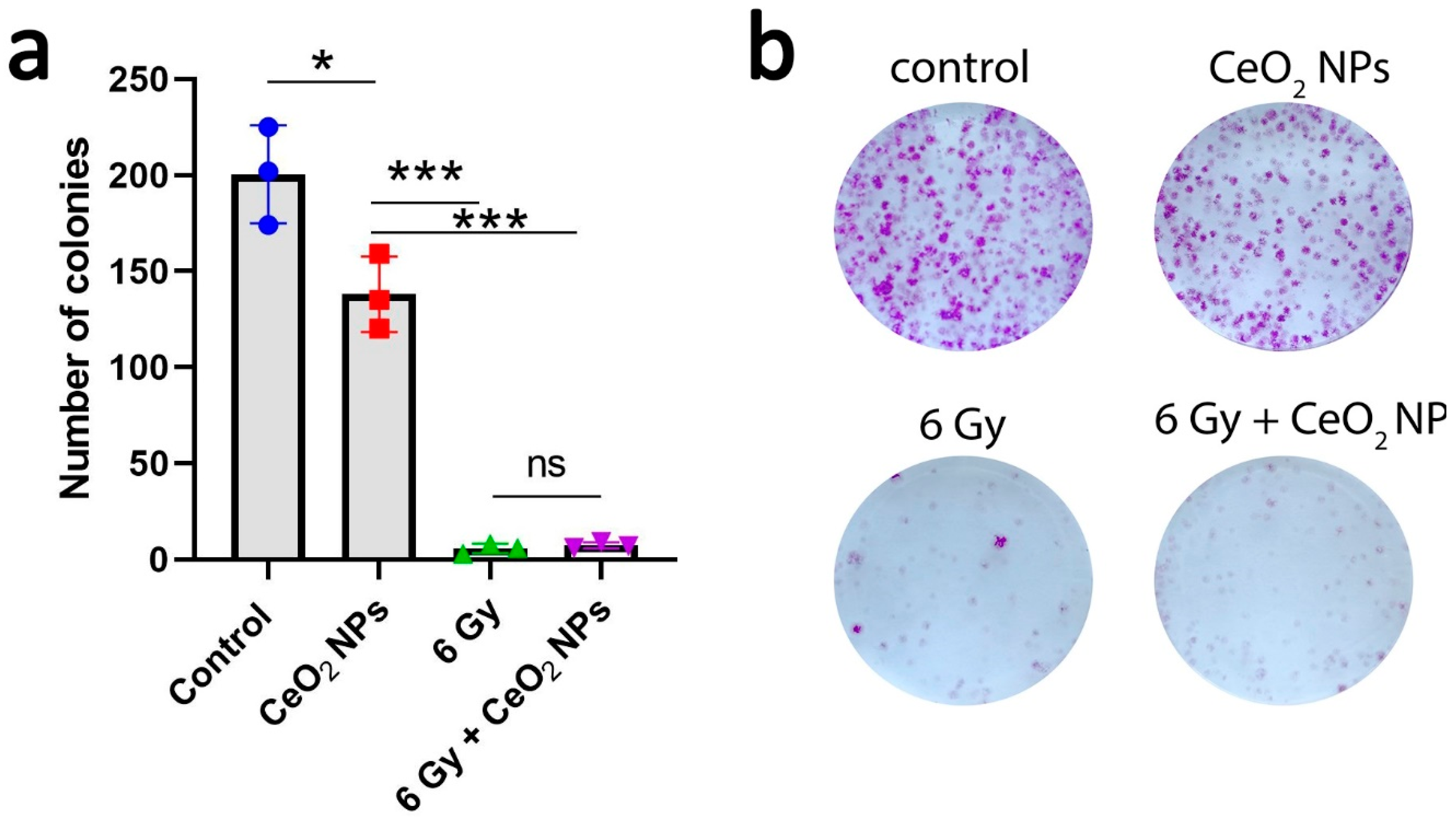
The analysis of clonogenic activity is one of the «gold standards» for determining the proliferative activity of cells after exposure to toxic agents or different types of radiation [44]. We analyzed the clonogenic activity of cells obtained from irradiated spheroids (Figure 5). A decrease in the clonogenic activity of cells for the group with nanoparticles (0.172 µg/mL) was revealed by 25% relative to the untreated control. Irradiation of spheroids at a dose of 6 Gy resulted in a 90-95% fold decrease in clonogenic activity. The combined effect of CeO2 NPs and X-ray irradiation almost completely supressed the clonogenic activity of cells. The inhibitory effect of CeO2 NPs may be related with their redox activity, which is realized both in the necrosis zone and in the hypoxia zone due to the used spheroid formation scheme.
Discussion
Cerium-containing nanoparticles are considered as a promising antitumor agent with selective cytotoxic activity against tumor cells and a high degree of biocompatibility with respect to normal cells. A key feature of cerium-containing nanomaterials, that allows them to be used in anticancer therapy, is their sensitivity to pH of the microenvironment, which makes it possible to damage tumor cells while maintaining a safe environment for normal cells under irradiation conditions. The chemical mechanism of controlled catalytic activity of cerium oxide nanoparticles is based on the ability of cerium ions located in the near-surface layer to easily and reversibly change their degree of oxidation from Ce3+ to Ce4+ and back. Using the reaction of CeO2 nanoparticle with hydrogen peroxide as an example, this phenomenon can be explained as follows: under neutral conditions, a redox reaction is initiated between the Ce4+ and the hydrogen peroxide molecule, adsorbed on the surface of nanoparticle. Then products of the hydrogen peroxide decomposition are released, exposing the surface of the nanoparticle again, and Ce3+ is oxidized to Ce4+ for the next antioxidant catalytic cycle. Under acidic conditions, an excess of protons can inhibit the transition from Ce4+ to Ce3+, which, in turn, disrupts the reactivity of catalytic centers and blocks the antioxidant cycles of the nanoparticle [41]. Thus, the microenvironment of cerium oxide can determine the direction of its bioactivity. The cellular spheroid, like the tumor node in vivo, has different zones, which differ not only in the level of ATP, the degree of oxygenation, but also in the pH level. Considering that all approved clinical nanoradiosensitizers are administered to patients intratumorally, a very important aspect determining the effectiveness of radiotherapy is the control of their localization and accumulation directly in the tumor node. In this work, it is shown that the preloading of cerium oxide nanoparticles into a spheroid ensures their uniform distribution in all zones of the spheroid, which ensures prooxidant activity in the zone of necrosis and hypoxia. We conducted a study of radiation-induced ROS generation in various microenvironment conditions (pH 6.5 and 7.4), which showed a prooxidant effect in slightly acidic conditions. Despite the fact that we have not experimentally confirmed the generation of ROS directly in spheroid cells, we can assume the probability of prooxidant activity of CeO2 NPs in acidified spheroid structures, which leads to a decrease in the migration activity of cells from irradiated spheroids. We confirmed that the proposed approach for preloading cells with nanoparticles ensures their effective accumulation in all zones of the spheroid, which provides a new opportunity to study the radiosensitizing effect of nanoparticles in vivo tumor models.
Funding
This research was funded by the Ministry of Science and Higher Education of the Russian Federation for the Institute of Theoretical and Experimental Biophysics of the Russian Academy of Sciences (State Assignment: 075-01025-23-01).
Acknowledgements
We express our gratitude to Shcherbakov A.B. for discussion of the experimental results.
References
- McNamara, K.; Tofail, S.A.M. Nanoparticles in Biomedical Applications. Adv. Phys. X 2017, 2, 54–88. [Google Scholar] [CrossRef]
- Anselmo, A.C.; Mitragotri, S. Nanoparticles in the Clinic: An Update. Bioeng. Transl. Med. 2019, 4, e10143. [Google Scholar] [CrossRef]
- Bonvalot, S.; Le Pechoux, C.; De Baere, T.; Kantor, G.; Buy, X.; Stoeckle, E.; Terrier, P.; Sargos, P.; Coindre, J.M.; Lassau, N.; et al. First-in-Human Study Testing a New Radioenhancer Using Nanoparticles (NBTXR3) Activated by Radiation Therapy in Patients with Locally Advanced Soft Tissue Sarcomas. Clin. Cancer Res. Off. J. Am. Assoc. Cancer Res. 2017, 23, 908–917. [Google Scholar] [CrossRef] [PubMed]
- Müller, S. Magnetic Fluid Hyperthermia Therapy for Malignant Brain Tumors--an Ethical Discussion. Nanomedicine Nanotechnol. Biol. Med. 2009, 5, 387–393. [Google Scholar] [CrossRef] [PubMed]
- Lux, F.; Mignot, A.; Mowat, P.; Louis, C.; Dufort, S.; Bernhard, C.; Denat, F.; Boschetti, F.; Brunet, C.; Antoine, R.; et al. Ultrasmall Rigid Particles as Multimodal Probes for Medical Applications. Angew. Chem. Int. Ed Engl. 2011, 50, 12299–12303. [Google Scholar] [CrossRef] [PubMed]
- Lux, F.; Tran, V.L.; Thomas, E.; Dufort, S.; Rossetti, F.; Martini, M.; Truillet, C.; Doussineau, T.; Bort, G.; Denat, F.; et al. AGuIX® from Bench to Bedside-Transfer of an Ultrasmall Theranostic Gadolinium-Based Nanoparticle to Clinical Medicine. Br. J. Radiol. 2019, 92, 20180365. [Google Scholar] [CrossRef] [PubMed]
- Shin, H.Y.; Park, T.J.; Kim, M.I. Recent Research Trends and Future Prospects in Nanozymes. J. Nanomater. 2015, 2015, 756278. [Google Scholar] [CrossRef]
- Zhang, Y.; Jin, Y.; Cui, H.; Yan, X.; Fan, K. Nanozyme-Based Catalytic Theranostics. RSC Adv. 2019, 10, 10–20. [Google Scholar] [CrossRef] [PubMed]
- Yang, Z.; Zhou, X.; Wang, L.; Guo, H.; Han, M.; Guo, H.; Chen, Y.; Wu, A.; Li, H.; Chen, S.; et al. Mn3O4 Nanozyme Loaded Thermosensitive PDLLA-PEG-PDLLA Hydrogels for the Treatment of Inflammatory Bowel Disease. ACS Appl. Mater. Interfaces 2023, 15, 33273–33287. [Google Scholar] [CrossRef] [PubMed]
- Li, S.; Chen, Z.; Yang, F.; Yue, W. The Age of Vanadium-Based Nanozymes: Synthesis, Catalytic Mechanisms, Regulation and Biomedical Applications. Chin. Chem. Lett. 2024, 35, 108793. [Google Scholar] [CrossRef]
- Lin, Y.; Ren, J.; Qu, X. Nano-Gold as Artificial Enzymes: Hidden Talents. Adv. Mater. Deerfield Beach Fla 2014, 26, 4200–4217. [Google Scholar] [CrossRef] [PubMed]
- Pedone, D.; Moglianetti, M.; De Luca, E.; Bardi, G.; Pompa, P.P. Platinum Nanoparticles in Nanobiomedicine. Chem. Soc. Rev. 2017, 46, 4951–4975. [Google Scholar] [CrossRef] [PubMed]
- Gao, L.; Fan, K.; Yan, X. Iron Oxide Nanozyme: A Multifunctional Enzyme Mimetic for Biomedical Applications. Theranostics 2017, 7, 3207–3227. [Google Scholar] [CrossRef]
- Popov, A.L.; Shcherbakov, A.B.; Zholobak, N.M.; Baranchikov, A.E.; Ivanov, V.K. Cerium Dioxide Nanoparticles as Third-Generation Enzymes (Nanozymes). Nanosyst. Phys. Chem. Math. 2017, 760–781. [Google Scholar] [CrossRef]
- Wei, H.; Wang, E. Nanomaterials with Enzyme-like Characteristics (Nanozymes): Next-Generation Artificial Enzymes. Chem. Soc. Rev. 2013, 42, 6060–6093. [Google Scholar] [CrossRef]
- Shcherbakov, A.B.; Ivanov, V.K.; Zholobak, N.M.; Ivanova, O.S.; Krysanov, E.Yu.; Baranchikov, A.E.; Spivak, N.Ya.; Tretyakov, Yu.D. Nanocrystalline Ceria Based Materials—Perspectives for Biomedical Application. Biophysics 2011, 56, 987–1004. [Google Scholar] [CrossRef]
- Ivanov, V.; Shcherbakov, A.; Usatenko, A.V. Structure-Sensitive Properties and Biomedical Applications of Nanodispersed Cerium Dioxide. Russ. Chem. Rev. 2009, 78, 855–871. [Google Scholar] [CrossRef]
- Orera, V.M.; Merino, R.I.; Peña, F. Ce3+↔Ce4+ Conversion in Ceria-Doped Zirconia Single Crystals Induced by Oxido-Reduction Treatments. Solid State Ion. 1994, 72, 224–231. [Google Scholar] [CrossRef]
- Heckert, E.; Karakoti, A.; Seal, S.; Self, W.T. The Role of Cerium Redox State in the SOD Mimetic Activity of Nanoceria. Biomaterials 2008, 29, 2705–2709. [Google Scholar] [CrossRef] [PubMed]
- Pirmohamed, T.; Dowding, J.M.; Singh, S.; Wasserman, B.; Heckert, E.; Karakoti, A.S.; King, J.E.S.; Seal, S.; Self, W.T. Nanoceria Exhibit Redox State-Dependent Catalase Mimetic Activity. Chem. Commun. 2010, 46, 2736–2738. [Google Scholar] [CrossRef] [PubMed]
- Du, J.; Qi, S.; Chen, J.; Yang, Y.; Fan, T.; Zhang, P.; Zhuo, S.; Zhu, C. Fabrication of Highly Active Phosphatase-like Fluorescent Cerium-Doped Carbon Dots for in Situ Monitoring the Hydrolysis of Phosphate Diesters. RSC Adv. 2020, 10, 41551–41559. [Google Scholar] [CrossRef] [PubMed]
- Filippova, A.D.; Sozarukova, M.M.; Baranchikov, A.E.; Kottsov, S.Y.; Cherednichenko, K.A.; Ivanov, V.K. Peroxidase-like Activity of CeO2 Nanozymes: Particle Size and Chemical Environment Matter. Molecules 2023, 28, 3811. [Google Scholar] [CrossRef]
- Wu, Y.; Ta, H.T. Different Approaches to Synthesising Cerium Oxide Nanoparticles and Their Corresponding Physical Characteristics, and ROS Scavenging and Anti-Inflammatory Capabilities. J. Mater. Chem. B 2021, 9, 7291–7301. [Google Scholar] [CrossRef] [PubMed]
- Seal, S.; Jeyaranjan, A.; Neal, C.J.; Kumar, U.; Sakthivel, T.S.; Sayle, D.C. Engineered Defects in Cerium Oxides: Tuning Chemical Reactivity for Biomedical, Environmental, & Energy Applications. Nanoscale 2020, 12, 6879–6899. [Google Scholar] [CrossRef] [PubMed]
- Huang, Y.; Zhang, M.; Jin, M.; Ma, T.; Guo, J.; Zhai, X.; Du, Y. Recent Advances on Cerium Oxide-Based Biomaterials: Toward the Next Generation of Intelligent Theranostics Platforms. Adv. Healthc. Mater. 2023, 12, e2300748. [Google Scholar] [CrossRef]
- Tang, J.L.Y.; Moonshi, S.S.; Ta, H.T. Nanoceria: An Innovative Strategy for Cancer Treatment. Cell. Mol. Life Sci. CMLS 2023, 80, 46. [Google Scholar] [CrossRef] [PubMed]
- Alpaslan, E.; Yazici, H.; Golshan, N.H.; Ziemer, K.S.; Webster, T.J. pH-Dependent Activity of Dextran-Coated Cerium Oxide Nanoparticles on Prohibiting Osteosarcoma Cell Proliferation. ACS Biomater. Sci. Eng. 2015, 1, 1096–1103. [Google Scholar] [CrossRef] [PubMed]
- Yao, C.; Wang, W.; Wang, P.; Zhao, M.; Li, X.; Zhang, F. Near-Infrared Upconversion Mesoporous Cerium Oxide Hollow Biophotocatalyst for Concurrent pH-/H2 O2 -Responsive O2 -Evolving Synergetic Cancer Therapy. Adv. Mater. Deerfield Beach Fla 2018, 30. [Google Scholar] [CrossRef]
- Xu, Y.; Wang, K.; Chen, Z.; Hu, R.; Zhao, Y.; Li, X.; Qu, J.; Liu, L. Oxygen Self-Supplied Upconversion Nanoplatform Loading Cerium Oxide for Amplified Photodynamic Therapy of Hypoxic Tumors. Biomater. Sci. 2022, 11, 119–127. [Google Scholar] [CrossRef] [PubMed]
- Xu, K.; Cheng, Y.; Yan, J.; Feng, Y.; Zheng, R.; Wu, X.; Wang, Y.; Song, P.; Zhang, H. Polydopamine and Ammonium Bicarbonate Coated and Doxorubicin Loaded Hollow Cerium Oxide Nanoparticles for Synergistic Tumor Therapy. Nano Res. 2019, 12, 2947–2953. [Google Scholar] [CrossRef]
- Sack, M.; Alili, L.; Karaman, E.; Das, S.; Gupta, A.; Seal, S.; Brenneisen, P. Combination of Conventional Chemotherapeutics with Redox-Active Cerium Oxide Nanoparticles--a Novel Aspect in Cancer Therapy. Mol. Cancer Ther. 2014, 13, 1740–1749. [Google Scholar] [CrossRef] [PubMed]
- Koula, G.; Yakati, V.; Rachamalla, H.K.; Bhamidipati, K.; Kathirvel, M.; Banerjee, R.; Puvvada, N. Integrin Receptor-Targeted, Doxorubicin-Loaded Cerium Oxide Nanoparticles Delivery to Combat Glioblastoma. Nanomed. 2024, 19, 1389–1406. [Google Scholar] [CrossRef] [PubMed]
- Saranya, J.; Sreeja, B.S.; Senthil Kumar, P. Microwave Assisted Cisplatin-Loaded CeO2/GO/c-MWCNT Hybrid as Drug Delivery System in Cervical Cancer Therapy. Appl. Nanosci. 2023, 13, 4219–4233. [Google Scholar] [CrossRef]
- Popov, A.L.; Abakumov, M.A.; Savintseva, I.V.; Ermakov, A.M.; Popova, N.R.; Ivanova, O.S.; Kolmanovich, D.D.; Baranchikov, A.E.; Ivanov, V.K. Biocompatible Dextran-Coated Gadolinium-Doped Cerium Oxide Nanoparticles as MRI Contrast Agents with High T1 Relaxivity and Selective Cytotoxicity to Cancer Cells. J. Mater. Chem. B 2021, 9, 6586–6599. [Google Scholar] [CrossRef] [PubMed]
- Chukavin, N.N.; Ivanov, V.K.; Popov, A.L. Calcein-Modified CeO2 for Intracellular ROS Detection: Mechanisms of Action and Cytotoxicity Analysis In Vitro. Cells 2023, 12, 2416. [Google Scholar] [CrossRef]
- Goryacheva, O.A.; Tsyupka, D.V.; Pigarev, S.V.; Strokin, P.D.; Kovyrshina, A.A.; Moiseev, A.A.; Popova, N.R.; Goryacheva, I.Y. Сerium Dioxide Nanoparticles for Luminescence Based Analytical Systems: Challenging Nanosensor and Effective Label. TrAC Trends Anal. Chem. 2024, 174, 117665. [Google Scholar] [CrossRef]
- Zanoni, M.; Piccinini, F.; Arienti, C.; Zamagni, A.; Santi, S.; Polico, R.; Bevilacqua, A.; Tesei, A. 3D Tumor Spheroid Models for in Vitro Therapeutic Screening: A Systematic Approach to Enhance the Biological Relevance of Data Obtained. Sci. Rep. 2016, 6, 19103. [Google Scholar] [CrossRef] [PubMed]
- Daunys, S.; Janonienė, A.; Januškevičienė, I.; Paškevičiūtė, M.; Petrikaitė, V. 3D Tumor Spheroid Models for In Vitro Therapeutic Screening of Nanoparticles. Adv. Exp. Med. Biol. 2021, 1295, 243–270. [Google Scholar] [CrossRef] [PubMed]
- Sambale, F.; Lavrentieva, A.; Stahl, F.; Blume, C.; Stiesch, M.; Kasper, C.; Bahnemann, D.; Scheper, T. Three Dimensional Spheroid Cell Culture for Nanoparticle Safety Testing. J. Biotechnol. 2015, 205, 120–129. [Google Scholar] [CrossRef] [PubMed]
- Vakhshiteh, F.; Bagheri, Z.; Soleimani, M.; Ahvaraki, A.; Pournemat, P.; Alavi, S.E.; Madjd, Z. Heterotypic Tumor Spheroids: A Platform for Nanomedicine Evaluation. J. Nanobiotechnology 2023, 21, 249. [Google Scholar] [CrossRef] [PubMed]
- Weng, Q.; Sun, H.; Fang, C.; Xia, F.; Liao, H.; Lee, J.; Wang, J.; Xie, A.; Ren, J.; Guo, X.; et al. Catalytic Activity Tunable Ceria Nanoparticles Prevent Chemotherapy-Induced Acute Kidney Injury without Interference with Chemotherapeutics. Nat. Commun. 2021, 12, 1436. [Google Scholar] [CrossRef] [PubMed]
- Ahmed-Cox, A.; Pandzic, E.; Johnston, S.T.; Heu, C.; McGhee, J.; Mansfeld, F.M.; Crampin, E.J.; Davis, T.P.; Whan, R.M.; Kavallaris, M. Spatio-Temporal Analysis of Nanoparticles in Live Tumor Spheroids Impacted by Cell Origin and Density. J. Control. Release Off. J. Control. Release Soc. 2022, 341, 661–675. [Google Scholar] [CrossRef] [PubMed]
- Chen, W.; Wang, W.; Xie, Z.; Centurion, F.; Sun, B.; Paterson, D.J.; Tsao, S.C.-H.; Chu, D.; Shen, Y.; Mao, G.; et al. Size-Dependent Penetration of Nanoparticles in Tumor Spheroids: A Multidimensional and Quantitative Study of Transcellular and Paracellular Pathways. Small Weinh. Bergstr. Ger. 2024, 20, e2304693. [Google Scholar] [CrossRef] [PubMed]
- Franken, N.A.P.; Rodermond, H.M.; Stap, J.; Haveman, J.; van Bree, C. Clonogenic Assay of Cells in Vitro. Nat. Protoc. 2006, 1, 2315–2319. [Google Scholar] [CrossRef] [PubMed]
Figure 1.
a) Schematic representation of the synthesis process b) TEM images of СeO2 NPs and electron diffraction pattern of СeO2 NPs c) Visual appearance of dried СeO2 NPs and the corresponding colloidal solution; d) UV spectra of СeO2 NPs; e) Size distribution of СeO2 NPs by dynamic light scattering (DLS) in deionized water, HBSS and cell culture medium DMEM/F-12 with and without 10% FBS.
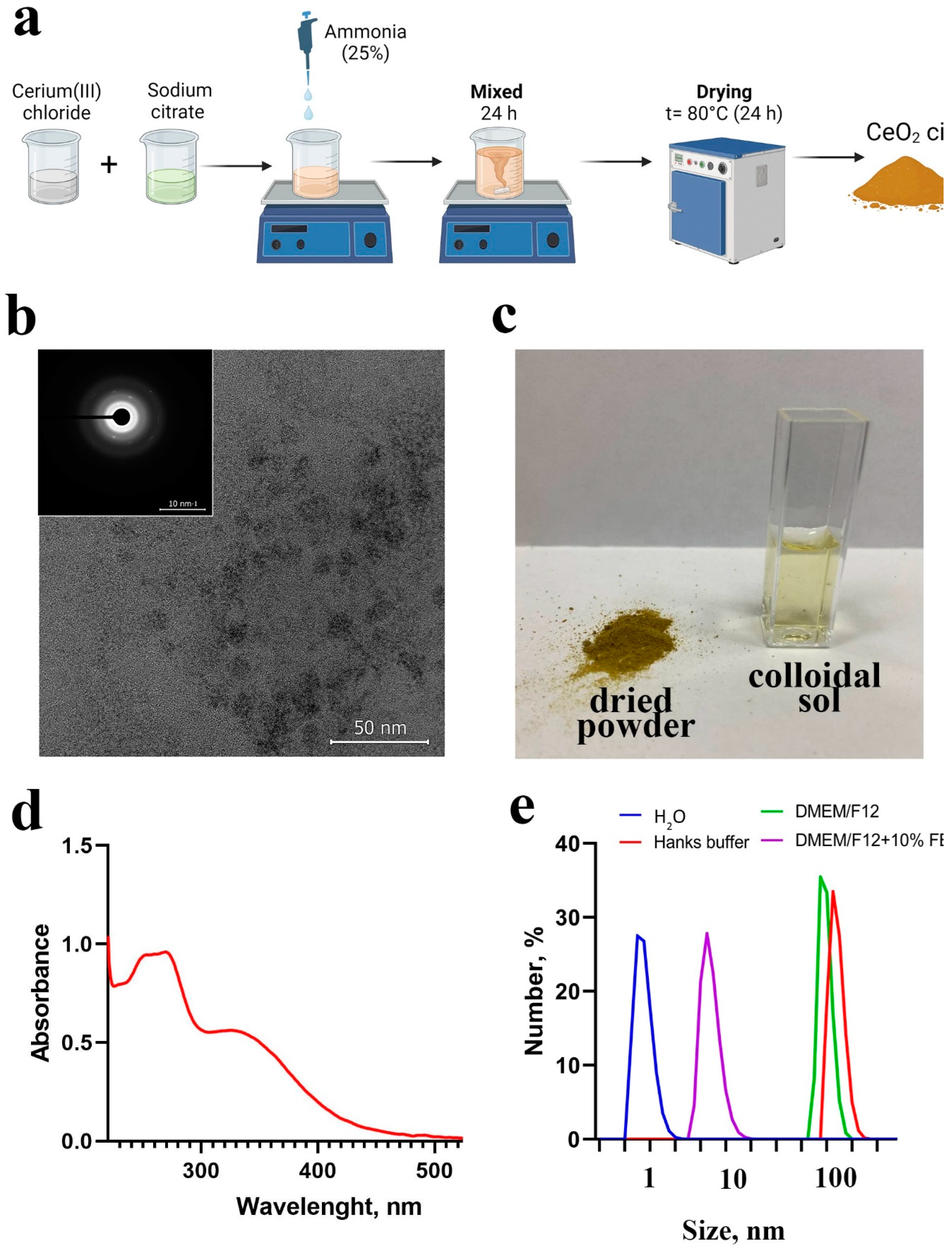
Figure 2.
ROS generation by CeO2 NPs in solutions with slightly acidic (6.5) and physiological (7.4) pH under X-ray irradiation at doses of 2 and 4 Gy.
Figure 2.
ROS generation by CeO2 NPs in solutions with slightly acidic (6.5) and physiological (7.4) pH under X-ray irradiation at doses of 2 and 4 Gy.
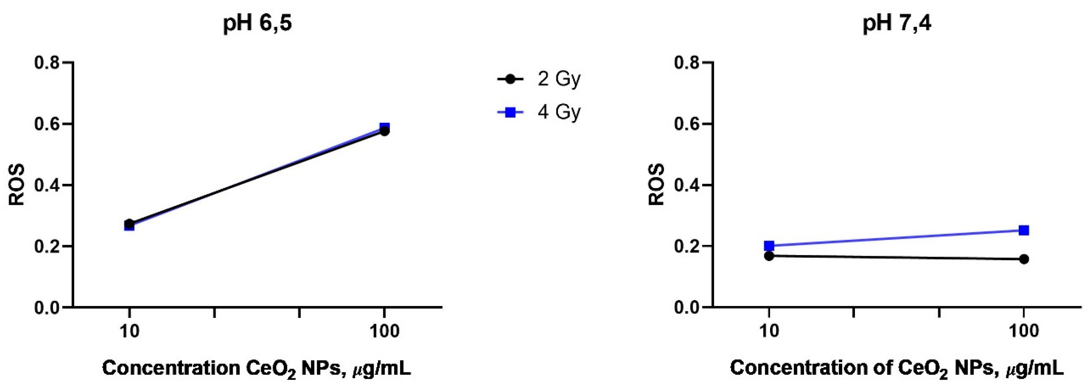
Figure 3.
Principal experiment scheme: loading of CeO2 NPs into tumor cells followed by the formation of 3D spheroids from the cells. Subsequently, the obtained spheroids are irradiated under X-ray and then analyzed for their morphology, cell migration and clonogenic activity.
Figure 3.
Principal experiment scheme: loading of CeO2 NPs into tumor cells followed by the formation of 3D spheroids from the cells. Subsequently, the obtained spheroids are irradiated under X-ray and then analyzed for their morphology, cell migration and clonogenic activity.
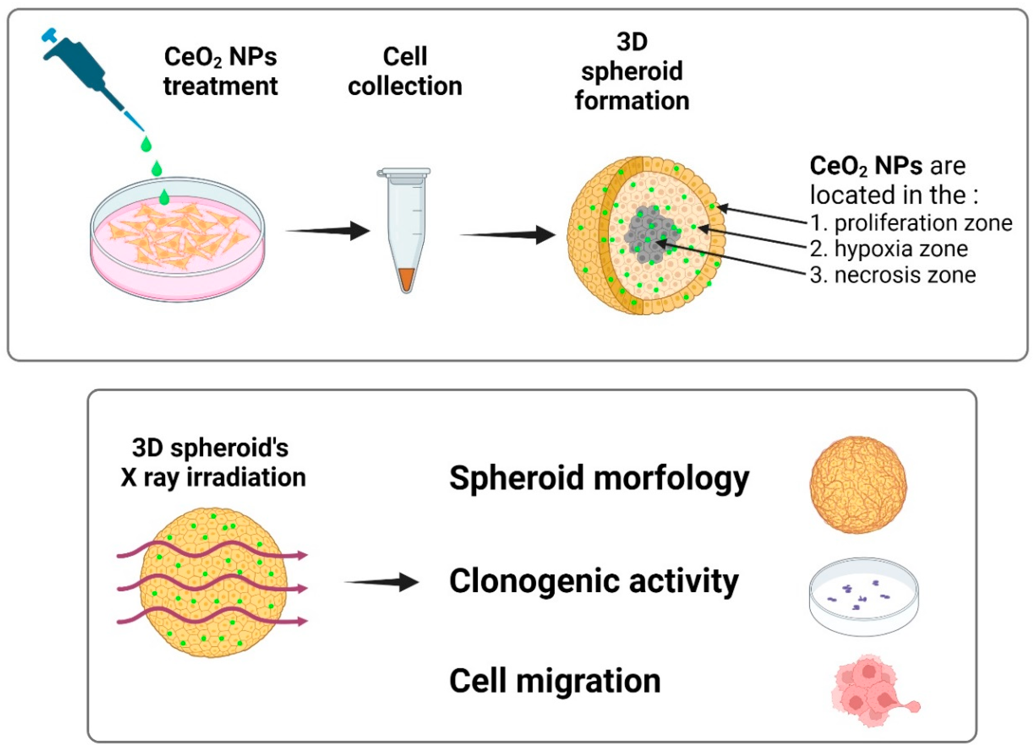
Figure 4.
Migration activity of cells from a 3D cellular spheroid 5 days after irradiation at a dose of 6 Gy in the presence CeO2 NPs (0.172 µg/mL). The data is presented as the violin plot with Mean ± Standard Deviation (SD). The statistical significance of differences between the values in experimental groups was determined using the Welch’s t-test. Differences were considered statistically significant at p < 0.05 (*), p < 0.01 (**), p < 0.005 (***), p < 0.001 (****).
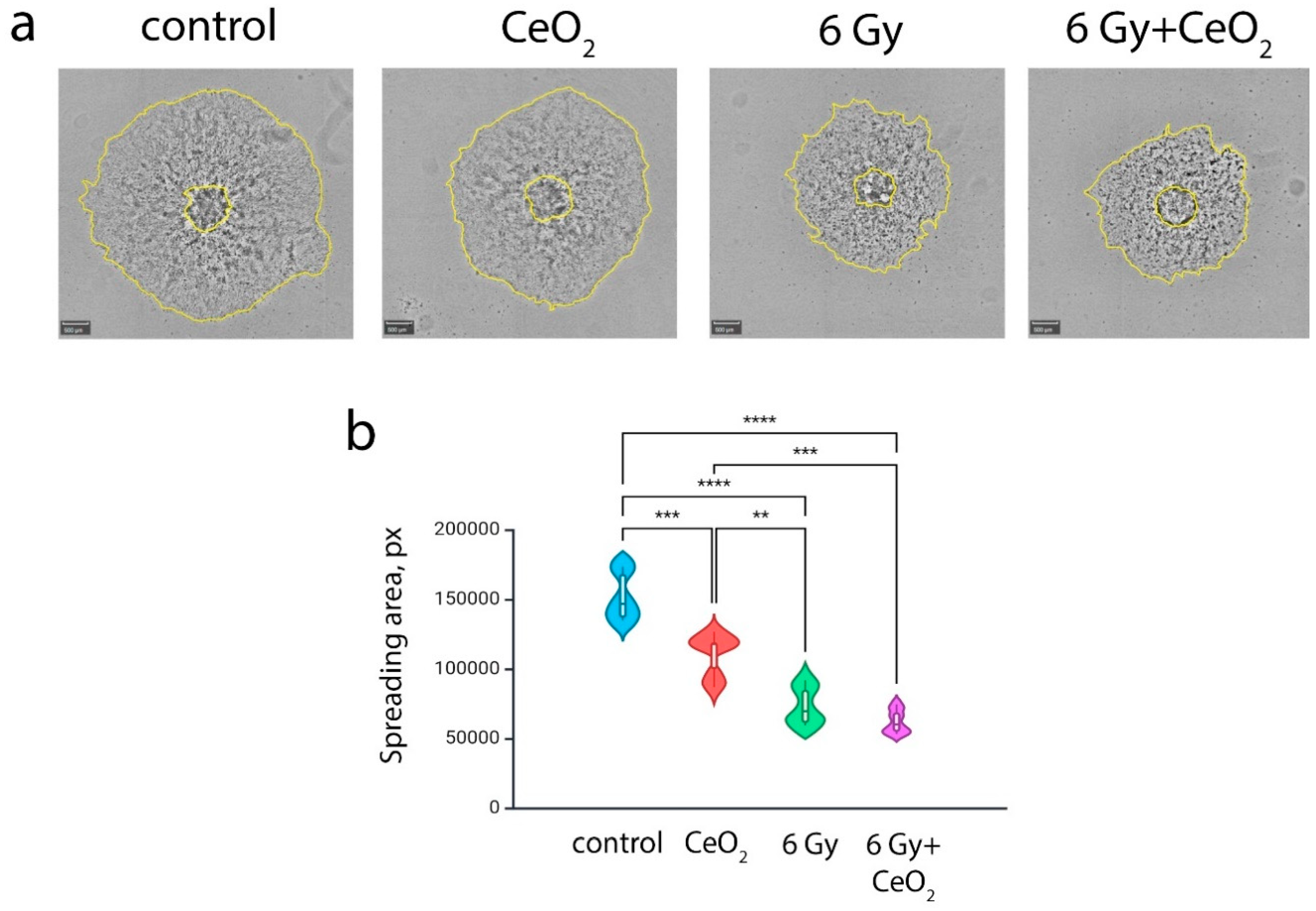
Figure 5.
Clonogenic assay after X-ray irradiation of 3D spheroids in the presence of CeO2 NPs: quantitative assessment (a) and photographs of colonies (b). The data is presented as the Mean ± Standard Deviation (SD) with experimental points. The statistical significance of differences between the values in experimental groups was determined using the Welch’s t-test. Differences were considered statistically significant at p < 0.05 (*), p < 0.01 (**), p < 0.005 (***), p < 0.001 (****).
Disclaimer/Publisher’s Note: The statements, opinions and data contained in all publications are solely those of the individual author(s) and contributor(s) and not of MDPI and/or the editor(s). MDPI and/or the editor(s) disclaim responsibility for any injury to people or property resulting from any ideas, methods, instructions or products referred to in the content. |
© 2025 by the authors. Licensee MDPI, Basel, Switzerland. This article is an open access article distributed under the terms and conditions of the Creative Commons Attribution (CC BY) license (http://creativecommons.org/licenses/by/4.0/).
Copyright: This open access article is published under a Creative Commons CC BY 4.0 license, which permit the free download, distribution, and reuse, provided that the author and preprint are cited in any reuse.


