Submitted:
17 January 2025
Posted:
17 January 2025
You are already at the latest version
Abstract
Chronic osteoarthritis (OA) is distinguished by the deterioration of cartilage within joints, resulting in pain and reduced joint function.With the aging of the population, the prevalence of OA is increasing, becoming a public health issue. Despite ongoing research, the exact pathogenesis of OA remains unclear, limiting the development of effective treatment strategies.The system involving ubiquitin and proteasomes is of vital importance in the pathogenesis of osteoarthritis, with deubiquitinating enzymes (DUBs) regulating protein stability, activity, and localization, which are essential for maintaining cellular homeostasis. Aberrant DUB expression or dysfunction is closely associated with OA. DUBs exert a key role in modulating cellular signaling, inflammatory responses, and extracellular matrix degradation, which are processes related to OA, making them a new target for therapeutic research. However, DUB research is still in its infancy, and more basic research and clinical trials are needed to verify their safety and efficacy. This analysis condenses the operational mechanisms of Deubiquitinating Enzymes (DUBs) in osteoarthritis (OA) and explores prospective treatment approaches, discussing the challenges and future prospects of modulating the expression of E3 ubiquitin ligases and deubiquitinating enzymes to improve the therapeutic effects for patients with osteoarthritis, aiming to promote the development of more effective OA treatment strategies.
Keywords:
osteoarthritis
; deubiquitinating enzymes
; Mechanisms
1. Introduction
OA, a progressive joint disorder, is distinguished by the deterioration of the cartilage within the joints, accompanied by joint pain, swelling, tenderness, and a decrease in mobility [1,2]. As the population ages, the incidence of OA is gradually increasing, becoming an important issue affecting public health [3,4]. Although research on OA has deepened in recent years, its exact pathogenesis is still not fully understood, which limits the development of effective treatment strategies for the disease. In the development of OA, a variety of cytokines, enzymes, and signaling pathways are involved, among which the ubiquitin-proteasome system plays a significant role in the occurrence and development of the disease [5,6].
Inside the cell, the ubiquitin-proteasome system is an important regulatory mechanism for protein degradation, with ubiquitination marking proteins for degradation. Correspondingly, deubiquitinating enzymes (DUBs) regulate protein stability, activity, and localization by removing ubiquitin chains from proteins. This process is essential for maintaining cellular homeostasis [7,8]. In recent years, increasing evidence has demonstrated that aberrant gene expression or dysfunction of deubiquitinating enzymes is intricately connected to various diseases, including inflammatory diseases and degenerative joint diseases [9,10,11]. Due to the key role of DUBs in regulating cellular signaling, inflammatory responses, and extracellular matrix (ECM) degradation, which are processes related to OA, they have become emerging targets in OA therapeutic research [12].
Early studies focused on the role of deubiquitinating enzymes in cancer, but with a deeper understanding of the function of the ubiquitin system, researchers have gradually begun to explore the role of DUBs in other complex diseases. In the mid-2000s, scientists first discovered the connection between DUBs and inflammation-related signaling pathways, and subsequent extensive research was conducted in the field of osteoarthritis. In recent years, more and more research has focused on specific DUB subtypes, such as USP and CYLD, which have been found to participate in the regulation of chondrocyte differentiation, extracellular matrix degradation, and the control of inflammatory responses.
Research on DUBs in osteoarthritis has gradually narrowed down from the overall regulation of signaling pathways to the functional validation of specific enzymes, especially some specific DUBs such as USP7 and USP15, which have been found to alleviate joint degeneration by inhibiting chondrocyte apoptosis and reducing inflammatory responses. Currently, the development of DUB inhibitors has entered the preclinical research stage, showing their potential as targets for osteoarthritis treatment. In the future, as the functions of DUBs are further elucidated, precise regulation of these enzymes' activity is expected to bring new treatment options for OA patients. However, research in this field is still in its infancy, and more basic research and clinical trials are needed in the future to verify the safety and efficacy of these potential therapeutic strategies. In light of this, this article aims to review the current understanding of the mechanisms of action of deubiquitinating enzymes in osteoarthritis and how they affect pathological processes, and to explore potential therapeutic strategies based on these findings. By revealing the specific roles of deubiquitinating enzymes in the pathogenesis of OA and exploring their feasibility and safety as potential therapeutic targets, the ultimate goal is to promote the development of more effective treatment strategies for OA.
2. Overview of the Biological Functions of Deubiquitinating Enzymes
Deubiquitinating enzymes (DUBs) are a class of enzymes that can remove ubiquitin molecules from proteins, and their biological functions within the cell are crucial. Ubiquitination is a process that regulates the stability, activity, and localization of target proteins by covalently attaching ubiquitin to them. This mechanism plays an important role in cellular physiology, affecting protein degradation, signal transduction, and the cell cycle, among other aspects, while DUBs regulate the fate of target proteins by removing these ubiquitin tags, thus maintaining protein homeostasis within the cell.
The process of ubiquitination is mediated by a trio of essential enzymes: the E1 activating enzyme, the E2 conjugating enzyme, and the E3 ligase. Initially, the E1 enzyme catalyzes the activation of ubiquitin and its subsequent transfer to the E2 enzyme. Following this, the E2 enzyme collaborates with the E3 ligase to catalyze the transfer of ubiquitin from the E2 enzyme to target substrate proteins. This post-translational modification enables the protein to be identified and subsequently degraded by the proteasome. Conversely, deubiquitination, which entails the removal of ubiquitin from these substrate proteins, is executed by deubiquitinating enzymes (DUBs), effectively counteracting the effects of ubiquitination [13,14].
DUBs primarily function through two mechanisms. Firstly, DUBs can specifically remove ubiquitin from target proteins, promoting protein stability and function. For example, USP7 (Ubiquitin-Specific Peptidase 7) can remove ubiquitin marks associated with apoptosis, inhibiting cell death and thus being crucial for maintaining cell survival and proliferation, especially under stress conditions. It regulates the stability of multiple substrates, including p53 and Mdm2, which is significant for the regulation of the cell cycle and apoptosis.
Secondly, DUBs play a significant role in regulating cellular signal transduction. Take CYLD (Cylindromatosis) as an example; this deubiquitinating enzyme can promote the nuclear transport of NF-κB complexes by removing ubiquitin marks, thereby activating the transcription of downstream genes. CYLD is also involved in regulating the activity of the Wnt signaling pathway, affecting cell proliferation and differentiation [15,16]. This signal modulation is crucial for maintaining cellular physiology and responding to external stimuli.
Additionally, DUBs are closely related to cellular stress responses. USP15 can remove ubiquitination modifications when cells are under stress, promoting autophagy and helping cells clear damaged proteins and organelles. Additionally,DUBs play a pivotal role in the modulation of diverse biological activities, including cellular migration and inflammatory reactions, further emphasizing their importance in cellular functions.
The function of DUBs is not only related to their catalytic activity but also influenced by the cellular environment, substrate specificity, and other regulatory factors. Abnormal expression or dysfunction of DUBs is closely related to various diseases, including cancer, neurodegenerative diseases, and autoimmune diseases [17,18,19,20]. Therefore, studying the biological functions of DUBs not only helps understand their importance in cellular physiology but also provides potential targets for disease treatment.
3. The Mechanism of Action of Deubiquitinating Enzymes in Osteoarthritis
3.1. Deubiquitinating Enzymes and Cellular Signal Transduction
In the pathological process of osteoarthritis, deubiquitinating enzymes participate in the regulation of cellular signal transduction through multiple pathways, which play an important role in maintaining joint health and responding to pathological conditions. Deubiquitinating enzymes can regulate the stability and function of signaling molecules related to inflammatory responses, cell proliferation, differentiation, and apoptosis.
Research has demonstrated that in the pathological state of OA, the expression and activity of certain deubiquitinating enzymes change, which may affect the ubiquitination levels of their substrates and thereby regulate related signaling molecules and cellular responses. This regulatory effect may exacerbate joint inflammation, affect the degradation and repair processes of cartilage, or alter the balance of other cellular signaling networks, thus playing a pivotal function in the progression of OA.
In recent years, the association between deubiquitinating enzymes (DUBs) and cellular signal transduction has been widely studied. The diverse roles of DUBs in processes of cellular transformation, such as oncogenesis, cell cycle regulation, cell metabolism, inflammation, and cell death are being continuously revealed [21,22]. In cancer, DUBs have been found to have significant impacts on the tumor microenvironment, cell cycle regulation, metabolic pathways, and drug resistance [23] . In inflammation and cell death signaling, the regulatory role of DUBs is also significant. Studies have shown that OTULIN and LUBAC act together in the TNF receptor signaling pathway, affecting inflammation and cell death signals [24] . OTUB1 regulates the degradation of cIAP1 by deubiquitination, inhibiting TNF-induced cell apoptosis, thus playing an important role in the regulation of inflammatory signals [24,25]. At the same time, the OTU family of DUBs has attracted the attention of researchers due to their specific functions and regulatory mechanisms in signaling pathways. These DUBs regulate protein stability, immune signaling, and cell cycles by modulating the ubiquitination status of proteins.
Deubiquitinating enzymes (DUBs) play a key role in cellular signal transduction, affecting fundamental processes such as cell cycles, apoptosis, and inflammatory responses. Reyes-Turcu et al. summarized how DUBs dynamically regulate various signaling pathways through the recognition of ubiquitin chain dynamics and emphasized their potential as targets in diseases. A20 is a classic DUB that regulates NF-κB signaling, and Catrysse et al. showed that it achieves negative feedback regulation of inflammatory signals through ubiquitin chain editing [26] . In addition, DUBs crucial in the process of DNA repair, and Nijman et al. revealed that they maintain genomic stability by regulating ubiquitination [27]. USP7 is closely related to cancer development by affecting p53 homeostasis, and Cummins et al. pointed out that it may become a therapeutic target [28]. Regarding antiviral immunity, Gack et al. found that the ubiquitination regulation of RIG-I achieves balance in activation and degradation [29]. DUBs affect cellular signal transduction through various mechanisms and play a key role in multiple biological processes.Despite the considerable advancements achieved in the field of DUBs research, there remains a need to further elucidate the mechanisms that are specific to various diseases,integration of upstream and downstream networks, and drug development.
3.2. Deubiquitinating Enzymes and Cell Cycle
Deubiquitinating enzymes (DUBs) play a crucial role in the precise regulation of the cell cycle, affecting multiple processes from DNA replication, centrosome replication to mitotic exit. Recent studies have revealed the importance of DUBs in maintaining cell cycle stability and genomic integrity, suggesting their potential as therapeutic targets.
DUBs function by dynamically regulating key proteins in the ubiquitination process. It has been found that USP7 regulates the stability of Mdm2 through deubiquitination, indirectly controlling p53 activity, thereby affecting G1 phase cell cycle arrest and DNA repair [30]. Similarly, USP37 directly deubiquitinates Cyclin A, promoting the G1/S transition and participating in the regulation of DNA replication initiation licensing to prevent premature DNA replication [31,32]. In addition, USP15 maintains the function of key proteins in the DNA replication process during the S phase checkpoint, ensuring the integrity of DNA replication and preventing genomic instability [33].
During mitosis, DUBs also play an important role. USP28 regulates the ubiquitination of Aurora kinase, affecting the exit from mitosis and preventing cell division errors [34] . USP39 regulates the stability of proteins in the spindle checkpoint, ensuring the correct separation of chromosomes [35]. Moreover, USP13 is involved in the regulation of centrosome replication, and its functional abnormalities may lead to polyploidy and cell cycle disorders [36] . These studies indicate that DUBs play a crucial role in maintaining cellular homeostasis at multiple key points in the cell cycle [37].
At the same time, certain DUBs further maintain the normal operation of the cell cycle by participating in DNA damage repair signaling pathways. It has been found that OTUB1 prevents the ubiquitination and degradation of DNA damage-related proteins, preventing premature entry or arrest of the cell cycle, protecting cells from genomic damage. In addition, USP7 and OTUB1 play an important protective role in DNA damage-induced G1/S or G2/M checkpoints, providing a new perspective for understanding ubiquitin signal regulation in DNA damage response.
Although the aforementioned studies have significantly improved the understanding of the role of DUBs in cell cycle regulation, there are still some shortcomings. First, most studies focus on a single DUB and its specific functions, neglecting the global regulatory effects of the DUB network. Second, current research is mainly completed in vitro, lacking validation in vivo or in clinical models. Finally, the specific mechanisms of DUBs as potential therapeutic targets still need further exploration, especially in the field of how to selectively inhibit specific DUBs to avoid off-target effects.
3.3. Deubiquitinating Enzymes and Apoptosis
The role of deubiquitinating enzymes (DUBs) in apoptosis has gradually attracted the attention of researchers. Apoptosis is a form of programmed cell death that is widely involved in tissue development, immune regulation, and the development of various diseases (such as cancer and neurodegenerative diseases). DUBs affect the transmission of cell death signals by modulating the levels of protein ubiquitination, thereby playing an important role in the process of apoptosis. Recent studies have revealed that multiple DUBs regulate apoptotic pathways through different mechanisms, including intervention in the ubiquitin-proteasome system and interactions with other intracellular signaling pathways.
A study summarized the multiple roles of DUBs in cell death, pointing out that DUBs not only play a role in apoptosis but are also involved in other forms of regulated cell death, such as necroptosis, pyroptosis, and ferroptosis [38]. Another study explored the functions of DUBs in urinary system cancers, pointing out that DUBs can promote tumor cell survival by regulating apoptosis-related pathways and can also inhibit tumor development by activating apoptotic pathways [39].
USP7 (also known as HAUSP) is an important DUB, and studies have shown that it regulates apoptosis by modulating the p53-MDM2 pathway. USP7 can remove ubiquitin modifications from p53, thereby stabilizing p53 and activating the apoptotic program, and polyubiquitinates MCL-1 (an anti-apoptotic protein) to maintain cell survival.The elevated expression of this gene is intimately linked to the initiation and progression of a spectrum of malignancies.Another study found that a key protein regulates the balance between autophagy and apoptosis, further revealing the complex role of DUBs in cell life and death decisions [40].
CYLD (Cylindromatosis DUB) has been proven to play a key role in regulating the NF-κB signaling pathway and apoptosis. CYLD inhibits the activity of NF-κB through deubiquitination, thereby promoting apoptosis. In addition, UCH-L1 (Ubiquitin Carboxy-drolase L1) also plays a role in neurodegenerative diseases. It regulates the apoptosis process of neurons through deubiquitination andis intricately linked to the genesis of conditions including Alzheimer's disease.
Regarding the therapeutic potential of DUBs, researchers have begun to focus on the development of small molecule inhibitors. Although some DUBs have been confirmed, these studies are mostly focused on cancer cells or specific disease models and lack broad clinical validation. Moreover, although there are studies on some DUBs as potential targets, their mechanisms of action in different types of cells are not fully understood. Therefore, future research should pay more attention to the function of DUBs across various cellular contexts and within the spectrum of pathological conditions.
3.4. Deubiquitinating Enzymes and Cellular Metabolism
The role of deubiquitinating enzymes (DUBs) in the regulation of cellular metabolism has received widespread attention in recent years, affecting glucose metabolism, lipid metabolism, oxidative stress responses, and cancer, among others. In cancer metabolism research, DUBs promote cancer cell growth and metabolic adaptation by regulating protein stability and signal pathway activity, becoming potential therapeutic targets. For example, the Machado-Joseph family of deubiquitinating enzymes plays a key role in tumor metabolism, regulating the PTEN and AKT/mTOR pathways, affecting cancer cell proliferation and metabolic balance [41]. USP14 supports the energy metabolism of cancer cells by stabilizing proteins related to mitochondrial function [42]. In addition, USP7 has been proven to play an important role in glucose metabolism by regulating key enzymes of glycolysis, maintaining the high metabolic level of cancer cells [43].
Studies have shown that DUBs not only regulate tumor metabolism but also play a protective role in oxidative stress responses. Deubiquitinating enzymes can enhance metabolic homeostasis by alleviating oxidative stress, thereby improving cell survival capabilities. In lipid metabolism, DUBs support the membrane biosynthesis needs of tumor cells by regulating the ubiquitination status of enzymes related to lipid synthesis and degradation [44]. In addition, research on DUB complexes further reveals their synergistic effects in multiple metabolic pathways, emphasizing their position as core nodes in the metabolic regulation network.
Although significant progress has been made in the study of the role of DUBs in cellular metabolism, many mechanisms are still unclear. For example, the role of USP7 in glucose metabolism has preliminary evidence, but the detailed mechanism by which it regulates key enzymes of glycolysis still needs in-depth study [45]. Similarly, the impact of USP14 on mitochondrial function is mostly based on single cell line studies, and the universality of the results needs more validation [46].
Further research should combine metabolomics, gene editing, and animal model experiments to reveal the multi-level functions of DUBs and assess their clinical potential as therapeutic targets.
The regulatory role of DUBs in cellular metabolism provides important insights for basic research and clinical applications. Research on them as potential therapeutic targets is still in its infancy, and future research should explore their fine regulatory mechanisms in normal cells and tumor metabolism through multidisciplinary cooperation, thereby providing new strategies for disease treatment.
4. The Role of Various Deubiquitinating Enzymes in Osteoarthritis
4.1. USP3
USP3, as a deubiquitinating enzyme, plays a key role in the regulation of inflammatory responses and aging in chondrocytes. According to literature reports, ubiquitin-specific peptidase 3 (USP3) can deubiquitinate through the RIG-I-like receptor pathway, thereby inhibiting type I interferon signal transduction and participating in antiviral immune responses [47]. Studies have revealed that in the pathological process of osteoarthritis, the expression level of USP3 shows a downward trend. The enhancement of USP3 function can alleviate chondrocyte apoptosis mediated by IL-1βand lead to the inhibition of the NF-kB signaling pathway [48]. Tumor necrosis factor receptor-associated factor 6 (TRAF6), as an adapter protein in the NF-kB signaling pathway, plays a central role in immune and inflammatory responses. IL-1β promotes the ubiquitination of TRAF6, and USP3 alleviates the aging process of chondrocytes by inhibiting the NF-κB signaling pathway induced by IL-1β and reducing the ubiquitination of TRAF6 [49]. In addition, the upregulation of USP3 also helps maintain the stability of SIRT3 protein, reducing cell damage caused by oxidative stress. This process involves inhibiting the ubiquitination of SIRT3, thereby maintaining its protein level. In turn, the inhibition of SIRT3 activity weakens the upregulation effect of USP3 in the aging process of chondrocytes [50]. Similarly, the upregulation of SIRT3 can counteract the production of reactive oxygen species (ROS) and cell aging caused by the absence of USP3. At the molecular mechanism level, SIRT3 partially reduces chondrocyte aging by deacetylating FOXO3 .
4.2. USP5
According to existing research, USP5 plays a crucial role in the development of inflammatory responses. Specifically, USP5 regulates neuropathic and inflammatory pain by enhancing the function of CaV3.2 channels [51]. Disrupting the interaction between USP5 and Cav3.2 has been proven to alleviate these two types of pain [52]. In addition, a peptide that can penetrate cells and target the cUBP domain of USP5 has been found to effectively relieve neuropathic and inflammatory pain . In pain signal transduction, IL-1b, as a key medium, modulates the communication between Cav3.2 and USP5 [53]. Experimental data also show that blocking the binding of USP5 and Cav3.2 can protect female mice from mechanical hyperalgesia caused by peripheral inflammation [54]. Further research reveals that USP5 is closely related to the pro-inflammatory function of RA-FLS [55]. Rheumatoid Arthritis (RA) is a prevalent chronic condition characterized by autoimmune-mediated inflammation, in which RA-FLS shows an increase in USP5 expression, while OA-FLS (osteoarthritis fibroblast-like synoviocytes) shows a decrease in USP5 expression. The stimulation of IL-1b leads to an increase in USP5 expression over time [56]. The increased expression of USP5 significantly enhances the activity of the NF-kB signaling cascade, thereby stimulating the synthesis of pro-inflammatory cytokines. On the contrary, the inhibition of USP5 reduces the release of cytokines and inhibits the activation of NF-kB. USP5 can interact with TRAF6 and stabilize TRAF6 through deubiquitination, thereby maintaining its stability in RA-FLS and controlling the inflammatory process [57].
4.3. USP7
In the field of osteoarthritis research, the mechanism of action of USP7 is gradually beingrevealed. Studies have shown that hydrogen peroxide (H2O2) can induce the expression of USP7 in chondrocytes of rats, and subsequently increase the level of reactive oxygen species (ROS), thereby inhibiting the proliferation of chondrocytes. The removal of USP7 can eliminate cell pyroptosis and ROS induction mediated by H2O2, and inactivate the activation of NLRP3 inflammasomes. In addition, the upregulation of USP7 enhances pyroptosis, IL-1β and IL-18 levels, MMP-1, MMP13, and the activation of NLRP3 inflammasomes in chondrocytes of rats by increasing the level of ROS in chondrocytes of rats. USP7 interacts with NOX4 to promote its ubiquitination in chondrocytes of rats. In patients with osteoarthritis, USP7 and NOX4 are highly expressed, and the inhibition of NOX4 can eliminate the functions of chondrocyte pyroptosis, ROS induction, and NLRP3 activation mediated by USP7. P22077, a USP7 inhibitor, inhibits the process of osteoarthritis in mice by injecting monosodium iodoacetate (MIA), indicating that USP7 promotes the progression of osteoarthritis by regulating the NOX4/ROS/NLRP3 pathway. In osteoarthritis, the ubiquitination of LKB1 increases the activation of the AMPK pathway, inhibits the NLRP3 inflammasome response, and blocks chondrocyte pyroptosis. Within the articular cartilage of mice afflicted with osteoarthritis, the expression of USP7 is reduced, and silencing USP7 by siRNA or its inhibitor can promote the proliferation of chondrocytes and accelerate chondrocyte apoptosis. USP7 reduces the inflammatory response in the inflammatory process.
The inhibitors of USP7 promote the destruction of cartilage in osteoarthritis mice by activating the BiP-eIF2a-ATF4-CHOP pathway in endoplasmic reticulum stress (ERS) and promoting the NF-kB/p65 pathway. Some compounds, including QNZ and 4-PBA, as well as CHOPsinas, reduce the expression of USP7, which helps which helps to suppress the proliferation of chondrocytes and promote cell death, while mitigating the inflammatory reaction triggered by TNF-α [58].
ADMA promotes the instability of SOX9, which is mediated by DDAH1. In patients with osteoarthritis, the level of DDAH1 expression is diminished, conversely, there is an augmentation in the expression of DDAH1. Mice with a global or chondrocyte-specific deletion of the DDAH1 gene, which functions as an ADMA hydrolase, exhibit a swift advancement of osteoarthritis.ADMA promotes the progression of osteoarthritis by inducing the degeneration and aging of chondrocytes and destroying ECM deposition. ADMA engages with SOX9 and USP7, safeguarding SOX9 from deubiquitination by USP7, which subsequently facilitates the degradation of SOX9. Consequently, the elevation of DDAH1 expression could suppress ADMA concentrations, modulate the USP7-dependent deubiquitination of SOX9, potentially offering a therapeutic approach for osteoarthritis management [59].
4.4. USP13
In the pathological process of osteoarthritis, the expression level of USP13 is closely related to inflammatory responses. In the synovial specimens of patients with rheumatoid arthritis (RA), the expression of USP13 increases, while under the stimulation of LPS, TNF-α, and IL-1β, the expression of USP13 in human fibroblast-like synoviocytes (H-FLS) decreases, indicating that USP13 may play a key role in the regulation of inflammatory responses. USP13 inhibits pulmonary inflammation by stabilizing the anti-inflammatory receptor IL-1R8/Sigirr, a mechanism that also applies to osteoarthritis [60]. USP13 also regulates PTEN by reducing oxidative stress, regulating apoptosis, and inflammation, thereby improving osteoarthritis. In osteoarthritis, oxidative stress and apoptosis are key factors leading to chondrocyte damage and joint inflammation. USP13 plays a protective role by upregulating Nrf-2 and downregulating Caspase-3, indicating that USP13 has potential therapeutic value in regulating oxidative stress and apoptosis [61].
In addition, USP13 functions by binding to PTEN and regulating the activation of AKT. Induced by IL-1β or TNF-α, the increased expression of USP13 mitigates the inflammatory reactions in H-FLS. This modulation is likely due to the heightened PTEN activity, the reduced phosphorylation of AKT, and the dampened NF-κB signaling pathway [62]. This finding reveals the important role of USP13 in regulating inflammatory responses, especially in the pathological process of osteoarthritis. An increase in the levels of USP13 is associated with the suppression of genes that are pivotal to osteoclast function and inhibits the occurrence of osteoclasts. In osteoarthritis, excessive activity of osteoclasts can lead to bone loss, thereby exacerbating joint damage. USP13 may provide a new strategy for the treatment of osteoarthritis by regulating the occurrence of osteoclasts.
In the collagen-induced arthritis (CIA) mouse model, USP13 alleviates synovial hyperplasia, cartilage damage, inflammation, and bone loss. These in vivo experimental results further confirm the protective effect of USP13 in osteoarthritis, especially in reducing joint inflammation and protecting cartilage.
USP13 exerts a complex influence on the development of osteoarthritis. It has a significant impact on the progression of osteoarthritis by regulating inflammatory responses, oxidative stress, apoptosis, and the occurrence of osteoclasts. The upregulation of USP13 may provide a new target for the treatment of osteoarthritis, especially in regulating inflammation and inhibiting bone loss.
4.5. USP14
It is well known that USP14 can regulate protein degradation and is related to. Li et al. have shown that USP14 augments the activation of the NF-kB signaling pathway and accelerates the process of chondrocyte dedifferentiation induced by IL-1, by regulating the degradation of IkBa [63]. USP14 is highly elevated in the articular cartilage of patients with osteoarthritis and in chondrocytes induced by il-1b. ACHP, an inhibitor of IKK-b, can inactivate the NF-kB pathway and eliminate the upregulation of USP14. In turn, USP14 enhances the deubiquitination of IkBa, promoting the activation of NF-kB. Inhibiting NF-kB can effectively eliminate the dedifferentiating effect of USP14 mediated by IL-1b on chondrocytes. Consequently, targeting USP14 could represent a promising therapeutic strategy for managing osteoarthritis.
Studies have found that USP14 exacerbates the activation of the NF-kB signaling pathway by affecting the degradation of IkBa, thereby accelerating the dedifferentiation process of chondrocytes induced by IL-1. In the articular cartilage of patients with osteoarthritis, the expression level of USP14 is significantly increased, especially in chondrocytes induced by IL-1β, and this trend is more pronounced.
Further mechanistic studies have shown that USP14 can enhance the deubiquitination of IkBa, thereby promoting the activation of NF-kB. This activation is crucial for chondrocyte dedifferentiation, and the engagement of the NF-kB signaling cascade is intricately linked to both the inflammatory reactions and the breakdown of chondrocytes. Therefore, inhibiting the activity of the NF-kB signaling pathway can effectively eliminate the effect of USP14 mediated by IL-1β on chondrocyte dedifferentiation. In terms of therapeutic strategy, ACHP, as an inhibitor of IKK-b, can inactivate the NF-kB pathway, thereby inhibiting the upregulating effect of USP14. This inhibitory effect has potential therapeutic value for controlling the progression of osteoarthritis.
4.6. USP15
In the field of osteoarthritis (OA) research, the role of USP15, a deubiquitinating enzyme, is increasingly receiving attention. USP15 is not only involved in various cellular processes and tumorigenesis but is also closely related to the development of non-cancer diseases. Especially in the pathological process of osteoarthritis, USP15 plays a key role in the progression of the disease by finely regulating multiple signaling pathways through its deubiquitinating activity.
USP15 affects the activity of signaling pathways by regulating the deubiquitination status of target proteins, thereby playing a role in various diseases. In immune and inflammatory responses, USP15 is believed to be involved in modulating immune and inflammatory responses through the regulation of multiple signaling cascades, such as the TLR signaling pathways, the NF-kB pathway, the RIG-1 signaling pathway, the TGF-β pathway, and the p53 pathway [64]. In particular, the role of USP15 in the TGF-β/SMAD2 signaling pathway is crucial for the pathogenesis of osteoarthritis. Studies have shown that USP15 can stimulate the TGF-β/SMAD2 signaling pathway, thereby affecting the phenotype and function of chondrocytes.
In the context of osteoarthritis, the interaction between USP15 and ERK2 is particularly important. ERK2 relies on USP15 to regulate the TGF-β/SMAD2 signaling pathway and control the phenotype of chondrocytes. At the molecular mechanism level, USP15 forms a complex with ERK2, which regulates the ubiquitination status of ERK2. USP15 activates the TGF-β/SMAD2 signaling pathway by increasing the level of phosphorylated-ERK1/2 but does not increase the stability of ERK2 [65].
The role of USP15 in osteoarthritis is multifaceted. It not only inhibits the progression of osteoarthritis by targeting the ERK and TGF-β/SMAD2 signaling pathways but may also affect the overall course of the disease by regulating immune and inflammatory responses.These discoveries offer novel therapeutic targets for the management of osteoarthritis, that is, by regulating the activity of USP15 or its interaction with ERK2, effective control of the progression of osteoarthritis may be achieved.
4.7. USP49
USP49, full name ubiquitin-specific peptidase 49, is an enzyme that has received attention in the field of oncology, and its research in osteoarthritis (OA) is also gradually receiving attention. Research has demonstrated that USP49 is a crucial factor in the modulation of cellular proliferation, invasion, and programmed cell death [66]. In the pathological process of osteoarthritis, the expression level of USP49 is significantly lower compared to healthy individuals, which may be related to its role in chondrocyte apoptosis.
In patients with osteoarthritis, IL-1β, as a key inflammatory factor, is capable of activating primary chondrocytes derived from rats, which results in the suppression of USP49 protein expression. The action of IL-1β may promote the degradation of the chondrocyte extracellular matrix, thereby affecting the integrity of articular cartilage. The downregulation of USP49 is closely related to chondrocyte apoptosis induced by IL-1β, which has been confirmed in vitro [67]. In addition, overexpression of USP49 can reduce chondrocyte apoptosis induced by IL-1β, and this effect may be related to the augmentation of Axin's deubiquitination process. The enhancement of axin protein expression results in the subsequent breakdown of β-catenin, thereby suppressing the Wnt/β-catenin signaling pathway, which plays a pivotal role in the maintenance of chondrocyte viability and functionality.
5. Osteoarthritis Treatment Strategies Based on Deubiquitinating Enzymes
In recent years, research on deubiquitinating enzymes (DUBs) in osteoarthritis (OA) has attracted widespread attention. DUBs, by regulating ubiquitination modifications, affect cartilage homeostasis, inflammatory responses, apoptosis, and matrix degradation, providing potential targets for OA treatment.
Cartilage homeostasis is central to maintaining joint health. USP4 and CYLD play important roles in regulating chondrocyte survival and matrix metabolism, and studies have found that CYLD can significantly reduce cartilage damage by inhibiting ubiquitinated inflammatory signaling pathways, suggesting its potential as an ideal therapeutic target. Research on USP19 has emphasized its potential to promote chondrocyte repair by enhancing autophagy activity, indicating that autophagy pathway regulation may be a new direction for treatment.
Inflammation is one of the important pathological characteristics of OA. DUBs, such as USP15 and USP22, stand out in regulating inflammatory signaling pathways, and they reduce synovial inflammation and cartilage damage by inhibiting the expression of NF-κB and related pro-inflammatory factors. In addition, USP7 has been recognized as a crucial element in regulating apoptosis, and its inhibitors can reduce programmed cell death of chondrocytes, thereby delaying disease progression.
Oxidative stress is one of the key drivers of OA progression. The role of USP15 in antioxidant defense has been widely studied, and its function has been shown to significantly alleviate oxidative damage both in vitro and in vivo. At the same time, USP2 is involved in the process of angiogenesis in diseased joints by regulating angiogenic factors such as VEGF, providing a theoretical basis for anti-angiogenesis targeted therapy.
Matrix degradation is another significant feature of OA pathology, mainly caused by the abnormal activation of matrix metalloproteinases (MMPs). USP5 directly regulates the ubiquitination degradation of MMPs to reduce their activity, thereby protecting the integrity of the cartilage matrix. In addition, the impact of USP10 on matrix stability has also been a focus of attention, and its inhibitors have shown the potential to protect cartilage.
Osteophyte (osteophytes) formation is a sign of late-stage OA changes. Research has demonstrated that USP21 is pivotal in modulating the development of osteophytes, and targeting its activity may effectively control the progression of osteoarticular changes. In addition, the regulatory mechanisms of synovitis and related pain signals have also become new hotspots in DUBs research, and CYLD and USP11 are considered potential therapeutic targets.
Although existing research provides much theoretical support for the targeted application of DUBs in OA, there are still some challenges. First, many studies focus on in vitro experiments and animal models, lacking sufficient clinical data. Second, the selectivity and long-term safety of DUB inhibitors still need to be optimized. In addition, the functional interactions between different DUBs and the clear mechanisms of specific targets still need in-depth exploration.
DUBs, as emerging targets for osteoarthritis treatment, show a broad research and application prospect. Future research should strengthen its mechanism research and drug development, especially promoting multicenter clinical research to achieve safer and more efficient targeted treatment strategies.
6. Discussion
6.1. Main Findings of the Study
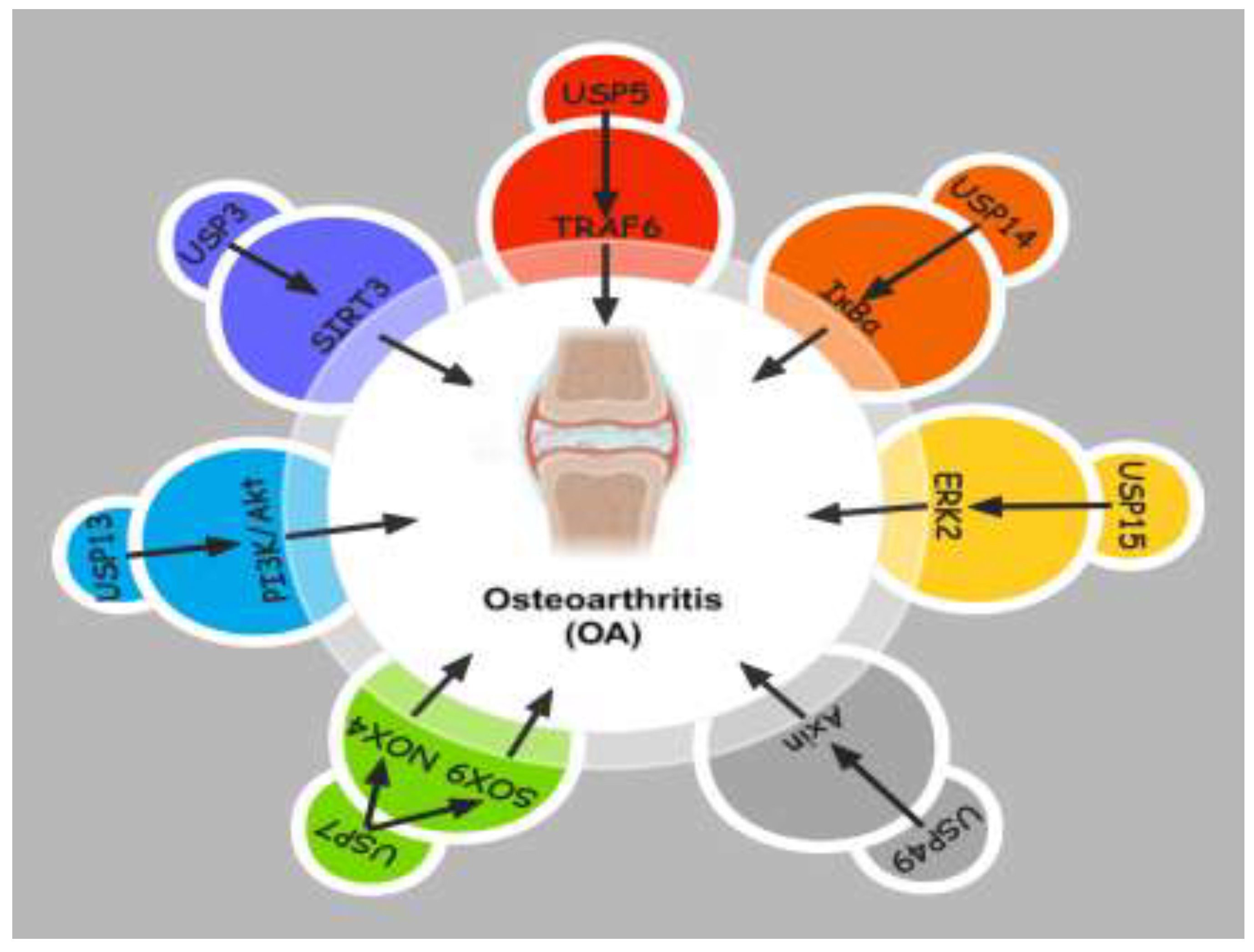
This article summarizes and analyzes the latest research progress on the relationship between deubiquitinating enzymes and osteoarthritis (OA), related pathogenesis, and therapeutic strategies. As a class of important molecules in protein degradation and recycling, deubiquitinating enzymes serve as a crucial component in the occurrence and development of OA. By engaging in the modulation of intracellular signaling pathways, apoptosis, and cellular metabolism, deubiquitinating enzymes may become important molecules affecting the pathological process of OA(Figure 1).
Studies have shown that the mechanisms of action of deubiquitinating enzymes in the pathological mechanisms of OA are complex and diverse. They may affect the stability of the extracellular matrix, regulate the expression of cytokines, and participate in inflammatory responses through various pathways. In addition, abnormal activity of deubiquitinating enzymes may lead to an imbalance in cellular signal transduction, thereby promoting the development of OA.
In response to the abnormal role of deubiquitinating enzymes in OA, researchers have proposed new therapeutic strategies based on these molecules. These strategies include the development of small molecule inhibitors targeting specific deubiquitinating enzymes and the mitigation or reversal of OA progression by intervening in the activity of deubiquitinating enzymes. The development of these strategies not only provides new targets for OA treatment but also opens up new research directions.
However,the prevailing comprehension of the function of deubiquitinating enzymes in OA and their use as therapeutic targets is still in its infancy. Future research needs to address the specific mechanisms of these molecules, their interactions with other signaling molecules, and how these actions lead to the development of OA. In addition, clinically safe and effective drugs that intervene in the activity of deubiquitinating enzymes are still urgently needed for development. Through in-depth research, we can not only comprehensively understand the pathophysiological mechanisms of OA but also achieve more effective disease prevention and treatment strategies.
6.2. Limitations of the Study and Future Research Directions
Although the connection between deubiquitinating enzymes and OA has been valued, the current understanding of their specific roles in the disease is still limited. The challenges faced by researchers include how to accurately locate the specific roles of deubiquitinating enzymes in OA and how they interact with other molecular pathways to regulate the progression of OA. In addition, the regulation of deubiquitinating enzyme activity holds substantial importance for the identification of novel therapeutic targets, but how to achieve this goal specifically, especially without affecting other important physiological processes, remains a challenge.
Future research directions include but are not limited to: in-depth exploration of the specific roles of deubiquitinating enzymes in the pathogenesis of OA, revealing how they regulate the activity of OA-related cytokines and proteases; confirming the impact of specific deubiquitinating enzyme inhibitors on OA progression through animal models and cell culture; and developing small molecule drugs targeting specific deubiquitinating enzymes, and assessing their safety and efficacy in preclinical settings. In addition, considering the multifactorial characteristics of OA, future research should also pay attention to the interactions between deubiquitinating enzymes and other molecules, in the hope of finding more effective comprehensive treatment strategies.
Author Contributions
YL wrote the manuscript and collated the articles.ZY and GC consulted the literature, participated in the drafting of the manuscript. HZ provided oversight for the project and was responsible for conducting the manuscript evaluation. All authors read and approved the final manuscript.
Funding
This study was funded by Dongguan Science and Technology of Social Development Program (No. 20231800940422, No. 20231800935592, No. 20231800935582).
Institutional Review Board Statement
Not applicable.
Informed Consent Statement
Not applicable.
Data Availability Statement
No new data were created or analyzed in this study.
Conflicts of Interest
The authors declare no conflicts of interest.
References
- Bernabei, I.; So, A.; Busso, N.; Nasi, S. Cartilage calcification in osteoarthritis: mechanisms and clinical relevance. Nat Rev Rheumatol 2023, 19, 10–27. [Google Scholar] [CrossRef]
- Synovial inflammation in osteoarthritis progression - PubMed. Available from: https://pubmed.ncbi.nlm.nih.gov/35165404/. [CrossRef]
- A review of quality-of-life in elderly osteoarthritis - PubMed. Available from: https://pubmed.ncbi.nlm.nih.gov/36803292/. [CrossRef]
- Jansen, M.P.; Mastbergen, S.C. Joint distraction for osteoarthritis: clinical evidence and molecular mechanisms. Nat Rev Rheumatol 2022, 18, 35–46. [Google Scholar] [CrossRef]
- Mechanism of HIFs in osteoarthritis - PubMed. Available from: https://pubmed.ncbi.nlm.nih.gov/37020556/. [CrossRef]
- Rosado, S.E. Osteoarthritis affects us too: an expert panel survey of factors important for younger adult wellbeing. Soc Work Health Care 2023, 62, 73–92. [Google Scholar] [CrossRef] [PubMed]
- Bhat, K.P.; Ümit Kaniskan, H.; Jin, J.; Gozani, O. Epigenetics and beyond: targeting writers of protein lysine methylation to treat disease. Nat Rev Drug Discov 2021, 20, 265–286. [Google Scholar] [CrossRef] [PubMed]
- Functions and mechanisms of non-histone protein acetylation - PubMed. Available from: https://pubmed.ncbi.nlm.nih.gov/30467427/. [CrossRef]
- Ubiquitylation at the crossroads of development and disease - PubMed. Available from: https://pubmed.ncbi.nlm.nih.gov/28928488/. [CrossRef]
- Yang, X.; Qian, K. Protein O-GlcNAcylation: emerging mechanisms and functions. Nat Rev Mol Cell Biol 2017, 18, 452–465. [Google Scholar] [CrossRef] [PubMed]
- Ubiquitination in disease pathogenesis and treatment - PubMed. Available from: https://pubmed.ncbi.nlm.nih.gov/25375928/. [CrossRef]
- Liu, J.; Chen, T.; Li, S.; Liu, W.; Wang, P.; Shang, G. Targeting matrix metalloproteinases by E3 ubiquitin ligases as a way to regulate the tumor microenvironment for cancer therapy. Semin Cancer Biol 2022, 86, 259–268. [Google Scholar] [CrossRef] [PubMed]
- Deubiquitylating enzymes and drug discovery: emerging opportunities - PubMed. Available from: https://pubmed.ncbi.nlm.nih.gov/28959952/. [CrossRef]
- Parihar, N.; Bhatt, L.K. Deubiquitylating enzymes: potential target in autoimmune diseases. Inflammopharmacology 2021, 29, 1683–1699. [Google Scholar] [CrossRef]
- Targeting the ubiquitination/deubiquitination process to regulate immune checkpoint pathways - PubMed. Available from: https://pubmed.ncbi.nlm.nih.gov/33479196/. [CrossRef]
- Cellular functions regulated by deubiquitinating enzymes in neurodegenerative diseases - PubMed. Available from: https://pubmed.ncbi.nlm.nih.gov/34023421/. [CrossRef]
- Deubiquitinating PABPC1 by USP10 upregulates CLK2 translation to promote tumor progression in pancreatic ductal adenocarcinoma - PubMed. Available from: https://pubmed.ncbi.nlm.nih.gov/37757903/. [CrossRef]
- Deubiquitinase PSMD7 promotes the proliferation, invasion, and cisplatin resistance of gastric cancer cells by stabilizing RAD23B - PubMed. Available from: https://pubmed.ncbi.nlm.nih.gov/34512150/. [CrossRef]
- Corrigendum: USP5 Promotes Metastasis in Non-Small Cell Lung Cancer by Inducing Epithelial-Mesenchymal Transition via Wnt/β-Catenin Pathway - PubMed. Available from: https://pubmed.ncbi.nlm.nih.gov/32670065/. [CrossRef]
- USP12 promotes breast cancer angiogenesis by maintaining midkine stability - PubMed. Available from: https://pubmed.ncbi.nlm.nih.gov/34759262/. [CrossRef]
- Deubiquitinating enzymes: potential regulators of the tumor microenvironment and implications for immune evasion - PubMed. Available from: https://pubmed.ncbi.nlm.nih.gov/38715050/. [CrossRef]
- Tanguturi, P.; Kim, K.-S.; Ramakrishna, S. The role of deubiquitinating enzymes in cancer drug resistance. Cancer Chemother Pharmacol 2020, 85, 627–639. [Google Scholar] [CrossRef] [PubMed]
- J D, L F, Y S, L Z. The function and regulation of OTU deubiquitinases. Frontiers of medicine 2020;14. [CrossRef]
- The emerging role of deubiquitylating enzymes as therapeutic targets in cancer metabolism - PubMed. Available from: https://pubmed.ncbi.nlm.nih.gov/35307036/. [CrossRef]
- Deubiquitinases in cell death and inflammation - PubMed. Available from: https://pubmed.ncbi.nlm.nih.gov/35608338/. [CrossRef]
- The Ubiquitin System and A20: Implications in Health and Disease - PubMed. Available from: https://pubmed.ncbi.nlm.nih.gov/32853526/. [CrossRef]
- Cellular functions of the DUBs - PubMed. Available from: https://pubmed.ncbi.nlm.nih.gov/22357969/. [CrossRef]
- The USP7 protein interaction network and its roles in tumorigenesis - PubMed. Available from: https://pubmed.ncbi.nlm.nih.gov/35005106/. [CrossRef]
- Regulation of RIG-I Activation by K63-Linked Polyubiquitination - PubMed. Available from: https://pubmed.ncbi.nlm.nih.gov/29354136/. [CrossRef]
- Dissenting degradation: Deubiquitinases in cell cycle and cancer - PubMed. Available from: https://pubmed.ncbi.nlm.nih.gov/32201366/. [CrossRef]
- Huang X, Summers MK, Pham V, et al. Deubiquitinase USP37 is activated by CDK2 to antagonize APC(CDH1) and promote S phase entry. Mol Cell 2011;42:511–23. [CrossRef]
- Zhang Y, Jost M, Pak RA, et al. Adaptive exchange sustains cullin-RING ubiquitin ligase networks and proper licensing of DNA replication. Proc Natl Acad Sci U S A 2022;119:e2205608119. [CrossRef]
- USP15 Represses Hepatocellular Carcinoma Progression by Regulation of Pathways of Cell Proliferation and Cell Migration: A System Biology Analysis - PubMed. Available from: https://pubmed.ncbi.nlm.nih.gov/36900163/. [CrossRef]
- Zhang, H.; Huang, H.; Feng, X.; et al. Deubiquitinase USP28 inhibits ubiquitin ligase KLHL2-mediated uridine-cytidine kinase 1 degradation and confers sensitivity to 5’-azacytidine-resistant human leukemia cells. Theranostics 2020, 10, 1046–1059. [Google Scholar] [CrossRef]
- An, Y.; Yang, S.; Guo, K.; Ma, B.; Wang, Y. Reduced USP39 expression inhibits malignant proliferation of medullary thyroid carcinoma in vitro. World J Surg Oncol 2015, 13, 255. [Google Scholar] [CrossRef] [PubMed]
- Liu, X.; Moussa, C. Regulatory Role of Ubiquitin Specific Protease-13 (USP13) in Misfolded Protein Clearance in Neurodegenerative Diseases. Neuroscience 2021, 460, 161–166. [Google Scholar] [CrossRef] [PubMed]
- Ubiquitin ligases and cell cycle control - PubMed. Available from: https://pubmed.ncbi.nlm.nih.gov/23495935/. [CrossRef]
- The Emerging Role of Deubiquitinases in Cell Death - PubMed. Available from: https://pubmed.ncbi.nlm.nih.gov/36551253/. [CrossRef]
- Roles of deubiquitinases in urologic cancers (Review) - PubMed. Available from: https://pubmed.ncbi.nlm.nih.gov/39525605/. [CrossRef]
- USP19 modulates autophagy and antiviral immune responses by deubiquitinating Beclin-1 - PubMed. Available from: https://pubmed.ncbi.nlm.nih.gov/26988033/. [CrossRef]
- Machado-Joseph Deubiquitinases: From Cellular Functions to Potential Therapy Targets - PubMed. Available from: https://pubmed.ncbi.nlm.nih.gov/32982735/. [CrossRef]
- The Function of Drosophila USP14 in Endoplasmic Reticulum Stress and Retinal Degeneration in a Model for Autosomal Dominant Retinitis Pigmentosa - PubMed. Available from: https://pubmed.ncbi.nlm.nih.gov/33053617/. [CrossRef]
- Gu J, Xiao X, Zou C, et al. Ubiquitin-specific protease 7 maintains c-Myc stability to support pancreatic cancer glycolysis and tumor growth. J Transl Med 2024;22:1135. [CrossRef]
- Role of Deubiquitinases in Parkinson’s Disease-Therapeutic Perspectives - PubMed. Available from: https://pubmed.ncbi.nlm.nih.gov/36831318/. [CrossRef]
- Ubiquitination and deubiquitination: Implications on cancer therapy - PubMed. Available from: https://pubmed.ncbi.nlm.nih.gov/37633647/. [CrossRef]
- Deubiquitinases in cancer - PubMed. Available from: https://pubmed.ncbi.nlm.nih.gov/37935888/. [CrossRef]
- USP3 inhibits type I interferon signaling by deubiquitinating RIG-I-like receptors - PubMed. Available from: https://pubmed.ncbi.nlm.nih.gov/24366338/. [CrossRef]
- USP3 inhibits type I interferon signaling by deubiquitinating RIG-I-like receptors - PubMed. Available from: https://pubmed.ncbi.nlm.nih.gov/24366338/. [CrossRef]
- Ubiquitin-specific protease 3 targets TRAF6 for deubiquitination and suppresses IL-1β induced chondrocyte apoptosis - PubMed. Available from: https://pubmed.ncbi.nlm.nih.gov/31056254/. [CrossRef]
- Ubiquitin-specific protease 3 attenuates interleukin-1β-mediated chondrocyte senescence by deacetylating forkhead box O-3 via sirtuin-3 - PubMed. Available from: https://pubmed.ncbi.nlm.nih.gov/34847835/. [CrossRef]
- A G-C, Vm G, P S, et al. The deubiquitinating enzyme USP5 modulates neuropathic and inflammatory pain by enhancing Cav3.2 channel activity. Neuron 2014;83. [CrossRef]
- Small organic molecule disruptors of Cav3.2 - USP5 interactions reverse inflammatory and neuropathic pain - PubMed. Available from: https://pubmed.ncbi.nlm.nih.gov/25889575/. [CrossRef]
- Identification of interleukin-1 beta as a key mediator in the upregulation of Cav3.2-USP5 interactions in the pain pathway - PubMed. Available from: https://pubmed.ncbi.nlm.nih.gov/28741432/. [CrossRef]
- Disrupting USP5/Cav3.2 interactions protects female mice from mechanical hypersensitivity during peripheral inflammation - PubMed. Available from: https://pubmed.ncbi.nlm.nih.gov/30340616/. [CrossRef]
- Proinflammatory Effects of Ubiquitin-Specific Protease 5 (USP5) in Rheumatoid Arthritis Fibroblast-Like Synoviocytes - PubMed. Available from: https://pubmed.ncbi.nlm.nih.gov/32214906/. [CrossRef]
- Linear ubiquitination of LKB1 activates AMPK pathway to inhibit NLRP3 inflammasome response and reduce chondrocyte pyroptosis in osteoarthritis - PubMed. Available from: https://pubmed.ncbi.nlm.nih.gov/36514784/. [CrossRef]
- USP7 Attenuates Endoplasmic Reticulum Stress and NF- κ B Signaling to Modulate Chondrocyte Proliferation, Apoptosis, and Inflammatory Response under Inflammation - PubMed. Available from: https://pubmed.ncbi.nlm.nih.gov/35432716/. [CrossRef]
- USP7 Inhibition Alleviates H2O2-Induced Injury in Chondrocytes via Inhibiting NOX4/NLRP3 Pathway - PubMed. Available from: https://pubmed.ncbi.nlm.nih.gov/33584299/. [CrossRef]
- Metabolite asymmetric dimethylarginine (ADMA) functions as a destabilization enhancer of SOX9 mediated by DDAH1 in osteoarthritis - PubMed. Available from: https://pubmed.ncbi.nlm.nih.gov/36753544/. [CrossRef]
- USP13-mediated IRAK4 deubiquitination disrupts the pathological symptoms of lipopolysaccharides-induced sepsis - PubMed. Available from: https://pubmed.ncbi.nlm.nih.gov/34298177/. [CrossRef]
- USP13 reduces septic mediated cardiomyocyte oxidative stress and inflammation by inducing Nrf2 - PubMed. Available from: https://pubmed.ncbi.nlm.nih.gov/36916102/. [CrossRef]
- USP13 mediates PTEN to ameliorate osteoarthritis by restraining oxidative stress, apoptosis and inflammation via AKT-dependent manner - PubMed. Available from: https://pubmed.ncbi.nlm.nih.gov/33378983/. [CrossRef]
- USP15 in Cancer and Other Diseases: From Diverse Functionsto Therapeutic Targets - PubMed. Available from: https://pubmed.ncbi.nlm.nih.gov/35203682/. [CrossRef]
- USP15: a review of its implication in immune and inflammatory processes and tumor progression - PubMed. Available from: https://pubmed.ncbi.nlm.nih.gov/33824497/. [CrossRef]
- Positive feedback regulation between USP15 and ERK2 inhibits osteoarthritis progression through TGF-β/SMAD2 signaling - PubMed. Available from: https://pubmed.ncbi.nlm.nih.gov/33726807/. [CrossRef]
- Fbxo45 facilitates pancreatic carcinoma progression by targeting USP49 for ubiquitination and degradation - PubMed. Available from: https://pubmed.ncbi.nlm.nih.gov/35279684/. [CrossRef]
- Yang, L.; Wang, Z.; Zou, C.; Mi, Y.; Tang, H.; Wu, X. Ubiquitin-specific protease 49 attenuates IL-1β-induced rat primary chondrocyte apoptosis by facilitating Axin deubiquitination and subsequent Wnt/β-catenin signaling cascade inhibition. Mol Cell Biochem 2020, 474, 263–275. [Google Scholar] [CrossRef]
Figure 1.
deubiquitination play a potential role in osteoarthritis progression.
Disclaimer/Publisher’s Note: The statements, opinions and data contained in all publications are solely those of the individual author(s) and contributor(s) and not of MDPI and/or the editor(s). MDPI and/or the editor(s) disclaim responsibility for any injury to people or property resulting from any ideas, methods, instructions or products referred to in the content. |
© 2025 by the authors. Licensee MDPI, Basel, Switzerland. This article is an open access article distributed under the terms and conditions of the Creative Commons Attribution (CC BY) license (http://creativecommons.org/licenses/by/4.0/).
Copyright: This open access article is published under a Creative Commons CC BY 4.0 license, which permit the free download, distribution, and reuse, provided that the author and preprint are cited in any reuse.


