Preprint
Hypothesis

This version is not peer-reviewed.

A Novel Thermodynamic Perspective on the Female Reproductive Axis: The Wayback Machine Theory

Submitted:

15 January 2025

Posted:

17 January 2025

You are already at the latest version

Abstract

Female reproductive physiology is characterized by its unique complexity. Despite advancements in research tools and resources, improvements in women’s healthcare remain limited. This qualitative study focuses on the fundamuntal principles of hemodynamics of the female reproductive system. Using a quasi-experimental setting, we applied several biophysical laws within the broader framework of the second law of thermodynamics: entropy. Contrary to previous findings which focus on the ovary, our results suggest that the myometrium actually plays the main role in regulating cervical mucus secretion, endometrial growth and even ovarian function, positioning the uterus as the key component of menstrual cycle regulation. In the light of our findings, we suggest a new approach in the hierarchy of the hypothalamic-pituitary-gonadal axis and the development of a potential new drug.

Keywords: 
;  ;  ;  ;  ;  ;  

Introduction

Recent years have witnessed the emergence of sophisticated research techniques, such as advanced imaging and genetic analyses. While these methodologies have provided valuable insights, they can sometimes divert attention from the basic principles of physiology that underpin these processes. As a result, there is a need to revisit these foundational concepts to gain a more holistic understanding of the female reproductive cycle. In this study, we shift our focus back to these fundamental principles, specifically examining the hemodynamics of the female reproductive system. By applying established biophysical laws within the framework of thermodynamics, we aim to uncover insights that may have been overlooked in contemporary research. Our investigation centers on the myometrium, proposing that it is the primary regulator of cervical mucus production, endometrial growth, and ovarian function.

Materials and Methods

Study Design

This study employs a quasi-experimental design that integrates a theoretical reinterpretation of existing findings with practical validation to investigate the regulation of the female reproductive cycle. We applied relevant biophysical laws, such Ohm’s law, Laplace law, Pascal law, along with the second thermodynamics principle, including equilibrium states, to analyze the hemodynamics of the female reproductive system. We confirmed our theoretical findings through the use of a specific substance, available as a nutraceutical which allowed us to observe its effects on cervical mucus production, endometrial growth and ovarian function.
The study focuses mainly on the theoretical aspect of the reproduction cycle. The use of the substance is a proof of concept and not a clinical trial per say.
Contrary to the many studies before which approach female organs separately, we have decided to merge the whole reproductive female system as a single network as that can be seen in an electrical circuit.

Entropy

Entropy is the second law of thermodynamics. This rule states that without external intervention, every organized system will inevitably tend to reach a disordered equilibrium state [1]. The application of this law is very broad in physics and may be abstract. To better understand it, we shall simplify it and use analogy in practical physics.

Ohm’s Law

In hemodynamics, Ohm’s law defines how an organ is perfused by a blood flow, considering the arterial pressure, the venous pressure and the several resistances that flow shall encounter, as the following:
Q = Δ P R Q = B l o o d   f l o w   Δ P = D i f f e r e n c e   b e t w e e n   a r t e r i a l   a n d   v e n o u s   p r e s s u r e   R = F l o w   r e s i s t a n c e
Before applying this in physiology, we shall make a brief explanation of this law in physics, as it is way clearer in an electrical circuit.
The electrical current according to Ohm’s law in a circuit is defined by the difference in potential of a generator, divided by the resistances, where the difference in potential is the voltage.
The voltage itself is defined by the difference of the potential between the anode and the cathode of an electricity generator, such as a battery.
Practically, this means that a flow of electrons (electrical current) needs a source (the positive terminal of the battery), a “swallowing” destination (the negative terminal of the battery) and a permissive path to move in with the least resistances possible, this includes the cabling and all the devices in the circuit.
The sole electron flow purpose here is to restore the equilibrium of the system (entropy), the generated electric current being only a by-product that can be used. Let’s take a basic circuit as an example.
In Figure 1, minimal cabling resistance allows electrons to flow easily and light the bulb correctly.
In Figure 2, however, when we add a resistor, such as a heater, to the circuit, we notice that the light emitted from the bulb dims, despite the resistor being positioned downstream from the bulb.
This paradoxical effect can be explained by the resistor creating a kind of electron “traffic jam” delaying them from reaching the negative terminal. This delay slows down the electric current, not only within the resistor but throughout the entire circuit, causing the light bulb to dim. This highlights the importance of studying devices—and even biological organs—not in isolation, but as interconnected parts of a single system.
Now we are going to apply the same physical rules to the female reproductive system, where the positive terminal is the arterial pressure, the negative terminal is the venous pressure, the uterus and the ovary being the “electric devices”.
The blood flow (arterial pressure) generated by the cardiac output will first pass through the ovarian artery, originating from the abdominal aorta (C, Figure 3), reaching the ovary (D, Figure 3), then going through the utero-ovarian artery (E, Figure 3) reaching finally the uterus through the myometrium (F, Figure 3).
At first glance, we shall not consider the lower uterine artery.
According to Ohm’s law, the blood flow must encounter the least resistances possible to reach the negative terminal for perfusing correctly all the organs.
However the current disposition of the organs highlights a potential obstacle to the blood flow here, which is represented by the myometrium (F, Figure 3). Indeed, a retracted myometrium of the fundic region of the uterus, as we can encounter in the immediate postpartum uterine involution, can seriously slow down the blood flow, not only for the uterus, but for all the circuit, including the ovary, exactly like we have seen in the electrical circuit. For the analogy, we can consider the retracted myometrium as a heater at full power, the increasing resistance will create a blood “traffic jam”. This slow down will reach the ovary (the light bulb), altering significantly its perfusion. As for the venous pressure, like the resistance, we can see in the equation that it is inversely proportional for a sufficient blood flow and must be then as low as possible as well.
In hemodynamics, we know that venous pressure can be significantly reduced in certain medical conditions, such as acute hemorrhage. However, it’s unlikely that physiology would employ this reduction as a means to enhance blood flow. Instead, there is a physiological phenomenon that grants this venous pressure lowering: the production of cervical mucus.
Indeed, the cervical mucus production can loosely be compared to the production of urine by the kidney, as there is a reabsorption of blood cells and excretion of proteins and fluid which shall be the matrix of that mucus, so there will be always a missing fraction of the returning (venous) blood, not in form of hemorrhage, but as cervical mucus. Furthermore, the myometrium reaches the cervical epithelium which produces the mucus. We can suggest that the myometrial relaxation at the cervical level will encourage the production of mucus and help maintain a continuous blood flow, not only for the uterus, but for the ovary as well. We can now close the blood circuit by considering that the cervical mucus production (G, Figure 3) is the negative terminal of the Ohm’s law. The endometrial hemostasis being achieved in the late luteal phase of the previous cycle, the is no hemorrhagic risk.
As a preliminary result, our study suggests that the myometrial relaxation, also known as the infamous uterine inertia, is the key condition for the perfusion for not only the uterus, but also for the ovary, especially in the follicular phase.
To better understand the regulation of the female cycle, we decided to investigate the role of hormones, taking in consideration these new findings.

Clinical Investigation

Our first “suspect” in the hormonal investigation was the FSH hormone. Studying the FSH itself did not really help, however, focusing on its receptor (FSHr) have opened some doors.
Indeed, the FSHr belongs to the family of the G Protein Coupled Receptors (GPRCs). The activation of such receptors leads in many cases, to the excretion, after a complex process, of the intra cytoplasmic calcium through the activation of the AMP cyclase (cAMP)[2]. Among other roles, GPRCs have been found to have an adrenergic effect, by relaxing smooth muscles caused by intracellular calcium excretion[3].
We suspect that FSH actually does not act on the ovarian granulosa cells, but rather on the myometrium. However, in contrast to previous literature, this action may go beyond tocolysis[4], literally provoking an inertia and enhancing the blood flow of the whole system as described earlier. As there are few works in the literature about activating the GPRCs in gynecology and knowing the heavy side effects of FSH hormones medication themselves, we decided to look for a nutraceutical substance that may activate GPRCs to confirm our findings. Being aware that not every GPRCs activation leads to myometrial relaxation, we have used more restrictive inclusion criteria.
We looked in the literature for a substance that acts as a smooth muscle relaxant, involving GPRCs activation and has a potential action on mucus production. Our very specific search has found only one substance that fills all these criteria: Alpha-Hederin. Indeed, Alpha-Hederin, and through its metabolites such Hederacoside C, has been found to inhibit GPRCs internalization, which leads to smooth muscle relaxation after its activation by a Beta2-adrenergic neurotransmitter[5] . This substance is naturally extracted from a plant, Hedera Helix (Common Ivy) and is used as an expectorant and a cough reliever.
The expectorant effect is closely linked to the myorelaxant action: the inhibition of GPRCs internalization will make those receptors available for adrenergic neurotransmitters, the release of the intra cytoplasmic calcium will lead to the myorelaxation of the bronchial Ressessen muscle, this will provide the bronchial epithelium a sufficient blood flow for producing mucus, the fresh mucus will dilute the dehydrated older one, helping in its expulsion which will at the end relieve the productive cough.
After filing for a patent in Algeria, we have decided to formulate a nutraceutical composition based on Alpha-Hederin with the higher dosage, so it can reach the myometrium instead of the thinner Ressesen bronchial muscle.
We administered the substance, after a clear explanation of the effect and the explicit oral consortment of the patients, in the form of a syrup of 1,2 grams of Hedera Helix extract (HH) containing 1.8% of Alpha Hederin, from day 01 to day 14 of the cycle, with a daily intake of 0,3 grams of HH extract depending on the medical indication.
  • Patient Groups and Conditions
  • Group 01:
    • o Patients: 53
    • o Cycle Days: Day 01 to Day 03
    • o Medical Condition: Dysmenorrhea
  • Group 02:
    • o Patients: 20
    • o Cycle Days: Day 01 to Day 07
    • o Medical Condition: Unexplained recurrent genital infection
  • Group 03:
    • o Patients: 15
    • o Cycle Days: Day 01 to Day 14
    • o Medical Condition: Irregular menstrual cycle
  • Inclusion Criteria
  • Group 01: Women aged 18 to 35 experiencing pelvic cramps during the first two days of the cycle.
  • Group 02: Women aged 18 to 43 with abnormal vaginal secretion during the second week of the cycle, with long menstruation day count (7 or more), with or without bacteriological proof of infection.
  • Group 03: Women aged 18 to 35 experiencing an abnormally long or anovulatory cycle. Note: Polycystic Ovarian Syndrome (PCOS) was not specifically targeted as its complexity requires a more organized clinical trial.
As the design of this work is a quasi-experimental study, we insist on the fact that this is not a clinical trial for a new drug, but rather a proof of concept of the earlier described theory. As there is no randomization, we clearly explained the effect of the substance to each patient. We did not file for an ethical approval of a recognized institution, nor the consentment of the local regulatory authorities here in Algeria, as the substance used is well known nutracetic and has negligible side effects. We are willing, in the near future, to provide this substance as a drug approved by our local “FDA” after a validated clinical trial.

Results

  • Group 01
  • Patients: 53
  • Outcome: 45 patients (85%) reported significant pain relief 20 minutes after the first intake.
  • Follow-up Questions and Responses:
    • o Q: Did you notice anything about the menstrual flow?
      • A: Of the 45 patients:
        • 40 (89%) reported a significant increase in menstrual flow, requiring more frequent changes of hygienic pads.
        • 20 (44%) noted a shortening of the menstrual period day count.
    • o Q: Did you notice anything else accompanying menstruation?
      • A: Of the 45 patients, 35 (78%) reported an increase in cervical mucus during menstruation.
  • Group 02
  • Patients: 20
  • Outcome: 13 patients (65%) experienced a significant reduction in symptoms (leucorrhea, itching) and required no further medical treatment in subsequent cycles.
  • Group 03
  • Patients: 15
  • Outcome: 10 patients (67%) observed a regularization of their cycle, with durations stabilizing between 25 and 28 days.
One patient in particular benefited from further investigation:
A 28-year-old married woman had been experiencing unexplained infertility with her partner for three years. Her clinical examination showed no significant findings, and no notable male factors were identified. On day 12 of her cycle, an ultrasound examination revealed multifollicular large ovaries without a dominant follicle.
Although the patient did not meet all criteria for polycystic ovarian syndrome (PCOS), we opted to medically induce ovulation with clomiphene citrate (CC) at a daily dose of 100 mg from day 2 to day 6. However, a follow-up ultrasound on day 12 showed no dominant follicle in either ovary.
We continued ovulation induction with a daily dose of 150 mg of CC over the next three cycles, but these attempts were unsuccessful. For the fourth cycle, we switched to induction with follitropin alfa (Gonal) at a daily dose of 75 IU from day 2 to day 12, conducting multiple ultrasounds during this period, yet no dominant follicle was observed.
In the following cycle, we decided to administer our substance, HH, alone at a dose of 0.3 g from day 1 to day 12. This time, a left dominant follicle measuring 22 mm was detected.
To gain further insight into the action of HH, we conducted elastography examinations before and after its administration.
Elastography is an emerging ultrasound imaging technique used to measure tissue elasticity and stiffness[6] . Although commonly used in hepatology and endocrinology, it is still relatively rare done in obstetrics and gynecology.

Discussion

The pain relief, increased blood and mucus flow, and shortened menstruation observed in Group 01 can be explained by the fact that HH caused a global uterine relaxation. The myometrial softening at the cervical stage shall not only dilate the cervical os, but also restore the blood flow for the mucus secretory epithelium.
These two events combined shall facilitate the elimination of the menstrual content without the need of uterine contractions, responsible for dysmenorrhea.
The reduction of the vaginal infectious symptoms in the group 02 may be connected to the same effect: the facilitation of the menstrual content elimination will remove a crucial culture medium for various kinds of pathogens.
As for the patient of group 03, we can explain the responsiveness to the treatment by the fact that the myorelaxation by HH removed a significant downstream obstacle for the ovary, which is the retracted myometrium. The restored blood flow will bring the necessary material for a convenient follicular growth.

The Redshift Theory

In the imagery, we can see a difference in the elasticity of the myometrium before and after the treatment:
Predominance of blue shades (Figure 4) before HH administration indicates stiffness of the uterus (retracted myometrium).
Green, yellow and even red shades in the uterus (Figure 5) obtained after HH administration indicates that there was a myorelaxation like described before. We can even notice a considerable attenuation of blue shades in the fundus. From these findings, we suggest that the uterine myometrial relaxation, from blue to red is actually responsible for follicular growth, the red shift nomenclature is a tribute to a cosmological phenomenon related to the observation of galaxies fading away due to the expansion of the universe.
Now, to explain how blood flow irrigates ovarian follicles, we shall have to use another biophysical principle.

Laplace Law

Δ P = 2 T r Δ P = T r a n s m u r a l   p r e s s u r e   d i f f e r e n c e i n t e r n a l e x t e r n a l T = S u r f a c e   t e n s i o n   a t   t h e   w a l l   o f   t h e   s p h e r e   r = R a d i u s   o f   t h e   s p h e r e
Laplace law is a fundamental principle that explains how fluid moves inside confined environments. It is widely applied in pulmonary physiology, and can by itself explain ventilation mechanics.
Indeed, any fluid tends to move from the higher to the lower pressure area, in a goal to achieve the equilibrium of the entropy described earlier.
To better understand this, we shall see how it works in pulmonary physiology.
During inspiration, the diaphragm muscle shall expand the thoracic cage. This will lower the pressure inside the body, making the external air “leak” into the body.
However, once inside, air circulation shall face a major problem, that may in some cases, lead to death.
Indeed, taking in consideration that internal alveolar pressure is inversely related to its radius and the fact that all alveoli are in direct communication together, this will make small and weak alveoli to literally spill their modest air countenance in the larger ones, causing them to collapse and drastically reducing gas exchange surface[7].
To prevent this, physiology has equipped small alveoli with a special component that can maintain them open: the surfactant.
If this “unfair” law can be problematic in pulmonary physiology, it can be very useful in gynecology. Since there is no direct communication between follicles, no one shall collapse.
However the blood flow generated earlier by myometrial relaxation favors larger follicles, bypassing small follicles due to their higher internal pressure.
Surface tension is the force that rises at the top of a surface (interface) when two different phases of the matter encounter.
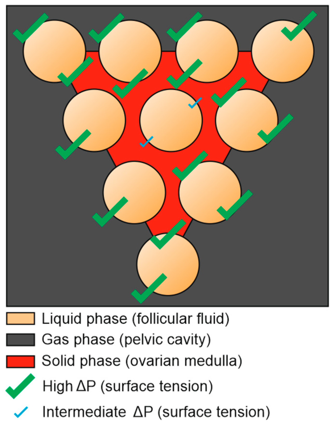
The different three phases we are facing in gynecology are the gas phase (peritoneal pressure), the liquid phase (follicular fluid) and the solid phase (the ovarian medulla). There is a positive correlation between surface tension and pressure gradient. Pressure gradient across a sphere is maximal when the state of the phase is different: a gas/liquid confrontation will generate more pressure gradient (tension) than a liquid/liquid encounter.

Pascal Law

According to Pascal law, the force applied to an incompressible liquid will be transmitted instantly and evenly to all the walls of a confined space, as the following:
P = P a p p l i e d + Δ P P = T o t a l   i n t e r n a l   p r e s s u r e   a t   a n y   p o i n t   i n s i d e   t h e   s p h e r e   P a p p l i e d = I n i t i a l   p r e s s u r e   o f   t h e   f l u i d   Δ P = C h a n g e   i n   p r e s s u r e   a p p l i e d   t o   t h e   f l u i d   s p h e r e
When we combine both of Pascal and Laplace laws, wall thickness, surface tension, we can understand that a small follicle tends to generate a higher internal pressure, similarly to a small alveoli[8], making it unwelcoming for the blood stream supposed to make it grow, the latter will then favor larger follicles with lesser internal pressure.
When the blood flow enters the follicle in the form of plasma (follicular fluid), the rise of follicular pressure will be counterbalanced by an elevation of follicular radius seeking the equilibrium state (entropy). However, the follicular wall thickness is a limited physical property: the radius growth is in fact a stretching made at the cost of the wall being thinner. Furthermore, The incompressibility of the fluid will this time act on the wall making it thinner too. When the elasticity of the thin wall cannot handle the radius growth anymore for sake of the equilibrium described earlier, the follicular shell (albuginea) will break leading to ovulation.
In summary, when the blood flow is moderate, follicular growth, dominance and ovulation are three phenomena that may be controlled by the laws of Laplace and Pascal.
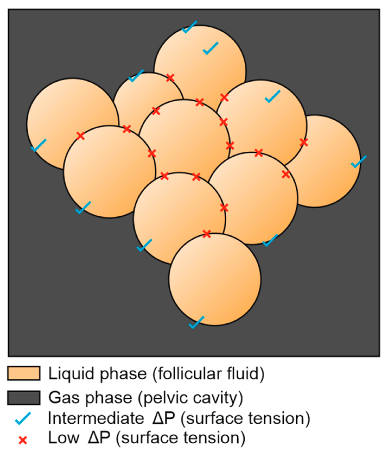
However, when the blood flow gets stronger, such as in the case of hyperstimulation, something else happens. The predominant follicle will grow, but its rapid saturation will make other follicles grow too (Figure 7).
If a “challenging” follicle reaches the predominant one, this can be very counterproductive for ovulation. Indeed, we have seen that the surface tension rise is crucial for ovulation, and it is closely correlated to the nature of the surrounding phase. When a challenging follicle touches the predominant one, it will swap its surrounding solid medulla by a similar follicular fluid. The contact of two same phases (liquid/liquid in this case) will drop the surface tension of the dominant follicle which will seriously compromise its ovulation potential.
In this case, the predominant follicle, which was promised a bright destiny, finds itself brought to the “past” once touched by the growth of other challenging follicles. From these findings, we can presume that a lonely, offcentered follicle with a diameter of 15mm, facing several phases of the matter, especially the gas phase, has a better chance to ovulate than a 21mm follicle surrounded by lesser peers (Figure 7).
This leads us to the conclusion that the current follicular monitoring technique may be incomplete, since it takes in account the diameter only, excluding the other key parameter represented by the pressure gradient generated by the follicular environment.
The current monitoring technique which takes in account follicular diameter only, could have been sufficient in the case the follicles were totally independent from each other (Figure 6), in a manner of a grapefruit: each unit of the fruit would be completely surrounded by different states of the matter, which will grant a sufficient pressure gradient. in this case, the pressure would be constant and the surface tension would be factorial to diameter only.
Figure 6. The confrontation of the 3 different states of the matter by its own (fluid-gas-solid) tends to generate a higher surface tension, whatever the follicular diameter.
Preprints 146226 g006
Figure 7. The growth of multiple follicles (hyperstimulation) at the expense of the ovarian medulla replaces a solid phase by a liquid one, causing a general drop of surface tension.
Preprints 146226 g007
This follicular reckless “race to ovulation” may have very useful roles in physiology: The first one is to prevent multiple pregnancies by favoring only one ovulation. The second one, by the way, is the key evidence of this manuscript: preventing ovarian depletion in the molar pregnancy.
The molar pregnancy is a medical condition where the trophoblast undergoes pathological cystic transformation, leading ultimately to an abortion.
It is well established that in the molar pregnancy, the ovaries are often hyperstimulated, this condition being assigned to BHCG action[9].
Based on the findings of this work and after reinterpretation of hormonal actions, we are not in agreement with this theory.
Indeed, the problem with the molar pregnancy is not the level of the BHCG itself, but its elevation compared to the term of the pregnancy. For instance, we may very well find a BHCG level of 300.000 IU/ml in an 8 weeks pregnancy and consider it molar, but the problem is that we shall find similar rates in a physiological pregnancy at 26 weeks as well. The question: Why do we never find hyperstimulated ovaries in a physiological pregnancy of 26 weeks? Answer: Because BHCG may be not responsible for hyperstimulation. The actual answer resides in the physical exam which is often neglected: a soft uterus fundus. For some reason, practitioners think they are sensing the vesicles themselves during palpation, this may in fact be a common misconception since regular trophoblast and amniotic fluid of a physiological pregnancy have similar, even softer consistency than the molar vesicles. In fact, they are dealing with a surrendered soft myometrium.
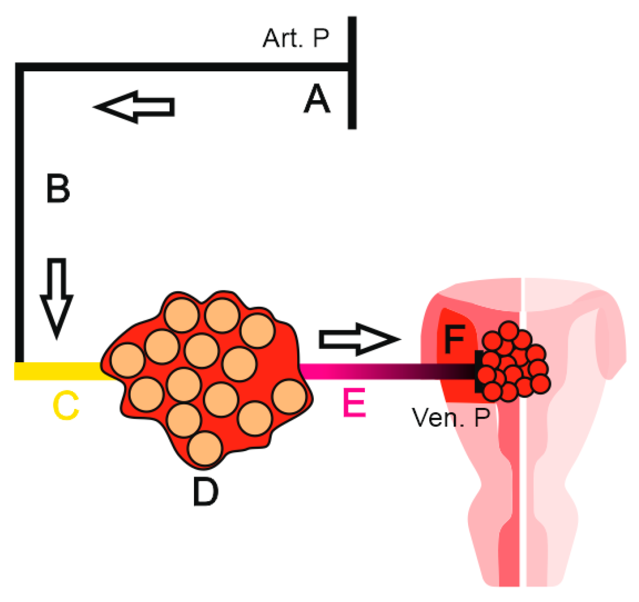
There is no need to practice an elastography here since the elevated fundal height made it clinically obvious. This aligns with our first theory of the relationship between a soft myometrium and ovarian stimulation. In this case, the FSH is not the responsible of the inertia, but the uncontrolled mitosis of the vesicular trophoblast caused myometrial neovascularization that gained backward access to the ovary through the utero ovarian artery. Despite myometrial efforts to retract, the aggressive trophoblastic activity “won” the duel and relaxed the uterine fundus, literally opening the “gates of hell” for the ovary(Figure 8).
The ovary in this case has little margin of maneuvers and can only activate a final defensive mode: the growth of multiple follicles will cause a general drop of surface tension and prevent follicular depletion (Figure 7).
This “airbag” (or water bag) mode will be the last resort of the ovary: if the molar pregnancy continues, the flow will remove all the medulla, as there will be no internal surface tension, the ovary will behave as a giant single cyst leading to an ovarian rupture and potentially the death of the patient. If the current technique of follicular monitoring, which relies exclusively on follicular diameter was correct, many hyperstimulation cases would result in an ovarian menopause.
We decided to make further investigations and revisit the iatrogenic ovarian hyperstimulation syndrome of in vitro fertilization (OHSS). When we compare these two hyperstimulations (molar and IVF) we find a key difference in the symptomatology.
Indeed, in the OHSS of the IVF, we encounter an ascites[10], which sometimes needs to be aspirated and can lead to an elevation of the thromboembolic risk, requiring preventive anticoagulant medication. Question: Why don’t we find ascites in molar pregnancy, despite the similarity of the background ? Answer: It is due to the “negative terminal” of the Ohm’s law described earlier.
In the molar pregnancy, the final generator of the blood flow (negative terminal) that assures the drop of the venous pressure is the pregnancy itself, where there is only a partial sequestration of the blood by the trophoblast. Whereas in the IVF OHSS, the drop of venous pressure is probably caused by the production of the cervical mucus, but rather than a partial blood sequestration, there may be a major leak of proteins for the production of the mucus, which leads to a drop of the oncotic pressure, leading to this ascites.
As a final analogy, we can take an example of water circulation inside a pipe: How do water companies bill water consumption? There are two options: 1- There is a person in each household that collects all the used water in a big container and then takes it to the water company for billing. 2- There is a water meter that measures the consumption (pretty reliable option).
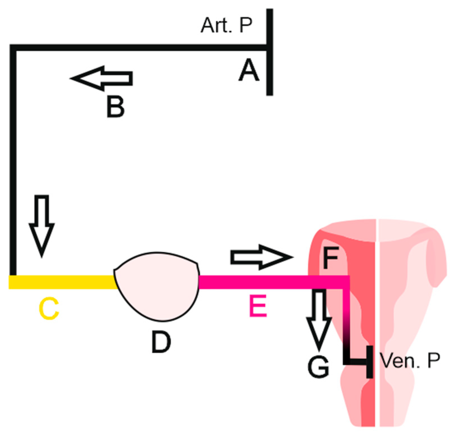
How does the water meter measure the consumption ? Actually, it doest it, not by measuring the final water output, but using the flow of the circulating water. When the tap is open, the water going outside generates a flow in the water meter despite its upstream location. The tap here would be the myometrium, the water meter being the ovary.
FSH would manually open the tap, while molar pregnancy shall force it in a manner of introducing a metallic rod backwards into the nozzle, reaching the mechanism and breaking it. In both cases, a flow will be generated for the water meter (ovary). We can go further in our interpretation and consider that even a small cervical polyp, or a cervical narrowing can be sufficient to stop follicular growth by blocking mucus production, which may be a key condition for maintaining a correct blood flow for the ovary.

Conclusions

Our findings suggest that myometrial inertia during the follicular phase is not only the active form of the uterus, but the whole reproductive female system, including the ovary.
The uterus, through the myometrium, may in fact be the “time machine” controlling the ovary. This action is probably caused by the FSH stimulation, positioning the granulosa cells and the whole ovary as passive organs in the menstrual cycle, subjected to the uterine flow.
Uterine inertia may have suffered an unjustified backlash in the literature due to the major vital risk associated with it in obstetrics.
However, further investigations may have to be done on the retracted myometrium globe state, as this one may be causing a lot of problems in female reproductive physiology despite its “reputation”.
The pituitary gland, through FSH, may in fact activate the uterus first, before the ovary. We are even questioning whether FSH has a direct action on the ovary itself.
During ultrasound monitoring, the follicle diameter measured alone may be insufficient, since its direct surroundings have to be taken in account too: The enemies of a dominant follicle are adjacent follicles.
Myometrial elastography should be considered for monitoring, especially in case of poor ovarian response.
A cervical polyp or even a simple narrowing can literally be the grain of sand that jam the whole machine by cutting a substantial blood flow caused by the venous pressure increasing.
FSH receptors externalization should be considered as an option in case of poor ovarian response using gonadotropin drugs.
Myometrial retraction and cervical obstacles hindering mucus production should be considered in the PCOS physiopathology as potential etiologies.
We suggest that, in the hierarchy of the reproductive axis, the uterus (myometrium) position should be reconsidered, as “The hypothalamic-pituitary-uterine-gonadal axis” would be a more inclusive nomenclature.

Conflicts of Interests

The author declares no conflict of interest.

References

  1. Wehrl, Alfred. General properties of entropy. Reviews of Modern Physics, vol. 50, no. 2, 1978, pp. 221–260. [CrossRef]
  2. Dhyani, Vaibhav, Gare, Suman, Gupta, Rishikesh Kumar, Swain, Sarpras, Venkatesh, K.V., and Giri, Lopamudra. GPCR mediated control of calcium dynamics: A systems perspective. Cellular Signalling, vol. 74, 2020, pp. 109717. [CrossRef]
  3. Billington, Charlotte K, and Penn, Raymond B. Signaling and regulation of G protein-coupled receptors in airway smooth muscle. Respir. Res., vol. 4, no. 1, 2003, pp. 2. [CrossRef]
  4. Stilley, J. A. W., Guan, R., Santillan, D. A., Mitchell, B. F., Lamping, K. G., and Segaloff, D. L. Differential Regulation of Human and Mouse Myometrial Contractile Activity by FSH as a Function of FSH Receptor Density. Biology of Reproduction, vol. 95, no. 2, 2016, pp. 36–36. [CrossRef]
  5. Schulte-Michels, Janka, Wolf, Anne, Aatz, Stefan, Engelhard, Katharina, Sieben, Anne, Martinez-Osuna, Manuel, Haberlein, Felix, and Haberlein, Hanns. α-Hederin inhibits G protein-coupled receptor kinase 2-mediated phosphorylation of β2-adrenergic receptors. Phytomedicine, vol. 23, no. 1, 2016, pp. 52–57. [CrossRef]
  6. Sarvazyan, Armen, Hall, Timothy J, Urban, Matthew W, Fatemi, Mostafa, Aglyamov, Salavat R, and Garra, Brian S. An overview of Elastography - an emerging branch of medical imaging. Curr. Med. Imaging Rev., vol. 7, no. 4, 2011, pp. 255–282. [CrossRef]
  7. Walton, Jon J. Advanced ventilation management. Surgery (Oxford), vol. 33, no. 10, 2015, pp. 485–490. [CrossRef]
  8. Grune, Jana, Tabuchi, Arata, and Kuebler, Wolfgang M. Alveolar dynamics during mechanical ventilation in the healthy and injured lung. Intensive Care Med. Exp., vol. 7, Suppl 1, 2019, pp. 34. [CrossRef]
  9. Alhalabi, Kinan, Lampl, Brooke S., and Behr, Gerald. Ovarian hyperstimulation syndrome as a complication of molar pregnancy. Cleveland Clinic Journal of Medicine, vol. 83, no. 7, 2016, pp. 504–506. [CrossRef]
  10. Chen, Chin-Der, Wu, Ming-Ying, Chao, Kuang-Han, Lien, Yih-Ron, Chen, Shee-Uan, and Yang, Yu-Shih. Update on management of ovarian hyperstimulation syndrome. Taiwanese Journal of Obstetrics and Gynecology, vol. 50, no. 1, 2011, pp. 2–10. [CrossRef]
Figure 1. Circuit diagram showing a battery (A), light bulb (B), and electron flow (C).
Preprints 146226 g001
Figure 2. Circuit diagram showing a battery (A), light bulb (B), electron flow (C), resistor (D).
Preprints 146226 g002
Figure 3. Circuit diagram representing arterial pressure (A), blood flow (B), ovarian artery (C), ovary (D), utero-ovarian artery (E), myometrium (F), venous return (G).
Preprints 146226 g003
Figure 4. Elastography exam, before Hedera Helix administration.
Preprints 146226 g004
Figure 5. Elastography exam, two hours after Hedera Helix administration.
Preprints 146226 g005
Figure 8. Circuit diagram representing arterial pressure (A), blood flow (B), ovarian artery (C), hyper-stimulated ovary (D), utero-ovarian artery (E), soft myometrium (F), venous retrun (molar pregnancy) (G).
Preprints 146226 g008
Disclaimer/Publisher’s Note: The statements, opinions and data contained in all publications are solely those of the individual author(s) and contributor(s) and not of MDPI and/or the editor(s). MDPI and/or the editor(s) disclaim responsibility for any injury to people or property resulting from any ideas, methods, instructions or products referred to in the content.
Copyright: This open access article is published under a Creative Commons CC BY 4.0 license, which permit the free download, distribution, and reuse, provided that the author and preprint are cited in any reuse.
Prerpints.org logo

Preprints.org is a free preprint server supported by MDPI in Basel, Switzerland.

Subscribe

Disclaimer

Terms of Use

Privacy Policy

Privacy Settings

© 2025 MDPI (Basel, Switzerland) unless otherwise stated