Preprint
Case Report

This version is not peer-reviewed.

Interstitial 1q Deletion Syndrome: A New Patient with Congenital Diaphragmatic Hernia and Multiple Midline Anomalies

A peer-reviewed article of this preprint also exists.

Submitted:

14 January 2025

Posted:

16 January 2025

You are already at the latest version

Abstract

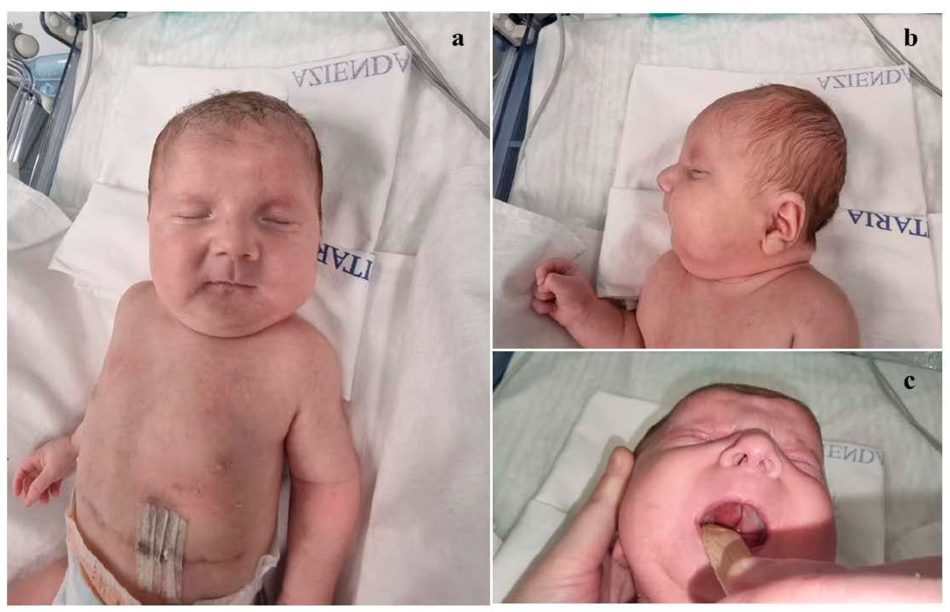
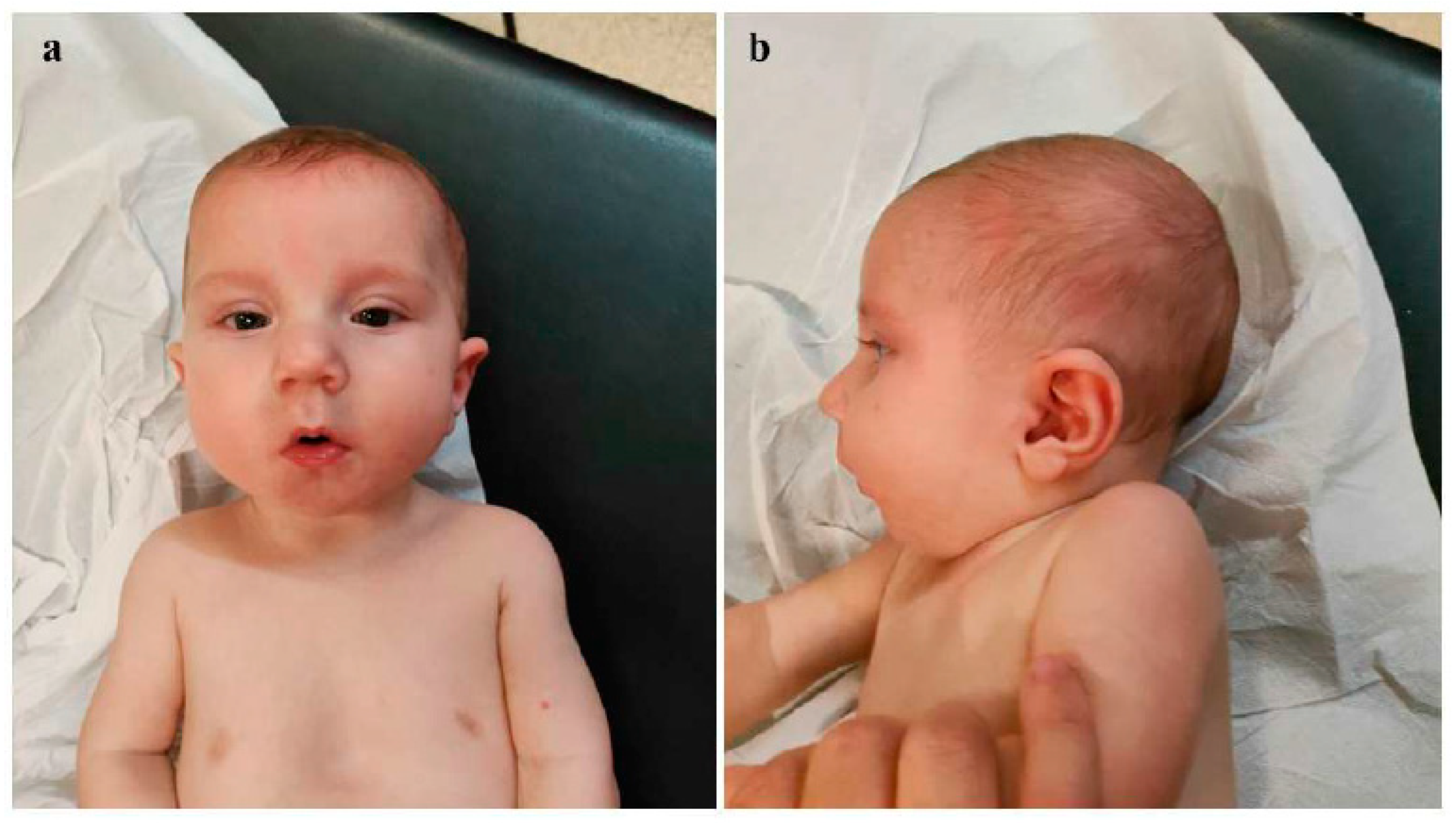
Background: Congenital diaphragmatic hernia (CDH) represents one of the most critical neonatal emergencies, whose timely recognition and appropriate management are essential to ensure patient survival. Genetic investigations play a crucial role for accurate diagnostic assessment, especially in those cases associated with other congenital defects and/or dysmorphic features. Interstitial deletions of chromosome 1q are rare, with about 30 cases reported in the literature. The phenotypical features of the affected subjects described so far include microcephaly, pre- and postnatal growth retardation, psychomotor delay, ear anomalies, brachydactyly, in addition to small hands and feet and rarely congenital diaphragmatic hernia (CDH). Here, we report on a neonate with CDH, dysmorphic features, and multiple midline anomalies, in whom array-comparative genomic hybridization (a-CGH) analysis allowed the identification of an interstitial deletion of the long arm of chromosome 1. Case presentation: The proband is a male infant, born late preterm at 36+1 weeks, with prenatal diagnosis of congenital diaphragmatic hernia (CDH) for which parents refused further genetic investigations. At birth physical examination revealed dysmorphic features and multiple midline anomalies, including cleft palate, pectus excavatum, and ostium primum type atrial septal defect, in addition to hypotonia. Cranial ultrasound showed poor gyral development. a-CGH identified a deletion of approximately 12 Mb on the long arm of chromosome 1, within the region 1q31.1-q32.1, thereafter documented as de novo. On the fourth day of life, he underwent surgical correction of CDH. At about one month of age, the infant was discharged and enrolled in a multidisciplinary follow-up, during which an impaired growth has been evidenced. During the last neuropsychiatric evaluation at the age of 8 months, the child showed mild hypotonia and immature manual exploration. Head control was present but inconsistent. Trunk control was not yet developed, and lower limb positioning exhibited flexion, external rotation and varus attitude of feet. He does not show any further clinical anomalies, and laboratory tests as well as US multiorgan and neurosensorial evaluations do not put in evidence other abnormalities to date. The surgical correction of cleft palate is currently planned to be performed at one year of age. Conclusions: The few cases of chromosome 1q deletions reported to date, along with the clinical and genetic profile of the present neonate, point out that 1q deletions should be considered within the context of "interstitial 1q deletion syndrome". Comparing our case with those described in previous studies, the involved genomic regions and the phenotypic traits are partially overlapping, although the clinical picture of the present patient is among the few ones including congenital diaphragmatic hernia within the phenotypical spectrum. A more extensive comparative analysis of a larger number of patients with similar genetic profiles may allow for a more precise clinical and genomic characterization of this rare syndrome, and for genotype-phenotype correlations.

Keywords: 
;  ;  

Background

Congenital diaphragmatic hernia (CDH) results from abnormal diaphragm development, leading to the herniation of abdominal viscera into the thoracic cavity. CDH is associated with pulmonary hypoplasia and pulmonary hypertension [1]. Its incidence is estimated at approximately 1:4000 live births. In about 5-10% of cases, CDH is related with genetic syndromes (such as Fryns, Donnai-Barrow, Cornelia de Lange, Beckwith-Wiedemann, Pallister-Killian, and Wolf-Hirschhorn) [2,3,4,5] or other chromosomal anomalies, such as deletions of chromosome 1q41q42, 8p23.1, and 15q26 [5]. In recent years, the development of new diagnostic techniques [6,7] and therapeutic strategies has significantly improved the prognosis and survival of patients affected [1,2]. Therefore, a proper diagnostic assessment including appropriate genetic testing, and individualized treatment is crucial for these patients.
Deletions of the long arm of chromosome 1 are rare and, particularly, cases of 1q31.1-q31.2 deletion are exceptionally observed. The clinical features of affected individuals include microcephaly, pre- and postnatal growth retardation, psychomotor delay, ear anomalies, brachydactyly, small hands and feet. Cases reported in the literature so far present some overlapping phenotypic traits, with CDH being documented only in individuals with 1q43-q44 deletions. Here, we report on a neonate with CDH, facial dysmorphisms, and multiple midline anomalies, in whom molecular cytogenetic analysis (array-comparative genomic hybridization, a-CGH) identified a deletion of the long arm of chromosome 1 (1q31.1-q32.1).

Case Presentation

The proband is a male, second child of healthy and non-consanguineous parents. Family history was unremarkable. During the first trimester of pregnancy, prenatal screening (bi-test) indicated an intermediate risk of trisomy 21, and a high one of fetal growth restriction (FGR). The couple declined any prenatal genetic testing. During the third trimester, polyhydramnios and a left congenital diaphragmatic hernia (CDH) were identified. He was born late preterm, at 36+1 weeks of gestation, via emergency cesarean section due to failure of labor progression. At birth (which occurred at a birthing center of Western Sicily, in a nearby province with respect to our Mother and Child Department of the University of Palermo, Palermo, Italy), Apgar scores were 0, 4 and 6, at 1, 5 and 10 minutes respectively. The patient underwent advanced resuscitation maneuvers, including intubation, and was transferred to the Neonatal Intensive Care Unit (NICU). There, invasive mechanical ventilation, total parenteral nutrition, and cardiovascular support through inotropic drugs were initiated. After stabilization, he was transferred to our Department, in which the Divisions of Pediatric Surgery, Medical Genetics and Child Neuropsychiatry are present, to continue diagnostic and therapeutic management. At admission, anthropometric measures were as follows: weight 3050 g (76th centile; +0.72 standard deviations, SD), length 52 cm (99th centile; +2.21 SD), and occipitofrontal circumference (OFC) 35.5 cm (96th centile; +1.75 SD), according with the Italian INeS Growth Charts [8]. Physical examination revealed a broad and receding forehead, hypertelorism, wide nasal root, bulbous tip, anteverted nares, long and thick philtrum, thin lips, dysplastic, low-set and posteriorly rotated ears with thickened helices, complete (primary and secondary) cleft palate, microretrognathia, pectus excavatum (Figure 1, a, b, c). Left cryptorchidism and bilateral clinodactyly of the fifth toe completed his clinical profile.
Neurological examination revealed generalized hypotonia, reduced deep tendon reflexes (DTR), diminished primitive reflexes, and decreased responsiveness. Brain ultrasound showed poor gyral development and millimetric cystic lesions within the periventricular white matter. Echocardiography revealed ostium primum type atrial septal defect (0.8 cm), patent foramen ovale and ductus arteriosus, as well as moderate pulmonary hypertension (pulmonic artery pressure [PAP] values of about 50 mmHg) which subsequently decreased after medical treatment. Testicular ultrasound identified only right testis (volume reduced than normal, resulting from the following measures 1 x 0.6 x 0.3 cm, [9,10]) being visualized along the inguinal canal. Ophthalmological examination did not reveal any anomalies. In light of the congenital malformations and dysmorphic features found, a molecular cytogenetic analysis (a-CGH) was performed. The genetic test allowed the identification of a deletion of approximately 12 Mb on the long arm of chromosome 1 (average resolution 50 kb), within the region 1q31.1-q32.1 (GRCh37, proximal and distal breakpoints between 187,956,640 and 199,996,777 bp, respectively), thereafter documented as de novo, and classified as probably pathogenic according to the American College of Medical Genetics and Genomics guidelines [11]. On the fourth day of life, the patient underwent surgical correction of CDH. The defect was large, due to near-complete agenesis of the left hemidiaphragm, with herniation of stomach, colon, small intestine, spleen, and left lobe of the liver. After repositioning the viscera into the abdomen, a patch placement was necessary. The postoperative course was complicated by a significant pleural effusion (4x4 cm), which was resolved after conservative treatment (diuretic therapy with furosemide, plasma administration, and correction of hypoproteinemia with fresh plasma and albumin). Subsequently, the clinical course worsened due to a sepsis caused by Staphylococcus epidermidis, from which the patient recovered after antibiotic therapy (vancomycin). The following clinical evolution was regular, with enteral nutrition with breast milk being initiated approximately two weeks after surgery, first via nasogastric tube and then using a special bottle suitable for cleft palate. After about one month, and following specific speech therapy and mother-infant bonding in rooming-in setting, complete and exclusive bottle feeding was achieved. However, feeding difficulties persisted, associated with frequent regurgitation, for which special anti-regurgitation formula, meal fractionation, and postural therapy have been started, finally leading to beneficial effects and improvement of symptoms. The patient was then discharged and enrolled in a multidisciplinary follow-up. Neurodevelopmental assessment at 2 months of age (corrected age of 1 month and 13 days) showed poor and repetitive spontaneous movements, generalized hypotonia, but normal passive muscle tone, as well as primitive and deep tendon reflexes. At approximately five months of age (corrected age 4 months; Figure 2, a, b), the testes were palpable in both inguinal regions. At cardiological follow-up evaluation only the persistence of patent foramen ovale was observed. The surgical scar was flat, with no evidence of excessive healing.
At 8 months of age, chest X-ray revealed a reduction in the opacification of the left hemithorax, with persistence of parenchymal consolidation in the mid-to-upper lung field, with partial sparing of the marginal zone. The arterial blood analysis evidenced a balanced profile. Neuropsychiatric evaluation disclosed an alert and cooperative patient. Visual engagement, social smiling and response to name were present. The infant was looking for contact with the operator. Pupils were isochoric, isocyclic and normoreactive to light stimuli. Cranial nerves were intact upon exploration. Visual tracking of the object was present both for visual and sound stimuli. The baby recognized familiar faces. Gestures were poor. He manipulated one cube at a time and threw objects on the ground. The exploration was predominantly oral. Generalized hypotonia was still present although mild: wide excursion at the scarf maneuver was noted along with a popliteal angle between 150 and 160°. Head control, but not trunk one, was nearly completely acquired. In the supine position, he had a tendency towards flexion and external rotation of the lower limbs. Muscular trophism was normal, feet were in varus attitude. Osteo-tendineous reflexes were normally elicitable. Landau reaction was elicitable, Babinski sign was not. Good positioning of the ventral suspension was observed; furthermore, if placed in a prone position he tended to free himself. Good manual grip and lively free motor skills were present. The proband is now 10 months old (9 months of corrected age), and shows a poor growth: weight 7.5 Kg (6th centile, -1.57 SD), length 71 cm (33rd centile, -0.44 SD), head circumference 45 cm (-0.01 SD), according to World Health Organization growth charts [12]. He does not show any further clinical anomalies, and laboratory tests as well as US multiorgan and neurosensorial evaluations do not put in evidence other abnormalities to date.

Discussion

Deletions of the long arm of chromosome 1 are rare. They are classified into three groups: 1q21-25, 1q25-32, and 1q42-44. The genomic regions involved and the phenotypic features of the reported individuals carrying such rearrangements are partially overlapping with those observed in the present patient [13,14,15]. However, thoraco-abdominal anomalies, which are present in our proband, have been rarely observed in these cases, suggesting that phenotypic expression may vary depending on the extent and type of genes involved in the rearrangement, according with contiguous gene syndromes [16,17,18,19,20]. Cases of 1q31.1-q31.2 deletions are exceptionally observed. Among the first patients reported in the literature, there is a 10-year-old child with a q25q32.1 deletion, exhibiting the following phenotypic features: dwarfism, severe intellectual disability, low-set ears, short neck, brachydactyly, clinodactyly of the 5th fingers, and bilateral cryptorchidism [21]. Recently, Milani et al. described a 6-year-old child with a de novo 1q31.1-q32.1 deletion (breakpoints between 187,437,627 and 203,015,924 bp) presenting with minor facial anomalies (broad forehead, laterally sparse eyebrows, slightly downward slanting palpebral fissures, broad and high nasal bridge, hypoplastic nares, long philtrum, thin upper lip, thick and slightly protruding lower lip, and posteriorly rotated ears), as well as motor, social, and cognitive developmental delays, hyperactivity, and behavioral disturbances [13]. Hyder et al. reported a case of a 31-year-old subject with a 1q31.2q32.1 deletion (breakpoints between 191,590,110 and 201,139,395 bp), who showed at birth hypotonia and feeding difficulties, which persisted until about two years of age, and followed by progressive weight gain and obesity. The patient manifested also dysmorphic features (broad forehead, downward slanting palpebral fissures, epicanthus, hypertelorism, prominent nose, long philtrum with a prominent groove, thin upper lip, everted lower lip, gynecomastia, single palmar crease on the left hand, small fingers, clinodactyly, bilateral depressed nails, hyperextensible distal interphalangeal joints), neurodevelopmental and speech delay, learning difficulties, feeding behavior disorders, hyperactivity, aggressiveness, disinhibition, and sleep disturbances [14]. Carter et al. described a case of a 10-year-old child with a 1q32.1 deletion (breakpoints between 199,985,888 and 203,690,832 bp) exhibiting global developmental delay, social skills and speech difficulties, reduced IQ, dysmorphic features (long face, narrow jaw, slanted palpebral fissures, highly arched eyebrows, low-set ears, thick lower lip, bilateral clinodactyly of the fifth digit, and proximally placed thumb), generalized hypotonia, and reduced deep tendon reflexes on neurological examination [15]. Comparing the cases reported in the literature with our patient (Table 1), similarities can be found both on phenotype (broad forehead, hypertelorism, broad nasal bridge, bulbous nasal tip, anteverted nares, long and thick philtrum, thin lips, low-set and posteriorly rotated auricles, bilateral clinodactyly of the fifth toe) and on a neurological asset (hypotonia, feeding problems). Additionally, psychiatric issues such as aggressiveness, disinhibition, school difficulties, and sleep disturbances have been disclosed, which are not yet evaluable in our patient due to his young age. However, they should be carefully monitored over time, to promptly identify their potential occurrence. In addition, thoraco-abdominal anomalies are also observed in our proband. Actually, these latter anomalies are present in patients with proximal and terminal deletions of chromosome 1. Specifically, congenital diaphragmatic hernia (CDH) is described in 18% of cases of telomeric deletions of chromosome 1, precisely in the q43-q44 region, along with other congenital defects such as cleft palate, clubfoot, and abnormalities of the cerebral gyri [22].
Hemming et al. described a case of 1q43-q44 deletion characterized by microcephaly, neonatal hypotonia, feeding difficulties, gastroesophageal reflux, corpus callosum abnormalities, tetraplegia, bilateral inguinal hernia, and facial dysmorphic features (prominent frontal bossing, downward-slanting palpebral fissures, flattened nasal bridge, thin philtrum, microretrognathia, carp-shaped mouth, and cleft palate) [23]. Terminal deletions of the long arm of chromosome 1 are also associated with brain anomalies, particularly hypoplasia (or even agenesis in severe cases) of the corpus callosum and reduced representation of cerebral gyri, with the latter being noted also in the present patient [24]. The literature also shows two cases that, due to their clinical features (congenital diaphragmatic hernia, pulmonary hypoplasia, and dysmorphic facies), were initially misdiagnosed with Fryns Syndrome. Subsequently, array-CGH revealed a microdeletion of 1q41-q42 [25]. A clinical overlap (hypotonia, autism, and facial dysmorphisms such as broad forehead, hypoplastic nasal wings, broad palate, mild retrognathia, everted lower lip, posteriorly rotated ears, pectus excavatum, and clinodactyly of the fifth digit) is also observed in cases of proximal long arm deletions of chromosome 1, specifically of 1q23.3-q24.2 [26]. These cases also show midline anomalies like umbilical hernia, as well as inguinal hernia and renal abnormalities [27,28]. Therefore, the cases reported so far in the literature (both those with overlapping genomic alterations and those with interstitial rearrangements of proximal or distal regions with respect to the one identified in the proband), as well as the clinical and genomic features of our baby, point out and confirm that 1q deletions should be classified within the "interstitial 1q deletion syndrome" [28]. It is likely that the phenotype associated with interstitial 1q microdeletions is influenced not only by the loss of function of the involved genes, but also by other factors such as expression variations of non-deleted genes and/or modifier and/or regulatory genes with effects on other nearby genomic regions. Additionally, epigenetic and environmental factors may play a role in phenotypic variability. This hypothesis is supported by literature data reporting patients who, in addition to the classic 1q deletion phenotype, exhibited atypical or additional clinical manifestations. In this regard, we refer to the case of an 8-year-old girl with a 1q31-q42 deletion associated with congenital glaucoma [29].
In the deleted region of our patient, several disease-associated genes are present (CDC73, KCNT2, CFH, CFHR1, CFHR5, F13B, ASPM, CRB1, PTPRC), which are associated with various conditions. Pathogenic variants of KCNT2 are linked with developmental and epileptic encephalopathy; a recent study analyzed 25 patients carrying mutations of this gene with a broad phenotypic spectrum, including intellectual disability, psychomotor/developmental delay, epilepsy, altered muscle tone, and facial dysmorphic features [30]. The literature also documents cases of KCNT2 variants associated with developmental delay and intellectual disability but without epilepsy [31]. CDC73 encodes parafibromin, a protein with dual function as both oncogene and tumor suppressor, but in cases of haploinsufficiency it may promote tumor development [32]. Its variations are related to a rare syndrome, known as Hyperparathyroidism-Jaw Tumor Syndrome (HPT-JT), characterized by hyperparathyroidism, ossifying fibromas of the mandible and maxilla, increased risk of parathyroid tumors, renal cysts or tumors, thyroid neoplasms, and uterine polyps. CFHR1 and CFHR5 are involved in the development of atypical hemolytic uremic syndrome [33] and type II membranous glomerulonephritis [34], respectively. F13B encodes the B subunit of coagulation factor XIII; mutations in this gene can be associated with factor XIII deficiency and consequently with coagulation disorders [35]. Variants of ASPM are associated with primary hereditary microcephaly, characterized by microcephaly and intellectual disability of varying severity in the absence of other congenital anomalies [36]. Mutations in CRB1 are associated with various forms of inherited retinal dystrophies, ranging from milder types such as cone dystrophy to more severe ones such as Leber congenital amaurosis or early-onset retinal dystrophy [37]. In addition to the aforementioned genes, many others are mapped in the 1q31.1-31.2 region (including TPR [38] and BRINP2/3), whose roles and potential pathogenic implications are not yet fully understood. BRINP3 is mapped to 1q31.1, and encodes a neuron-specific protein belonging to the BRINP protein family. In murine models where its expression was inhibited, mice showed an altered response to potential danger, particularly a reduction in anxiety, while Brinp2 knockout mice (1q25.2) manifested hyperactivity [39].
A more extensive comparative analysis of a larger number of patients with overlapping genetic profiles will allow for a more precise clinical and genomic characterization of the interstitial 1q deletion syndrome, enabling better genotype-phenotype correlations, also for rare manifestations such as CDH, which could improve the genomic and clinical characterization of the syndrome.

Conclusions

Congenital diaphragmatic hernia (CDH) is a neonatal emergency. If not promptly identified and treated, CDH can compromise the survival of the patient, especially when it is associated with syndromic polymalformative conditions. The patient described here is among the few cases of 1q31.1-q32.1 deletion reported in the literature to include CDH. Our observation, thus, expands the database of patients with interstitial 1q deletion syndrome, enhancing its genomic and phenotypic characterization and highlighting the clinical variability of this rare disease. In the diagnostic work-up of patients with CDH, particularly when facial dysmorphic features and/or multiple congenital defects are present, molecular genetic analyses play a decisive role. Such investigations are crucial also for family counseling. Actually, they enable to formulate more precise prognosis (including recurrence risk) evaluations, as well as individualized management and follow-up.

References

  1. Chatterjee, D.; Ing, R.J.; Gien, J. Update on Congenital Diaphragmatic Hernia. Anesth Analg. 2020, 131, 808–821. [Google Scholar] [CrossRef] [PubMed]
  2. Mehollin-Ray, A.R. Congenital diaphragmatic hernia. Pediatr Radiol. 2020, 50, 1855–1871. [Google Scholar] [CrossRef]
  3. Serra, G.; Antona, V.; Schierz, M.; Vecchio, D.; Piro, E.; Corsello, G. Esophageal atresia and Beckwith-Wiedemann syndrome in one of the naturally conceived discordant newborn twins: first report. Clin Case Rep. 2018, 6, 399–401. [Google Scholar] [CrossRef] [PubMed]
  4. Hedrick, H.L.; Adzick, N.S. Congenital diaphragmatic hernia (CDH) in the neonate: Clinical features and diagnosis. In: UpToDate, Post TW (Ed), UpToDate, Waltham, MA.
  5. Scott, D.A.; Gofin, Y.; Berry, A.M.; Adams, A.D. Underlying genetic etiologies of congenital diaphragmatic hernia. Prenat Diagn. 2022, 42, 373–386. [Google Scholar] [CrossRef] [PubMed]
  6. Piccione, M.; Serra, G.; Sanfilippo, C.; Andreucci, E.; Sani, I.; Corsello, G. A new mutation in EDA gene in X-linked hypohidrotic ectodermal dysplasia associated with keratoconus. Minerva Pediatr. 2012, 64, 59–64. [Google Scholar] [PubMed]
  7. Serra, G.; Corsello, G.; Antona, V.; D'Alessandro, M.M.; Cassata, N.; Cimador, M.; Giuffrè, M.; Schierz, I.A.M.; Piro, E. Autosomal recessive polycystic kidney disease: case report of a newborn with rare PKHD1 mutation, rapid renal enlargement and early fatal outcome. Ital J Pediatr. 2020, 46, 154. [Google Scholar] [CrossRef] [PubMed]
  8. Valutazione Antropometrica neonatale. Riferimento carte INeS. http://www.inescharts.com.
  9. Goede, J.; Hack, W.W.; Sijstermans, K.; van der Voort-Doedens, L.M.; Van der Ploeg, T.; Meij-de Vries, A.; Delemarre-van de Waal, H.A. Normative values for testicular volume measured by ultrasonography in a normal population from infancy to adolescence. Horm Res Paediatr. 2011, 76, 56–64. [Google Scholar] [CrossRef] [PubMed]
  10. Kaplan, S.L.; Edgar, J.C.; Ford, E.G.; Adgent, M.A.; Schall, J.I.; Kelly, A.; Umbach, D.M.; Rogan, W.J.; Stallings, V.A.; Darge, K. Size of testes, ovaries, uterus and breast buds by ultrasound in healthy full-term neonates ages 0-3 days. Pediatr Radiol. 2016, 46, 1837–1847. [Google Scholar] [CrossRef] [PubMed]
  11. Richards S, Aziz N, Bale S, Bick D, Das S, Gastier-Foster J, Grody WW, Hegde M, Lyon E, Spector E, Voelkerding K, Rehm HL; ACMG Laboratory Quality Assurance Committee. Standards and guidelines for the interpretation of sequence variants: a joint consensus recommendation of the American College of Medical Genetics and Genomics and the Association for Molecular Pathology. Genet Med. 2015, 17, 405–24. [Google Scholar] [CrossRef]
  12. World Health Organization. Child growth standards. 2021. https://www.who.int/tools/child-growth-standards/standards.
  13. Milani, D.; Bedeschi, M.F.; Iascone, M.; Chiarelli, G.; Cerutti, M.; Menni, F. De novo deletion of 1q31.1-q32.1 in a patient with developmental delay and behavioral disorders. Cytogenet Genome Res. 2012, 136, 167–70. [Google Scholar] [CrossRef] [PubMed]
  14. Hyder, Z.; Fairclough, A.; Douzgou, S. Chromosome 1q31.2q32.1 deletion in an adult male with intellectual disability, dysmorphic features and obesity. Clin Dysmorphol. 2019, 28, 131–136. [Google Scholar] [CrossRef] [PubMed]
  15. Carter, J.; Zombor, M.; Máté, A.; Sztriha, L.; Waters, J.J. De Novo Interstitial Microdeletion at 1q32.1 in a 10-Year-Old Boy with Developmental Delay and Dysmorphism. Case Rep Genet. 2016, 2016, 2501741. [Google Scholar] [CrossRef] [PubMed]
  16. Serra, G.; Antona, V.; Giuffrè, M.; Piro, E.; Salerno, S.; Schierz, I.A.M.; Corsello, G. Interstitial deletions of chromosome 1p: novel 1p31.3p22.2 microdeletion in a newborn with craniosynostosis, coloboma and cleft palate, and review of the genomic and phenotypic profiles. Ital J Pediatr. 2022, 48, 38. [Google Scholar] [CrossRef] [PubMed]
  17. Piccione M, Serra G, Consiglio V, Di Fiore A, Cavani S, Grasso M, Malacarne M, Pierluigi M, Viaggi C, Corsello G. 14q13.1-21.1 deletion encompassing the HPE8 locus in an adolescent with intellectual disability and bilateral microphthalmia, but without holoprosencephaly. Am J Med Genet A. 2012, 58, 1427–33. [Google Scholar]
  18. Piro E, Serra G, Giuffrè M, Schierz IAM, Corsello G. 2q13 microdeletion syndrome: Report on a newborn with additional features expanding the phenotype. Clin Case Rep. 2021, 9, e04289. [Google Scholar] [CrossRef]
  19. Serra, G.; Giambrone, C.; Antona, V.; Cardella, F.; Carta, M.; Cimador, M.; Corsello, G.; Giuffrè, M.; Insinga, V.; Maggio, M.C.; Pensabene, M.; Schierz, I.A.M.; Piro, E. Congenital hypopituitarism and multiple midline defects in a newborn with non-familial Cat Eye syndrome. Ital J Pediatr. 2022, 48, 170. [Google Scholar] [CrossRef]
  20. Schierz, I.A.M.; Giuffrè, M.; Cimador, M.; D’Alessandro, M.M.; Serra, G.; Favata, F.; Antona, V.; Piro, E.; Corsello, G. Hypertrophic pyloric stenosis masked by kidney failure in a male infant with a contiguous gene deletion syndrome at Xp22.31 involving the steroid sulfatase gene: case report. Ital J Pediatr 2022, 48, 19. [Google Scholar] [CrossRef] [PubMed]
  21. Hamano, S.; Fukushima, Y.; Yamada, T.; Shimizu, H.; Okuyama, M.; Ito, F.; Maekawa, K. A case of interstitial 1q deletion [46,XY,del(q25q32.1)]. Ann Genet. 1987, 30, 105–8. [Google Scholar] [PubMed]
  22. Hill, A.D.; Chang, B.S.; Hill, R.S.; Garraway, L.A.; Bodell, A.; Sellers, W.R.; Walsh, C.A. A 2-Mb critical region implicated in the microcephaly associated with terminal 1q deletion syndrome. Am J Med Genet A. 2007, 143A, 1692–8. [Google Scholar] [CrossRef]
  23. Hemming, I.A.; Forrest, A.R.; Shipman, P.; Woodward, K.J.; Walsh, P.; Ravine, D.G.; Heng, J.I. Reinforcing the association between distal 1q CNVs and structural brain disorder: A case of a complex 1q43-q44 CNV and a review of the literature. Am J Med Genet B Neuropsychiatr Genet. 2016, 171B, 458–67. [Google Scholar] [CrossRef]
  24. Nagamani, S.C.; Erez, A.; Bay, C.; Pettigrew, A.; Lalani, S.R.; Herman, K.; Graham, B.H.; Nowaczyk, M.J.; Proud, M.; Craigen, W.J.; Hopkins, B.; Kozel, B.; Plunkett, K.; Hixson, P.; Stankiewicz, P.; Patel, A.; Cheung, S.W. Delineation of a deletion region critical for corpus callosal abnormalities in chromosome 1q43-q44. Eur J Hum Genet. 2012, 20, 176–9. [Google Scholar] [CrossRef]
  25. Shaffer, L.G.; Theisen, A.; Bejjani, B.A.; Ballif, B.C.; Aylsworth, A.S.; Lim, C.; McDonald, M.; Ellison, J.W.; Kostiner, D.; Saitta, S.; Shaikh, T. The discovery of microdeletion syndromes in the post-genomic era: review of the methodology and characterization of a new 1q41q42 microdeletion syndrome. Genet Med. 2007, 9, 607–16. [Google Scholar] [CrossRef] [PubMed]
  26. Della Monica, M.; Lonardo, F.; Faravelli, F.; Pierluigi, M.; Luquetti, D.V.; De Gregori, M.; Zuffardi, O.; Scarano, G. A case of autism with an interstitial 1q deletion (1q23.3-24.2) and a de novo translocation of chromosomes 1q and 5q. Am J Med Genet A. 2007, 143A, 2733–7. [Google Scholar] [CrossRef]
  27. Lo, L.J.; Noordhoff, M.S.; Huang, C.S.; Chen, K.T.; Chen, Y.R. Proximal deletion of the long arm of chromosome 1: [del(1)(q23-q25)]. Cleft Palate Craniofac J. 1993, 30, 586–9. [Google Scholar] [CrossRef] [PubMed]
  28. Libotte, F.; Bizzoco, D.; Gabrielli, I.; Tamburrino, C.; Ernandez, C.; Carpineto, L.; D'Aleo, M.P.; Cima, A.; Mesoraca, A.; Cignini, P.; Aloisi, A.; Angioli, R.; Vitale, S.G.; Giorlandino, C. A new case of interstitial 1q 25.3-32.1 deletion: cytogenetic analysis molecular characterization and ultrasound findings. J Prenat Med. 2015, 9, 8–11. [Google Scholar] [CrossRef]
  29. AlSaad, R.; ElMansoury, J.; AlHazzaa, S.A.F.; Dirar, Q.S. Chromosome 1q Terminal Deletion and Congenital Glaucoma: A Case Report. Am J Case Rep. 2020, 21, e918128. [Google Scholar] [CrossRef] [PubMed]
  30. Cioclu MC, Mosca I, Ambrosino P, Puzo D, Bayat A, Wortmann SB, Koch J, Strehlow V, Shirai K, Matsumoto N, Sanders SJ, Michaud V, Legendre M, Riva A, Striano P, Muhle H, Pendziwiat M, Lesca G, Mangano GD, Nardello R; KCNT2-study group; Lemke JR, Møller RS, Soldovieri MV, Rubboli G, Taglialatela M. KCNT2-Related Disorders: Phenotypes, Functional, and Pharmacological Properties. Ann Neurol. 2023, 94, 332–349. [Google Scholar] [CrossRef]
  31. Jackson A, Banka S, Stewart H; Genomics England Research Consortium; Robinson H, Lovell S, Clayton-Smith J. Recurrent KCNT2 missense variants affecting p.Arg190 result in a recognizable phenotype. Am J Med Genet A. 2021, 185, 3083–3091. [Google Scholar] [CrossRef] [PubMed]
  32. Nishimura, N.; Murakami, H.; Saito, T.; Masuno, M.; Kurosawa, K. Tumor predisposition in an individual with chromosomal rearrangements of 1q31.2-q41 encompassing cell division cycle protein 73. Congenit Anom (Kyoto). 2020, 60, 128–130. [Google Scholar] [CrossRef] [PubMed]
  33. Sugawara Y, Kato H, Nagasaki M, Yoshida Y, Fujisawa M, Minegishi N, Yamamoto M, Nangaku M. CFH-CFHR1 hybrid genes in two cases of atypical hemolytic uremic syndrome. J Hum Genet. 2023, 68, 427–430. [Google Scholar] [CrossRef] [PubMed]
  34. Fei, Z.G.; Zhen, K.; Zhang, F.J.; Liu, P.Y.; Xu, H.Y.; Chen, H.H. Clinical research advances of CFHR5 nephropathy: a recent review. Eur Rev Med Pharmacol Sci. 2023, 27, 9987–10000. [Google Scholar] [PubMed]
  35. Biswas, A.; Ivaskevicius, V.; Thomas, A.; Oldenburg, J. Coagulation factor XIII deficiency. Diagnosis, prevalence and management of inherited and acquired forms. Hamostaseologie. 2014, 34, 160–6. [Google Scholar]
  36. Verloes A, Drunat S, Passemard S.. In: Adam MP, Feldman J, Mirzaa GM, Pagon RA, Wallace SE, Bean LJH, Gripp KW, Amemiya A, ASPM Primary Microcephaly. 2020 Apr 2. GeneReviews® [Internet]. Seattle (WA): University of Washington, Seattle; 1993–2024.
  37. Daher, A.; Banjak, M.; Noureldine, J.; Nehme, J.; El Shamieh, S. Genotype-phenotype associations in CRB1 bi-allelic patients: a novel mutation, a systematic review and meta-analysis. BMC Ophthalmol. 2024, 24, 167. [Google Scholar] [CrossRef]
  38. Van Bergen NJ, Bell KM, Carey K, Gear R, Massey S, Murrell EK, Gallacher L, Pope K, Lockhart PJ, Kornberg A, Pais L, Walkiewicz M, Simons C; MCRI Rare Diseases Flagship; Wickramasinghe VO, White SM, Christodoulou J. Pathogenic variants in nucleoporin TPR (translocated promoter region, nuclear basket protein) cause severe intellectual disability in humans. Hum Mol Genet. 2022, 31, 362–375. [Google Scholar] [CrossRef]
  39. Berkowcz, S.R.; Featherby, T.J.; Whisstock, J.C.; Bird, P.I. Mice Lacking Brinp2 or Brinp3, or both, exhibit behaviors consistent with neurodevelopmental disorders. Front.Behav. Neurosci. 2016, 10, 196. [Google Scholar]
Figure 1. Patient’s phenotype at birth. A. Frontal view: broad and receding forehead, hypertelorism, broad nasal root, bulbous tip, anteverted nares, long and thick philtrum, thin lips and pectus excavatum. B. Lateral view: dysplastic, low-set and posteriorly rotated ears with thickened helices, microretrognathia. C. Primary and secondary cleft palate.
Preprints 146129 g001
Figure 2. Patient's phenotype at 5 months. A. Frontal view. B. Lateral view.
Preprints 146129 g002
Table 1. Comparison between our patient and the few cases described in the literature with 1q interstitial deletion syndrome and overlapping deleted genomic regions.
Authors Deletion (breakpoints, GRCh37) Dysmorphic features Involved genes Genetic test Cardio-vascular system Respiratory system Neuropsychomotor profile Hands and/or feet anomalies Outcome
Milani et al. (2012) [13] Del 1q 31.1-q32.1 ( 187,437,627-203,015,924) broad forehead, laterally sparse eyebrows, slightly downward-slanted palpebral fissures, broad and high nasal bridge, hypoplastic nostrils, long philtrum, thin upper lip and slightly protruding lower lip, retroverted ears F13B, ASPM, CRB1, PTPRC, PKP1,
CDC73, CACNA 1S, TNNT2
Array-CGH n.r. n.r. Motor, social and cognitive developmental delay At age 6 years, normal physical growth parameters; mild motor and cognitive developmental delay, hyperactivity and behavioral disorders.
Hyder et al
(2019) [14]
del 1q 31.2-q32.1(191,590,110-201,139,395) frontal upsweep, hypertelorism,
epicanthic folds, broad nasal bridge, prominent nose, low columella, thin upper lip and everted lower lip, prominent ears, short chin.
DDX59, ASPM, CRB1, F13B, CDC73,
CFHR5, CACNA1S, UCHL5, TROVE2, B3GALT, ZBTB41, CAMSA2, KIF21B,
TMEM9
Array- CGH n.r. n.r. At birth, hypotonia and feeding difficulties. Subsequently, developmental delay, hyperactivity, aggression, disinhibition, and sleep disturbances.
Clinodactyly, single palmar crease on left hand, tapering fingers, deep-set small nails. At 31 years, head circumference 57.4cm (50th–75th centile), height 174.8cm (25th–50th centile) and weight 140.6kg (>99th centile). Downslanting palpebral fissures, broad nasal bridge, low-hanging columella, thin upper lip, thick lower lip, deep-set small nails and tapering fingers. He currently lives independently in a flat with supported living. His main difficulties are with arithmetic and finances, but his memory is good and he is able to read and write independently
Carter et al.
(2016) [15]
Del. 1q32.1 (199,985,888 – 203,690,832)


Long face, narrow jaw, down-slanted palpebral fissures, highly arched eyebrows, low-set ears, thick lower lip. KDM5B, NAV1, KIF21B, GPR37L, SYT2 Array-CGH n.r. n.r. Global developmental delay, social skills and language difficulties, reduced IQ.
Generalized hypotonia and decreased deep tendon reflexes
Bilateral clinodactyly of the fifth finger and proximal positioning of the thumb Neuropsychological evaluation at 7 years of age: full scale IQ of about 50 (Woodcock-Johnson Tests of Cognitive Abilities), difficulties in visual-motor coordination. Significant difficulties with receptive and expressive language; slow improvement in language acquisition. At 10 years, he requires special education and support in everyday life
​Our patient Del.1q31.1-q32.1 (187,95,640 – 199,996,777) Broad and sloping forehead, hypertelorism, wide nasal bridge, bulbous nasal tip, anteverted nares, long and thick philtrum, thin lips, dysplastic auricles with thickened helices, low-set and posteriorly rotated ears, complete cleft palate, microretrognathia.
CDC73, KCNT2, CFH, CFHR1, CFHR5, F13B, ASPM, CRB1, PTPRC Array-CGH ostium primum-type atrial septal defect
diaphragmatic hernia Generalized hypotonia, diminished deep tendon reflexes, reduced primitive reflexes and reactivity, poor cortical gyration, and millimeter-sized cystic lesions in the periventricular white matter
clinodactyly of the fifth finger At age 8 months, generalized mild hypotonia. Nearly completely acquired head control, not that of the trunk. In the supine position, tendency towards flexion and external rotation of the lower limbs. Normal muscular trophism, feet in varus attitude. Good manual grip and lively free motor skills. Normally elicitable osteo-tendineous reflexes and Landau reaction, not the Babinski sign.
n.r. = not reported.
Disclaimer/Publisher’s Note: The statements, opinions and data contained in all publications are solely those of the individual author(s) and contributor(s) and not of MDPI and/or the editor(s). MDPI and/or the editor(s) disclaim responsibility for any injury to people or property resulting from any ideas, methods, instructions or products referred to in the content.
Copyright: This open access article is published under a Creative Commons CC BY 4.0 license, which permit the free download, distribution, and reuse, provided that the author and preprint are cited in any reuse.
Prerpints.org logo

Preprints.org is a free preprint server supported by MDPI in Basel, Switzerland.

Subscribe

Disclaimer

Terms of Use

Privacy Policy

Privacy Settings

© 2025 MDPI (Basel, Switzerland) unless otherwise stated