Submitted:
08 January 2025
Posted:
08 January 2025
You are already at the latest version
Abstract
Human central nervous system infections due to free-living nematodes, although extremely rare, are usually fatal. Immunodeficiency has not been a feature of most of these cases, unlike the situation pertaining to disseminated Strongyloides stercoralis infection. An elderly immunocompetent man presented with a history of tinnitus and otalgia, progressing to central nervous system involvement with confusion, weakness and other neurological signs. Examination revealed a unilateral external auditory canal soft tissue mass, and radiological evidence of ipsilateral temporal bone destruction and brain parenchymal disease. Biopsy of the ear canal mass revealed the presence of an unidentified nematode species, and treatment with anthelminthics was started. The patient’s clinical condition deteriorated and he demised shortly after admission to the intensive care unit. The immediate cause of death was bronchopneumonia. At autopsy extensive involvement of the right middle cranial fossa was found, with destruction of the squamous and petrous parts of the temporal bone. We identified adult, larval and egg stages of a free-living nematode in the ante-mortem external auditory canal tissue mass and in the post-mortem brain samples. Polymerase chain reaction assays, with Sanger and whole genome sequencing, identified Cephalobus cubaensis. This is a free-living species not previously known to be pathogenic to humans, although nematodes of the same genus have caused mastitis in horses. Microscopic appearance and the invasive behaviour of the pathogen evoked a putative diagnosis of Halicephalobus gingivalis, the most frequently reported free-living nematode infecting humans. However, this the nematode’s size and anatomical features, and the clinical presentation and duration of illness, prompted consideration of an alternative species. We speculate that an initial bacterial otitis externa provided the opportunity for colonisation by the nematode from an environmental source, and subsequent invasion.
Keywords:
Free-living nematode
; Cephalobus cubaensis
; central nervous system
; meningoencephalitis
Introduction
Nematodes are a phylum of non-segmented roundworms. They are among the most numerous and diverse metazoan organisms on the planet, comprising more than 27 000 species that inhabit soil, the aquatic environment, plants, and animals. Free-living nematodes are the majority and feed on environmental microorganisms; parasitic species can cause disease in various plants and animals. Cephalobus cubaensis is a free-living nematode belonging to the Order Rhabditida, Family Cephalobidae [1]. Cephalobs (members of this family) are abundant inhabitants of soil. Verminous mastitis in mares due to Cephalobus species has been reported in United States of America and South Africa [2,3]. No previous human cases of cephalob infection have been reported.
Case report
In August 2022, a clergyman in his mid-seventies travelled from the Democratic Republic of Congo to Durban, South Africa to seek medical care, having developed otalgia and tinnitus eight months prior. He had been well previously, did not use tobacco or alcohol, and had no known history of skin, ear or eye problems. There had been no exposure to farm animals or pets. Initial symptoms were followed by a growing mass in the external auditory canal, swelling in the temporomandibular region, otorrhoea, hearing loss and right facial muscle weakness. Over the three months prior to admission he developed confusion, drowsiness, slurred speech, eating difficulties, urge incontinence and inability to walk. Examination showed mild generalised wasting, but no obvious pallor, no head and neck lymphadenopathy, and no skin lesions. A right-side lower motor neuron seventh nerve palsy was present. All other cranial nerves were intact. Otorhinolaryngological examination found trismus, a prominent firm swelling over the right temporomandibular area, and an exophytic fleshy mass in the ipsilateral external auditory canal, with a serosanguinous discharge (Video 1, supplementary information). Laboratory investigations revealed microcytic anaemia (haemoglobin of 8.8 g/dL) and mild eosinophilia of 0.53 x 10⁹/L. Computed tomography of the brain showed erosions of the right temporal bone with a soft tissue mass extending through the middle ear into the external auditory canal (Figure 1 A, B). There was also a minimally-enhancing 3.1 cm diameter hyperdense lesion with surrounding vasogenic oedema in the left parietal lobe (Figure 1 C, D). The differential diagnoses included temporal bone squamous cell carcinoma and lymphoma. Other malignancies or cholesteatoma were thought less likely. Infectious causes considered were necrotising otitis externa, tuberculosis, and fungal skull base osteomyelitis. Histopathology of tissue biopsies of the ear mass revealed an inflammatory background with granuloma formation, central microabscesses and associated nematode larvae, possibly Strongyloides stercoralis. Bone destruction by the inflammatory process was observed, without evidence of malignancy. Lumbar puncture revealed a normal opening pressure and clear colourless cerebrospinal fluid (CSF), with an elevated protein (0.83 g/L), normal glucose (3.9 mmol/L), lymphocyte count of 44/µL, erythrocyte count of 90/µL and no polymorphonuclear cells. No organisms were observed on Gram stain and bacterial culture was negative. C-reactive protein was 69 mg/L, and glycated haemoglobin 5.4 %. HIV-1/2 ELISA and HTLV-1 serology were negative. Cerebrospinal fluid, auditory canal fluid and fresh tissue from the biopsied ear mass were referred to the Parasitology Reference Laboratory (PRL) at the National Institute for Communicable Diseases (NICD) for further investigation. Empiric treatment included albendazole (400 mg bd) and intravenous methylprednisone (750 mg daily for three days), followed by dexamethasone (8 mg daily IVI). The level of consciousness declined. On day six of admission praziquantel (1500 mg tds) and oral ivermectin (18 mg daily) were added. Subsequently the patient developed respiratory failure and died after transfer to the Intensive Care Unit for ventilatory support. At autopsy, external examination showed right temporomandibular area swelling with necrotic material in the external auditory canal. Further dissection revealed extradural necrotic tissue involving the middle and inner ear with erosion of the petrous and squamous parts of the temporal bone extending externally, eroding the mastoid portion of temporal bone (Figure 2 A). Microscopic examination of the brain and meninges showed extensive necrosis, inflammation, and nematodes primarily affecting the left temporal lobe and to a lesser extent, the right temporal lobe, pons, and cerebellum. Meningoencephalitis, with perivascular inflammation comprising lymphocytes and macrophages was present. Adult female nematodes, larvae, and eggs surrounded by a granulomatous inflammatory response were observed within the brain parenchyma, some within blood vessels (Figure 2 B, C). The necrotic material from the right temporomandibular region and brain biopsies showed abundant nematodes. The spinal cord was not examined. The lungs showed bronchopneumonic changes. Nematodes were not seen in any other organs. The cause of death and final diagnosis was that of progressive parasitic meningoencephalitis with terminal bronchopneumonia.
Materials and Methods
Laboratory methods
Samples
Samples received at PRL for microscopy, culture and molecular analysis included antemortem CSF, formalin-fixed paraffin-embedded and unfixed tissue biopsies from ear mass and ear canal fluid. Autopsy specimens included inner ear mass and brain tissue.
Microscopy
We performed light microscopy on preparations of ear discharge fluid, CSF and ear tissue mass. Direct wet preparations were viewed under low-power magnification (100x). Giemsa-stained preparations were examined at low and higher (500x, 1000x) magnifications.
Nematode culture
Samples were planted on blood and malt extract agar plates with lawns of E. coli (ATCC 25922) and incubated at 30°C. Antibiotics were added to inhibit a Proteus sp. bacterial contamination. Minimum Essential Medium (MEM) (ThermoFisher, Johannesburg, SA) with and without gentamicin (0.5 µg/ml), streptomycin (0.1 µg/ml) and ampicillin (50 µg/ml) (Merck, Johannesburg, SA) was also inoculated. Cultures were monitored microscopically.
PCR assays
DNA was extracted from all samples except ear tissue, using the QIAamp DNA mini kit (Qiagen, Hilden, Germany). Strongyloides stercoralis PCR targeting a non-coding repeat region was initially performed, [1] followed by nested PCR using primers targeting the large nuclear subunit ribosomal RNA gene of cephalob nematodes [4]. (Table, supplementary information).
Sanger sequencing
Amplicons from the cephalob PCR were purified using the QIAquick Gel Extraction Kit (Qiagen, Hilden, Germany), and submitted for Sanger sequencing (Inqaba Biotec, Pretoria, South Africa). Raw data from all samples were edited and cleaned, to generate a consensus sequence (BioEdit, Informer Technologies, Los Angeles, USA).
Whole genome sequencing
The ear mass biopsy sample was prepared using the NEBNext Microbiome DNA Enrichment Kit (New England Biolabs, Massachusetts, USA) to remove human DNA. Original and cultured samples were subjected to whole genome sequencing. Paired-end libraries (2 ×150 bp) were prepared using the Illumina DNA Prep kit (Illumina, San Diego, USA), followed by sequencing on the Illumina NextSeq 2000 platform (Illumina, San Diego, USA). Raw sequence reads were processed with quality checks and removal of low quality and adapter regions by Trim galore v0.6.2 (Babraham Bioinformatics, Cambridge, UK), followed by removal of human host reads using Bowtie2 v2.4.2. [5]. Remaining reads were used for de novo metagenome assembly with MetaSPAdes v3.14.1. [6].The genome completeness of the targeted nematode was evaluated using benchmarking universal single-copy orthologs (BUSCO) [7].
Phylogenetic analysis
The 18S and 28S rRNA sequences of species from the same nematode clade were accessed from the National Center for Biotechnology Information (NCBI). The rRNA sequences of the sample were extracted from the de novo metagenome assembly results using BAsic Rapid Ribosomal RNA Predictor version 0.9 (https://github.com/tseemann/barrnap). The 18S rRNA sequence alignment (1759 bp) was used for maximum likelihood phylogenetic tree construction with RAxMLv8.2.12, [8] with 1000 bootstrap and GTRGAMMAX model. The partial sequence of the 28S rRNA sequence alignment (1080 bp) was used for maximum likelihood phylogenetic tree construction using IQ-TREE v.2 [9] with 1000 bootstrap and GTR+I+G model.
Results
Microscopy
All tissue samples showed the presence of nematodes. Serous fluid (negative for beta-2-transferrin, therefore excluding CSF) from the auditory canal demonstrated nematode adults, larvae and ova on microscopy (Figure 3). Rhabditiform morphology of the larvae initially suggested Strongyloides or Halicephalobus species. Adult nematodes measured 70 to 270 μm by 10 to 20 μm, showing a smooth cuticle, a rhabditiform oesophagus and a female genital tract; however, absence of a dorsiflexed ovary and double-bulbed oesophagus, and the measured size of all stages, indicated a different free-living nematode species than first thought.
Culture
Motile larvae and ova were observed microscopically on the blood agar plate after three days’ incubation (Video 2, supplementary information). There was overgrowth of Proteus sp., which potentially led to failure of subsequent culture attempts.
Molecular identification and confirmation
PCR and Sanger sequencing
For all samples, the S. stercoralis PCR was negative and the cephalob PCR was positive. Sanger sequencing results on ear tissue and ear canal fluid samples passed quality checks for editing. Since the entire PCR amplicon could not be sequenced due to its length (~1200 bp), forward and reverse sequences were concatenated to generate one fragment. BLAST analysis showed alignment with regions 21-275 and 766-981 of the 28S ribosomal RNA gene of Cephalobus cubaensis (GenbankDQ903102.1) with 99% query cover, 4E-120 E-value and percentage identity of 98.06%.
Whole genome sequencing
One hundred and eighty million paired-end raw reads were generated. After data trimming and removal of host sequences, a 13x sequencing coverage for the 18S rRNA and 28S rRNA gene sequences was obtained. However, only 6.2% completeness of the genome was retrieved based on BUSCO v5.2.2 assessment, using the eukaryote odb10 dataset. From the combined data analysis, the 18S rRNA (1,660 bp) sequence similarity was 98.83% to C. cubaensis with 96% BLAST query coverage, and the 28S rRNA (4,029 bp) sequence had a similarity of 92.09% to Zeldia punctata with 85% query coverage (891 bp – 4,002 bp). Part of the 28S rRNA sequence (1,612 bp – 3,510 bp) showed a 93.72% similarity to the partial 28S rRNA gene region of Heterocephalobellus sp. JB-8, and another partial region of the 28S rRNA (489 bp – 1487 bp) showed a higher 95.80% similarity to the partial 28S rRNA gene of C. cubaensis strain PS-1197 (Table 1).
Phylogenetic analysis
Based on the ML 18S and 28S rRNA phylogenetic tree results, the ear mass biopsy isolate (B3311/M641) grouped closely with the species Cephalobus cubaensis (Figure 4 and supplementary information).
Discussion
This report describes the first human case of infection caused by a Cephalobus species of free-living nematode, and the first such infection found in Africa. Also unique is that an antemortem diagnosis was rendered and antihelminthic treatment commenced. Invasive free-living nematode infections in humans are very rare. There have been only six such cases described over the past four decades, all attributable to Halicephalobus species [10,11]. All previous cases had fatal central nervous system involvement, with no pathogen-specific treatment commenced before death. Halicephalobus infections have been described in equines, [12,13] with uniformly fatal central nervous system infections despite anthelminthic therapy. In humans, the diagnosis was confirmed by molecular testing in only two described instances. A single case of bilateral otitis externa associated with a free-living nematode, Rhabditis sp., was documented in Germany [14]. The origin of the helminth infection remained unclear, but as was possible in our case, bacterial otitis externa may have facilitated nematode superinfection. Rhabditis spp. nematodes have been reported in stool, urine and vaginal swab samples, but their role as pathogens, as opposed to environmental contaminants, is typically obscure [14]. In our patient, the history did not provide clues to the mode of acquisition of infection, although localised exposure to environmental organic matter was likely. The unusual clinical presentation of an ear mass facilitated the tissue biopsy. The recognition of larvae in histopathological sections and access to a reference laboratory were key to establishing the diagnosis. Morphological features of larvae and molecular testing expedited the exclusion of S. stercoralis and H. gingivalis. Identification of the pathogen via whole genome sequencing on tissue samples (ante- and post-mortem) was challenging. The 18S rRNA gene sequence results were the same across all sample types and showed high similarity and coverage of C. cubaensis. The 28S rRNA gene sequence had lower query coverage and specificity, indicating that this genomic region may be less useful for identification; however, these data also indicated C. cubaensis and other members of the Cephalobidae. Failure to culture the nematode limited the availability of genomic material for analysis. To date, the most comprehensive phylogenetic analysis of nematodes within the family Cephalobidae [1] showed the utility of 18S and 28S rRNA gene sequencing for species identification.
Supplementary Materials
The following supporting information can be downloaded at the website of this paper posted on Preprints.org. Figure S1. Maximum likelihood 28S rRNA phylogenetic tree showing Cephalobus cubaensis’ relationship to related species with bootstrap support above 70% (GenBank accession numbers in parentheses). The isolate sample in this study is in black text. The scale bar represents the inferred substitutions per nucleotide position. Outgroup comprises Cervidellus alutus and Acrobeles complexus (green text). Table S1. Primer sequences for nested PCR targeting the large nuclear subunit ribosomal RNA gene of cephalob nematodes. Video S1. Otoscopy of the right external auditory canal, showing the exudative inflammatory tissue mass obstructing the canal. Video S2. External auditory canal exudate cultured on blood agar with E. coli lawn, showing an adult gravid female nematode moving on the surface.
Author Contributions
Conceptualization, C.C., M-Y.M. and C.S-N.; Clinical management, C.C., M.M., T.N., N.M. and P.B.; Microbiological Investigations, L.M.S. and B.M.; Pathological Investigations, A.M., M.Z.M. and AE; Molecular Investigations, S.B., W.Y.C. and A.I.; Writing – Original Draft Preparation: C.C., C.S-N.; Writing – Review & Editing, C.S-N., J.F.; Visualization, A.M., M.Z.M., P.B., L.M.S., J.F. and C.S-N. All authors approved the final version of the manuscript.
Funding
Funding: No external funding was obtained. Reference tests conducted at the Parasitology Reference Laboratory were covered by NICD operational funding.
Institutional Review Board Statement
Approval for publication of this report was provided by the University of KwaZulu-Natal Biomedical Research Ethics Committee (BREC reference number: EXM001/2023).
Informed Consent Statement
Patient demised. Exemption from ethics review was granted by the IRB above for this case report. The clinical investigators made every effort to contact the next-of-kin and anonymized the case.
Data Availability Statement
The original contributions presented in this study are included in the article/supplementary material. Further inquiries can be directed to the corresponding author.
Conflicts of Interest
The authors declare no conflicts of interest.
Abbreviations
The following abbreviations are used in this manuscript:
| MDPI | Multidisciplinary Digital Publishing Institute |
| DOAJ | Directory of open access journals |
References
- van Megen H, van den Elsen S, Holterman M, et al. A phylogenetic tree of nematodes based on about 1200 full-length small subunit ribosomal DNA sequences. Nematology 2009;11(6):927-950.
- Greiner EC, Mays MB, Smart GC Jr, Weisbrode SE. Verminous mastitis in a mare caused by a free-living nematode. J Parasitol 1991;77(2):320-2.
- Gratwick Z, Frean J, du Plessis D, Hewetson M, Schwan V. A case of verminous mastitis in a mare. Equine Vet Educ 2021;33:e248-e252.
- Pilotte N, Papaiakovou M, Grant JR, et al. Improved PCR-based detection of soil transmitted helminth infections using a next-generation sequencing approach to assay design. PLoS Negl Trop Dis 2016;10(3):e0004578.
- Langmead B, Salzberg SL. Fast gapped-read alignment with Bowtie 2. Nat Methods 2012;9(4):357-9.
- Bankevich A, Nurk S, Antipov D, et al. SPAdes: a new genome assembly algorithm and its applications to single-cell sequencing. J Comput Biol 2012;19(5):455-77.
- Simão FA, Waterhouse RM, Ioannidis P, Kriventseva EV, Evgeny M. Zdobnov EM. BUSCO: assessing genome assembly and annotation completeness with single-copy orthologs. Bioinformatics 2015;31:3210–12.
- Stamatakis A. RAxML version 8: a tool for phylogenetic analysis and post-analysis of large phylogenies. Bioinformatics 2014;30(9):1312-3.
- Nguyen LT, Schmidt HA, von Haeseler A, Minh BQ. IQ-TREE: a fast and effective stochastic algorithm for estimating maximum-likelihood phylogenies. Mol Biol Evol 2015;32(1):268-74.
- Lim CK, Crawford A, Moore CV, Gasser RB, Nelson R, Koehler AV, Bradbury RS, Speare R, Dhatrak D, Weldhagen GF. First human case of fatal Halicephalobus gingivalis meningoencephalitis in Australia. J Clin Microbiol 2015;53(5):1768-74.
- Monoranu CM, Müllges W, Keppler M, et al. Fatal human meningoencephalitis due to Halicephalobus nematodes, Germany. Open Forum Infect Dis 2015;2(2):ofv061.
- Onyiche TE, Okute TO, Oseni OS, Okoro DO, Biu AA, Mbaya AW. Parasitic and zoonotic meningoencephalitis in humans and equids: Current knowledge and the role of Halicephalobus gingivalis. Parasite Epidemiol Control 2017;3(1):36-42.
- Nadler SA, Carreno RA, Adams BJ, Kinde H, Baldwin JG, Mundo-Ocampo M. Molecular phylogenetics and diagnosis of soil and clinical isolates of Halicephalobus gingivalis (Nematoda: Cephalobina: Panagrolaimoidea), an opportunistic pathogen of horses. Int J Parasitol 2003;33(10):1115–1125.
- Teschner M, Würfel W, Sedlacek L, Suerbaum S, Tappe D, Hornef MW. Outer ear canal infection with Rhabditis sp. nematodes in a human. J Clin Microbiol 2014;52(5):1793-5.
Figure 1.
Ante-mortem computed tomography (CT) brain scans. A. Non-enhanced CT showing bony destruction and erosion of the right-side mastoid bone and facial nerve canal, the petrous and squamous temporal bones, and zygomatic process. B. Non-enhanced CT at orbital cavity level showing extracranial soft tissue swelling and temporal bone erosion. C. Contrast-enhanced CT demonstrating the hyperdense left parietal parenchymal brain lesion with surrounding oedema that extends inferiorly, shown in D.
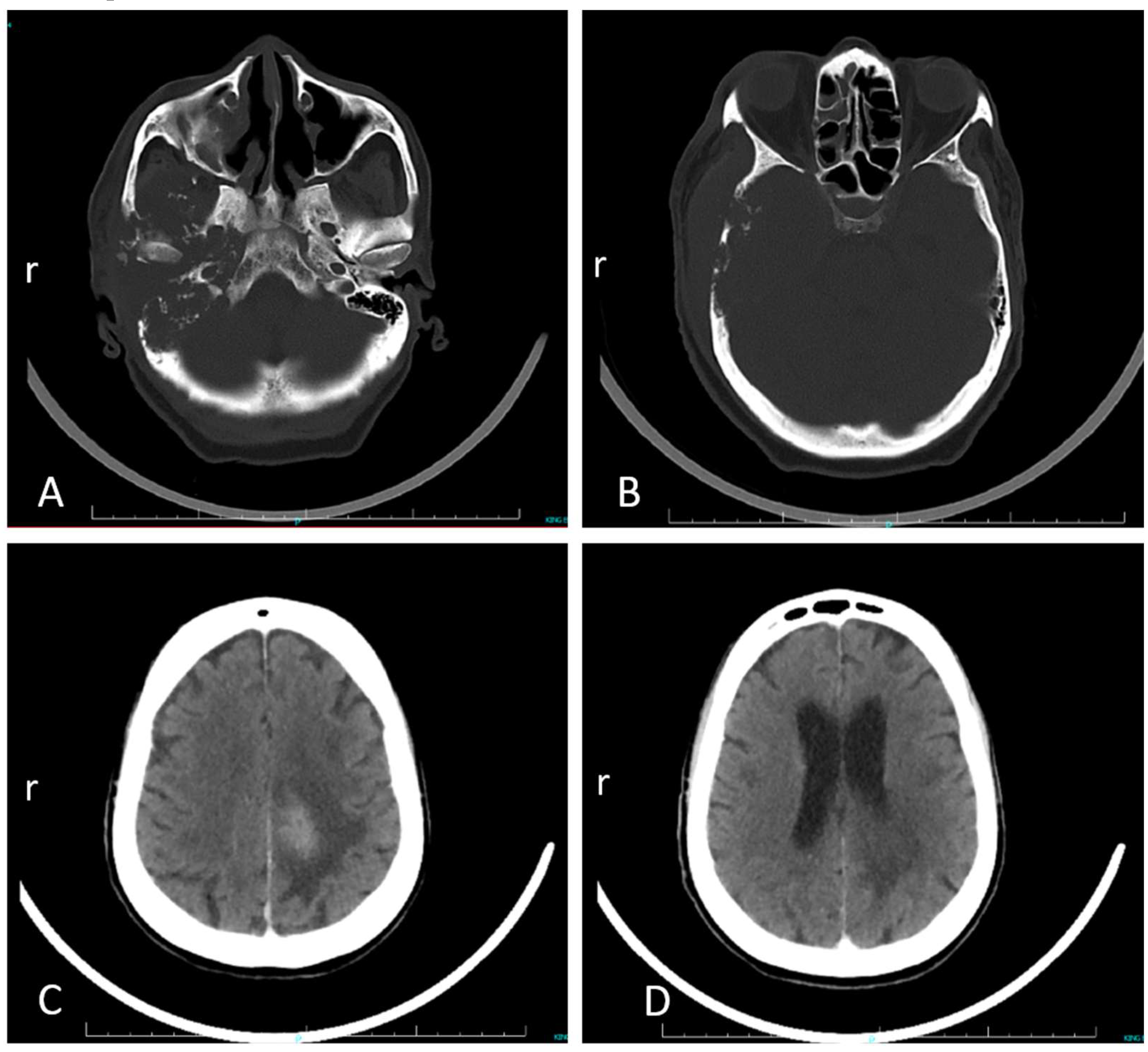
Figure 2.
Pathological features at autopsy. A. Extradural necrotic material in the right side of the middle cranial fossa base, and external auditory meatus. B, C. Histopathological images of brain tissue, showing nematode adults, larvae and eggs in various section planes (arrows) and a mixed inflammatory infiltrate (H&E stain; bars = 100 µm).
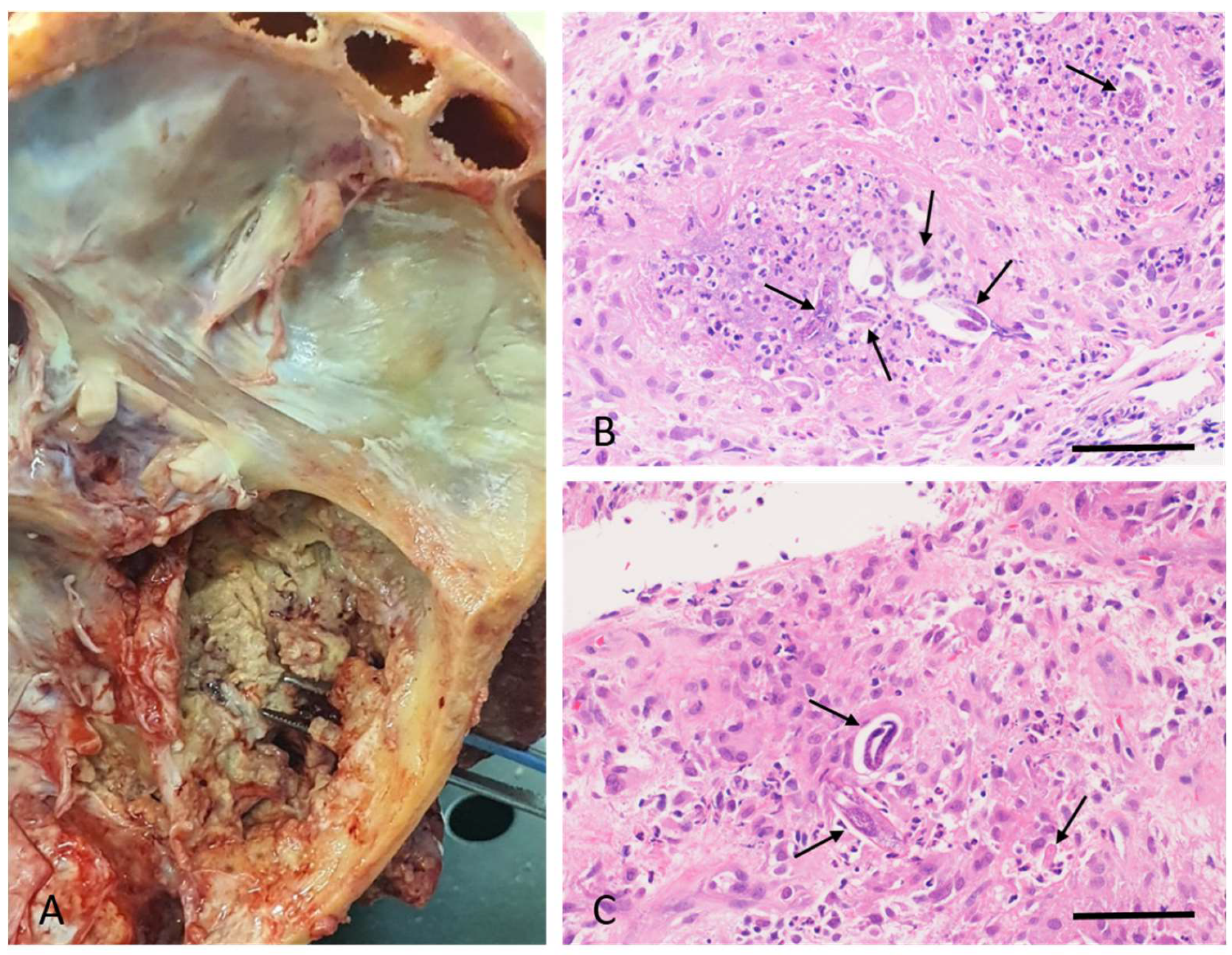
Figure 3.
Free-living Cephalobus cubaensis nematodes in ear canal exudate sample. A. Low-power view, showing adults, larvae and eggs, standard illumination. B. Adult female and larval stages, interference contrast illumination. Note the rhabitiform single-bulb oesophagus (arrow). C. Adult female stage, with egg in uterus; free eggs in the exudate. D. Embryonated egg. All bars = 100 µm.
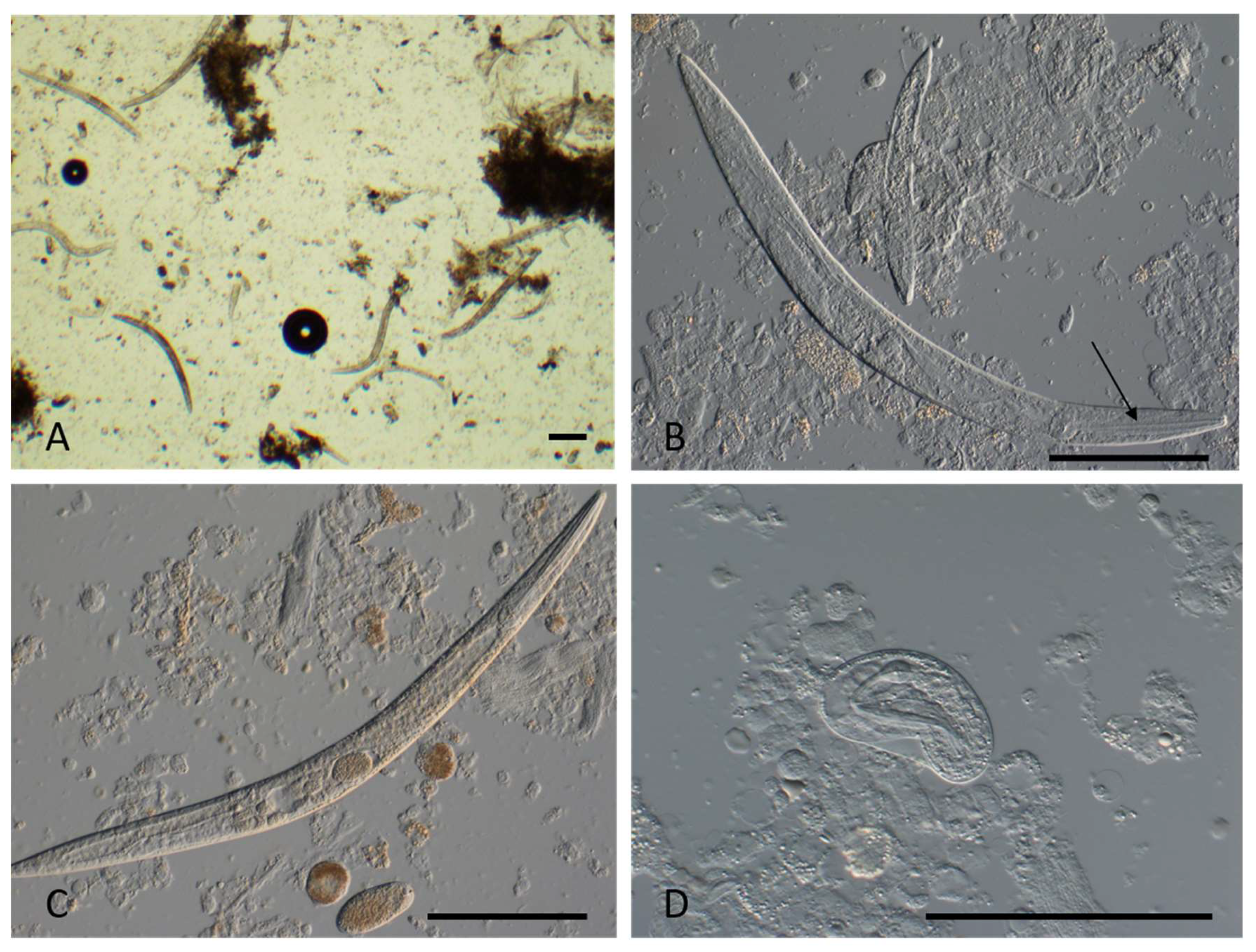
Figure 4.
Maximum likelihood 18S rRNA phylogenetic tree showing Cephalobus cubaensis’ relationship to related species with bootstrap support above 70% (GenBank accession numbers in parentheses). The isolate sample in this study is in black text. The scale bar represents the inferred substitutions per nucleotide position. Outgroup is Drilocephalobus species (green text).
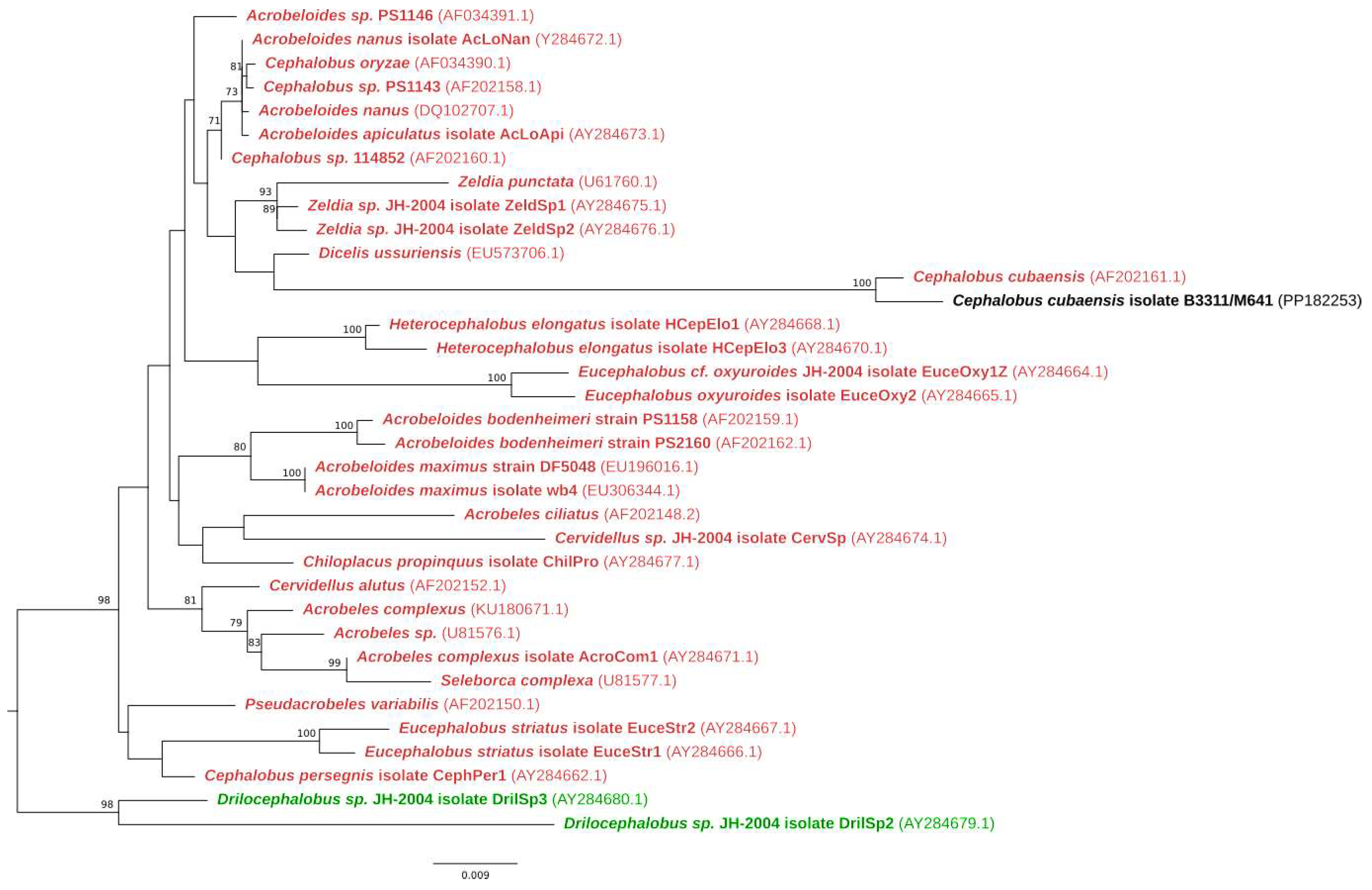

Table 1.
BLAST analysis results obtained from whole genome sequencing.
| Combined data analysis | |||
|---|---|---|---|
| Gene | Species | Identity | BLAST Query Coverage |
| 18S | Cephalobus cubaensis | 98.83% | 96% |
| 28S | Cephalobus cubaensis | 95.80% | 25% |
| Heterocephalobellus sp. | 93.72% | 47% | |
| Zeldia punctata | 92.09% | 85% | |
Disclaimer/Publisher’s Note: The statements, opinions and data contained in all publications are solely those of the individual author(s) and contributor(s) and not of MDPI and/or the editor(s). MDPI and/or the editor(s) disclaim responsibility for any injury to people or property resulting from any ideas, methods, instructions or products referred to in the content. |
© 2025 by the authors. Licensee MDPI, Basel, Switzerland. This article is an open access article distributed under the terms and conditions of the Creative Commons Attribution (CC BY) license (http://creativecommons.org/licenses/by/4.0/).
Copyright: This open access article is published under a Creative Commons CC BY 4.0 license, which permit the free download, distribution, and reuse, provided that the author and preprint are cited in any reuse.


