
Submitted:
08 January 2025
Posted:
08 January 2025
You are already at the latest version
Abstract
Fluoride, a trace element, plays a crucial role in dental health, primarily by preventing dental caries and promoting enamel remineralization. However, excessive exposure to fluoride can result in adverse health effects, including dental and skeletal fluorosis. This review provides a critical analysis of the clinical manifestations of fluorosis, highlighting the physiological signs associated with chronic fluoride exposure, such as osteocondensation, cortical thickening, and skeletal deformities.
Keywords:
Fluoride
; fluorosis
; biomarkers
; public health
1. Introduction
Fluoride is abundantly found in the Earth’s crust, predominantly in the form of fluoride compounds that can combine with other elements. This trace element is essential for humans, playing various physiological roles and participating in metabolic processes; it is considered fundamental for development and growth [1,2]. In appropriate concentrations, fluoride is linked to the prevention and treatment of dental caries, as well as to reducing microbial infections, among other benefits [3]. According to the World Health Organization (WHO), acceptable fluoride concentrations are below 1 ppm in warm regions and 1.2 ppm in cold regions. While WHO recommends a maximum limit of 1.5 mg/L of fluoride in drinking water, higher concentrations can be associated with toxicity [4].
Fluoride commonly enters water sources through both natural and anthropogenic activities, including volcanic activity, erosion of fluoride-containing rocks, and industrial activities such as chemical manufacturing and mining. Fluoride intake is not only from water but also from other products like food and beverages, which can add to the body’s total fluoride burden. Consequently, monitoring fluoride content in both water and other commonly consumed products is crucial [5,6]. Some countries have set limits for fluoride concentrations. For instance, the Bureau of Indian Standards has established a limit of 1.0 mg/L for fluoride in drinking water, significantly preventing fluoride buildup in the body. Excessive fluoride intake can lead to health issues like dental fluorosis, arthritis, osteoporosis, and other musculoskeletal disorders, and it is estimated that over 200 million people across more than 25 countries face health problems related to high levels of fluoride in drinking water [7,8,9]. Globally, many countries have reported water contamination by fluorides, leading to excessive intake and systemic toxicity, linked to previously mentioned systemic manifestations [2,3,10].
Evaluating fluoride toxicity in exposed populations is essential, as it helps identify toxicity levels, which can be assessed through biomarkers. Urine is considered the most suitable biomarker for assessing acute fluoride intake due to its easy collection and measurement process. Nails and hair are used to assess systemic subchronic or chronic fluoride exposure, while other biomarkers like plasma and saliva can also be used; however, these measurements may be affected by various factors, and in the case of saliva, topical applications and/or food intake may alter concentrations [11]. This study aims to review the literature on fluoride toxicity, focusing on both acute and chronic biomarkers of fluoride exposure and their significance for public health.
2. Fluoride Toxicity
Acute fluoride toxicity can occur from excessive intake of this element, potentially becoming life-threatening. This type of toxicity is characterized by nonspecific symptoms, such as abdominal pain, vomiting, diarrhea, excessive sweating, and joint pain. If untreated, acute exposure can result in death within the first 72 hours [12]. In contrast, chronic toxicity develops from prolonged ingestion of small doses of fluoride, which can lead to dental, skeletal, and nonskeletal fluorosis. The most severe cases of fluorosis typically occur in warm regions, where drinking water contains high concentrations of fluoride [13].
Fluoride contamination presents a critical issue in many parts of the world, especially where water contains high levels of this element. Sources of exposure include drinking water, as well as food and beverages, particularly sugary products like juices and soft drinks, which can account for up to 20% of daily fluoride intake. This elevated exposure is associated with multiple health problems, such as kidney damage, cardiometabolic risk, and neurological disorders [14,15,16,17,18]. Fluoride affects children particularly, as the element accumulates more easily in calcified tissues during this stage of development [19]. Various studies of children chronically exposed to fluoride through drinking water have shown a reduction in IQ, dental fluorosis, and an increased risk of kidney disease, cardiovascular issues, and hormonal disorders [19,20,21,22,23]. Other effects of prolonged exposure include endocrine dysfunction, hepatotoxicity, and nephrotoxicity [19,24,25,26].
3. Diet-Related Toxicity
Fluoride intake through the diet has been associated with the consumption of products typical of the “Urban Convenience Diet,” a dietary pattern linked to urban lifestyles. This diet includes beverages such as water, coffee, soft drinks, tea, milk, and juices; cereals like tortillas, rice, bread, and pasta; and foods like fish, in addition to sweets, snacks, dairy products, sauces, and dental hygiene products containing fluoride, such as toothpaste and mouthwashes. Among these products, fish is particularly relevant, as it can contain high concentrations of fluoride, reaching up to 12 ppm. This type of diet, common in urban areas, has been linked to an increased intake of fluoride, which inhibits its intestinal absorption and reduces urinary excretion. This reduction in excretion is associated with greater retention of fluoride in the body, which in turn is related to the development of fluorosis [3,27,28,29].
4. Toxicity Related to Cellular Damage, Organs, and Tissues
Fluoride-induced cellular damage is associated with multiple mechanisms that compromise cell integrity and function, with oxidative stress being one of the most significant. Elevated fluoride exposure has been linked to an increase in the production of reactive oxygen species (ROS), which generate damage to the lipids in cellular membranes. This alters membrane integrity, promotes lipid peroxidation, and induces apoptosis. Lipid peroxidation involves the oxidation of unsaturated fatty acids in cellular membranes, generating toxic products that trigger cellular apoptosis [30,31].
Fluoride toxicity can also inhibit the activity of antioxidant enzymes, such as superoxide dismutase (SOD) and catalase (CAT), which are essential for neutralizing ROS. The reduction of these enzymes increases oxidative stress, thereby exacerbating lipid membrane damage [32]. This damage compromises the structure and functionality of cellular membranes, leading to loss of cell integrity, disruption in cellular signaling, and activation of apoptotic pathways due to the accumulation of lipid peroxidation products that act as pro-apoptotic signals. Moreover, the accumulation of ROS and lipid peroxidation can impair mitochondrial membrane integrity, potentially triggering the opening of the mitochondrial permeability transition pore (mPTP) and the release of pro-apoptotic proteins [33,34].
Mitochondrial dysfunction has also been documented in cases of fluoride toxicity, as it interferes with the electron transport chain, affecting ATP production and increasing ROS accumulation. This oxidative damage impacts cellular structures, including mitochondria, and contributes to apoptosis. Additionally, alterations in the endoplasmic reticulum (ER) have been observed, causing stress in this organelle and affecting the synthesis of pro-apoptotic proteins such as caspases, Bcl-2, and cytochrome C [34,37]. The ER, along with lysosomes and the Golgi apparatus, also undergoes alterations that compromise cellular transport and molecular degradation processes, leading to DNA damage and an increased risk of mutations and carcinogenesis [37,38,39,40,41,42].
At the systemic level, fluoride is associated with renal damage, particularly affecting tubular epithelial tissues, resulting in the flattening of renal tubules and glomerular atrophy—indicators of severe renal injury. In the central nervous system, fluoride can induce an inflammatory response mediated by pro-inflammatory factors that impair cognitive function. In the gastrointestinal system, alterations in the microbiota and damage to the intestinal mucosa have been observed, increasing susceptibility to infections and causing intestinal atrophy, which compromises the integrity of the epithelial barrier and enhances its permeability. In the liver, fluoride exposure induces oxidative stress, causing structural and functional damage, including abnormal cellular morphology, cell fragmentation, and focal necrosis, as well as atrophy of the central hepatic cord and dilation of the central vein [43,44,45,46,47,48].
Dental Fluorosis
Dental fluorosis is a public health problem that occurs due to excessive fluoride exposure during dental development, particularly in children. This condition manifests as changes in the appearance of tooth enamel, ranging from white spots to more severe discoloration, resulting from enamel with lower mineral content and increased porosity. It is a condition that arises from excessive fluoride exposure during the development of the teeth, leading to alterations in enamel mineralization. This condition presents through opacities in the enamel, which can vary in severity, from mild forms showing subtle color changes to more severe forms that may result in stains and an irregular texture [49].
The aesthetic concerns associated with dental fluorosis are significant, as they can affect an individual’s perception of their smile and, consequently, their self-esteem. In particular, moderate to severe forms of fluorosis can be especially problematic, as the changes in dental appearance can be noticeable and affect the patient’s quality of life [50].
Fluorosis is commonly associated with the consumption of water containing high concentrations of fluoride, especially in areas where groundwater contaminated by local industries is used, with water consumption being one of the most significant indicators linked to fluorosis [51]. In many communities, groundwater presents fluoride concentrations exceeding the recommended limit of 1.5 ppm established by the World Health Organization. This is especially concerning in areas where people rely on wells for their water supply, as fluoride concentrations can be considerably higher due to local geology and industrial pollution [52].
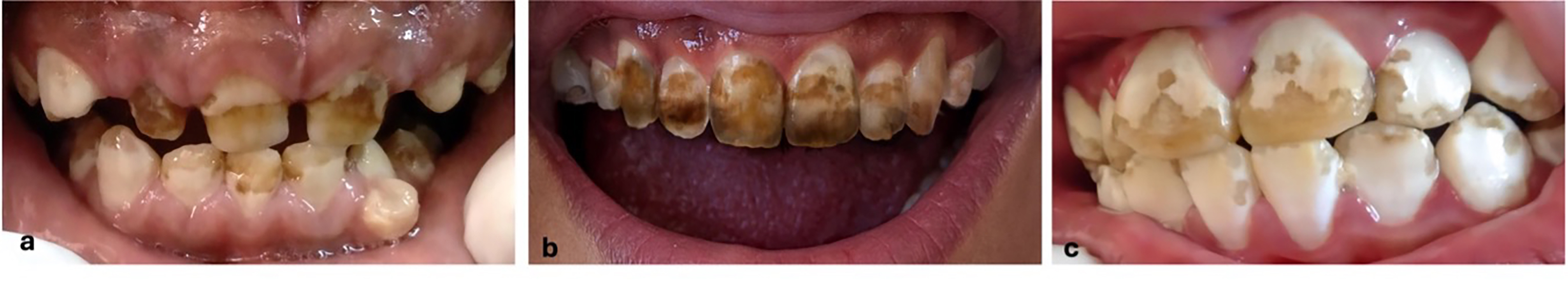
In dental tissues, excess fluoride manifests as symmetrical and diffuse hypomineralization, related to the alteration of ameloblasts. This condition is irreversible and occurs in situations of overexposure to fluoride during enamel development [10]. The enamel manifestations include a white-cream surface, white and yellow spots, as well as lines or striations, with a porous texture. In more severe cases, the enamel becomes weak, brittle, and prone to fractures, presenting significant areas of wear. This issue is common in children aged three to six years and is associated with chronic toxicity from prolonged intake of small amounts of fluoride from water and food. If excessive intake persists for many years, toxicity can manifest at the skeletal level, showing clinical features similar to rheumatoid arthritis or osteoarthritis. Figure 1 shows dental fluorosis in children aged 6 and 12 years from Durango, Durango, Mexico [53,54].
Skeletal Fluorosis
Skeletal fluorosis is a public health issue in several regions around the world, affecting approximately 100 million people, particularly in areas of Africa and Asia where fluoride levels in water exceed permissible limits [55]. It is considered a pathological condition that results from the chronic accumulation of fluoride in bones and joints, typically due to prolonged intake of fluoridated drinking water and diet with high fluoride levels. This disease is characterized by a series of symptoms that impact the mobility and quality of life of those affected. It primarily develops from exposure to fluoride concentrations greater than 1.5 ppm in drinking water and in the diet. Chronic intake causes an imbalance in bone mineral metabolism, leading to bone resorption and alterations in calcium levels in bone tissue [55]. Symptoms include chronic joint pain, stiffness, bone deformities, and ligament calcification. As the disease progresses, there may be significant loss of mobility, particularly affecting major joints such as the neck, shoulders, knees, and hips [56].
This disease is classified into three categories according to its severity: i. Mild fluorosis, which is associated with general pain and discomfort in bones and joints; ii. Moderate fluorosis, characterized by stiffness and periosteal formation with restricted movement; and iii. Severe fluorosis, associated with significant deformities in the spine and extremities, with very limited mobility [57]. In Table 1, the main manifestations of skeletal fluorosis according to Sellami et al. 2019 are shown [58].
Nail fluorosis
Leo Spira describes nail fluorosis as a condition characterized by various changes in the nails, indicative of chronic fluoride exposure. Spira states that nail alterations serve as a visible sign of fluorosis, akin to the enamel mottling observed in dental fluorosis. Affected nails may exhibit features that reflect the severity of fluoride intoxication [59]. Manifestations include nail mottling, which appears as opaque spots and transverse bands. Longitudinal striations, abnormal curvatures (such as claw-like nails), and color changes ranging from dark gray to brown are also observed [59]. The condition may include onychia, which is inflammation of the nails, and may be accompanied by onycholysis (separation of the nail from its bed) and onychomadesis (shedding of the nail). These alterations can lead to nail deformities [59]. Spira notes that affected nails tend to grow abnormally, often curving or developing pitting on their surface. This can result in an irregular appearance and increased nail fragility [59]. The author suggests that the presence of these nail changes may serve as an early indicator of fluorosis and that appropriate treatment can restore normal nail appearance, unlike the permanent changes observed in dental fluorosis [59]. In summary, Spira highlights the importance of nail manifestations as a reflection of general health and as a clinical sign of fluoride exposure, suggesting that these alterations should be considered in the diagnosis and management of fluorosis. Table 2 summarizes the clinical nail manifestations associated with fluorosis, as described by Spira [59].
5. Biomarkers in Fluorosis
Biomarkers are defined as biological indicators that reflect physiological, pathological, or pharmacological processes within the body. They can be classified into acute and chronic biomarkers, depending on the exposure duration and the nature of the biological response they represent. Acute biomarkers are used to assess recent exposure to external agents, such as pollutants or toxic substances, and are characterized by their ability to detect biological changes that occur shortly after exposure. For example, the concentration of fluoride in plasma and urine is considered an acute biomarker, as it can be quantified shortly after ingestion, reflecting immediate exposure [60]. In contrast, chronic biomarkers indicate long-term exposure and are associated with more permanent biological changes. These may include the accumulation of substances in specific tissues, such as fluoride in bones and teeth, which reflects prolonged historical exposure [60]. Additionally, hair and nail analysis has been proposed as chronic biomarkers due to their ability to accumulate fluoride and other elements over time, thus providing information about prolonged exposure [61]. However, the reliability of these biomarkers is debated, as they may be subject to external contamination and variability in growth rates [62]. Bone tissue and teeth are considered chronic but invasive biomarkers due to the nature of their collection. The assessment of fluoride in bones is performed through invasive procedures, such as bone biopsies. Bones can accumulate fluoride over extended periods, which allows for the study of long-term effects on bone health, as fluoride levels in bone tissue reflect cumulative intake over time. However, collecting bone samples carries risks, and therefore, its use as a biomarker is not recommended [63,64]. Similarly, teeth are considered chronic and invasive biomarkers, as their analysis involves the extraction of dental tissue samples. Fluoride is incorporated into the enamel structure and can be used to assess chronic exposure to this element. Fluoride concentrations in teeth correlate with fluoride intake throughout life, providing information about prolonged exposure. However, like bone samples, dental sample collection is an invasive procedure [65,66].
6. Acute Biomarkers in Fluorosis
Urinary Fluoride Excretion
Urinary fluoride excretion (UFE) has become a key acute biomarker for assessing recent fluoride exposure in various populations. This biomarker quantifies the amount of fluoride that has been absorbed and subsequently excreted by the body, providing an objective measure of fluoride intake [60]. UFE is commonly determined by collecting urine samples over a 24-hour period, which allows for an accurate estimate of total fluoride excretion. This method is particularly useful in epidemiological studies, where researchers aim to establish correlations between fluoride intake and the occurrence of health effects, such as dental fluorosis [63]. UFE has been shown to be a sensitive indicator of changes in fluoride intake, reflecting short-term exposure fluctuations, which makes it a valuable tool for public health surveillance [64]. Studies have demonstrated a significant positive correlation between UFE and total fluoride intake, suggesting that as fluoride exposure increases, so does the amount excreted in the urine [66]. This finding is especially relevant in areas with high fluoride concentrations in drinking water, where populations tend to have significantly higher UFE levels compared to those in low-fluoride regions [67]. The World Health Organization has established reference values for UFE in children, with normal values typically being below 0.5 mg/L in 24-hour urine samples. However, in areas with high fluoride exposure, these values can be higher, with reported values reaching up to 1.0 mg/L or more in children consuming fluoridated water or having a diet rich in fluoride. In adults, UFE reference values are usually similar, with a normal range of about 0.5 to 1.0 mg/L, although in areas of high exposure, levels can be higher, reaching 1.0 mg/L in children and 2.0 mg/L or more in adults [68,69]. It is important to note that UFE not only allows for exposure assessment but can also be used to study the association between fluoride intake and health outcomes, contributing to the development of public health policies aimed at preventing fluorosis and other fluoride-related adverse effects. Rango et al. (2015) observed a significant positive correlation between fluoride concentrations in drinking water and urinary fluoride excretion, with a correlation coefficient of r = 0.78 (p < 0.001) [70]. This suggests that urinary fluoride levels reliably reflect environmental exposure. Additionally, the authors emphasized that urinary fluoride excretion is a more sensitive biomarker compared to fluoride content in nails, which reflects average fluoride concentrations in plasma over a longer period [70]. Therefore, the ability of urine to provide data on acute fluoride exposure makes it a valuable tool for early detection of fluoride toxicity in populations, aiding in the mitigation of fluoride-related risks [61]. Although urine is a useful biomarker for assessing acute fluoride exposure, it has limitations that must be considered: i. Excretion Variability: The amount of fluoride excreted in the urine can vary significantly among individuals due to factors such as diet, hydration, renal function, and the time elapsed since the last fluoride intake. This variability can complicate comparisons of results between individuals or populations and lead to inconsistent findings [61]. ii. Temporary Exposure: Urine reflects recent exposure, not necessarily long-term cumulative exposure. This means it may not be a suitable indicator of chronic fluoride exposure, which is important for evaluating long-term health effects, such as dental fluorosis [70]. iii. Dilution from Fluid Intake: Fluid intake prior to sample collection can dilute fluoride concentrations in the urine, potentially underestimating actual exposure. This is particularly problematic in populations that consume large quantities of water or other liquids [61,70]. iv. Sample Collection: Collecting urine samples can be challenging, especially in field studies. The need to gather samples within a specific period (e.g., 12-hour urine) can pose logistical difficulties, affecting data quality. Variability in sample collection, influenced by factors such as hydration, diet, and physical activity, may also impact fluoride concentrations. Furthermore, differences in sample collection times throughout the day can result in variations in fluoride levels, complicating comparisons across individuals or groups [61,70]. v. Interpretation of Results: Interpreting fluoride levels in urine can be complex, as multiple factors—such as age, health status, and exposure to other fluoride sources—must be considered [70]. vi. Limitations in Specific Populations: In populations with specific health conditions (e.g., kidney disease), fluoride excretion may not accurately represent true exposure, limiting the usefulness of urine as a biomarker in these cases [70]. These limitations highlight the importance of using urine in conjunction with other biomarkers, such as fluoride in nails or hair, to provide a more comprehensive assessment of fluoride exposure.
Plasma
One of the main benefits of using plasma as a biomarker is its ability to provide a quick and accurate assessment of fluoride exposure. Unlike other biomarkers, such as hair or nails, which reflect exposure over longer periods, plasma can indicate changes in exposure over a short period of time. This characteristic is particularly useful in epidemiological studies that aim to correlate fluoride intake with adverse health effects, such as fluorosis [69]. Plasma analysis using advanced techniques, such as mass spectrometry, allows for precise quantification of fluoride concentration, thereby facilitating comparison of results across different studies and populations and contributing to a better understanding of the effects of fluoride on public health [71]. However, using plasma as a biomarker has some disadvantages. Firstly, fluoride concentration in plasma can vary considerably depending on dietary intake and fluoride bioavailability, which makes interpretation of results more difficult [63]. Additionally, there is notable temporal variability in plasma fluoride levels, as they reflect acute exposure rather than cumulative long-term exposure [72]. Another significant limitation is that plasma sample collection involves invasive procedures, which can be a challenge in studies involving pediatric populations or communities with limited resources, potentially reducing participation rates. Furthermore, sample analysis requires a specialized laboratory, which increases both costs and the time needed to obtain results, limiting the feasibility of plasma as a biomarker in large-scale studies [61]. Although plasma provides information on recent fluoride exposure, its ability to predict adverse health effects, such as dental fluorosis, is not fully established. This is because the relationship between plasma fluoride levels and biological effects can be influenced by multiple factors, such as age, nutritional status, and the presence of other contaminants [66].
7. Chronic Biomarkers in Fluorosis
Hair
Hair is considered a suitable biomarker for assessing exposure to various substances, including fluoride, due to several scientific reasons. Firstly, hair provides a more stable and lasting record of long-term exposure compared to other biological fluids like blood or urine, which reflect recent exposure and may vary significantly over short periods. As hair grows, chemical compounds like fluoride become incorporated into its structure, allowing for a retrospective evaluation of exposure [73]. Collecting hair samples is a non-invasive and relatively simple procedure, which is particularly useful in epidemiological studies as it facilitates sample collection from large populations under various conditions. Hair can also accumulate and concentrate chemical compounds, enabling the detection of exposure levels that might not be evident in other biological media. This is especially relevant in the case of fluoride, where levels in hair can correlate with environmental and dietary exposure [74].
Studies have shown that fluoride levels in hair are directly related to fluoride concentration in drinking water and other sources of exposure. This suggests that hair analysis can be an effective indicator of environmental fluoride exposure. The use of hair as a biomarker can help assess exposure in populations exposed to fluoride, such as workers in related industries or residents in highly fluoridated areas [75,76]. Thus, hair serves as a valuable biomarker for monitoring fluoride exposure, presenting several positive characteristics that support its use in public health studies. For instance, it has been found that hair can effectively reflect fluoride exposure under certain conditions. Selma Elekdag-Turk et al., in Turkey, evaluated hair as a biomarker of fluoride exposure in the cities of Isparta (endemic region) and Samsun (non-endemic region). Results showed that the mean fluoride concentration in the hair of participants from the endemic region (Isparta City, IC) was higher than in the non-endemic region (Samsun City, SC). This difference was statistically significant, suggesting that hair can be used as an appropriate biomarker for detecting fluoride exposure in large populations [77]. Joshi and Ajithkrishnan reported higher fluoride levels in the hair of individuals from endemic fluorosis areas, indicating that hair may be suitable for monitoring chronic fluoride exposure. This capacity to accumulate fluoride over time allows hair to serve as a reflection of long-term exposure, which is crucial for assessing the health effects of chronic exposure [78]. Using hair as a biomarker is non-invasive, which simplifies sample collection in various populations, including children and adults. This is particularly important in public health studies where participant acceptance and comfort are key factors. Therefore, hair analysis presents itself as a useful tool for assessing chronic fluoride exposure, a public health issue affecting various regions, especially in endemic areas. Joshi et al. demonstrated a significant correlation between fluoride content in drinking water and fluoride levels in the participants’ hair, suggesting that hair can act as a biomarker for evaluating long-term fluoride exposure. Moreover, using this biomarker complements measurements in urine and blood, which are related to short-term exposures [78,79]. Unlike urine and blood analyses, which can present variations related to urinary flow and pH, hair reflects average plasma fluoride concentrations over time, making it an indicator of chronic exposure [80]. Hair sample collection is easy, painless, and non-invasive [81]. While biomarkers like urine and blood show fluctuations in fluoride levels due to recent intake, hair provides a more continuous representation of exposure over an extended period [82]. Therefore, it is suggested to perform fluoride concentration assessments using both acute and chronic biomarkers.
Nails
The use of nails as a biomarker has emerged as a relevant approach in assessing exposure to various chemical substances, including heavy metals and organic compounds. Nails, primarily composed of keratin, have the ability to accumulate and store metabolites and contaminants over time, allowing for a retrospective evaluation of environmental and occupational exposure [83]. This attribute differentiates them from other biomarkers, such as blood and urine, which reflect more immediate exposures and may be influenced by temporary and physiological factors [66]. Collecting nail samples is a non-invasive and relatively simple procedure, making it feasible for application in epidemiological studies and in vulnerable populations, such as children or individuals with medical conditions that limit blood extraction [69]. Additionally, nails can provide information on long-term exposure, which is crucial for establishing correlations between exposure to toxic substances and health effects [84]. Specifically in the context of fluoride exposure, research has begun exploring the feasibility of nails as biomarkers for measuring fluoride intake and its relationship to dental health. Recent studies have shown that fluoride concentrations in nails can significantly correlate with fluoride intake, suggesting that this biomarker could be useful for public health monitoring and the prevention of fluoride-related diseases [83]. However, it is essential to continue researching the validity and reliability of nails as biomarkers, given that heterogeneity in collection and analysis methods may influence results [84]. Thus, the use of nails as biomarkers represents a valuable tool in research on chemical exposure, offering insights into the relationship between long-term exposure and health effects. Validating this approach could have significant implications for public health monitoring and policy formulation. However, Oliveira Lima-Arsati et al. note that the sensitivity of nails for detecting variations in fluoride intake is limited, as a clear dose-response relationship between the amount of fluoride ingested and its concentration in nails has not been established. Additionally, the authors observed that despite a significant increase in fluoride intake when toothpaste was used, fluoride concentrations in nails did not decrease after discontinuing toothpaste use, suggesting that nails do not adequately reflect recent fluoride exposure. Moreover, fluoride intake assessment relies on the brushing frequency reported by parents and the total fluoride concentration in toothpaste, without considering fluoride bioavailability, which may lead to misinterpretations regarding actual exposure. Therefore, the authors conclude that nails do not meet the sensitivity and reliability criteria required for an effective biomarker of fluoride exposure. The study by Oliveira Lima-Arsati et al. focuses on evaluating the concentration of fluoride in nails in relation to fluoride intake during two distinct phases: one phase in which children are exposed to fluoride from both diet and toothpaste, and another phase in which toothpaste use is discontinued, and only dietary fluoride intake is considered. The study does not directly address chronic fluoride exposure, as it focuses on variations in nail fluoride concentration in response to short-term intake changes, which strengthens the use of nails as a biomarker for chronic use [85]. A systematic review conducted in 2021 reports that nails have been identified as relevant biomarkers for monitoring fluoride exposure in various populations, particularly through fluoridated drinking water intake. The systematic review indicates that several studies agree that nails are suitable for assessing chronic and subchronic fluoride exposure, as they can reflect systemic levels of this compound over time [86]. It has been observed that toenails are more effective than fingernails and hair for this purpose. This difference is attributed to toenails’ lower environmental exposure and slower growth rate, making them a more reliable indicator of long-term exposure [69]. Additionally, nail sample collection and processing are less invasive and more accessible compared to other biomarkers, such as mineralized tissues (bones and teeth), which require surgical procedures for their collection [87]. Therefore, it can be suggested that nails represent a viable alternative for monitoring fluoride exposure, providing a useful tool for risk assessment in public health related to fluoride intake through drinking water [88,89].
8. Discussion
Fluoride exposure and its relationship with public health reveal a complex landscape encompassing both the benefits and risks associated with this element. In terms of benefits, fluoride has proven to be an effective agent in preventing dental caries, which has led to its inclusion in public health programs and water fluoridation initiatives. The WHO has set guidelines for optimal fluoride concentrations, emphasizing its importance in dental health, particularly in vulnerable populations. However, excessive fluoride exposure can lead to adverse effects, such as dental and skeletal fluorosis, raising concerns about the safety of its use. Assessing fluoride exposure through biomarkers such as urine, plasma, hair, and nails has both strengths and weaknesses. Plasma, for instance, stands out for its ability to reflect short-term changes in exposure, which is crucial in epidemiological studies. On the other hand, urine, while useful, may not accurately represent actual exposure in individuals with specific health conditions, limiting its effectiveness as a standalone biomarker. Hair emerges as a promising biomarker, providing a retrospective assessment of fluoride exposure and reflecting levels that can correlate with environmental and dietary exposure. However, using nails as a biomarker has been questioned, as a clear relationship between fluoride intake and its concentration in this tissue has not been established, suggesting that its usefulness may be limited. While fluoride is essential for dental health, its management must be cautious to avoid adverse effects. Continued research on exposure biomarkers is essential for improving the understanding of the relationship between fluoride and health and informing public health policies that balance the benefits and risks of fluoride exposure. Integrating multiple biomarkers in exposure studies could provide a more comprehensive and accurate assessment, thus contributing to the development of effective strategies for preventing fluorosis and other fluoride-related adverse effects.
9. Conclusion
Evaluating fluoride exposure through acute and chronic biomarkers is essential for understanding its impact on public health, especially in the context of fluorosis. Acute biomarkers, such as plasma and urine, offer insight into recent exposure, enabling the detection of changes in fluoride concentration over short periods. Plasma, in particular, is notable for its capacity to reflect rapid variations in exposure, which is crucial in epidemiological studies that seek to correlate fluoride intake with adverse health effects. In contrast, chronic biomarkers like hair and nails provide a more stable and long-lasting assessment of fluoride exposure over time. Hair, in particular, has been identified as a valuable biomarker due to its ability to accumulate and concentrate chemical compounds, allowing for a retrospective assessment of exposure. Its collection is a non-invasive and relatively simple procedure, making it suitable for use in studies with large populations. Additionally, fluoride levels in hair have been shown to correlate with environmental and dietary exposure, making it an effective indicator of chronic fluoride exposure. Nails also emerge as a promising biomarker. Their ability to reflect long-term exposure, combined with their non-invasive collection, positions them as a useful tool in public health monitoring. However, further research is needed to validate their efficacy and establish clear correlations between fluoride intake and its concentration in nails. Therefore, combining acute and chronic biomarkers such as plasma, urine, hair, and nails provides a comprehensive approach to evaluating fluoride exposure.
This strategy enables a more complete understanding of fluoride’s effects on health, facilitating the identification of at-risk populations and the development of more effective preventive policies. Future research should focus on standardizing collection and analysis methods and exploring the interaction between these biomarkers to maximize their utility in public health surveillance related to fluoride exposure.
Author Contributions
R.G-.G and JM.S.-P: writing original draft, conceptualization, performed the digital search, selection, analysis, and extraction of the information; O.T-M and G.O.-S: perfomed the digital search, analysis and extraction of information; O.A.-O and SM.S.-P: writing orginal draft, conceptualization and formal analysis; N.M.-F and S.L-.V: performed the digital search, selection and analysis; F.M.-C; performed the digital search and extraction of information. R.B.-M; Formal analysis; Supervision and Conceptualization.
Funding
This research received no external funding.
Institutional Review Board Statement
Not applicable.
Informed Consent Statement
Not applicable.
Data Availability Statement
Not applicable.
Conflicts of Interest
The authors declare no conflict of interest.
References
- Pramanik, S.; Saha, D. The Genetic Influence in Fluorosis. Environ. Toxicol. Pharmacol. 2017, 56, 157–162. [Google Scholar] [CrossRef] [PubMed]
- Wei, W.; Pang, S.; Sun, D. The Pathogenesis of Endemic Fluorosis: Research Progress in the Last 5 Years. J. Cell. Mol. Med. 2019, 23, 2333–2342. [Google Scholar] [CrossRef] [PubMed]
- Sharma, D.; Singh, A.; Verma, K.; Paliwal, S.; Sharma, S.; Dwivedi, J. Fluoride: A Review of Pre-clinical and Clinical Studies. Environ. Toxicol. Pharmacol. 2017, 56, 297–313. [Google Scholar] [CrossRef]
- World Health Organization. Guidelines for Drinking-Water Quality, 4th ed.; WHO: Geneva, 2011; p. 340. [Google Scholar] [CrossRef]
- Kanduti, D.; Sterbenk, P.; Artnik, B. Fluoride: A Review of Use and Effects on Health. Mater. Socio Med. 2016, 28, 133. [Google Scholar] [CrossRef] [PubMed]
- Mohapatra, M.; Rout, K.; Gupta, S.K.; Singh, P.; Anand, S.; Mishra, B.K. Facile Synthesis of Additive-Assisted Nano Goethite Powder and Its Application for Fluoride Remediation. J. Nanopart. Res. 2010, 12, 681–686. [Google Scholar] [CrossRef]
- Halder, A.; Singh, S.; Adhikari, A.; Singh, P.; Sarkar, P.K.; Pal, U.; Ghosh, R.; Shikha, D.; Solanki, Y.S.; Agarwal, M.; Gupta, A.B.; Chakraborty, R.; Saha-Dasgupta, T.; Das, R.; Pal, S.K. Selective and Fast Responsive Sensitized Micelle for Detection of Fluoride Level in Drinking Water. ACS Sustain. Chem. Eng. 2019, 7, 16355–16363. [Google Scholar] [CrossRef]
- Ayoob, S.; Gupta, A.K. Fluoride in Drinking Water: A Review on the Status and Stress Effects. Crit. Rev. Environ. Sci. Technol. 2006, 36, 433–487. [Google Scholar] [CrossRef]
- Paudyal, H.; Inoue, K.; Kawakita, H.; Ohto, K. Removal of Fluoride by Effectively Using Spent Cation Exchange Resin. J. Mater. Cycles Waste Manag. 2018, 20, 975–984. [Google Scholar] [CrossRef]
- Dhar, V.; Bhatnagar, M. Physiology and Toxicity of Fluoride. Indian J. Dent. Res. 2009, 20, 350–355. [Google Scholar] [CrossRef] [PubMed]
- Lavalle-Carrasco, J.; Vergara-Onofre, M.; González-González, R.; Bologna-Molina, R.; Isiordia-Espinoza, M.A.; Gaona, E.; Molina-Frechero, N. A Systematic Review and Meta-Analysis of the Relationship Between the Severity of Dental Fluorosis and Fluoride Biomarkers in Endemic Areas. Biol. Trace Elem. Res. 2023, 201, 1051–1062. [Google Scholar] [CrossRef] [PubMed]
- Khairnar, M.R.; Dodamani, A.S.; Jadhav, H.C.; Naik, R.G.; Deshmukh, M.A. Mitigation of Fluorosis—A Review. J. Clin. Diagn. Res. 2015, 9, ZE05–ZE09. [Google Scholar] [CrossRef] [PubMed]
- Patil, M.M.; Lakhkar, B.B.; Patil, S.S. Curse of Fluorosis. Indian J. Pediatr. 2018, 85, 375–383. [Google Scholar] [CrossRef] [PubMed]
- Wasana, H.M.; Perera, G.D.; Gunawardena, P.S.; Fernando, P.S.; Bandara, J. WHO Water Quality Standards vs Synergic Effects of Fluoride, Heavy Metals, and Hardness in Drinking Water on Kidney Tissues. Sci. Rep. 2017, 14, 42516. [Google Scholar] [CrossRef]
- Malin, J.A.; Lesseur, C.; Busgang, S.A.; Curtin, P.; Wright, R.O.; Sanders, A.P. Fluoride Exposure and Kidney and Liver Function Among Adolescents in the United States: NHANES, 2013–2016. Environ. Int. 2019, 132, 105012. [Google Scholar] [CrossRef] [PubMed]
- Liu, L.; Wang, M.; Li, Y.; Liu, H.; Hou, C.; Zeng, Q.; Li, P.; Zhao, Q.; Dong, L.; Yu, X.; Liu, L.; Zhang, S.; Wang, A. Low-to-Moderate Fluoride Exposure in Relation to Overweight and Obesity Among School-Age Children in China. Ecotoxicol. Environ. Saf. 2019, 15, 109558. [Google Scholar] [CrossRef] [PubMed]
- Rocha-Amador, D.; Navarro, M.E.; Carrizales, L.; Morales, R.; Calderón, J. Decreased Intelligence in Children and Exposure to Fluoride and Arsenic in Drinking Water. Cad. Saúde Pública 2007, 23, S579–S587. [Google Scholar] [CrossRef]
- Mohammadi, A.A.; Yousefi, M.; Yaseri, M.; Jalilzadeh, M.; Mahvi, A.H. Skeletal Fluorosis in Relation to Drinking Water in Rural Areas of West Azerbaijan, Iran. Sci. Rep. 2017, 7, 17300. [Google Scholar] [CrossRef] [PubMed]
- Das, K.; Mondal, N.K. Dental Fluorosis and Urinary Fluoride Concentration as a Reflection of Fluoride Exposure and Its Impact on IQ Level and BMI of Children of Laxmisagar, Simlapal Block of Bankura District, W.B., India. Environ. Monit. Assess. 2016, 188, 218. [Google Scholar] [CrossRef]
- Kanduti, D.; Sterbenk, P.; Artnik, A. Fluoride: A Review of Use and Effects on Health. Mat. Soc. Med. 2016, 28, 133. [Google Scholar] [CrossRef]
- Valdez, J.L.; Calderón, H.J.; Córdova, A.R.I.; Sandoval, A.S.Y.; Alegría, T.J.A.; Costilla, S.R.; Rocha, A.D. Level of Exposure to Fluorides by the Consumption of Different Types of Milk in Residents from an Area of Mexico with Endemic Hydrofluorosis. An. Pediatr. (Engl. Ed.) 2019, 90, 342–348. [Google Scholar] [CrossRef]
- Bashash, M.; Thomas, D.; Hu, H.; Martinez-Mier, E.A.; Sanchez, B.N.; Basu, N.; Peterson, K.E.; Ettinger, A.S.; Wright, R.; Zhang, Z.; Liu, Y.; Schnaas, L.; Mercado-García, A.; Téllez-Rojo, M.M.; Hernández-Ávila, M. Prenatal Fluoride Exposure and Cognitive Outcomes in Children at 4 and 6–12 Years of Age in Mexico. Environ. Health Perspect. 2017, 125, 097017. [Google Scholar] [CrossRef] [PubMed]
- Valdez, J.L.; López Guzmán, O.D.; Cervantes, F.M.; Costilla-Salazar, R.; Calderón, H.J.; Alcaraz, C.Y.; Rocha-Amador, D.O. In Utero Exposure to Fluoride and Cognitive Development Delay in Infants. Neurotoxicology 2017, 59, 65–70. [Google Scholar] [CrossRef] [PubMed]
- Barbier, O.; Arreola, L.; Del Razo, L. Molecular Mechanisms of Fluoride Toxicity. Chem. Biol. Interact. 2010, 188, 319–333. [Google Scholar] [CrossRef] [PubMed]
- Lennon, M.A.; Whelton, H.; O’Mullane, D.; Ekstrand, J. Fluoride. Rolling Revision of the WHO Guidelines for Drinking-Water Quality. World Health Organization, Switzerland, 2004.
- Nanayakkara, S.; Senevirathna, S.T.M.L.D.; Harada, K.H.; Chandrajith, R.; Nanayakkara, N.; Koizumi, A. The Influence of Fluoride on Chronic Kidney Disease of Uncertain Aetiology (CKDu) in Sri Lanka. Chemosphere 2020, 257, 127186. [Google Scholar] [CrossRef] [PubMed]
- McLure, F.J. Ingestion of Fluoride and Dental Caries Quantitative Relations Based on Food and Water Requirements of Children One to Twelve Years Old. Am. J. Dis. Child. 1943, 66, 362–369. [Google Scholar] [CrossRef]
- Yadav, K.K.; Kumar, S.; Pham, Q.B.; Gupta, N.; Rezania, S.; Kamyab, H.; et al. Fluoride Contamination, Health Problems and Remediation Methods in Asian Groundwater: A Comprehensive Review. Ecotoxicol. Environ. Saf. 2019, 182, 109362. [Google Scholar] [CrossRef]
- Castiblanco-Rubio, G.A.; Baston, M.; Hernandez-F, M.; Martinez-Mier, E.A.; Cantoral, A. Associations Between Dietary Patterns, Fluoride Intake and Excretion in Women Exposed to Fluoridated Salt: A Preliminary Study. Nutrients 2024, 16, 3404. [Google Scholar] [CrossRef]
- Li, M.; Cao, J.; Zhao, Y.; Wu, P.; Li, X.; Khodaei, F.; Han, Y.; Wang, J. Fluoride Impairs Ovary Development by Affecting Oogenesis and Inducing Oxidative Stress and Apoptosis in Female Zebrafish (Danio rerio). Chemosphere 2020, 256, 127105. [Google Scholar] [CrossRef]
- Li, M.; Wang, J.; Wu, P.; Manthari, R.K.; Zhao, Y.; Li, W.; Wang, J. Self-Recovery Study of the Adverse Effects of Fluoride on Small Intestine: Involvement of Pyroptosis Induced Inflammation. Sci. Total Environ. 2020, 742, 140533. [Google Scholar] [CrossRef] [PubMed]
- Sinha, M.; Manna, P.; Sil, P.C. Aqueous Extract of the Bark of Terminalia arjuna Plays a Protective Role Against Sodium-Fluoride-Induced Hepatic and Renal Oxidative Stress. J. Nat. Med.-Tokyo 2007, 61, 251–260. [Google Scholar] [CrossRef]
- Gao, J.; Wang, Y.; Xu, G.; Wei, J.; Chang, K.; Tian, X.; Liu, M.; Yan, X.; Huo, M.; Song, G. Selenium Attenuates Apoptosis and p-AMPK Expressions in Fluoride-Induced NRK-52E Cells. Environ. Sci. Pollut. Res. 2019, 26, 15685–15697. [Google Scholar] [CrossRef] [PubMed]
- Burgos Aceves, M.A.; Migliaccio, V.; Lepretti, M.; Paolella, G.; Di Gregorio, I.; Penna, S.; Faggio, C.; Lionetti, L. Dose-Dependent Response to the Environmental Pollutant Dichlorodiphenylethylene (DDE) in HepG2 Cells: Focus on Cell Viability and Mitochondrial Fusion/Fission Proteins. Toxics 2021, 9, 270. [Google Scholar] [CrossRef]
- Sang, H.; Zhang, L.; Li, J. Anti-Benzopyrene-7,8-Diol-9,10-Epoxide Induces Apoptosis via Mitochondrial Pathway in Human Bronchiolar Epithelium Cells Independent of the Mitochondrial Permeability Transition Pore. Food Chem. Toxicol. 2012, 50, 2417–2423. [Google Scholar] [CrossRef] [PubMed]
- Ol, M.S.; Nawaz, M.; Ahsan, H. Role of Bcl-2 Family Proteins and Caspases in the Regulation of Apoptosis. Mol. Cell. Biochem. 2011, 351, 41–58. [Google Scholar] [CrossRef] [PubMed]
- Wei, Y.; Chiang, W.; Sumpter, R.; Mishra, P.; Levine, B. Prohibitin 2 is an Inner Mitochondrial Membrane Mitophagy Receptor. Cell 2017, 168, 224–238. [Google Scholar] [CrossRef] [PubMed]
- Li, Y.; Liu, Y.; Yi, J.; Li, Y.; Yang, B.; Shang, P.; Mehmood, K.; Bilal, R.M.; Zhang, H.; Chang, Y.; Tang, Z.; Wang, Y.; Li, Y. The Potential Risks of Chronic Fluoride Exposure on Nephrotoxicity via Altering Glucolipid Metabolism and Activating Autophagy and Apoptosis in Ducks. Toxicology 2021, 461, 152906. [Google Scholar] [CrossRef]
- Zhao, L.; Yu, Y.; Deng, C. Protein and mRNA Expression of Shh, Smo, and Gli 1 and Inhibition by Cyclopamine in Hepatocytes of Rats with Chronic Fluorosis. Toxicol. Lett. 2014, 225, 318–324. [Google Scholar] [CrossRef] [PubMed]
- Barbier, O.; Arreola-Mendoza, L.; Del Razo, L.M. Molecular Mechanisms of Fluoride Toxicity. Chem. Biol. Interact. 2010, 188, 319–333. [Google Scholar] [CrossRef] [PubMed]
- Wei, Q.; Deng, H.; Cui, H.; Fang, J.; Zuo, Z.; Deng, J.; Li, Y.; Wang, X.; Zhao, L. A Mini Review of Fluoride-Induced Apoptotic Pathways. Environ. Sci. Pollut. Res. Int. 2018, 25, 33926–33935. [Google Scholar] [CrossRef] [PubMed]
- Wei, Q.; Luo, Q.; Liu, H.; Chen, L.; Cui, H.; Fang, J.; Zuo, Z.; Deng, J.; Li, Y.; Wang, X.; Zhao, L. The Mitochondrial Pathway is Involved in Sodium Fluoride (NaF)-Induced Renal Apoptosis in Mice. Toxicol. Res. 2018, 7, 792–808. [Google Scholar] [CrossRef]
- Wang, J.; Yue, B.; Zhang, X.; Guo, X.; Sun, Z.; Niu, R. Effect of Exercise on Microglial Activation and Transcriptome of Hippocampus in Fluorosis Mice. Sci. Total Environ. 2021, 760, 143376. [Google Scholar] [CrossRef]
- Liu, Y.; Sun, B.; Zhang, S.; Li, J.; Qi, J.; Bai, C.; Zhang, J.; Liang, S. Glycine Alleviates Fluoride-Induced Oxidative Stress, Apoptosis, and Senescence in a Porcine Testicular Sertoli Cell Line. Reprod. Domest. Anim. 2021, 56, 884–896. [Google Scholar] [CrossRef] [PubMed]
- Wang, H.W.; Liu, J.; Zhao, W.P.; Zhang, Z.H.; Li, S.Q.; Li, S.H.; Zhu, S.Q.; Zhou, B.H. Effect of Fluoride on Small Intestine Morphology and Serum Cytokine Contents in Rats. Biol. Trace Elem. Res. 2019, 189, 511–518.46. [Google Scholar] [CrossRef]
- Moyson, S.; Liew, H.J.; Fazio, A.; Van Dooren, N.; Delcroix, A.; Faggio, C.; Blust, R.; De Boeck, G. Kidney activity increases in copper exposed goldfish(Carassius auratus auratus). Comp. Biochem. Physiol. C Toxicol. Pharmacol. 2016, 190, 32–37. [Google Scholar] [CrossRef]
- Akinrinde, A.S.; Tijani, M.; Awodele, O.A.; Oyagbemi, A.A. Fluoride-induced hepatotoxicity is prevented by L-Arginine supplementation via suppressionof oxidative stress and stimulation of nitric oxide production in rats. Toxicology and Environmental Health Sciences 2021, 13, 57–64. [Google Scholar] [CrossRef]
- Sinha, M.; Manna, P.; Sil, P.C. Aqueous extract of the bark of Terminalia arjuna plays a protective role against sodium-fluoride-induced hepatic and renal oxidative stress. J NAT MED 2007, 61, 251–260. [Google Scholar] [CrossRef]
- Thylstrup, A.; Fejerskov, O. Clinical Appearance of Dental Fluorosis in Permanent Teeth in Relation to Histologic Changes. Community Dent. Oral Epidemiol. 1978, 6, 315–328. [Google Scholar] [CrossRef] [PubMed]
- Abanto Alvarez, J.; Rezende, K.M.; Marocho, S.M.; Alves, F.B.; Celiberti, P.; Ciamponi, A.L. Dental Fluorosis: Exposure, Prevention, and Management. Med. Oral Patol. Oral Cir. Bucal. 2009, 14, E103–E107. [Google Scholar] [PubMed]
- Oliveira Chagas, F.; Rocha Valadas, L.A.; Sorazabal, A.; Dayo, A.; Botelho Dantas, T.C.F.; Squassi, A. Fluoride in Drinking Groundwater and Prevalence of Fluorosis in Children and Adolescents: A Systematic Review. Acta Odontol. Latinoam. 2023, 36, 169–176. [Google Scholar] [CrossRef] [PubMed]
- Rompante, P. Mecanismos Preventivos do Fluór e Cárie Dentária. Acta Pediatr. Port. 2009, 40, 223–228. [Google Scholar] [CrossRef]
- Mellberg, J.R.; Ripa, L.W. Fluoride Metabolism. In Fluorides in Preventive Dentistry: Theory and Clinical Applications; Quintessence Publishing Co Limited: 1983; pp. 81–102.
- Majumdar, K.K. Health Impact of Supplying Safe Drinking Water Containing Fluoride Below Permissible Level on Fluorosis Patients in a Fluoride-Endemic Rural Area of West Bengal. Indian J. Public Health 2011, 55, 303–308. [Google Scholar] [CrossRef] [PubMed]
- Srivastava, S.; Flora, S.J.S. Fluoride in Drinking Water and Skeletal Fluorosis: A Review of the Global Impact. Curr. Environ. Health Rep. 2020, 7, 140–146. [Google Scholar] [CrossRef] [PubMed]
- Kurdi, M.S. Chronic Fluorosis: The Disease and Its Anaesthetic Implications. Indian J. Anaesth. 2016, 60, 157–162. [Google Scholar] [CrossRef] [PubMed]
- Mohammadi, A.A.; Yousefi, M.; Yaseri, M.; Jalilzadeh, M.; Mahvi, A.H. Skeletal Fluorosis in Relation to Drinking Water in Rural Areas of West Azerbaijan, Iran. Sci. Rep. 2017, 7, 17300. [Google Scholar] [CrossRef] [PubMed]
- Sellami, M.; Riahi, H.; Maatallah, K.; Ferjani, H.; Bouaziz, M.C.; Ladeb, M.F. Skeletal Fluorosis: Don’t Miss the Diagnosis! Skeletal Radiol. 2020, 49, 345–357. [Google Scholar] [CrossRef] [PubMed]
- Spira, L. Mottled Nails. An Early Sign of Fluorosis. J. Hyg. 1943, 43, 69–71. [Google Scholar] [CrossRef] [PubMed]
- Buzalaf, M.A.; Rodrigues, M.H.; Pessan, J.P.; Leite, A.L.; Arana, A.; Villena, R.S.; et al. Biomarkers of Fluoride in Children Exposed to Different Sources of Systemic Fluoride. J. Dent. Res. 2011, 90, 215–219. [Google Scholar] [CrossRef]
- Whitford, G.M. Monitoring Fluoride Exposure with Fingernail Clippings. Schweiz. Monatsschr. Zahnmed. 2005, 115, 685–689. [Google Scholar] [PubMed]
- Joshi, N.A.; Ajithkrishnan, C.G. Scalp Hair as Biomarker for Chronic Fluoride Exposure among Fluoride Endemic and Low-Fluoride Areas: A Comparative Study. Int. J. Trichol. 2018, 10, 71–75. [Google Scholar] [CrossRef]
- Pessan, J.P.; Buzalaf, M.R. Historical and Recent Biological Markers of Exposure to Fluoride. Monogr. Oral Sci. 2011, 22, 52–65. [Google Scholar]
- Rugg-Gunn, A.J.; Villa, A.E.; Buzalaf, M.R. Contemporary Biological Markers of Exposure to Fluoride. Monogr. Oral Sci. 2011, 22, 37–51. [Google Scholar] [CrossRef] [PubMed]
- Levy, F.M.; Bastos, J.R.; Buzalaf, M.A. Nails as Biomarkers of Fluoride in Children of Fluoridated Communities. J. Dent. Child. 2004, 71, 121–125. [Google Scholar]
- Villa, A.; Anabalon, M.; Zohouri, V.; Maguire, A.; Franco, A.M.; Rugg-Gunn, A. Relationships Between Fluoride Intake, Urinary Fluoride Excretion, and Fluoride Retention in Children and Adults: An Analysis of Available Data. Caries Res. 2010, 44, 60–68. [Google Scholar] [CrossRef]
- Thomas, D.B.; Basu, N.; Martinez-Mier, E.A.; Sánchez, B.N.; Zhang, Z.; Liu, Y.; et al. Urinary and Plasma Fluoride Levels in Pregnant Women from Mexico City. Environ. Res. 2016, 150, 489–495. [Google Scholar] [CrossRef] [PubMed]
- Zipkin, I.; Likins, R.C.; McClure, F.; Steere, A.C. Urinary Fluoride Levels Associated with Use of Fluoridated Drinking Waters. Public Health Rep. 1956, 71, 767–772. [Google Scholar] [CrossRef] [PubMed]
- Idowu, O.S.; Duckworth, R.M.; Valentine, R.A.; Zohoori, F.V. Biomarkers for the Assessment of Exposure to Fluoride in Adults. Caries Res. 2021, 55, 292–300. [Google Scholar] [CrossRef] [PubMed]
- Rango, T.; Jeuland, M.; Manthrithilake, H.; McCornick, P. Nephrotoxic Contaminants in Drinking Water and Urine, and Chronic Kidney Disease in Rural Sri Lanka. Sci. Total Environ. 2015, 518–519, 574–585. [Google Scholar] [CrossRef]
- Buzalaf, M.A.; Whitford, G.M. Fluoride Metabolism. Monogr. Oral Sci. 2011, 22, 20–36. [Google Scholar] [CrossRef] [PubMed]
- Schamschula, R.G.; Sugár, E.; Un, P.S.; Tóth, K.; Barmes, D.E.; Adkins, B.L. Physiological Indicators of Fluoride Exposure and Utilization: An Epidemiological Study. Community Dent. Oral Epidemiol. 1985, 13, 104–107. [Google Scholar] [CrossRef]
- Hulka, B.S. Overview of Biological Markers. In Biological Markers in Epidemiology; Hulka, B.S., Wilcosky, T.C., Griffith, J.D., Eds.; Oxford University Press: New York, 1990; pp. 3–15. [Google Scholar]
- Czarnowski, W.; Krechniak, J. Fluoride in the Urine, Hair, and Nails of Phosphate Fertiliser Workers. Br. J. Ind. Med. 1990, 47, 349–351. [Google Scholar] [CrossRef]
- Boivin, G.; Chapuy, M.C.; Baud, C.A.; Meunier, P.J. Fluoride Content in Human Iliac Bone: Results in Controls, Patients with Fluorosis, and Osteoporotic Patients Treated with Fluoride. J. Bone Miner. Res. 1988, 3, 497–502. [Google Scholar] [CrossRef]
- Kokot, Z.; Drzewiecki, D. Fluoride Levels in Hair of Exposed and Unexposed Populations in Poland. Fluoride 2000, 33, 196–204. [Google Scholar]
- Elekdag-Turk, S.; Almuzian, M.; Turk, T.; Buzalaf, M.A.R.; Alnuaimi, A.; Dalci, O.; Darendeliler, M.A. Big Toenail and Hair Samples as Biomarkers for Fluoride Exposure: A Pilot Study. BMC Oral Health 2019, 19, 82. [Google Scholar] [CrossRef] [PubMed]
- Joshi, N.A.; Ajithkrishnan, C.G. Scalp Hair as Biomarker for Chronic Fluoride Exposure among Fluoride Endemic and Low Fluoride Areas: A Comparative Study. Int. J. Trichol. 2018, 10, 71–75. [Google Scholar] [CrossRef]
- Mehta, A. Biomarkers of Fluoride Exposure in the Human Body. Indian J. Dent. 2013, 4, 207–210. [Google Scholar] [CrossRef]
- Czarnowski, W.; Krechniak, J. Fluoride in the Urine, Hair, and Nails of Phosphate Fertiliser Workers. Br. J. Ind. Med. 1990, 47, 349–351. [Google Scholar] [CrossRef] [PubMed]
- Sampaio, F.C. Biomarkers of Exposure to Fluorides. J. Appl. Oral Sci. 2006, 14, 41–43. [Google Scholar]
- Kotecha, P.V.; Patel, S.V.; Bhalani, K.D.; Shah, D.; Shah, V.S.; Mehta, K.G.; et al. Prevalence of Dental Fluorosis and Dental Caries in Association with High Levels of Drinking Water Fluoride Content in a District of Gujarat, India. Indian J. Med. Res. 2012, 135, 873–877. [Google Scholar]
- Eskandari, F.; Kumah, E.A.; Azevedo, L.; Stephenson, J.; John, S.; Zohoori, F.V. Fluoride Exposure in Community Prevention Programmes for Oral Health Using Nail Clippings and Spot Urine Samples: A Systematic Review and Meta-Analysis. Caries Res. 2023, 57, 197–210. [Google Scholar] [CrossRef]
- Kumah, E.A.; Eskandari, F.; Azevedo, L.B.; John, S.; Zohoori, F.V. Mapping the Evidence for Monitoring Fluoride Exposure in Community Prevention Programmes for Oral Health Using Nail Clippings and Spot Urine Samples: A Scoping Review. BMC Oral Health 2022, 22, 575–584. [Google Scholar] [CrossRef] [PubMed]
- Oliveira Chagas, F.; Rocha Valadas, L.A.; Sorazabal, A.; Dayo, A.; Botelho Dantas, T.C.F.; Squassi, A. Fluoride in Drinking Groundwater and Prevalence of Fluorosis in Children and Adolescents: A Systematic Review. Acta Odontol. Latinoam. 2023, 36, 169–176. [Google Scholar] [CrossRef] [PubMed]
- Lavalle-Carrasco, J.; Molina-Frechero, N.; Nevárez-Rascón, M.; Sánchez-Pérez, L.; Hamdan-Partida, A.; González-González, R.; Cassi, D.; Isiordia-Espinoza, M.A.; Bologna-Molina, R. Recent Biomarkers for Monitoring the Systemic Fluoride Levels in Exposed Populations: A Systematic Review. Int. J. Environ. Res. Public Health 2021, 18, 317. [Google Scholar] [CrossRef]
- Buzalaf, M.A.; Massaro, C.S.; Rodrigues, M.H.; Fukushima, R.; Pessan, J.P.; Whitford, G.M.; et al. Validation of Fingernail Fluoride Concentration as a Predictor of Risk for Dental Fluorosis. Caries Res. 2012, 46, 394–400. [Google Scholar] [CrossRef] [PubMed]
- Corrêa Rodrigues, M.H.; de Magalhães Bastos, J.R.; Rabelo Buzalaf, M.A. Fingernails and Toenails as Biomarkers of Subchronic Exposure to Fluoride from Dentifrice in 2- to 3-Year-Old Children. Caries Res. 2004, 38, 109–114. [Google Scholar] [CrossRef] [PubMed]
- Fukushima, R.; Rigolizzo, D.S.; Maia, L.P.; Sampaio, F.C.; Lauris, J.R.; Buzalaf, M.A. Environmental and Individual Factors Associated with Nail Fluoride Concentration. Caries Res. 2009, 43, 147–154. [Google Scholar] [CrossRef] [PubMed]
Figure 1.
Children aged six (a) and 12 years (b and c), with a) moderate fluorosis (TF4), b and c) severe fluorosis (TF7 and TF8). Durango city, Durango, Mexico.
Figure 1.
Children aged six (a) and 12 years (b and c), with a) moderate fluorosis (TF4), b and c) severe fluorosis (TF7 and TF8). Durango city, Durango, Mexico.
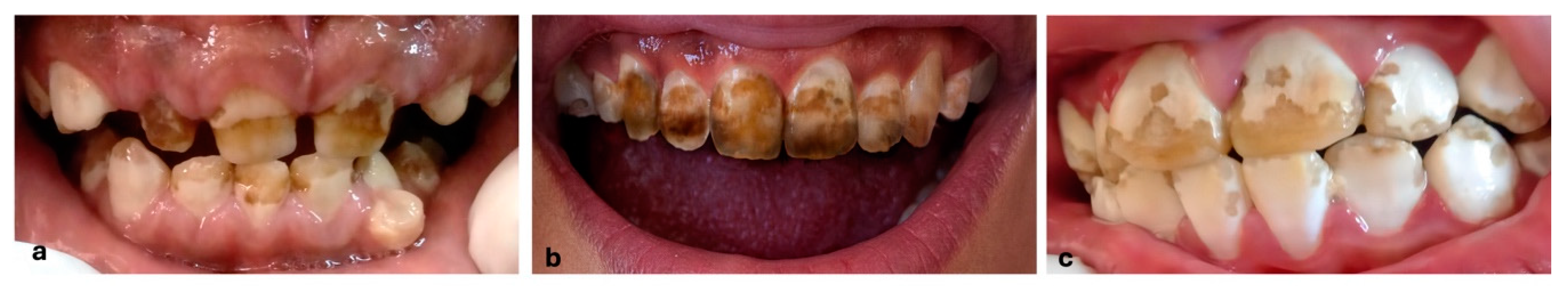

Table 1.
Shows the main manifestations of skeletal fluorosis according to Sellami et al., 2019 [58].
Table 1.
Shows the main manifestations of skeletal fluorosis according to Sellami et al., 2019 [58].
| Symptoms | Definition | Risk Factors |
|---|---|---|
| Ostecondensation | Increase in bone density, observed radiographically | Excessive fluoride deposition in bone matrix |
| Cortical thickening: | Increased thickness of cortical bone | Prolonged fluoride exposure |
| Ligament and interosseous ossification | Ossification of ligaments and interosseous membranes | Fluoride accumulation |
| Diffuse skeletal pain | Structural changes and inflammation, associated with toxicity | Symptom linked to chronic fluoride exposure |
| Limited mobility | Decreased joint movement due to stiffness and ossification of surrounding tissues | Related to chronic exposure |
| Skeletal deformity | Abnormal changes in the shape or structure bone | Related to chronic exposure |
Table 2.
Clinical nail manifestations related to fluorosis [59].
Table 2.
Clinical nail manifestations related to fluorosis [59].
| Manifestation | Description |
|---|---|
| Nail Mottling | Appears as opaque spots and transverse bands |
| Longitudinal striations | Vertical lines running along the length of the nail |
| Abnormal curvatures | Curved or claw-like nail shapes |
| Color Changes | Ranges from dark gray to brown |
| Onychia | Nail inflammation can affect both the growth and appearance of the nails |
| Onycholysis | Separation of the nail from its bed, which can lead to nail loss |
| Onychomadesis | Shedding of the nail, which can be partial or complete |
| Nails fragility | Nails that break or crack easily due to their weakness |
| Development of pitting on the nails. | Formation of small depressions on the surface of the nails |
Disclaimer/Publisher’s Note: The statements, opinions and data contained in all publications are solely those of the individual author(s) and contributor(s) and not of MDPI and/or the editor(s). MDPI and/or the editor(s) disclaim responsibility for any injury to people or property resulting from any ideas, methods, instructions or products referred to in the content. |
© 2025 by the authors. Licensee MDPI, Basel, Switzerland. This article is an open access article distributed under the terms and conditions of the Creative Commons Attribution (CC BY) license (http://creativecommons.org/licenses/by/4.0/).
Copyright: This open access article is published under a Creative Commons CC BY 4.0 license, which permit the free download, distribution, and reuse, provided that the author and preprint are cited in any reuse.


