Preprint
Article

This version is not peer-reviewed.

A Cost-Effective and Easy to Assemble 3D Human Microchannel-Blood-Brain Barrier Model and Its Application in Tumor Metastasis

A peer-reviewed article of this preprint also exists.

Submitted:

06 January 2025

Posted:

07 January 2025

You are already at the latest version

Abstract

By utilizing PDMS, collagen hydrogel and a cell line for human cerebral microvascular endothelial cells, we generated a 3D microchannel-BBB model under physiological flows. This 3D BBB has a circular shaped cross-section and a diameter of ~100m, which can properly mimic the cerebral microvessel responsible for material exchange between the circulating blood and brain tissue. The permeability of the 3D microchannel-BBB to a small molecule (sodium fluorescein with molecular weight 376) and that to a large molecule (Dex-70k) are the same as those of rat cerebral microvessels. This 3D BBB model can replicate the response to a plasma protein, orosomucoid, a cytokine, VEGF, and an enzyme, heparinase III, in either rat cerebral or mesenteric microvessesels in terms of permeability and glycocalyx (heparan sulfate). It can also replicate the adhesion of a breast cancer cell, MDA-MB-231, in rat mesenteric microvessels with no treatment and with treatments with VEGF, orosomucoid and heparinase III. Because of difficulties in accessing human cerebral microvessels, this inexpensive and easy to assemble 3D human BBB model can be applied to investigate the BBB modulating mechanisms in health and in disease and to develop therapeutic interventions targeting tumor metastasis to the brain.

Keywords: 
;  ;  ;  ;  ;  ;  
Subject: 
Engineering  -   Bioengineering

1. Introduction

The blood-brain barrier (BBB) is a highly selective interface that separates the central nervous system from the peripheral circulation, playing a critical role in maintaining brain homeostasis [1]. Proper in vivo and in vitro BBB models are indispensable tools for advancing our understanding of brain physiology, investigating disease mechanisms, and overcoming challenges in drug delivery [2,3,4]. Due to the differences in cerebrovascular structures and compositions between humans and commonly used rodents, animal models often fail to fully replicate human diseases [5,6]. Therefore, in vitro models utilizing human cells serve as a crucial bridge between human physiology and animal studies. They can offer valuable insights into disease mechanisms and aid in the development of novel strategies for drug and gene delivery to the brain [7,8,9,10,11,12,13,14,15].
The 2D BBB model generated on the Transwell filter has been the most widely utilized cell-based in vitro BBB models over the last decades [2,3,4].Although the Transwell filter can generate the BBB with the comparable permeability as in vivo data, the generated BBB is flat and without the influence of the blood flow. The flow not only more efficiently brings fresh nutrients and carries away the cell generated wastes, it also induces shear stresses, which are important in endothelial responses to various stimulations via mechano-sensors and transcription factors [16,17] and in circulating cell adhesion and transmigration [18,19,20]. Studies by Cucullo et al [21] found that the laminar flow induced shear stress promote the formation of a tight and highly selective BBB generated by HBMECs (human brain microvascular endothelial cells), as well as increasing the RNA level of multidrug resistance transporters and ion channels at the BBB. Though the flow can be generated in some 2D BBB models as described in [22,23,24,25], they are not able to fully reproduce the 3D BBB anatomical structure such as the circular shaped cross-section.
Recent advancements in microfluidics have led to the development of BBB-on-a-chip platforms, which aim to mimic the structural and functional properties of the BBB more closely to the cerebral microvessels. Many microfluidic models in use are polydimethylsiloxane (PDMS)-based systems because they confer several advantages: biocompatible, inexpensive, easy to manipulate, transparent and gas permeable. PDMS devices can be prepared by a photolithography mask, and the replication step allows mass-production of wanted structures. However, the main drawback of PDMS, in particular for biomedical applications, is hydrophobic properties, which may induce the undesired adsorption of organic molecules [26]. In addition, the untreated PDMS surface has poor affinity for live cells and the uncross-linked free PDMS monomers can leach out into the culture medium and affect cell growth [27]. Due to the nature of the photolithography fabrication process, PDMS-based 3D BBB models usually have microchannels with a rectangular cross-section [28,29]. This geometry results in different flow and shear stress profiles as in a circular shaped real blood microvessel.
In the last several years, many 3D BBB models utilizing PDMS and collagen hydrogel have been generated. Tube-like human BBB models have been generated either from the hollow spaces in 3D hydrogel produced with a needle pull-out method [7,30,31], or within hollow channels in microfluidic systems [32,33,34]. However, the tube-like structures produced vessels have a diameter of 600-800 μm [35] which is much larger than that of the capillary and post-capillary venule whose wall is defined as the BBB. Hajal et al. [36] and Winkelman et al. [37] recently developed an in vitro model of the human BBB self-assembled within microfluidic devices from HBMECs, human brain pericytes and astrocytes. Zhao et al. [38] also engineered the human BBB at the capillary scale using a double-templating technique. Their approaches can generate the microvessels with the diameter 10-40μm. However, their techniques are quite complicated and need to fabricate the devices in a clean room. Linville at el. [7] generated a tube-like BBB of diameter ~150 μm in the PDMS and hydrogel by the needle pull-out method. Although their manufacture procedure is less complicated than the self-assembled method, it still needs relatively high skill and sophisticated apparatus. In addition, the high cost of these 3D BBB systems limits its widespread use, especially in labs with budget constraints [21]. Therefore, the main aim of this study is to generate a simple, easy to assemble and cost-effective 3D microchannel-BBB with a circular cross-section and a diameter ~100 μm, mimicking the flow and shear stress profiles in real microvessels, particularly the post-capillary venules in the brain. This inexpensive 3D BBB can be constructed at a general wet lab without using a clean room or other microfabrication techniques.
Collagen type I is a popular hydrogel used in many models of the microvasculature due to its biocompatibility and physiological resemblance with the natural ECM. In this study, a 3D human BBB-on-chip in PDMS-hydrogel is generated. The hydrogel made of 4-5 mg/ml collagen I simulates the brain tissue with the comparable mechanical and transport properties [7,39,40,41]. In the current study, the collagen gel-based 3D microtube structure is constructed by using a microneedle of diameter ~100 μm. After the 3D microchannel BBB is generated with hCMECs (human cerebral microvascular endothelial cells) under a physiological flow, its permeability to small and large solutes are quantified as well as its surface glycocalyx, one of the crucial components in regulating the BBB function.As an application of the generated 3D BBB, the effects of glycocalyx (heparan sulfate, HS)-modulating agents on the HS and the permeability of the 3D microchannel-BBB are evaluated. To test the HS-modulating effects on tumor cell metastasis in the microcirculation, the adhesion of a malignant breast cancer cell, MDA-MB-231 (or MB231) in the 3D BBB under flow is quantified with no treatment and after treatments with HS-modulating agents.

2. Materials and Methods

2.1. Cell Culture

Human cerebral microvascular endothelial cells (hCMEC/D3 or hCMEC) from Millipore Sigma (Burlington, MA, USA) (passage 8 to 19 after purchase) were cultured using EBMTM-2 Basal Medium (Lonza, Basel, Switzerland), supplemented with EGMTM-2 MV Microvascular Endothelial Cell Growth Medium SingleQuotsTM kit (Lonza) [42,43]. Human breast carcinoma cells MDA-MB-231 (or MB231) from ATCC (Manassas, VA, USA) (passage 11 to 16 after purchase) were cultured in Dulbecco’s Modified Eagle’s Medium/Nutrient Mixture F-12 Ham (DMEM/F-12), 2 mM L-glutamine, 100 U/ml penicillin and 1 mg/ml streptomycin, all from Sigma-Aldrich (St. Louis, MO, USA), supplemented with 10% fetal bovine serum (FBS, Atlanta Biologicals, Flowery Branch, GA, USA) [43,44]. All cells were cultured in the incubator with 5% CO2 at 37 °C.

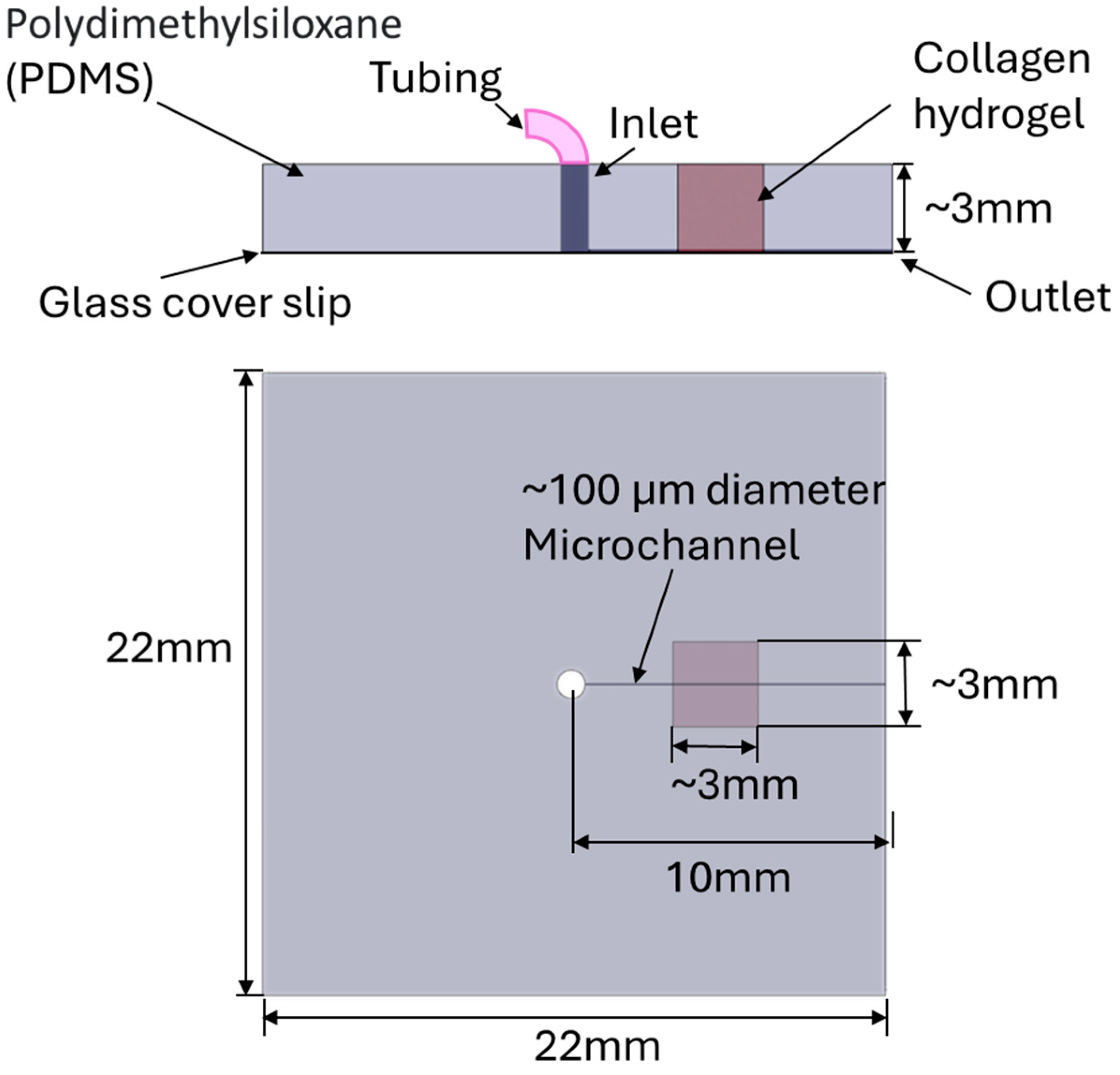
2.2. Generation of a 3D PDMS-Hydrogel Microchannel

Polydimethylsiloxane (PDMS; Sylgard, Dow Corning) pre-polymer was mixed with a curing agent (10:1 w/w ratio of PDMS to curing agent). The bubbles generated during the mixing were removed in a vacuum chamber. Then the liquid PDMS was poured on top of a glass coverslip (22 x 22 mm, Dow Corning) with a 120 μm-diameter acupuncture needle (SEIRIN, Thermo Fisher Scientific, Waltham, MA) of length 10 mm in a hexagon container. Before placed on the coverslip, the needle was coated with the 1% BSA/PBS overnight at 4 °C. The PDMS layer was cured for 35 mins at 100 °C in an oven or overnight in the 37 °C incubator. After solidification, the needle was pulled out to generate a microchannel in the PDMS device. An 18G tubing adapter was used to drill a hole perpendicularly on the PDMS base to form an inlet (Figure 1). A segment of PDMS (~ 3 x 3 mm) in the middle of the microchannel was removed and the needle was reinserted. Next, the neutralized collagen type I from rat tail (Advanced Biomatrix, Thermo Fisher Scientific, Waltham, MA) was created at ~5 mg/ml [7,45]. 1 mM genipin (Sigma-Aldrich St. Louis, MO, USA) was also added for the crosslink to stabilize the collagen hydrogel [46]. The collagen mixture was then added in the cut segment of the PDMS. After incubation overnight at 370C, the needle was pulled out to create a microchannel in the collagen part of the device. The inlet was connected to a PE-50 tubing (BD, Thermo Fisher Scientific, Waltham, MA) for perfusion which is driven by a syringe pump (NE-1800, New Era Pump system, NY, USA). Then the PBS solution was perfused to the microchannel for at least 1h to remove the residual genipin [47], followed by perfusion with cell culture medium overnight. Figure 1 shows the schematic of the generated PDMS-collagen hydrogel microchannel device. The distance between the microchannel to the cover slip was estimated to be ~ 5μm. Kim et al. [45] found that collagen I at 4-5 mg/ml has the elastic modulus similar to that of mouse hippocampus.Grifno et al. [48] found that 7 mg/ml collagen crosslinked by genipin generated a Young’s modulus of ~3kPa which is comparable to that of the mouse brain tissue. Chrobak et al [49] also reported that the gel formed by the collagen with concentration 5-7 mg/ml can prevent invasion of the cells into the gel.
The flow rate was set up to generate 1-3 dyne cm-2 wall shear stress (WSS) for the microchannel of about 100 μm diameter, which is the physiological condition in the post-capillary venule of cerebral microcirculation [7,50,51,52]. Figure 2 presents the relation between the perfusion rate and the flow velocity, and that between perfusion rate and the WSS. The equation to calculate the WSS is given by τ = (4μV)/D, where τ is the wall shear stress, μ is the viscosity of the perfusate which was estimated as 2 mPaS for 1million cells/ml in 1%BSA Ringer or the cell culture medium [44], V is the flow velocity in the channel, and D is the microchannel diameter. The calibration relation shown in Figure 2A was done by tracing a cell tracker red labeled MB231 cell by a Nikon imaging system. The flow velocity was estimated as the centerline MB231 velocity [53]. The perfusion rate of 1-1.3 μl/min in our system can generate the same WSS in the post-capillary venule of the cerebral microcirculation (Figure 2B). Guo et al. [54] found that MB231 cells prefer to adhere to the wall of post-capillary venules after overcoming the mechanical trapping of the capillaries with diameter less than 10 μm.

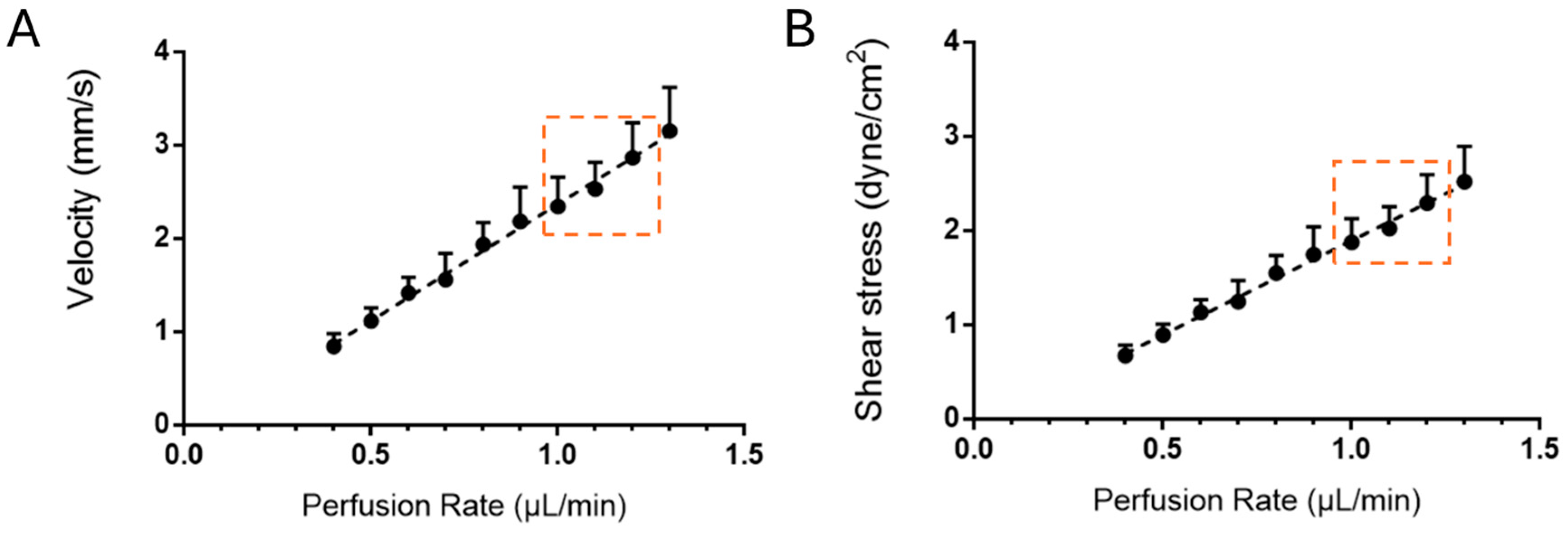
2.3. Generation of a 3D Microchannel-BBB Under Flow

4-5 million cells/ml hCMECs suspension was made in EGMTM-2 MV culture medium (Lonza, Basel, Switzerland), along with 8% dextran-70k which can increase the viscosity of the loading medium and promote vascular stability [55,56]. Then the cell suspension was slowly perfused into the microchannel via the inlet by a pipettor with a 10µL tip. After checking by microscope to make sure that the cells were attached in the microchannel, the device was incubated and flipped over every 15 min to ensure cells being seeded evenly on the microchannel surface. After 1h incubation, the suspension medium was replaced by a fresh EGMTM-2 MV medium and the microchannel was linked to a PE50 tubing system and perfused at a rate of 1.0-1.3 μl/min by a syringe pump starting at 6h after seeding. The 3D microchannel-BBB was formed in 4-5 days under this physiological flow (Figure 2B).

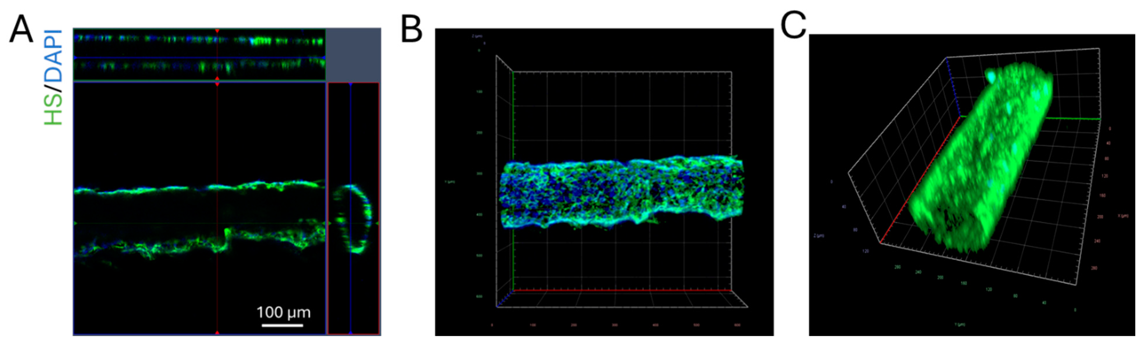
2.4. Quantification of Heparan Sulfate (HS) at the 3D Microchannel-BBB

To verify that glycocalyx (HS) was successfully generated in the 3D microchannel-BBB and to quantify the effect of orosomucoid, VEGF, and heparinase III on the HS of the microchannel-BBB, the microchannel-BBB wasimmuno-stained with anti-HSas described in [44]. Briefly, the microchannel-BBB was first rinsed by 10 mg/ml bovine serum albumin (BSA, Sigma-Aldrich) in PBS (1%BSA/PBS) and fixed with 2% paraformaldehyde and 0.1% glutaraldehyde for 20min. Then 0.1% NaBH4 (Sigma-Aldrich) was used to treat the BBB for 7 min. After rinsing with 1%BSA/PBS, the BBB was blocked by 2% normal goat serum for 30 min at room temperature (RT). Then HS on the microchannel-BBB was labeled by FITC-conjugated antibody (1:100) at 4 °C for ∼2.5 h [44]. The 2.5 h was long enough to allow FITC-anti-HS to infiltrate the entire depth of the glycocalyx [57]. Rinsed by 1%BSA/PBS to remove the free dye, the microchannel-BBB was scanned by Zeiss LSM 800 confocal laser scanning microscope with a 20×/NA0.8 objective lens. Two fields (each field 465 µm × 465 µm) (2048 x 2048) from each microchannel were captured as a z-stack of 20-30 images with a z-step of 5.28 μm. Image projection and intensity quantification for HS were performed by Zeiss ZEN and NIH ImageJ (version 1.53t). Figure 3 demonstrates the confocal images of the 3D microchannel-BBB labeled with the FITC-anti-HS and DAPI.

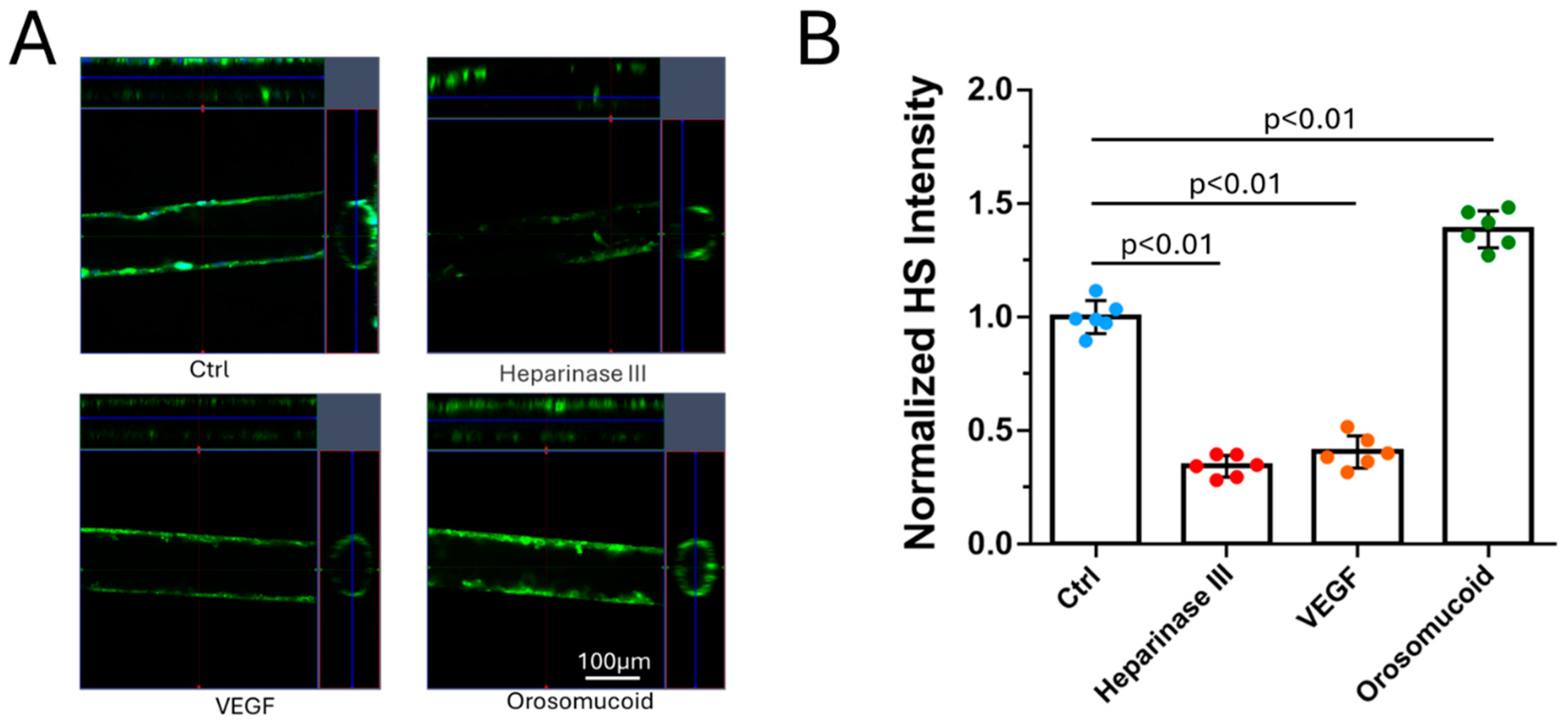
2.5. Modulation of HS of the 3D BBB and MB231 by Various Agents

Zeng et al. [58] applied different concentrations of heparinase III for 2h to digest HS on the rat fat pad endothelial cell monolayer and Cai et al. [44] perfused 1% BSA Ringer solution with 50 mU/ml heparinase III into a rat mesenteric postcapillary venule for 1h to disrupt HS at the luminal surface of a microvessel in vivo. Based on their studies, we applied 50 mU/ml heparinase III (Sigma-Aldrich) for 2h to manipulate the HS at the 3D BBB and MB231 cells before investigating MB231 adhesion to the BBB. Xia et al. [42] applied 1nM VEGF for 2h on a 2D BBB and MB231 to demonstrate a differential effect on their respective HS. In a study for tumor cell adhesion to the wall of a microvessel on rat mesentery, Shen et al. [59] pretreated the microvessel with 1nM VEGF for 1h.They showed that VEGF increased tumor cell adhesion. Corresponding to these studies, we pretreated the 3D BBB and MB231 with 1nM VEGF (recombinant human VEGF165, Peprotech, Rocky Hill, NJ, USA) for 2h.Cai et al. [44] enhanced HS of the microvessel to 1.4 folds of the control by perfusing 0.1 mg/ml orosomucoid in 1% BSA-Ringer into a rat mesenteric postcapillary venule for 30 min. In our study, we pretreated the 3D BBB and MB231 for 1h with 0.1 mg/ml orosomucoid (G3643, α1-acid glycoprotein from bovine plasma, Sigma-Aldrich). The method for modulating the HS on the 3D BBB is the same for the 2D BBB which is introduced in [43]. The only difference is that the treatment is under physiological flow in the 3D BBB while it is static in the 2D BBB.

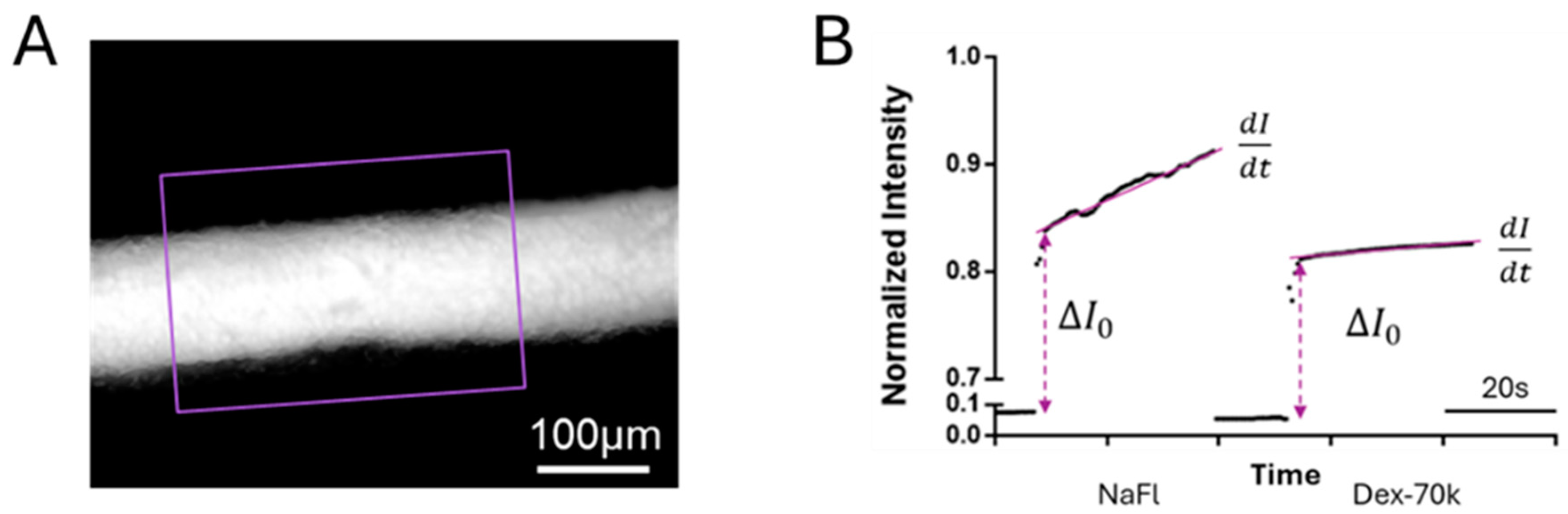
2.6. Quantification of 3D Microchannel-BBB Permeability

The method for quantifying solute permeability of the 3D microchannel-BBB was the same as that described in ref. [60]. Briefly, 5 μM sodium fluorescein (NaFl, MW 376, Sigma-Aldrich) or 50 μM FITC-dextran-70k (Dex-70k, MW 70 kD, Sigma-Aldrich) in 1% BSA Ringer was perfused into the microchannel-BBB at a flow rate of ~1.5 μl/min. Calibration of the NaFl and FITC-Dex-70k concentration vs. intensity curves using our imaging system for the permeability measurement is shown in Figure A1. The 5 μM for NaFl and 50 μM for FITC-Dex-70k used in the permeability measurement are in the linear range of the calibration curve. Simultaneously, a Nikon TE2000-S imaging system with an objective lens 20x/NA0.75 was used to capture the image every 0.5s for 1-2 min. NIH ImageJ (version 1.53t) was used for measuring the intensity profiles of ROI (purple line enclosed region) shown in Figure 4A. The permeability ( P ) to NaFI or Dex-70k was calculated by P = 1 I 0 d I d t r 2 , where I 0 is the fluorescent intensity in the ROIwhen the dye just fills up the lumen of the microchannel-BBB, d I d t is the slope of the intensity vs time curve, and r is the radius of the microchannel-BBB (Figure 4B). To determine the effect of HS modulation on the permeability of the microchannel-BBB, VEGF, orosomucoid and heparinase III were used to treat the microchannel-BBB. For the effect of orosomucoid or heparinase III, after measuring the control permeability (P), 0.1mg/mL orosomucoid or 50 mU/mL heparinase III in 1% BSA Ringer was injected into the microchannel-BBB for 1h or 2h, respectively, then P was measured on the same microchannel-BBB after the treatment. Since 1nM VEGF has an acute effect on the permeability of the BBB in vivo in the rat cerebral microvessels [61], to test the acute effect of VEGF on the in vitro 3D BBB, after the baseline P measurement, NaFl or FITC-Dex-70k in 1 nM VEGF in 1%BSA Ringer was continuously perfused into the microchannel-BBB, the image was collected every 0.5 sec for 8 min. The permeability of the microchannel-BBB was calculated every 30 sec.

2.7. Quantification of MB231 Cell Adhesion to the 3D Microchannel-BBB Under Flow

For the tumor cell adhesion experiments, MB231 cells were first fluorescently labeled with 10 μM cell tracker red, EX/EM=577/602 nm (Invitrogen, Thermo Fisher Scientific) for 30min and filtered using 40 µm cell strainer before perfused into the 3D microchannel-BBB [44]. Under no treatment, pretreatment with 50 mU/mL heparinase III for 2h to microchannel-BBB only, to both BBB and MB231, pretreatment with1nM VEGF for 2h and 0.1 mg/ml orosomucoid for 1h to both BBB and MB231, as described in Li et al. [43], MB231 cells at ~1 million/ml in 1%BSA-Ringer were perfused to the microchannel-BBB at 1-1.3 μl/min flow rate for 1h at 370C.The microchannel-BBB with adherent MB231 cells were imaged by a Nikon Eclipse TE2000-S microscope with an objective lens 20x/NA0.75 [44]. 2 fields (436 μm × 334 μm for each field) were imaged for each sample with 3 independent experiments analyzed for each case. The number of adherent MB231 was counted for each case and presented as the number per 50,000 μm2 mid-plane area of the microchannel segment.

2.8. Statistical Analysis

For All the measurements, data were presented as mean ± standard deviation (SD). A T-test or two-way ANOVA was used for comparisons between treatments and non-treatment and among different treatments. The samples were from at least 3 independent experiments. A level of p-value < 0.05 was considered statistical significance in all the experiments.

3. Results

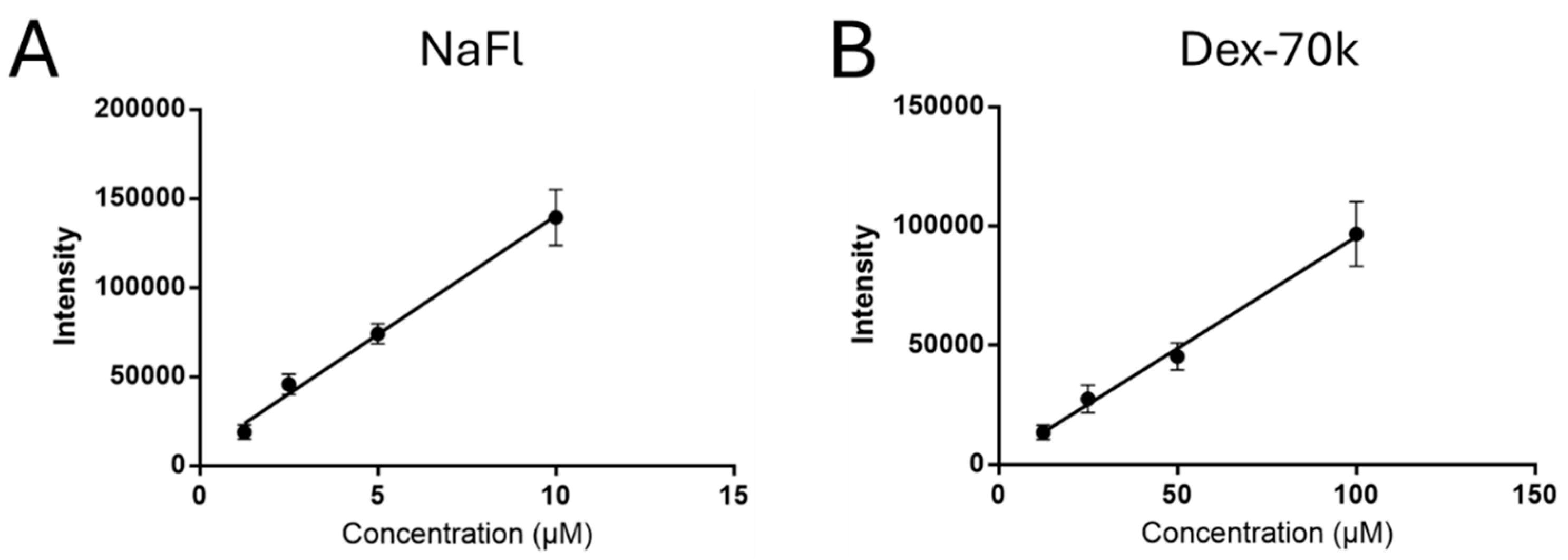
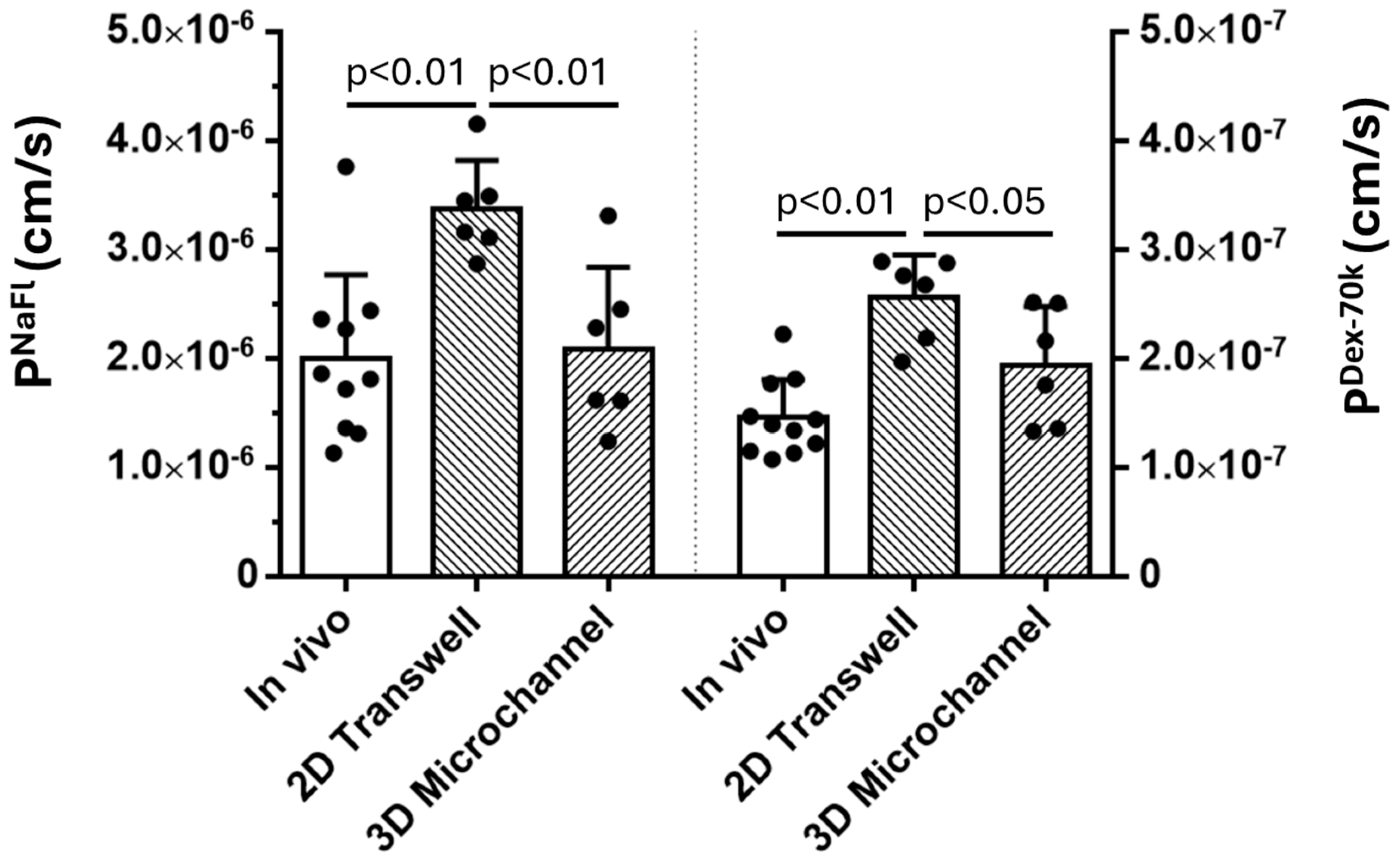
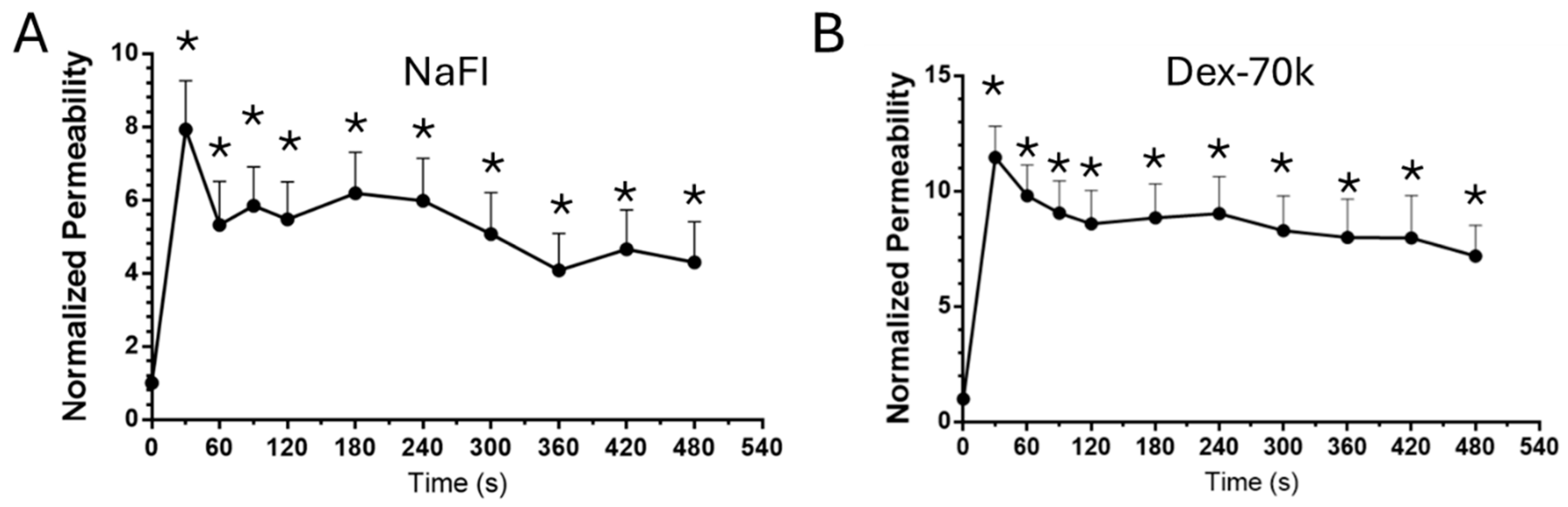
3.1. Comparison of the Solute Permeability of the 3D Microchannel-BBB with That of the 2D BBB and That of Rat Cerebral Microvessels

Unlike for the 2D BBB generated on a Transwell filter [43], it is hard to measure the TEER (indicator for BBB permeability to ions or small molecules) for the 3D microchannel-BBB, instead, the permeability (P) to a small molecule NaFl (MW=376) (PNaFl) was quantified. Figure 5 shows that PNaFl of the microchannel-BBB is 2.09 ± 0.75 × 10⁻⁶ cm/s, which is not significantly different from that of rat cerebral microvessels, 2.00 ± 0.76 × 10⁻⁶ cm/s [62](p > 0.83) but significantly smaller than that of the 2D BBB, 3.37 ± 0.44× 10⁻⁶ cm/s (p < 0.007). Similarly, P to a large molecule, Dex-70k (PDex-70k) of the microchannel-BBB is 1.94 ± 0.53 × 10⁻⁷ cm/s, which has no significant difference with that of rat cerebral microvessels, 1.46 ± 0.35 × 10⁻⁷ cm/s [62] (p > 0.08), but significantly smaller than that of the 2D BBB, 2.56 ± 0.38 × 10⁻⁷ cm/s (p < 0.05). These results indicate that the flow plays a crucial role in generating in vivo-like BBB.

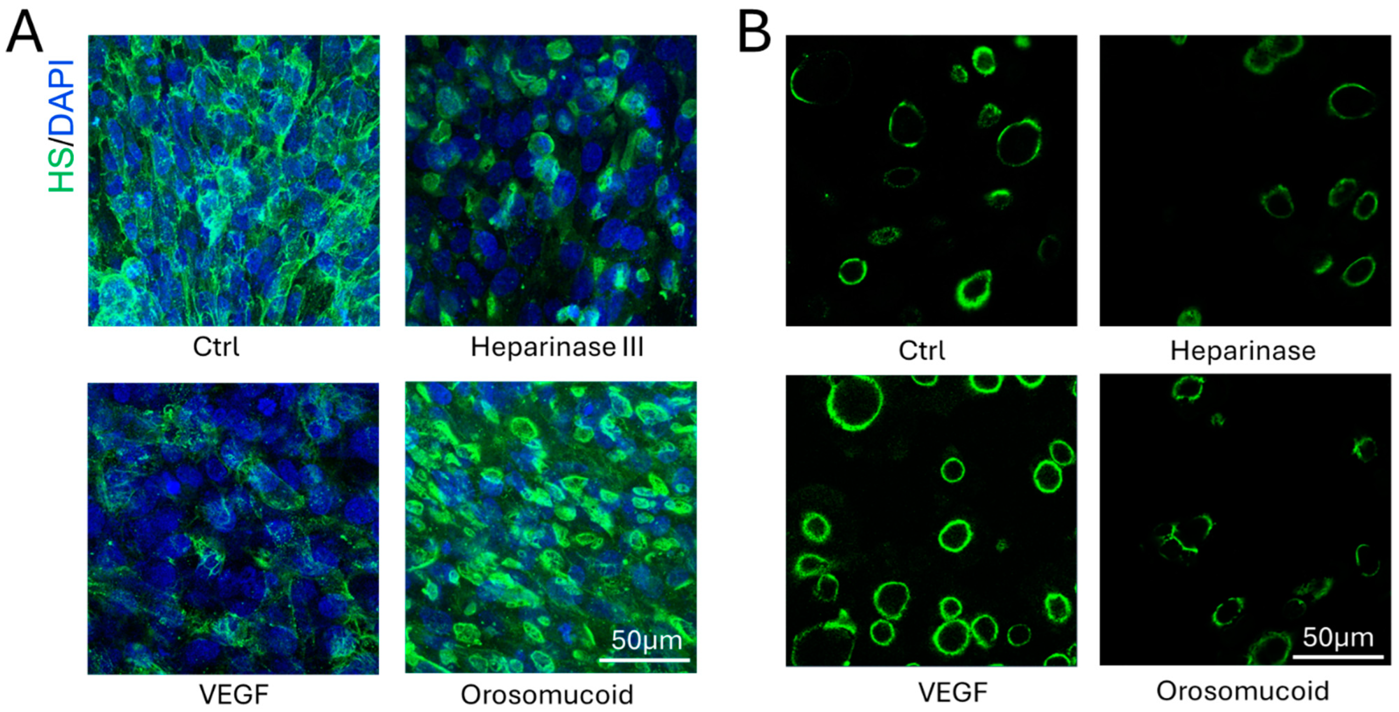
3.2. Effects of Heparinase III, VEGF and Orosomucoid on the HS of the 3D Microchannel-BBB

To investigate the effects of HS modulating agents on the 3D microchannel-BBB generated under flow, same as for the 2D BBB generated on the Transwell filters under static conditions, we treated the 3D BBB with 50 mU/mL heparinase III or 1 nM VEGF for 2h, or 0.1 mg/mL orosomucoid for 1h under flow. Figure 6 shows the confocal images (Figure 6A) and intensity quantification of the HS (Figure 6B) at the microchannel-BBB under no treatment (control) and after the treatment with these HS modulating agents. Heparinase III and VEGF reduced the HS at the microchannel-BBB to 34.3 ± 4 % and 40.6 ± 7 % of the control, respectively, comparable but different from their effects on the HS at the 2D BBB, which are 63 ± 8% (p < 0.001) and 29 ± 6% (p < 0.01) of the control [43], also see Figure A2A. In contrast, orosomucoid increased HS at the microchannel-BBB to 1.4 ± 0.1 folds of the control, which is the same for the HS at post-capillary venules in rat mesentery [44]. However, orosomucoid increased HS of the 2D BBB generated on the Transwell filter under static conditions to 3.4 ± 0.6 folds of the control, 2.4 folds of that for 3D BBB. It seems that static conditions favor the HS enhancing effect of orosomucoid.

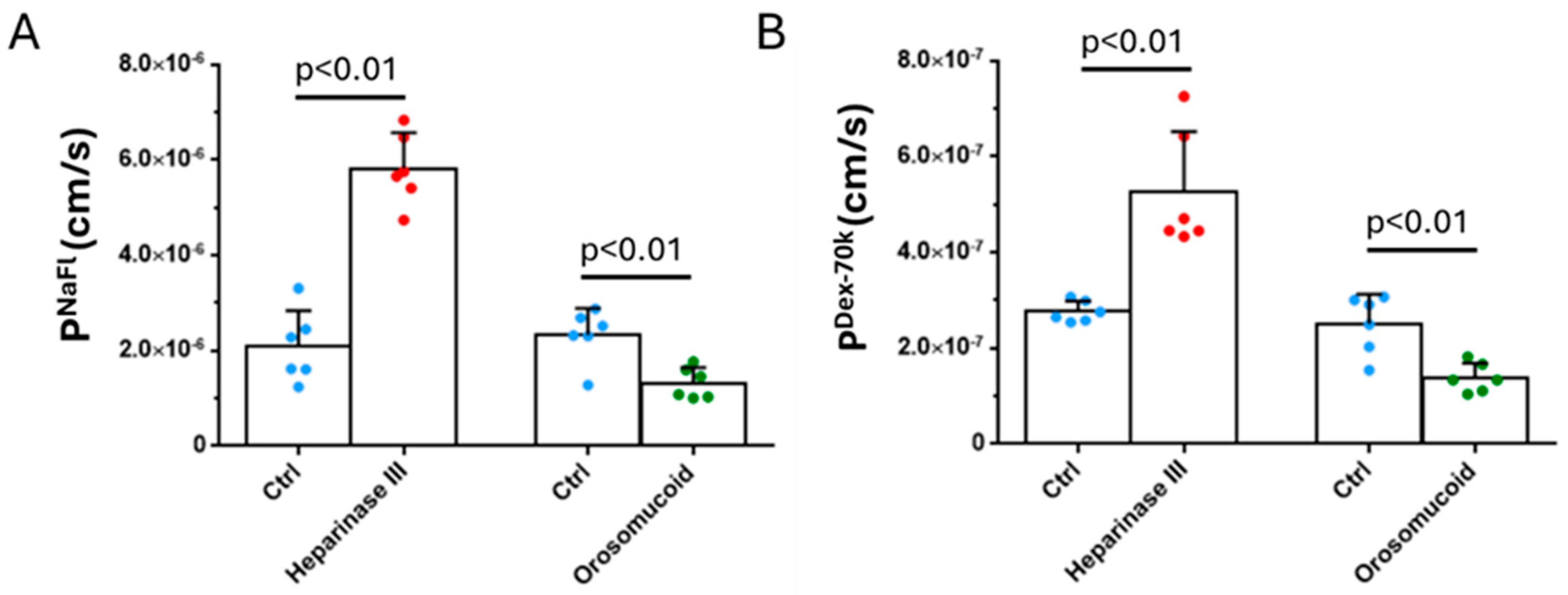
3.3. Effects of Heparinase III, Orosomucoid and VEGF on the Solute Permeability of the 3D Microchannel-BBB

To investigate the effect of HS modulation on the barrier function of the 3D BBB, we measured the solute permeability of the 3D BBB to a small molecule, sodium fluorescein (NaFl) and to a large molecule, Dex-70k. Figure 7 shows, after reduction of HS by heparinase III, PNaFl increases to 2.8 folds, from 2.09 ± 0.75 × 10⁻⁶ cm/s to 5.81 ± 0.75 × 10⁻⁶ cm/s, PDex-70k increases to 1.9 folds, from 2.77 ± 0.22×10⁻⁷ cm/s to 5.27 ± 0.12 × 10⁻⁷ cm/s, respectively. In contrast, the orosomicoid enhances HS and decreases PNaFl to 56.7%, from 2.33 ± 0.56 × 10⁻⁶ cm/s to 1.32 ± 0.33 × 10⁻⁶ cm/s, decreases PDex-70k to 55.2%, from 2.51 ± 0.61 × 10⁻⁷ cm/s to 1.39 ± 0.31 × 10⁻⁷ cm/s, respectively. The effects of heparinase III and orosomucoid on PDex-70k of 3D BBB are comparable to those on PDex-70k of the 2D BBB [43].
It was found that VEGF has an acute effect on the permeability (P) of rat cerebral microvessels [61]. We tested this acute effect of VEGF on the permeability of the 3D BBB. Figure 8 shows that for both small and large molecules, there is a transient increase in their P within 30s, peaks at 30s. The peak PNaFl is 1.7 × 10⁻⁵ cm/s, a 7.9-fold increase from its baseline P of 2.1 × 10⁻⁶ cm/s (p < 0.001). Unlike in rat cerebral microvessels, PNaFl does not return to the baseline in 2 min. Instead, it maintains at ~ 6-fold that of the baseline at 2 min and ~5-fold at 8 min (p < 0.01) at which time, we ended the measurement. Same pattern applies to PDex-70k, which peaks at 30s with the value of 2.7× 10⁻⁶ cm/s, a 11.4-fold increase from its baseline P of 2.4 × 10⁻⁷ cm/s (p < 0.001). It keeps at ~9-fold that of the baseline at 2min and ~7-fold at 8 min (p < 0.02).

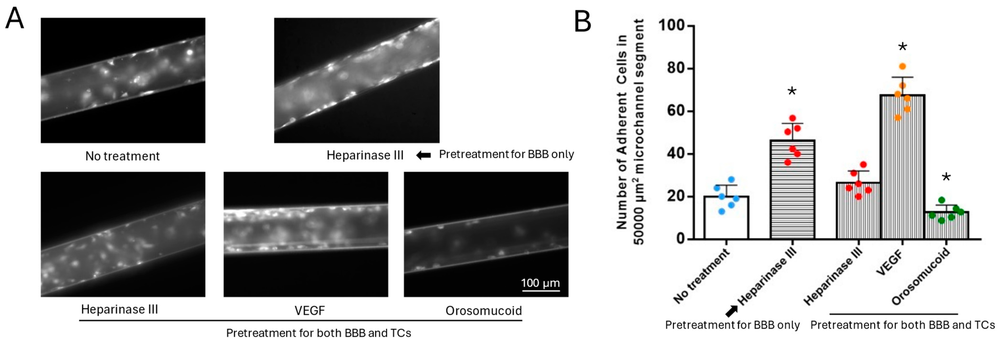
3.4. Effects of HS Modulation on MB231 Adhesion to the 3D Microchannel-BBB Under Flow

Our recent study [43] reported that heparinase III reduces HS at both MB231 and the BBB, however, VEGF and orosomucoid have differential effects on the HS of MB231 and the BBB. While VEGF increases HS of MB231, it decreases that of the BBB. In contrast, orosomucoid decreases HS of MB231, it increases that of the BBB. Figure A2 from Li et al. [43] summarizes these effects. Based on their observations, to investigate the effects of HS modulation on MB231 adhesion to the 3D microchannel-BBB under flow, we had 4 types of modulation: 1) only the 3D BBB was pretreated with heparinase III to reduce HS of the BBB but keep HS of MB231 intact; 2) both MB231 and the BBB were pretreated with heparinase III to reduce HS of both MB231 and the BBB; 3) both MB231 and the BBB were pretreated with VEGF which enhances HS of MB231 but reduces that of the BBB; and 4) both MB231 and the BBB were pretreated with orosomucoid which reduces HS of MB231 but enhances that of the BBB. Figure 9 shows the effects of HS modulation on MB231 adhesion to the microchannel-BBB under flow. Figure 9A demonstrates the typical fluorescent microscopic images for adherent MB231 cells to the BBB under various pretreatments. Figure 9B presents the quantification results. Under no pretreatment condition, 20±5 MB231 cells were adherent to the microchannel BBB per 50,000 μm2 mid-plane area of a microchannel segment. For the first modulation by pretreating only the BBB with heparinase III, the adherent MB231 cells increased to 2.3 folds compared to no treatment, indicating that reducing the HS of the BBB increases MB231 adhesion. In the second modulation by pretreating both BBB and MB231 with heparinase III, the adherent MB231 cells did not differ from that with no treatment, indicating that reducing HS on both BBB and MB231 neutralize the effect on MB231 adhesion. For the third modulation by pretreating both the BBB and MB231 with VEGF, the adherent MB231 cells increased to 3.4 folds compared to no treatment, indicating that enhancing HS of MB231 but reducing that of the BBB favor MB 231 adhesion. Finally, for the fourth modulation by pretreating both the BBB and MB231 with orosomucoid, the MB231 adhesion reduced to 63% that of no treatment, indicating that reducing HS of MB231 but enhancing that of the BBB retards MB231 adhesion.
For the MB231 adhesion to the 2D BBB under static conditions, pretreatment with heparinase III to the BBB only increases the MB231 adhesion to 1.3 folds, pretreatment with VEGF to both the BBB and MB231 increases the adhesion to 2.3 folds, while pretreatment with orosomucoid to both the BBB and MB231 reduces the adhesion to 68%. Compared with these data under static conditions, the effects of HS modulation on the MB231 adhesion appear to be augmented by the physiological flow.

4. Discussion

By utilizing PDMS, collagen hydrogel and a cell line for human cerebral microvascular endothelial cells (hCMECs), we generated a 3D microchannel-BBB under physiological flows. This 3D BBB has a circular shaped cross-section and a diameter of ~100μm, which can properly mimic the cerebral microvessel, i.e., the post-capillary venule, responsible for material exchange between the circulating blood and brain tissue (the blood-brain barrier). The permeability of the 3D microchannel-BBB to a small molecule (sodium fluorescein with molecular weight 376) and that to a large molecule (Dex-70k) are the same as what were measured in vivo for the rat cerebral microvessels [61,62]. They are smaller than those of the 2D BBB using the same hCMECs but generated on the Transwell filter under static conditions [43]. Our results indicate that a proper flow is crucial in generating an in vivo-like BBB. The flow or flow-induced shear stress effect is consistent with other studies. Cucullo et al. [21] reported that shear stress increases the TEER and RNA levels of a variety of tight and adherens junctions, resulting in enhanced BBB integrity. Winkelman et al. [37] also found that interstitial flow enhances the barrier function of the 3D microvascular networks generated within a microfluidic device.
Since glycocalyx contributes significantly to the function of the BBB including modulating the BBB permeability to water and solutes, serving as the barrier between circulating cells and endothelium as well as a mechanosensor to the blood flow [17,63]. We examined the glycocalyx at the 3D microchannel-BBB generated under flow. The same as for the 2D BBB, this 3D BBB demonstrates significant glycocalyx, specifically, heparan sulfate (HS), comparable to what observed in rat mesenteric microvessels [44,57]. Modulations of the HS on the 3D BBB as well as its permeability by VEGF, heparinase III and orosomucoid are also similar to what were found in either rat mesenteric microvessels [44,59] or rat cerebral microvessels [61]. The transient increase by VEGF observed in rat cerebral microvessels shows that exposure to 1 nM VEGF transiently increased the permeability to sodium fluorescein (PNaFl) and that to Dex-70k (PDex-70k) to 2.2 and 9.8 times their control values, respectively, within 30 s, and both returned to the control in 2 min. Unlike 2D BBB in which transient effects of VEGF on its permeability are not able to be measured, 3D BBB enables the measurement for the transient response. Although it was observed that exposure of the 3D BBB to 1 nM VEGF transiently increased PNaFl and PDex-70k to 7.9-fold and 11.4-fold of their control values in 30 s, respectively, however, the increased PNaFl and PDex-70k did not return to the control in 2 min, even in 8 min when the measurement was stopped. The increases were kept at 5-fold and 7-fold of their baselines, respectively. The reason for this observation is unknown but possibly is because the 3D BBB currently formed does not include pericytes and astrocytes that can help to maintain the BBB integrity especially in its recovery after insults [3,35,64,65,66,67].
Many in vivo and in vitro models [9,43,44,54,59,68,69,70] have been employed to investigate tumor cell adhesion and transmigration across vascular barriers, the two critical steps in tumor hematogenous metastasis [71]. The 2D BBB generated on the Transwell filter is the most convenient model for investigating tumor cell adhesion and transmigration. However, this 2D BBB under static conditions does not replicate the real physiological conditions in the cerebral microvessels. The blood flow not only brings fresh nutrients and carries away the cell generated wastes, it also induces shear stresses, which are important in endothelial responses to various stimulations via mechano-sensors and transcription factors [16,17] and in circulating cell adhesion and transmigration [18,19,20]. The 3D microchannel-BBB generated in the current study has a circular shaped cross-section and a diameter of ~100 μm, which enable us to mimic the post-capillary venules in which tumor cells prefer to adhere after they escape the size trapping from the capillary [54].Compared to adhesion to the 2D BBB under static conditions, MB231 adhesion to the 3D BBB under normal physiologic flows increases by about 50%. After the treatment with 1nM VEGF, the MB231 adhesion increases to 3.4 folds of that without treatment. However, the increase by VEGF is only 1.4 folds for the 2D BBB. This observation for MB231 adhesion under flow is consistent with that reported in Shen et al [59] for MB435 cell (another type of breast cancer cells) adhesion in the post capillary venules of rat mesentery. They found that the adherent MB435 cells under normal flow (1 mm/s) is 1.2-1.4 folds that of under a reduced flow with and without VEGF treatments [59]. Flow-induced shear stresses can enhance tumor cell adhesion to endothelial cells by activating cell adhesion molecules including integrins [72], and inducing cluster of adhesion molecules on cell surface [73]. Hajal et al reported that laminar flows in the microcirculation play an important role in tumor cell adhesion and extravasation, which could determine the local metastatic potential of tumor cells [11]. Using the 3D BBB under flow, we found that treatment with heparinase III to only BBB increases MB231 adhesion to 2.3 folds, and treatment with orosomucoid reduces MB231 adhesion to 63%, respectively, compared to no treatments. These observations are also consistent with that reported in Cai et al. [44] that heparinase III treatment increased MB231 adhesion to ~2.8 folds and orosomucoid treatment reduced the adhesion to ~54% in the post-capillary venules on rat mesentery, respectively, compared to no treatments.
The glycocalyx of tumor cells also mediates their adhesion and extravasation during metastatic dissemination [43,69]. As mentioned in the earlier section, heparinase III, VEGF and orosomucoid are employed to modulate the glycocalyx, specifically, heparan sulfate (HS) at the BBB and MB231 cells. As found in the recent study by Li et al [43] that although heparinase III reduces the HS of both MB231 and the BBB, VEGF and orosomucoid have differential effects on the HS of MB231 and that of the BBB. While VEGF increases/decreases the HS of MB231/BBB, orosomucoid decreases/increases the HS of MB231/ BBB. For the 2D BBB under static conditions, heparinase III decreases HS to 63%, while it decreases HS to 34% for 3D BBB under flow. The less HS at the 3D BBB enables more MB231 adhesion, 2.3 folds that of no treatment while the increased MB231 adhesion is only 1.3 folds for the 2D BBB. The plasma protein, orosomucoid is essential for the maintenance of stable microvessel solute permeability by enhancing the charge and organization of the endothelial glycocalyx [74]. Orosomucoid increases the HS of 2D BBB and that of 3D BBB to 3.4 folds and 1.4 folds, it decreases the MB231 adhesion to 68% and 63%, respectively, compared to no treatment. VEGF (a tumor secretion) reduces HS to 40% at the 3D BBB, to 29% at the 2D BBB, respectively from that without treatment. However, VEGF increases MB231 adhesion to 3.4 folds for the 3D BBB under flow, to 2.3 folds for the 2D BBB under static conditions. The flow seems to have a larger effect than the HS for MB231 adhesion. The flow induced forces may activate the cell adhesion molecules on both MB231 and the BBB to promote MB231 adhesion. But the enhanced HS at the BBB may shield the flow effect on the cell adhesion molecules of the endothelial cells. HS proteoglycans (HSPGs) on the endothelial cell surface interact with various adhesion molecules and extracellular matrix (ECM) proteins, including fibronectin, laminin, thrombospondin, and collagen [75]. Syndecan family of HSPGs, particularly syndecan-1 and syndecan-4, are involved in regulating cell adhesion and migration through interactions with integrins and the actin cytoskeleton [76]. On the other hand, the HS at MB231 cells may behave as a cell adhesion molecule. The reduced HS on MB231 by orosomucoid likely weakens the increased adhesive ability by the flow.
During the last decade, in addition to primary cells and cell lines, the human iPSCs (induced pluripotent cells) have been widely used to derive endothelial cells, astrocytes, pericytes and neurons, which are used to generate in vivo-like 3D human BBB [2,7,32,48,64,77,78,79,80]. These 3D human BBB models do provide a crucial tool in understanding cerebral vasculature in health and in disease, but they are expensive and need access to a clean room for manufacture. In our 3D microchannel-BBB, we utilized inexpensive materials including PDMS, collagen I and microneedles. It can be conveniently constructed in any wet lab without using a clean room. We also used the cell line hCMECs in our 3D BBB models, which are stable, easy to grow, and they can maintain normal BBB phenotypes and properties of primary cells [81,82]. Compared to the commonly used Transwell filter for 2D BBB generation, which is more than $5 each, the cost for the material used in one 3D microchannel BBB is estimated to be ~$2. In addition to inexpensive materials, our 3D microchannel BBB generation system is convenient and enables continuous perfusion of the fresh cell culture medium just like in the real blood circulation. Due to the small diameter and physiological flow rate, the required cell culture medium for generating one microchannel-BBB is 8-10ml, comparable to what is needed for generating one 2D BBB on a Transwell filter.
Although the currently generated 3D microchannel-BBB has comparable shape, size, and barrier functions as the cerebral microvessels, it does not have surrounding astrocytes and pericytes, which not only are components of the BBB but also play an important role in maintaining the BBB integrity and function. The current design can be modified to seed the astrocytes and pericytes first on the luminal surface of the collagen gel before hCMECs in a larger diameter microchannel. Besides, the current 3D microchannel-BBB model can only simulate a single microvessel of ~100 μm diameter, the complex vascular networks observed in the brain tissue are difficult to create using the current platform. More sophisticated platform will be developed to mimic the real cerebral microvasculature in the future.

5. Conclusions

We have developed a 3D human microchannel-BBB model in a PDMS-collagen hydrogel device under physiological flows. This microchannel-BBB has a circular shaped cross section with a diameter of ~100μm. It has the same permeability to both small and large solutes as that of the rat cerebral microvessels and can replicate the response to a plasma protein, orosomucoid, a cytokine, VEGF, and an enzyme, heparinase III, in either rat cerebral or mesenteric microvessesels in terms of permeability and glycocalyx (heparan sulfate). It can also replicate the tumor cell (MB231) adhesion in rat mesenteric microvessels under no treatment and under treatments with VEGF, orosomucoid and heparinase III. Because of difficulties in accessing human cerebral microvessel, this cost-effective and easy to assemble 3D human BBB model can be applied to investigate the BBB modulating mechanisms in health and in disease as well as to develop therapeutic interventions targeting tumor metastasis to the central nervous system.

Author Contributions

Conceptualization, B.M.F.; methodology, Y.L. and B.M.F.; validation, Y.L., and B.M.F.; formal analysis, Y.L; investigation, Y.L.; writing—original draft preparation, Y.L. and B.M.F.; writ-ing—review and editing, B.M.F.; visualization, Y.L.; supervision, B.M.F.; project administration, B.M.F.; funding acquisition, B.M.F. All authors have read and agreed to the published version of the manuscript.

Funding

This work was partially supported by NIH grants (RO1NS101362 and 1UG3UH3TR002151), a grant from ARO (Army Research Office) (W911NF2310189) and a grant from NSF (NSF 2324052).

Institutional Review Board Statement

Human cerebral microvascular endothelial cells (hCMEC/D3 or hCMEC) from Millipore Sigma (Burlington, MA, USA) and human breast carcinoma cells MDA-MB-231 (or MB231) from ATCC (Manassas, VA, USA).

Data Availability Statement

Data are contained withing the article.

Conflicts of Interest

The authors declare no conflicts of interest. The funders had no role in the design of the study; in the collection, analyses, or interpretation of data; in the writing of the manuscript; or in the decision to publish the results.

Appendix A

Figure A1 shows the calibration for concentration vs. intensity for sodium fluorescein and FITC-Dex-70k using the same imaging system for the solute permeability measurement. Figure A2 is from Li et al (2024) [43] for the confocal images of modulation of HS at a 2D BBB and at MB231.
Figure A1. Calibration for concentration vs. intensity for NaFl (A) and FITC-Dex-70k (B) using the imaging system for determining the solute permeability of the 3D microchannel-BBB.
Preprints 145384 g0a1
Figure A2. Modulation of heparan sulfate (HS) at a 2D BBB (A) and HS at MB231 by various agents. From Li et al, 2024. [43].
Preprints 145384 g0a2

References

  1. Abbott, N.J.; Patabendige, A.A.K.; Dolman, D.E.M.; Yusof, S.R.; Begley, D.J. Structure and function of the blood-brain barrier. Neurobiol. Dis. 2010, 37, 13–25. [Google Scholar] [CrossRef]
  2. Pérez-López, A.; Torres-Suárez, A.I.; Martín-Sabroso, C.; Aparicio-Blanco, J. An overview of in vitro 3D models of the blood-brain barrier as a tool to predict the in vivo permeability of nanomedicines. Adv. Drug Deliv. Rev. 2023, 196, 114816. [Google Scholar] [CrossRef]
  3. Tran, M.; Heo, C.; Lee, L.P.; Cho, H. Human mini-blood–brain barrier models for biomedical neuroscience research: a review. Biomater. Res. 2022, 26, 1–20. [Google Scholar] [CrossRef] [PubMed]
  4. Jamieson, J.J.; Searson, P.C.; Gerecht, S. Engineering the human blood-brain barrier in vitro. J. Biol. Eng. 2017, 11, 37–37. [Google Scholar] [CrossRef]
  5. Watase, K.; Zoghbi, H.Y. Modelling brain diseases in mice: the challenges of design and analysis. Nat. Rev. Genet. 2003, 4, 296–307. [Google Scholar] [CrossRef] [PubMed]
  6. Bogorad, M.I.; DeStefano, J.; Wong, A.D.; Searson, P.C. Tissue-engineered 3D microvessel and capillary network models for the study of vascular phenomena. Microcirculation 2017, 24. [Google Scholar] [CrossRef] [PubMed]
  7. Linville, R.M., et al., Human iPSC-derived blood-brain barrier microvessels: validation of barrier function and endothelial cell behavior. Biomaterials, 2019. 190-191: p. 24-37.
  8. Lee, S., et al., 3D brain angiogenesis model to reconstitute functional human blood-brain barrier in vitro. Biotechnol Bioeng 2020, 117, 748–748. [CrossRef]
  9. Jeon, J.S.; Bersini, S.; Gilardi, M.; Dubini, G.; Charest, J.L.; Moretti, M.; Kamm, R.D. Human 3D vascularized organotypic microfluidic assays to study breast cancer cell extravasation. Proc. Natl. Acad. Sci. USA 2015, 112, 214–219. [Google Scholar] [CrossRef] [PubMed]
  10. Shin, Y.; Choi, S.H.; Kim, E.; Bylykbashi, E.; Kim, J.A.; Chung, S.; Kim, D.Y.; Kamm, R.D.; Tanzi, R.E. Blood–Brain Barrier Dysfunction in a 3D In Vitro Model of Alzheimer's Disease. Adv. Sci. 2019, 6, 1900962. [Google Scholar] [CrossRef] [PubMed]
  11. Hajal, C.; Ibrahim, L.; Serrano, J.C.; Offeddu, G.S.; Kamm, R.D. The effects of luminal and trans-endothelial fluid flows on the extravasation and tissue invasion of tumor cells in a 3D in vitro microvascular platform. Biomaterials 2020, 265, 120470–120470. [Google Scholar] [CrossRef] [PubMed]
  12. Jagtiani, E.; Yeolekar, M.; Naik, S.; Patravale, V. In vitro blood brain barrier models: An overview. 2022, 343, 13–30. [CrossRef]
  13. Cucullo, L.; Hossain, M.; Rapp, E.; Manders, T.; Marchi, N.; Janigro, D. Development of a Humanized In Vitro Blood–Brain Barrier Model to Screen for Brain Penetration of Antiepileptic Drugs. Epilepsia 2007, 48, 505–516. [Google Scholar] [CrossRef]
  14. Yeon, J.H.; Na, D.; Choi, K.; Ryu, S.-W.; Choi, C.; Park, J.-K. Reliable permeability assay system in a microfluidic device mimicking cerebral vasculatures. Biomed. Microdevices 2012, 14, 1141–1148. [Google Scholar] [CrossRef] [PubMed]
  15. Sivandzade, F.; Cucullo, L. In-vitro blood–brain barrier modeling: A review of modern and fast-advancing technologies. J. Cereb. Blood Flow Metab. 2018, 38, 1667–1681. [Google Scholar] [CrossRef] [PubMed]
  16. Atkins, G.B. and M.K. Jain, Role of Krüppel-Like Transcription Factors in Endothelial Biology. Circulation Research, 2007. 100(12): p. 1686-1695.
  17. Fu, B.M. and J.M. Tarbell, Mechano-sensing and transduction by endothelial surface glycocalyx: composition, structure, and function. Wiley Interdiscip Rev Syst Biol Med, 2013. 5(3): p. 381-90.
  18. Coisne, C.; Lyck, R.; Engelhardt, B. Live cell imaging techniques to study T cell trafficking across the blood-brain barrier in vitro and in vivo. Fluids Barriers CNS 2013, 10, 7–7. [Google Scholar] [CrossRef]
  19. Slattery, M.J.; Dong, C. Neutrophils influence melanoma adhesion and migration under flow conditions. Int. J. Cancer 2003, 106, 713–722. [Google Scholar] [CrossRef] [PubMed]
  20. Slattery, M.J.; Liang, S.; Dong, C.; Zhang, L.; Zeng, M.; Fu, B.M.; Cai, B.; Fan, J.; Ozdemir, T.; Zhang, P.; et al. Distinct role of hydrodynamic shear in leukocyte-facilitated tumor cell extravasation. Am. J. Physiol. Physiol. 2005, 288, C831–C839. [Google Scholar] [CrossRef] [PubMed]
  21. Cucullo, L.; Hossain, M.; Puvenna, V.; Marchi, N.; Janigro, D. The role of shear stress in Blood-Brain Barrier endothelial physiology. BMC Neurosci. 2011, 12, 40–40. [Google Scholar] [CrossRef] [PubMed]
  22. Griep, L.M.; Wolbers, F.; de Wagenaar, B.; ter Braak, P.M.; Weksler, B.B.; Romero, I.A.; Couraud, P.O.; Vermes, I.; van der Meer, A.D.; Berg, A.v.D. BBB ON CHIP: microfluidic platform to mechanically and biochemically modulate blood-brain barrier function. Biomed. Microdevices 2012, 15, 145–150. [Google Scholar] [CrossRef]
  23. Sellgren, K.L.; Hawkins, B.T.; Grego, S. An optically transparent membrane supports shear stress studies in a three-dimensional microfluidic neurovascular unit model. Biomicrofluidics 2015, 9, 061102. [Google Scholar] [CrossRef]
  24. Booth, R.; Kim, H. Characterization of a microfluidic in vitro model of the blood-brain barrier (μBBB). Lab a Chip 2012, 12, 1784–1792. [Google Scholar] [CrossRef]
  25. Wang, Y.I.; Abaci, H.E.; Shuler, M.L. Microfluidic blood–brain barrier model provides in vivo-like barrier properties for drug permeability screening. Biotechnol. Bioeng. 2017, 114, 184–194. [Google Scholar] [CrossRef]
  26. Staicu, C.E.; Jipa, F.; Axente, E.; Radu, M.; Radu, B.M.; Sima, F. Lab-on-a-Chip Platforms as Tools for Drug Screening in Neuropathologies Associated with Blood–Brain Barrier Alterations. Biomolecules 2021, 11, 916. [Google Scholar] [CrossRef]
  27. Du, G.; Fang, Q.; den Toonder, J.M.J. Microfluidics for cell-based high throughput screening platforms—A review. Anal. Chim. Acta 2016, 903, 36–50. [Google Scholar] [CrossRef] [PubMed]
  28. Oddo, A.; Peng, B.; Tong, Z.; Wei, Y.; Tong, W.Y.; Thissen, H.; Voelcker, N.H. Advances in Microfluidic Blood–Brain Barrier (BBB) Models. 2019, 37, 1295–1314. [CrossRef]
  29. Gao, F.; Sun, H.; Li, X.; He, P. Leveraging avidin-biotin interaction to quantify permeability property of microvessels-on-a-chip networks. Am. J. Physiol. Circ. Physiol. 2022, 322, H71–H86. [Google Scholar] [CrossRef] [PubMed]
  30. Li, X.; Xu, J.; Bartolák-Suki, E.; Jiang, J.; Tien, J. Evaluation of 1-mm-diameter endothelialized dense collagen tubes in vascular microsurgery. J. Biomed. Mater. Res. Part B: Appl. Biomater. 2020, 108, 2441–2449. [Google Scholar] [CrossRef] [PubMed]
  31. Linville, R.M.; Boland, N.F.; Covarrubias, G.; Price, G.M.; Tien, J. Physical and Chemical Signals That Promote Vascularization of Capillary-Scale Channels. Cell. Mol. Bioeng. 2016, 9, 73–84. [Google Scholar] [CrossRef] [PubMed]
  32. Adriani, G., et al., A 3D neurovascular microfluidic model consisting of neurons, astrocytes and cerebral endothelial cells as a blood–brain barrier. Lab on a Chip, 2017. 17(3): p. 448-459.
  33. Brown, J.A.; Pensabene, V.; Markov, D.A.; Allwardt, V.; Neely, M.D.; Shi, M.; Britt, C.M.; Hoilett, O.S.; Yang, Q.; Brewer, B.M.; et al. Recreating blood-brain barrier physiology and structure on chip: A novel neurovascular microfluidic bioreactor. Biomicrofluidics 2015, 9, 054124. [Google Scholar] [CrossRef] [PubMed]
  34. Park, T.-E.; Mustafaoglu, N.; Herland, A.; Hasselkus, R.; Mannix, R.; FitzGerald, E.A.; Prantil-Baun, R.; Watters, A.; Henry, O.; Benz, M.; et al. Hypoxia-enhanced Blood-Brain Barrier Chip recapitulates human barrier function and shuttling of drugs and antibodies. Nat. Commun. 2019, 10, 1–12. [Google Scholar] [CrossRef] [PubMed]
  35. Herland, A.; van der Meer, A.D.; FitzGerald, E.A.; Park, T.-E.; Sleeboom, J.J.F.; Ingber, D.E. Distinct Contributions of Astrocytes and Pericytes to Neuroinflammation Identified in a 3D Human Blood-Brain Barrier on a Chip. PLOS ONE 2016, 11, e0150360. [Google Scholar] [CrossRef] [PubMed]
  36. Hajal, C.; Offeddu, G.S.; Shin, Y.; Zhang, S.; Morozova, O.; Hickman, D.; Knutson, C.G.; Kamm, R.D. Engineered human blood–brain barrier microfluidic model for vascular permeability analyses. Nat. Protoc. 2022, 17, 95–128. [Google Scholar] [CrossRef] [PubMed]
  37. Winkelman, M.A.; Kim, D.Y.; Kakarla, S.; Grath, A.; Silvia, N.; Dai, G. Interstitial flow enhances the formation, connectivity, and function of 3D brain microvascular networks generated within a microfluidic device. Lab a Chip 2021, 22, 170–192. [Google Scholar] [CrossRef] [PubMed]
  38. Zhao, N.; Guo, Z.; Kulkarni, S.; Norman, D.; Zhang, S.; Chung, T.D.; Nerenberg, R.F.; Linville, R.M.; Searson, P. Engineering the Human Blood–Brain Barrier at the Capillary Scale using a Double-Templating Technique. Adv. Funct. Mater. 2022, 32. [Google Scholar] [CrossRef] [PubMed]
  39. Morin, F., et al., Chapter 6 - Biomechanical Modeling of Brain Soft Tissues for Medical Applications, in Biomechanics of Living Organs, Y. Payan and J. Ohayon, Editors. 2017, Academic Press: Oxford. p. 127-146.
  40. Raub, C.; Putnam, A.; Tromberg, B.; George, S. Predicting bulk mechanical properties of cellularized collagen gels using multiphoton microscopy. Acta Biomater. 2010, 6, 4657–4665. [Google Scholar] [CrossRef] [PubMed]
  41. Shi, L.; Zeng, M.; Sun, Y.; Fu, B.M. Quantification of Blood-Brain Barrier Solute Permeability and Brain Transport by Multiphoton Microscopy. J. Biomech. Eng. 2014, 136, 031005. [Google Scholar] [CrossRef]
  42. Xia, Y.; Li, Y.; Fu, B.M. Differential effects of vascular endothelial growth factor on glycocalyx of endothelial and tumor cells and potential targets for tumor metastasis. APL Bioeng. 2022, 6, 016101. [Google Scholar] [CrossRef] [PubMed]
  43. Li, Y.; Shteyman, D.B.; Hachem, Z.; Ulay, A.A.; Fan, J.; Fu, B.M. Heparan Sulfate Modulation Affects Breast Cancer Cell Adhesion and Transmigration across In Vitro Blood–Brain Barrier. Cells 2024, 13, 190. [Google Scholar] [CrossRef]
  44. Cai, B.; Fan, J.; Zeng, M.; Zhang, L.; Fu, B.M. Adhesion of malignant mammary tumor cells MDA-MB-231 to microvessel wall increases microvascular permeability via degradation of endothelial surface glycocalyx. J. Appl. Physiol. 2012, 113, 1141–1153. [Google Scholar] [CrossRef]
  45. Kim, J.A., et al., Collagen-based brain microvasculature model in vitro using three-dimensional printed template. Biomicrofluidics, 2015. 9(2).
  46. Výborný, K.; Vallová, J.; Kočí, Z.; Kekulová, K.; Jiráková, K.; Jendelová, P.; Hodan, J.; Kubinová, Š. Genipin and EDC crosslinking of extracellular matrix hydrogel derived from human umbilical cord for neural tissue repair. Sci. Rep. 2019, 9, 1–15. [Google Scholar] [CrossRef] [PubMed]
  47. Roy, S.; Rhim, J.-W. Genipin-Crosslinked Gelatin/Chitosan-Based Functional Films Incorporated with Rosemary Essential Oil and Quercetin. Materials 2022, 15, 3769. [Google Scholar] [CrossRef] [PubMed]
  48. Grifno, G.N.; Farrell, A.M.; Linville, R.M.; Arevalo, D.; Kim, J.H.; Gu, L.; Searson, P.C. Tissue-engineered blood-brain barrier models via directed differentiation of human induced pluripotent stem cells. Sci. Rep. 2019, 9, 1–13. [Google Scholar] [CrossRef]
  49. Chrobak, K.M.; Potter, D.R.; Tien, J. Formation of perfused, functional microvascular tubes in vitro. 2006, 71, 185–196. [CrossRef]
  50. Gould, I.G., et al., The capillary bed offers the largest hemodynamic resistance to the cortical blood supply. J Cereb Blood Flow Metab, 2017. 37(1): p. 52-68.
  51. Koutsiaris, A.G.; Tachmitzi, S.V.; Batis, N.; Kotoula, M.G.; Karabatsas, C.H.; Tsironi, E.; Chatzoulis, D.Z. Volume flow and wall shear stress quantification in the human conjunctival capillaries and post-capillary venules in vivo. Biorheology 2007, 44, 375–386. [Google Scholar] [CrossRef] [PubMed]
  52. Santisakultarm, T.P.; Cornelius, N.R.; Nishimura, N.; Schafer, A.I.; Silver, R.T.; Doerschuk, P.C.; Olbricht, W.L.; Schaffer, C.B. In vivo two-photon excited fluorescence microscopy reveals cardiac- and respiration-dependent pulsatile blood flow in cortical blood vessels in mice. Am. J. Physiol. Circ. Physiol. 2012, 302, H1367–H1377. [Google Scholar] [CrossRef] [PubMed]
  53. Liu, Q.; Mirc, D.; Fu, B.M. Mechanical mechanisms of thrombosis in intact bent microvessels of rat mesentery. J. Biomech. 2008, 41, 2726–2734. [Google Scholar] [CrossRef] [PubMed]
  54. Guo, P.; Cai, B.; Lei, M.; Liu, Y.; Fu, B.M. Differential arrest and adhesion of tumor cells and microbeads in the microvasculature. Biomech. Model. Mechanobiol. 2013, 13, 537–550. [Google Scholar] [CrossRef] [PubMed]
  55. Katt, M.E., et al., Functional brain-specific microvessels from iPSC-derived human brain microvascular endothelial cells: the role of matrix composition on monolayer formation. Fluids Barriers CNS, 2018. 15(1): p. 7.
  56. Xu, S.; Li, X.; Liu, Y.; He, P. Development and Characterization of In Vitro Microvessel Network and Quantitative Measurements of Endothelial [Ca2+]i and Nitric Oxide Production. J. Vis. Exp. 2016, e54014–e54014. [Google Scholar] [CrossRef]
  57. Yen, W.-Y.; Cai, B.; Zeng, M.; Tarbell, J.M.; Fu, B.M. Quantification of the endothelial surface glycocalyx on rat and mouse blood vessels. Microvasc. Res. 2012, 83, 337–346. [Google Scholar] [CrossRef]
  58. Zeng, Y.; Ebong, E.E.; Fu, B.M.; Tarbell, J.M. The Structural Stability of the Endothelial Glycocalyx after Enzymatic Removal of Glycosaminoglycans. PLOS ONE 2012, 7, e43168. [Google Scholar] [CrossRef] [PubMed]
  59. Shen, S.; Fan, J.; Cai, B.; Lv, Y.; Zeng, M.; Hao, Y.; Giancotti, F.G.; Fu, B.M. Vascular endothelial growth factor enhances cancer cell adhesion to microvascular endothelium in vivo. Exp. Physiol. 2010, 95, 369–379. [Google Scholar] [CrossRef]
  60. Yuan, W.; Lv, Y.; Zeng, M.; Fu, B.M. Non-invasive measurement of solute permeability in cerebral microvessels of the rat. Microvasc. Res. 2008, 77, 166–173. [Google Scholar] [CrossRef]
  61. Shi, L., M. Zeng, and B.M. Fu, Temporal effects of vascular endothelial growth factor and 3,5-cyclic monophosphate on blood-brain barrier solute permeability in vivo. J Neurosci Res, 2014. 92(12): p. 1678-89.
  62. Shin, D.W.; Fan, J.; Luu, E.; Khalid, W.; Xia, Y.; Khadka, N.; Bikson, M.; Fu, B.M. In Vivo Modulation of the Blood–Brain Barrier Permeability by Transcranial Direct Current Stimulation (tDCS). Ann. Biomed. Eng. 2020, 48, 1256–1270. [Google Scholar] [CrossRef] [PubMed]
  63. Kutuzov, N., H. Flyvbjerg, and M. Lauritzen, Contributions of the glycocalyx, endothelium, and extravascular compartment to the blood–brain barrier. Proceedings of the National Academy of Sciences, 2018. 115(40): p. E9429-E9438.
  64. Jamieson, J.J., et al., Role of iPSC-derived pericytes on barrier function of iPSC-derived brain microvascular endothelial cells in 2D and 3D. Fluids and Barriers of the CNS, 2019. 16(1): p. 15.
  65. Helms, H.C.; Abbott, N.J.; Burek, M.; Cecchelli, R.; Couraud, P.-O.; Deli, M.A.; Förster, C.; Galla, H.J.; Romero, I.A.; Shusta, E.V.; et al. In vitro models of the blood–brain barrier: An overview of commonly used brain endothelial cell culture models and guidelines for their use. J. Cereb. Blood Flow Metab. 2016, 36, 862–890. [Google Scholar] [CrossRef] [PubMed]
  66. Abbott, N.J.; Friedman, A. Overview and introduction: The blood–brain barrier in health and disease. Epilepsia 2012, 53, 1–6. [Google Scholar] [CrossRef] [PubMed]
  67. Abbott, N.J., L. Rönnbäck, and E. Hansson, Astrocyte–endothelial interactions at the blood–brain barrier. Nature reviews neuroscience, 2006. 7(1): p. 41.
  68. Offeddu, G.S.; Cambria, E.; Shelton, S.E.; Haase, K.; Wan, Z.; Possenti, L.; Nguyen, H.T.; Gillrie, M.R.; Hickman, D.; Knutson, C.G.; et al. Personalized Vascularized Models of Breast Cancer Desmoplasia Reveal Biomechanical Determinants of Drug Delivery to the Tumor. Adv. Sci. 2024, 11, e2402757. [Google Scholar] [CrossRef] [PubMed]
  69. Offeddu, G.S.; Hajal, C.; Foley, C.R.; Wan, Z.; Ibrahim, L.; Coughlin, M.F.; Kamm, R.D. The cancer glycocalyx mediates intravascular adhesion and extravasation during metastatic dissemination. Commun. Biol. 2021, 4, 1–10. [Google Scholar] [CrossRef]
  70. Meena, M.; Vandormael, R.; De Laere, M.; Pintelon, I.; Berneman, Z.; Watts, R.; Cools, N. A Microfluidic In Vitro Three-Dimensional Dynamic Model of the Blood–Brain Barrier to Study the Transmigration of Immune Cells. Brain Sci. 2022, 12, 1293. [Google Scholar] [CrossRef] [PubMed]
  71. Achrol, A.S., et al., Brain metastases. Nat Rev Dis Primers, 2019. 5(1): p. 5.
  72. Fu, Y.; Li, A.; Wu, J.; Kunz, R.F.; Sun, R.; Ding, Z.; Wu, J.; Dong, C. Fibrinogen and Fibrin Differentially Regulate the Local Hydrodynamic Environment in Neutrophil–Tumor Cell–Endothelial Cell Adhesion System. Appl. Sci. 2020, 11, 79. [Google Scholar] [CrossRef]
  73. Julia, C.G., et al., CD44 engagement enhances acute myeloid leukemia cell adhesion to the bone marrow microenvironment by increasing VLA-4 avidity. Haematologica, 2021. 106(8): p. 2102-2113.
  74. Schnitzer, J.E.; Pinney, E. Quantitation of specific binding of orosomucoid to cultured microvascular endothelium: role in capillary permeability. Am. J. Physiol. Circ. Physiol. 1992, 263, H48–H55. [Google Scholar] [CrossRef]
  75. Nagarajan, A.; Malvi, P.; Wajapeyee, N. Heparan Sulfate and Heparan Sulfate Proteoglycans in Cancer Initiation and Progression. Front. Endocrinol. 2018, 9, 483. [Google Scholar] [CrossRef]
  76. Lim, H.C.; AB Multhaupt, H.; Couchman, J.R. Cell surface heparan sulfate proteoglycans control adhesion and invasion of breast carcinoma cells. Mol. Cancer 2015, 14, 1–18. [Google Scholar] [CrossRef] [PubMed]
  77. Lippmann, E.S.; Al-Ahmad, A.; Palecek, S.P.; Shusta, E.V. Modeling the blood–brain barrier using stem cell sources. Fluids Barriers CNS 2013, 10, 2–2. [Google Scholar] [CrossRef] [PubMed]
  78. Stebbins, M.J.; Gastfriend, B.D.; Canfield, S.G.; Lee, M.-S.; Richards, D.; Faubion, M.G.; Li, W.-J.; Daneman, R.; Palecek, S.P.; Shusta, E.V. Human pluripotent stem cell–derived brain pericyte–like cells induce blood-brain barrier properties. Sci. Adv. 2019, 5, eaau7375. [Google Scholar] [CrossRef] [PubMed]
  79. Campisi, M.; Shin, Y.; Osaki, T.; Hajal, C.; Chiono, V.; Kamm, R.D. 3D self-organized microvascular model of the human blood-brain barrier with endothelial cells, pericytes and astrocytes. Biomaterials 2018, 180, 117–129. [Google Scholar] [CrossRef]
  80. Lippmann, E.S.; Azarin, S.M.; Kay, J.E.; Nessler, R.A.; Wilson, H.K.; Al-Ahmad, A.; Palecek, S.P.; Shusta, E.V. Derivation of blood-brain barrier endothelial cells from human pluripotent stem cells. Nat. Biotechnol. 2012, 30, 783–791. [Google Scholar] [CrossRef] [PubMed]
  81. Weksler, B.; A Romero, I.; Couraud, P.-O. The hCMEC/D3 cell line as a model of the human blood brain barrier. Fluids Barriers CNS 2013, 10, 16–16. [Google Scholar] [CrossRef]
  82. Daniels, B.P.; Cruz-Orengo, L.; Pasieka, T.J.; Couraud, P.-O.; Romero, I.A.; Weksler, B.; Cooper, J.A.; Doering, T.L.; Klein, R.S. Immortalized human cerebral microvascular endothelial cells maintain the properties of primary cells in an in vitro model of immune migration across the blood brain barrier. J. Neurosci. Methods 2012, 212, 173–179. [Google Scholar] [CrossRef] [PubMed]
Figure 1. Schematic of the PDMS-collagen hydrogel microchannel device. PDMS solidification on a glass coverslip to form a 22 mm x 22 mm x ~3 mm base. A microchannel of diameter ~100 μm is formed by pulling out a microneedle from the collagen hydrogel. The inlet is formed by an 18 Gauge tubing adapter and connected to a PE-50 tubing for perfusion which is driven by a syringe pump.
Preprints 145384 g001
Figure 2. Calibration plots for perfusion rate vs. velocity (A) and perfusion rate vs. wall shear stress (B) in a circular microchannel of diameter ~100 μm. The range of perfusion rate for generating appropriate physiological wall shear stresses in post-capillary venules is indicated by an orange dash line enclosed region.
Preprints 145384 g002
Figure 3. Confocal images showing a 3D microchannel-BBB labeled with anti-HS (heparan sulfate) and DAPI. (A) multiview orthographic projections, (B) top perspective view and (C) 3D view of the 3D BBB labeled with anti-HS.
Preprints 145384 g003
Figure 4. Schematic for determining permeability of the 3D microchannel-BBB to fluorescently labeled solutes. (A) Fluorescent microscopic image showing a 3D microchannel-BBB filled with fluorescently labeled solutes. (B) Total fluorescence intensity in ROI (the purple frame enclosed region) as a function of the perfusion time. Fluorescence intensity in the ROI is proportional to the total amount of the solutes in ROI. The slope of the intensity vs. time line (indicated by a purple line) is dI/dt, which is used to calculate the solute permeability of microchannel-BBB: P=1/ΔI0*(dI/dt)* r/2, where ΔI0 (purple dotted line with arrowheads) is the step increase of the intensity in the ROI when the fluorescently labeled solutes just fill up the lumen of the microchannel-BBB, and r is the radius of the vessel. The smaller the solute (NaFl), the larger the slope.
Preprints 145384 g004
Figure 5. Comparison of PNaFl and PDex-70k of the in vitro 2D Transwell BBB, 3D microchannel-BBB and in vivo BBB. The in vivo data are from the measured permeability of rat cerebral microvessels (Shin et al, 2020, [62]). Values are mean ± SD.
Preprints 145384 g005
Figure 6. Modulation of heparan sulfate (HS) at a 3D microchannel-BBB by various agents. (A) Confocal images showing HS at the BBB under control and after the treatment by various agents. (B) Normalized HS intensity at the BBB under control (Ctrl) and various treatments. Values are mean ± SD. n = 3 samples with 2 fields (each field 465 µm × 465 µm) per sample analyzed for each case.
Preprints 145384 g006
Figure 7. Effects of heparan sulfate (HS) modulation by various agents on the 3D microchannel-BBB permeability to NaFl (A) and dextran-70k (B). Values are mean ± SD. n = 6 samples from 3 independent experiments for each case.
Preprints 145384 g007
Figure 8. Acute effects of 1 nM VEGF on microchannel-BBB permeability. Permeability to NaFl (A) and Dextran-70k (B) relative to the baseline as a function of time. *p < 0.05 compared with the baseline. Values are mean ± SD. n = 6 samples from 3 independent experiments for each case.
Preprints 145384 g008
Figure 9. Effects of heparan sulfate (HS) modulation by various agents on MB231 adhesion to 3D microchannel-BBB under flow. (A) Fluorescent microscopic images for adherent MB231 cells to the BBB after various treatments. (B) Number of adherent MB231 cells to 3D microchannel-BBB under no treatment (control) and after various treatments. * p < 0.05, comparison between the labeled case with control (no treatment). Values are mean ± SD. n = 6 fields (436 µm x 334 µm for each field) from 3 independent experiments analyzed for each case.
Preprints 145384 g009
Disclaimer/Publisher’s Note: The statements, opinions and data contained in all publications are solely those of the individual author(s) and contributor(s) and not of MDPI and/or the editor(s). MDPI and/or the editor(s) disclaim responsibility for any injury to people or property resulting from any ideas, methods, instructions or products referred to in the content.
Copyright: This open access article is published under a Creative Commons CC BY 4.0 license, which permit the free download, distribution, and reuse, provided that the author and preprint are cited in any reuse.
Prerpints.org logo

Preprints.org is a free preprint server supported by MDPI in Basel, Switzerland.

Subscribe

Disclaimer

Terms of Use

Privacy Policy

Privacy Settings

© 2025 MDPI (Basel, Switzerland) unless otherwise stated