
Submitted:
27 December 2024
Posted:
27 December 2024
You are already at the latest version
Abstract
Historical tempera paints exposed to pollutant gases suffer chemical and mineralogical deterio-ration which manifest through physical changes. Knowledge about these changes is fundamental to develop strategies for preventive conservation of wall paintings. In this research, binary tem-pera mock-ups composed of calcite, gypsum or lead white mixed with a proteinaceous binder (i.e., egg yolk or rabbit glue) were exposed to an ageing test by SO2-rich atmosphere exposure to learn about the degradation mechanisms and forms related to the pigment-binder interaction. Reference (unaltered) and aged mock-ups were studied from a physical point of view, characterizing the morphological changes by stereomicroscope and profilometry, colour variations by spectropho-tometry, gloss changes, and reflectance changes by hyperspectral camera. Also, mineralogical and chemical changes were studied by means of x-ray diffraction, Fourier transform infrared spec-troscopy and scanning electron microscopy with energy-dispersive x-ray spectroscopy.
Egg yolk-based paints showed higher chromatic changes than their counterparts made of rabbit glue binder. Also, sulphate and sulphite salts precipitated on the surface of the aged paints re-gardless of their binder, influencing the painting reflectance which subsequently increased. Egg yolk-based mock-ups exhibited roughness increases while the rabbit glue-based paints showed roughness reduction, with exception of lead white-based paints. Therefore, an important influence of the type of binder and the interaction between the binder and the pigment on the durability of tempera paints in atmospheres rich in SO2 was confirmed.
Keywords:
wall paintings
; preventive conservation
; tempera painting
; calcite
; Bianco di San Giovanni
; gypsum
; lead white
; binder
1. Introduction
Over the last decades, part of the research studies carried out in the field of conservation of cultural heritage has been focused on studying the influence of atmospheric contamination on the construction materials used in architectural heritage [1,2,3,4,5]. Monuments and historical buildings are frequently located in urban environments where air contamination coming from traffic, industrial and construction activities, and domestic heating have negative consequences on the materials used in the structures and decorative elements. It is known that, among cultural heritage materials, outdoor historical wall paintings are also sensitive to anthropogenic pollutants, suffering, consequently, deterioration and subsequent loss [6]. Hence, it is important to characterize the decay forms and clarify the degradation mechanisms of such historical paintings when exposed to contaminated atmospheres in order to develop respectful conservation and restoration strategies [7,8,9].
Even though SO2 levels have been reduced over the last decades [10], sulphur dioxide continues to be the main pollutant gas involved in the deterioration of building stones [7,8], and the main responsible of the sulfation process of historical paintings [9,11]. In this respect, the high porosity of the paint layers and their underlying mortars will contribute their susceptibility to pollution-induced chemical decay. This deterioration also involves the action of particulate pollutants, as well as other atmospheric factors such as moisture [1,2,3,12].
Tempera is an old painting technique that consists of an emulsion where pigments are mixed with a water-soluble binder typically of proteinaceous nature such as egg yolk, rabbit glue, casein (milk) or gums [13]. The tempera technique was replaced by oil painting in the Late Renaissance and Baroque periods; however, a remarkable revival of the tempera technique has taken place since the 20th century [14]. Tempera wall paintings are widely spread along the south of Spain following the so-called a secco technique where, unlike buon fresco, pigments are fixed to the surface mixed with an organic binder [6,14,15]. Tempera paintings are long-lasting since good examples from the ancient Egyptian period still stand [16]. Nonetheless, they can suffer deterioration processes due to exposure to SO2 atmospheres, as reported in the literature, both in real paintings and mock-ups [9,17,18,19,20]. Several studies have confirmed the degradation of tempera paint materials in contact with contaminated atmospheres, though research concerning the degradation mechanisms is still sparse. Gypsum (CaSO4·2H2O) has been identified in wall paintings [17], in lime-rich mortars (i.e., made of calcite, CaCO3) exposed to medium/high SO2 levels in south Italy [6,18], and in lime-based tempera mock-ups artificially aged in SO2 rich atmospheres in presence of moisture [9]. In the case of lead white (2PbCO3·Pb(OH)2), a pigment very susceptible to saponification by reaction with oil binders [21], blackening occurs when is present in fresco paintings and subjected to SO2 exposure. This is due to the formation of i) PbO2, due to the interaction with oxidizing agents, ii) or to the formation of PbS [22,23]. The exposure of paintings made with this pigment to sulphur pollutants and humidity results in the formation of Pb sulphides and Pb sulphates [24,25].
All these degradation products trigger colorimetric changes that can be increased by chemical modifications promoted by the interaction of the paint components with sunlight and atmospheric pollutants, as well as by physical changes that take place mostly due to pigment-binder interactions [20,26]. Regarding this interaction, recent studies have shown that the susceptibility to deterioration due to exposure to a SO2 atmosphere depends greatly on the type of binder and its interaction with the pigment. Firstly, it seems that the susceptibility to deterioration of the binder varies depending on its composition. For instance, Elert and Cardell [20] observed that egg yolk-based mock-ups were more stable than rabbit glue-based paints. Indeed, SO2-exposed egg yolk-based paint mock-ups prepared with blue and green pigments, i.e., azurite, malachite, blue smalt and lapis lazuli [27,28], remained physically intact, although sulphate salts precipitated on their surfaces. In addition, a yellowing of the binder was also noticed. Instead, the corresponding rabbit glue-based paint mock-ups developed intense cracking and binder loss, where sulphated salts were present in lesser amounts. Secondly, in these works it was found that the effects of the deterioration of the binder depended on the pigment with which it was mixed. All these studies addressed blue and green pigments, but not white pigments. Therefore, this paper gathers information on the stability of binary tempera paint mock-ups made of a proteinaceous binder (egg yolk or rabbit glue) mixed with a white pigment, more precisely with calcite, also named Bianco San Giovanni (CaCO3), gypsum (CaSO4·2H2O) or white lead (2PbCO3·Pb(OH)2) under exposure to SO2 atmospheric conditions in a climatic chamber. Reference (unaltered) and artificially aged mock-ups were studied from a physical point of view, characterizing textural changes under stereomicroscope and by means of profilometry, colour changes using a spectrophotometer, gloss changes by a glossmeter, and reflectance changes using a hyperspectral camera. Also, mineralogical and chemical changes after SO2 exposure were studied by using x-ray diffraction (XRD), Fourier transform infrared spectroscopy (FTIR), and scanning electron microscopy with energy-dispersive x-ray spectroscopy (SEM-EDS). The results presented here are part of an on-going investigation which aims to address the effect of SO2 pollutant on the physical-chemical stability of tempera paints considering the pigment-binder interaction, as well as the pigment grain size [27,28].
2. Materials and Methods
2.1. Materials
In this study, fourteen tempera mock-ups were studied made with one inorganic pigment, namely calcite, gypsum or lead white (the former two with different grain sizes) mixed with either egg yolk or rabbit glue (proteinaceous binders). Traditionally, gypsum has been mixed with other pigments in paint layers or used as white ground in e.g., wall paintings [13]. Calcite was usually used as a filler pigment in paint layers, either to reduce cost or to add bulk and consistency to paint layers when combined with other pigments [13]. Lead white was the most important white pigment since ancient times until the end of the 19th century, both in grounds and in paint layers, due to its dense opacity and bright shade. Pigments were supplied by Kremer Pigments GmbH & Co. KG (Germany) (Table 1). Two organic binders of a proteinaceous nature were used: pearls of rabbit glue (collagen, ref. 63028) from Kremer Pigments GmbH & Co. KG, and egg yolk (albumin, ovalbumin and fatty acids) from eggs purchased locally.
Paint mock-ups were prepared according to Old Master recipes [29]. The procedure can be consulted in [30] for the egg yolk-based paints and in [19] for the rabbit glue-based paints. The obtained paints were then spread onto glass slides (ca. 75 mm x 25 mm x 1 mm) using a paintbrush. Several layers were applied once the previous layer was completely dried. The paint mock-ups were labelled by adding the letter E for egg yolk or R for rabbit glue to the pigments label to distinguish the pigments from the binary paint mixtures. Table 1 shows the commercial name, identification code, mineral composition and grain size of the pigments reported by the manufacturer, and the mineralogy, grain size and identification code of the paint mock-ups according to the authors. Next, each paint mock-up was cut in two pieces: one part to be used in the SO2 exposure aging test and the other part to be kept as the reference (unaltered) paint sample. Before starting the aging test, samples were kept under laboratory-controlled conditions (18 ± 5 ºC; 60 ± 10% RH) for 1 month.
2.2. SO2 Exposure Aging Test
The paint samples were exposed to SO2 gas in a FITOCLIMA 300EDTU climatic chamber (25 ºC and 45% RH) for two months. SO2 was diluted at 3% in 3000 ppm of nitrogen and dosed at a concentration of 200 ppm, a value more than 250,000 times higher than the current SO2 levels in most Europe cities (average value 0.00076 ppm). As was reported in previous works [27,28], the water used in the chamber was characterized by High-resolution liquid chromatography (Metrohm instrument with Metrosep A Supp5-250 column) to determine content in Cl−, NO2−, NO3− and SO42− anions and by ICP-OES (inductively coupled plasma-optical emission spectrometry) using a Perkin Elmer Optima 4300 DV ICP-EPS, in order to determine Ba2+, Ca2+, K+, Mg2+ and Na+ cations content. These analyses showed the following composition: 15.8 mg/L Cl−, <0.05 mg/L NO2−, 2.15 mg/L NO3−, 23.7 mg/L SO42−, 0.017 mg/L Ba2+, 17.35 mg/L Ca2+, 2.37 mg/L K+, 3.74 mg/L Mg2+ and 13.18 mg/L Na+.
2.3. Analytical Techniques
The pigments were characterized from a mineralogical point of view by means of XRD using a Siemens D5000 equipped with an x-ray generator and Cu-Kα radiation by using the random powder method. Each mineral phase was identified using the X'Pert HighScore software.
The particle size of the pigments was also studied by laser diffraction using a Mastersizer 2000LF. For this, samples were dispersed in alcohol and results were expressed on volume distribution.
Regarding the paintings, the same analytical approach used in [27,28] was applied to characterize them before and after the SO2 exposure ageing test:
A stereomicroscope (SMZ 1000, Nikon) was used to examine the appearance features of the paint mock-ups (references and aged samples).
The colour of the references and the aged samples were characterized using the CIELAB and CIELCH colour spaces [31] measuring L*, a* and b* coordinates by means of a Minolta CM-700d spectrophotometer. L* is the lightness ranging from 0 (absolute black) to 100 (absolute white); a* indicates the colour position between red (positive values) and green (negative values), and b* between yellow (positive values) and blue (negative values). Nine measurements for each sample were randomly taken. The measurements were made in the Specular Component Included (SCI) mode, for a spot diameter of 3 mm, using D65 as the illuminant and an observer angle of 10◦. In order to evaluate the colour changes, colour data were processed as colour differences (ΔL*, Δa* and Δb*) and colour difference (ΔE*ab) between the colour of the reference and the aged sample [31].
The gloss variation (ΔG) between the references and the aged paints were obtained with a glossmeter Konica Minolta Unigloss 60Plus. A reflection angle of 60° was used and 3 measurements per sample were taken to obtain the average.
Changes on the reflectance of the paints were also studied with a hyperspectral camera, a combination of an imaging spectrograph with a monochrome matrix array sensor. The equipment consisted of a CCD sensor Pulnix TM-1327 GE (1040 rows, 1392 columns) with an objective lens, and a focal length of 10 mm. An ImSpector V10 spectrograph with a spectral resolution of 4.55 nm was positioned between the sensor and the lens. The spectral camera recorded a linear array of 1,392 pixels at 1,040 wavelengths in the range 400-1000 nm. By moving the target sample vertically, the camera scans the surface line by line to obtain an image at each of the 1040 wavelengths. A Schott DCR® III incandescent lamp with a rectangular head that is 51 mm long and 0.89 mm wide was used as a light source. A cylindrical lens placed in front of the lamp focused the light so that the illuminated area would be 15 cm long and 1 cm wide. In order to move the sample, it was placed on a motorized XYZ translation stage in which the Z- axis is perpendicular to the sample surface. The paint mock-ups were fully scanned. Once the hyperspectral images were acquired, the data were processed in a MATLAB programming environment to display the respective reflectance graphs.
Roughness variation was characterized by a profilometer (Mitutoyo SJ400) to assess the morphological degradation of the surfaces of the aged paint mock-ups. Average maximum profile height (Rz, µm) [32] was obtained tracing a scan length of 2 cm; 3 profiles per sample were obtained.
Hydrophobic character of the tempera paints before aging was characterized by static contact angle (º) measurements by means of a goniometer SEO Phoenix-300 Touch [33] by applying the sessile drop method. A droplet of 6 µL of deionized water was felt of the surfaces. The measurements were made at room temperature (18±5 ºC) and a total of three measurements per sample were considered.
The mineralogical composition of the tempera paints (references and aged samples) was obtained by XRD using a Siemens D5000 equipped with an X-ray generator and Cu-Kα radiation by using the random powder method. Identification of each mineral phase was determined using the X'Pert HighScore software.
Attenuated Transmittance Reflectance –Fourier transform infrared spectroscopy (ATR-FTIR) was applied in order to chemically characterize the changes after the ageing test. A Thermo Nicolet 6700 equipment was used, recording infrared (IR) spectra at a 2 cm-1 resolution over 100 scans from 400 to 4000 cm-1.
In order to evaluate the chemical and micro-morphology of the paint mock-ups before and after the aging test, carbon-sputtered surfaces were studied under SEM-EDS (with a Philips XL30) in both Secondary Electron (SE) and Back Scattered Electron (BSE) modes. Optimum conditions of observation were obtained at an accelerating potential of 20 kV, a working distance of 9–11 mm and specimen current of 60 mA. The acquisition time to record the EDS spectra (dwell time) was 40–60s.
3. Results
3.1. Pigment Characterization
XRD confirmed that the mineralogical composition of the white pigments differed to that reported by the manufacturer (Table 1); a similar finding was obtained in [34]. The exception was for Bianco di San Giovanni pigments (BSG-ST and BSG-C) which were composed by portlandite (Ca(OH)2) and calcite (CaCO3) as informed by the manufacturer. CA-EF pigment was composed of calcite, though dolomite (CaMg(CO3)2) was identified as impurity; a similar finding was reported in [9]. In gypsum pigments G-EF and G-F, rather than gypsum (as reported by the manufacturer), bassanite (CaSO4·1/2H2O) and anhydrite (CaSO4) were identified (Table 1). Bassanite was present in GM (according to the manufacturer), however anhydrite was also identified. LW pigment, which is sold as basic lead carbonate (Pb(CO3)2·Pb(OH)2), is actually composed by hydrocerussite (2PbCO3·Pb(OH)2) and cerussite (PbCO3), as reported in the literature [35,36].
Regarding the pigments grain size, some inaccuracies were found between values reported by the manufacturer and the experimental data (Table 1), as found elsewhere [19,20,27,28,37]. For example, for pigment CA-EF, a grain size of 20 µm was reported by the manufacturer, while the laser diffraction detected a maximum particle size of 25 µm and a range from 0.25 to 100 µm. Moreover, it is noticeable that for pigment BSG-C, we detected a grain size range between 0.3 and 250 µm whereas the manufacturer stated a range between 120 and 1000 µm. In the case of gypsum pigments, differences with the sizes reported by the manufacturer were also detected. For G-EF pigment, a grain size lower than 75 μm was reported, while a maximum grain size of 7 μm and a range from 0.2 to 85 μm were experimentally obtained. For the G-F pigment, the grain size stated by the manufacturer was lower than 32 μm while the size range here found was from 0.2 to 75 μm, with a main maximum particle at 9 μm. For the G-M pigment, a grain size lower than 40 μm was reported by Kremer while the real maximum grain size was 16 μm, being the range between 1 and 160 μm. Likewise, for pigment LW, the manufacturer indicated a grain size lower than 45 µm, although the primary particle size was 3 µm with a size range from 0.1 to 10 µm.
3.2. Physical, Mineralogical and Chemical Changes of the Mock-Ups After SO2 Exposure Aging Test
Stereomicroscopy observations made on the paintings before tested revealed an influence of the grain size on the roughness of the paints, since paints made with coarser pigments had rougher surfaces (Figure 1). For example, the surfaces of BSG-C-based paints showed greater irregularity since the pigment used was the coarsest. Paint surfaces showed surfaces with circular pockmarks, assigned to relics of air bubble. Moreover, in LW-E paint, where the pigment had the smallest grain size (3 µm), it was possible to see the brush strokes (Figure 1). On the other hand, some paint mock-ups containing egg yolk often showed a higher yellow colouring (due to the binder tint) than their counterparts made with rabbit glue (see in Figure 1, CA-EF-E, BSG-ST-E, BSG-C-E, G-EF-E, G-F-E).
The SO2 aged paints did not show any visible modification of their surfaces in comparison with the reference paints (Figure 1). However, the spectrophotometry results (Figure 2) revealed that some paints (i.e., CA-EF-E, BSG-ST-E and G-F-R) suffered ΔE*ab higher than 3.5 CIELAB units, which is the threshold above which a colour change is visible by a non-expert observer [38]. Therefore, considering ΔE*ab, the aged paint CA-EF-E showed the highest colour change, while the aged paint G-M-R showed the lowest colour change. The most affected colorimetric parameter after the SO2 test was b* for most of the samples, except for the rabbit glue-based paints G-EF-R and WL-R and the egg yolk-based paint with BSG-C. The b* parameter in the egg yolk paints typically experimented a decrease (except for paints BSG-C-E and WL-E) suggesting the loss of the characteristic yellow coloration of the egg yolk paints, as was in [19]. Conversely, in the rabbit glue paints, b* suffered an increase (except G-M-R) resulting in a yellowing effect. In BSG-C-E, G-EF-R and WL-R, the most affected parameter was L*, showing a decrease trend.
L* parameter increased in the egg yolk-based paints, with exception of the BSG paints, demonstrating a whiting effect on their colour. Conversely, L* diminished in all rabbit glue-based paints, except for CA-EF-R. Colour difference ΔE*ab was higher in the egg yolk-based paints comparatively to those made with rabbit glue, with exception of paints made with G-F. Therefore, egg yolk-based paints experienced the most intense colorimetric change in comparison to the rabbit glue-based paints.
Gloss variations (ΔG) after SO2 exposure test were not important except for two samples: the egg yolk-BSG ST paint and the rabbit glue-G EF paint. It was not possible to find out a relation between the gloss variation and the type of binder (Figure 3). The data for this property showed no relationship between the magnitude of the change and the type of binder or pigment grain size.
Reflectance spectra of the reference and the aged paint mock-ups are depicted in Figure 4. In agreement with [39], we found that before the ageing test, the morphology of the spectral curve of the reference mock-ups prepared with the same pigment was similar regardeless of the binder used. However, differences were found in the reflectance intensity between samples made with the same pigment though different binder. Thus, the reference paints made with rabbit glue (Figure 4B and D) showed in the lower region of the spectrum (450-500 nm) higher reflectance values than those prepared with egg yolk (Figure 4A and C), except for the paints made with G-M and G-F (Figure 4C and D). However, and contrary to what has been found in previous works [40], the reflectance intensity did not show a relation with the grain size of the pigment. After the SO2 aging test, the intensity of the reflectance increased in all the mock-ups, except in paints prepared with pigments BSG-ST and BSG-C mixed with egg yolk (Figure 4A).
Figure 5 shows the roughness variation after the ageing test. In general, egg yolk-based paints suffered roughness increases and rabbit glue-based paints experienced roughness decreases, except BSG-C-R paint mock-up (Figure 5C) and at lesser extent LW-R paint mock-up (Figure 5G). Moreover, although there were not statistical differences between the paints made with egg yolk or rabbit glue, those prepared with egg yolk showed higher Rz variations than those made with rabbit glue, except for BSG-C and G-F paints.
The mineralogical analyses carried out on the paints before being subjected to the test yielded interesting results when comparing the x-ray diffractograms of the paints with those of the original pigments before mixing them with the binders (Table 1). Thus, new mineral phases were identified, which suggest an interaction between the pigment and the binder during the drying process of the paint, as reported in previous studies for other pigments [28]. In paints made with G-F, gypsum and anhydrite were detected, though the pigment contained bassanite and anhydrite. This occurred in both egg yolk- and rabbit glue-based paints. Something similar happened to the GE-F-R paint since the original pigment was made of bassanite and anhydrite, though only gypsum was detected in the paint.
After the SO2 aging test, the formation of new mineral phases in all the paint mock-ups was detected by means of XRD (Table 1). In CA-EF paints, calcium sulphite (Ca3(SO3)2SO4·2H2O) was formed in the egg yolk mock-up whereas gypsum was formed in rabbit glue mock-up. In BSG-ST and BSG-C mock-ups (both in egg yolk- and rabbit glue-based paints), calcium sulphite hemihydrate (CaSO3·1/2H2O) formed.
In G-based aged paints, in addition to the diverse calcium sulphates detected in the corresponding reference paints (i.e., bassanite, gypsum and/or anhydrite), different neo-formed calcium-sulphate minerals were identified. Accordingly, in G-EF-E aged paint, bassanite, anhydrite, gypsum and K2SO4 were detected (Table 1), while bassanite, anhydrite, gypsum and CaSO3 were found in G-EF-R aged paint. Neo-formed minerals were more varied in the G-F-based aged paints where calcium (Ca), potassium (K), sodium (Na) and magnesium (Mg) sulphates and sulphites were identified (Table 1). Instead, gypsum and CaSO3 were the only minerals detected in the G-M aged paints. Finally, in the LW-based aged paints, in addition to hydrocerussite and cerussite, neo-formed Pb-based sulphates and carbonates were found, i.e., Pb4O3SO4·H2O, Pb2O(SO4) and Pb4SO4(CO3)·2(OH)2.
FTIR analyses allowed the identification of absorption bands assigned to the binders, the inorganic pigments and the neo-formed minerals identified in the aged paint mock-ups (Figure 6). On the one hand, FTIR spectra of the egg yolk binder showed a broad band at 3266 cm−1 assigned to O–H stretching or/and N–H stretching from proteins, together with bands at 2921 cm-1 assigned to C–H asymmetric stretching, and at 2850 cm-1 assigned to C–H symmetric stretching from long chain fatty acids. Also, bands between 1785-1720 cm-1 assigned to C=O asymmetric stretching were detected, as well as a band at 1620 cm-1 due to C=O stretching from amide I, in addition to a band at 1530 cm-1 assigned to N–H bending from amide II. Bands at 1440 and 1365 cm-1 were also observed, assigned to bending vibrations of CH2 groups and CH3 groups of amino acid side chains respectively. Likewise, a band at 1230 cm-1 due to C–N stretching and N–H bending vibrations of amide III, bands between 1160 and 1041 cm-1 (stretching vibration of the C–O group of glycerol) and 956 cm-1 (twisting vibration of CH2 group of glycerol) [21,41,42] were found.
On the other hand, FTIR spectra of the rabbit glue binder showed bands at 3290 cm-1 assigned to O–H symmetric stretching or/and N–H stretching from proteins, at 2871 cm-1 assigned to C–H symmetric stretching, and at 1620 cm-1 associated to C=O stretching from amide I. A band at 1520 cm-1 assigned to C–N stretching and N–H bending from amide II was also found. Finally, bands between 1400-1000 cm-1 were associated to collagen absorption features attributed to CH2 wagging, CH3 deformation, C–N stretching and C–O stretching [19,43,44,45].
Regarding the reference paints, in the CA- and BSG-based paints, calcite was identified through their characteristic C–O bands: the most intense at 1409 cm-1, and weaker ones at 874 and 725 cm-1 assigned to CO32- stretching [46]. Compared to calcite, dolomite displays characteristic FTIR absorption bands at 3020, 2626 and 730 cm−1 [47]. Surprisingly, although by XRD dolomite was identified in the reference CA-EF-E paint, the FTIR analysis did not allow to detect the aforementioned bands assigned to this mineral. Portlandite was identified by XRD in several reference and aged mock-ups (Table 1). In the FTIR spectra, the presence of this mineral was confirmed through a strong band around 3680 cm-1, which was assigned to O–H stretching vibration of portlandite [48].
For the reference G-based paints, the sulphate ion (S–O) of gypsum was detected through four fundamental vibration modes: bands ̴980 cm-1 due to SO42- v1 symmetric stretching vibration, ̴450 cm-1 due to SO42- v2 symmetric bending vibration, ̴1100 cm-1 for SO42- v3 symmetric bending, and ̴610 cm-1 for the SO42- v4 asymmetric bending vibration [49,50,51]. Considering the diverse calcium sulphate minerals (gypsum, bassanite and anhydrite), small shifts can be found among the FTIR spectra bands [51]. The SO42- v1 vibration was found at 1017, 1015 and 1008 cm-1 for anhydrite, bassanite and gypsum respectively, being more intense in bassanite and almost inappreciable in anhydrite [51,52]. Gypsum, bassanite, and anhydrite all exhibited a doublet for v2 symmetric bending of the SO42- tetrahedra at 439 and 415 cm-1, 489 and 427 cm-1 and 499 and 416 cm-1 respectively. The SO42- v3 symmetric bending was identified through two bands at 667 and 595 cm-1 for gypsum, at 659 and 594 cm-1 for bassanite, and three bands at 672, 610 and 591 cm-1 for anhydrite [52]. For v3 antisymmetric stretching vibration, the strongest absorption peaks were found at 1111, 1102 and 1094 cm-1 for bassanite, gypsum and anhydrite respectively [52]. For the v4 antisymmetric bending vibration modes, the identified bands were those at 676, 629 and 612 cm-1 in anhydrite, 668 and 628 cm-1 in bassanite, and 670 and 620 cm-1 in gypsum [52]. Additionally, we found i) for gypsum, O–H stretching vibrations at 3500 and 3394 cm-1 and O–H bending vibration at 1680 and 1619 cm-1, and ii) for bassanite, O–H stretching vibrations around 3600 and 3540 cm-1, and O–H v1 bending vibration at 1617 cm-1 [52].
For the reference LW-based paints, the presence of hydrocerussite was detected due to the presence O–H stretching vibrations assigned to the band at 3530 cm-1, C–O vibrations modes of CO32- anions at 1733 cm-1 corresponding to the v1 + v4 combination mode of CO32- anions, 1384 cm-1 (v3 asymmetric C–O stretching vibrations), 1041 cm-1 (v1 symmetric C–O stretching vibrations), 837 cm-1 (v2 out-of-plane bending vibrations) and 676 cm-1 (v4 in-plane bending vibrations) [53,54]. The shoulder at 1052 cm-1 and the bands at 1041 and 960 cm-1 can be assigned to SO42- anions [54]. Regarding the bands assigned to the cerussite could be overlapped by hydrocerussite [54].
After the SO2 test, increases on the absorbance of the S–O FTIR characteristic bands were observed in the spectra of samples BSG- ST, BSG-C, G-M, G-F, G-EF and, to a lesser extent, in the WL spectra. In all these cases, these modifications were found both in the paints made with egg yolk and in those made with rabbit glue. These S–O FTIR bands were attributed to the different species of sulphites and sulphates that formed in the samples after the test. In BSG- ST, BSG-C, G-M and G-EF paints, new FTIR bands appeared around 640 cm-1 (v4 asymmetric bending vibration of S–O group), 980 cm-1 (v1 symmetric stretching vibration of S–O) and 1100 cm-1 (v3 symmetric bending vibration). In the paints made with LW, it was noticeable the identification of only one new peak at 640 cm-1, considering that two species of lead sulphate were found by XRD.
By SEM-EDS, the formation of deposits of different nature was observed after the SO2 exposure test (Figure 7). On CA-based paints (Figure 7A and B), scarce calcium-rich deposits, and to a lesser extent, deposits rich Na, Mg, silica (Si), sulphur (S) and chlorine (Cl) (pointed with an arrow in Figure 7B), were found on the aged surfaces of egg yolk and rabbit glue-based paints. On the BSG-based paints, micrometric particles, already found in the reference paints (pointed with a rectangle in Figure 7C and E), were also detected in the aged ones (pointed with a rectangle in Figure 7D and F). Moreover, deposits rich in S and Ca on the surfaces (with atomic ratio S:Ca similar to that of gypsum) were also identified on the BSG aged samples. As for the BSG-C aged paints made with egg yolk we observed that grain minerals were more intensely agglomerated when compared to the reference paints (Figure 7F and Figure 7E respectively).
No remarkable differences were detected after exposure to SO2 on the G-based paints (Figure 7G-J). Regarding the aged LW paint mock-ups, SEM observations did not allow the identification of neo-formed minerals, although different phases of lead sulphates were already identified by XRD. Nonetheless, EDS analyses confirmed an enrichment of S on the surface of the paints. Moreover, no major differences in texture or in the amount and type of surface deposits were detected in LW paints made of either rabbit glue or egg yolk.
4. Discussion
Attending to the colorimetric changes, paints made with egg yolk showed higher ΔE*ab in comparison to their rabbit glue counterparts, suffering a loss of their original yellowish coloration and a whitening of the surface. It is suggested that this whitening effect could be related to the physical degradation of the binder (which gave to unaltered paints a subtle yellowish colour) and to the formation of white sulphate and sulphite salts (identified by XRD, FTIR and SEM) due to the SO2 aging test. In previous articles focused on the study of the deterioration of blue and green paint mock-ups (rabbit glue or egg yolk-based paints with lapis lazuli, smalt, azurite, or malachite) under the same ageing test conditions [27,28], it was detected that paints made with egg yolk experimented higher colorimetric changes (i.e. whitening) than those with rabbit glue. In the referred works, this colorimetric change was also attributed to the sulphate salts deposited on the paint surfaces, which were more abundant in egg yolk paints. In the current research, XRD revealed the formation of diverse phases of sulphites and sulphates after the ageing test, which confirms the sulfation of the paint surfaces due to their interaction with SO2. Through this interaction process, the dissolution of the pigments contained in the paint samples, i.e. calcium carbonate in the CA and BSG-based paints, calcium sulphate in the G-based paints, and lead carbonate in the WL-based paints, could take place leading to the subsequent precipitation of sulphites and sulphates, whether calcium (preferably in paints made with calcium carbonate and gypsum) or lead (in the case of WL-based paints).
After the SO2 exposure of BSG-based paints, calcium sulphite hemihydrate was formed, which could subsequently transform into gypsum [9]. In these paints, portlandite was still detected in the aged samples regardless of the binder, revealing that the carbonation was not complete after 2 months of exposure. In the case of the egg yolk-based paints, several authors [9] assigned the presence of portlandite to the existence of a long-lasting superficial film of binder surrounding the portlandite particles, impeding the diffusion of CO2 and consequently lowering the carbonation rates.
In the reference CA-EF-R paint, calcite and portlandite were found, however the latter was lacking in the corresponding aged paint, where gypsum was identified. As was reported elsewhere [9,55,56], portlandite is much more hygroscopic and soluble than calcite and, thus, in the absence of egg yolk binder (which according to [9] protects the portlandite grains against carbonation) can also react with SO2 to form calcium sulphite/sulphate. This reaction only occurs in the presence of water or sufficiently high relative humidity levels [55,57]. Cultrone et al. [58] already recognized the important role of portlandite in the fixation of SO2 as sulphates. According to these authors, the high pH created by the dissolution of portlandite facilitates SO2 absorption and hydrolysis, leading to the formation of sulphite and subsequent oxidation to sulphate ions which react with Ca2+ and, ultimately, precipitate as gypsum.
On SO2 aged LW mock-ups, lead sulphates were identified, which would indicate that the lead carbonate cerussite was partially dissolved, a process that is expected considering the slightly acidic environment involving sulfation by the deposition of SO2.
Moreover, K, Na and Mg sulphates were detected by means of XRD on the G-EF and G-F paints. In addition, Cl was also identified in the calcium-based samples using SEM-EDS. These elements (K, Na, Mg and Cl) could be derived from the water used in the SO2 test environment, leading to the precipitation of sulphated or hydrated salts, such as K2SO4, K0.67Na1.33SO4 and MgS2O3·6H2O (detected by XRD).
Concerning the reflectance measurements, results confirmed changes on the aged paints. Indeed, the increase on the intensity of the reflectance detected in the aged paints could be explained considering the formation of new sulphur-based phases [27,28]. In this work, however, a greater susceptibility of the egg yolk-based paints to the SO2 deposition process was not confirmed, unlike to what was found in previous works [27,28] where blue (smalt, azurite, lapis lazuli) and green (malachite) tempera paints were subjected to the same SO2 aging test. Firstly, neither by XRD nor by SEM-EDS, a greater quantity of S-rich deposits was detected on the egg yolk-based paints, unlike in [28] where a greater quantity of sulphated salts (as product of SO2 deposition) was found in egg-based paints. Furthermore, the deterioration of the binder was unequivocally confirmed in the previously cited works [27,28], namely, a higher degree of cracking in rabbit glue-based paints and an opacification of the egg yolk-based paints surfaces [27]. The egg yolk binder opacification was explained as a result of the photo-oxidation of their lipids which, following several studies [9,56], lead to a formation of peroxyl radicals and the denaturation of proteins (polymerization of proteins and/or crosslinking of peptide chains). In our work, only differences between egg yolk- and rabbit glue-based paints were detected in terms of colour and roughness. That is, in egg yolk-based paints, colour suffered a whitening, and roughness varied more intensely (showing increases) than in rabbit glue-based paints.
Considering that less salt deposits were detected in egg yolk-based paints compared to those made with rabbit glue, these changes in colour and roughness were due to a differential degradation of the binders (organic phase) during the aging test. However, no evidence of this deterioration was detected by FTIR or SEM. FTIR only identified changes in the bands assigned to the S–O group (which were associated with the newly formed sulphates and sulphites), and no changes were detected in the functional groups of the binder. On the other hand, SEM did not detect a more intense physical deterioration in egg yolk-based paints. Probably the analytical techniques here applied were not the most suitable for detecting minor chemical or physical modifications in the binders. However, it is indisputable from the point of view of roughness and colour, that a greater physical deterioration in egg yolk-based paints took place. This greater deterioration could be explained by differences in the degree of hydrophilicity of the paints. The determination of the static contact angle in our tempera mock-ups allowed to confirm that paints made with egg yolk were hydrophilic (static contact angle lower than 90º, [59]) while paints made with rabbit glue showed static contact angle values above 90º, thus showing an hydrophobic behaviour (Table 2). This would determine a greater susceptibility to deterioration of the egg yolk binder that could manifest itself in a greater change in roughness and colour.
5. Conclusions
In this research, white tempera mock-ups composed by a pigment (calcite, Bianco di San Giovanni, gypsum or lead white) mixed with an organic binder (rabbit glue or egg yolk) were exposed to a SO2-rich atmosphere in order to evaluate the physical, chemical and mineralogical changes. It was confirmed that these tempera paints were susceptible to sulfation resulted from SO2 deposition. This sulfation is revealed through the formation of sulphur-based salts (both sulphates and sulphites) because of the dissolution of the pigments. The amount of salts formed was similar in paints made with egg yolk and rabbit glue, contrary to what was found in tempera paint mock-ups made with other pigments subjected to the same SO2 aging test. Therefore, it can be concluded that, in the case of tempera paints made with calcite, gypsum and lead carbonate, the nature of the binder (egg yolk or rabbit glue) did not influence the reactivity to sulfation by means of SO2 gas exposure. Spectrophotometry confirmed that the colour of the paints slightly changed after ageing, more intensely in egg yolk-based paints. In these paints, a greater modification of roughness was also detected after the SO2 test. Given that the amount and type of salts formed was similar in the paints regardless of the binder, the changes in colour and roughness in egg yolk-based tempera could be due to the deterioration of the egg yolk binder, which would be more sensitive than rabbit glue to the sulfation process by wet deposition of SO2. However, no chemical changes were detected on the binders. Therefore, it could be highlighted that contrary to what happens with temperas made with other pigments exposed to the same ageing test, temperas made with the selected white pigments showed higher resistance to the SO2 test. Finally, more sensitive analytical techniques would be necessary to be applied capable of detecting minimal changes in the organic binders to explain the slight changes in colour and roughness.
Author Contributions
Conceptualization, T.R, J.S.P-A. and C.C.; methodology, T.R, J.S.P-A. and C.C.; software, J.S.P-A.; validation, T.R, J.S.P-A. and C.C.; formal analysis, T.R, J.S.P-A., D.J-D. and C.C.; investigation, T.R, J.S.P-A., A.D. and C.C.; resources, T.R, J.S.P-A., A.D. and C.C.; data curation, J.S.P-A., D.J-D.; writing—original draft preparation, T.R., J.S.P-A. and D.J-D.; writing—review and editing, A.D. and C.C.; visualization, T.R. and C.C.; supervision, T.R, J.S.P-A. and C.C.; project administration, T.R, J.S.P-A. and C.C.; funding acquisition, T.R, J.S.P-A. and C.C. All authors have read and agreed to the published version of the manuscript.
Funding
This research was funded by the Spanish Research Projects AERIMPACT (CGL2012-30729), EXPOAIR (P12-FQM-1889), LASERING-PH (PID2021-1233950A-100), the European Regional Development Fund (ERDF) and the Andalusian Research Group RNM-179. Funding for open access charge: Universidade de Vigo/CISUG.
Acknowledgments
Analyses were performed in the CACTI (Centro de Apoyo a la Investigación) Research Support Centre at the University of Vigo. J.S. Pozo-Antonio was supported by the RYC2020-028902-I project funded by MICIU/AEI/10.13039/501100011033 and, by “ESF Investing in your future”. Daniel Jiménez-Desmond was supported by the ED481A-2023/086 predoctoral contract through “Programa de axudas á etapa predoutoral da Xunta de Galicia” cofinanced by the European Union within the framework of the FSE+ Galicia 2021-2027 programme.
Conflicts of Interest
The authors declare no conflicts of interest.
References
- Rivas, T.; Pozo-Antonio, J.S.; Paz, M. Sulphur and oxygen isotope analysis to identify sources of sulphur in gypsum-rich black crusts developed on granites. Sci. Total Environ. 2014, 482-483(1), 137-147. [CrossRef]
- La Russa, M.F.; Fermo, P.; Comite, V.; Belfiore, C.M.; Barca, D.; Cerioni, A.; De Santis, M.; Barbagallo, L.F.; Ricca, M.; Ruffolo, S.A. The Oceanus statue of the Fontana di Trevi (Rome): The analysis of black crust as a tool to investigate the urban air pollution and its impact on the stone degradation. Sci. Total Environ. 2017, 593-594, 297-309. [CrossRef]
- Comite, V.; Miani, A.; Ricca, M.; La Russa, M.; Pulimeno, M.; Fermo, P. The impact of atmospheric pollution on outdoor cultural heritage: an analytic methodology for the characterization of the carbonaceous fraction in black crusts present on stone surfaces. Environ. Res. 2021, 201, 111565. [CrossRef]
- Rovella, N.; Aly, N.; Comite, V.; Randazzo, L.; Fermo, P.; Barca, D.; Alvarez de Buergo, M.; La Russa, M.F. The environmental impact of air pollution on the built heritage of historic Cairo (Egypt). Sci. Total Environ. 2021, 764, 142905. [CrossRef]
- Pozo-Antonio, J.S.; Cardell, C.; Comite, V.; Fermo, P. Characterization of black crusts developed on historic stones with diverse mineralogy under different air quality environments. Environ. Sci. Pollut. Res. 2022, 29, 1-17. [CrossRef]
- Coccato, A.; Moens, L.; Vandenabeele, P. On the stability of mediaeval inorganic pigments: a literature review of the effect of climate, material selection, biological activity, analysis and conservation treatments. Herit. Sci. 2017, 5(1), 1-25. [CrossRef]
- Vazquez, P.; Carrizo, L.; Thomachot-Schneider, C.; Gibeaux, S.; Alonso, F.J. Influence of surface finish and composition on the deterioration of building stones exposed to acid atmospheres. Constr. Buil. Mater. 2016, 106, 392-403. [CrossRef]
- Urosevic, M.; Yebra-Rodríguez, A.; Sebastián-Pardo, E.; Cardell, C. Black soiling of an architectural limestone during two-year term exposure to urban air in the city of Granada (Spain). Sci. Total Environ. 2012, 414, 564–575. [CrossRef]
- Herrera, A.; Cardell, C.; Pozo-Antonio, J.S.; Burgos-Cara, A.; Elert, K. Effect of proteinaceous binder on pollution-induced sulfation of lime-based tempera paints. Prog. Org. Coat. 2018, 123, 99-110. [CrossRef]
- Naqvi, A. Decoupling trends of emissions across EU regions and the role of environmental policies. J. Clean. Prod. 2021, 323, 129130, . [CrossRef]
- Bevilacqua, N.; Borgioli, L.; Gracia, I.A. I Pigmenti Nell'Arte Dalla Preistoria All Rivoluzi Industriale. Il Prato: Saonara, Italy, 2003.
- Horemans, B.; Cardell, C.; Bencs, L.; Kontozova-Deutsch, V.; De Wael, K.; Van Grieken, R. Evaluation of airborne particles at the Alhambra monument in Granada, Spain. Microchem. J. 2011, 99(2), 429-438. [CrossRef]
- Palet, A. Tratado de Pintura. Color, pigmentos y ensayo. Edicions de la Universitat de Barcelona: Barcelona, Spain, 2002.
- Asenjo Rubio, E. Las arquitecturas pintadas en las ciudades europeas: Aportaciones desde Málaga: la secuencia cronológica y estilística. Boletín de arte 2005, (26), 117-138.
- Del Pino Díaz, C. La pintura mural: conservación y restauración. CIE Inversiones Editoriales Dossat: Madrid, Spain, 2003.
- Ambers J. Raman analysis of pigments from the Egyptian Old Kingdom. J. Raman Spectrosc. 2004, 35(8–9), 768–73. [CrossRef]
- Cotte, M.; Susini, J.; Metrich, N.; Moscato, A.; Gratziu, C.; Bertagnini, A.; Pagano, M. Blackening of Pompeian Cinnabar Paintings: X-ray Microspectroscopy Analysis. Anal. Chem. 2006, 78, 7484–7492. [CrossRef]
- Maguregui, M.; Knuutinen, U.; Martínez-Arkarazo, I.; Castro, K.; Madariaga, J.M. Thermodynamic and spectroscopic speciation to explain the blackening process of hematite formed by atmospheric SO2 impact: The case of Marcus Lucretius House (Pompeii). Anal. Chem. 2011, 83, 3319–3326. [CrossRef]
- Cardell, C.; Herrera, A.; Guerra, I.; Navas, N.; Rodríguez-Simón, L.; Elert, K. Pigment-size effect on the physico-chemical behavior of azurite-tempera dosimeters upon natural and accelerated photo aging. Dyes Pigm. 2017, 141, 53-65. [CrossRef]
- Elert, K.; Cardell, C. Weathering behavior of cinnabar-based tempera paints upon natural and accelerated aging. Spectrochim. Acta A Mol. Biomol. Spectrosc. 2019, 216, 236-248. [CrossRef]
- Mazzeo, R.; Prati, S.; Quaranta, M.; Joseph, E.; Kendix, E.; Galeotti, M. Attenuated total reflection micro FTIR characterisation of pigment– binder interaction in reconstructed paint films. Anal. Bioanal. Chem. 2008, 392(1–2), 65–76. [CrossRef]
- Franceschi, C.M.; Costa, G.A.; Franceschi, E. Aging of the paint palette of Valerio Castello (1624–1659) in different paintings of the same age (1650–1655). J. Therm. Anal. Calorim. 2010, 103(1), 69–73. [CrossRef]
- Gutman, M.; Lesar-Kikelj, M.; Mladenovič, A.; Čobal-Sedmak, V.; Križnar, A.; Kramar, S. Raman microspectroscopic analysis of pigments of the Gothic wall painting from the Dominican Monastery in Ptuj (Slovenia). J. Raman Spectrosc. 2014, 45(11–12), 1103–9. [CrossRef]
- Smith, G.D.; Clark, R.J. The role of H2S in pigment blackening. J. Cult Herit. 2002, 3(2), 101–5. [CrossRef]
- Pérez-Rodrı́guez, J.L.; Maqueda, C.; De Haro, M.J.; Rodríguez-Rubio, P. Effect of pollution on polychromed ceramic statues. Atmos. Environ. 1998, 32(6), 993-8. [CrossRef]
- Manzano, E.; Romero-Pastor, J.; Navas, N.; Rodríguez-Simón, L.R.; Cardell, C. A study of the interaction between rabbit glue binder and blue copper pigment under UV radiation: a spectroscopic and PCA approach. Vib. Spectrosc. 2010, 53(2), 260-268. [CrossRef]
- Pozo-Antonio, J.S.; Rivas, T.; Dionísio, A.; Barral, D.; Cardell, C. Effect of a SO2 Rich Atmosphere on Tempera Paint Mock-Ups. Part 1: Accelerated Aging of Smalt and Lapis Lazuli-based Paints. Minerals, 2020, 10(5), 427. [CrossRef]
- Pozo-Antonio, J.S.; Cardell, C.; Barral, D.; Dionísio, A.; Rivas, T. Effect of a SO2 Rich Atmosphere on Tempera Paint Mock-Ups. Part 2: Accelerated Aging of Azurite-and Malachite-based Paints. Minerals, 2020, 10(5), 424. [CrossRef]
- Pacheco, F. Arte de la pintura. Cátedra: Madrid, Spain, 12, 1990.
- Herrera, A.; Navas, N.; Cardell, C. An evaluation of the impact of urban air pollution on paint dosimeters by tracking changes in the lipid MALDI-TOF mass spectra profile. Talanta 2016, 155, 53–61. [CrossRef]
- CIE S014-4/E:2007, Colorimetry Part 4: CIE 1976 L*A*b* Colour Space. Commission Internationale de l'eclairage, CIE Central Bureau, Vienna, 2007.
- UNE-EN USO 4288:1998. Geometrical product specifications (GPS) - Surface texture: Profile method - Rules and procedures for the assessment of surface texture. Asociación Española de Normalización y Certificación.
- UNE-EN 828:2013. Adhesives - Wettability - Determination by measurement of contact angle and surface free energy of solid surface.
- Pozo-Antonio, J.S.; Barral, D.; Herrera, A.; Elert, K.; Rivas, T.; Cardell, C. Effect of tempera paint composition on their superficial physical properties- application of interferometric profilometry and hyperspectral imaging techniques. Prog. Org. Coat. 2018, 117, 56-68. [CrossRef]
- Eastaugh, N.; Walsh, V.; Chaplin, T.; Siddall, R. Pigment compendium: a dictionary of historical pigments. Elsevier Butterworth-Heinemann publications: Oxford, United Kingdom, 2007.
- Gettens, R.J.; Kühn, H.; Chase, W.T. Lead white. Stud Conserv. 1967, 12(4), 125–39. [CrossRef]
- Elert, K.; Benavides-Reyes, C.; Cardell, C. Effect of animal glue on mineralogy, strength and weathering resistence of calcium sulfate-based composite materials. Cem. Concr. Compos. 2019, 96, 274-283. [CrossRef]
- Mokrzycki, W.; Tatol, M. Color difference DeltaE-A survey. Mach. Graph. Vis. 2011, 20, 383–411.
- Liang, H.; Keita, K.; Peric, B.; Vajzovic, T. Pigment identification with optical coherence tomography and multispectral imaging. In Proceedings of the 2nd Int. Topical Meeting on Optical Sensing and Artificial Vision, Saint Petersburg, Russia, 12–15 May 2008.
- Liang, H. Advances in multispectral and hyperspectral imaging for archaeology and art conservation. Appl. Phys. A 2012, 106(2), 309-323. [CrossRef]
- Nodari, L.; Ricciardi, P. Non-invasive identification of paint binders in illuminated manuscripts by ER-FTIR spectroscopy: A systematic study of the influence of different pigments on the binders’ characteristic spectral features. Herit. Sci. 2019, 7, 7. [CrossRef]
- Fuertes, S.; Laca, A.; Oulego, P.; Paredes, B.; Rendueles, M.; Díaz, M. Development and characterization of egg yolk and egg yolk fractions edible films. Food Hydrocoll. 2017, 70, 229–239. [CrossRef]
- Wang, Q.; Sanad, W.; Miller, L.M.; Voigt, A.; Klingel, K.; Kandolf, R.; Stangl, K.; Baumann, G. Infrared imaging of compositional changes in inflammatory cardiomyopathy. Vib. Spectrosc. 2005, 38, 217–222. [CrossRef]
- Navas, N.; Romero-Pastor, J.; Manzano, E.; Cardell, C. Benefits of applying combined diffuse reflectance FTIR spectroscopy and principal component analysis for the study of blue tempera historical painting. Anal. Chim. Acta 2008, 630, 141–149. [CrossRef]
- Pellegrini, D.; Duce, C.; Bonaduce, I.; Biagi, S.; Ghezzi, L.; Colombini, M.P.; Tinè, M.R.; Bramanti, E. Fourier transform infrared spectroscopic study of rabbit glue/inorganic pigments mixtures in fresh and aged reference paint reconstructions. Microchem. J. 2016, 124, 31–35. [CrossRef]
- Rodríguez Blanco, J.D.; Shaw, S; Benning, L.G. The kinetics and mechanisms of amorphous calcium carbonate (ACC) crystallization to calcite, via vaterite. Nanoscale 2010, 3(1), 265-71. http://doi.org/10.1039/C0NR00589D.
- Ji, J.; Ge, Y.; Balsam, W.; Damuth, J.E.; Chen, J. Rapid identification of dolomite using a Fourier Transform Infrared Spectrophotometer (FTIR): A fast method for identifying Heinrich events in IODP Site U1308. Mar.Geol. 2009, 258(1-4), 60-68. [CrossRef]
- Horgnies, M.; Chen, J.J.; Bouillon, C. Overview about the use of fourier transform infrared spectroscopy to study cementitious materials. WIT Trans. Eng. Sci. 2013, 77, 251-262. http://doi.org/10.2495/MC130221.
- Socrates, G. Infrared and Raman Characteristic Group Frequencies: Tables and Charts. Third edition, John Wiley and Sons: Hoboken, New Jersey, 2001.
- Lane, M.D. Mid-infrared emission spectroscopy of sulfate and sulfate-bearing minerals. Am. Min. 2008, 92(1), 1-18. [CrossRef]
- Bishop, J.; Lane, M.; Dyar, M.; King, S.; Brown, A.; Swayze, G. What Lurks in the Martian Rocks and Soil? Investigations of Sulfates, Phosphates, and Perchlorates. Spectral properties of Ca-sulfates: Gypsum, bassanite, and anhydrite. Am. Min. 2014, 99. 2105-2115. [CrossRef]
- Liu, Y. Raman, Mid-IR, and NIR spectroscopic study of calcium sulfates and mapping gypsum abundances in Columbus crater, Mars. Planet. Space Sci. 2018, 163, 35-41. [CrossRef]
- Brooker, M.H.; Sunder, S.; Taylor, P.; Lopata, V.J. Infrared and Raman spectra and X-ray diffraction studies of solid lead(II) carbonates. Can. J. Chem.1983, 61, 494–502. [CrossRef]
- Siidra, O.; Nekrasova, D.; Depmeier, W.; Chukanov, N.; Zaitsev, A.; Turner, R. Hydrocerussite-related minerals and materials: structural principles, chemical variations and infrared spectroscopy. Acta Crystallographica Section B: Structural Science, Crystal Engineering and Materials 2018, 74(2), 182-195. [CrossRef]
- Camuffo, D.; Del Monte, M.; Sabbioni, C. Origin and growth of the sulfated crusts on urban limestone. Water Air Soil Pollut. 19, 1983, 351–359. [CrossRef]
- Elert, K.; Herrera, A.; Cardell, C. Pigment-binder interactions in calcium-based tempera paints. Dyes Pig. 2018, 148, 236-248. [CrossRef]
- Charola A.E.; Ware, R. Acid depostion and the deterioration of stone: a brief review of a broad topic. In Natural Stone, Weathering Phenomena, Conservation Strategies and Case Studies, Siegesmund, S.; Weiss, T.; Vollbrecht, A. (Eds.); Geological Society Special Publication No. 205: London, United Kingdom, 2002; pp. 393–406.
- Cultrone G.; Arizzi, A.; Sebastián, E.; Rodriguez-Navarro, C. Sulfation of calcitic and dolomitic lime mortars in the presence of diesel particulate matter. Environ. Geol. 2008, 56, 741–752. [CrossRef]
- Bico, J.; Thiele U.; Quere, D. Wetting of textured surfaces. Colloids Surf. A Physicochem. Eng. Asp. 2002, 206, 41–46. [CrossRef]
Figure 1.
Photomicrographs taken with stereomicroscopy of the references (Ref, on the right) and the aged paint mock-ups (SO2, on the left). Check Table 1 for the identification code of samples.
Figure 1.
Photomicrographs taken with stereomicroscopy of the references (Ref, on the right) and the aged paint mock-ups (SO2, on the left). Check Table 1 for the identification code of samples.
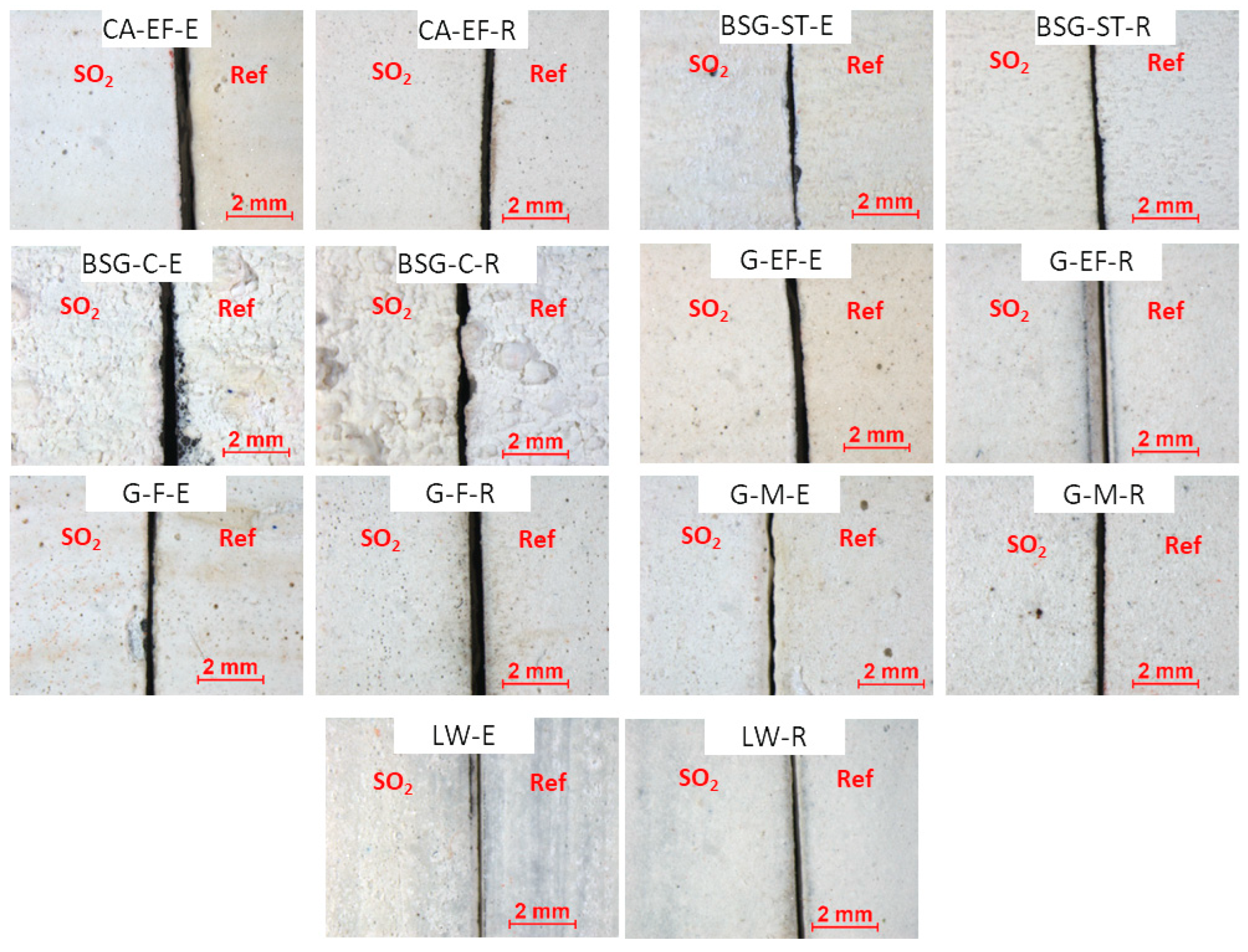
Figure 2.
Differences on colorimetric parameters ΔL*, Δa*and Δb* and colour difference ΔE*ab suffered by the aged paints. Check Table 1 for the identification code of samples.
Figure 2.
Differences on colorimetric parameters ΔL*, Δa*and Δb* and colour difference ΔE*ab suffered by the aged paints. Check Table 1 for the identification code of samples.
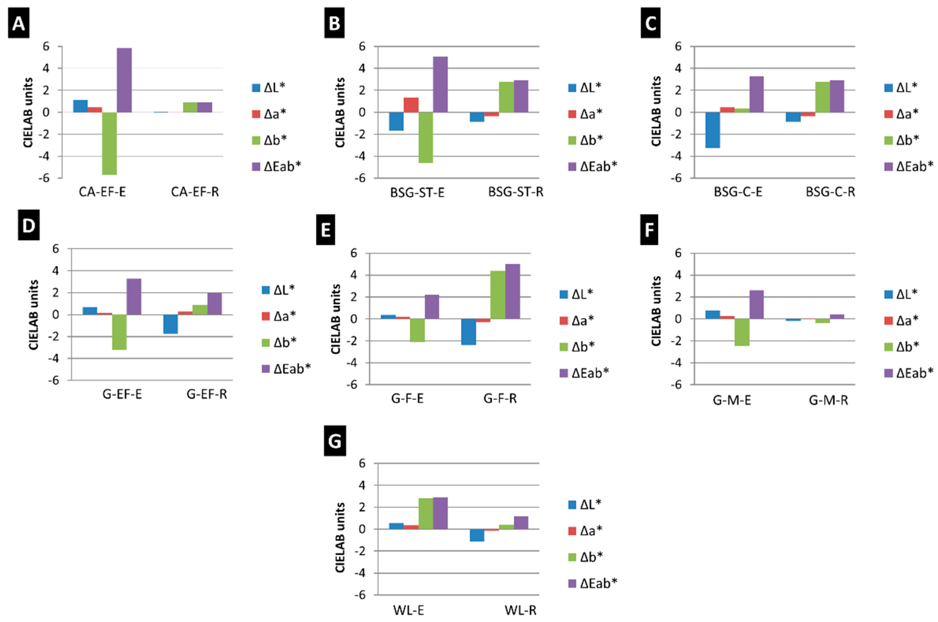
Figure 3.
Gloss variations (ΔG) of the aged paint mock-ups. Check Table 1 for the identification code of samples.
Figure 3.
Gloss variations (ΔG) of the aged paint mock-ups. Check Table 1 for the identification code of samples.
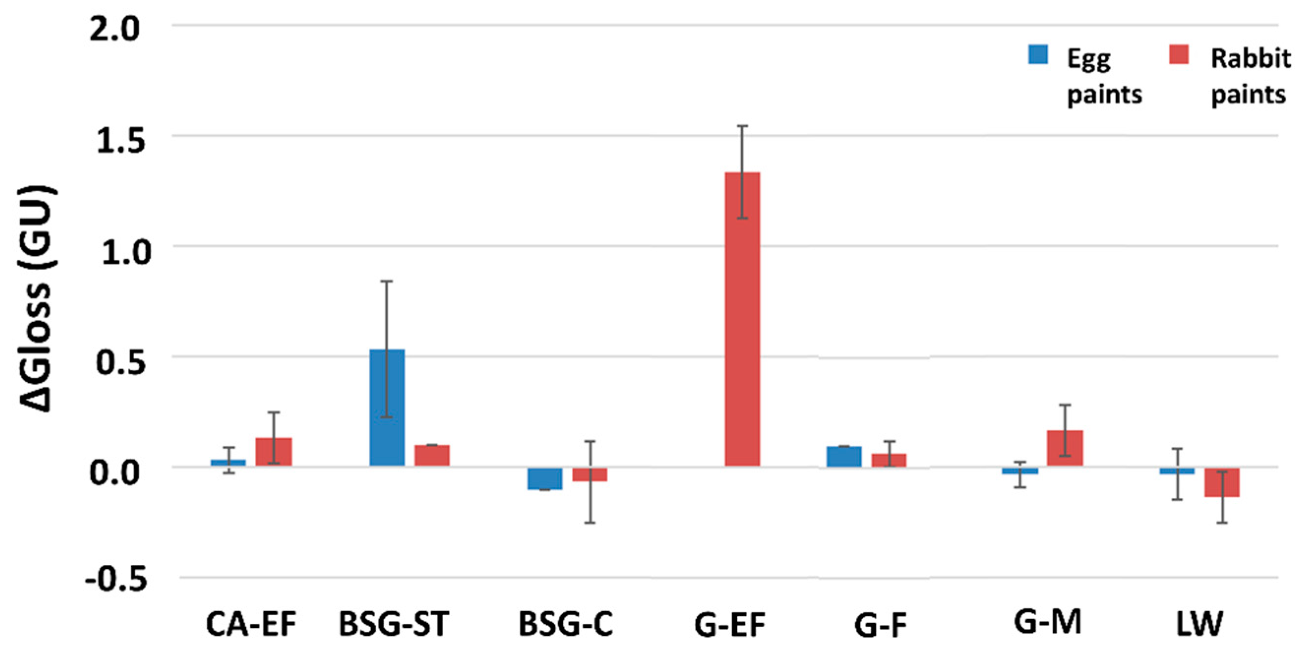
Figure 4.
Reflectance spectra of reference (-Ref) and aged (-SO2) paint mock-ups. Check Table 1 for the identification codes of samples. A and C: egg yolk-based paints. B and D: rabbit glue-based paints.
Figure 4.
Reflectance spectra of reference (-Ref) and aged (-SO2) paint mock-ups. Check Table 1 for the identification codes of samples. A and C: egg yolk-based paints. B and D: rabbit glue-based paints.
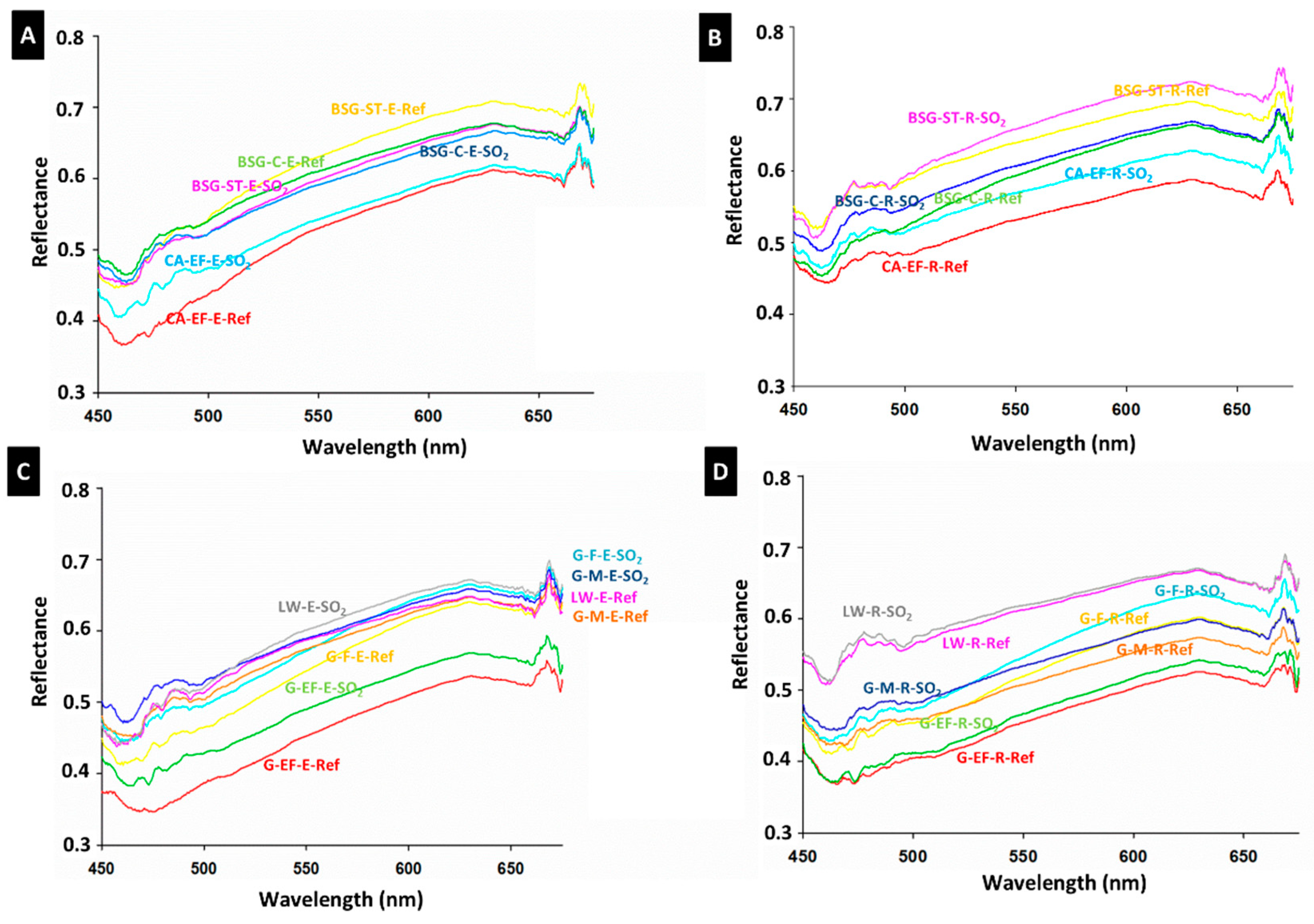
Figure 5.
Variation of the average maximum profile height (ΔRz, µm) of the tempera paint mock-ups after SO2 aging. Check Table 1 for the identification codes of samples.
Figure 5.
Variation of the average maximum profile height (ΔRz, µm) of the tempera paint mock-ups after SO2 aging. Check Table 1 for the identification codes of samples.
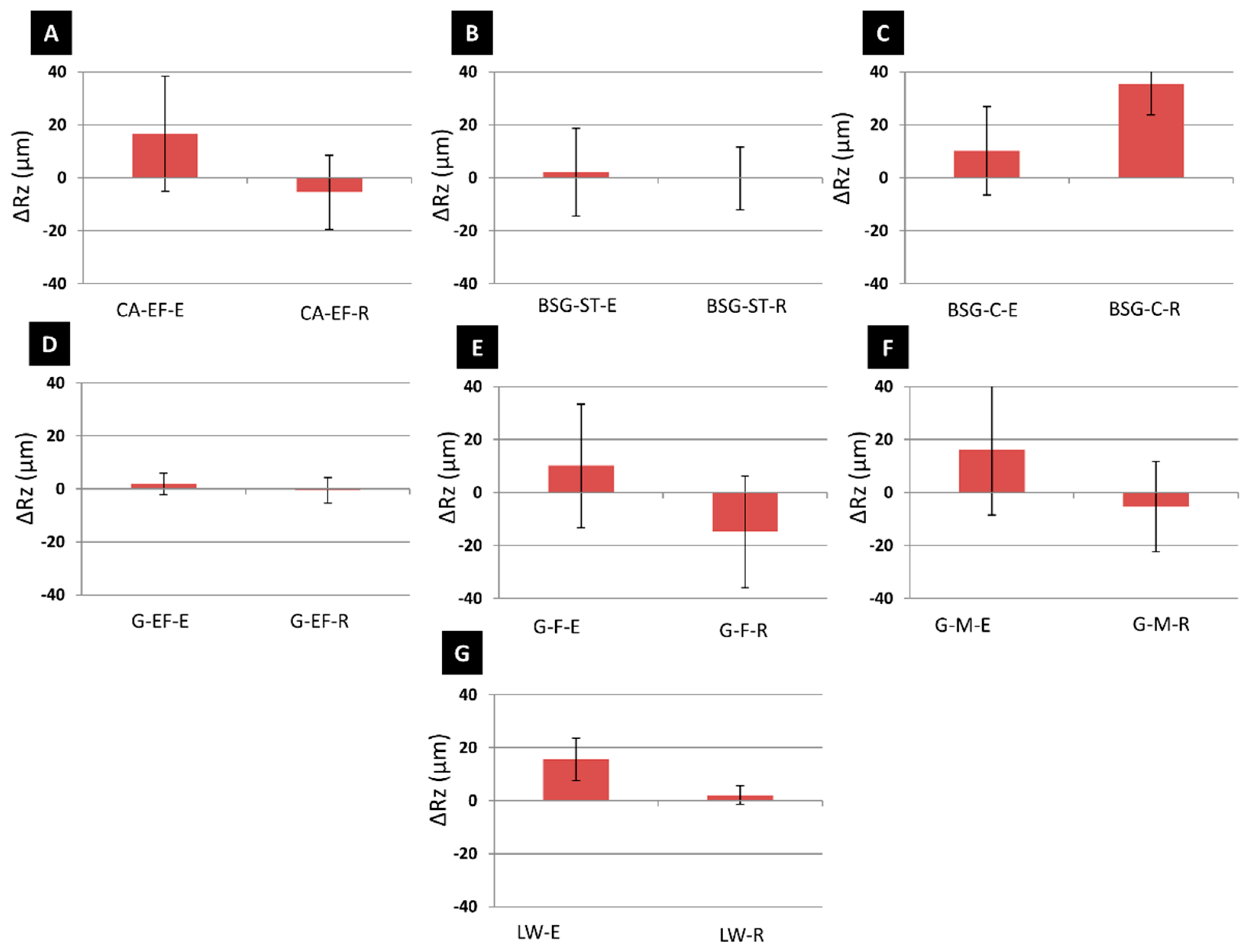
Figure 6.
FTIR (absorbance) spectra of the reference (-Ref) and aged (-SO2) mock-ups. Check Table 1 for the identification code of samples. In red, new FTIR bands, not present in the reference samples, are indicated. Letters between brackets, a: anhydrite, b: bassanite and g: gypsum.
Figure 6.
FTIR (absorbance) spectra of the reference (-Ref) and aged (-SO2) mock-ups. Check Table 1 for the identification code of samples. In red, new FTIR bands, not present in the reference samples, are indicated. Letters between brackets, a: anhydrite, b: bassanite and g: gypsum.
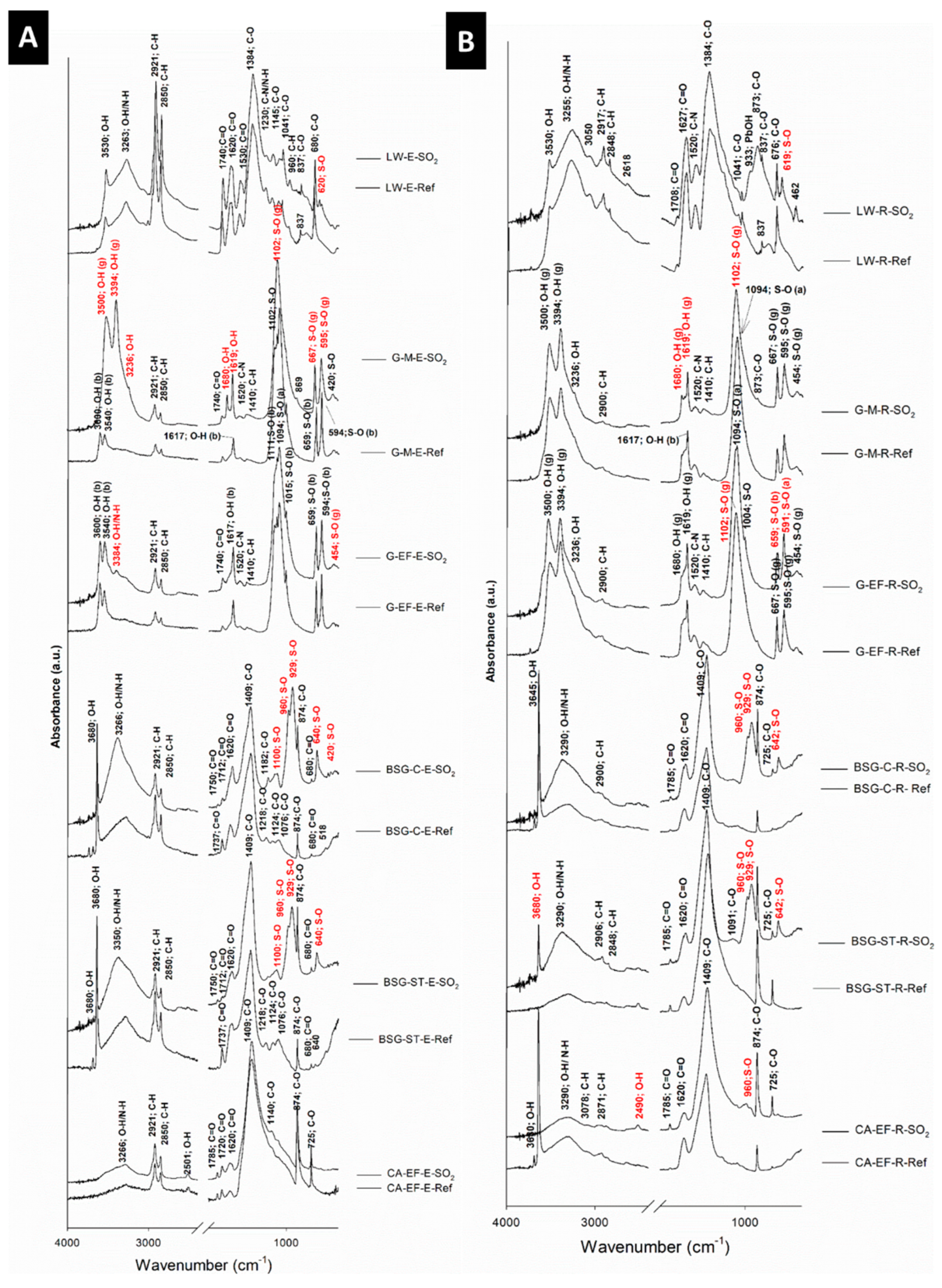
Figure 7.
SEM micrographs of CA-, BSG- and G- based paint mock-ups made with egg yolk or rabbit glue. -Ref = reference paints; -SO2 = aged paints. Check Table 1 for the identification codes of the samples. Arrow pointed out deposits rich in Ca and at lesser extent Na, Mg, Si, S and Cl. Rectangles shown micrometric particles.
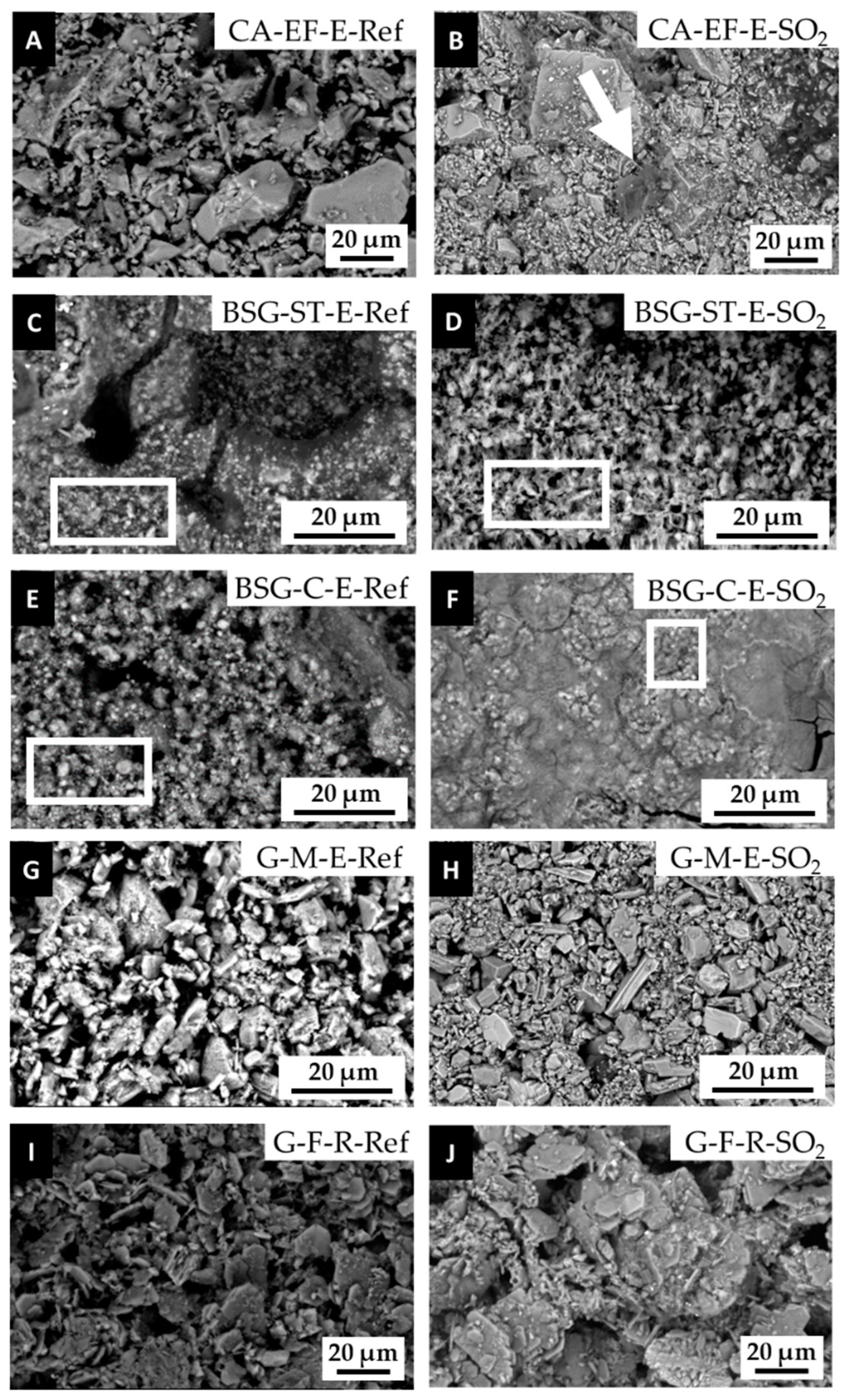

Table 1.
Properties of the white pigments and tempera paint mock-ups used in this study according to Kremer manufacturer and the authors.
Table 1.
Properties of the white pigments and tempera paint mock-ups used in this study according to Kremer manufacturer and the authors.
| Kremer pigment reference | Authors’ identification code | Kremer mineralogical composition | Authors’ mineralogical composition | Kremer grain size (µm) | Authors’ grain size (µm)* | Mineralogical composition of paints before the aging test | Mineralogical composition of paints after the aging test | ||
|---|---|---|---|---|---|---|---|---|---|
| Egg yolk mock-ups | Rabbit glue mock-ups | Egg yolk mock-ups | Rabbit glue mock-ups | ||||||
| Calcite 58720 | CA-EF | Calcite | Calcite Dolomite |
20 | 25 (0.25-100) |
Calcite Dolomite |
Calcite Portlandite |
Calcite Dolomite Ca3(SO3)2SO4·2H2O |
Calcite Gypsum |
| Bianco San Giovanni 11415 | BSG-ST | Portlandite Calcite |
Portlandite Calcite |
120 | 60 (0.25-120) |
Portlandite Calcite |
Portlandite Calcite |
Portlandite Calcite CaSO3·1/2H2O |
Portlandite Calcite CaSO3·1/2H2O |
| Bianco San Giovanni 11416 | BSG-C | Portlandite Calcite |
Portlandite Calcite |
120-1000 | 120 (0.3-250) |
Portlandite Calcite |
Portlandite Calcite |
Portlandite Calcite CaSO3·1/2H2O |
Portlandite Calcite CaSO3·1/2H2O |
| Gypsum Alabaster, Italian plaster 58340 |
G-EF | Gypsum | Bassanite Anhydrite |
<75 | 7 (0.2-85) |
Bassanite Anhydrite |
Gypsum |
Bassanite Anhydrite Gypsum K2SO4 |
Gypsum Bassanite Anhydrite CaSO3 |
| Natural gypsum 58300 Selenite (Terra Alba) |
G-F | Gypsum | Bassanite Anhydrite |
80% <20 18% <25 1.9% <32 1.9% <32 |
9 (0.2-75) |
Anhydrite Gypsum | Anhydrite Gypsum |
Anhydrite Gypsum CaSO3 K0.67Na1.33SO4 MgS2O3·6H2O |
Anhydrite Gypsum CaSO3 K0.67Na1.33SO4 MgS2O3·6H2O |
| Gypsum alabaster 58343 |
G-M | Bassanite | Bassanite Anhydrite |
85% <40 | 16 (1-160) |
Bassanite Anhydrite |
Bassanite Anhydrite |
Gypsum CaSO3 |
Gypsum CaSO3 |
| Lead white 46000 | LW | Basic lead carbonate | Hydrocerussite Cerussite |
<45 | 3 (0.1-10) |
Hydrocerussite Cerussite |
Hydrocerussite Cerussite |
Hydrocerussite Cerussite Pb4O3SO4·H2O Pb2O(SO4) Pb4SO4(CO3)·2(OH)2 |
Hydrocerussite Cerussite Pb4O3SO4·H2O Pb2O(SO4) Pb4SO4(CO3)·2(OH)2 |
*: Main maximum particle size and particle size range into parenthesis. EF: extra fine size; ST: standard size; C: coarse size; F: fine size; M: medium size; Calcite: CaCO3; Portlandite: Ca(OH)2; Gypsum: CaSO4·2H2O; Bassanite: CaSO4·1/2H2O; Anhydrite: CaSO4; Basic lead carbonate: Pb(CO3)2·Pb(OH)2; Hydrocerussite: 2PbCO3·Pb(OH)2; Cerussite: PbCO3; Dolomite: CaMg(CO3)2; Quartz: SiO2.
Table 2.
Static contact angle (θº) of the reference paint mock-ups. Check Table 1 for the identification codes of the samples.
Table 2.
Static contact angle (θº) of the reference paint mock-ups. Check Table 1 for the identification codes of the samples.
| Authors’ identification code | Egg yolk-based samples | Rabbit glue-based samples |
|---|---|---|
| CA-EF | 56.82 ±6.33 | 120.58 ±2.68 |
| BSG-ST | 83.20 ±1.83 | 127.63 ±3.94 |
| BSG-C | 96.97 ±2.28 | 114.12 ±3.73 |
| G-EF | 111.93 ±2.05 | 114.84 ±1.34 |
| G-F | 86.45 ±2.13 | 115.55 ±6.65 |
| G-M | 90.17 ±4.14 | 113.39 ±1.41 |
| WL | 93.70 ±1.87 | 98.67 ±1.03 |
Disclaimer/Publisher’s Note: The statements, opinions and data contained in all publications are solely those of the individual author(s) and contributor(s) and not of MDPI and/or the editor(s). MDPI and/or the editor(s) disclaim responsibility for any injury to people or property resulting from any ideas, methods, instructions or products referred to in the content. |
© 2024 by the authors. Licensee MDPI, Basel, Switzerland. This article is an open access article distributed under the terms and conditions of the Creative Commons Attribution (CC BY) license (http://creativecommons.org/licenses/by/4.0/).
Copyright: This open access article is published under a Creative Commons CC BY 4.0 license, which permit the free download, distribution, and reuse, provided that the author and preprint are cited in any reuse.


