

Submitted:
26 December 2024
Posted:
26 December 2024
You are already at the latest version
Abstract
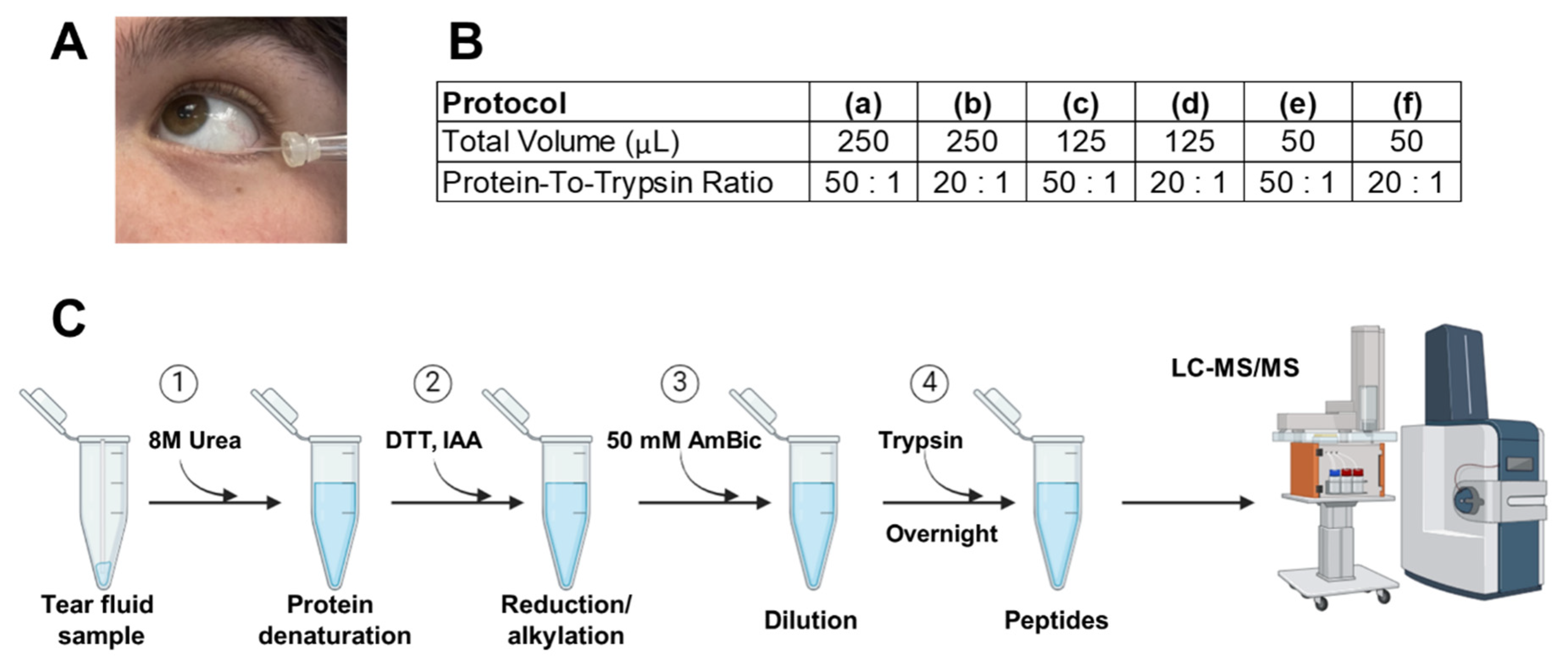
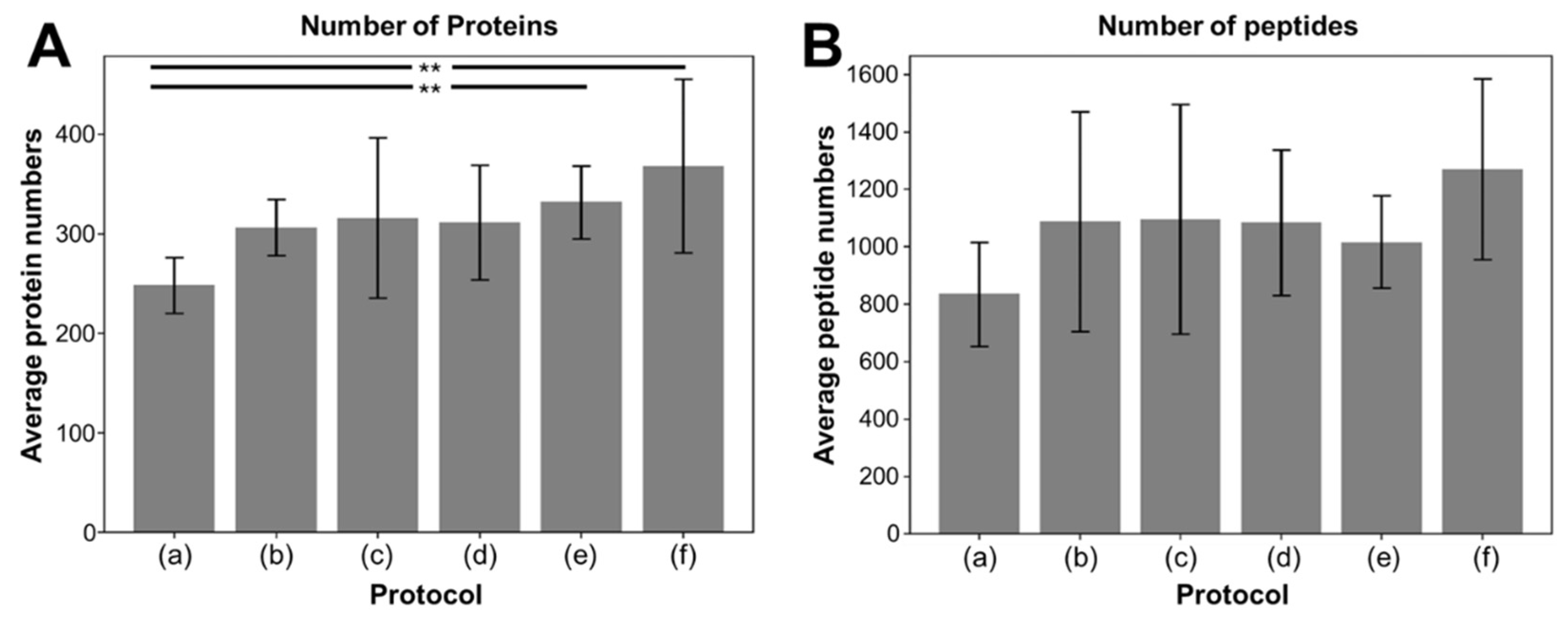
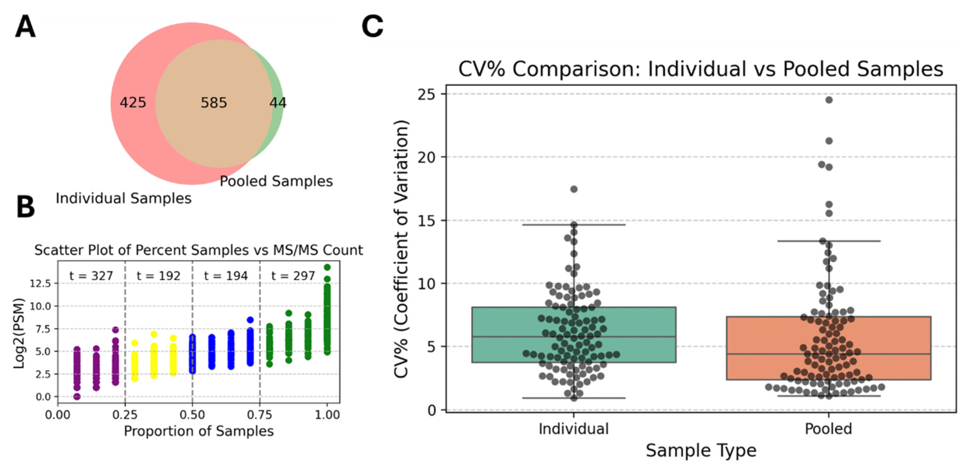
Background/Objectives: Tear fluid, a complex biofluid that contains thousands of proteins and can be collected non-invasively, has emerged as a promising source of biomarkers for ocular and systemic health. Liquid chromatography-tandem mass spectrometry (LC-MS/MS) is currently the primary method for discovering novel biomarkers in tear fluid. However, the method of tear collection can significantly impact LC-MS/MS analysis outcomes. Tear fluid is commonly collected using either Schirmer strips or capillary tubes. While capillary tubes offer distinct advantages, such as reduced extracellular contamination and reflex tearing, most LC-MS/MS protocol development has focused on optimizing protocols for Schirmer strips. This study addresses this gap by evaluating digestion protocols for tear fluid collected with capillary tubes, focusing on biomarker discovery using small-volume samples. In this study, we evaluated multiple digestion protocols for the shotgun quantitative LC-MS/MS analysis of small-volume tear fluid samples collected using glass capillary tubes. Protocol optimization was performed using pooled samples and then compared with the analysis of individual samples. With the optimized protocol, we identified an average of 368 ± 87 proteins in pooled samples and 502 ± 127 proteins in individual small-volume tear fluid samples, using less than 1µL of total fluid volume. This protocol highlights the practicality of using glass capillary tubes for comprehensive LC-MS/MS-based tear proteomics analysis, paving the way for detailed proteomics characterization of individual tear fluid samples rather than pooled samples. By shifting from pooled to individual samples, this approach greatly accelerates tear biomarker discovery, advancing precision and personalized medicine.
Keywords:
1. Introduction
2. Materials and Methods
2.1. Sample Collection
2.2. In-Solution Protein Digestion
2.3. LC-MS/MS
2.4. Protein Identification and Analysis
2.5. Statistical Analysis
2.6. Selection of Optimal Protocol
3. Results
3.1. Protein and Peptide Yield
3.2. Unique Protein Identification
3.3. Frequency of Detection
3.4. Protocol Variance
3.5. Protocol Selection
3.6. Comparison of Pooled and Individual Samples
3.5. Gene Ontology (GO) Enrichment Analysis and Tear Protein Classification
4. Discussion
Supplementary Materials
Author Contributions
Funding
Institutional Review Board Statement
Informed Consent Statement
Data Availability Statement
Acknowledgments
Conflicts of Interest
References
- Li, N.; Wang, N.; Zheng, J.; Liu, X.M.; Lever, O.W.; Erickson, P.M.; Li, L. Characterization of Human Tear Proteome Using Multiple Proteomic Analysis Techniques. J. Proteome Res. 2005, 4, 2052–2061. [Google Scholar] [CrossRef] [PubMed]
- Hagan, S.; Martin, E.; Enríquez-De-Salamanca, A. Tear fluid biomarkers in ocular and systemic disease: potential use for predictive, preventive and personalised medicine. EPMA J. 2016, 7, 1–20. [Google Scholar] [CrossRef] [PubMed]
- Murube, J. Basal, Reflex, and Psycho-emotional Tears. Ocul. Surf. 2009, 7, 60–66. [Google Scholar] [CrossRef] [PubMed]
- Bron, A.J.; Tomlinson, A.; Foulks, G.N.; Pepose, J.S.; Baudouin, C.; Geerling, G.; Nichols, K.K.; Lemp, M.A. Rethinking Dry Eye Disease: A Perspective on Clinical Implications. Ocul. Surf. 2014, 12, S1–S31. [Google Scholar] [CrossRef] [PubMed]
- D. A. Dartt, R. R. Hodges, and D. Zoukhri, “Tears and Their Secretion,” in Advances in Organ Biology, vol. 10, J. Fischbarg, Ed., in The Biology of the Eye, vol. 10. Elsevier, 2005, pp. 21–82. [CrossRef]
- Aluru, S.V.; Agarwal, S.; Srinivasan, B.; Iyer, G.K.; Rajappa, S.M.; Tatu, U.; Padmanabhan, P.; Subramanian, N.; Narayanasamy, A. Lacrimal Proline Rich 4 (LPRR4) Protein in the Tear Fluid Is a Potential Biomarker of Dry Eye Syndrome. PLOS ONE 2012, 7, e51979. [Google Scholar] [CrossRef]
- Srinivasan, S.; Thangavelu, M.; Zhang, L.; Green, K.B.; Nichols, K.K. iTRAQ Quantitative Proteomics in the Analysis of Tears in Dry Eye Patients. Investig. Opthalmology Vis. Sci. 2012, 53, 5052–9. [Google Scholar] [CrossRef]
- Csősz. ; Boross, P.; Csutak, A.; Berta, A.; Tóth, F.; Póliska, S.; Török, Z.; Tőzsér, J. Quantitative analysis of proteins in the tear fluid of patients with diabetic retinopathy. J. Proteom. 2012, 75, 2196–2204. [Google Scholar] [CrossRef]
- Kim, H.-J.; Kim, P.-K.; Yoo, H.-S.; Kim, C.-W. Comparison of tear proteins between healthy and early diabetic retinopathy patients. Clin. Biochem. 2012, 45, 60–67. [Google Scholar] [CrossRef]
- Boerger, M.; Funke, S.; Leha, A.; Roser, A.-E.; Wuestemann, A.-K.; Maass, F.; Bähr, M.; Grus, F.; Lingor, P. Proteomic analysis of tear fluid reveals disease-specific patterns in patients with Parkinson's disease – A pilot study. Park. Relat. Disord. 2019, 63, 3–9. [Google Scholar] [CrossRef]
- R. J. Fullard and D. L. Tucker, “Evaluation of commonly used tear sampling methods and their relevance in subsequent biochemical analysis,” Invest. Ophthalmol. Vis. Sci., vol. 32, no. 8, pp. 2290–2301, Jul. 1991.
- Fullard, R.; Snyder, C. PROTEIN-LEVELS IN NONSTIMULATED AND STIMULATED TEARS OF NORMAL HUMAN-SUBJECTS. 1990, 31, 1119–1126.
- Fullard, R.J.; Tucker, D.L. Changes in human tear protein levels with progressively increasing stimulus. Invest. Ophthalmol. Vis. Sci 1991, 32, 2290–301. [Google Scholar]
- Krajčíková, K.; Glinská, G.; Tomečková, V. Effect of tear fluid sampling and processing on total protein quantity and electrophoretic pattern. Taiwan J. Ophthalmol. 2022, 12, 88–92. [Google Scholar] [CrossRef] [PubMed]
- Tham, M.L.; Mahmud, A.; Abdullah, M.; Saleh, R.M.; Razali, A.M.; Cheah, Y.K.; Taib, N.M.; Ho, K.L.; Mahmud, M.; Isa, M.M. Tear Samples for Protein Extraction: Comparative Analysis of Schirmer's Test Strip and Microcapillary Tube Methods. Cureus 2023, 15, e50972. [Google Scholar] [CrossRef] [PubMed]
- Nättinen, J.; Aapola, U.; Nukareddy, P.; Uusitalo, H. Clinical Tear Fluid Proteomics—A Novel Tool in Glaucoma Research. Int. J. Mol. Sci. 2022, 23, 8136. [Google Scholar] [CrossRef] [PubMed]
- Jones, G.; Lee, T.J.; Glass, J.; Rountree, G.; Ulrich, L.; Estes, A.; Sezer, M.; Zhi, W.; Sharma, S.; Sharma, A. Comparison of Different Mass Spectrometry Workflows for the Proteomic Analysis of Tear Fluid. Int. J. Mol. Sci. 2022, 23, 2307. [Google Scholar] [CrossRef]
- Vergouwen, D.P.C.; Schotting, A.J.; Endermann, T.; van de Werken, H.J.G.; Grashof, D.G.B.; Arumugam, S.; Nuijts, R.M.M.A.; Berge, J.C.T.; Rothova, A.; Schreurs, M.W.J.; et al. Evaluation of pre-processing methods for tear fluid proteomics using proximity extension assays. Sci. Rep. 2023, 13, 1–8. [Google Scholar] [CrossRef]
- Bachhuber, F.; Huss, A.; Senel, M.; Tumani, H. Diagnostic biomarkers in tear fluid: from sampling to preanalytical processing. Sci. Rep. 2021, 11, 1–9. [Google Scholar] [CrossRef]
- J. Xiao et al., “Tear fluid proteomic analysis with improved LC-MS/MS protocol,” Invest. Ophthalmol. Vis. Sci., vol. 65, no. 7, p. 6557, Jun. 2024.
- Zhan, X.; Li, J.; Guo, Y.; Golubnitschaja, O. Mass spectrometry analysis of human tear fluid biomarkers specific for ocular and systemic diseases in the context of 3P medicine. EPMA J. 2021, 12, 449–475. [Google Scholar] [CrossRef]
- Bolaños-Jiménez, R.; Navas, A.; López-Lizárraga, E.P.; de Ribot, F.M.; Peña, A.; O Graue-Hernández, E.; Garfias, Y. Ocular Surface as Barrier of Innate Immunity. Open Ophthalmol. J. 2015, 9, 49–55. [Google Scholar] [CrossRef]
- Green-Church, K.B.; Nichols, K.K.; Kleinholz, N.M.; Zhang, L.; Nichols, J.J. Investigation of the human tear film proteome using multiple proteomic approaches. 2008, 14, 456–470.
- Huang, Z.; Du, C.-X.; Pan, X.-D. The use of in-strip digestion for fast proteomic analysis on tear fluid from dry eye patients. PLOS ONE 2018, 13, e0200702. [Google Scholar] [CrossRef]
- Lépine, M.; Zambito, O.; Sleno, L. Targeted Workflow Investigating Variations in the Tear Proteome by Liquid Chromatography Tandem Mass Spectrometry. ACS Omega 2023, 8, 31168–31177. [Google Scholar] [CrossRef]
- Ponzini, E.; Santambrogio, C.; De Palma, A.; Mauri, P.; Tavazzi, S.; Grandori, R. Mass spectrometry-based tear proteomics for noninvasive biomarker discovery. Mass Spectrom. Rev. 2021, 41, 842–860. [Google Scholar] [CrossRef]
- Rossi, C.; Cicalini, I.; Cufaro, M.C.; Agnifili, L.; Mastropasqua, L.; Lanuti, P.; Marchisio, M.; De Laurenzi, V.; Del Boccio, P.; Pieragostino, D. Multi-Omics Approach for Studying Tears in Treatment-Naïve Glaucoma Patients. Int. J. Mol. Sci. 2019, 20, 4029. [Google Scholar] [CrossRef] [PubMed]
- Tong, L.; Zhou, X.Y.; Jylha, A.; Aapola, U.; Liu, D.N.; Koh, S.K.; Tian, D.; Quah, J.; Uusitalo, H.; Beuerman, R.W.; et al. Quantitation of 47 human tear proteins using high resolution multiple reaction monitoring (HR-MRM) based-mass spectrometry. J. Proteom. 2014, 115, 36–48. [Google Scholar] [CrossRef] [PubMed]
- Nättinen, J.; Jylhä, A.; Aapola, U.; Parkkari, M.; Mikhailova, A.; Beuerman, R.W.; Uusitalo, H. Patient stratification in clinical glaucoma trials using the individual tear proteome. Sci. Rep. 2018, 8, 12038. [Google Scholar] [CrossRef]
- Gerber-Hollbach, N.; Plattner, K.; O'Leary, O.E.; Jenoe, P.; Moes, S.; Drexler, B.; Schoetzau, A.; Halter, J.P.; Goldblum, D. Tear Film Proteomics Reveal Important Differences Between Patients With and Without Ocular GvHD After Allogeneic Hematopoietic Cell Transplantation. Investig. Opthalmology Vis. Sci. 2018, 59, 3521–3530. [Google Scholar] [CrossRef]
- Pieragostino, D.; Agnifili, L.; Fasanella, V.; D'Aguanno, S.; Mastropasqua, R.; Di Ilio, C.; Sacchetta, P.; Urbani, A.; Del Boccio, P. Shotgun proteomics reveals specific modulated protein patterns in tears of patients with primary open angle glaucoma naïve to therapy. Mol. Biosyst. 2013, 9, 1108–1116. [Google Scholar] [CrossRef]
- Böhm, D.; Keller, K.; Pieter, J.; Boehm, N.; Wolters, D.; Siggelkow, W.; Lebrecht, A.; Schmidt, M.; Kölbl, H.; Pfeiffer, N.; et al. Comparison of tear protein levels in breast cancer patients and healthy controls using a de novo proteomic approach. Oncol. Rep. 2012, 28, 429–438. [Google Scholar] [CrossRef]
- López-López, M.; Regueiro, U.; Bravo, S.B.; Chantada-Vázquez, M.d.P.; Varela-Fernández, R.; Ávila-Gómez, P.; Hervella, P.; Lema, I. Tear Proteomics in Keratoconus: A Quantitative SWATH-MS Analysis. Investig. Opthalmology Vis. Sci. 2021, 62, 30–30. [Google Scholar] [CrossRef]
- Li, B.; Sheng, M.; Li, J.; Yan, G.; Lin, A.; Li, M.; Wang, W.; Chen, Y. Tear proteomic analysis of Sjögren syndrome patients with dry eye syndrome by two-dimensional-nano-liquid chromatography coupled with tandem mass spectrometry. Sci. Rep. 2014, 4, srep05772. [Google Scholar] [CrossRef]
- Kishazi, E.; Dor, M.; Eperon, S.; Oberic, A.; Hamedani, M.; Turck, N. Thyroid-associated orbitopathy and tears: A proteomics study. J. Proteom. 2018, 170, 110–116. [Google Scholar] [CrossRef]
- Angeles-Han, S.T.; Yeh, S.; Patel, P.; Duong, D.; Jenkins, K.; Rouster-Stevens, K.A.; Altaye, M.; Fall, N.; Thornton, S.; Prahalad, S.; et al. Discovery of tear biomarkers in children with chronic non-infectious anterior uveitis: a pilot study. J. Ophthalmic Inflamm. Infect. 2018, 8, 17. [Google Scholar] [CrossRef] [PubMed]
- Liang, A.; Qin, W.; Zhang, M.; Gao, F.; Zhao, C.; Gao, Y. Profiling tear proteomes of patients with unilateral relapsed Behcet’s disease-associated uveitis using data-independent acquisition proteomics. PeerJ 2020, 8, e9250. [Google Scholar] [CrossRef] [PubMed]
- Pieragostino, D.; Bucci, S.; Agnifili, L.; Fasanella, V.; D'Aguanno, S.; Mastropasqua, A.; Ciancaglini, M.; Mastropasqua, L.; Di Ilio, C.; Sacchetta, P.; et al. Differential protein expression in tears of patients with primary open angle and pseudoexfoliative glaucoma. Mol. Biosyst. 2011, 8, 1017–1028. [Google Scholar] [CrossRef]
- Dor, M.; Eperon, S.; Lalive, P.H.; Guex-Crosier, Y.; Hamedani, M.; Salvisberg, C.; Turck, N. Investigation of the global protein content from healthy human tears. Exp. Eye Res. 2018, 179, 64–74. [Google Scholar] [CrossRef]
- O'Leary, O.E.; Schoetzau, A.; Amruthalingam, L.; Geber-Hollbach, N.; Plattner, K.; Jenoe, P.; Schmidt, A.; Ullmer, C.; Drawnel, F.M.; Fauser, S.; et al. Tear Proteomic Predictive Biomarker Model for Ocular Graft Versus Host Disease Classification. Transl. Vis. Sci. Technol. 2020, 9, 3–3. [Google Scholar] [CrossRef]
- Zhou, L.; Beuerman, R.W.; Chan, C.M.; Zhao, S.Z.; Li, X.R.; Yang, H.; Tong, L.; Liu, S.; Stern, M.E.; Tan, D. Identification of Tear Fluid Biomarkers in Dry Eye Syndrome Using iTRAQ Quantitative Proteomics. J. Proteome Res. 2009, 8, 4889–4905. [Google Scholar] [CrossRef]
- Aluru, S.V.; Shweta, A.; Bhaskar, S.; Geetha, K.; Sivakumar, R.M.; Utpal, T.; Padmanabhan, P.; Angayarkanni, N. Tear Fluid Protein Changes in Dry Eye Syndrome Associated with Rheumatoid Arthritis: A Proteomic Approach. Ocul. Surf. 2016, 15, 112–129. [Google Scholar] [CrossRef]
- Aass, C.; Norheim, I.; Eriksen, E.F.; Thorsby, P.M.; Pepaj, M. Single unit filter-aided method for fast proteomic analysis of tear fluid. Anal. Biochem. 2015, 480, 1–5. [Google Scholar] [CrossRef]
- López-López, M.; Regueiro, U.; Bravo, S.B.; Chantada-Vázquez, M.d.P.; Pena, C.; Díez-Feijoo, E.; Hervella, P.; Lema, I. Shotgun Proteomics for the Identification and Profiling of the Tear Proteome of Keratoconus Patients. Investig. Opthalmology Vis. Sci. 2022, 63, 12–12. [Google Scholar] [CrossRef]
- Winiarczyk, M.; Kaarniranta, K.; Winiarczyk, S.; Adaszek, .; Winiarczyk, D.; Mackiewicz, J. Tear film proteome in age-related macular degeneration. Graefe's Arch. Clin. Exp. Ophthalmol. 2018, 256, 1127–1139. [Google Scholar] [CrossRef]
- Grus, F.H.; Podust, V.N.; Bruns, K.; Lackner, K.; Fu, S.; Dalmasso, E.A.; Wirthlin, A.; Pfeiffer, N. SELDI-TOF-MS ProteinChip Array Profiling of Tears from Patients with Dry Eye. Investig. Opthalmology Vis. Sci. 2005, 46, 863–876. [Google Scholar] [CrossRef] [PubMed]
- Liu, Y.-C.; Yam, G.H.-F.; Lin, M.T.-Y.; Teo, E.; Koh, S.-K.; Deng, L.; Zhou, L.; Tong, L.; Mehta, J.S. Comparison of tear proteomic and neuromediator profiles changes between small incision lenticule extraction (SMILE) and femtosecond laser-assisted in-situ keratomileusis (LASIK). 2020, 29, 67–81. [CrossRef]
- Pieragostino, D.; Lanuti, P.; Cicalini, I.; Cufaro, M.C.; Ciccocioppo, F.; Ronci, M.; Simeone, P.; Onofrj, M.; van der Pol, E.; Fontana, A.; et al. Proteomics characterization of extracellular vesicles sorted by flow cytometry reveals a disease-specific molecular cross-talk from cerebrospinal fluid and tears in multiple sclerosis. J. Proteom. 2019, 204, 103403. [Google Scholar] [CrossRef] [PubMed]
- D'Souza, S.; Petznick, A.; Tong, L.; Hall, R.C.; Rosman, M.; Chan, C.; Koh, S.K.; Beuerman, R.W.; Zhou, L.; Mehta, J.S. Comparative Analysis of Two Femtosecond LASIK Platforms Using iTRAQ Quantitative Proteomics. Investig. Opthalmology Vis. Sci. 2014, 55, 3396–3402. [Google Scholar] [CrossRef]
- Vaajanen, A.; Nättinen, J.; Aapola, U.; Gielen, F.; Uusitalo, H. The effect of successful trabeculectomy on the ocular surface and tear proteomics—a prospective cohort study with 1-year follow-up. Acta Ophthalmol. 2020, 99, 160–170. [Google Scholar] [CrossRef] [PubMed]
- Funke, S.; Beck, S.; Lorenz, K.; Kotterer, M.; Wolters, D.; Perumal, N.; Pfeiffer, N.; Grus, F.H. Analysis of the effects of preservative-free tafluprost on the tear proteome. Am. J. Transl. Res. 2016, 8, 4025–4039. [Google Scholar]
- Wong, T.T.; Zhou, L.; Li, J.; Tong, L.; Zhao, S.Z.; Li, X.R.; Yu, S.J.; Koh, S.K.; Beuerman, R.W. Proteomic Profiling of Inflammatory Signaling Molecules in the Tears of Patients on Chronic Glaucoma Medication. Investig. Opthalmology Vis. Sci. 2011, 52, 7385–7391. [Google Scholar] [CrossRef] [PubMed]
- Balasubramanian, S.A.; Wasinger, V.C.; Pye, D.C.; Willcox, M.D.P. Preliminary identification of differentially expressed tear proteins in keratoconus. 2013, 19, 2124–2134.
- Acera, A.; Vecino, E.; Rodríguez-Agirretxe, I.; Aloria, K.; Arizmendi, J.M.; Morales, C.; A Durán, J. Changes in tear protein profile in keratoconus disease. Eye 2011, 25, 1225–1233. [Google Scholar] [CrossRef]
- Ananthi, S.; Prajna, N.V.; Lalitha, P.; Valarnila, M.; Dharmalingam, K. Pathogen Induced Changes in the Protein Profile of Human Tears from Fusarium Keratitis Patients. PLOS ONE 2013, 8, e53018. [Google Scholar] [CrossRef]
- Soria, J.; Acera, A.; Merayo-Lloves, J.; Durán, J.A.; González, N.; Rodriguez, S.; Bistolas, N.; Schumacher, S.; Bier, F.F.; Peter, H.; et al. Tear proteome analysis in ocular surface diseases using label-free LC-MS/MS and multiplexed-microarray biomarker validation. Sci. Rep. 2017, 7, 17478–17478. [Google Scholar] [CrossRef]
- Lee, S.H.; Park, M.-Y.; Kim, K.W.; Wee, S.W.; Kim, J.C. Zinc Finger Protein in Severe Dry Eye Syndrome. Curr. Eye Res. 2013, 39, 431–438. [Google Scholar] [CrossRef]
- Funke, S.; Azimi, D.; Wolters, D.; Grus, F.H.; Pfeiffer, N. Longitudinal analysis of taurine induced effects on the tear proteome of contact lens wearers and dry eye patients using a RP-RP-Capillary-HPLC–MALDI TOF/TOF MS approach. J. Proteom. 2012, 75, 3177–3190. [Google Scholar] [CrossRef] [PubMed]
- Ananthi, S.; Chitra, T.; Bini, R.; Prajna, N.V.; Lalitha, P.; Dharmalingam, K. Comparative analysis of the tear protein profile in mycotic keratitis patients. 2008, 14, 500–507.
- Yenihayat, F.; Altıntaş, .; Kasap, M.; Akpınar, G.; Güzel, N.; Çelik, O.S. Comparative proteome analysis of the tear samples in patients with low-grade keratoconus. Int. Ophthalmol. 2017, 38, 1895–1905. [Google Scholar] [CrossRef]
- A de Souza, G.; de Godoy, L.M.; Mann, M. Identification of 491 proteins in the tear fluid proteome reveals a large number of proteases and protease inhibitors. Genome Biol. 2006, 7, R72–R72. [Google Scholar] [CrossRef] [PubMed]
- Ponzini, E.; Ami, D.; Duse, A.; Santambrogio, C.; De Palma, A.; Di Silvestre, D.; Mauri, P.; Pezzoli, F.; Natalello, A.; Tavazzi, S.; et al. Single-Tear Proteomics: A Feasible Approach to Precision Medicine. Int. J. Mol. Sci. 2021, 22, 10750. [Google Scholar] [CrossRef] [PubMed]
- Nichols, J.J.; Green-Church, K.B. Mass Spectrometry-Based Proteomic Analyses in Contact Lens-Related Dry Eye. Cornea 2009, 28, 1109–1117. [Google Scholar] [CrossRef]
- Kalló, G.; Emri, M.; Varga, Z.; Ujhelyi, B.; Tőzsér, J.; Csutak, A.; Csősz. Changes in the Chemical Barrier Composition of Tears in Alzheimer’s Disease Reveal Potential Tear Diagnostic Biomarkers. PLOS ONE 2016, 11, e0158000. [Google Scholar] [CrossRef]
- Chen, X.; Rao, J.; Zheng, Z.; Yu, Y.; Lou, S.; Liu, L.; He, Q.; Wu, L.; Sun, X. Integrated Tear Proteome and Metabolome Reveal Panels of Inflammatory-Related Molecules via Key Regulatory Pathways in Dry Eye Syndrome. J. Proteome Res. 2019, 18, 2321–2330. [Google Scholar] [CrossRef]
- Technology overview | TearLab osmolarity system for diagnosing dry eye disease | Advice | NICE.” Accessed: Sep. 17, 2024. [Online]. Available: https://www.nice.org.uk/advice/mib47/chapter/technology-overview.
- Ritorto, M.S.; Borlak, J. A simple and reliable protocol for mouse serum proteome profiling studies by use of two-dimensional electrophoresis and MALDI TOF/TOF mass spectrometry. Proteome Sci. 2008, 6, 25–25. [Google Scholar] [CrossRef]
- Millioni, R.; Tolin, S.; Puricelli, L.; Sbrignadello, S.; Fadini, G.P.; Tessari, P.; Arrigoni, G. High Abundance Proteins Depletion vs Low Abundance Proteins Enrichment: Comparison of Methods to Reduce the Plasma Proteome Complexity. PLOS ONE 2011, 6, e19603. [Google Scholar] [CrossRef]
- Zhou, L.; Zhao, S.Z.; Koh, S.K.; Chen, L.; Vaz, C.; Tanavde, V.; Li, X.R.; Beuerman, R.W. In-depth analysis of the human tear proteome. J. Proteom. 2012, 75, 3877–3885. [Google Scholar] [CrossRef]

| Protocol | Peptides | Proteins | ||
|---|---|---|---|---|
| Average | STD | Average | STD | |
| (a) | 835 | 181 | 248 | 28 |
| (b) | 1088 | 381 | 306 | 28 |
| (c) | 1095 | 400 | 316 | 80 |
| (d) | 1085 | 253 | 311 | 57 |
| (e) | 1017 | 160 | 332 | 36 |
| (f) | 1270 | 314 | 368 | 87 |
| Unique Proteins Identified | Protocol | |||||
|---|---|---|---|---|---|---|
| (a) | (b) | (c) | (d) | (e) | (f) | |
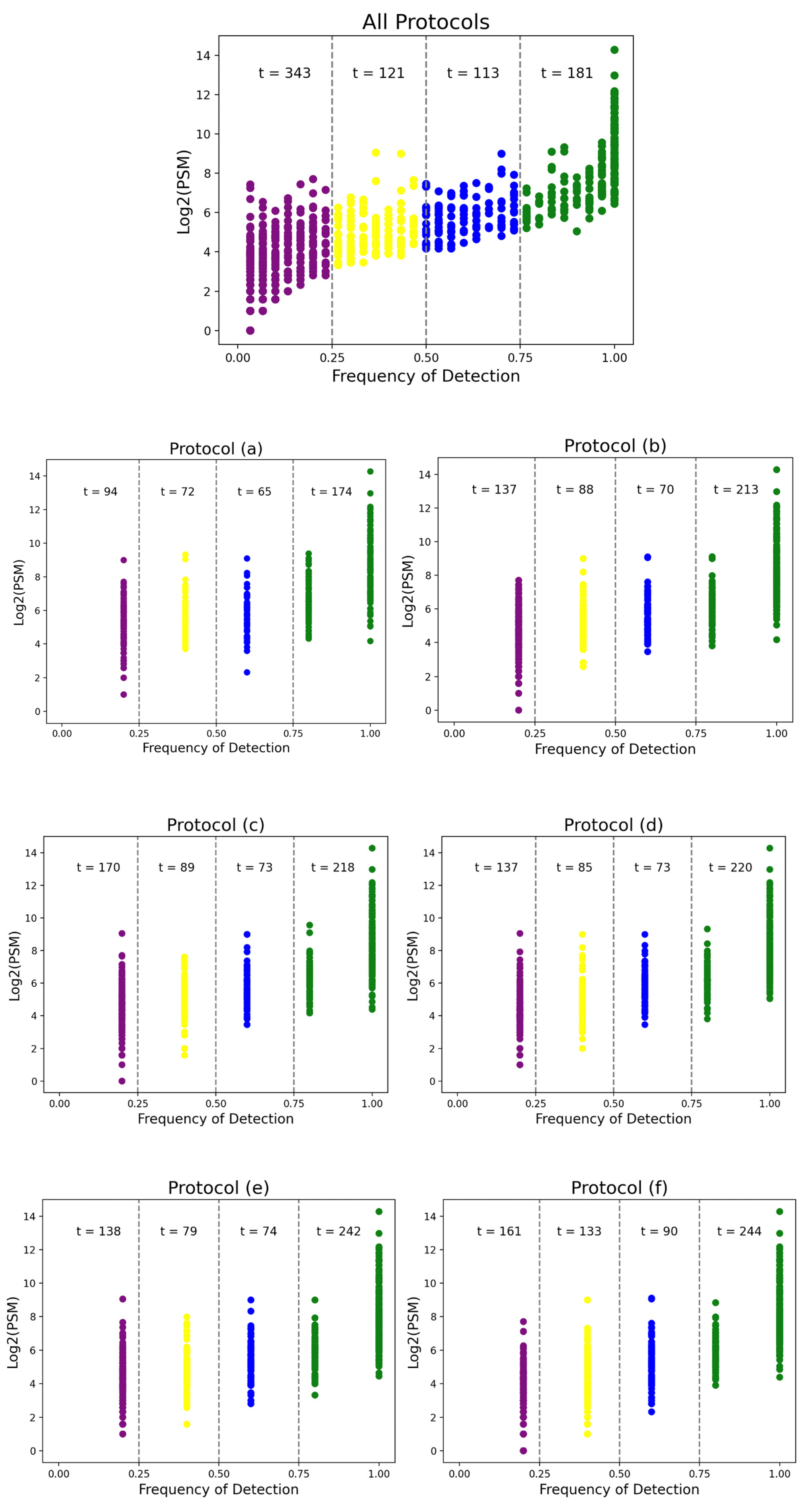
| High Abundance (>75%) | 174 | 213 | 218 | 220 | 242 | 244 |
| Medium Abundance (50%-75%) | 65 | 70 | 73 | 73 | 74 | 90 |
| Low Abundance (25%-49%) | 72 | 88 | 89 | 85 | 80 | 133 |
| Rare (< 25%) | 95 | 137 | 170 | 138 | 138 | 162 |
| Total | 406 | 508 | 550 | 516 | 534 | 629 |
| Protein Name | (a) | (b) | (c) | (d) | (e) | (f) |
|---|---|---|---|---|---|---|
| Lactotransferrin | 1.09 | 1.83 | 1.96 | 1.68 | 1.04 | 1.67 |
| Lysozyme C | 2.08 | 1.91 | 2.55 | 2.79 | 1.95 | 2.29 |
| Basement membrane-specific heparan sulfate proteoglycan core protein | 2.05 | 1.35 | 3.56 | 2.30 | 1.45 | 2.25 |
| Albumin | 1.84 | 2.06 | 3.25 | 2.28 | 1.44 | 2.47 |
| Mammaglobin-B | 2.51 | 6.04 | 5.44 | 7.53 | 3.68 | 3.22 |
| Cystatin-S | 4.37 | 5.36 | 6.37 | 3.61 | 3.64 | 4.91 |
| Proline-rich protein 4 | 3.25 | 4.55 | 4.31 | 5.81 | 1.39 | 2.70 |
| Cystatin-C | 5.08 | 5.29 | 8.15 | 5.04 | 7.96 | 6.04 |
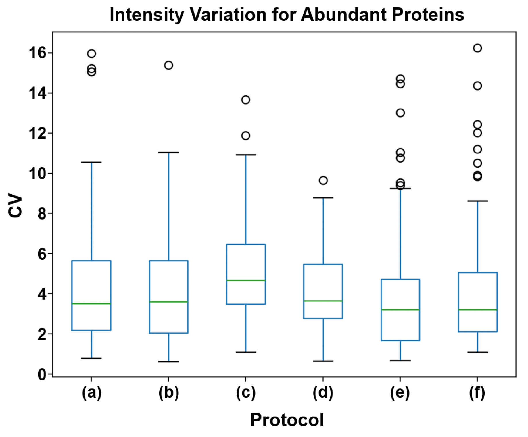
| Average | 4.56 | 4.06 | 5.20 | 4.19 | 3.96 | 4.36 |
| STD | 3.47 | 2.64 | 2.52 | 2.12 | 3.15 | 3.26 |
Disclaimer/Publisher’s Note: The statements, opinions and data contained in all publications are solely those of the individual author(s) and contributor(s) and not of MDPI and/or the editor(s). MDPI and/or the editor(s) disclaim responsibility for any injury to people or property resulting from any ideas, methods, instructions or products referred to in the content. |
© 2024 by the authors. Licensee MDPI, Basel, Switzerland. This article is an open access article distributed under the terms and conditions of the Creative Commons Attribution (CC BY) license (http://creativecommons.org/licenses/by/4.0/).


