Preprint
Review

This version is not peer-reviewed.

MicroRNAs’ Impact on Cardiovascular Diseases

A peer-reviewed article of this preprint also exists.

Submitted:

22 December 2024

Posted:

23 December 2024

You are already at the latest version

Abstract

Cardiovascular diseases (CVDs) are the most prevalent cause of global mortality, highlight-ing the importance of understanding their molecular bases. Recently, small non-coding RNAs (miR-NAS) were shown to affect messenger RNA (mRNA) stability, either by inhibiting translation or by causing degradation through base pairing with mRNAs, being negative regulators of protein transla-tion. Moreover, miRNAs modulate many signaling pathways and cellular processes, including cell-to-cell communication. In the cardiovascular system, miRNAs control functions in cardiomyo-cytes, endothelial cells, smooth muscle cells and fibroblasts. Because miRNA expression was detected in the blood of patients with various cardiovascular diseases, they are considered attractive candidates for noninvasive biomarkers. This study reviews the literature on the role played by miRNAs in CVDs. The findings suggest that miRNA regulation may offer new perspectives for therapeutic interventions in heart diseases.

Keywords: 
;  ;  ;  

1. Introduction

Cardiovascular diseases are the most prevalent cause of death worldwide. Estimates indicate that around 19.8 million people die every year due to cardiovascular diseases [1,2]. Despite advances in the prevention and treatment of those diseases, there is still much to be discovered regarding their underlying molecular mechanisms.
According to the Mendelian theory, an organism's set of phenotypic characteristics is essentially determined by its genotype, so that the gene encoded from nuclear DNA is transcribed into RNA which, in turn, is translated into a protein, specific of each cell. This mechanism of gene expression is highly regulated, being possible to modulate the expression of genes in specific tissues and cells without altering the genome. The reprogramming of gene expression, with hyper- or hypo- regulation of a set of genes, is a critical process in the maintenance of homeostasis and functional modulation of tissues, and its deregulation can trigger pathologies.
Nevertheless, more recent evidence shows the existence of changes in expressed proteins without corresponding changes in their respective genes [3,4,5]. The mechanisms involved in those modifications are described in the literature as epigenetic factors, the main ones being DNA methylation reactions, histone modification and non-coding RNA (ncRNA) synthesis [3,5,6].
ncRNA, that corresponds to 80% of the 3 billion base pairs in the human genome [7], are now recognized as an essential part of the mechanisms regulating gene expression [7] with functions that lie outside the confines of the central dogma of molecular biology [8,9]. In recent years, there has been growing interest in the function of small ncRNA called microRNAs (miRNAs) mainly because some studies have shown that they may be related to cardiovascular diseases [10,11]. To date, about 2500 miRNAs have been identified in the human genome [12].
MicroRNAs were shown to affect messenger RNA (mRNA) stability, either by inhibiting translation or by causing degradation through base pairing with mRNAs, being negative regulators of protein translation. Because microRNAs modulate up to 90% of mammalian genes, they play fundamental roles in regulating cellular function and cell-to-cell communication [13,14]. Moreover, miRNAs also interact with long non-coding RNAs (lncRNAs), circular RNAs (circRNAs), and pseudogenes to either induce miRNA suppression or increase cellular competition for miRNA binding sites [15,16,17,18].
As a result of their role in various biological systems [18] and their stability in human fluids, miRNAs are promising biomarkers for diagnosis as well as prognosis and treatment of many diseases [19,20,21]. In the cardiovascular system, miRNAs control functions of cardiomyocytes, such as growth and contractility, endothelial cells, smooth muscle cells and fibroblasts [22]. miRNAs also control the cardiac rhythm [23,24,25], and their expression is altered in the blood of patients with various cardiovascular diseases [26,27].
Because mRNA molecule may be regulated by multiple miRNAs, a single miRNA may influence the expression of hundreds of genes [28]. Many miRNAs are tissue specific while others are ubiquitously expressed [29,30,31]. Both transcriptional and post-transcriptional regulation of miRNA precursors within the cell determine their pattern of distribution. All known sequences of miRNAs are available in the database at the website www.mirbase.org.
Here we present a literature review concerning miRNAs and their role in cardiovascular diseases. To compile this bibliographic review, scientific articles indexed in the PubMed (https://pubmed.ncbi.nlm.nih.gov/) and Google Scholar databases, published between 2018 and 2024, were consulted. The following keywords were employed: microRNAs, cardiovascular diseases, cardiovascular research, heart.

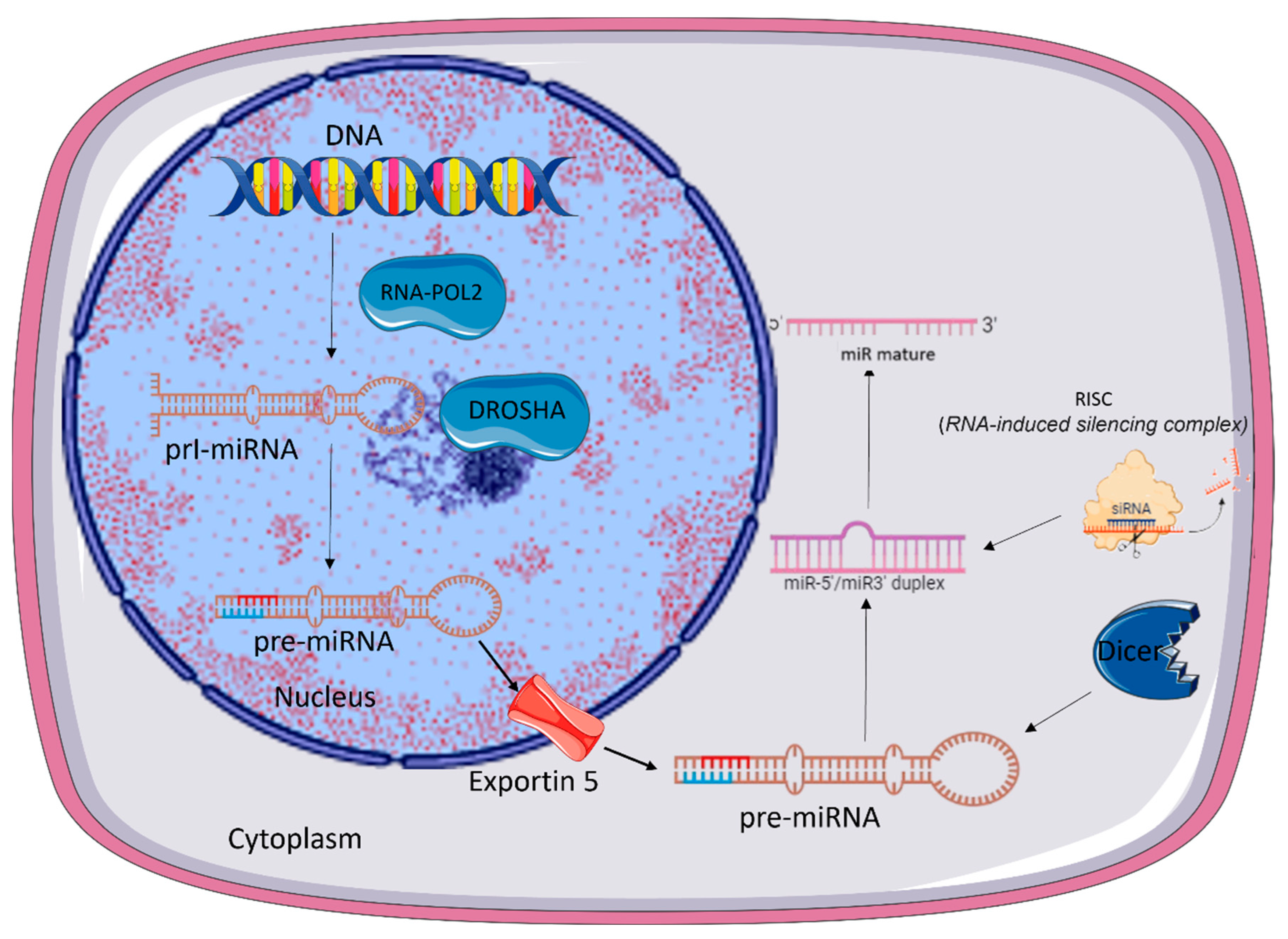
2. microRNA Biogenesis

The process of biogenesis of a miRNA occurs in two parts: one in the nucleus, and the other in the cytoplasm (Figure 1). In the nucleus, starts the transcription that generates primary transcripts (pri-microRNAs) [16] with over 1000 nucleotides in length organized in three major domains: a 33- to 35-nucleotide-long stem, terminal loop, and two single-stranded RNA (ssRNA) segments flaking both ends [32,33].
Depending on the location of the miRNA gene within the genome, identified miRNAs are intragenic or intergenic [15,26]. Intergenic-derived miRNAs are transcribed either by RNA polymerase II or III, come from noncoding regions of the DNA between genes, and have unique promoter regions [32,33,34,35]. By contrast, the genes of intragenic-derived miRNA are transcribed by RNA polymerase II. They are located within exons or introns of protein-coding genes, and are co-expressed with their host gene [32,33,34].
The biogenesis of miRNA is classified into canonical and non-canonical pathways. In the canonical pathway (Figure 1), those pri-micro RNAs are processed by the enzyme Drosha (RNAse III) and its cofactor DGCR8 (DiGeorge Syndrome Critical Region 8). The resulting precursor microRNA (pre-microRNA) is exported to the cytoplasm by exportin-5 through pores in the nuclear membrane [36]. In the cytoplasm, those pre-microRNAs are processed by Dicer, another RNAse [37], forming a double-stranded mature miRNA.
Both strands derived from the mature microRNA duplex can be loaded into the Argonaute (AGO) [38], incorporated into the RNA-induced silencing complex (RISC) and then separated into two strands. One of the strands is degraded, while the other remains associated with RISC, forming the mature microRNA [12,13,39,40].
The strand with lower thermodynamic stability is selected to be loaded into AGO and is deemed the guide strand [41]. The unloaded strand is called the passenger strand. The passenger strands are cleaved by AGO2 and degraded by cellular machinery [42]. Mature microRNA is identified by the prefix “miR” while pre-microRNAs are identified by “mir-”.
Several non-canonical miRNA biogenesis pathways use the same proteins involved in the canonical pathway and can be grouped into Drosha/DGCR8-independent and Dicer-independent pathways [43].
Drosha/DGCR8-independent pathway may process the pre-miRNA splicing machinery from the intron of protein-coding gene [26,28]. Those microRNAs, called miRtrons, are correlated with the host gene expression based on their location in either the introns or splice site junctions. MiRtrons are continuously transported to the cytoplasm and processed by Dicer [44,45,46].
Conversely, in the Dicer-independent pathway miRNAs are processed by Drosha from endogenous short hairpin RNA (shRNA) transcripts [47]. These pre-miRNAs require AGO2 to complete their maturation within the cytoplasm because they are of insufficient length to be Dicer-substrates [48]. This in turn promotes loading of the entire pre-miRNA into AGO2 and AGO2-dependent slicing of the 3p strand. The 3′-5′ trimming of the 5p strand completes their maturation [47].

3. MicroRNAs and Intercellular Communication

miRNAs were first discovered as acting inside the cells by controlling gene expression at a post-transcriptional level. More recently, however, they have also been detected in circulating blood. MicroRNAs which migrate outside the cells play a role in intercellular communication [48,49]. Those miRNAs are called circulating miRNAs [48].
In order to be protected from digestion by RNAses [50], the circulating miRNAs are secreted in exosomes or extracellular vesicle (EVs) [51,52], or form complexes with proteins including Ago2 [47], nucleophosmin 1 (NPM 1) and high-density lipoprotein [53]. Unlike intracellular miRNAs, circulating miRNA shows remarkable stability and resistance to degradation by endogenous RNAse activity [54,55]. Exosomes containing miRNAs enter neighboring cells where they affect mRNA targets remotely from their origin throughout endocytic uptake, membrane fusion, or integrating with specific receptors in the cell surface [56,57]. The circulating miRNAs in exosomes can reflect the specific cell of origin of exosome content, and the variation depending on the physiological and pathological condition [56]. Precisely because their content reflects their origin and pathophysiological state, exosomes and EVs have been widely used as valuable biomarkers for diagnostic and therapeutic strategies.
The main source of circulating miRNAs seems to be the adipose tissue although they may be derived from many other tissues. Adipose tissue-secreted extracellular vesicles containing miRNA may arise from different cells within the fat pad and may be differentially regulated by various stimuli, thus being trigger of some diseases, such as metabolic diseases [14].
Obesity changes the profile of exosomal miRNAs in mice [52]. Moreover, obesity-associated-exosomal miRNAs are active players in the first stages of the metabolic syndrome characterized by development of glucose intolerance, dyslipidemia, and central obesity [58].
miRNAs are secreted by pancreatic islet cells along with secretion of insulin and glucagon [59]. In that case, miRNA would modulate insulin action in target tissues, while peripheral cells could send signals back to islets via extracellular vesicles containing miRNAs. MiR-122, miR-142–3p, miR-192, miR-222, and miR-378a were upregulated, and miR-138 and miR-221 were downregulated in obese patients or obese animals [59,60,61,62,63].
Circulating extracellular vesicles may also cross the ependymal layer and the blood brain barrier (BBB), by a transcytosis mechanism, thus acting on the central nervous system [64,65]. Neurodegenerative diseases which alter BBB permeability could facilitate the exchange of circulating miRNAs from the brain to the blood and vice-versa, reviewed in reference [62].
Communication between the different cell types that make up the heart, such as cardiomyocytes, fibroblasts, endothelial cells and macrophages, has already been described as important for maintaining cardiac homeostasis [65]. This intercellular communication can occur through direct contact between cells, such as gap junctions, or through the secretion of soluble factors or EVs [66].
In cardiovascular diseases, such as myocardial infarction, miRNAs transfer by extracellular vesicles play a key role in driving the healing and remodeling response [65]. They can modulate phenotype and function of macrophages, which in turn modulate fibroblast proliferation and the inflammatory response (reviewed by [65]). EVs from ischemic cardiomyocytes can also stimulate endothelial cell angiogenesis by miRNAs transfers, mainly miRNA-222 and miRNA-143 [66].

4. miRNAs in Cardiac Diseases

Epigenetic mechanisms are present at all stages of cardiac development, and miRNAs regulate the initial process of cardiac differentiation and cardiomyocyte proliferation [67]. Mature miRNAs participate in cell cycling, cell proliferation, cellular aging, apoptosis, angiogenesis, autophagy, mitochondrial metabolism, hematopoiesis, and cardiovascular development.
That is why dysregulated miRNA expression is directly involved in many CVDs and diseases’ development [68]. Altered expression of miRNAs have been described in cardiomyopathy, atrial fibrillation, hypertension, metabolic syndrome, and stroke, angiogenesis, inflammation, and cardiac remodeling, which are central to the development and progression of CVDs [68,69]. Some miRNAs and the related CVDs are summarized in Figure 2 [70].
The first described miRNA was lin-4 in C. elegans [71]. The second, also found in C. elegans, was let7. This one is highly expressed in cells of the cardiovascular system [72,73,74,75,76,77]. Exacerbated expression of let7 has been linked to cardiac fibrosis, myocardial infarction, cardiac hypertrophy, arrhythmia, atherosclerosis, and hypertension. Let 7 also acts in cardiovascular differentiation from embryonic stem cells [67,78].
The regulatory potential of miRNAs in the cardiac tissue homeostasis was demonstrated by the loss of a component of miRNA´s biogenesis machinery in animal model, which was incompatible with properly cardiac function and maintenance of life. Mutant mice presented premature lethality displaying dilated cardiomyopathy and heart failure [79], showing the importance of miRNA pathway in the heart.
Circulating levels, as well as the source, of miRNAs have pattern altered in cardiovascular diseases [68]. In coronary artery disease´s patients, circulating levels of miR-126, miR-17, miR-92a, miR-155 and miR-145 are markedly reduced while miRNAs from cardiac muscle, mainly miR-133a and miR-208a are higher [68]. Furthermore, expression levels of circulating miR-423, miR-320, miR-765, miR-149, miR-21, miR-126, and miR-1 are altered in stable and unstable coronary artery disease and myocardial infarction patients [80,81].
MiR-126, which plays an important role in angiogenesis process, is reduced in patients with atherosclerosis and coronary artery disease [80]. Progression of atherosclerosis is related to several miRNAs, including miR-15a-5p, miR-199a-3p, miR-34a, miR-146a, and miR-217. Those miRNAs are also involved in cellular senescence, endothelial dysfunction, and inflammation, whereas miR-342-5p protects against endothelial cellular injury during atherosclerosis [82].
MiR-21 is a key molecule involved in cardiac hypertrophy, promotes angiogenesis and cardiomyocyte survival post-myocardial infarction [83], inhibits apoptosis and inflammation [83]. Within the myocardium, miR-21 has its strongest expression in cardiac macrophages that controls myocardial fibrosis through intercellular signals to fibroblasts (Ramanujam et al., 2021). Overexpression of miR-21 triggered by hypoxia in rat hearts has been linked to pathological cell growth and cellular stress, indicating the emergence of cardiac hypertrophy [85]. Elevated expression of miR-21 has also been observed in cardiac tissue from patients with heart failure, and this miRNA has been shown to promote cardiac fibrosis and hypertrophy in animal models. MiR-133 mitigates cardiac hypertrophy by blocking the AKT/mTOR pathway and the beta-adrenergic receptor (β-AR), inhibiting activation cascades that could lead to atrophy, apoptosis, and autophagy [86]. miR-1 is the most abundant miRNA in the heart being implicated in the cardiac homeostasis and function [87]. When overexpressed, miR-1 reduces hypertrophy and fibrosis [87], however, when reduced triggers multiple impairments in heart function [78,80].
MiR-484 plays a role in cardiomyocyte apoptosis [88], and its direct interaction with miR-361 has implications on cardiac diseases such as myocardial infarction [89]. Reduced expression of miR-125b-5p in both humans and mice is related to cardiac ischemia and has been proposed as a diagnostic biomarker as well as a biomarker of progression to heart failure [90]. miR-127-3p has been identified as a pro-fibrogenic agent and was up-regulated in the rat heart during cardiac fibrosis. When overexpressed, miR-127-3p increased fibrogenic differentiation and proliferation, microRNA inhibition suppressed fibroblast activation [91].
MiR-208 is one of the most relevant miRNAs implicated in cardiovascular diseases. MiR-208 is related to various cardiac processes, including increased cardiac fibrosis via THRAP-1 (Thyroid Hormone Receptor Associated Protein 1) inhibition, right ventricular hypertrophy by inhibiting the Mef2 axis, and negative regulation of the SOX6 (SRY-Box Transcription Factor 6) gene associated with cardiac hypertrophy [92]. miR208a has been described as specifically expressed in cardiac tissue promoting cardiac hypertrophy in animal models [93].
When exposed to stress conditions such as elevated blood pressure or workload, miR-208 is activated, triggering a series of adaptive responses. Furthermore, miR-208 is also involved in regulating the expression of genes associated with cardiac function, such as the Myosin Heavy Chain β (β-MHC) gene, contributing to cardiac remodeling aimed at enhancing performance under challenging conditions. Ultimately, the interaction between miR-208 and cell stress reveals an intricate network of molecular signaling aimed at optimizing cardiac capacity in face of demands [94]. miR-208 is encoded within the introns of various myosin genes. The expression of miR-208a, encoded within a fast myosin gene, is capable to regulate the expression of miR-499 and miR-208b, encoded within slow myosin gene, which together forms feedback loops that regulate miRNA levels and, consequently, muscle contraction in response to physiological changes [91].
From the above-mentioned interactions of miRNAs with cardiac diseases it is possible to figure out the complexity of miRNAs actions and the big challenge deciphering the precise roles of specific miRNAs actions in physiological and pathological contexts. The regulation of mRNA expression by miRNAs is highly specific for cells and organs, depends on the metabolic state and level of stress on the organism. Furthermore, usually there is no specific correlation between the level of miRNA expression and its action, since its expression can be intermittently altered in pathophysiological contexts and its activity and availability can also be regulated [95].
Nevertheless, there are substantial efforts to determine the mechanism underlying miRNAs action and their targets. Table 1 summarizes some information about miRNA and their targets in cardiovascular diseases.
Unravel how miRNA expression and its ability to regulate cells and tissues responses, as well as their behavior in the development of diseases is not only necessary as well as mandatory. It should facilitate the design of new therapeutic approaches based on miRNA targeting molecular mechanisms underlying complex diseases.
Therefore, due to their marked implication in cardiovascular diseases many miRNAs, mainly circulating miRNAs, have been pointed out as cardiovascular diseases biomarkers. Their use as therapeutic tools is on horizon.

5. miRNA and Other Diseases

miRNA has been implicated in research of allergy, carcinogenesis, obesity, metabolic syndrome, neurodegenerative diseases [92]. Once they enter the extravascular space or blood, miRNAs can act on other cells within the organ or tissues even at a distance, to establish metabolic homeostasis and energy balance.
Many miRNAs have been considered as biomarkers in neurodegenerative diseases. It has been demonstrated that the aging process affects secretion of miRNAs extracellular vesicles by hypothalamic stem cells and that the intracerebroventricular injection of extracellular vesicles containing miRNAs from these cells slows hypothalamic aging [96,97]. Extracellular vesicles containing miRNAs have also been implicated in the crosstalk between neurons, astrocytes, microglia and endothelial cells [reviewed in reference 98]. For example, after brain injury, the increment on miR-124 level was associated to inhibition of neuronal inflammation [99].
The mature let-7 miRNA is controlled by miR-107. Because let-7 is a tumor suppressor, its downregulation and suppression by miR-107 leads to an increase in the abundance of its target oncogenes, contributing to downstream tumorigenesis [100]. Furthermore, the downregulation of pri-miR-9 by miR-503 and miR-484 promotes cellular lineage commitment [101], however, the interruption of this interaction leads to miR-9 upregulation, leading to an undifferentiated state typical of cancer cells [102].
Environmental stress due to the modern life style has been identified as a relevant cardiovascular risk factor [103,104]. The stress response is characterized by activation of the sympathetic nervous system and the adrenal gland, which release catecholamines and glucocorticoids, classical circulating stress markers. We have extensively reported the consequences of stress in endocrine, metabolic, behavioral and cardiovascular parameters in humans and animal models [103,104,105,106]. In the heart, repeated stress promotes changes in the β-adrenergic receptors population with consequences to heart function [107,108,109,110].
Cardiac expression of several genes related to signaling pathways of the β-adrenergic receptors and glucocorticoid receptors are altered as well [111]. The large number of genes with significant differences in expression suggests the influence of stress on phenotypic changes in cardiac cells [110]. Thus, it is reasonable to predict participation of some epigenetic factors, such as miRNAs, in stress-induced gene regulation. In fact, microarray data have suggested the participation of some miRNAs in the modulation of cardiac response to stress. miR-331-5p, miR-331-3p, miR-127-3p, miR-125b-5p, miR-191-5p, miR-30c-5p were predicted to participate as upstream regulators of those genes (unpublished data), and further study may clarify it.

6. Methodological approaches

Considering that miRNAs have been proposed as fine-tuners of gene and protein expression profiles during pathological conditions, investigation concerning the expression of selected miRNAs and their predictive targets have used animal models [112].
qPCR, microarrays, and next-generation sequencing (NGS) are the techniques mainly used for studying miRNAs, each one with advantages and limitations. qPCR is frequently used in studies that require precise validation of target miRNA expression. The processing time is up to 6 hours, it has high sensitivity and accuracy in quantifying specific miRNAs. qPCR can identify around 754 human miRNAs per sample with minimal infrastructure requirements. However, its analysis capacity is lower compared to other techniques.
Otherwise, microarrays allow the simultaneous profiling of hundreds of miRNAs in a single sample, making them useful for large-scale exploratory studies, although their sensitivity is lower than that of qPCR. Microarray experiments take approximately 2 days to complete and can identify 950 miRNAs per sample, requiring moderate infrastructure.
Finally, NGS provides a comprehensive analysis of the miRNA transcriptome, with the ability to detect new miRNAs and sequence variants. Despite being a more expensive technique that requires sophisticated infrastructure, NGS is highly advantageous for studies aiming to explore the diversity of miRNAs at a deep level and with high throughput, theoretically identifying all miRNAs per sample. Due to its complexity, NGS experiments can take from one to two weeks to complete [113].

7. The clinical potential of miRNAs: diagnostic, prognostic and therapeutic implications

The discovery that miRNAs showed organ- and cell-specific expression patterns [114] has created great expectations as promising new biomarkers in the field of several diseases. MiRNA quantification and in vitro findings suggest groups of miRNAs being specifically up and down regulated in different diseases [115], while polymorphisms in the miRNA regulatory pathway—so called miRSNPs—have shown association with different types of disease [116].
Circulating miRNAs can be isolated from body fluids such as serum, plasma and saliva. Exosomes present in body fluids can have a potential role in immunotherapy and vaccination modalities and as a potential vector for gene therapy. Healthy saliva contains approximately 50 miRNAs and some of them can be used clinically to detect various salivary gland diseases [117], including oral cancer [117]. Two miRNAs in particular, miR-125a and miR-200a, have been found exclusively in the saliva of oral cancer patients and are diagnostic markers of the disease [69].
Another promising application of miRNAs is to utilize their immunomodulatory functions to promote antimicrobial pathways during infection and control dysregulated inflammatory responses during sepsis. Most infection studies compare the extracellular miRNA profile of patients with healthy controls. Many identify extracellular miRNA signatures which are highly predictive of infection.
A promising application of miRNA biomarker work may be to differentiate viral from bacterial infection, identify or prognosticate sepsis, and in monitoring of response to antimicrobial treatment.
MiRNAs are essential mediators of host response to pathogens. They have pleiotropic roles which microbes have evolved to exploit. Elucidating the roles miRNAs in host response to infectious disease is inherently interesting at it provides a tool for identifying key genes and pathways that must be activated, enhanced, repressed, or silenced to facilitate an effective immune response. The complex regulatory network within which miRNAs are embedded, make unpicking the roles of miRNAs tough but not impossible. Integrating large miRNA and mRNA datasets using advanced statistical techniques (in a “systems biology” approach) will facilitate the unpicking of these complex networks.
The diagnostic value of miRNAs in cardiovascular diseases has been examined in numerous studies and animal models with respect to coronary artery disease and myocardial infarction. Various miRNAs are described to complement protein-based biomarkers or classical risk factors in the diagnosis of coronary artery disease or myocardial infarction and even represent potential new biomarkers in the discrimination of unstable angina pectoris. Signatures consisting of sets of multiple miRNAs seem to improve the predictive power compared to single miRNAs. Defined groups of circulating miRNAs being quantitatively altered were detected also in other disease entities such as atherosclerosis, heart failure, atrial fibrillation, hypertrophy and fibrosis (118-122).
So, clinical applications of miRNAs are the next step. As the literature on miRNAs grows, the potential for new miRNA therapeutics, diagnostics/prognostics, and vaccines becomes tangibly closer. Translating the insights of miRNA studies into improving the lives of patients is the critical next step now.

8. Conclusions

MiRNAs play a significant role in cardiovascular system maturation, regulating heart function, and participating in the development of cardiac diseases. They also hold potential as biomarkers and therapeutic targets. The future of healthcare could be closely tied to miRNA research, as they are implicated in various organ systems and numerous diseases. However, further research is required to identify the role played by miRNAs in physiological and pathological processes. Thus, miRNA investigation remains a rapidly expanding and promising field for the development of novel therapies and treatments.

Acknowledgments

Figures were partly generated using Servier Medical Art, provided by Servier, licensed under a Creative Commons Attribution 3.0 unported license (https://creativecommons.org/licenses/by/3.0/). This work was supported by the São Paulo Research Foundation (FAPESP grant 2022/12159-3), Coordenação de Aperfeiçoamento de Pessoal de Nível Superior (CAPES).

Conflicts of Interest

The authors declare no competing interests.

References

  1. Mensah, G.A.; Fuster, V.; Roth, G.A. A Heart-Healthy and Stroke-Free World: Using Data to Inform Global Action. J. Am. Coll. Cardiol. 2023, 82 (25), 2343-2349.
  2. Kalayinia, S.; Arjmand, F.; Maleki, M.; Malakootian, M.; Singh, C.P. MicroRNAs: roles in cardiovascular development and disease. Cardiovasc. Pathol. 2021, 50:107296. [CrossRef]
  3. Feeney, A.; Nilsson, E.; Skinner, M.K. Epigenetics and transgenerational inheritance in domesticated farm animals. J. Anim. Sci. Biotechnol. 2014, 5(1):48. [CrossRef]
  4. Oliveira, N.F.P.; Planello, A.C.; Andia, D.C.; Pardo, A.P. de S. Metilação de DNA e Câncer. Rev. Bras. Cancerol. 2010, 56, 493-499.
  5. Kiefer, J.C. Epigenetics in development. Developmental dynamics: an official publication of the American Association of Anatomists, 2007; 236(4), 1144-1156.
  6. Morris, J.R. Genes, genetics, and epigenetics: a correspondence. Science, 2001, 293(5532), 1103-1105. [CrossRef]
  7. Greco, C.M.; Condorelli, G. Epigenetic modifications and noncoding RNAs in cardiac hypertrophy and failure. Nat. Ver. Cardiol. 2015,12 (8), 488-497.
  8. Sen, G.L.; Blau, H.M. A brief history of RNAi: the silence of the genes. FASEB. J. 2016, 20:1293–1299. [CrossRef]
  9. Setten, R.L.; Rossi, J.J.; Han, S.P. The current state and future directions of RNAi-based therapeutics. Nat. Rev. Drug. Discov. 2019, 18:421–446. [CrossRef]
  10. Quiat, D.; Olson, E.N. MicroRNAs in cardiovascular disease: from pathogenesis to prevention and treatment. J. Clin. Invest. 2013,123 (1),11-18. [CrossRef]
  11. Thum, T. Noncoding RNAs and myocardial fibrosis. Nat. Rev. Cardiol. 2014, 11(11), 655-663. [CrossRef]
  12. Yang, Z.; Wu, L.; Wang, A. dbDEMC 2.0: updated database of differentially expressed miRNAs in human cancers. Nucleic. Acids. Res. 2017 45, 812-D818. [CrossRef]
  13. Lu, T.X.; Rothenberg, M.E. MicroRNA. J. Allergy Clin. Immunol. 2018, 141 (4), 1202-1207.
  14. Mori, M.A.; Ludwig R.G.; Garcia-Martin R.; Brandão B.B.; Kahn C.R. Extracellular miRNAs: From Biomarkers to Mediators of Physiology and Disease. Cell. Metab. 2019, 30 (4), 656-673. [CrossRef]
  15. Ransohoff, J.D.; Wei, Y.; Khavari, P.A. The functions and unique features of long intergenic non-coding RNA. Nat. Rev. Mol. Cell. Biol. 2018,19 (3),143-157. [CrossRef]
  16. Ulitsky, I. Interactions between short and long noncoding RNAs. FEBS Lett. 2018, 592 (17), 2874-2883. [CrossRef]
  17. Gebert, L.F.R., MacRae, I.J. Regulation of microRNA function in animals. Nat. Rev. Mol. Cell. Biol. 2019, 20 (1), 21-37. [CrossRef]
  18. Hill, M.; Tran, N. miRNA interplay: mechanisms and consequences in cancer. Dis. Model. Mech. 2021,14 (4). [CrossRef]
  19. Lu, T.X.; Rothenberg, M.E. MicroRNA. J. Allergy Clin. Immunol. 2018, 141 (4), 1202-1207.
  20. Citron, F.; Armenia, J.; Franchin, G. An Integrated Approach Identifies Mediators of Local Recurrence in Head and Neck Squamous Carcinoma. Clin. Cancer Res. 2017, 23 (14): 3769-3780.
  21. Yoon, A.J.; Wang, S.; Kutler, D.I.; et al. MicroRNA-based risk scoring system to identify early-stage oral squamous cell carcinoma patients at high-risk for cancer-specific mortality. Head Neck. 2020, 42(8),1699-1712.
  22. Wojciechowska, A.; Braniewska, A.; Kozar-Kamińska, K. MicroRNA in cardiovascular biology and disease. Adv. Clin. Exp. Med. 2017, 26 (5), 865-874.
  23. Zernecke, A.; Bidzhekov, K.; Noels, H.; et al. Delivery of microRNA-126 by apoptotic bodies induces CXCL12-dependent vascular protection. Sci. Signal. 2009, 2(100), ra81.
  24. Hata, A. Functions of microRNAs in cardiovascular biology and disease. Annu. Ver. Physiol. 2013, 75, 69-93.
  25. Condorelli, G.; Latronico, M.V.; Cavarretta, E. microRNAs in cardiovascular diseases: current knowledge and the road ahead. J. Am. Coll. Cardiol. 2014, 63(21), 2177-2187.
  26. Bronze-da-Rocha, E. MicroRNAs expression profiles in cardiovascular diseases. Biomed. Res. Int. 2014, 2014, 985408.
  27. Klimczak, D.; Pączek, L.; Jażdżewski, K.; Kuch, M. MicroRNAs: powerful regulators and potential diagnostic tools in cardiovascular disease. Kardiol. Pol. 2015, 73 (1), 1-6.
  28. Anglicheau, D.; Muthukumar, T.; Suthanthiran, M. MicroRNAs: small RNAs with big effects. Transplantation 2010, 90 (2),105-112.
  29. Lagos-Quintana, M.; Rauhut, R.; Yalcin, A.; Meyer J.; Lendeckel, W.; Tuschl T. Identification of tissue-specific microRNAs from mouse. Curr. Biol. 2002, 12 (9), 735-739.
  30. Ludwig, N.; Leidinger, P.; Becker K.; et al. Distribution of miRNA expression across human tissues. Nucleic Acids Res. 2016, 44 (8), 3865-3877. [CrossRef]
  31. Nalbant, E.; Akkaya-Ulum, Y.Z. Exploring regulatory mechanisms on miRNAs and their implications in inflammation-related diseases. Clin. Exp. Med. 2024, 24 (1), 142.
  32. Ha, M.; Kim.; V.N. Regulation of microRNA biogenesis. Nat. Rev. Mol. Cell. Biol. 2014, 15:509–524. [CrossRef]
  33. Wu, K.L.; Tsai, Y.M.; Lien, C.T.; Kuo, P.L.; Hung, A.J. The Roles of MicroRNA in Lung Cancer. Int. J. Mol. Sci. 2019, 20 (7), 1611. [CrossRef]
  34. Valinezhad, O.A; Safaralizadeh, R.; Kazemzadeh-Bavili, M. Mechanisms of miRNA-mediated gene regulation from common downregulation to mRNA- specific upregulation. Int. J. Genomics. 2014, 2014:970607.
  35. Liu, B.; Shyr, Y.; Cai, J.; Liu, Q. Interplay between miRNAs and host genes and their role in cancer. Brief. Funct. Genomics. 2018, 18:255–266.
  36. Matsumoto. J; Stewart, T.; Banks, W.A.; Zhang, J. The Transport Mechanism of Extracellular Vesicles at the Blood-Brain Barrier. Curr. Pharm. Des. 2017, 23(40), 6206-6214. [CrossRef]
  37. Bernstein, E.; Caudy, A.A.; Hammond, S.M.; Hannon, G.J. Role for a bidentate ribonuclease in the initiation step of RNA interference. Nature 2001, 409 (6818), 363-366.
  38. Song, J.J.; Liu, J.; Tolia, N.H.; et al. The crystal structure of the Argonaute2 PAZ domain reveals an RNA binding motif in RNAi effector complexes. Na.t Struct. Biol. 2003, 10 (12), 1026-1032.
  39. Schwarz, D.S.; Hutvágner, G.; Du, T.; Xu, Z.; Aronin, N; Zamore, P.D. Asymmetry in the assembly of the RNAi enzyme complex. Cell 2003, 115 (2), 199-208.
  40. Wu, K.L.; Tsai, Y.M.; Lien, C.T.; Kuo, P.L.; Hung, A.J. The Roles of MicroRNA in Lung Cancer. Int. J. Mol. Sci. 2019, 20 (7), 1611.
  41. Khvorova. A.; Reynolds. A.; Jayasena. S.D. Functional siRNAs and miRNAs exhibit strand bias. Cell. 2003, 115 209–16.
  42. Ha. M.; Kim. V.N. Regulation of microRNA biogenesis. Nat. Rev. Mol. Cell. Biol. 2014, 15 509–24.
  43. O'Brien. J.; Hayder. H.; Zayed. Y. Peng. C. Overview of MicroRNA Biogenesis, Mechanisms of Actions, and Circulation Front. Endocrinol. 2018, 3 9:402.
  44. Isik, M.; Korswagen, H.C.; Berezikov, E. Expression patterns of intronic microRNAs in Caenorhabditis elegans. Silence 2010, 1 (1), 5.
  45. Westholm, J.O.; Lai, E.C. Mirtrons: microRNA biogenesis via splicing. Biochimie 2011, 93 (11), 1897-1904.
  46. Ramalingam, P.; Palanichamy, J.K.; Singh, A.; et al. Biogenesis of intronic miRNAs located in clusters by independent transcription and alternative splicing. RNA. 2014, 20 (1), 76-87.
  47. Yang. J.S.; Maurin. T.; Robine. N.; Rasmussen. K.D.; Jeffrey. K.L.; Chandwani. R. Conserved vertebrate mir-451 provides a platform for Dicer-independent, Ago2-mediated microRNA biogenesis. Proc. Natl. Acad. Sci. USA. 2010, 107 15163–8.
  48. Cheloufi. S.; Dos Santos. C.O.; Chong. M.M.W.; Hannon. G.J. A dicer-independent miRNA biogenesis pathway that requires Ago catalysis. Nature. 2010, 465 584–9.
  49. Weber, J.A.; Baxter, D.H.; Zhang, S.; et al. The microRNA spectrum in 12 body fluids. Clin Chem. 2010, 56(11), 1733-1741.
  50. Arroyo, J.D.; Chevillet, J.R.; Kroh, E.M.; et al. Argonaute2 complexes carry a population of circulating microRNAs independent of vesicles in human plasma. Proc. Natl. Acad. Sci. USA. 2011, 108 (12), 5003-5008. [CrossRef]
  51. Weickmann, J.L.; Glitz, D.G. Human ribonucleases. Quantitation of pancreatic-like enzymes in serum, urine, and organ preparations. J. Biol. Chem. 1982, 257 (15), 8705-8710.
  52. Théry, C. Exosomes: secreted vesicles and intercellular communications. F1000 Biol. Rep. 2011, 3: 15.
  53. Théry, C.; Zitvogel, L.; Amigorena, S. Exosomes: composition, biogenesis and function. Nat. Ver. Immunol. 2002, 2 (8), 569-579.
  54. Tsui. N.B.; Ng E.K.; Lo. Y.M.; Stability of endogenous and added RNA in blood specimens, serum, and plasma. Clin. Chem. 2002.
  55. 55. Wang. K.; Zhang. S.; Marzolf. B.; Troisch. P.; Brightman. A.; Hu. Circulating microRNAs, potential biomarkers for drug-induced liver injury. Proc. Natl. Acad. Sci. U S A. 2009, 106 4402–7. [CrossRef]
  56. Valadi, H.; Ekström, K.; Bossios, A.; Sjöstrand. M.; Lee, J.J.; Lötvall, J.O. Exosome-mediated transfer of mRNAs and microRNAs is a novel mechanism of genetic exchange between cells. Nat. Cell Biol. 2007, 9 (6), 654-659.
  57. Morello, M.; Minciacchi, V.R.; de Candia, P.; et al. Large oncosomes mediate intercellular transfer of functional microRNA. Cell Cycle. 2013, 12 (22), 3526-3536.
  58. Castaño. C.; Kalko. S.; Novials. A.; Parrizas. M. Obesity-associated exosomal miRNAs modulate glucose and lipid metabolism in mice. PNAS. 2018, 15 12158-12163.
  59. Guay, C.; Kruit, J.K.; Rome, S.; et al. Lymphocyte-Derived Exosomal MicroRNAs Promote Pancreatic β Cell Death and May Contribute to Type 1 Diabetes Development. Cell Metab. 2019, 29 (2), 348-361.e6.
  60. Can, U.; Buyukinan, M.; Yerlikaya, F.H. The investigation of circulating microRNAs associated with lipid metabolism in childhood obesity. Pediatr. Obes. 2016, 11 (3), 228-234.
  61. Jones, A.; Danielson, K.M.; Benton, M.C.; et al. miRNA Signatures of Insulin Resistance in Obesity. Obesity (Silver Spring). 2017, 25 (10), 1734-1744.
  62. Sweeney, M.D.; Sagare, A.P.; Zlokovic, B.V. Blood-brain barrier breakdown in Alzheimer disease and other neurodegenerative disorders. Nat. Ver. Neurol. 2018, 14 (3), 133-150. [CrossRef]
  63. Chen, C.C.; Liu, L.; Ma, F.; et al. Elucidation of Exosome Migration across the Blood-Brain Barrier Model In Vitro. Cell. Mol. Bioeng. 2016, 9 (4), 509-529. [CrossRef]
  64. Chen, L.; Qian, H.; Xue, J.; Zhang, J.; Chen, H. MicroRNA-152 regulates insulin secretion and pancreatic β cell proliferation by targeting PI3Kα. Mol. Med. Rep. 2018, 18 (4), 4113-4121.
  65. Martins-Marques, T.; Hausenloy, D.J.; Sluijter, J.P.G.; Leybaert, L.; Girao, H. Intercellular Communication in the Heart: Therapeutic Opportunities for Cardiac Ischemia. Trends Mol. Med. 2021, 27 (3), 248-262.
  66. Ribeiro-Rodrigues, T.M.; Laundos, T.L.; Pereira-Carvalho, R; et al. Exosomes secreted by cardiomyocytes subjected to ischemia promote cardiac angiogenesis. Cardiovasc. Res. 2017, 113 (11), 1338-1350.
  67. Bao. M.; H., Feng. X.; Zhang. Y. W.; Lou. X. Y.; Cheng. Y. U.; Zhou. H. H. Let-7 in cardiovascular diseases, heart development and cardiovascular differentiation from stem cells. Int. J. Mol. Sci. 2013, 14(11), 23086-23102.
  68. Fichtlscherer, S.; De Rosa, S.; Fox, H.; et al. Circulating microRNAs in patients with coronary artery disease. Circ. Res. 2010, 107: 677-684.
  69. Small, E. M.; Frost, R. J.; Olson, E. N. MicroRNAs add a new dimension to cardiovascular disease. Circulation 2010, 121:1022-1032. [CrossRef]
  70. Mauri, M.; Elli, T.; Caviglia, G.; Uboldi, G.; Azzi, M. RAWGraphs: A Visualisation Platform to Create Open Outputs. In Proceedings of the 12th Biannual Conference on Italian SIGCHI. 2017, p. 28:1–28:5.
  71. Lee. R.C.; Feinbaum. R.L.; Ambros. V. The C. elegans heterochronic gene lin-4 encodes small RNAs with antisense complementarity to lin-14. Cell. 1993, 75 843–854.
  72. Reinhart. B.J.; Slack. F.J.; Basson. M.; Pasquinelli. A.E.; Bettinger. J.C.; Rougvie. A.E.; Horvitz. H.R.; Ruvkun. G. The 21-nucleotide let-7 RNA regulates developmental timing in Caenorhabditis elegan s. Nature. 2000, 403 901–906.
  73. Ding. Z.; Wang. X.; Schnackenberg. L.; Khaidakov. M.; Liu. S.; Singla. S.; Dai. Y.; Mehta. J.L. Regulation of autophagy and apoptosis in response to ox-LDL in vascular smooth muscle cells, and the modulatory effects of the microRNA hsa-let-7g. Int. J. Cardiol. 2013.
  74. Chen. Z.; Lai. T.C.; Jan. Y.H.; Lin. F.M.; Wang. W.C.; Xiao. H.; Wang. Y.T.; Sun. W.; Cui. X.; Li. Y.S. Hypoxia-responsive miRNAs target argonaute 1 to promote angiogenesis. J. Clin. Investig. 2013 123 1057–1067.
  75. Mamoru. S.; Yoshitaka. M.; Yuji. T.; Tsuyoshi. T.; Motoyuki. N. A cellular microRNA, let-7i, is a novel biomarker for clinical outcome in patients with dilated cardiomyopathy. J. Card. Fail. 2011, 17 923–929.
  76. Rao. P.K.; Toyama. T.; Chiang. H.R.; Gupta. S.; Bauer. M.; Medvid. R.; Reinhardt. F.; Liao. R.; Krieger. M.; Jaenisch. R. Loss of cardiac microRNA-mediated regulation leads to dilated cardiomyopathy and heart failure. Circ. Res. 2009, 105 585–594.
  77. Ji. R.; Cheng. Y.; Yue. J.; Yang. J.; Liu. X.; Chen. H.; Dean. D.B.; Zhang. C. MicroRNA expression signature and antisense-mediated depletion reveal an essential role of microRNA in vascular neointimal lesion formation. Circ. Res. 2007, 100 1579–1588.
  78. Topkara, V.K.; Mann, D.L. Role of microRNAs in cardiac remodeling and heart failure. Cardiovasc. Drug Ther 2011, 25, 171–182. [CrossRef]
  79. Rao. P.K.; Toyama. T.; Chiang. H.R.; Gupta. S.; Bauer. M.; Medvid. R.; Reinhardt. F.; Liao. R.; Krieger. M.; Jaenisch. R. Loss of cardiac microRNA-mediated regulation leads to dilated cardiomyopathy and heart failure. Circ. Res. 2009, 105 585–594.
  80. Jayawardena. E.; Medzikovic. L.; Ruffenach. G. Role of miRNA-1 and miRNA-21 in acute myocardial ischemia-reperfusion injury and their potential as therapeutic strategy. Int. J. Mol. Sci. 2022, 23 1512.
  81. Yu, B.; Jiang, Y.; Wang, X.; Wang, S. An integrated hypothesis for miR-126 in vascular disease. Medical research archives, 2020, 8(5), 2133.
  82. Gonzalez-Lopez. P.; Alvarez-Villarreal. M.; Ruiz-Simon. R. Role of miR-15a-5p and miR-199a-3p in the inflammatory path- way regulated by NF-jB in experimental and human atherosclerosis. Clin. Transl. Med. 2023 13 1363.
  83. Surina, S.; Fontanella, R.A.; Scisciola, L.; Marfella, R.; Paolisso, G.; Barbieri, M.; miR-21 in Human Cardiomyopathies [published correction appears in Front. Cardiovasc. Med. 2022 25 9:913429.
  84. Ramanujam, D.; Schön, A.P.; Beck, C. et al. MicroRNA-21-dependent macrophage-to-fibroblast signaling determines thecardiac response to pressure overload. Circulation, 2021, 143: 1513-1525.
  85. Krichevsky. A. M.; Gabriely. G. miR-21: a small multi-faceted RNA. J. Cell. Mol. Med. 2009, 13 39-53.
  86. Li, N.; Zhou, H.; Tang, Q. MiR-133: a suppressor of cardiac remodeling? Frontiers in Pharmacology, 2018, 9, 903.
  87. Çakmak. H. A.; Demir. M. MicroRNA and cardiovascular diseases. Balkan medical journal, 2020, 37(2), 60.
  88. Li. J.; Li. L.; Li. X.; Wu. S. Long noncoding RNA LINC00339 aggravates doxorubicin-induced cardiomyocyte apoptosis by targeting MiR-484. Biochemical and biophysical research communications, 2018. 503(4), 3038-3043.
  89. Wang. K., Liu. C. Y., Zhang. X. J., Feng. C., Zhou. L. Y., Zhao. Y.; Li. P. F. miR-361-regulated prohibitin inhibits mitochondrial fission and apoptosis and protects heart from ischemia injury. Cell death and differentiation, 2015, 22(6), 1058–1068.
  90. Bayoumi. A. S.; Park, K. M.; Wang. Y.; Teoh. J. P.; Aonuma. T.; Tang. Y.; Kim. I. M. A carvedilol-responsive microRNA, miR-125b-5p protects the heart from acute myocardial infarction by repressing pro-apoptotic bak1 and klf13 in cardiomyocytes. JMCC. 2018, 114 72-82.
  91. Zhou. Q.; Liu. Z.; Bei. Y.; Zhang. Z.; L. Xiao. J. P4751 Suppression of miR-127-3p prevents cardiac fibrosis. Eur. Heart J., 2018. 39, 563-P4751.
  92. Zhao. X.; Wang. Y.; Sun. X. The functions of microRNA-208 in the heart. Diabetes. Res. Clin. Pract. 2020, 160:108004.
  93. Callis. T. E.; Pandya. K.; Seok. H. Y.; Tang. R. H.; Tatsuguchi. M.; Huang. Z. P.; Chen. J. F.; Deng. Z.; Gunn. B.; Shumate. J.; Willis. M. S.; Selzman. C. H.; Wang. D. Z. MicroRNA-208a is a regulator of cardiac hypertrophy and conduction in mice. The Journal of clinical investigation, 2009, 119(9), 2772–2786.
  94. Ho, P.T.B.; Clark, I.M.; Le, L.T.T. MicroRNA-Based Diagnosis and Therapy. Int. J. Mol. Sci. 2022, 23 (13), 7167.
  95. Correia de Sousa. M.; Gjorgjieva. M.; Dolicka. D.; Sobolewski. C.; Foti. M. Deciphering miRNAs' Action through miRNA Editing. Int. J. Mol. Sci. 2019, 20(24), 6249.
  96. Zhang, Y.; Kim, M.S.; Jia, B.; et al. Hypothalamic stem cells control ageing speed partly through exosomal miRNAs [published correction appears in Nature. 2018 Aug, 560 (7719):E33.
  97. Zagrean, A.M.; Hermann, D.M.; Opris, I.; Zagrean, L.; Popa-Wagner, A. Multicellular Crosstalk Between Exosomes and the Neurovascular Unit After Cerebral Ischemia. Therapeutic Implications. Fron.t Neurosci. 2018, 12:811.
  98. Huang, S.; Ge, X.; Yu, J.; et al. Increased miR-124-3p in microglial exosomes following traumatic brain injury inhibits neuronal inflammation and contributes to neurite outgrowth via their transfer into neurons [published correction appears in FASEB J. 2018 Apr, 32 (4):2315.
  99. Chen, P.S.; Su, J.L.; Cha, S.T.; et al. miR-107 promotes tumor progression by targeting the let-7 microRNA in mice and humans [published correction appears in J Clin Invest. 2017 Mar 1, 127 (3), 1116. doi: 10.1172/JCI92099]. .J Clin. Invest. 2011, 121 (9), 3442-3455.
  100. Forrest, A.R.; Kanamori-Katayama, M.; Tomaru, Y.; et al. Induction of microRNAs, mir-155, mir-222, mir-424 and mir-503, promotes monocytic differentiation through combinatorial regulation. Leukemia. 2010, 24 (2), 460-466. [CrossRef]
  101. Hill, M.; Tran, N. MicroRNAs Regulating MicroRNAs in Cancer. Trends Cancer. 2018, 4 (7), 465-468.
  102. Levine, G.N. Psychological Stress and Heart Disease: Fact or Folklore? Am. J. Med. 2022, 135 (6), 688-696.
  103. Ortolani, D.; Oyama, L. M.; Ferrari, E. M.; Melo, L. L., Spadari-Bratfisch, R. C. Effects of comfort food on food intake, anxiety-like behavior and the stress response in rats. Physiol. Behav. 2011, 103(5), 487-492.
  104. Ortolani, D.; Garcia, M.C.; Melo-Thomas, L.; Spadari-Bratfisch, R.C. Stress-induced endocrine response and anxiety: the effects of comfort food in rats. Stress. 2014, 17(3):211-218.
  105. Spadari-Bratfisch, R. C.; dos Santos, I. N. Adrenoceptors and adaptive mechanisms in the heart during stress. Annals of the New York Academy of Sciences, 2008 1148, 377–383.
  106. Moura, A. L. D.; Hyslop, S.; Grassi-Kassisse, D. M.; Spadari, R. C. Functional β2-adrenoceptors in rat left atria: effect of foot-shock stress. CJPP, 2017, 95(9), 999-1008.
  107. Cordeiro, M.A.; Rodrigues, L.S.; Ortolani, D.; de Carvalho, A.E.T.; Spadari, R.C. Persistent Effects of Subchronic Stress on Components of Ubiquitin-Proteasome System in the Heart. J. Clin. Exp. Cardiolog. 2020, 11:676.
  108. Moura, A.L.; Brum, P.C.; de Carvalho, A.E.T.S.; Spadari, R.C. Effect of stress on the chronotropic and inotropic responses to β-adrenergic agonists in isolated atria of KOβ2 mice. Life Science. 2023, 322: 121644.
  109. Rodrigues, L.S.M.; Cordeiro, M.A.; de Carvalho, A.E.T.S.; Caceres, M.V.; Spadari, R.C.; Stress induced cardiac dysfunction in rats, unpublished.
  110. de Carvalho, A.E.T.S.; Cordeiro, M.A.; Rodrigues, L.S.; Ortolani, D.; Spadari, R.C. Stress-induced differential gene expression in cardiac tissue. Scientific Reports, 2021, 11: 9129.
  111. Johnson. A.M.F.; Olefsky. J.M. The origins and drivers of insulin resistance. Cell. 2013, 152: 673–684.
  112. Baker. M. MicroRNA profiling: separating signal from noise. Nat. Methods. 2010, 7: 687–692.
  113. van Rooij, E. The art of microRNA research. Circ. Res. 2011, 108: 219-234.
  114. Creemers, E. E.; Tijsen, A. J.; Pinto, Y. M. Circulating microRNAs: novel biomarkers and extracellularcommunicators in cardiovascular disease? Circ. Res. 2012, 110: 483-495.
  115. Dzikiewicz-Krawczyk, A. MicroRNA polymorphisms as markers of risk, prognosis and treatment response in hematological malignancies. Crit. Rev. Oncol. Hematol. 2015, 93: 1-17.
  116. Etheridge A, Lee I, Hood L, Galas D, Wang K. Extracellular microRNA: A new source of biomarkers. Mutat. Res. 2011, 717: 85–90.
  117. Park, N. J.; Zhou, H.; Elashoff, D.; Henson, B. S.; Kastratovic, D.A.; Abemayor, E.; et al. Salivary microRNA: Discovery, characterization, and clinical utility for oral cancer detection. Clin. Cancer Res. 2009, 15: 5473–5477.
  118. Michael, A.; Bajracharya, S. D.; Yuen, P. S.; Zhou, H.; Star, R. A.; Illei, G. G.; et al. Exosomes from human saliva as a source of microRNA biomarkers. Oral Dis. 2010, 16: 34–38.
  119. Small, E. M.; Frost, R. J.; Olson, E. N. MicroRNAs add a new dimension to cardiovascular disease. Circulation 2010, 121:1022-1032.
  120. Corsten, M. F.; Dennert, R.; Jochems, S.; et al. Circulating MicroRNA-208b and MicroRNA-499 reflect myocardial damage in cardiovascular disease. Circ. Cardiovasc. Genet. 2010, 3: 499-506.
  121. Gomes da Silva, A. M.; Silbiger, V. N. miRNAs as biomarkers of atrial fibrillation. Biomarkers 2014, 19: 631-636.trial fibrillation. Biomarkers 2014, 19: 631-636.
  122. Kalayinia. S.; Arjmand. F.; Maleki. M.; Malakootian. M.; Singh. C. P. MicroRNAs: roles in cardiovascular development and disease. Cardiovascular pathology: the official journal of the Society for Cardiovascular Pathology, 2021, 50, 107296.
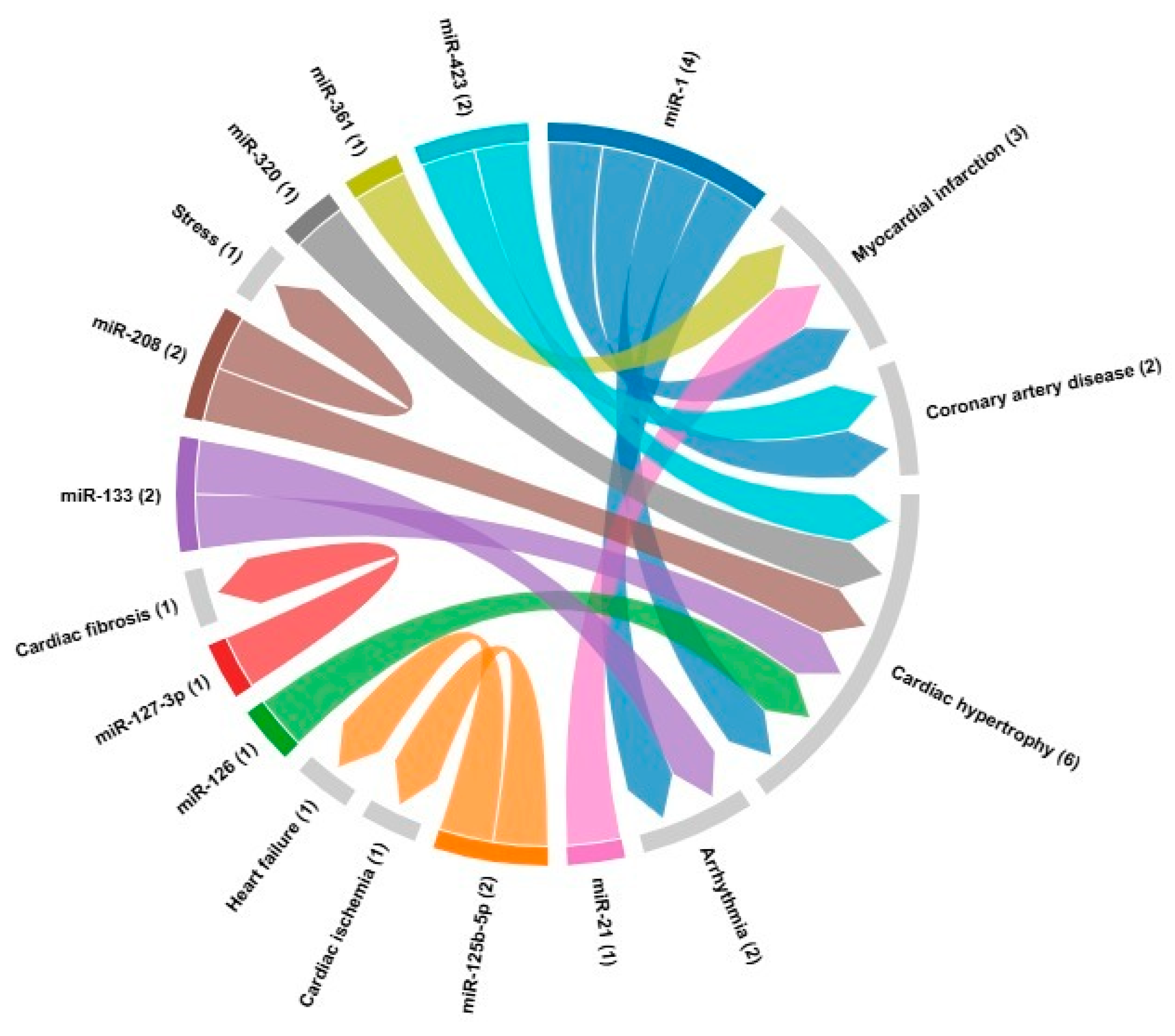
Figure 1. MicroRNA biogenesis. In the nucleus, primary transcripts (pri-miRNA) are processed by the Drosha enzyme, and a double-stranded RNA-binding protein forms a precursor microRNA (pre-miRNA), which is exported to the cytoplasm by exportin-5 located on the nuclear membrane. These pre-miRNAs are processed by Dicer, a nuclease, forming a double-stranded RNA that is incorporated into the RNA-induced silencing complex (RISC) and then separated into two strands. One of these strands is degraded, and the other remains associated with RISC, forming the mature microRNA (miR-).
Preprints 143790 g001
Figure 2. Chord diagram illustrating the associations between different RNAs and specific cardiac conditions, such as cardiac hypertrophy, myocardial infarction and arrhythmia, highlighting the complexity of molecular interactions in the context of cardiovascular diseases. Each color represents one miRNA and arrows indicate the linkage with the cardiovascular disease.
Preprints 143790 g002
Table 1. miRNA and targets in cardiovascular diseases.
microRNA Cardiovascular disease Target References
miR-1 Myocardial infarction
Coronary artery disease Cardiac hypertrophy Arrhythmia
GJA1, KCNJ2, HAND2, IRX5, HSP70, HSP60, HDAC4, CDK9, KCNE1, RASGAP, RHEB, NPTB, biomarker [24,121]
miR-21 Myocardial infarction Biomarker [24]
miR-133 ArrhythmiasCardiac hypertrophy GJA1, KCNJ2, AKT/mTOR [20,82]
miR-361 Myocardial infarction PHB1 [85]
miR-125b-5p Cardiac ischemia
Heart failure
Biomarker, bak1 and klf13 [86]
miR-127-3p Cardiac fibrosis TGF-β, Ang II [87]
miR-208 Cardiac fibrosis
Cardiac hypertrophy
Stress
THRAP-1, Mef2, SOX6, βMHC, αMHC [88]
miR-320 Cardiac hypertrophy Biomarker [24]
miR-423 Coronary artery disease
Cardiac hypertrophy
Biomarker [24]
miR-126 Cardiac hypertrophy Biomarker [24]
Disclaimer/Publisher’s Note: The statements, opinions and data contained in all publications are solely those of the individual author(s) and contributor(s) and not of MDPI and/or the editor(s). MDPI and/or the editor(s) disclaim responsibility for any injury to people or property resulting from any ideas, methods, instructions or products referred to in the content.
Copyright: This open access article is published under a Creative Commons CC BY 4.0 license, which permit the free download, distribution, and reuse, provided that the author and preprint are cited in any reuse.
Prerpints.org logo

Preprints.org is a free preprint server supported by MDPI in Basel, Switzerland.

Subscribe

Disclaimer

Terms of Use

Privacy Policy

Privacy Settings

© 2025 MDPI (Basel, Switzerland) unless otherwise stated