1. Introduction
In cases of recurrent shoulder dislocations with anterior glenoid rim bone loss, the transfer of the coracoid process to the anteroinferior glenoid was described in 1954 by Latarjet [
1]. This technique involves a free-hand transfer of the coracoid process through a split in the subscapularis muscle to the glenoid. The success rate of this procedure is directly related to the proper placement of the bone block. The recurrence rate can increase to 83% with a medial transfer of more than 10mm [
2]. A lateral placement can result in the development of osteoarthritis [
3]. In Latarjet cases performed with the classic free-hand technique, poor graft position can result from improper screw placement and angle. It is expected that results will be better when the screw placement is parallel to the glenoid. The angle made by the screws with the glenoid is defined as the alpha angle. Graft fixation with an alpha angle greater than 15 degrees adversely affects the results of the procedure [
4].
Even in cases performed by the same surgeon, achieving similar alpha angles each time is challenging. Our observation of higher alpha angles in patients with longer anterior-posterior thoracic dimensions on postoperative chest computed tomography (CT) scans led to this study. We hypothesize that thoracic morphology and scapular position result in different alpha angles due to intraoperative difficulties.
This study was designed to fill this knowledge gap in the Latarjet procedure, which is gaining popularity for recurrent shoulder dislocation.
2. Material and Methods
Patients who underwent the Latarjet procedure for recurrent anterior shoulder instability between 2022 and 2024 were included in the study. As part of the routine protocol, we perform post-operative CT evaluation to assess graft and screw placement in all patients. Of these patients, those in whom CT evaluation of the hemithorax was possible were included in the study. Post-Latarjet procedure thoracic CT scans were examined based on hemithorax measurement standards, as described in the radiographic evaluation section. A total of 74 patients meeting the criteria were included in the study. The institutional review board of the Istanbul Medical Faculty of Istanbul University (24.05.2024-2573187) has approved this study. All cases have been performed by the same surgeon with the same surgical procedure, which is addressed under the surgical procedure section.
3. Surgical Preparation and Procedure
All patients were prepared in the beach chair position under general anesthesia, with the upper extremity freely prepared to allow for movement. Scapular retraction was achieved by placing a supportive pillow between the scapular regions. The back of the operated side was left exposed. After palpating and marking the coracoid process, skin, and subcutaneous tissue incision was made through an anterior axillary approach. Following the opening of the deltopectoral fascia and osteotomy of the coracoid block from its root, provisional drilling was performed using the free-hand technique. Subsequently, a transverse incision was made at the level of the anterior-inferior glenoid rim, opening the capsule and subscapularis muscle. After provisional drilling using the free-hand technique, bone graft transfer to the glenoid anterior was achieved through holes obtained, and fixation was performed using one partial-threaded and one fully threaded 4mm cannulated screw.
4. Radiographic Evaluation
In the chest CT scans taken in the first postoperative week, the following evaluations were performed for all patients: (1) alpha angle, (2) scapular inclination angle, (3) thoracoscapular angle, (4) glenoid version angle, (5) thoracic anterior-posterior diameter, (6) hemithoracic transverse diameter. All assessments were conducted using the PACS (Picture Archiving and Communication Systems) system. Measurements were taken from axial sections in the chest CT scans. A three-window system displaying sagittal and coronal sections simultaneously was employed to standardize the axial section.
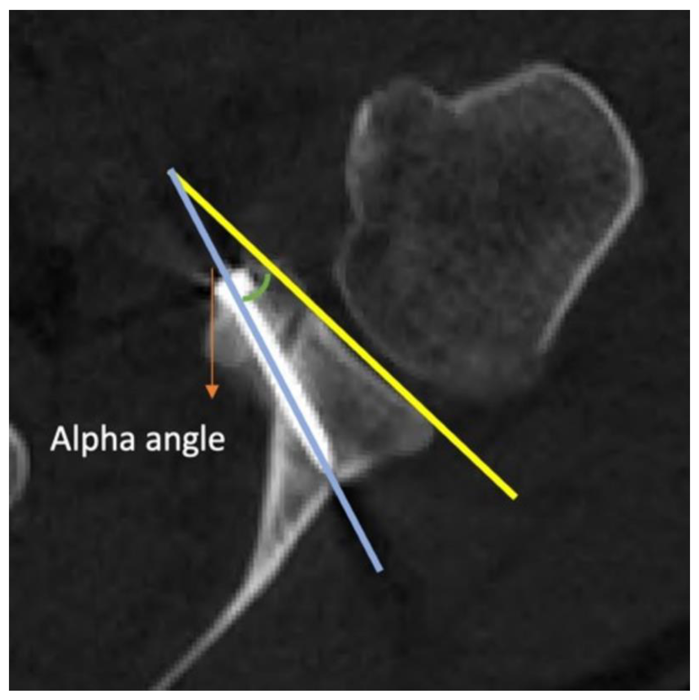
Measurement of Alpha Angle: An axial CT image of the glenohumeral joint is captured. The most prominent subcortical points of the anterior and posterior corners of the glenoid are connected. In the axial section, the angle between this line and the body of the screw is referred to as the alpha angle (
Figure 1).
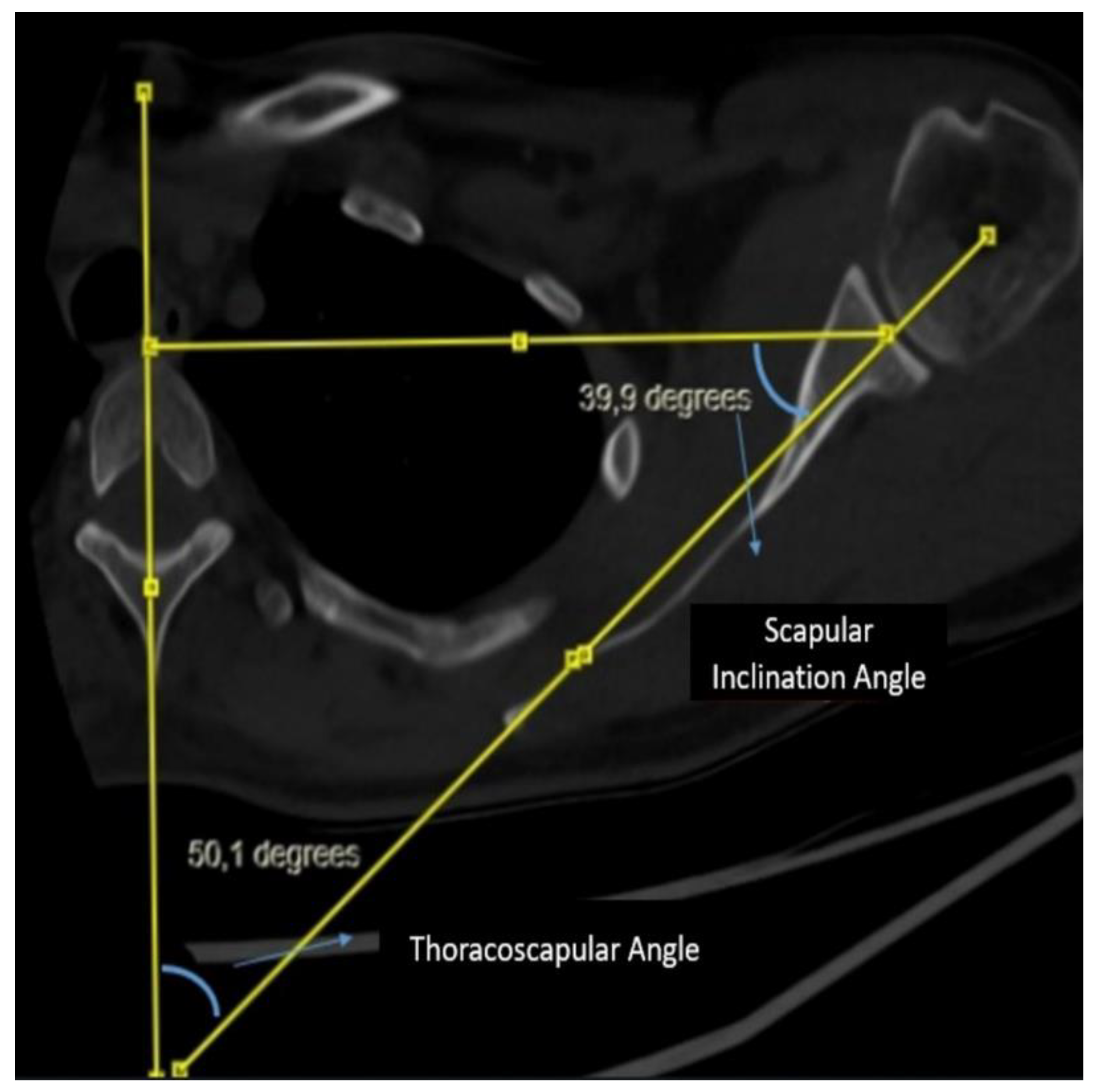
Measurement of Scapular Inclination Angle and Thoracoscapular Angle: A vertical line is drawn from the midline of the corpus and spinous process of the thoracic vertebra at the level of the spine of the scapula. Subsequently, a second line connecting the center of the glenoid to the spine of the scapula is drawn, intersecting the line through the midline of the thoracic vertebra. A triangle is formed by drawing a line from the center of the glenoid perpendicular to the vertical line on the vertebra. The angle between the line connecting the center of the glenoid to the spine of the scapula and the line drawn perpendicular from the center of the glenoid to the vertical line on the vertebra is evaluated as the scapular inclination angle. The angle between the line connecting the center of the glenoid to the spine of the scapula and the vertical line on the midline of the vertebra is evaluated as the thoracoscapular angle. It is noteworthy that the scapular inclination angle and thoracoscapular angle together complement each other to form a total of 90 degrees (
Figure 2).
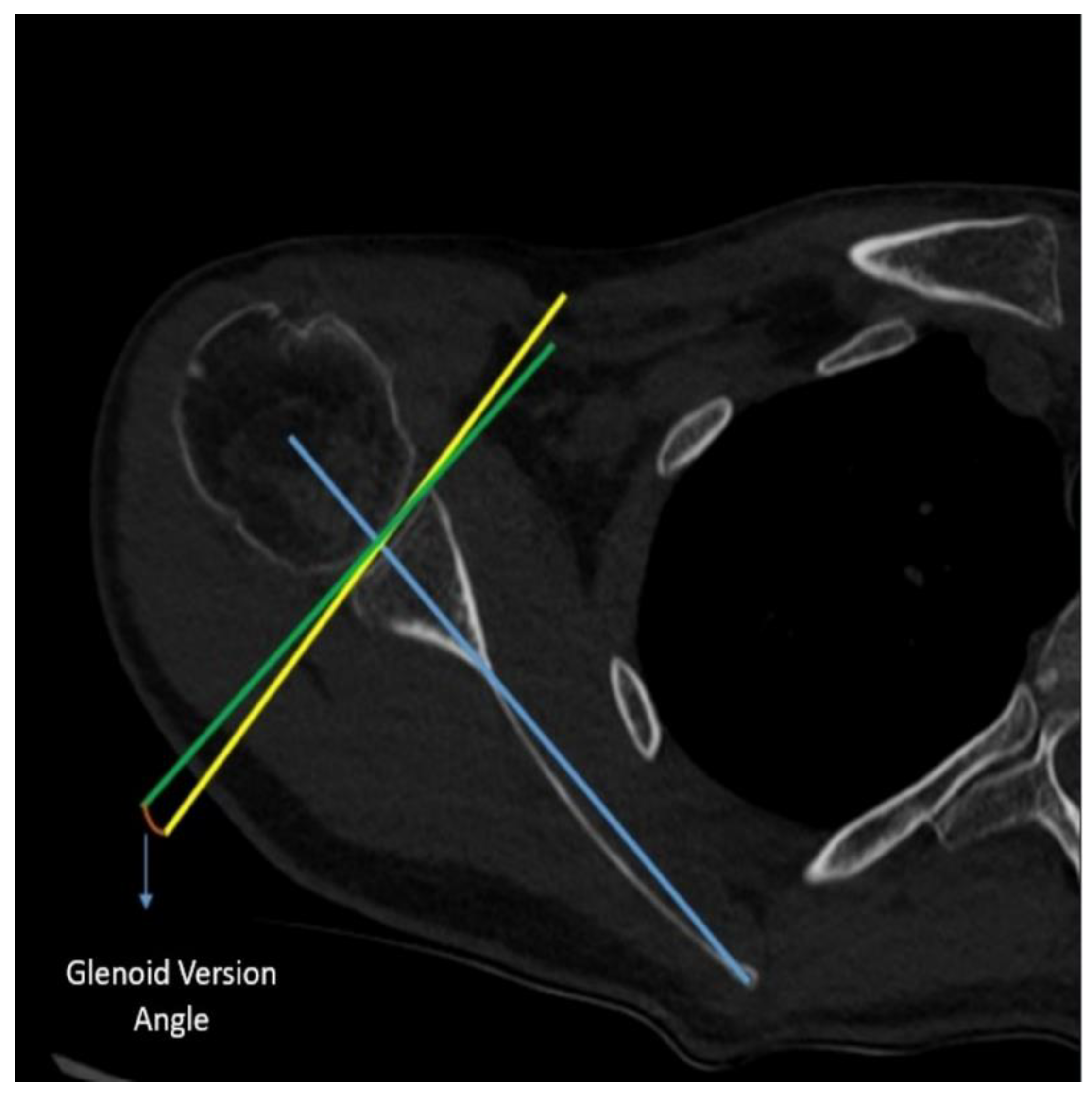
Measurement of Glenoid Version Angle: In the axial CT imaging, a line (referred to as the Friedman line) is drawn from the most medial border of the scapula to the center of the glenoid fossa. Subsequently, a line perpendicular to this scapular axis is drawn through the center of the glenoid. Following that, a line connecting the anterior and posterior rims of the glenoid is drawn. The angle formed between the line drawn perpendicular to the scapular axis and the line connecting the corners of the glenoid is assessed as the glenoid version angle (
Figure 3).
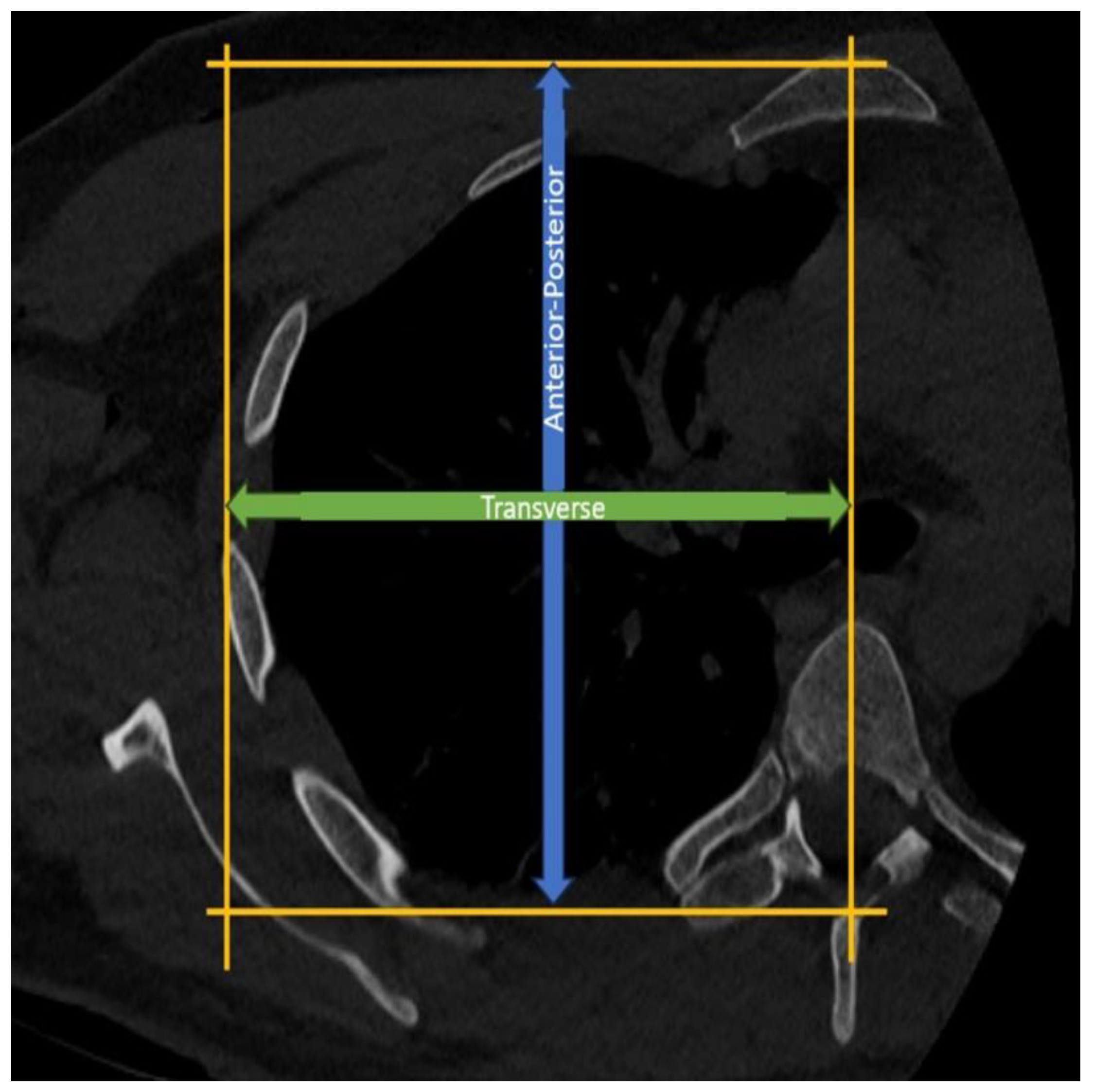
Measurement of Thoracic Anterior-Posterior Diameter: In the axial CT sections at the T4-5 level, the length between a line drawn parallel to the sternum from the anterior surface of the sternum and another line drawn parallel to this line from the apex of the spinous process of the corresponding vertebra is assessed as the thoracic anterior-posterior diameter (
Figure 4).
Measurement of Hemithoracic Transverse Diameter: In the axial CT sections at the T4-5 level, a vertical line is drawn from the midline of the vertebra, connecting a line drawn parallel to the sternum from the anterior surface of the sternum and another line drawn parallel to this line from the lateralmost part of the corresponding rib. The distance between these lines is then assessed as the thoracic transverse radius (
Figure 4).
The measurements of thoracic diameter were conducted based on morphometric standards employed in the literature by disciplines such as thoracic surgery and pulmonology [
5]. The reason for standardizing the method to the T4-5 level was also based on the level of the scapular spine.
4. Statistical Analysis
Descriptive statistical methods were employed to analyze the study data. The normal distribution of the data was assessed using the Shapiro-Wilk test. For data with a normal distribution, Pearson correlation analysis was utilized, whereas Spearman correlation analysis was employed for data without a normal distribution. To determine the threshold value for AP/T, the ROC curve was used. IBM SPSS v28.0 was used for statistical analysis. A p-value of <0.05 was considered statistically significant.
5. Results
Patients who underwent the Latarjet procedure for recurrent shoulder instability between 2022 and 2024 were studied in a single center by a single surgeon. Among the 74 patients, 90% (n=67) were male, and 10% (n=7) were female. Their ages ranged from 16 to 45, with an average age of 26.4±6.4 years. The surgical procedures were performed on the right side in 60% of the cases and on the left side in 40%. A significant relationship was noted between thoracic morphology and the alpha angle in postoperative CT scans. There was a significant correlation between the anterior-posterior/transverse diameter ratio (AP/T) and the alpha angle (r=0.407, p<0.001). A significant correlation was also found between scapular inclination and the alpha angle (r=0.275, p=0.018), as well as between the glenoid version and the alpha angle (r=0.241, p=0.039). As expected, a significant negative correlation was observed between the thoracoscapular angle and the alpha angle (r= -0.288, p=0.013).
An alpha angle value of 15 degrees was set as the threshold, and the patients were divided into two groups. Below the alpha angle of 15, there were 39 patients, while above 15, there were 35 patients. An alpha angle above 15 degrees was determined to be associated with poor outcomes. Descriptive statistical data of the two groups divided according to alpha angle are shown in
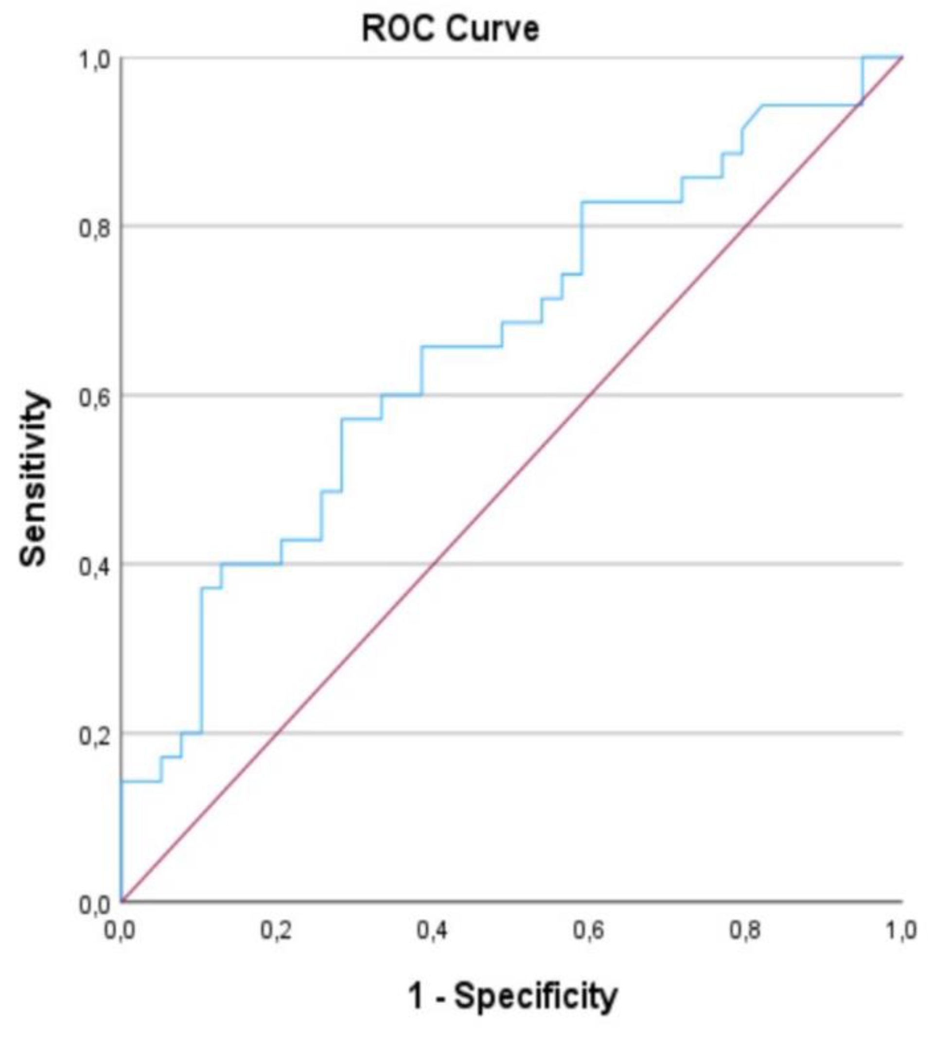
Table 1. According to the ROC curve, with an area under the curve (AUC) value of 0.660, the AP/T threshold value was determined to be “1.2545” with statistical significance at p=0.018, it was found to be correlated with poor outcomes (
Figure 5). These findings suggest that the thoracic wall morphology directly influences the alpha angle.
6. Discussion
To our knowledge, this is the first study to demonstrate a direct relationship between thoracic morphology and the alpha angle. Our findings indicate that the position of the scapula on the thoracic wall directly affects the placement of screws during surgery. In patients with a high AP/T diameter ratio, an increase in the alpha angle was observed, supporting our hypothesis.
Postoperative changes in scapular inclination and thoracoscapular angle following the Latarjet procedure have been shown in the literature [
6]. Cerciello et all observed a decrease in the thoracoscapular angle in the early postoperative period, indicating a more vertical positioning of the scapula on the thoracic wall. Despite the release of the pectoralis minor muscle, they hypothesized that this change was amplified by the coracobrachialis muscle's protraction due to the coracoid block transfer. This reported change, measured at 4 degrees, was observed to normalize and become equal to the unaffected side by the 6th postoperative month. Our study considered chest CT scans taken in the early postoperative period, not accounting for these early changes in scapular inclination. Since the thoracic AP/Transverse diameter measurements were taken from bony landmarks, we believe they are not affected by postoperative changes.
Literature shows that the postoperative alpha angle directly affects the procedure's outcome. Hsu [
4] et all, in a cadaveric biomechanical study, examined the stability of the coracoid block transfer. Their findings revealed no significant difference at angles between 0-15 degrees, but stability markedly decreased under cyclic loadings in samples positioned at a 30-degree alpha angle. Other authors in the literature have also indicated 15 degrees as the stability limit [
7]. In our study, taking 15 degrees as a limit was made in line with these studies.
Some suggestions have been made in the literature to reduce this angle. There are several studies in this context. Barth [
8] et all suggested using a drill guide in addition to the free-hand technique to reduce this angle. Tang [
9] et all demonstrated using a longer drill guide could effectively reduce this angle. We find it useful to use one of these methods in patients with a high AP/T diameter ratio on preoperative CT evaluation.
In our study, a large portion of the sample (90%) consisted of male patients, and differences in male and female thoracic morphology were not taken into account. Existing literature suggests that the AP/Lateral diameter ratio is similar in both sexes [
10]. Despite variations in thoracic volume ratios between the sexes, the proportional similarity of the AP/Lateral diameter is the reason for not assessing this distinction in our study.
Our study has at least 1 limitation. In the study, alpha angle above 15 was associated with poor results. The reason for the limit of the alpha value of 15 is based on biomechanical studies in the literature and should be supported by clinical results. Investigating the effect of alpha angle on clinical outcome may be the subject of another study.
7. Conclusions
Our study clearly demonstrates that thoracic morphology directly affects the alpha angle observed in chest CT scans following the Latarjet procedure. Specifically, in patients with a higher anterior posterior to transverse thoracic diameter ratio (AP/T), there is a proportional increase in the alpha angle. Given that a deviation beyond a certain degree in the alpha angle during the coracoid process transfer directly influences the procedure's outcome, a preoperative understanding of thoracic morphology becomes crucial. In preoperative evaluation, if the AP/T ratio is above 1.25 on the chest CT scan, the surgeon may have difficulty achieving the target alpha angle (≤15). This knowledge can be advantageous for the surgeon in anticipating and addressing intra-operative challenges. Armed with this information, a surgeon can gain an edge in achieving the target alpha angle through the application of appropriate techniques.
List of Abbreviations
| CT |
Computed Tomography |
| PACS |
Picture Archiving and Communication Systems |
| AP/T |
Anterior-Posterior/Transverse Diameter Ratio |
References
- M. LATARJET, “Treatment of recurrent dislocation of the shoulder.,” Lyon Chir., vol. 49, no. 8, pp. 994–997, 1954.
- L. Hovelius, B. Sandström, A. Olofsson, O. Svensson, and H. Rahme, “The effect of capsular repair, bone block healing, and position on the results of the Bristow-Latarjet procedure (study III): long-term follow-up in 319 shoulders.,” J. shoulder Elb. Surg., vol. 21, no. 5, pp. 647–660, May 2012. [CrossRef]
- S. L. Schmid, M. Farshad, S. Catanzaro, and C. Gerber, “The Latarjet procedure for the treatment of recurrence of anterior instability of the shoulder after operative repair: a retrospective case series of forty-nine consecutive patients.,” J. Bone Joint Surg. Am., vol. 94, no. 11, p. e75, Jun. 2012. [CrossRef]
- K. L. Hsu et al., “Biomechanical comparison between various screw fixation angles for Latarjet procedure: a cadaveric biomechanical study,” J. Shoulder Elb. Surg., vol. 31, no. 9, pp. 1947–1956, 2022. [CrossRef]
- N. Sverzellati et al., “Computed Tomography Measurement of Rib Cage Morphometry in Emphysema,” PLoS One, vol. 8, no. 7, 2013. [CrossRef]
- S. Cerciello, T. B. Edwards, G. Cerciello, and G. Walch, “Scapular position after the open Latarjet procedure: Results of a computed tomography scan study,” J. Shoulder Elb. Surg., vol. 24, no. 2, pp. 199–202, 2015. [CrossRef]
- L. Hovelius, B. Sandström, and M. Saebö, “One hundred eighteen Bristow-Latarjet repairs for recurrent anterior dislocation of the shoulder prospectively followed for fifteen years: Study II-the evolution of dislocation arthropathy,” J. Shoulder Elb. Surg., vol. 15, no. 3, pp. 279–289, 2006. [CrossRef]
- J. Barth, A. Boutsiadis, L. Neyton, L. Lafosse, and G. Walch, “Can a drill guide improve the coracoid graft placement during the latarjet procedure?: A prospective comparative study with the freehand technique,” Orthop. J. Sport. Med., vol. 5, no. 10, pp. 1–7, 2017. [CrossRef]
- T. Rebecca Lauren Emma et al., “Optimising Screw Trajectory in the Bristow-Latarjet Procedure: A Simple Technique to Improve the Alpha Angle,” J. Orthop. Surg. Tech., vol. 5, no. 2, pp. 487–493, 2022. [CrossRef]
- F. Bellemare, A. Jeanneret, and J. Couture, “Sex differences in thoracic dimensions and configuration,” Am. J. Respir. Crit. Care Med., vol. 168, no. 3, pp. 305–312, 2003. [CrossRef]
|
Disclaimer/Publisher’s Note: The statements, opinions and data contained in all publications are solely those of the individual author(s) and contributor(s) and not of MDPI and/or the editor(s). MDPI and/or the editor(s) disclaim responsibility for any injury to people or property resulting from any ideas, methods, instructions or products referred to in the content. |
© 2024 by the authors. Licensee MDPI, Basel, Switzerland. This article is an open access article distributed under the terms and conditions of the Creative Commons Attribution (CC BY) license (http://creativecommons.org/licenses/by/4.0/).