Preprint
Review

This version is not peer-reviewed.

Caffeine: A Neuroprotectant and Neurotoxin in Traumatic Brain Injury (TBI)

A peer-reviewed article of this preprint also exists.

Submitted:

10 December 2024

Posted:

11 December 2024

You are already at the latest version

Abstract

Caffeine is a weak, nonselective adenosine receptor antagonist. At low-to-moderate doses, caffeine has a stimulating effect, however, at higher doses, it can act as a depressant. It can act both as a neuroprotectant and a neurotoxin. In experimental Traumatic Brain Injury (TBI), administration of this psychoactive drug has been associated with beneficial or detrimental effects, depending on the dose, model, and timing. In a healthy brain, it can boost alertness and promote wakefulness. On the other hand, its consumption during late adolescence and early adulthood disrupts normal pruning processes in the context of repetitive moderate TBI (mTBI), leading to changes in dendritic spine morphology resulting in neurological and behavioral impairments. Caffeine can potentially reduce TBI-associated intracranial pressure, oxidative stress, lipid peroxidation, cytotoxic edema, inflammation, and apoptosis. It can enhance alertness and reduce mental fatigue, which is critical for the cognitive rehabilitation of TBI patients. It has positive effects on immune cells and recovery post-TBI. It can improve cognitive function by antagonizing adenosine receptors involved in controlling synaptic transmission, synaptic plasticity, and synapse toxicity. On the contrary, studies have also reported caffeine consumers had significantly higher somatic discomfort compared to non-consumers. Therefore, we bring forth this review with the objective of exploring various studies and thoroughly examining the positive and negative role of caffeine in TBI.

Keywords: 
;  ;  ;  ;  ;  ;  

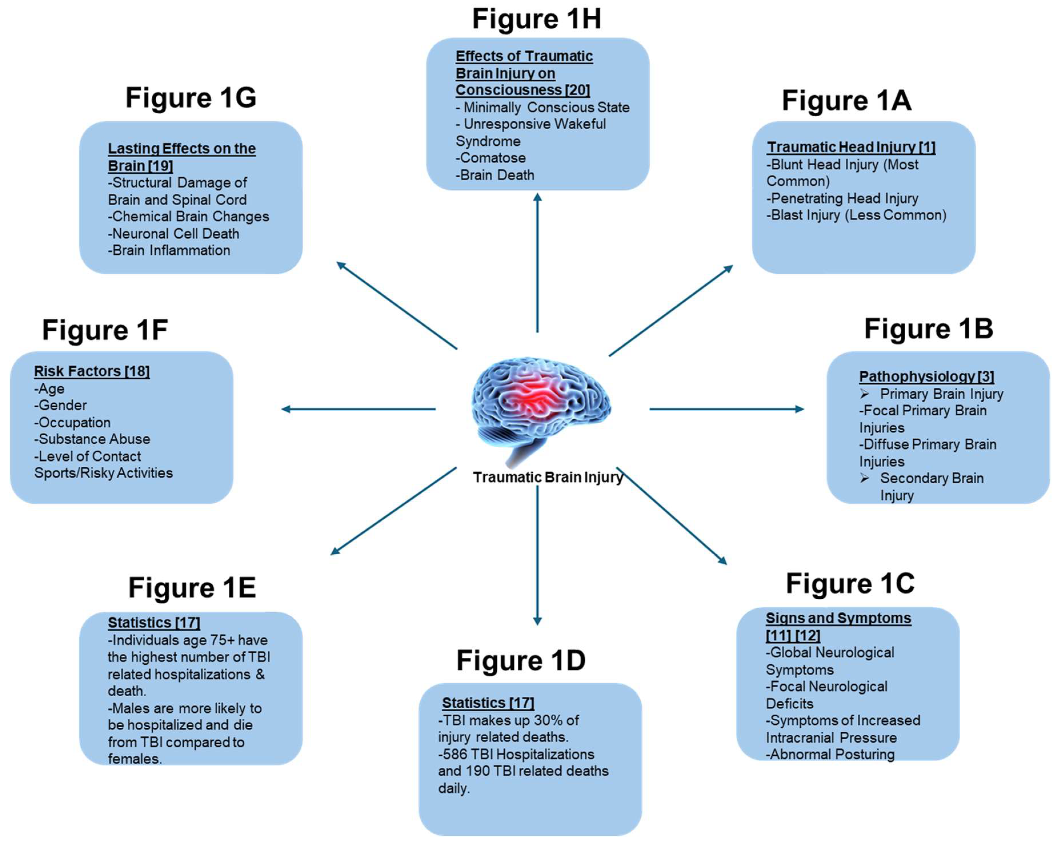
1. Introduction to Traumatic Brain Injury (TBI)

Traumatic Brain Injury (TBI) is being acknowledged more and more as a substantial and growing issue for public health, affecting millions worldwide. TBI is characterized by physiological or structural disruption of cerebral homeostasis, resulting from either penetrating injury or blunt force trauma to the cranium, as summarized in Figure 1-1A[1]. Head injuries can be stratified into three primary categories: blunt head trauma, penetrating cranial injuries, and blast-related neurotrauma.[1]. Blunt head injury represents the predominant etiology of TBI and typically arises from direct blunt force trauma to the cranium, often resulting from falls, motor vehicle collisions, or contact sports such as football.[2]. Penetrating cranial trauma occurs when a foreign object breaches the cranial vault. This can happen due to gunshot wounds, physical assaults, or accidental head injuries. Blast-induced neurotraumas are less common and result from high-pressure blast waves typically seen in military conflict zones due to large-scale explosions.[2].
Pathophysiologically, TBI can be delineated into primary and secondary brain injuries, contingent upon whether the initiating trauma induces a direct or indirect insult to cerebral structures, as described in Figure 1-1B[3]. A primary brain injury is defined as the immediate damage inflicted at the moment of impact.[3]. Primary brain injuries are further stratified into focal and diffuse categories[3]. Focal primary brain injuries encompass subarachnoid hemorrhages, subdural hemorrhages, epidural hemorrhages, intracerebral hemorrhages, cerebral contusions, brain parenchymal lacerations, coup-contrecoup injuries, and intracerebral and intracerebellar hematomas[3]. Subarachnoid hemorrhage, predominantly precipitated by cranial trauma, is classified as a type of intracranial hemorrhage characterized by hemorrhaging within the subarachnoid space, situated between the pia mater and the arachnoid mater of the meninges[4] . This pathological bleeding disrupts cerebrospinal fluid dynamics and can lead to elevated intracranial pressure, cerebral vasospasm, and secondary ischemic injury[4]. Subdural hemorrhage, typically induced by cranial trauma, is categorized as an intracranial hemorrhage involving the accumulation of blood between the arachnoid membrane and the dura mater [5]. This condition arises from the rupture of bridging veins traversing the subdural space, leading to hematoma formation and potential subsequent increases in intracranial pressure, cerebral compression, and herniation syndromes[5]. Epidural hemorrhage, predominantly resulting from head trauma, is classified as a type of intracranial hemorrhage characterized by the accumulation of blood between the dura mater and the inner table of the calvarium[6]. This condition is typically precipitated by a laceration of the middle meningeal artery, leading to the rapid formation of an epidural hematoma[6]. The ensuing mass effect can cause significant intracranial pressure elevation, brain tissue displacement, and potentially life-threatening herniation[6].
Intracerebral hemorrhage, which can be precipitated by traumatic head injury, is defined as a hemorrhagic insult occurring within the brain parenchyma [7]. This pathological condition involves the extravasation of blood into the cerebral tissue, often resulting in focal neurological deficits, increased intracranial pressure, and secondary injury mechanisms, such as edema and ischemia, exacerbating the initial trauma.[7]. Cerebral contusion is characterized as a localized area of brain injury that can range from a minor parenchymal bruise to a severe focal area of necrosis[8]. This pathology involves the disruption of the neural tissue, which may include hemorrhage, edema, and tissue infarction, often resulting in significant functional impairment and secondary complications such as increased intracranial pressure and cerebral edema [8]. Brain parenchymal lacerations are characterized by the physical disruption of brain parenchyma through cuts or tears resulting from blunt or shearing forces [9]. Coup-contrecoup injuries can be delineated into coup and contrecoup injuries[9]. Coup injuries occur at the site of initial impact, whereas contrecoup injuries manifest on the contralateral side of the brain, opposite the point of impact [9]. Intracerebral and intracerebellar hematomas are defined as localized collections of blood within the cerebrum or cerebellum, respectively, leading to elevated intracranial pressure due to resultant swelling [9]. These hematomas typically arise from the rupture of cerebral vessels secondary to TBI [9]. Examples of diffuse primary brain injury encompass concussions, diffuse axonal injury (DAI), and cerebral edema[10]. Concussions are classified as mild TBI characterized by transient neurological dysfunction without discernible focal deficits, typically stemming from rapid acceleration-deceleration forces or direct head trauma [10]. Diffuse axonal injury (DAI), alternatively known as traumatic axonal injury, refers to the biomechanical disruption and shearing of axons within the brain, stemming from traumatic acceleration-deceleration or rotational forces applied to the cranium, often occurring in severe high-impact vehicular collisions[11].This condition results in widespread axonal damage across multiple brain regions, leading to profound neurological impairment and potential long-term cognitive deficits[11,12] .Cerebral edema, medically termed brain swelling, denotes the pathological accumulation of fluid within the brain parenchyma[13].This condition is commonly observed in the initial phases following TBI contributing to increased intracranial pressure and potential secondary brain damage [13]. A secondary brain injury is characterized by multifaceted disturbances in cerebral homeostasis, encompassing alterations in systemic parameters such as blood pressure, oxygenation, and intracranial pressure, which evolve after an initial acute insult to the brain or spinal cord[3]. Examples of secondary brain injury include hypoxic-ischemic encephalopathy, involving cerebral ischemia due to inadequate oxygen supply, and disruption to the blood-brain barrier, exacerbating neuronal vulnerability to systemic toxins and pathogens [3].
Figure 1. It illustrates a comprehensive summary introducing traumatic brain injury (TBI) entailing the following categories: types of head injury which can lead to TBI Figure 1-1A [1], pathophysiology of TBI Figure 1-1B [3], signs and symptoms of TBI Figure 1-1C [11,12], statistics of TBI Figure 1-1D, Figure 1-1E [16], risk factors associated with TBI Figure 1-1F [17], the lasting effect of TBI on the brain Figure 1-1G [18], and the effect of TBI on the consciousness Figure 1-1H [19].
Preprints 142430 g001
The clinical manifestations of TBI exhibit variability by the site, severity, and specific subtype of TBI incurred by the patient [11,12]. General symptoms of TBI can be categorized into subgroups, including global neurological symptoms, focal neurological deficits, signs indicative of heightened intracranial pressure, and abnormal posturing, as summarized in Figure 1-1C [11,12]. Global neurological symptoms encompass headache, amnesia, loss of consciousness, or altered consciousness accompanied by disorientation, confusion, or potentially a lucid interval [11,12]. Focal neurological deficits typically correlate with the specific anatomical region of brain involvement, yielding symptoms such as ataxia, dysmetria, dysarthria, cranial nerve palsies affecting visual and auditory acuity, sensory deficits, and hemiparesis or hemiplegia contingent on injury severity [11,12]. Symptoms indicative of heightened intracranial pressure encompass vertigo, emesis, Cushing’s triad, alterations in affect or behavior, and potential manifestations of cerebral herniation syndromes [11,12]. Symptoms of abnormal posturing are indicative of significant brain injury and typically manifest as either decorticate or decerebrate posturing [11,12]. Decorticate posturing presents with bilateral upper extremity flexion and lower extremity extension, reflecting corticospinal tract dysfunction typically localized above the red nucleus [11,12]. Conversely, decerebrate posturing is characterized by bilateral extension of both upper and lower extremities, suggesting more profound damage involving structures below the red nucleus or brainstem [11,12]. In cases where head injury leads to penetrating trauma, additional clinical signs and symptoms may arise, contingent upon the specific anatomical site affected[14,15]. These may encompass periorbital ecchymosis (raccoon eyes), cerebrospinal fluid (CSF) leakage from the nasal passages (CSF rhinorrhea) or ears (CSF otorrhea), epistaxis, hemotympanum, hematoma formation, and a palpable localized mass suggestive of an expanding lesion [14,15]. These indicators are crucial in the diagnostic evaluation and management of penetrating cranial injuries [14,15].
In the United States, TBI contribute to 30% of all injury-related fatalities, as summarized in Figure 1-1D[16]. According to the Centers for Disease Control and Prevention (2023), approximately 2.8 million individuals sustain TBI annually, a figure comparable to the population of Mississippi [16]. In 2020, there were 214,110 hospitalizations directly attributed to TBI, and in 2021, 69,473 deaths were recorded due to TBI[16]. These statistics equate to an average of 586 TBI-related hospitalizations and 190 TBI-related deaths each day, underscoring the significant public health burden associated with TBI in the United States [16]. Individuals aged 75 and older exhibit the highest incidence of TBI-related hospitalizations and mortality, primarily attributable to increased susceptibility to falls and accidents inherent to this demographic, as summarized in Figure 1-1E [16]. Approximately 32% of all TBI-related hospitalizations and 28% of TBI-related fatalities are reported within this age cohort, highlighting age-associated vulnerabilities and the need for targeted preventive measures and clinical management strategies [16]. Between genders, males exhibit a significantly higher propensity for hospitalization (age-adjusted rate: 79.9 vs. 43.7) and mortality (age-adjusted rate: 28.3 vs. 8.4) due to TBI, with a twofold and threefold greater likelihood, respectively, compared to females [16]. This disparity is attributed to the greater engagement of males in high-risk activities predisposing them to brain injury [16]. Globally, TBI impacts an estimated 69 million individuals annually, a figure roughly equivalent to twice the population of California, underscoring the magnitude of this public health concern worldwide [16].
Numerous risk factors are linked to TBI, encompassing demographic variables such as age and gender, occupational hazards, substance abuse patterns, and engagement in high-contact sports or hazardous activities, as summarized in Figure 1-1F[17]. These factors collectively contribute to the varying incidence and severity of TBI observed across populations, highlighting the multifactorial nature of TBI risk assessment and prevention strategies [17]. In terms of age demographics, epidemiological data consistently indicate that elderly adults and very young children exhibit heightened susceptibility to TBI[16,17], primarily attributable to increased incidences of falls and accidents within these age brackets [17]. Regarding gender disparities, statistical analyses demonstrate a higher incidence of TBI among males compared to females, largely influenced by occupational factors involving physically demanding roles and participation in high-risk physical activities [16,17]. Occupationally, individuals employed in military service, athletics (e.g., football), transportation, or construction sectors exhibit heightened susceptibility to TBI, necessitating rigorous adherence to safety protocols [16,17]. Substance abuse, encompassing both drug and alcohol misuse, further escalates the risk of accidents predisposing individuals to TBI, underscoring the critical importance of preventive measures and intervention strategies aimed at mitigating TBI incidence in high-risk populations [16,17] .Finally, individuals engaged in high-contact sports, activities prone to significant falls, or tasks involving heavy lifting inherently elevate their risk of experiencing TBI [16,17].These pursuits necessitate heightened awareness of injury prevention strategies and comprehensive management protocols to mitigate the incidence and severity of TBI associated with such activities [17,18]
TBI exert profound and enduring impacts on cerebral function, as summarized in Figure 1-1G[18]. Following brain injury, individuals may endure structural alterations within the brain parenchyma or brainstem, encompassing physical trauma to blood vessels, neural tissue, nerves, and the spinal cord, precipitating conditions such as hemorrhage, ischemia, and potentially fatal outcomes [18]. These consequences underscore the critical need for comprehensive neuroprotective strategies and rehabilitative interventions to mitigate long-term neurological sequelae associated with TBI [18]. Another mechanism through which TBI impacts the brain involves biochemical alterations [18]. These changes encompass disturbances in neurotransmitter dynamics, leading to aberrant release of cytotoxic substances and harmful chemical byproducts secondary to the injury [18]. These biochemical cascades contribute significantly to secondary injury mechanisms and neuroinflammatory responses, highlighting their pivotal role in the pathophysiology of TBI and emphasizing the necessity for targeted therapeutic interventions to mitigate detrimental outcomes [18]. TBI exerts additional detrimental effects on the brain through mechanisms such as neuronal cell death induced by hypoxia or direct trauma [18]. Furthermore, TBI precipitates inflammatory responses in the brain parenchyma, exacerbating neuronal damage and fostering a cascade of secondary debilitating symptoms for affected individuals [18]. These inflammatory processes underscore the critical need for targeted anti-inflammatory strategies and neuroprotective therapies to mitigate the long-term consequences of TBI [18].
TBI exert profound effects on consciousness, categorizable into various severity levels, as described in Figure 1-1G[19]. Following mild TBI, the initial altered consciousness state often observed is the minimally conscious state (MCS), characterized by limited responsiveness where individuals demonstrate the ability to inconsistently follow simple commands, respond with affirmative or negative answers, and demonstrate fluctuating awareness of self and environment [19]. These individuals may experience transient periods of unconsciousness but retain the capacity for appropriate responsiveness and demonstrate varying degrees of self-awareness concerning their surroundings [19]. The second altered state of consciousness, often associated with moderate TBI, is characterized by unresponsive wakefulness syndrome (UWS), clinically denoting a condition where individuals exhibit wakefulness without awareness of their environment [19]. It is noteworthy that individuals in this state may exhibit motor movements, vocalizations, or reflexive responses despite their lack of responsiveness to external stimuli [19]. The third altered state of consciousness, typically associated with severe TBI, manifests as a coma, characterized by profound unconsciousness, complete unresponsiveness to external stimuli, and absence of environmental awareness [19]. For these individuals, the duration of a coma can vary significantly, spanning days, weeks, months, or even years [19]. Prognostically, the outcome following coma duration may entail emergence into consciousness, transition into a vegetative state characterized by wakefulness without awareness, or mortality [19].
To assess the level of consciousness following TBI, clinicians utilize the Glasgow Coma Scale (GCS), a standardized tool categorized into several domains[20]. These domains encompass eye-opening responses, verbal responses, and motor responses, providing a quantitative framework for evaluating neurological status and guiding clinical management decisions [20]. Eye-opening is graded according to a four-point scale: one signifies no response, two indicates a response to pain stimuli, three denotes responsiveness to verbal commands, and four signifies spontaneous eye-opening [20]. Verbal response is assessed using a five-point scale: one denotes no response, two indicates incomprehensible sounds, three reflects inappropriate word usage, four indicates confused responses, and five signifies orientation to person, place, and time [20]. The motor response is assessed using a six-point scale: one signifies no response, two indicates decerebrate posturing characterized by extension and rigidity of extremities, three denotes decorticate posturing marked by flexion of upper extremities and extension of lower extremities, four indicates withdrawal from painful stimuli, five signifies localization of pain, and six denotes the ability to follow complex instructions [20]. The Glasgow Coma Scale (GCS) score is derived by summing the individual scores from each category, with a minimum score of three indicating severe coma or brain death, and a maximum score of fifteen signifying full consciousness[21]. TBI severity correlates closely with GCS scores, reflecting the extent of neurological impairment and aiding in clinical stratification and prognostication of patient outcomes [21]. The correlation involves categorizing TBI based on their Glasgow Coma Scale (GCS) scores: mild TBI typically ranges from thirteen to fifteen, moderate TBI ranges from nine to twelve, and severe TBI manifests with a GCS score of eight or less, necessitating immediate consideration for intubation due to compromised airway protection and potential respiratory compromise[21]. This classification system aids in clinical decision-making and prioritization of therapeutic interventions based on the severity of neurological impairment following TBI [21]. Finally, the fourth altered state of consciousness is brain death, defined as the irreversible cessation of cerebral function, confirmed by the absence of measurable brain activity over an extended period and validated through diagnostic tests demonstrating diminished cerebral blood flow [19]. Hence, in this review, we present a literature search that provides insights into both the positive and negative roles of caffeine in traumatic brain injury (TBI).

2. Introduction to Caffeine

Caffeine (C8H10N4O2), also known as 1,3,7-trimethyl xanthine, is a naturally occurring alkaloid that belongs to the methylxanthine class[22]. It is one of the most used psychoactive stimulants worldwide and can be found in a variety of foods and beverages such as coffee, tea, cacao beans, energy drinks, and soda, as well as some over-the-counter medications[23]. It is found to contain stimulatory, antioxidant, anti-inflammatory, and pain management properties [24]. Caffeine has been used since the 17th century, but caffeine was discovered by Ferdinand Runge when he first isolated caffeine from coffee beans[25].

2.1. Mechanism of Action

Caffeine works by stimulating the central nervous system through multiple mechanisms. One mechanism caffeine works by is acting as A1 and A2A adenosine receptor antagonists. Adenosine and adenosine receptors regulate neurotransmitter release which plays a role in the regulation of sleep, arousal, memory, cognition, and learning[24]. Another mechanism includes caffeine’s ability to mobilize calcium within cells, which influences neurotransmitter release in the nervous system through the endoplasmic reticulum and plasma membrane, with effects varying based on the caffeine concentration [24]. Lastly, caffeine can work by inhibiting phosphodiesterase which leads to increased cAMP and promotes the release of neurotransmitters like dopamine and adrenaline, which affect mood, memory, alertness, and cognitive function [24].

2.2. Route of Administration

FDA-approved indications for caffeine include IV caffeine to treat apnea of maturity and oral caffeine for restoring mental alertness or wakefulness [23]. For apnea, IV caffeine citrate is used which contains 20mg of caffeine citrate per milliliter [26]. Orally, caffeine can be ingested in food and beverages as well as in medication forms. A moderate daily caffeine intake of 300-400mg is considered to not cause harm to healthy individuals [27]. Caffeine has also been used off-label for the treatment of migraines as well as in athletes as a performance enhancer [23]. Since caffeine produces a vasoconstrictive effect which leads to decreased cerebral blood flow, it may help in acute settings of headaches [27]. Migraine sufferers should limit their caffeine intake to 200mg to avoid risking worsening of symptoms [27].

2.3. Metabolism of Caffeine

Once orally ingested, caffeine is rapidly absorbed from the GI tract and reaches its peak plasma concentration at 30-60 minutes[28]. Due to its lipophilic and hydrophilic nature, caffeine crosses cell membranes including the blood-brain barrier, which gives rise to its CNS stimulant effects [28]. Caffeine metabolism in humans is done through the demethylation reaction of caffeine (1,3,7-trimethyl xanthine) to paraxanthine (1,7-dimethylxanthine), theobromine (1-demethylated product), and theophylline (7-demethylated product)[29]. This reaction takes place in the liver and is facilitated by the activity of CYP1A2, a cytochrome p450 isoform.[8] The half-life and clearance of caffeine are dependent on many outside factors including environment, genetics, age, medications, smoking status, etc. [28]. In healthy adults, the half-life of caffeine ranges from about 3-5 hours [28]. Caffeine increases the bioavailability of some drugs such as aspirin, ergotamine, and levodopa by decreasing gastric pH, which helps accelerate the absorption rate of the drugs mentioned[30].

2.4. Caffeine Toxicity

When caffeine is consumed at 3-5mg/kg it has been associated with a lower risk of Alzheimer’s disease and Parkinson’s Disease[31]. It has been shown that caffeine can protect cells by reducing damage caused by oxidative stress and reactive oxygen species, giving it its antioxidant properties[32]. Studies in rats have demonstrated that caffeine can reverse oxidative stress and inflammation caused by certain compounds[33]. In TBI a major concern is neuroinflammation and toxicity, so by using nano coffee particles, significant improvements in behavior, proteins, and dendritic cells can be seen during treatment for TBI [28].

3. Numerous Effects of Caffeine in TBI

3.1. Caffeine as a Neuroprotectant

Caffeine is a substance consumed globally and may have neuroprotective effects against different brain injuries, including neurotrauma[34]. Caffeine (1,3,7-trimethyl xanthine) is a broad-spectrum antagonist of adenosine receptors[35]. It may provide neuroprotection through long-term upregulation of adenosine A1 receptors or acute inhibition of A2a receptors[36]. Research on adenosine receptor knockout (KO) mice indicates that many of caffeine’s acute effects might be mediated by the adenosine A2a receptor (A2AR)[37]. Blocking the A2a receptor reduces brain damage after a TBI (TBI)[38]. Under normal circumstances, adenosine levels are regulated for receptor interaction, but during severe metabolic stress like TBI, adenosine surges and excessively stimulates A1 and A2A receptors[39]. Leading to prolonged apnea and suppressed respiratory and cardiovascular function[40].
Administering caffeine after injury can entirely prevent fatal apnea associated with trauma[28]. Caffeine has also been demonstrated to play a significant role in protecting the brain against various types of damage, including neurotoxicity, seizures, and cognitive dysfunction, through its antioxidant mechanisms [38]. A single acute dose of caffeine administered after the injury can prevent lethal apnea, regardless of prior chronic caffeine exposure [38]. In an experiment done by, Lusardi et al 2012, involving the use of caffeine in adenosine receptor blockade and knockout mice, they found that post-injury caffeine treatment quickly restored spontaneous breathing [38]. In another study done by Lusardi et al 2020, administering a single acute bolus of caffeine (25 mg/kg) to rats 10 seconds after a severe TBI nearly completely prevented lethal apnea, in conditions where 40% of the animals would have otherwise succumbed [38].
While caffeine is a widely used psychoactive substance, and its chronic consumption is known to impact adenosine receptor expression, long-term caffeine consumption is linked to positive outcomes following TBI [38]. The favorable effects of chronic caffeine consumption might be due to the upregulation of adenosine A1 receptor expression[41]. Activation of A1 receptors plays a role in neuroprotection by mediating neuronal excitability blood vessel dilation, decrease in heart rate, and sleep induction[42]. In an experiment conducted by Ning et al 2019 where they studied the contribution of different methods of caffeine application on outcomes in whole body blast injury models (WBBI) in mice, it was found that the chronic caffeine group showed alleviated neurological deficits and higher locomotor activity 24 hours post-trauma compared to the acute caffeine and water groups [38]. In another preclinical study administering caffeine to mice for three weeks significantly protected against neuro damage from a cortical contusion injury, whereas a single dose of caffeine in the same model had no effect [38].
Caffeine administration has been shown to have beneficial effects on the central nervous system against various cerebral insults, including acute brain damage such as TBI, in both experimental animals and clinical patients [38]. Caffeine administered before injury is likely to impact the entire brain, while caffeine perfusion into the injured brain may be concentrated in the most severely affected areas [38]. In a rat model study treatment with caffeine, a combination of caffeine (3.3 mg/kg) and ethanol (0.65 g/kg), administered 15 minutes post-TBI, improved working memory and reduced contusion volume[43]. The dose-response results after diffuse injury show that a 25 mg/kg caffeine treatment in rats significantly reduces acute mortality by 75% without impairing motor function, indicating that quality of life can be maintained with caffeine intervention following diffuse injury [38]. Clinical studies found that there is a significant association between a CSF caffeine concentration of at least 1 mmol/L (194 ng/mL) and a favorable 6-month outcome [38].
Another clinical study found that giving 2.5 mg/kg/day of caffeine for up to a month after injury improved consciousness and performance in children and adolescents with moderate TBI[44]. In a multicenter prospective study on patients with TBI and intracranial injury, a significant association between serum caffeine concentrations of 0.01 to 1.66 µg/mL and favorable functional recovery at the 6-month follow-up was observed[45]. Data suggest that injecting coffee extracts after a TBI may effectively reduce cognitive deficits and improve overall neuronal health and recovery[46]. Caffeine’s stability at room temperature makes it suitable for emergency kits, and intramuscular delivery could be a viable administration method in human trauma settings [38].
Figure 2. Effect of caffeine on the adenosine receptors to mediate neuroprotection in TBI.
Preprints 142430 g002

Caffeine as a Neurotoxin

Although caffeine can be neuroprotective, a study investigating the influence of caffeine on brain function, swelling, and permeability found that it significantly worsens neurological deficits and increases mortality in rats after induced head injury [38]. The harmful effects of immediate caffeine consumption are likely due to the blockade of A1 receptors, supported by evidence showing that TBI in mice lacking A1 receptors resulted in fatal status epilepticus [38]. In an experiment conducted by Ning et al 2019 where they studied the contribution of different methods of caffeine application on outcomes in whole-body blast injury models (WBBI) in mice, they found that mortality was significantly higher in mice with acute and chronic caffeine treatment than mice in the caffeine withdrawal and water groups [38]. In vitro studies show that stretch injury of neurons reduces caffeine’s impact on calcium-induced calcium release, implying that pre-injury caffeine in caffeine-naïve neurons adds to the injury [38]. In a 2020 study by Christensen et al., it was found that caffeine consumption during late adolescence and early adulthood disrupts normal pruning processes in the context of repetitive moderate TBI (mTBI), leading to changes in dendritic spine morphology resulting in neurological and behavioral impairments[47]. In an animal model study conducted by Al Moutaery et al 2003, caffeine-treated rats exhibited more severe hemorrhage and neuronal degeneration in the injured hemisphere compared to rats in the injury-alone group [38]. This study’s results showed that caffeine significantly worsens neurological deficits and increases mortality in animals following head injury [38].

3.2. Effects of Caffeine on a Healthy Brain and an Injured Brain

3.2.1. Healthy Brain

Caffeine enhances cognitive performance, boosts alertness, and promotes wakefulness[48]. A 75 mg dose of caffeine can improve reaction time and enhance visual and sustained attention, especially during long, demanding tasks[49]. Caffeine acts as a potent inhibitor of A1 and A2A adenosine receptors in the brain, particularly at low concentrations equivalent to those achieved after drinking a single cup of coffee (a few mmol/L)[48]. This inhibition leads to the release of predominantly excitatory neurotransmitters, which are more strongly affected by adenosine compared to inhibitory neurotransmitters[50]. A study done by Yang, J.-N et al, 2009 using knockout mice indicates that blocking A2A receptors with caffeine affects sleep and motor activity while blocking both A1 and A2A receptors influences heart rate, body temperature, and oxygen consumption[51].
In a crossover study done by Powers, 2015, participants including men and women, were evaluated across three conditions: supplementation (5.5 g of Jacked 3D, including caffeine and 1,3-dimethylamylamine), placebo, and control, with each condition separated by one week[52]. Ingesting a stimulant 30 minutes before testing led to enhancements in memory, visual processing speed, and reaction time [52]. However, consumption of caffeine at higher doses can lead to anxiety[53]. Animal models of anxiety have validated caffeine’s tendency to induce anxiety [48]. Two human studies have reported that caffeine consumption increases self-ratings of anxiety[54]. Additionally, research has found that a variant of the ADORA2A gene influences caffeine-induced anxiety. Regular caffeine consumption can lead to tolerance to its anxiety-inducing effects through central mechanisms [48].
Caffeine influences sleep by delaying sleep onset, reducing total sleep time, and increasing light sleep phases[55]. This occurs with doses as low as 100 mg [48]. These effects persist when caffeine is consumed in the morning, leading to overall less sleep and a longer time to enter stage 2 sleep [48]. Habitual caffeine users may not experience these sleep disruptions as severely[55]. Individual sensitivity to caffeine’s effects on sleep varies significantly[56]. This variability may be influenced by genetic factors such as polymorphisms in the CYP1A2 enzyme and more crucially, variations in the brain adenosine A2A receptor (ADORA2A) [56]. Sensitive individuals may experience nearly twice the incidence of insomnia when consuming caffeine compared to those who do not[57].

3.2.2. Injured Brain

Caffeine, the most commonly consumed psychoactive substance, can have both positive and negative effects following TBI[36]. Caffeine’s stimulant properties can help alleviate fatigue, hypersomnia, and reduced energy levels commonly experienced by TBI[58,59]. An animal model by Lusardi et al, 2020 of severe TBI revealed that caffeine treatment prevents lethal apnea and decreases epileptiform EEG activity, while also demonstrating no adverse effects on neuromuscular behavior or histological outcomes [39].
Stimulants such as caffeine improve cognitive performance, auditory vigilance, and reaction times[60]. However, they can hinder the brain’s ability to adapt and disrupt normal sleep, two factors that are critical for recovering from TBI[61]. An animal study done by Yamakawa et al, 2017, examined the impact of adolescent caffeine use on recovery from repetitive mild TBI (RmTBI) and post-traumatic symptoms[62]. In male rats, combining caffeine with RmTBI exacerbated negative outcomes in tests like open field and forced swimming, compared to either alone [62]. Females showed similar disruption from RmTBI and caffeine exposure in tests including open field [62].
Li et al, 2008 performed a study aimed to assess the impact of acute and chronic caffeine treatment on TBI using the weight-dropped model in mice[63]. It revealed that chronic caffeine treatment has been observed to decrease cell apoptosis in the brain following TBI [62]. A rat study done by Lusardi et al, 2020 found that a single intraperitoneal dose of caffeine administered immediately after severe TBI restored normal breathing patterns and prevented lethal injury-induced apnea[40]. Researchers investigated the potential applicability of caffeine treatments in humans with varying histories of caffeine consumption, testing acute administration post-injury with different doses and patterns [62]. They found that administering a single dose of caffeine after TBI could potentially prevent fatal outcomes in various scenarios [62].

3.3. Effect of Caffeine on Oxidative Stress and Mortality in TBI

Caffeine is a widely used stimulant[64]. Using mice models, the stimulatory effect of caffeine was found to be primarily by the adenosine1 and adenosine2A receptors[65] . Adenosine receptors are of interest in TBI because they are involved in various brain injury pathways [66,67]. Using mice models, the adenosine2A receptor has been linked to the neuroinflammation seen in TBI[68]. Li et al. found that the inactivation of the adenosine2A receptor in mice models protects against acute TBI by decreasing glutamate levels[69]. Caffeine has been shown to have a neuroprotective effect on Parkinson’s Disease pathways in mouse models[70,71]. Further studies using mice models, found benefits of chronic caffeine treatment, rather than acute treatment, before TBI through regulating glutamate release and inflammatory cytokine production[63]. One study using rat models found caffeine given 12 - 18 hours later after TBI was able to reduce intracranial pressure[72]. Early animal studies found that adenosine antagonist was shown to decrease the extent of brain damage in hypoxic-ischemic brain injuries[73]. A later study using rat models found similar results where caffeine pre-treatment before hypoxic-ischemic brain injury decreased brain injury and improved EEG activity [74]. Caffeine has also been shown to decrease neuronal apoptosis in hypoxic-ischemic brain injuries in rat models[74]. A different study by Washington et al. found chronic caffeine treatment was associated with decreased neuronal cell death in the hippocampus following TBI[75]. In a case study using two adolescent patients, one with acquired brain injury and another who had a TBI, caffeine was shown to be a useful neuro-stimulant that improved the patient’s cognitive impairments[76]. One study examined the effects of caffeine on GCS and GOSE scores in children and adolescent patients with moderate brain trauma and found that it improved consciousness and performance in these patients[44]. Blocking of the adenosine2A receptor with caffeine or through gene knockout was shown to improve cognitive function in TBI through inhibition of tau hyperphosphorylation in mice models[77]. Yamakawa et al. observed behavioral and recovery differences between genders in rat models following repetitive mild traumatic brain injuries (TBI) when caffeine was administered [62]. Everson et al. found caffeine caused changes to microstructure regions of gray and white matter regions following mild TBI in rat models [59]. A 2023 review article found through reviewing clinical, experimental, and epidemiological studies, there were benefits of treating patients with Parkinson’s Disease and TBI with caffeine specifically through targeting the adenosine2A receptor[78].
Knockout of the adenosineA1 receptor in mice models was shown to decrease post-traumatic seizures and status epileptics after TBI[79]. Additional studies using mice models showed that blocking the adenosineA1 receptor after TBI leads to worsened neuroinflammation [80]. The effects of caffeine on mortality in traumatic brain injury (TBI) show mixed results. Mountaery et al. discovered increased mortality and microscopic brain injury in mouse models that were pre-treated with caffeine before experiencing a concussive head injury[35]. Ning et al. found mixed results of caffeine on the morality of blast-induced TBI in mice models[34] . Another study examining blast-induced TBI in mice models found that caffeine treatment showed cognitive memory improvements [41]. Lusardi et al. found that treatment of mice with caffeine one minute after TBI decreased mortality and did not alter motor function in the recovery period[40]. A similar study found that an acute dose of caffeine given 10 seconds after TBI decreased mortality and morbidity in mice models[40]. Sanjakdar et al. discovered that both acute and chronic caffeine use before a penetrating ballistic-type injury provided neuroprotective and motor benefits in rat models[81]. One multi-drug randomized control trial study using neonate rat models with hypoxic-ischemic brain injury reported that caffeine did not have any adverse effects like death and had high neuroprotection compared to other drugs tested[82]. Sachse et al. found that patients who had increased CSF concentration of caffeine with TBI, specifically the metabolites theobromine and paraxanthine, had more favorable 6-month outcomes [36]. A prospective cohort study by Yoon et al. found that patients with TBI who had a serum caffeine concentration between 0.01 to 1.66 μg/mL had better recovery after 6 months of injury compared to the no-caffeine group [45].
A human research clinical trial found that one complication of TBI is acute lung injury (TBI-ALI) is associated with high mortality [84]. Wei et al. using a mice model found that inhibition of the adenosine2A after TBI decreased the severity of TBI-ALI but further studies are needed to better understand the role between adenosine2A, glutamate levels, and TBI-ABI[85]. An early study by Strong et al. using rat models found that a combination of low-dose ethanol with caffeine right before or 2 hours after brain ischemia had protective properties for the brain [43]. A later study by Dash et al. found that caffeine with ethanol was able to decrease cognitive deficit and cortical tissue loss in rat models [43]. A dosage of 8mg/kg caffeine and 0.4g/kg were associated with neuroprotective properties in human trials with ischemic stroke [86]. Kontos et al. are one of the first groups to examine the pathophysiology of oxygen radicals in TBI using animal models [87]. Another clinical study reported the potential role adenosine has on cerebral blood flow and oxidative metabolism [88]. An early study using a rat model found evidence suggesting that caffeine made brain injury worse through a free radical damage mechanism [74]. In a study blockage of the adenosine2A receptor rather than the adenosine1 receptor was shown to prevent mitochondrial damage by not inhibiting the release of guanosine, a neuroprotective molecule[89]. White tea, which contains caffeine, was shown to have neuroprotective effects in striatal cell cultures against oxidative stress cell death[90]. Caffeine in rat models has been shown to decrease oxidative stress and specifically decrease lipid peroxidation in the hippocampus[91]. Rabbit models found that caffeine could protect against oxidative stress and Alzheimer’s dementia-like pathology[92]. One study found positive neuronal changes when treating mice with nano coffee injections after a TBI[46].
Various caffeine-associated preclinical and clinical findings for this section are shown in Table 1 of this paper.

1.1. Effect of Caffeine on Behavioral, Cognitive, and Motor outcomes in TBI

Caffeine primarily affects the central nervous system, including the brain[39] . Research has shown the myriad of effects caffeine has on the brain. Caffeine works by blocking adenosine receptors [39] . Adenosine and adenosine receptors regulate the release of neurotransmitters and also play an important role in the regulation of sleep, arousal, cognition, memory, and learning[42]. Caffeine binds to adenosine receptors, which does not allow for the binding of adenosine, particularly the A1 And A2A receptors, indirectly affecting the release of dopamine, serotonin, glutamate, and other neurotransmitters [39,93]. These neurotransmitters have been shown to alter mood, memory, alertness, motor controls, and cognitive function[94]. Caffeine’s ability to block adenosine receptors and modulate neurotransmitter release can have potential neuroprotective effects, which might be beneficial in the context of TBI. TBI is associated with a large increase of adenosine in the brain[40]. Adenosine has a dual role in the brain, acting as a neuroprotective agent under normal conditions but potentially contributing to neurodegeneration under pathological conditions such as TBI. By blocking adenosine receptors, caffeine may reduce the negative effects of excessive adenosine signaling following TBI, potentially mitigating neuronal damage. The effects of caffeine on TBI are vast, but here we will discuss the cognitive, motor, and behavioral outcomes of TBI.
Cognitive deficits, particularly in attention and alertness, are prevalent in TBI patients. Studies have also shown that caffeine can enhance attention and reduce mental fatigue, which are critical for the cognitive rehabilitation of TBI patients. To test cognitive performance, a 2019 animal study evaluated the cognitive performance of mice with penetrating ballistic-like brain injuries testing acute and chronic caffeine exposure. This study used a Morris water maze task, which tested for the retention of a platform location after its removal, post-injury. There were observed cognitive deficits in all injury groups[81]. Acute caffeine dosing did not alter the cognitive ability in the task, however chronic caffeine exposure significantly increased the delay in performing the task at all injury levels [81]. Therefore, caffeine pretreatment worsened cognitive outcomes. A 2020 human study, focused on the effect of caffeine on adolescents and young adults in TBI. Adolescent and young adult age groups exhibit the highest incidence of TBI[47]. Mild TBI and caffeine consumption are prevalent in these age groups. During young adulthood caffeine consumption and repetitive mild TBI induced the greatest impairments in males on cognitive tasks [47].
Motor impairments, including reduced coordination and strength, are common in TBI patients. As mentioned earlier, caffeine influences neurotransmitter systems involved in motor control, such as dopamine and glutamate. Adenosine A1 and A2A receptors form functional heteromers with dopamine D1 and D2 receptors. In the striatopallidal enkephalinergic neurons, D2 and A2A subtype receptors for dopamine and adenosine form functional heteromers[95]. These interactions between A2A and D2 receptors modulate the function of enkephalinergic neurons such that blocking adenosine A2A receptors with caffeine directly enhances the excitatory activity of D2 receptors, thereby increasing psychomotor activity in animals[96]. It has been shown that at low-to-moderate doses, caffeine has a stimulating effect, however, at higher doses it has a depressive effect[97,98]. In a 2021 animal study, twenty-four adult male mice were given caffeine in low doses, caffeine in high doses, and water control daily for eight weeks. Then, motor function was assessed by open field, pole, and inverted square grid tests. In the high-dose group, there was an increase in location, but not muscular strength. In both low-dose and high-dose groups, cell density, dendrite length in the sensorimotor cortex, and dendritic arborization increased. Overall, prolonged caffeine improved motor function by increasing the number of neurons and advancing the growth of dendrites[99]. Specific to TBI, an animal 2019 study, previously mentioned, assessed the effect of caffeine on motor outcomes on penetrating ballistic-like brain injury. Rats were randomly assigned to pretreatment groups, acute and chronic caffeine pretreatments. Motor coordination was evaluated using a rotarod task. The animals were tested 7 to 10 days post-injury. The average latency decreased in all the animals, regardless of treatment, compared to acute caffeine treatment. Even though acute caffeine treatment did not show a significant decrease, chronic caffeine treatments significantly reduced the average delay. Thus, the results suggested that chronic caffeine composition before brain injury improved motor performance after brain injury [81]. An animal 2020 study, demonstrated the effects of caffeine treatment pre-injury and post-injury. Regardless of pre-injury caffeine exposure, administering an acute dose of caffeine immediately after the injury dose did not have detrimental effects on motor performance following sublethal injuries. In conclusion, contrary to the 2019 study, pre-exposure to caffeine did not have a major impact. Additionally, chronic caffeine treatment after injury impairs motor function recovery, whereas caffeine withdrawal does not[40].
Figure 3. The Effects of Acute and Chronic Caffeine on Cognitive, Motor and Behavioral Outcomes in TBI.
Preprints 142430 g003
Behavioral changes, including depression and apathy, are frequently observed in TBI patients. Caffeine’s impact on serotonin and dopamine levels can enhance mood and motivation, potentially alleviating depressive symptoms. As mentioned before, adolescents and children are at the highest risk for mild TBI and repetitive mild TBI, which may lead to the development of long-term psychological and neurological deficits. Longitudinal measurements of patients who have experienced repetitive mild TBI showed poor executive function, depression scores, and cognitive changes that were related to the number of injuries received[100]. In a 2017 animal study, male and female rats were given either caffeinated or non-caffeinated drinking water and were then randomly assigned to receive three mild brain injuries. Behavioral outcomes were assessed using a test battery for post-concussion syndrome (PCS) symptoms, including balance, motor coordination, anxiety, short-term working memory, and depressive-like behaviors. In the open field test, non-caffeinated males increased the distance traveled after repetitive mild TBI, while chronic caffeine exposure reduced it[62]. Non-caffeinated females showed reduced distance after repetitive mild TBI, with no change in the caffeine group [62]. Short-term working memory was significantly affected by sex, injury, and caffeine. Caffeine exacerbated memory deficits in males but mitigated them in females. Rats exposed to repetitive mild TBI or caffeine had increased immobility in the forced swim task, with the highest immobility in caffeine-exposed males with repetitive mild TBI. Poor behavioral regulation and performance on executive function tasks have been reported in adolescent populations with high caffeine consumption rates[101]. The behavioral results from this study suggest that adolescence may be a developmental period during which caffeine exposure is particularly detrimental to recovery.

3.4. Effect of Caffeine on the Immune System in TBI

TBI has a high incidence of mortality and morbidity[39]. Caffeine has been found to possess neuroprotective benefits in degenerative neurological disorders[45]. Caffeine is a widely consumed stimulant[102]. It affects immune cells such as lymphocytes [102], natural killer cells[103], mesenchymal stem cells, and neutrophils[104]. Caffeine also has been noted to play a role in TBI and works as an antagonist on the A2A receptor, potentially aiding recovery [102]. In this review, we will look at the effect of caffeine on the pathophysiology of an injured brain, its effects on immune cells as well as dendritic changes in TBI. Caffeine exerts neuroprotective effects primarily through its antagonism of adenosine A2A receptors. This action helps reduce neuroinflammation and oxidative stress, both critical factors in neurodegenerative diseases [102]. A cohort study looked at serum caffeine concentration in TBI patients who presented to the ED [45]. Caffeine levels within 4 hours of injury were analyzed [45]. Patients with established caffeine levels were categorized into groups; low (0.01-0.58 μg/mL), intermediate (0.59-1.66 μg/mL), and high (1.67-10.00 μg/mL) [45]. Recovery function was looked at over 6 months [45]. A regression analysis was performed, the low- and intermediate-caffeine groups corresponded to a higher likelihood of 6-month functional recovery compared with the no-caffeine group [45]. Caffeine’s antioxidative properties play a significant role in protecting neuronal cells from damage due to reactive oxygen species (ROS). It also modulates inflammatory responses, further contributing to its neuroprotective effects [102] . By inhibiting inflammatory pathways, caffeine lowers the production of pro-inflammatory cytokines [62]. It promotes synaptic plasticity and neurogenesis, aiding in cognitive recovery [81,102]. Adult female albino mice were used to investigate the impact of caffeine on immune cells and tumor growth [102]. Caffeine reduced the expression of PD1 on cytotoxic T lymphocytes, enhancing their ability to target tumor cells [102]. Mice treated with caffeine showed a significant decrease in tumor size, indicating enhanced anti-tumor immunity [102]. Caffeine also affects natural killer cells. A study looked at fourteen male cyclists who ingested either 0, 2, or 6 mg/kg of caffeine before engaging in prolonged cycling. Blood samples were collected to assess the activation of natural killer (NK) cells in response to antigen stimulation post-exercise [103]. Results showed that both low (2 mg/kg) and high (6 mg/kg) doses of caffeine significantly enhanced NK cell activation compared to the placebo [103]. No significant differences were observed between the two caffeine doses, suggesting that even a low dose is effective in boosting immune function during prolonged physical activity [103].
Another study analyzed the effects of caffeine on rats during development. It focused on how caffeine affects dendritic spine density in the prefrontal cortex and hippocampus and examined the behavioral and cognitive outcomes post-TBI[47]. Caffeine consumption during development resulted in altered spine density in the prefrontal cortex and hippocampus of rats [47]. These structural changes correlated with behavioral alterations, including impaired memory and cognitive function [47]. Rats exposed to caffeine showed altered spine density, impaired cognitive functions, and different recovery patterns from TBI compared to controls, indicating that developmental caffeine exposure impacts brain structure and function [47]. In conclusion, caffeine demonstrates significant neuroprotective and immune-modulating effects, aiding recovery in TBI and offering potential benefits in neurodegenerative conditions. Studies in humans and animals demonstrate caffeine’s positive effects on immune cells, cognitive function, and recovery post-TBI. Further research is necessary to fully understand the long-term implications and optimal dosing for therapeutic use.
Table 2. This table summarizes the effects of caffeine on TBI, including its neuroprotective and immune-modulating properties. Further research is necessary to fully understand the long-term implications and optimal dosing for therapeutic use.
Study/Experiment Details
TBI Mortality and Morbidity High incidence of mortality and morbidity in TBI patients [39]
Caffeine Neuroprotective Effects Caffeine has neuroprotective benefits in degenerative neurological disorders, antagonizes A2A receptors [45]
Serum Caffeine in TBI Patients Caffeine levels analyzed within 4 hours of injury; and categorized into low, intermediate, and high levels; higher likelihood of 6-month recovery in low- and intermediate-caffeine groups [45]
Caffeine Antioxidative Properties Caffeine protects neuronal cells from ROS damage, modulates inflammatory responses, lowers pro-inflammatory cytokine production [62,102]
Impact on Cytotoxic T Lymphocytes Caffeine reduced PD1 expression on cytotoxic T lymphocytes, enhanced tumor targeting, decreased tumor size [102]
Effect on Natural Killer Cells Caffeine enhanced NK cell activation post-exercise in cyclists, effective at low and high doses [103]
Developmental Exposure in Rats Developmental caffeine exposure in rats altered spine density, impaired memory and cognitive function, different TBI recovery patterns [47]

3.5. Effect of Caffeine on Various Physiological Proteins and Elements in TBI

Caffeine influences essential electrolytes, trace elements (iron, and specific amino acids as well as proteins such as podoplanin and morphogens, particularly in the context of TBI. Understanding these interactions is crucial for managing TBI patients, where metabolic and neurochemical balance is critical for recovery. Caffeine can both affect and impair TBI recovery[105]. One pre-clinical study examined how acute versus chronic caffeine treatment affects brain injury in a mouse model of TBI [63]. The study showed that chronic caffeine treatment decreased glutamate release and inflammatory cytokine production, therefore helping to mitigate brain injury through the A1 receptor-mediated mechanism [63]. Another pre-clinical study investigated the short-term effects of caffeine on L-arginine metabolism in rat brains[106]. It demonstrated that caffeine modulates L-arginine metabolism by decreasing arginase activity and increasing NO production, without affecting TNF-α levels [106].
Oxidative stress, a significant factor in neurotoxicity in TBI [107] and neurodegenerative diseases, arises from an imbalance between the production of free radicals, such as reactive oxygen species (ROS), and the body’s antioxidant defenses. The association between aluminum (Al) exposure and oxidative stress is crucial for cognitive well-being, as Al exhibits strong prooxidant characteristics [107]. A study designed an experimental model to investigate the effects of caffeinated coffee and caffeine on the survival of PC12 cells and the generation of reactive oxygen species (ROS) when exposed to neurotoxic agents such as aluminum maltolate (Almal) and/or hydrogen peroxide [107]. It concluded that caffeinated coffee can prevent Almal-induced neurotoxicity in PC12 cells, and caffeine resulted in a higher than fivefold increase in the survival rate of PC12 cells [107] .
Caffeine has a diuretic effect, which can increase urine output [108]. This can lead to higher excretion of both sodium and potassium[109]. A study found that high doses of caffeine significantly increased urinary excretion of these electrolytes over 24 hours [107]. The balance of sodium and potassium in the body is critical for nerve function and muscle contractions [107]. The northern university, tertiary care hospital in Thailand found that in TBI, post-operative death increased with high levels of blood glucose, hypernatremia, and acidosis[110], and hypokalemia was found to be the most common electrolyte imbalance [107]. A fundamental mechanism contributing to neuronal injury and death following TBI is the disruption of cellular calcium homeostasis, particularly involving intracellular calcium stores in the endoplasmic reticulum[111]. Using an in vitro model of stretch-induced traumatic injury and fura-2 digital calcium imaging, the study investigated changes in calcium-induced calcium release (CICR) and inositol (1,4,5)-trisphosphate (IP3)-linked signaling in cultured rat cortical neurons [107]. Caffeine, which stimulates CICR, caused a rapid increase in intracellular free calcium in 70% of uninjured neurons [107]. However, this response decreased to 30% fifteen minutes after injury[107]. These alterations could affect normal neurotransmission in the brain and may contribute to some of the pathology of TBI [107].

3.6. Effect of Caffeine on Various Signaling Pathways, Genes, and Proteins in TBI

TBI is a significant health concern worldwide, often leading to long-term disability and cognitive impairment[19]. Current therapeutic strategies focus on minimizing secondary injury mechanisms and promoting neuroprotection to improve outcomes[112]. One emerging area of interest is the potential neuroprotective effects of caffeine, a widely consumed psychoactive substance known for its stimulant properties[45]. This review explores the current understanding of how caffeine influences TBI pathophysiology and outcomes based on animal studies and preliminary human research. Caffeine acts primarily by antagonizing adenosine receptors in the central nervous system (CNS)[113]. Adenosine receptors play a crucial role in regulating neurotransmitter release, cerebral blood flow, and inflammatory responses following TBI[114]. By blocking adenosine receptors, caffeine modulates various signaling pathways involved in neuroinflammation and neuronal survival [114].
In animal models of TBI, caffeine has demonstrated multiple neuroprotective effects[115]. It reduces cytotoxic edema caused by free radicals and mitigates inflammation and apoptosis through its anti-inflammatory properties[116]. These effects contribute to a decrease in neuronal cell death and an improvement in cognitive function post-injury [116]. Long-term blockade of adenosine receptors by caffeine leads to a significant upregulation of these receptors, potentially enhancing its neuroprotective benefits over time [116]. Caffeine also downregulates pro-inflammatory pathways such as NF-κB and MAPK signaling, resulting in decreased expression of pro-inflammatory cytokines like TNF-α and IL-1β[117]. Additionally, it upregulates neurotrophic factors such as brain-derived neurotrophic factor (BDNF) and nerve growth factor (NGF), promoting neuronal survival and synaptic plasticity through cAMP/PKA signaling and calcium homeostasis[114,118]. Caffeine has also been found to improve cognitive function by antagonizing adenosine receptors involved in the control of synaptic transmission, synaptic plasticity, and synapse toxicity[119]. The impact of caffeine on the different signaling pathways, genes, and proteins in animal studies is summarized in Table 1.
Studies in human subjects are beginning to explore caffeine’s impact on TBI outcomes[36]. Early findings suggest a potential association between higher levels of caffeine and its metabolites in cerebrospinal fluid (CSF) and improved outcomes six months post-TBI [36]. This association aligns with observations in other neurodegenerative disorders, where caffeine consumption has been linked to neuroprotection[32,120]. However, the existing data on the role of caffeine in TBI in humans is limited and heterogeneous, emphasizing the need for larger-scale clinical trials to validate these preliminary findings and establish robust evidence [36]. In summary, while the neuroprotective effects of caffeine in animal models with TBI are well-documented, its impact on human TBI remains an area of active investigation. Preliminary evidence in humans suggests that caffeine may offer therapeutic benefits by modulating inflammatory responses, promoting neuronal survival, and enhancing cognitive recovery post-injury. However, conclusive evidence from well-designed clinical trials is necessary to fully understand caffeine’s potential in improving TBI outcomes and guiding future therapeutic strategies.
Table 3. This table summarizes numerous impacts of caffeine on various signaling pathways, genes, and proteins in TBI in animal studies.
Impact Mechanism Pathway Genes/Proteins
Neuroprotection [114] Reduces cell death and improves cognitive function post-injury [116] Activates adenosine receptors, leading to downstream neurotransmitter release and neuroinflammatory pathways [115] Modulates expression of genes involved in inflammation like TNF-α and IL-1β and neuronal survival like BDNF and NGF [117]
Anti-inflammatory [116] Inhibits microglial activation and reduces the release of pro-inflammatory cytokines [117]
Downregulates NF-κB signaling and modulates MAPK pathways [117]
Alters expression of inflammatory mediators (e.g., TNF-α, IL-1β) [114,117]
Improvement of Cognitive Function [119] Enhances synaptic plasticity and neurotransmitter systems (e.g., acetylcholine) [119]
Modulates cAMP/PKA signaling and calcium homeostasis [114,118] Upregulates neurotrophic factors (e.g., BDNF, NGF) [114,118]

3.7. How does Caffeine Interfere with Concussion and TBI Recovery?

According to the National Institute of Neurological Disorders and Stroke (NINDS), TBI is defined as a form of acquired brain injury that occurs when a sudden trauma causes damage to the brain and can result when the head suddenly and violently hits an object or when an object pierces the skull and enters brain tissue[121].The association between caffeine and its role in concussion and TBI recovery is contradictory. Pre-clinical studies have shown that chronic caffeine treatment can alleviate blast-induced TBI in mice[34] and also chronic caffeine exposure can reduce the effects of moderate blast wave-induced memory deficits in mice[41]. Another pre-clinical study on the effects of caffeine has also demonstrated that the inflammatory cell infiltration at a 24-hour time point post-TBI was significantly reduced in mice pretreated chronically with 0.25 g/L caffeine in drinking water, suggesting a neuroprotective role of caffeine in TBI and thus concussion recovery[63]. Furthermore, serum caffeine level has been identified as a biomarker to determine the likelihood of a positive TBI outcome in some clinical studies[45].
The chronic effect of caffeine conducted in a clinical study for post-TBI injury recovery indicated that it might provide beneficial effects to neurological recovery but may also worsen outcomes[81].Caffeine produces metabolites (theobromine, paraxanthine, and theophylline) that can be measured and are useful indicators to determine how it contributes to the pathophysiology and clinical outcomes of patients with TBI[36].The exact dosage of caffeine, its rate, and form of administration (intravenous, oral, etc.) should be studied further as this is presently not exact and will be a novel discovery[72].The timing of caffeine administration before or after TBI can affect the outcome. The chronic caffeine treatment after TBI can impair the recovery of motor function further exacerbating the clinical outcomes of patients[40].One of the mechanisms of how caffeine affects TBI recovery might be its effects on the brain’s microstructure in its gray and white matter regions[59] but further study is needed to understand how caffeine causes the dynamic change among brain structures and function. In conclusion, additional research is needed to recommend the amount of caffeine consumption that can be prescribed to patients with TBI [122].
Table 4. This table summarizes various pre-Clinical and Clinical Studies of How Caffeine Interferes with Concussion and TBI Recovery.
Type of Study Caffeine Treatment Conclusion
Pre-Clinical Study 0.25g/L Chronic caffeine treatment alleviated cerebral injury at 24h post severe blast-induced TBI (bTBI)[41]
Pre-Clinical Study 5mg/kg,15mg/kg and 50mg/kg Chronic caffeine treatment represses the release of glutamate and inhibits cytokine expression after TBI[63]
Clinical Study 0.01 - 10.00 μg/mL A statistically significant association between a serum caffeine concentration of 0.01 to 1.66 μg/mL and good functional recovery at 6 months after injury compared with the no-caffeine group of patients with TBI with intracranial injury[45]
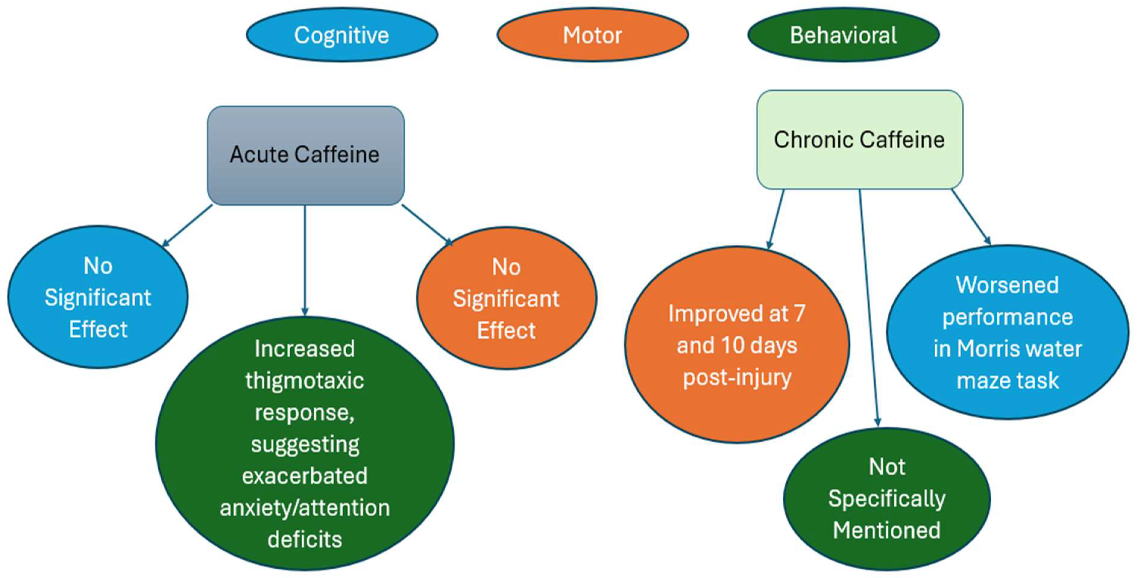
Pre-Clinical Study Oral bolus dose of 25mg/kg Regular caffeine consumption before a penetrating brain injury may moderately improve motor recovery but worsen the neurocognitive sequelae associated with a penetrating brain injury [81]
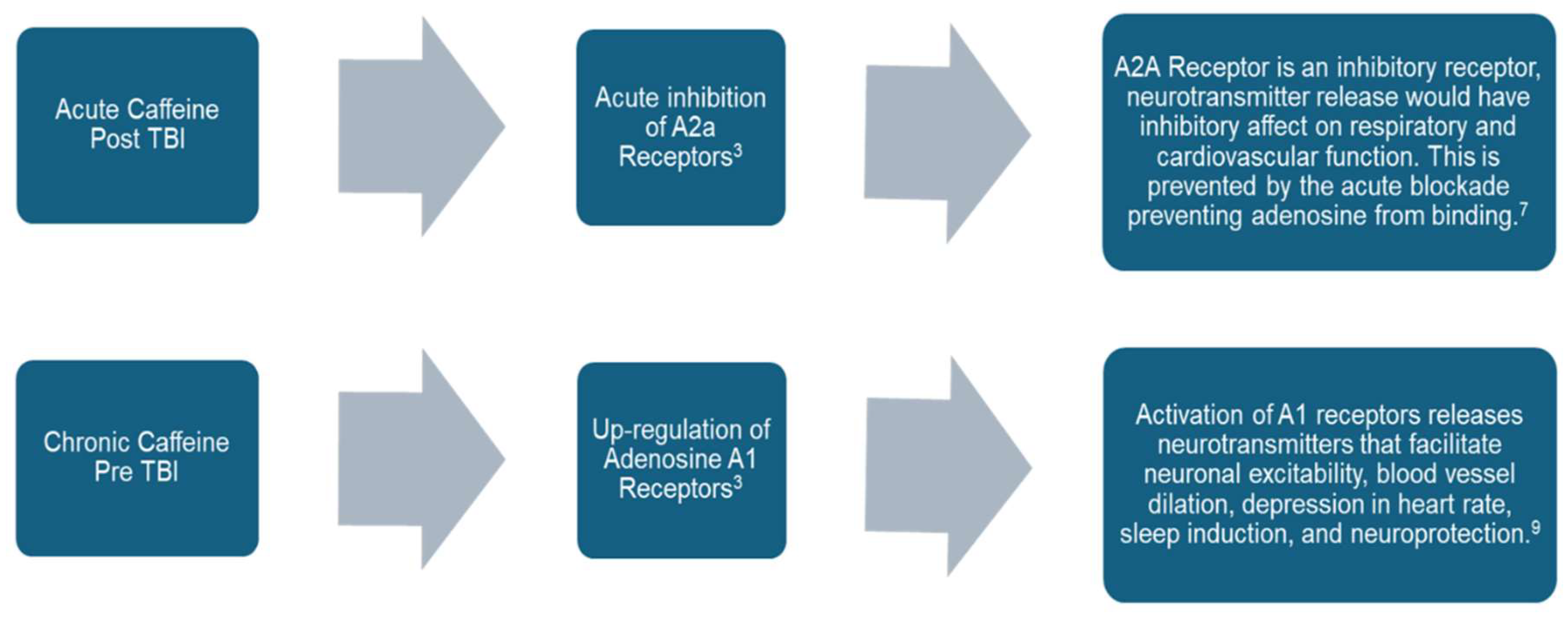
Clinical Study ≥1 μmol/L (194 ng/mL) Caffeine may be neuroprotective by long-term upregulation of adenosine A1 receptors or acute inhibition of A2a receptors[36]
Pre-Clinical Study 20mg/kg Intracranial pressure decreased by 11% from baseline value which can improve clinical outcomes post-TBI[72]
Pre-Clinical Study 25mg/kg Chronic treatment initiated after TBI suggested improved motor function with a nonspecific adenosine receptor agonist, but a slight decrease in motor function after an A1 receptor antagonist [40]
Pre-Clinical Study 36mg/kg The interventions of caffeine, sleep deprivation, sleep aids, and sedation during the acute post-mTBI period each changed the subclinical characteristics of the brain after mTBI and altered the return toward normal function[59].

3.8. Effect of Preinjury and Post-Injury Exposure to Caffeine

The effects of caffeine in rats with TBI have been studied extensively, focusing on motor function, cognitive function, and mortality. In the 2012 article “Survival and Injury Outcome After TBI: Influence of Pre- and Post-Exposure to Caffeine,” Sun et al. demonstrated that pre-exposure to caffeine provided the most significant protective effect against mortality and injury severity following TBI[123]. The study involved phases assessing chronic pre-injury caffeine use, the impact of pre- and post-injury caffeine exposure, and the dose-dependency and efficacy of caffeine as a rescue treatment [123]. The first phase sampled rats with chronic caffeine use before TBI and caffeine-naive rats [123]. The second phase monitored the effects of pre-injury caffeine for three weeks and post-injury caffeine use [123]. The third part examined if the caffeine rescue dosage was dose-dependent, the bolus’s efficacy, and possible negative effects [123]. The findings concluded that pre-exposure to caffeine was most beneficial, with post-injury and continuous exposure also offering benefits, though to a lesser extent [123]. Further research by Lusardi et al. in the same year examined the effects of caffeine on mortality and morbidity following TBI[39]. In the article “Caffeine prevents acute mortality after TBI in rats without increased morbidity,” caffeine was administered intraperitoneally at doses comparable to human consumption levels, both before and after injury [39] . The study monitored the rats’ mortality rates and various behavioral and histological outcomes to assess morbidity [39]. The findings showed a significant reduction in acute mortality following TBI in caffeine-treated rats compared to untreated controls, with no increase in morbidity [39]. Together, these studies underscore caffeine’s potential neuroprotective benefits in managing TBI [39]. While these studies highlighted the acute benefits of caffeine, it is crucial to consider the chronic impacts of caffeine on cognitive and motor functions, particularly in the context of TBI. In the 2017 article “Behavioral and Pathophysiological Outcomes Associated with Caffeine Consumption in RmTBI in Adolescent Rats,” Yamakawa et al. explored these chronic effects[62]. Adolescent rats were given either caffeinated or non-caffeinated water and subjected to three mild brain injuries [62]. Behavioral assessments included tests for balance and motor coordination, anxiety-like behaviors, short-term working memory, and depressive-like behaviors [62]. The findings revealed that chronic caffeine consumption during adolescence significantly altered normal developmental trajectories and the recovery process from repeated mild TBI [62] Behavioral differences were noted in balance, motor coordination, anxiety, memory, and depressive behaviors, influenced by the timing of caffeine exposure and the occurrence of injury [62]. The study underscores the importance of monitoring caffeine consumption in adolescents at high risk for repeated mild TBI due to its significant effects on both behavioral and pathophysiological outcomes [62].
Building on these insights, in 2019, Sanjakdar et al. researched the impacts of caffeine on motor and cognitive outcomes in rats with penetrating ballistic-like brain injury (PBBI). The study evaluated the effects of moderate (7%-PBBI) and severe (10%-PBBI) injuries on cognitive and motor functions, with rats receiving a 25 mg/kg caffeine bolus one hour before injury or no caffeine[81]. Results indicated that caffeine-pre-treated rats showed improved motor recovery compared to controls [81]. However, cognitive outcomes were worsened by caffeine pre-treatment, suggesting that chronic caffeine consumption before PBBI could enhance motor recovery but impair cognitive functions [81]. In 2020, Lusardi et al. expanded their research on the impacts of caffeine on motor and cognitive outcomes in rats with TBI. The study examined the acute and chronic effects of caffeine on mortality and morbidity following TBI[40]. Rats received either chronic caffeine pre-exposure or an acute dose of caffeine immediately following the injury [40]. The findings indicated that a single acute dose of caffeine administered immediately after TBI prevented lethal apnea, a significant cause of acute mortality, without negatively impacting motor performance following sublethal injuries [40]. However, chronic caffeine treatment post-injury impaired recovery of motor function [40]. The study also demonstrated that pre-exposure to caffeine did not significantly impact acute and delayed outcome parameters [40]. Importantly, the study revealed that caffeine withdrawal did not have detrimental effects on motor recovery, contrasting with the negative impact observed with chronic caffeine treatment post-injury [40]. Overall, the study highlighted that the timing and duration of caffeine exposure are critical factors in its effects on TBI outcomes [40].
Most recently, in 2023, Johnson et al. conducted an observational cohort study on the effects of caffeine on adolescents with mild TBI (mTBI). The study included eighty adolescent caffeine consumers (CAF+) and forty non-caffeine consumers (CAF-), averaging 15 years old[122]. Behavioral outcomes were assessed using the Rivermead Post-Concussion Symptoms Questionnaire (RPQ), the Depression subscale of the Beck Youth Inventories - Second Edition (BYI-D), and The Behavior Rating Inventory of Executive Function (BRIEF), along with heart rate variability (HRV) measures [122]. Results showed that caffeine consumers had significantly higher RPQ scores for emotional health and sleep, greater somatic discomfort, and higher depressive symptoms compared to non-consumers [122]. There were no significant differences in executive function or HRV metrics between the groups [122]. The study suggests that adolescent caffeine consumers with mTBI may experience poorer emotional health, sleep quality, and greater somatic discomfort, highlighting the need for careful management of caffeine consumption in this population [122]. Together, these studies provide a comprehensive understanding of the complex role caffeine plays in both acute and chronic settings following TBI. While acute caffeine administration can offer neuroprotective benefits and improve motor recovery, chronic consumption poses risks, particularly in developmental contexts such as adolescence. The findings emphasize the importance of tailored caffeine consumption guidelines for individuals at risk of or recovering from TBI.

4. Conclusions and Future Perspectives

This review provides a comprehensive analysis of the emerging connection between TBI and caffeine. Our primary objective was to comprehensively review all relevant preclinical and clinical studies with the highest degree of sensitivity. Caffeine has the potential to induce changes in various TBI-associated cellular proteins, receptors, signaling pathways, and molecules. A more comprehensive understanding of these interactions, beyond individual markers, could potentially offer a new approach to tailoring TBI management. Furthermore, in the realm of molecular biology, there remains a substantial need for further studies to decipher the specific mechanisms through which caffeine operates and to elucidate its therapeutic effects in the treatment of TBI. Various published findings demonstrating that caffeine can attenuate brain trauma may place personnel on the battlefield at high risk of casualties. Thus, in some scenarios re-evaluating the therapeutic strategy of caffeine application, particularly in multiple-organ-trauma settings should be thoroughly considered.  This exposition underscores the necessity for ongoing research, innovation, and the establishment of standardized protocols to optimize the therapeutic potential of caffeine in the context of TBI. Such findings can enhance understanding of key developments that can improve patient care and advance personalized medicine. Additionally, it will influence trauma quality improvement practices both regionally and nationally.

Author Contributions

Conceptualization by B.S., G.A., J.W., K.T., N.D.B, Z.S.; writing and draft preparation by B.S., R.B., M.S., A.S., S.C., S.B., B.P., J.C., A.C., L.C., J.C., F.J. review, and editing, by BS; figure and table design by B.S., R.B., M.S., A.S., S.C., S.B., B.P., J.C., A.C., L.C., J.C., F.J.; supervision by B.S.; project administration by B.S. All authors have read and agreed to this version of the manuscript.

Funding

There is no grant support or financial relationship for this manuscript

Institutional Review Board Statement

Not applicable.

Data Availability Statement

Information for this review was collected from various sources including PubMed, Web pages, Google Scholar, Scopus, Web of Science, and ClinicalTrials.gov.

Acknowledgments

Not applicable

Conflicts of Interest

Authors have no competing interest to declare

References

  1. Menon, D. K.; Schwab, K.; Wright, D. W.; Maas, A. I. Position Statement: Definition of Traumatic Brain Injury. Arch Phys Med Rehabil 2010, 91 (11), 1637–1640. [CrossRef]
  2. Accessed on December 6, 2024. CDC Surveillance Report of Traumatic Brain Injury-related Emergency Department Visits, Hospitalizations, and Deaths—United States, 2014. URL: https://www.cdc.gov/traumaticbraininjury/data/tbi-edhd.html .
  3. Saatman, K. E.; Duhaime, A.-C.; Bullock, R.; Maas, A. I. R.; Valadka, A.; Manley, G. T. Classification of Traumatic Brain Injury for Targeted Therapies. J Neurotrauma 2008, 25 (7), 719–738. [CrossRef]
  4. Bhaisora, K.; Behari, S.; Godbole, C.; Phadke, R. Traumatic Aneurysms of the Intracranial and Cervical Vessels: A Review. Neurol India 2016, 64 (7), 14. [CrossRef]
  5. Gerard, C.; Busl, K. M. Treatment of Acute Subdural Hematoma. Curr Treat Options Neurol 2014, 16 (1), 275. [CrossRef]
  6. Ron Walls, M. R. H. M. M. G.-H. M. F. F. T. B. E. M. F. F. F. and S. R. W. M. Rosen’s Emergency Medicine: Concepts and Clinical Practice, ISBN: 9780323757898 , 10th Edition.; Page count 2768, 2022; Vol. II.
  7. Joseph Loscalzo, A. F. D. K. S. H. D. L. J. L. J. Harrison’s Principles of Internal Medicine, 21st ed.; McGraw-Hill Education; 2022, 2022.
  8. Neurology and Clinical Neuroscience, 1626 Pages; Anthony Henry Vernon Schapira, Edward Byrne, Eds.; Mosby Elsevier, 2007 ISBN: 0323033547, 9780323033541.
  9. Mckee, A. C.; Daneshvar, D. H. The Neuropathology of Traumatic Brain Injury; 2015; pp 45–66. [CrossRef]
  10. Carney, N.; Ghajar, J.; Jagoda, A.; Bedrick, S.; Davis-O’Reilly, C.; du Coudray, H.; Hack, D.; Helfand, N.; Huddleston, A.; Nettleton, T.; Riggio, S. Concussion Guidelines Step 1. Neurosurgery 2014, 75 (Supplement 1), S3–S15. [CrossRef]
  11. Rincon, S.; Gupta, R.; Ptak, T. Imaging of Head Trauma. Handb Clin Neurol 2016, 135, 447–477. [CrossRef]
  12. Lerner, J. T.; Giza, C. C. Traumatic Brain Injury in Children. In Swaiman’s Pediatric Neurology; Elsevier, 2012; pp 1087–1125. [CrossRef]
  13. Marmarou, A. A Review of Progress in Understanding the Pathophysiology and Treatment of Brain Edema. Neurosurg Focus 2007, 22 (5), E1. [CrossRef]
  14. Drs. Richard G. Ellenbogen, L. N. S. and N. K. Principles of Neurological Surgery, 4th Edition.; Imprint: Elsevier. eBook ISBN: 9780323461276; Hardback ISBN: 9780323431408, 2017.
  15. Flood, L. HEAD AND NECK TRAUMA: AN INTERDISCIPLINARY APPROACH A Ernst, M Herzog, R Seidl Georg Thieme Verlag, 2006 ISBN 3 13 140001 3 Pp 222 Price Euro (D) 99.95 CHF 160.00 Before Receipt. J Laryngol Otol 2007, 121 (4), 408–408. [CrossRef]
  16. Centers for Disease Control and Prevention; National Center for Health Statistics: Mortality Data on CDC WONDER. Available online; accessed on December 6, 2024.
  17. Lafta, G.; Sbahi, H. Factors Associated with the Severity of Traumatic Brain Injury. Med Pharm Rep 2023, 96 (1), 58–64. [CrossRef]
  18. Bramlett, H. M.; Dietrich, W. D. Long-Term Consequences of Traumatic Brain Injury: Current Status of Potential Mechanisms of Injury and Neurological Outcomes. J Neurotrauma 2015, 32 (23), 1834–1848. [CrossRef]
  19. https://www.ninds.nih.gov/health-information/disorders/traumatic-brain-injury-tbi. National Institute of Neurological Disorders and Stroke; Traumatic Brain Injury (TBI). Available online; accessed on December 6, 2024.
  20. Teasdale, G.; Jennett, B. ASSESSMENT OF COMA AND IMPAIRED CONSCIOUSNESS. The Lancet 1974, 304 (7872), 81–84. [CrossRef]
  21. Teasdale, G.; Maas, A.; Lecky, F.; Manley, G.; Stocchetti, N.; Murray, G. The Glasgow Coma Scale at 40 Years: Standing the Test of Time. Lancet Neurol 2014, 13 (8), 844–854. [CrossRef]
  22. Saraiva, S. M.; Jacinto, T. A.; Gonçalves, A. C.; Gaspar, D.; Silva, L. R. Overview of Caffeine Effects on Human Health and Emerging Delivery Strategies. Pharmaceuticals 2023, 16 (8), 1067. [CrossRef]
  23. Justin Evans; John R. Richards; Amanda S. Battisti. Caffeine. In In: StatPearls [Internet] Available from: https://www.ncbi.nlm.nih.gov/books/NBK519490/; Treasure Island (FL): StatPearls Publishing, 2024.
  24. Fiani, B.; Zhu, L.; Musch, B. L.; Briceno, S.; Andel, R.; Sadeq, N.; Ansari, A. Z. The Neurophysiology of Caffeine as a Central Nervous System Stimulant and the Resultant Effects on Cognitive Function. Cureus 2021. [CrossRef]
  25. Burdan, F. Caffeine in Coffee. In Coffee in Health and Disease Prevention; Elsevier, 2015; pp 201–207. [CrossRef]
  26. Reddy, V. S.; Shiva, S.; Manikantan, S.; Ramakrishna, S. Pharmacology of Caffeine and Its Effects on the Human Body. European Journal of Medicinal Chemistry Reports 2024, 10, 100138. [CrossRef]
  27. Nowaczewska, M.; Wiciński, M.; Kaźmierczak, W. The Ambiguous Role of Caffeine in Migraine Headache: From Trigger to Treatment. Nutrients 2020, 12 (8), 2259. [CrossRef]
  28. Sharma, V. K.; Sharma, A.; Verma, K. K.; Gaur, P. K.; Kaushik, R.; Abdali, B. A COMPREHENSIVE REVIEW ON PHARMACOLOGICAL POTENTIALS OF CAFFEINE. Journal of Applied Pharmaceutical Sciences and Research 2023, 6 (3), 16–26. [CrossRef]
  29. Grzegorzewski, J.; Bartsch, F.; Köller, A.; König, M. Pharmacokinetics of Caffeine: A Systematic Analysis of Reported Data for Application in Metabolic Phenotyping and Liver Function Testing. Front Pharmacol 2022, 12. [CrossRef]
  30. Belayneh, A.; Molla, F. The Effect of Coffee on Pharmacokinetic Properties of Drugs : A Review. Biomed Res Int 2020, 2020, 1–11. [CrossRef]
  31. Ruggiero, M.; Calvello, R.; Porro, C.; Messina, G.; Cianciulli, A.; Panaro, M. A. Neurodegenerative Diseases: Can Caffeine Be a Powerful Ally to Weaken Neuroinflammation? Int J Mol Sci 2022, 23 (21), 12958. [CrossRef]
  32. Kolahdouzan, M.; Hamadeh, M. J. The Neuroprotective Effects of Caffeine in Neurodegenerative Diseases. CNS Neurosci Ther 2017, 23 (4), 272–290. [CrossRef]
  33. Kumar, R. Treatment of Traumatic Brain Injury: Nanotherapeutics. J Clin Haematol 2024, 5 (1), 1–3. [CrossRef]
  34. Ning, Y.-L.; Yang, N.; Chen, X.; Tian, H.-K.; Zhao, Z.-A.; Zhang, X.-Z.; Liu, D.; Li, P.; Zhao, Y.; Peng, Y.; Wang, Z.-G.; Chen, J.-F.; Zhou, Y.-G. Caffeine Attenuates Brain Injury but Increases Mortality Induced by High-Intensity Blast Wave Exposure. Toxicol Lett 2019, 301, 90–97. [CrossRef]
  35. Al Moutaery, K.; Al Deeb, S.; Khan, H. A.; Tariq, M. Caffeine Impairs Short-Term Neurological Outcome after Concussive Head Injury in Rats. Neurosurgery 2003, 53 (3), 704–712. [CrossRef]
  36. Sachse, K. T.; Jackson, E. K.; Wisniewski, S. R.; Gillespie, D. G.; Puccio, A. M.; Clark, R. S.; Dixon, C. E.; Kochanek, P. M. Increases in Cerebrospinal Fluid Caffeine Concentration Are Associated with Favorable Outcome after Severe Traumatic Brain Injury in Humans. Journal of Cerebral Blood Flow & Metabolism 2008, 28 (2), 395–401. [CrossRef]
  37. Huang, Z.-L.; Qu, W.-M.; Eguchi, N.; Chen, J.-F.; Schwarzschild, M. A.; Fredholm, B. B.; Urade, Y.; Hayaishi, O. Adenosine A2A, but Not A1, Receptors Mediate the Arousal Effect of Caffeine. Nat Neurosci 2005, 8 (7), 858–859. [CrossRef]
  38. Moreira-de-Sá, A.; Lourenço, V. S.; Canas, P. M.; Cunha, R. A. Adenosine A2A Receptors as Biomarkers of Brain Diseases. Front Neurosci 2021, 15. [CrossRef]
  39. Lusardi, T. A.; Lytle, N. K.; Szybala, C.; Boison, D. Caffeine Prevents Acute Mortality after TBI in Rats without Increased Morbidity. Exp Neurol 2012, 234 (1). [CrossRef]
  40. Lusardi, T. A.; Lytle, N. K.; Gebril, H. M.; Boison, D. Effects of Preinjury and Postinjury Exposure to Caffeine in a Rat Model of Traumatic Brain Injury. J Caffeine Adenosine Res 2020, 10 (1), 12–24. [CrossRef]
  41. Ning, Y.-L.; Yang, N.; Chen, X.; Zhao, Z.-A.; Zhang, X.-Z.; Chen, X.-Y.; Li, P.; Zhao, Y.; Zhou, Y.-G. Chronic Caffeine Exposure Attenuates Blast-Induced Memory Deficit in Mice. Chinese Journal of Traumatology 2015, 18 (4), 204–211. [CrossRef]
  42. Sebastião, A. M.; Ribeiro, J. A. Adenosine Receptors and the Central Nervous System; 2009; pp 471–534. [CrossRef]
  43. Dash, P. K.; Moore, A. N.; Moody, M. R.; Treadwell, R.; Felix, J. L.; Clifton, G. L. Post-Trauma Administration of Caffeine Plus Ethanol Reduces Contusion Volume and Improves Working Memory in Rats. J Neurotrauma 2004, 21 (11), 1573–1583. [CrossRef]
  44. Ahmadipour, M.; Ahmadinejad, M. Effects of Caffeine Administration on GCS and GOSE in Children and Adolescent Patients with Moderate Brain Trauma. European Journal of Molecular & Clinical Medicine 2021, 08 (1).
  45. Yoon, H.; Ro, Y. S.; Jung, E.; Moon, S. B.; Park, G. J.; Lee, S. G. W.; Shin, S. Do. Serum Caffeine Concentration at the Time of Traumatic Brain Injury and Its Long-Term Clinical Outcomes. J Neurotrauma 2023, 40 (21–22), 2386–2395. [CrossRef]
  46. Ratliff, W. A.; Saykally, J. N.; Mervis, R. F.; Lin, X.; Cao, C.; Citron, B. A. Behavior, Protein, and Dendritic Changes after Model Traumatic Brain Injury and Treatment with Nanocoffee Particles. BMC Neurosci 2019, 20 (1), 44. [CrossRef]
  47. Christensen, J.; Yamakawa, G. R.; Salberg, S.; Wang, M.; Kolb, B.; Mychasiuk, R. Caffeine Consumption during Development Alters Spine Density and Recovery from Repetitive Mild Traumatic Brain Injury in Young Adult Rats. Synapse 2020, 74 (4). [CrossRef]
  48. Cappelletti, S.; Daria, P.; Sani, G.; Aromatario, M. Caffeine: Cognitive and Physical Performance Enhancer or Psychoactive Drug? Curr Neuropharmacol 2015, 13 (1), 71–88. [CrossRef]
  49. Nehlig, A. Is Caffeine a Cognitive Enhancer? In Journal of Alzheimer’s Disease; 2010; Vol. 20. [CrossRef]
  50. Fredholm, B. B.; Bättig, K.; Holmén, J.; Nehlig, A.; Zvartau, E. E. Actions of Caffeine in the Brain with Special Reference to Factors That Contribute to Its Widespread Use. Pharmacological Reviews. 1999.
  51. Yang, J.-N.; Chen, J.-F.; Fredholm, B. B. Physiological Roles of A 1 and A 2A Adenosine Receptors in Regulating Heart Rate, Body Temperature, and Locomotion as Revealed Using Knockout Mice and Caffeine. American Journal of Physiology-Heart and Circulatory Physiology 2009, 296 (4), H1141–H1149. [CrossRef]
  52. Powers, M. E. Acute Stimulant Ingestion and Neurocognitive Performance in Healthy Participants. J Athl Train 2015, 50 (5). [CrossRef]
  53. Rusted, J. Caffeine and Cognitive Performance: Effects on Mood or Mental Processing? In Caffeine and Behavior: Current Views & Research Trends: Current Views and Research Trends; 2020.
  54. Smith, J. E.; Lawrence, A. D.; Diukova, A.; Wise, R. G.; Rogers, P. J. Storm in a Coffee Cup: Caffeine Modifies Brain Activation to Social Signals of Threat. Soc Cogn Affect Neurosci 2012, 7 (7), 831–840. [CrossRef]
  55. Porkka-Heiskanen, T. Methylxanthines and Sleep. Handbook of Experimental Pharmacology. 2011. [CrossRef]
  56. Djordjevic, N.; Ghotbi, R.; Jankovic, S.; Aklillu, E. Induction of CYP1A2 by Heavy Coffee Consumption Is Associated with the CYP1A2 -163C>A Polymorphism. Eur J Clin Pharmacol 2010, 66 (7). [CrossRef]
  57. Rétey, J. V.; Adam, M.; Khatami, R.; Luhmann, U. F. O.; Jung, H. H.; Berger, W.; Landolt, H. P. A Genetic Variation in the Adenosine A2A Receptor Gene (ADORA2A) Contributes to Individual Sensitivity to Caffeine Effects on Sleep. Clin Pharmacol Ther 2007, 81 (5). [CrossRef]
  58. Coris, E. E.; Moran, B.; Sneed, K.; Del Rossi, G.; Bindas, B.; Mehta, S.; Narducci, D. Stimulant Therapy Utilization for Neurocognitive Deficits in Mild Traumatic Brain Injury. Sports Health: A Multidisciplinary Approach 2022, 14 (4), 538–548. [CrossRef]
  59. Everson, C. A.; Szabo, A.; Plyer, C.; Hammeke, T. A.; Stemper, B. D.; Budde, M. D. Sleep Loss, Caffeine, Sleep Aids and Sedation Modify Brain Abnormalities of Mild Traumatic Brain Injury. Exp Neurol 2024, 372, 114620. [CrossRef]
  60. Temple, J. L. Caffeine Use in Children: What We Know, What We Have Left to Learn, and Why We Should Worry. Neurosci Biobehav Rev 2009, 33 (6), 793–806. [CrossRef]
  61. Landolt, H.-P.; Rétey, J. V; Tönz, K.; Gottselig, J. M.; Khatami, R.; Buckelmüller, I.; Achermann, P. Caffeine Attenuates Waking and Sleep Electroencephalographic Markers of Sleep Homeostasis in Humans. Neuropsychopharmacology 2004, 29 (10), 1933–1939. [CrossRef]
  62. Yamakawa, G. R.; Lengkeek, C.; Salberg, S.; Spanswick, S. C.; Mychasiuk, R. Behavioral and Pathophysiological Outcomes Associated with Caffeine Consumption and Repetitive Mild Traumatic Brain Injury (RmTBI) in Adolescent Rats. PLoS One 2017, 12 (11), e0187218. [CrossRef]
  63. Li, W.; Dai, S.; An, J.; Li, P.; Chen, X.; Xiong, R.; Liu, P.; Wang, H.; Zhao, Y.; Zhu, M.; Liu, X.; Zhu, P.; Chen, J.-F.; Zhou, Y. Chronic but Not Acute Treatment with Caffeine Attenuates Traumatic Brain Injury in the Mouse Cortical Impact Model. Neuroscience 2008, 151 (4), 1198–1207. [CrossRef]
  64. Gupta, B. S.; Gupta, U. Caffeine and Behavior: Current Views and Research Trends; 2020.
  65. Kuzmin, A.; Johansson, B.; Gimenez, L.; Ögren, S.-O.; Fredholm, B. B. Combination of Adenosine A1 and A2A Receptor Blocking Agents Induces Caffeine-like Locomotor Stimulation in Mice. European Neuropsychopharmacology 2006, 16 (2), 129–136. [CrossRef]
  66. Lusardi, T. Adenosine Neuromodulation and Traumatic Brain Injury. Curr Neuropharmacol 2009, 7 (3), 228–237. [CrossRef]
  67. Kochanek, P. M.; Verrier, J. D.; Wagner, A. K.; Jackson, E. K. The Many Roles of Adenosine in Traumatic Brain Injury. In Adenosine; Springer New York: New York, NY, 2013; pp 307–322. [CrossRef]
  68. Dai, S.-S.; Zhou, Y.-G. Adenosine 2A Receptor: A Crucial Neuromodulator with Bidirectional Effect in Neuroinflammation and Brain Injury. revneuro 2011, 22 (2), 231–239. [CrossRef]
  69. Li, W.; Dai, S.; An, J.; Xiong, R.; Li, P.; Chen, X.; Zhao, Y.; Liu, P.; Wang, H.; Zhu, P.; Chen, J.; Zhou, Y. Genetic Inactivation of Adenosine A2A Receptors Attenuates Acute Traumatic Brain Injury in the Mouse Cortical Impact Model. Exp Neurol 2009, 215 (1), 69–76. [CrossRef]
  70. Chen, J.-F.; Xu, K.; Petzer, J. P.; Staal, R.; Xu, Y.-H.; Beilstein, M.; Sonsalla, P. K.; Castagnoli, K.; Castagnoli, N.; Schwarzschild, M. A. Neuroprotection by Caffeine and A 2A Adenosine Receptor Inactivation in a Model of Parkinson’s Disease. The Journal of Neuroscience 2001, 21 (10), RC143–RC143. [CrossRef]
  71. Chen, J.-F.; Chern, Y. Impacts of Methylxanthines and Adenosine Receptors on Neurodegeneration: Human and Experimental Studies; 2011; pp 267–310. [CrossRef]
  72. Bláha, M.; Vajnerová, O.; Bednár, M.; Vajner, L.; Tichý, M. Traumatic Brain Injuries--Effects of Alcohol and Caffeine on Intracranial Pressure and Cerebral Blood Flow. Rozhl Chir 2009, 88 (11).
  73. Bona, E.; Ådén, U.; Gilland, E.; Fredholm, B. B.; Hagberg, H. Neonatal Cerebral Hypoxia-Ischemia: The Effect of Adenosine Receptor Antagonists. Neuropharmacology 1997, 36 (9), 1327–1338. [CrossRef]
  74. Sun, H.; Gonzalez, F.; McQuillen, P. S. Caffeine Restores Background EEG Activity Independent of Infarct Reduction after Neonatal Hypoxic Ischemic Brain Injury. Dev Neurosci 2020, 42 (1), 72–82. [CrossRef]
  75. Washington, C. ; J. E. ; J. K. ; V. V. ; L. Z. ; J. L. ; C. R. ; D. C. E. ; K. P. Chronic Caffeine Administration Reduces Hippocampal Neuronal Cell Death after Experimental Traumatic Brain Injury in Mice. J Neurotrauma. J. Neurotrauma 2005, 22 1256–1256.
  76. Barrett, P. J.; Casey, E. K.; Sisung, C.; Gaebler-Spira, D. Caffeine as a Neurostimulant: Two Pediatric Acquired Brain Injury Cases. J Pediatr Rehabil Med 2010, 3 (3), 229–232. [CrossRef]
  77. Zhao, Z.-A.; Zhao, Y.; Ning, Y.-L.; Yang, N.; Peng, Y.; Li, P.; Chen, X.-Y.; Liu, D.; Wang, H.; Chen, X.; Bai, W.; Chen, J.-F.; Zhou, Y.-G. Adenosine A2A Receptor Inactivation Alleviates Early-Onset Cognitive Dysfunction after Traumatic Brain Injury Involving an Inhibition of Tau Hyperphosphorylation. Transl Psychiatry 2017, 7 (5), e1123–e1123. [CrossRef]
  78. Zhao, Y.; Zhou, Y.-G.; Chen, J.-F. Targeting the Adenosine A2A Receptor for Neuroprotection and Cognitive Improvement in Traumatic Brain Injury and Parkinson’s Disease. Chinese Journal of Traumatology 2024, 27 (3), 125–133. [CrossRef]
  79. Kochanek, P. M.; Vagni, V. A.; Janesko, K. L.; Washington, C. B.; Crumrine, P. K.; Garman, R. H.; Jenkins, L. W.; Clark, R. S.; Homanics, G. E.; Dixon, C. E.; Schnermann, J.; Jackson, E. K. Adenosine A1 Receptor Knockout Mice Develop Lethal Status Epilepticus after Experimental Traumatic Brain Injury. Journal of Cerebral Blood Flow & Metabolism 2006, 26 (4), 565–575. [CrossRef]
  80. Haselkorn, M. L.; Shellington, D. K.; Jackson, E. K.; Vagni, V. A.; Janesko-Feldman, K.; Dubey, R. K.; Gillespie, D. G.; Cheng, D.; Bell, M. J.; Jenkins, L. W.; Homanics, G. E.; Schnermann, J.; Kochanek, P. M. Adenosine A 1 Receptor Activation as a Brake on the Microglial Response after Experimental Traumatic Brain Injury in Mice. J Neurotrauma 2010, 27 (5), 901–910. [CrossRef]
  81. Sanjakdar, S. S.; Flerlage, W. J.; Kang, H. S.; Napier, D. A.; Dougherty, J. R.; Mountney, A.; Gilsdorf, J. S.; Shear, D. A. Differential Effects of Caffeine on Motor and Cognitive Outcomes of Penetrating Ballistic-Like Brain Injury. Mil Med 2019, 184 (Supplement_1), 291–300. [CrossRef]
  82. Sabir, H.; Maes, E.; Zweyer, M.; Schleehuber, Y.; Imam, F. B.; Silverman, J.; White, Y.; Pang, R.; Pasca, A. M.; Robertson, N. J.; Maltepe, E.; Bernis, M. E. Comparing the Efficacy in Reducing Brain Injury of Different Neuroprotective Agents Following Neonatal Hypoxia–Ischemia in Newborn Rats: A Multi-Drug Randomized Controlled Screening Trial. Sci Rep 2023, 13 (1), 9467. [CrossRef]
  83. Rincon, F.; Ghosh, S.; Dey, S.; Maltenfort, M.; Vibbert, M.; Urtecho, J.; McBride, W.; Moussouttas, M.; Bell, R.; Ratliff, J. K.; Jallo, J. Impact of Acute Lung Injury and Acute Respiratory Distress Syndrome After Traumatic Brain Injury in the United States. Neurosurgery 2012, 71 (4), 795–803. [CrossRef]
  84. Bai, W.; Li, P.; Ning, Y.-L.; Jiang, Y.-L.; Yang, N.; Chen, X.; Zhou, Y.-G. Reduction in Blood Glutamate Levels Combined With the Genetic Inactivation of A2AR Significantly Alleviate Traumatic Brain Injury-Induced Acute Lung Injury. Shock 2019, 51 (4), 502–510. [CrossRef]
  85. Strong, R.; Grotta, J. C.; Aronowski, J. Combination of Low Dose Ethanol and Caffeine Protects Brain from Damage Produced by Focal Ischemia in Rats. Neuropharmacology 2000, 39 (3), 515–522. [CrossRef]
  86. Piriyawat, P.; Labiche, L. A.; Burgin, W. S.; Aronowski, J. A.; Grotta, J. C. Pilot Dose-Escalation Study of Caffeine Plus Ethanol (Caffeinol) in Acute Ischemic Stroke. Stroke 2003, 34 (5), 1242–1245. [CrossRef]
  87. KONTOS, H. A.; POVLISHOCK, J. T. Oxygen Radicals in Brain Injury. Central Nervous System Trauma 1986, 3 (4), 257–263. [CrossRef]
  88. Clark, R. S. B.; Carcillo, J. A.; Kochanek, P. M.; Obrist, W. D.; Jackson, E. K.; Mi, Z.; Wisneiwski, S. R.; Bell, M. J.; Marion, D. W. Cerebrospinal Fluid Adenosine Concentration and Uncoupling of Cerebral Blood Flow and Oxidative Metabolism after Severe Head Injury in Humans. Neurosurgery 1997, 41 (6), 1284–1292. [CrossRef]
  89. Gerbatin, R. R.; Dobrachinski, F.; Cassol, G.; Soares, F. A. A.; Royes, L. F. F. A1 Rather than A2A Adenosine Receptor as a Possible Target of Guanosine Effects on Mitochondrial Dysfunction Following Traumatic Brain Injury in Rats. Neurosci Lett 2019, 704, 141–144. [CrossRef]
  90. Almajano, M. P.; Vila, I.; Gines, S. Neuroprotective Effects of White Tea Against Oxidative Stress-Induced Toxicity in Striatal Cells. Neurotox Res 2011, 20 (4), 372–378. [CrossRef]
  91. Caravan, I.; Sevastre Berghian, A.; Moldovan, R.; Decea, N.; Orasan, R.; Filip, G. A. Modulatory Effects of Caffeine on Oxidative Stress and Anxiety-like Behavior in Ovariectomized Rats. Can J Physiol Pharmacol 2016, 94 (9), 961–972. [CrossRef]
  92. Prasanthi, J. R. P.; Dasari, B.; Marwarha, G.; Larson, T.; Chen, X.; Geiger, J. D.; Ghribi, O. Caffeine Protects against Oxidative Stress and Alzheimer’s Disease-like Pathology in Rabbit Hippocampus Induced by Cholesterol-Enriched Diet. Free Radic Biol Med 2010, 49 (7), 1212–1220. [CrossRef]
  93. Fredholm, B. B.; Chen, J.-F.; Masino, S. A.; Vaugeois, J.-M. ACTIONS OF ADENOSINE AT ITS RECEPTORS IN THE CNS: Insights from Knockouts and Drugs. Annu Rev Pharmacol Toxicol 2005, 45 (1), 385–412. [CrossRef]
  94. &NA; Caffeine for the Sustainment of Mental Task Performance: Formulations for Military Operations. Nutr Today 2002, 37 (1). [CrossRef]
  95. Ferré, S. Mechanisms of the Psychostimulant Effects of Caffeine: Implications for Substance Use Disorders. Psychopharmacology (Berl) 2016, 233 (10), 1963–1979. [CrossRef]
  96. Ferré, S.; Ciruela, F.; Quiroz, C.; Luján, R.; Popoli, P.; Cunha, R. A.; Agnati, L. F.; Fuxe, K.; Woods, A. S.; Lluis, C.; Franco, R. Adenosine Receptor Heteromers and Their Integrative Role in Striatal Function. TheScientificWorldJournal. 2007. [CrossRef]
  97. Yacoubi, M. El; Ledent, C.; Ménard, J.; Parmentier, M.; Costentin, J.; Vaugeois, J. The Stimulant Effects of Caffeine on Locomotor Behaviour in Mice Are Mediated through Its Blockade of Adenosine A 2A Receptors. Br J Pharmacol 2000, 129 (7), 1465–1473. [CrossRef]
  98. Wilk, M.; Krzysztofik, M.; Filip, A.; Zajac, A.; Del Coso, J. The Effects of High Doses of Caffeine on Maximal Strength and Muscular Endurance in Athletes Habituated to Caffeine. Nutrients 2019, 11 (8), 1912. [CrossRef]
  99. Olopade, F. E.; Femi-Akinlosotu, O. M.; Adekanmbi, A. J.; Ighogboja, O. O.; Shokunbi, M. T. Chronic Caffeine Ingestion Improves Motor Function and Increases Dendritic Length and Arborization in the Motor Cortex, Striatum, and Cerebellum. J Caffeine Adenosine Res 2021, 11 (1), 3–14. [CrossRef]
  100. Vynorius, K. C.; Paquin, A. M.; Seichepine, D. R. Lifetime Multiple Mild Traumatic Brain Injuries Are Associated with Cognitive and Mood Symptoms in Young Healthy College Students. Front Neurol 2016, 7. [CrossRef]
  101. Van Batenburg-Eddes, T.; Lee, N. C.; Weeda, W. D.; Krabbendam, L.; Huizinga, M. The Potential Adverse Effect of Energy Drinks on Executive Functions in Early Adolescence. Front Psychol 2014, 5. [CrossRef]
  102. Venkata Charan Tej, G. N.; Neogi, K.; Verma, S. S.; Chandra Gupta, S.; Nayak, P. K. Caffeine-Enhanced Anti-Tumor Immune Response through Decreased Expression of PD1 on Infiltrated Cytotoxic T Lymphocytes. Eur J Pharmacol 2019, 859. [CrossRef]
  103. Fletcher, D. K.; Bishop, N. C. Effect of a High and Low Dose of Caffeine on Antigen-Stimulated Activation of Human Natural Killer Cells After Prolonged Cycling. Int J Sport Nutr Exerc Metab 2011, 21 (2), 155–165. [CrossRef]
  104. Abbasi, A.; Abtahi Froushani, S. M.; Delirezh, N.; Mostafaei, A. Caffeine Alters the Effects of Bone Marrow-Derived Mesenchymal Stem Cells on Neutrophils. Advances in Clinical and Experimental Medicine 2018, 27 (4), 463–468. [CrossRef]
  105. Nehlig, A. Are We Dependent upon Coffee and Caffeine? A Review on Human and Animal Data. Neurosci Biobehav Rev 1999, 23 (4), 563–576. [CrossRef]
  106. Ofluoglu, E.; Pasaoglu, H.; Pasaoglu, A. The Effects of Caffeine on L-Arginine Metabolism in the Brain of Rats. Neurochem Res 2009, 34 (3), 395–399. [CrossRef]
  107. Ismail, H.; Shakkour, Z.; Tabet, M.; Abdelhady, S.; Kobaisi, A.; Abedi, R.; Nasrallah, L.; Pintus, G.; Al-Dhaheri, Y.; Mondello, S.; El-Khoury, R.; Eid, A. H.; Kobeissy, F.; Salameh, J. Traumatic Brain Injury: Oxidative Stress and Novel Anti-Oxidants Such as Mitoquinone and Edaravone. Antioxidants 2020, 9 (10), 943. [CrossRef]
  108. Shirley, D. G.; Walter, S. J.; Noormohamed, F. H. Natriuretic Effect of Caffeine: Assessment of Segmental Sodium Reabsorption in Humans. Clin Sci 2002, 103 (5). [CrossRef]
  109. Seal, A. D.; Bardis, C. N.; Gavrieli, A.; Grigorakis, P.; Adams, J. D.; Arnaoutis, G.; Yannakoulia, M.; Kavouras, S. A. Coffee with High but Not Low Caffeine Content Augments Fluid and Electrolyte Excretion at Rest. Front Nutr 2017, 4. [CrossRef]
  110. Pin-on, P.; Saringkarinkul, A.; Punjasawadwong, Y.; Kacha, S.; Wilairat, D. Serum Electrolyte Imbalance and Prognostic Factors of Postoperative Death in Adult Traumatic Brain Injury Patients. Medicine 2018, 97 (45), e13081. [CrossRef]
  111. Weber, J. T.; Rzigalinski, B. A.; Ellis, E. F. Calcium Responses to Caffeine and Muscarinic Receptor Agonists Are Altered in Traumatically Injured Neurons. J Neurotrauma 2002, 19 (11), 1433–1443. [CrossRef]
  112. Buccilli, B.; Alan, A.; Baha’, A.; Shahzad, A.; Almealawy, Y.; Chisvo, N. S.; Ennabe, M.; Weinand, M. Neuroprotection Strategies in Traumatic Brain Injury: Studying the Effectiveness of Different Clinical Approaches. Surg Neurol Int 2024, 15, 29. [CrossRef]
  113. Tsutsui, S.; Schnermann, J.; Noorbakhsh, F.; Henry, S.; Yong, V. W.; Winston, B. W.; Warren, K.; Power, C. A1 Adenosine Receptor Upregulation and Activation Attenuates Neuroinflammation and Demyelination in a Model of Multiple Sclerosis. The Journal of Neuroscience 2004, 24 (6), 1521–1529. [CrossRef]
  114. Pugliese, A. M.; Coppi, E.; Spalluto, G.; Corradetti, R.; Pedata, F. A 3 Adenosine Receptor Antagonists Delay Irreversible Synaptic Failure Caused by Oxygen and Glucose Deprivation in the Rat CA1 Hippocampus in Vitro. Br J Pharmacol 2006, 147 (5), 524–532. [CrossRef]
  115. Mota-Rojas, D.; Villanueva-García, D.; Hernández-Ávalos, I.; Casas-Alvarado, A.; Domínguez-Oliva, A.; Lezama-García, K.; Miranda-Cortés, A.; Martínez-Burnes, J. Cardiorespiratory and Neuroprotective Effects of Caffeine in Neonate Animal Models. Animals 2023, 13 (11), 1769. [CrossRef]
  116. Vargas-Pozada, E. E.; Ramos-Tovar, E.; Rodriguez-Callejas, J. D.; Cardoso-Lezama, I.; Galindo-Gómez, S.; Talamás-Lara, D.; Vásquez-Garzón, V. R.; Arellanes-Robledo, J.; Tsutsumi, V.; Villa-Treviño, S.; Muriel, P. Caffeine Inhibits NLRP3 Inflammasome Activation by Downregulating TLR4/MAPK/NF-ΚB Signaling Pathway in an Experimental NASH Model. Int J Mol Sci 2022, 23 (17), 9954. [CrossRef]
  117. Tanaka, S.; Koike, T. Caffeine Promotes Survival of Cultured Sympathetic Neurons Deprived of Nerve Growth Factor through a CAMP-Dependent Mechanism. Biochimica et Biophysica Acta (BBA) - Molecular Cell Research 1992, 1175 (1), 114–122. [CrossRef]
  118. HAN, K.; JIA, N.; LI, J.; YANG, L.; MIN, L.-Q. Chronic Caffeine Treatment Reverses Memory Impairment and the Expression of Brain BNDF and TrkB in the PS1/APP Double Transgenic Mouse Model of Alzheimer’s Disease. Mol Med Rep 2013, 8 (3), 737–740. [CrossRef]
  119. Lopes, J. P.; Pliássova, A.; Cunha, R. A. The Physiological Effects of Caffeine on Synaptic Transmission and Plasticity in the Mouse Hippocampus Selectively Depend on Adenosine A1 and A2A Receptors. Biochem Pharmacol 2019, 166, 313–321. [CrossRef]
  120. Schapira, A. H. V.; Bezard, E.; Brotchie, J.; Calon, F.; Collingridge, G. L.; Ferger, B.; Hengerer, B.; Hirsch, E.; Jenner, P.; Novère, N. Le; Obeso, J. A.; Schwarzschild, M. A.; Spampinato, U.; Davidai, G. Novel Pharmacological Targets for the Treatment of Parkinson’s Disease. Nat Rev Drug Discov 2006, 5 (10), 845–854. [CrossRef]
  121. National Academies of Sciences, Engineering, and Medicine; Board on Health Care Services; Committee on the Review of the Department of Veterans Affairs Examinations for Traumatic Brain Injury. Evaluation of the Disability Determination Process for Traumatic Brain Injury in Veterans. Washington (DC): National Academies Press (US); 2019 Apr 10. B, Definitions of Traumatic Brain Injury. Nd Medicine Division; https://www.ncbi.nlm.nih.gov/books/NBK542588/.
  122. Johnson, J. M. ; E. J. M. ; M. R. D. Effects of Caffeine on Measures of Clinical Outcome and Recovery Following Mild Traumatic Brain Injury in Adolescents. Master’s Thesis, Available at: https://scholarcommons.sc.edu/etd/7497., University of South Carolina, 2023.
  123. Sun, D. ; D. T. E. ; P. Y. ; K. K. ; C. H. T. ; L. A. M. ; J. L. S. ; C. J. D. Survival and Injury Outcome After TBI: Influence of Pre- and Post-Exposure to Caffeine. Neuroscience 2012, 47–58.
Table 1. This table summarizes various caffeine-associated preclinical and clinical findings in Traumatic Brain Injury (TBI).
Reference number Study Model Findings
[64] N/A Caffeine acts as an adenosine antagonist, specifically the A1 and A2A receptors.
[65] N/A Adenosine receptors are of interest in TBI because they are involved in various brain injury pathways.
[66] Rodent Adenosine may have a protective role in recovery of TBI
[67] Rodent Caffeine may have a neuroprotective effect on Parkinson’s disease pathways.
[68] Mixed Caffeine may have a neuroprotective effect on neurodegenerative diseases
[69] Rodent Chronic caffeine treatment rather than acute caffeine treatment showed better recovery from TBI in mice models.
[70] Mixed Review article finding combined evidence of potential benefit of treating Parkinson’s Disease and TBI through targeting the adenosine2A receptor.
[71] Rodent Pretreatment of rats with caffeine before TBI showed an increase in mortality
[63] Rodent Chronic caffeine use may have some protective benefits in blast-induced TBI however chronic and acute caffeine use both increased mortality.
[72] Rodent Caffeine injection given immediately after TBI reduced TBI-induced mortality in rat models.
[73] Rodent An acute dose of caffeine given 10 seconds after TBI decreased mortality and morbidity in mice models.
[74] Humans An increase in CSF concentration of caffeine in patients with TBI was associated with more favorable outcomes.
[74] Human Patients with TBI who had a serum caffeine concentration between 0.01 to 1.66 μg/mL had better recovery after 6 months of injury compared to the no caffeine group.
[75] Cats One of the earliest studies that examined the pathophysiology of oxygen radicals in TBI
[76] Humans Found potential benefits of adenosine on cerebral blood flow and oxidative metabolism in patients with severe head injury
[44] Rodent Caffeine in female rats has been shown to decrease oxidative stress and specifically decrease lipid peroxidation in the hippocampus.
[77] Rabbits Caffeine was seen to protect against oxidative stress and Alzheimer’s dementia-like pathology in rabbit models.
[62] Rodent Positive neuronal changes were seen when treating mice with nano coffee injections after a TBI.
Disclaimer/Publisher’s Note: The statements, opinions and data contained in all publications are solely those of the individual author(s) and contributor(s) and not of MDPI and/or the editor(s). MDPI and/or the editor(s) disclaim responsibility for any injury to people or property resulting from any ideas, methods, instructions or products referred to in the content.
Copyright: This open access article is published under a Creative Commons CC BY 4.0 license, which permit the free download, distribution, and reuse, provided that the author and preprint are cited in any reuse.
Prerpints.org logo

Preprints.org is a free preprint server supported by MDPI in Basel, Switzerland.

Subscribe

Disclaimer

Terms of Use

Privacy Policy

Privacy Settings

© 2025 MDPI (Basel, Switzerland) unless otherwise stated