Preprint
Article

This version is not peer-reviewed.

Redirection of Care for Neonates with Hypoxic-Ischemic Encephalopathy Receiving Therapeutic Hypothermia

A peer-reviewed article of this preprint also exists.

Submitted:

03 December 2024

Posted:

04 December 2024

You are already at the latest version

Abstract

Background/Objectives: Hypoxic-ischemic encephalopathy (HIE) in (near)term neonates accounts for neonatal mortality and unfavorable neurodevelopmental outcome in survivors, despite therapeutic hypothermia (TH) for neuroprotection. The circumstances of death in neonates with HIE and involvement of neonatal palliative care (NPC) specialists warrant further evaluation. Methods: Retrospective multicenter cohort study including neonates ≥ 35 weeks gestational age with moderate to severe HIE receiving TH, registered in the Swiss National Asphyxia and Cooling Register between 2011 and 2021. Neurodevelopmental follow-up at 18-24 months in survivors was assessed. The groups of survivors and deaths were compared regarding perinatal demographic and HIE data. Prognostic factors leading to redirection of care (ROC) were depicted. Results: 137 neonates were included, 23 (16.8%) deaths and 114 (83.2%) survivors. All but one death (95.7%) occurred after ROC with death on median 3.5 (2-6) days of life. Severe encephalopathy indicated by Sarnat score 3 on admission and seizures were more frequent, and blood lactate values were higher on postnatal days 1 to 4 in neonates who died. Lactate in worst blood gas analysis (unit aOR 1.25, 95% CI 1.02-1.54, p=0.0352) was the only variable independently associated with ROC. NPC specialists were involved in one case. Of 114 survivors, 88 (77.2%) had neurodevelopmental assessments, and 21 (23.9%) of those had unfavorable outcome (moderate to severe disability). Conclusions: Death in neonates with moderate to severe HIE receiving TH almost exclusively occurred after ROC. Parents thus had to take critical decisions and accompany their neonate at end-of-life within the first week of life. To ensure continuity of care for the family, also beyond the death of the neonate, involvement of NPC specialists is encouraged in ROC.

Keywords: 
;  ;  ;  ;  ;  

1. Introduction

Hypoxic ischemic encephalopathy (HIE) is a leading cause of neonatal death, even in high income countries [1]. To date, therapeutic hypothermia (TH) is the only evidence-based neuroprotective therapy for HIE to reduce mortality and improve neurodevelopmental outcomes [2,3,4,5,6]. Despite TH, HIE often leads to poor neurodevelopmental outcome prognosis and thus evaluation for withdrawal of life-sustaining therapy (WLST) is a recurrent topic to discuss with families in context of HIE [7 Natarajan]. Apart from redirection of care (ROC) in face of devastating neurodevelopmental prognosis, some neonates with HIE die due to HIE-related multi-organ failure despite provision of neonatal intensive care [7].
HIE occurs unanticipated and parents thus face a sudden challenge to the life of their expectedly healthy term-born neonate. TH for HIE is standard of care for moderate to severe HIE in tertiary neonatal intensive care units (NICUs) [2,3,4,5,6]. Neonatal palliative care (NPC) is emerging as a dedicated subspecialty of pediatric palliative care offered in large tertiary neonatal centers [8,9]. However, involvement of NPC specialists seems underrepresented in context of neonatal death due to HIE in Switzerland [10].
The primary objective was to review circumstances of neonatal deaths of neonates with moderate to severe HIE receiving TH between 2011 and 2021 in two tertiary NICUs in Zurich, Switzerland, with standardized access to NPC. The secondary objective was to compare demographics and clinical prognostic parameters of survivors and deaths of moderate to severe HIE receiving TH, registered in the Swiss National Asphyxia and Cooling Register between 2011 and 2021.

2. Materials and Methods

This retrospective multicenter cohort study included neonates ≥ 35 weeks gestational age with HIE registered in the Swiss National Asphyxia and Cooling Register. All of them were admitted to the NICU at the University Hospital Zurich or to the neonatal/pediatric ICU of the University Children’s Hospital Zurich between January 2011 and December 2021 for TH due to moderate (Sarnat Score 2) or severe (Sarnat Score 3) HIE [11]. All neonates were treated according to the Swiss National Asphyxia and Cooling Register Protocol [12]. TH with whole-body cooling was initiated within 6 hours of birth, targeting 33.0°C to 34.0°C core temperature and was continued for 72 hours. Exclusion criteria for TH according to the national cooling protocol were applied [12,13].
Inclusion required available data in the Swiss National Asphyxia and Cooling Register. Available results of the 18-24 months neurodevelopmental follow-up (NDFU) assessment in survivors were included. Neonates were excluded if further use of data was refused by parents or if there was evidence of syndromic disease.
Maternal and neonatal demographics, data on pregnancy and delivery as well as neonatal characteristics during TH were extracted from the Swiss National Asphyxia and Cooling Register. Resuscitation was defined as any form of ventilation necessary at the age of 10 minutes. Worst blood gas analysis was defined as worst blood gas results (arterial, venous or capillary samples) within 60 minutes after birth, including umbilical gases [Appendix A]. Severity of neonatal encephalopathy was graded on admission and every 24 hours during TH. Corresponding Thompson scores [14] were converted to Sarnat scores [11] according to Thompson et al. [14]. All cranial magnetic resonance imaging (cMRI) and cranial ultrasound (cUS) recordings were evaluated by experienced pediatric neuro-radiologists. Data collection was amended manually by electronic medical chart review to collect information on circumstances of WLST and death.
Prognostic factors of unfavorable neurodevelopmental outcome [15,16,17] that were included in the discussion to redirect care were determined and depicted in three main categories:
  • Severe clinical neurological abnormalities, reflected in persistent Sarnat score 2 and 3 [11],
  • Severely abnormal neuro-monitoring with electroencephalogram (EEG), defined as significantly reduced or no cerebral activity or therapy-refractory clinical or subclinical seizures or status epilepticus [17,18] (amplitude-integrated EEG was not included)
  • Severely abnormal neuro-imaging (cMRI or cUS) depicting severe hypoxic-ischemic injury [19,20]
Data on NDFU at 18-24 months was extracted from the Swiss National Follow-up Register. NDFU assessments included a clinical examination, a structured neurological assessment which encompassed hearing and vision, and a developmental assessment, utilizing either the Bayley Scales of Infant and Toddler Development, Third Edition (BSID-III) [21] or the Griffiths Development Scales [22], following the guidelines of the follow-up center. BSID-III testing provided three main composite scores: Cognitive (CCS), language (LCS) and motor composite score (MCS) with a mean score of 100 and a SD of ± 15 [21]. The Griffiths Development Scales (Griffiths) comprised five subscales: Locomotor, personal-social, hearing and speech, eye and hand coordination and performance. Results were declared in months (developmental age). In order to compare Griffiths with BSID-III, a developmental quotient was calculated for each domain of the Griffiths (developmental age in relation to adjusted chronological age). Cerebral palsy was graded according to the Gross Motor Function Classification System (GMFCS) of Palisano et al. for children aged ≤ 2 years [23].
Neurodevelopmental outcome at 18-24 months was categorized as follows [2,3,4,5,24]:
Unfavorable outcome:
  • Death or
  • Severe disability, defined as a CCS, LCS or MCS (BSID-III) or one Griffiths developmental quotient more than 2 SD below the mean score (i.e. < 70), or a GMFCS grade of level 3 to 5, or hearing impairment (inability to follow commands despite amplification) or blindness or
  • Moderate disability, defined as a CCS, LCS or MCS (BSID-III) or one Griffiths developmental quotient of 1 to 2 SD below the mean score (i.e. 70 to 84) in addition to one or more of the following: GMFCS grade of level 2, hearing impairment (hearing deficit with the ability to follow commands after amplification) or persistent seizure disorder.
Favorable outcome:
  • Mild disability, defined as a CCS, LCS or MCS (BSID-III) or one Griffiths developmental quotient of 70-84 alone, or CCS or LCS ≥ 85 and GMFCS level 1 or 2, seizure disorder without anti-epileptic medication or hearing deficit with ability to follow commands with amplification or
  • No disability: Absence of previously listed disabilities.
All missing patient information was reported. All statistical analyses were conducted using R v4.4.1 (The R Foundation for Statistical Computing, Vienna, Austria). Descriptive data were reported as frequency (proportion) for categorical variables and as median (interquartile range) for continuous variables. Subgroups for the main outcomes death and survival were compared using Pearson’s Chi-squared and Fisher’s exact test for dichotomous variables and Wilcoxon rank sum test for continuous variables in unadjusted analysis. Binary logistic regression analysis was used to explore associations between maternal or neonatal characteristics and the main outcomes, and adjusted odds ratios (aOR) with corresponding 95% confidence intervals (CI) were calculated accordingly. Logistic regression analyses were adjusted for Apgar at 1 minute, resuscitation in the delivery room, umbilical artery blood pH, highest lactate in blood gas analysis, Sarnat score on admission (prior to TH), and seizures on the first postnatal day. Goodness-of-fit was assessed using the Hosmer-Lemeshow test. A p-value of <0.05 was considered statistically significant.
Data collection, analysis and publication was approved by the Swiss ethical committee of the Canton of Zurich (KEK-ZH Number 2024-00208). STROBE guidelines were followed for reporting this observational study.

3. Results

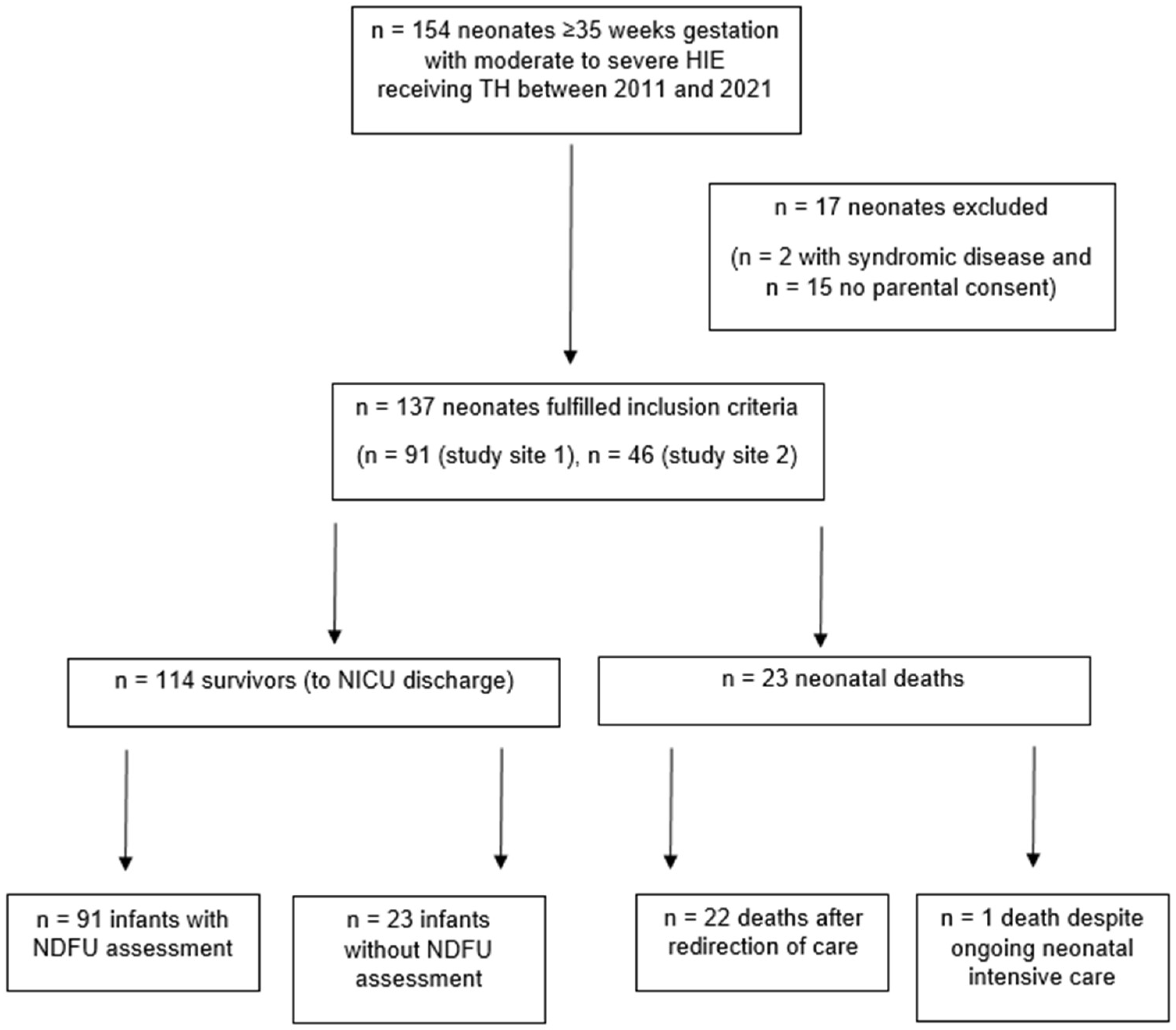
3.1. Participants and descriptive data

A total of 154 neonates were initially eligible. However, two neonates with syndromic diseases and 15 neonates whose parents refused participation were excluded, resulting in a final study population of 137 neonates (Figure 1).

3.2. Outcome data and main results

Twenty-three out of 137 (16.8%) neonates died in the NICU. Demographic and maternal characteristics did not differ between neonates who died and those who survived (Table 1 and Table 2).
Neonates who died had lower Apgar scores at 1 (0 vs. 2, p=0.0003), 5 (1 vs. 4, p=0.0032), and 10 (2 vs. 5, p=0.0056) minutes after birth as compared to survivors. Blood gas parameters revealed more severe acidosis in those who died (Table 2).
The majority of neonates who died had Sarnat score 3 before and during hypothermia, whereas most survivors had Sarnat score 2. Seizures were more prevalent in neonates who did not survive on day 1 (47.8% vs. 16.7%, p=0.0022), day 2 (33.3% vs. 12.3%, p = 0.0227), day 3 (42.9% vs. 6.1%, p=0.0006), and in total (60.9% vs. 24.6%, p=0.0014). Cerebral MRI scans were performed in almost all survivors, whereas the rate was low in neonates who died (97.4% vs. 30.4%, p<0.0001). Daily highest lactate values were higher during postnatal days 1 to 4 in neonates which deceased (Table 3a).
Of the 23 neonates who died, one neonate passed away on the first postnatal day despite intensive care interventions due to cardiorespiratory failure. Twenty-two (95.7%) neonates died after ROC. In addition to clinical neurological abnormalities according to Sarnat score, severely abnormal EEG (63.4%) [significantly reduced or no cerebral activity], seizures (59.1%) [therapy-refractory clinical or subclinical seizures or status epilepticus], severely abnormal cMRI (31.8%), and pathologic cUS (95.5%) [depicting severe hypoxic-ischemic injury] were reasons to redirect care. The decision to redirect care was made on median postnatal day 3 (2-5), and the neonates eventually died on median day 3.5 (2-6) of life, resulting in a time period of 0 (0-1) days from the decision to redirect care to death (Figure 2 and Table 3b).
Table 3. Prognostic factors of expected unfavorable neurodevelopmental outcome leading to redirection of care.
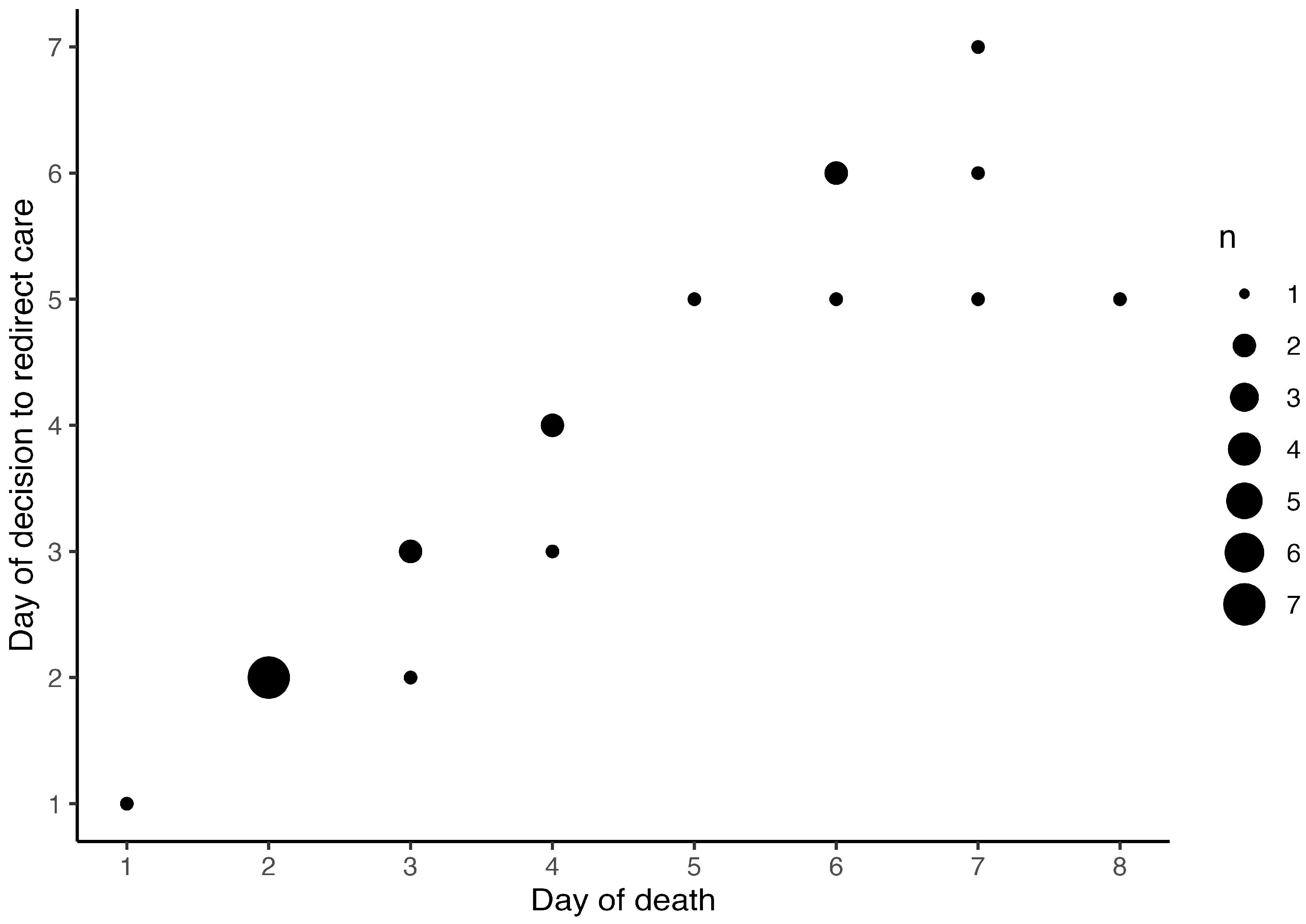
Table 3. b. Depiction of prognostic factors in cases of death.
Case Death EEG Seizure cMRI cUS Sarnat score ROC Death (DOL)
despite maximal support after ROC on admission DOL 1 DOL 2 DOL 3 Decision (DOL) Execution (DOL)
1 0 1 0 0 0 1 3 3 NA NA 1 1 1
2 0 1 1 0 1 1 3 3 3 3 6 6 6
3 0 1 1 1 0 1 2 3 2 2 5 5 5
4 0 1 0 1 0 0 3 3 NA 2 2 2
5 0 1 0 1 0 1 2 NA 2 2 2
6 0 1 0 1 0 1 3 3 NA 2 2 2
7 0 1 0 0 0 1 3 3 NA 2 2 2
8 0 1 1 1 1 1 2 2 2 3 5 6 6
9 0 1 1 1 1 1 3 3 3 3 5 7 8
10 0 1 1 1 1 1 2 2 3 3 6 7 7
11 0 1 0 1 0 1 2 2 3 3 4 4 4
12 0 1 0 1 0 1 3 3 3 3 3 3
13 0 1 1 1 0 1 3 NA 2 2 2
14 0 1 1 0 0 1 3 3 NA 2 2 2
15 0 1 1 0 0 1 3 3 NA 2 2 3
16 0 1 1 1 1 1 3 2 2 2 7 7 7
17 0 1 1 0 0 1 2 1 1 1 6 6 6
18 0 1 1 1 0 1 2 2 2 3 3 3
19 0 1 0 0 0 1 3 3 NA 2 2 2
20 0 1 1 0 0 1 2 3 3 3 4 4
21 0 1 1 0 1 1 3 3 3 3 4 4 4
22 0 1 1 1 1 1 2 2 3 3 5 7 7
23 1 0 0 0 0 0 3 3 NA NA NA NA 1
DOL: day of life; ROC: redirection of care; NA: not applicable. Empty fields correspond to missing values. EEG: severely abnormal electroencephalogram (EEG), defined as significantly reduced or no cerebral activity. Seizure: therapy-refractory clinical or subclinical seizures or status epilepticus. cMRI: cranial magnetic resonance imaging depicting severe hypoxic-ischemic injury. cUS: cranial ultrasound depicting severe hypoxic-ischemic injury; (note: continuous amplitude-integrated EEG monitoring in all cases, not reported).
The multiprofessional NPC team was involved in one (4.5%) case of ROC. Between the two centers studied, there were no differences in the frequency of ROC (13.3% vs. 17.6%, p = 0.6997), the age at decision to redirect care (2.5 vs. 3.5 days, p = 0.5434), and the time period from the decision to redirect care to death (0 vs. 0 days, p = 0.7058).
Eighty-eight (77.2%) of 114 surviving infants had neurodevelopmental assessments (n=111 BSID-III, n=3 Griffiths) at the age of 24 (23-25) months. Among these, 37 (42.0%) infants had no disability, 30 (34.1%) infants had mild disability, 11 (12.5%) infants had moderate disability and 10 (11.4%) infants had severe disability. Among the entire study cohort, 44 (32.1%) neonates had an unfavorable outcome and among the survivors 21 (23.9%) had a moderate or severe disability.
In the adjusted analysis, the decision to redirect care was independently associated with lactate in worst blood gas analysis (unit aOR 1.25, 95% CI 1.02-1.54, p=0.0352). The logistic regression model fit well to predict ROC (Hosmer-Lemeshow p = 0.920).

4. Discussion

This study revealed that almost all deaths in moderate to severe HIE receiving TH in the examined Swiss NICUs occurred in context of WLST during or shortly after TH. The decision for ROC was made due to severe clinical neurological abnormalities, severely abnormal neuro-monitoring and neuro-imaging, prognosticating an unfavorable neurodevelopmental outcome [15,16,17,18,19,20]. In 76% of the survivors, neurodevelopmental follow-up assessment at the age of two years confirmed favorable outcomes.
There was no difference in maternal and demographic characteristics between survivors and deaths. As expected, deceased neonates suffered from more severe HIE. This was confirmed by lower Apgar scores and blood gas parameters revealed more severe acidosis (and persistent lactate) compared to survivors, which is in line with previous reports [15,16]. Clinical encephalopathy was more severe (i.e. Sarnat score 3), often accompanied by seizures, in deaths compared to survivors.
Mortality was 17% in this study, which is comparable to an overall median mortality of 14% in moderate to severe HIE receiving TH in Switzerland [13]. Internationally, mortality rates of 14% (Canada) [7] to 27% (France) [17] to 31% (Turkey and Iran) [15,16] were reported. This Swiss cohort of HIE compared best to the Canadian multicenter data registry study (level IV NICUs) evaluating frequency and timing of WLST in moderate or severe HIE [7]. 96% of deaths occurred after ROC in this study, which is comparable to 88% in the Canadian cohort [7]. Moreover, 94% of parents of a neonate with severe HIE had “treatment limitation” discussions [7,26].
However, within Switzerland, mortality between cooling centers ranged from 0-25% in the national study of the Swiss National Asphyxia and Cooling Register on short-term outcomes [13]. Deveci et al. also reported mortality rates of 10-60% in Turkey, with a national average of 23% [16]. These differences in mortality are unlikely explained by deviating medical aspects in the care of HIE, especially since all Swiss cooling centers follow the same National Asphyxia and Cooling Protocol. These perceived discrepancies underline the importance of non-medical aspects in ROC evaluations.
As all but one neonate died after ROC, psychosocial aspects surrounding this decision need to be considered. The centers studied both follow a shared decision-making approach when counselling parents of neonates with HIE with expected poor neurodevelopmental prognosis. The discussion of finding a “threshold” for redirecting care versus accepting less favorable neurodevelopmental outcomes was beyond the scope of this report.
Regarding socioeconomic status, the SES score of parents did not differ between the groups. Other socioeconomic factors associated with ROC according to Natarajan et al., such as health insurance coverage and distant geographics, do not apply to Switzerland [7]. In this study, there was no center difference in the rate of ROC and timing was similar, in line with the Canadian report (day of life 3.5 (2-6) versus 4.0 (2-8)) [7].
In about two thirds of deaths, cMRI was not performed prior to ROC. However, neuro-imaging with cUS and neuro-monitoring with amplitude-integrated EEG was performed on all neonates prior to ROC. Most neonates also had an EEG recording before redirecting care. Interestingly, in the Canadian cohort, 57% of neonates had no cMRI and 25% no (amplitude-integrated) EEG prior to WLST and these examinations were not considered necessary to initiate end-of-life discussions with parents [7].
In this study, NPC specialists were involved in one case of ROC only. While an approach for many years in some countries has been to initiate palliative care from the point of diagnosis or recognition, in the corresponding healthcare setting the decision-making process in regards to referral to a NPC specialist has been more individual [27,28,29,30]. Team resources can be a restraint in countries in which NPC is still developing and core palliative care is provided by the NICU team [31,32]. In ROC, involving NPC specialists early in the NICU would facilitate continuous care and support NICU staff and families to create memories, have close ones present and perform rituals or baptism [33]. NPC teams provide ongoing grief support and optimized bereavement follow-up of families whose neonate died in the NICU, including sibling care [31,33].
All cases of moderate to severe HIE receiving TH should be reviewed systematically in a multidisciplinary, multiprofessional team meeting, reaching consensus for a (neurodevelopmental) prognosis [16], including prognostic indicators for ROC [34]. In some settings, admission of a neonate with HIE to the NICU is an indication for involving NPC specialists [27,31]. Consultation of NPC and comprehensive parental counselling with involvement of NPC specialists [35] is less standardized for parents of neonates with HIE in Switzerland [10,36]. There is still an unmet need for continuing NPC support both in ROC in neonates with HIE and in HIE survivors during and after NICU discharge [10]. In view of an increasing prevalence of perinatal life-limiting conditions, guidelines for NPC in the NICU are strongly recommended [37].
This multicenter study benefited from the prospective data entry in the Swiss National Asphyxia and Cooling Register over a decade with application of the standardized national cooling protocol. Data on 18-24 months neurodevelopmental follow-up assessment was available for 77% of survivors. However, assessors were not blinded since follow-up occurred as routine clinical care and was limited to 18-24 months. The documentation of end-of-life discussions with families in the medical charts varied notably and data on counselling was thus not included in the analysis. The findings on ROC should be interpreted mainly in context of Swiss NICUs and the Swiss society, however, some results seemed generalizable to high resource settings.

5. Conclusions

Death in neonates with moderate to severe HIE receiving TH mainly occurred in face of expected devastating neurodevelopmental prognosis, leading to WLST in the NICU. Involvement of NPC in cases of moderate and severe HIE, particularly in end-of-life decisions and ROC, warrant national guidelines endorsed by neonatologists and NPC specialists.

Author Contributions

Conceptualization, D.G., B.B. and B.G.; methodology, V.B., R.L., B.B. and B.G.; software, V.B.; validation, V.B., R.L., D.G. and B.G.; formal analysis, V.B.; investigation, A.W. and D.G.; resources, V.B. and B.G; data curation, A.W., V.B. and R.L.; writing—original draft preparation, A.W. and V.B.; writing—review and editing, V.B., D.G. and B.G.; visualization, V.B.; supervision, B.B, C.H. and B.G.; project administration, D.G. and B.G.; funding acquisition, not applicable. All authors have read and agreed to the published version of the manuscript.

Funding

This research received no external funding.

Institutional Review Board Statement

The study was conducted in accordance with the Declaration of Helsinki, and approved by the Ethics Committee of the Canton of Zurich (KEK-ZH-Nr. 2024-00208, May 17th, 2024).

Informed Consent Statement

Informed consent was obtained from all subjects involved in the study.

Data Availability Statement

The datasets used and/or analyzed during the current study are available from the corresponding author on reasonable request.

Conflicts of Interest

The authors declare no conflicts of interest.

Abbreviations

HIE – hypoxic ischemic encephalopathy; TH – therapeutic hypothermia; ROC – redirection of care; WLST - withdrawal of life-sustaining therapy; NICU – neonatal intensive care unit; NPC – neonatal palliative care; NDFU – neuro-developmental follow-up; cMRI – cranial magnetic resonance imaging; cUS – cranial ultrasound; EEG – electroencephalogram; aEEG – amplitude-integrated electroencephalogramBSID-III – Bayley Scales of Infant Development, Third Edition; CCS – cognitive composite score; LCS – language composite score; MCS – motor composite score; GMFCS – Gross Motor Function Classification System; aOR – adjusted Odds ratio; CI – confidence interval; SES – socio-economic status; DOL – day of life.

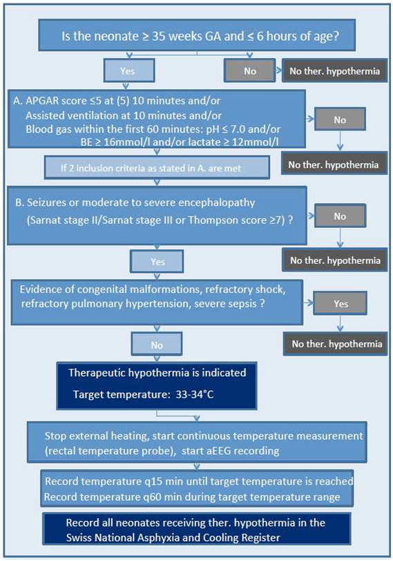
Appendix A. Swiss National Asphyxia and Cooling Flowchart

Preprints 141719 i001

References

  1. Jacobs, S.E.; Berg, M.; Hunt, R.; Tarnow-Mordi, W.O.; Inder, T.E.; Davis, P.G. Cooling for newborns with hypoxic ischaemic encephalopathy. Cochrane Database Syst. Rev. 2013, 2013, CD003311. [Google Scholar] [CrossRef]
  2. Shankaran, S.; Laptook, A.R.; Ehrenkranz, R.A.; Tyson, J.E.; McDonald, S.A.; Donovan, E.F.; Fanaroff, A.A.; Poole, W.K.; Wright, L.L.; Higgins, R.D.; et al. Whole-Body Hypothermia for Neonates with Hypoxic–Ischemic Encephalopathy. New Engl. J. Med. 2005, 353, 1574–1584. [Google Scholar] [CrossRef]
  3. Shankaran, S.; Pappas, A.; McDonald, S.A.; Vohr, B.R.; Hintz, S.R.; Yolton, K.; Gustafson, K.E.; Leach, T.M.; Green, C.; Bara, R.; et al. Childhood Outcomes after Hypothermia for Neonatal Encephalopathy. N. Engl. J. Med. 2012, 366, 2085–2092. [Google Scholar] [CrossRef]
  4. Azzopardi, D.V.; Strohm, B.; Edwards, A.D.; Dyet, L.; Halliday, H.L.; Juszczak, E.; Kapellou, O.; Levene, M.; Marlow, N.; Porter, E.; et al. Moderate Hypothermia to Treat Perinatal Asphyxial Encephalopathy. New Engl. J. Med. 2009, 361, 1349–1358. [Google Scholar] [CrossRef] [PubMed]
  5. Azzopardi, D.; Strohm, B.; Marlow, N.; Brocklehurst, P.; Deierl, A.; Eddama, O.; Goodwin, J.; Halliday, H.L.; Juszczak, E.; Kapellou, O.; et al. Effects of Hypothermia for Perinatal Asphyxia on Childhood Outcomes. N. Engl. J. Med. 2014, 371, 140–149. [Google Scholar] [CrossRef] [PubMed]
  6. Shankaran, S.; Laptook, A.R.; Pappas, A.; McDonald, S.A.; Das, A.; Tyson, J.E.; Poindexter, B.B.; Schibler, K.; Bell, E.F.; Heyne, R.J.; et al. Effect of Depth and Duration of Cooling on Deaths in the NICU Among Neonates With Hypoxic Ischemic Encephalopathy. JAMA 2014, 312, 2629–2639. [Google Scholar] [CrossRef]
  7. Natarajan, G.; Mathur, A.; Zaniletti, I.; DiGeronimo, R.; Lee, K.-S.; Rao, R.; Dizon, M.; Hamrick, S.; Rudine, A.; Cook, N.; et al. Withdrawal of Life-Support in Neonatal Hypoxic-Ischemic Encephalopathy. 2019, 91, 20–26. [Google Scholar] [CrossRef]
  8. McCarthy, F.T.; Kenis, A.; Parravicini, E. Perinatal palliative care: focus on comfort. Front. Pediatr. 2023, 11, 1258285. [Google Scholar] [CrossRef] [PubMed]
  9. Dombrecht, L.; Chambaere, K.; Beernaert, K.; Roets, E.; Keyser, M.D.V.D.; De Smet, G.; Roelens, K.; Cools, F. Components of Perinatal Palliative Care: An Integrative Review. Children 2023, 10, 482. [Google Scholar] [CrossRef] [PubMed]
  10. Pion, A.B.; Baenziger, J.; Fauchère, J.-C.; Gubler, D.; Hendriks, M.J. National Divergences in Perinatal Palliative Care Guidelines and Training in Tertiary NICUs. Front. Pediatr. 2021, 9, 673545. [Google Scholar] [CrossRef]
  11. Sarnat, H.B.; Sarnat, M.S. Neonatal Encephalopathy Following Fetal Distress. Arch. Neurol. 1976, 33, 696–705. [Google Scholar] [CrossRef]
  12. Brotschi, B.; Grass, B.; Ramos, G.; Beck, I.; Held, U.; Hagmann, C.; Meyer, P.; Zeilinger, G.; Schulzke, S.; Wellmann, S.; et al. The impact of a register on the management of neonatal cooling in Switzerland. Early Hum. Dev. 2015, 91, 277–284. [Google Scholar] [CrossRef]
  13. Grass, B.; Brotschi, B.; Hagmann, C.; Birkenmaier, A.; Schwendener, K.; Adams, M.; Kleber, M.; Register, S.N.A.A.C. Centre-specific differences in short-term outcomes in neonates with hypoxic-ischaemic encephalopathy. Swiss Med Wkly. 2021, 151, w20489. [Google Scholar] [CrossRef] [PubMed]
  14. Thompson, C.; Puterman, A.; Linley, L.; Hann, F.; van der Elst, C.; Molteno, C.; Malan, A. The value of a scoring system for hypoxic ischaemic encephalopathy in predicting neurodevelopmental outcome. Acta Paediatr. 1997, 86, 757–761. [Google Scholar] [CrossRef]
  15. Basiri, B.; Sabzehei, M.; Sabahi, M. Predictive factors of death in neonates with hypoxic-ischemic encephalopathy receiving selective head cooling. Clin. Exp. Pediatr. 2021, 64, 180–187. [Google Scholar] [CrossRef] [PubMed]
  16. Deveci̇, M.F.; Turgut, H.; Alagöz, M.; Kaya, H.; Gökçe, I.K.; Özdemi̇r, R. Mortality Related Factors on Hypoxic Ischemic Encephalopathic Patients Treated with Therapeutic Hypothermia: A 11-year Single Center Experience. Turk. J. Med Sci. 2022, 52, 796–802. [Google Scholar] [CrossRef] [PubMed]
  17. Labat, J.; Brocard, C.; Belaroussi, Y.; Bar, C.; Gotchac, J.; Chateil, J.; Brissaud, O. Hypothermia for neonatal hypoxic–ischemic encephalopathy: Retrospective descriptive study of features associated with poor outcome. Arch. De Pediatr. 2022, 30, 93–99. [Google Scholar] [CrossRef] [PubMed]
  18. \Glass, H.C.; Numis, A.L.; Comstock, B.A.; Gonzalez, F.F.; Mietzsch, U.; Bonifacio, S.L.; Massey, S.; Thomas, C.; Natarajan, N.; Mayock, D.E.; et al. Association of EEG Background and Neurodevelopmental Outcome in Neonates With Hypoxic-Ischemic Encephalopathy Receiving Hypothermia. Neurology 2023, 101, E2223–E2233. [Google Scholar] [CrossRef]
  19. Weeke, L.C.; Groenendaal, F.; Mudigonda, K.; Blennow, M.; Lequin, M.H.; Meiners, L.C.; van Haastert, I.C.; Benders, M.J.; Hallberg, B.; de Vries, L.S. A Novel Magnetic Resonance Imaging Score Predicts Neurodevelopmental Outcome After Perinatal Asphyxia and Therapeutic Hypothermia. J. Pediatr. 2018, 192, 33–40.e2. [Google Scholar] [CrossRef] [PubMed]
  20. Machie, M.; de Vries, L.S.; Inder, T. Advances in Neuroimaging Biomarkers and Scoring. Clin. Perinatol. 2024, 51, 629–647. [Google Scholar] [CrossRef] [PubMed]
  21. Bayley, N. Manual for the Bayley scales of infant and toddler development, 3rd ed. San Antonio, TX: Harcourt Assessment,2006.
  22. Griffith, R. The abilities of young children. High Wycombe, UK: The TestAgency Ltd, 1984.
  23. Palisano, R.; Rosenbaum, P.; Walter, S.; Russell, D.; Wood, E.; Galuppi, B. Development and reliability of a system to classify gross motor function in children with cerebral palsy. Dev. Med. Child Neurol. 1997, 39, 214–223. [Google Scholar] [CrossRef] [PubMed]
  24. Boerger, W.; Mozun, R.; Frey, B.; Liamlahi, R.; Grass, B.; Brotschi, B. Blood Lactate Levels during Therapeutic Hypothermia and Neurodevelopmental Outcome or Death at 18–24 Months of Age in Neonates with Moderate and Severe Hypoxic-Ischemic Encephalopathy. Neonatology 2024, 1–10. [Google Scholar] [CrossRef]
  25. Larg, R.H.; Graf, S.; Kundu, S.; Hunziker, U.; Molinari, L. Predicting Developmental Outcome At School Age From Infant Tests of Normal, At-Risk and Retarded Infants. Dev. Med. Child Neurol. 1990, 32, 30–45. [Google Scholar] [CrossRef]
  26. Brecht, M.; Wilkinson, D.J.C. The outcome of treatment limitation discussions in newborns with brain injury. Arch. Dis. Child. - Fetal Neonatal Ed. 2014, 100, F155–F160. [Google Scholar] [CrossRef] [PubMed]
  27. Parravicini, E. Neonatal palliative care. Curr. Opin. Pediatr. 2017, 29, 135–140. [Google Scholar] [CrossRef]
  28. A Guide to Children’s Palliative Care. 4th edition. Bristol: Together for Short Lives (TfSL). Last accessed th, 2024: https://www.togetherforshortlives.org. 27 November.
  29. Bergstraesser, E.; Hain, R.D.; Pereira, J.L. The development of an instrument that can identify children with palliative care needs: the Paediatric Palliative Screening Scale (PaPaS Scale): a qualitative study approach. BMC Palliat. Care 2013, 12, 1–20. [Google Scholar] [CrossRef] [PubMed]
  30. Bergstraesser, E.; Paul, M.; Rufibach, K.; Hain, R.D.; Held, L. The Paediatric Palliative Screening Scale: Further validity testing. Palliat. Med. 2013, 28, 530–533. [Google Scholar] [CrossRef] [PubMed]
  31. British Association of Perinatal Medicine. Recognising uncertainty: an integrated framework for palliative care in perinatal medicine. A BAPM Framework for Practice, July 2024. Last accessed November 16th, 2024: https://www.bapm.org/resources/palliative-care-in-perinatal-medicine-framework.
  32. Zimmermann, K.; on behalf of the PELICAN Consortium; Cignacco, E.; Engberg, S.; Ramelet, A.-S.; von der Weid, N.; Eskola, K.; Bergstraesser, E.; Ansari, M.; Aebi, C.; et al. Patterns of paediatric end-of-life care: a chart review across different care settings in Switzerland. BMC Pediatr. 2018, 18, 67. [CrossRef]
  33. Wyatt, J.; Hain, R. Redirecting Care: Compassionate Management of the Sick or Preterm Neonate at the End of Life. Children 2022, 9, 344. [Google Scholar] [CrossRef] [PubMed]
  34. Raghu, K.; Kalish, B.T.; Tam, E.W.; El Shahed, A.; Chau, V.; Wilson, D.; Tung, S.; Kazazian, V.; Miran, A.A.; Hahn, C.; et al. Prognostic Indicators of Reorientation of Care in Perinatal Hypoxic-Ischemic Encephalopathy Spectrum. J. Pediatr. 2024, 276, 114273. [Google Scholar] [CrossRef]
  35. Cawley, P.; Chakkarapani, E. Fifteen-minute consultation: Therapeutic hypothermia for infants with hypoxic ischaemic encephalopathy-translating jargon, prognosis and uncertainty for parents. Arch. Dis. Child. - Educ. Pr. Ed. 2020, 105, 75–83. [Google Scholar] [CrossRef]
  36. Grass, B.M.; Erlach, M.; Rathke, V.; Cippa, G.; Hagmann, C.; Brotschi, B. Parents' Experiences on Therapeutic Hypothermia for Neonates With Hypoxic-Ischemic Encephalopathy (HIE): A Single-Center Cross-Sectional Study. Qual. Manag. Heal. Care 2023, 33, 94–100. [Google Scholar] [CrossRef] [PubMed]
  37. Fraser, L.K.; Gibson-Smith, D.; Jarvis, S.; Norman, P.; Parslow, R.C. Estimating the current and future prevalence of life-limiting conditions in children in England. Palliat. Med. 2020, 35, 1641–1651. [Google Scholar] [CrossRef] [PubMed]
Figure 1. Flowchart of included neonates. HIE: Hypoxic-ischemic encephalopathy; TH: Therapeutic hypothermia; NICU: Neonatal Intensive Care Unit; NDFU: Neurodevelopmental follow-up assessment at 18-24 months of age.
Preprints 141719 g001
Figure 2. Comparison of age at decision to redirect care and age at death. Age (postnatal days) at decision to redirect care and occurrence of death. The size of the symbols refers to the absolute number of patients.
Preprints 141719 g002
Table 1. Maternal, pregnancy and delivery characteristics.
Characteristics Death (n=23) Survivor (n=114)
n(%) or median (IQR) n(%) or median (IQR) p value
Sex, male 14 (60.9) 58 (50.9) 0.5179
Gestational age (weeks) 40.0 (39.0, 40.6) 39.8 (38.4, 40.7) 0.5038
Birth weight (grams) 3450 (3050, 3658) 3380 (3000, 3724) 0.5542
Outborn 23 (100.0) 105 (92.1) 0.3561
Small for gestation (< 10. percentile) 2 (8.7) 14 (12.3) 1.0000
Multiple gestation 1 (4.3) 4 (3.5) 1.0000
Maternal age (years) 34 (31, 38) 33 (30, 35) 0.2930
Primiparae 15 (71.4) 68 (62.4) 0.5879
SES Score 4 (2, 10) 4 (3, 6) 0.8834
Mode of delivery
Vaginal delivery (all) 12 (52.2) 61 (53.5) 1.0000
spontaneous, cephalic 7 (30.4) 36 (31.6)
spontaneous, breech 0 (0.0) 2 (1.8)
instrumental 5 (21.7) 23 (20.2)
Caesarean delivery (all) 11 (47.8) 53 (46.5) 1.0000
elective CS 1 (4.3) 5 (4.4)
emergency CS 8 (34.8) 41 (36.0)
secondary CS 2 (8.7) 7 (6.1)
CS: caesarean section; SES: socioeconomic status [25 Largo]. Missing values: Maternal age = 3 and 19, primiparae = 2 and 5, and SES-Score = 5 and 24, in the death and survivor group, respectively.
Table 2. Neonatal characteristics.
Characteristics Death (n=23) Survivor (n=114)
(according to Swiss National Cooling Protocol) n(%) or Median (IQR) n(%) or Median (IQR) p value
Apgar score
at 1 minute 0 (0, 1) 2 (1, 3) 0.0003
at 5 minutes 1 (1, 4) 4 (2, 5) 0.0032
at 10 minutes 2 (1, 4) 5 (3, 6) 0.0056
Resuscitation in the delivery room* 18 (78.3) 65 (57.0) 0.0650
Umbilical artery pH 6.87 (6.79, 7.04) 6.99 (6.88, 7.10) 0.0934
Blood gas analysis, worst#
pH 6.78 (6.61, 6.80) 6.86 (6.79, 6.95) <0.0001
Base deficit (mmol/L) 24.4 (19.1, 28.5) 19.0 (14.8, 22.0) 0.0029
Lactate (mmol/L) 17.7 (15.0, 19.0) 14.1 (11.5, 16.0) 0.0003
* Resuscitation was defined as any form of ventilation necessary at the age of 10 minutes. # Worst blood gas analysis was defined as worst blood gas results within 60 min after birth, including umbilical gases. Missing values: Apgar at 1 minute = 1 and 2, Apgar at 5 minutes = 2 and 1, Apgar at 10 minutes = 2 and 1, umbilical artery pH = 5 and 15, pH = 1 and 5, base deficit = 6 and 12, lactate = 4 and 17, in the death and survivor group, respectively.
Table 3. Prognostic factors of expected unfavorable neurodevelopmental outcome leading to redirection of care.
Table 3. a. Comparison of prognostic factors between deaths and survivors.
Neurologic variables Death (n=23) Survivor (n=114)
n(%) or median (IQR) n(%) or median (IQR) pvalue
Sarnat Score
on admission (prior to TH) 0.0177
stage 1 0 (0.0) 5 (4.5)
stage 2 9 (39.1) 73 (65.8)
stage 3 14 (60.9) 33 (29.7)
day 1 <0.0001
stage 1 1 (5.0) 29 (26.9)
stage 2 6 (30.0) 62 (57.4)
stage 3 13 (65.0) 17 (15.7)
day 2* <0.0001
stage 1 1 (4.8) 33 (31.7)
stage 2 4 (19.0) 61 (58.7)
stage 3 9 (42.9) 10 (9.6)
day 3* <0.0001
stage 1 1 (7.1) 38 (38.8)
stage 2 2 (14.3) 53 (54.1)
stage 3 7 (50.0) 7 (7.1)
day 4* 0.0034
stage 1 1 (9.1) 45 (50.0)
stage 2 4 (36.4) 39 (43.3)
stage 3 4 (36.4) 6 (6.7)
Seizure
day 1 11 (47.8) 19 (16.7) 0.0022
day 2* 7 (33.3) 14 (12.3) 0.0227
day 3* 6 (42.9) 7 (6.1) 0.0006
day 4* 3 (27.3) 7 (6.1) 0.0436
total 14 (60.9) 28 (24.6) 0.0014
MRI done at discharge 7 (30.4) 111 (97.4) <0.0001
Highest lactate (mmol/L)
day 1 11.0 (10.5, 15.2) 6.8 (4.3, 12.0) 0.0233
day 2* 7.3 (5.5, 7.8) 3.4 (2.2, 4.7) 0.0063
day 3* 4.3 (3.7, 4.6) 2.0 (1.5, 3.4) 0.0245
day 4* 3.0 (2.6, 4.4) 1.5 (1.2, 2.1) 0.0297
TH: therapeutic hypothermia * The number of deaths on the first, second and third day of life was 2, 7 and 3, respectively. Therefore, the death group on the 2nd, 3rd and 4th day of life contains only 21, 14 and 11 surviving infants, respectively. Missing values: Sarnat before cooling = 3 in the survivor group. Sarnat day 1 = 3 and 6, Sarnat day 2 = 7 and 10, Sarnat day 3 = 4 and 16, Sarnat day 4 = 2 and 24, highest lactate on day 1 = 16 and 48, day 2 = 15 and 48, day 3 = 10 and 49, and day 4 = 8 and 51, in the death and survivor group, respectively.
Disclaimer/Publisher’s Note: The statements, opinions and data contained in all publications are solely those of the individual author(s) and contributor(s) and not of MDPI and/or the editor(s). MDPI and/or the editor(s) disclaim responsibility for any injury to people or property resulting from any ideas, methods, instructions or products referred to in the content.
Copyright: This open access article is published under a Creative Commons CC BY 4.0 license, which permit the free download, distribution, and reuse, provided that the author and preprint are cited in any reuse.
Prerpints.org logo

Preprints.org is a free preprint server supported by MDPI in Basel, Switzerland.

Subscribe

Disclaimer

Terms of Use

Privacy Policy

Privacy Settings

© 2025 MDPI (Basel, Switzerland) unless otherwise stated