Preprint
Article

This version is not peer-reviewed.

Mortality in Antinuclear Antibody-Positive Patients with and Without Rheumatologic Immune-Related Disorders: A Large-Scale Population-Based Study

A peer-reviewed article of this preprint also exists.

Submitted:

02 December 2024

Posted:

03 December 2024

You are already at the latest version

Abstract

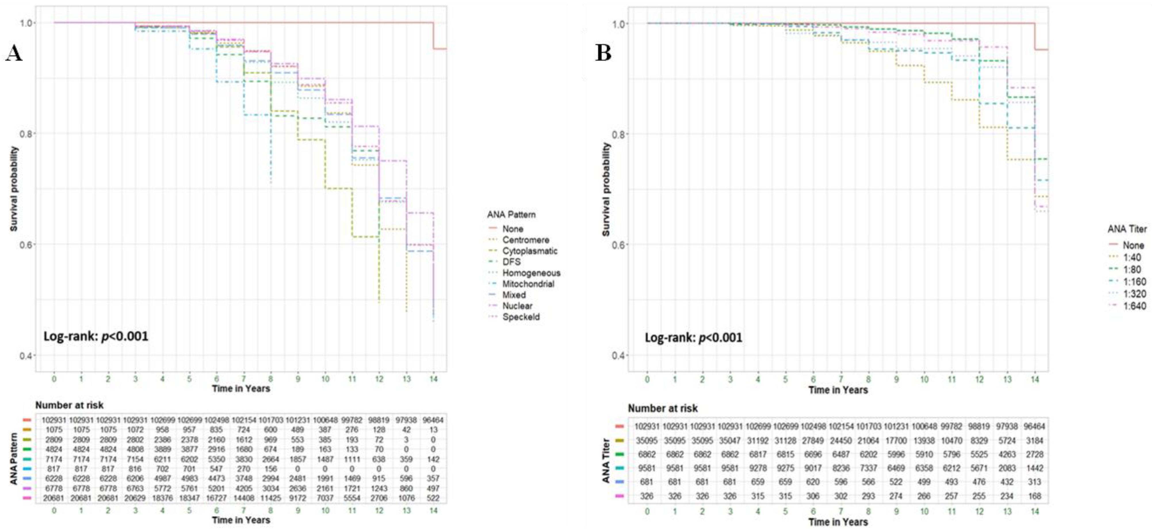
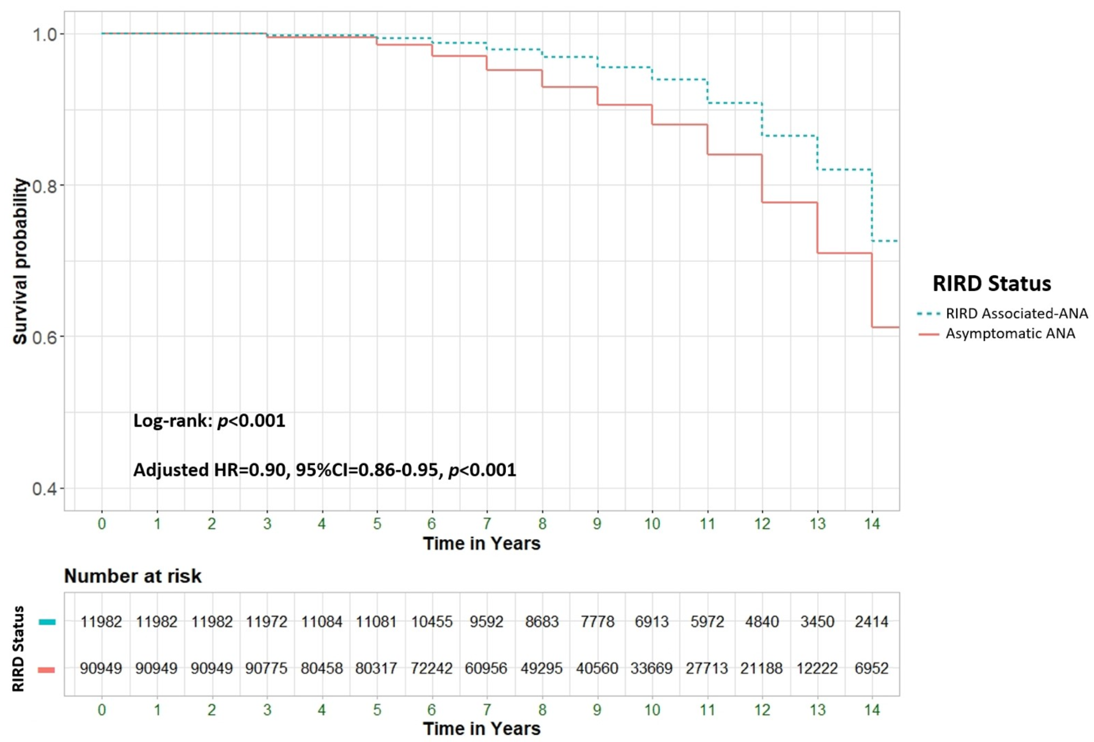
Background & Objectives: To explore the potential association between positive ANA serology and all-cause mortality in a large cohort of patients, including those with and without rheumatological conditions and other immune-related diseases. Material and Methods: A retrospective cohort study analyzed all-cause mortality among 205,862 patients from Clalit Health Services (CHS), Israel's largest health maintenance organization (HMO). We compared patients aged 18 and older with positive ANA serology (n=102,931) to an equal number of ANA-negative controls (n=102,931). Multivariable Cox regression models were used to assess hazard ratios (HR) for mortality, adjusting for demographic and clinical factors. Results: ANA positivity was strongly associated with increased mortality (adjusted HR [aHR] 4.62; 95% CI 4.5-4.7, p<0.001). Significant predictors of mortality included male gender (39.2% vs. 24.4%, p<0.001), older age at testing (72.4 ± 13.0 vs. 50.1 ± 17.3 years, p<0.001), and Jewish ethnicity (89.6% vs. 83.2%, p<0.001). Certain ANA patterns, such as mitochondrial and dense fine speckled (DFS), were highly predictive of mortality, with aHRs of 36.14 (95% CI 29.78-43.85) and 29.77 (95% CI 26.58-33.34), respectively. ANA-positive patients with comorbid rheumatological immune-related disorders (RIRDs) demonstrated a higher survival rate compared to those without such a condition (aHR 0.9, 95% CI 0.86–0.95, p < 0.001). This finding remained significant after adjusting for several parameters, including age. Conclusion: ANA positivity is associated with increased all-cause mortality, particularly in individuals without rheumatologic disorders, after adjusting for confounders such as age. This may indicate occult malignancies, cardiovascular pathology, or chronic inflammatory states, necessitating more vigilant surveillance.

Keywords: 
;  ;  ;  

Introduction

Antinuclear antibodies (ANAs) are autoantibody that targets nuclear of normal human cells, indicating a breakdown of immunologic tolerance [1]. The detection of ANA is important for diagnosing ANA-associated rheumatic diseases (AARD), including systemic erythematous lupus (SLE) and systemic sclerosis (SS) [2], but also and other autoimmune conditions [3], including autoimmune thyroid disorders [2], highlighting the complex interplay between various components of the immune system.
ANA positivity can appear years before the development of symptomatic autoimmune disease [4]. The prevalence of ANA positivity is high and nonspecific, found in approximately 12–16% of the general U.S. population, with higher occurrences among women, older individuals (ages ≥70 years), African Americans, and those of normal weight [4,5]. In China, ANA positivity rates are also high and correlate with sex and age groups [6].
The reasons for elevated ANA levels in healthy and aging individuals are not well understood, but genetic or environmental factors influencing immune regulation and activity may play a role. These antibodies can also be associated with non-autoimmune factors such as chronic diseases including heart disease [7], malignancies [8,9], infections [10], and certain medications [11], although evidence is limited. Notably, inflammation-related exposures such as obesity and smoking have not been linked to higher ANA prevalence in the U.S. population [4].
However, some research has explored potential links between ANA positivity and mortality due to cardiovascular disease (CVD) and cancer [4]. ANA has shown a weak overall link with all-cause mortality, but studies suggest stronger associations with increased mortality from cardiovascular disease and cancer in certain demographic groups. While ANA may predict cardiovascular events, its connection to cancer risk and prognosis remains unclear, with mixed evidence[1,5,9,12,13]. Unlike cardiovascular diseases and cancer, the impact of chronic inflammatory conditions on all-cause mortality in ANA-positive patients has been less thoroughly investigated.
Due to the limited and often contradictory existing literature, this study aimed to explore the potential association between positive ANA serology and all-cause mortality in a large cohort of patients, including those with and without rheumatological conditions and other immune-related diseases.

Material and Methods

Study Design and Population

A retrospective study was conducted in Israel between January 1st 2000 and December 31st, 2022. Inclusion criteria included all individuals aged 18 or older with positive ANA serology (i.e., the test group), who were then compared to ANA-negative patients (i.e., the control group). The Ethical Committee of Clalit Health Service (CHS) granted approval for the study, which was exempted from obtaining informed consent.

Data Collection

We utilized data from the electronic database of Clalit Health Services (CHS), the largest health maintenance organization (HMO) in Israel [14]. The CHS serves over 4.5 million patients across the country. Under the Israeli National Health Insurance Act of 1994, CHS provides comprehensive healthcare coverage to all citizens. Enrolment in one of the four HMOs is mandatory for every Israeli, ensuring access to healthcare services and insurance coverage regardless of gender, age, health status, or other factors. The extensive CHS database consolidates patient medical records from various healthcare providers, including diagnostic visits, pharmaceutical interventions, in-office procedures, laboratory test results, imaging studies, and comprehensive summaries of hospital encounters such as outpatient clinic visits, emergency department visits, and inpatient discharge records.

Outcomes and Definitions

The primary outcome of this study was to evaluate all-cause mortality rates among patients who tested positive for ANA serology compared to those who tested negative. Patients were defined as ANA-positive if they had an ANA titer of 1:40 or higher, measured by enzyme-linked immunosorbent assay (ELISA) on at least two separate occasions.
A secondary outcome of this study was to explore the potential relationship between specific ANA characteristics—such as titer and pattern—and all-cause mortality. ANA titers were assessed in a range from 1:40 to 1:640, with increments of twofold. The ANA patterns analyzed included the speckled pattern, which is the standard in ANA serology testing at CHS, as well as centromere, cytoplasmic, dense fine speckled (DFS), homogeneous, mitochondrial, mixed, and nuclear patterns.
Given the established association between ANA serology and systemic autoimmune diseases, an additional secondary outcome of this study was to investigate whether ANA positivity is significantly linked to increased mortality in patients diagnosed with various rheumatological conditions. Identified using recorded diagnoses from the International Classification of Diseases, 9th Revision (ICD-9) [15], and included systemic lupus erythematosus (SLE, code 710.0), systemic sclerosis (diffuse type, code 710.1), Sjögren's syndrome (code 710.2), rheumatoid arthritis (RA, code 714.0), and myositis (code 729.1).
In addition, we evaluated rheumatic diseases where ANA is not considered to play a pathogenic role, specifically psoriatic arthritis (PsA, code 696.0), reactive arthritis (ReA, code 711.1), and ankylosing spondylitis (AS, code 720.0). All the rheumatological conditions analyzed in this study, regardless of whether ANA was involved as a major pathogenic factor, were categorized collectively as Rheumatologic-Immune Related Disorders (RIRD).
Asymptomatic ANA patient was defined as any patient with a positive ANA titer of at least 1:40 who did not have an additional diagnosis of any rheumatological disorders at the time of analysis (i.e., in 2023) included in our data. JUST FOR ONE TIMEPOINT EVER?
The study also considered demographic parameters such as age (in years), gender (male or female), and ethnicity (classified as Arab or Jewish). Smoking status (current or past) was recorded. Metabolic comorbidities, including diabetes mellitus (both type 1 and type 2), hyperlipidaemia, hypertension, and obesity, were documented, along with cancer-related comorbidities.

Statistical Analysis

Descriptive statistics were used to analyse the attributes of the study population. Each variable was presented using the most appropriate central and dispersion measures: nominal variables (e.g., patient sex, comorbidities) were presented as number and percentage (%), while numerical (continuous) variables (e.g., patient age) were presented as mean ± standard deviation (SD). The normality of continuous variables was assessed using histograms, Q-Q plots, the Shapiro-Wilk test, and the Kolmogorov-Smirnov test.
Initially, we conducted a univariate analysis to evaluate the clinical and sociodemographic characteristics of the study cohort, stratified by ANA status, and to assess their association with all-cause mortality among ANA-positive patients. For continuous variables, the Mann-Whitney test was used due to their non-normal distribution, and for nominal variables, the Chi-square test was employed. Next, we examined the association between ANA characteristics (e.g., ANA status, pattern, and titer) and all-cause mortality using Kaplan-Meier curves and the Log-rank test. Finally, we used multivariable Cox regression to assess the hazard ratio (HR) of patients' ANA characteristics, adjusting for potential confounders. All analyses were conducted using SPSS Statistics V. 25 and R software. A two-sided test significance level of 0.05 was maintained throughout the entire study.

Ethical Considerations

The Ethical Committee of Clalit Health Services (CHS) granted approval for the study, which was exempted from obtaining informed consent.

Results

Study Population

The study included 205,862 patients, evenly divided into two groups: 102,931/205,862 patients (50%) tested positive for ANA (study group), and 102,931/205,862 patients (50%) tested negative for ANA (control group). Table 1 outlines the baseline characteristics of each group. Patients in the study group were younger, with a mean age of 53.2 years (SD ± 18.5), compared to 50.3 years (SD ± 18.6) in the control group (p<0.001). The gender distribution (male/female) was equal between the two group in the original dataset. Jewish ethnicity was more prevalent in the control group (90,402/102,931 patients, 88%) compared to the study group (86,446/102,931 patients, 84.1%) (p<0.001).
In terms of comorbidities, the prevalence of hypertension was higher in the study group (41,138/102,931 patients, 40%) compared to the control group (39,796/102,931 patients, 38.7%) (p<0.001). Conversely, hyperlipidemia was more prevalent in the control group (61,049/102,931 patients, 59.3%) compared to the study group (60,028/102,931 patients, 58.3%). As expected, the prevalence of the most rheumatological conditions was higher in the study group, except for ReA, AS and PsA, which the latter two was more prevalent in ANA-negative patients.
Table 1. Clinical and sociodemographic characteristics of Study Cohort stratified by ANA Status.
Variable All Patients(n=205,862) ANA Positive(N=102,931) ANA Negative(N=102,931) P-value
Patient Sex Male 54406(26.4) 27203(26.4) 27203(26.4) 1.00
Female 151456(73.6) 75728(73.6) 75728(73.6)
Age at ANA test, years 50.3±18.6 53.2±18.5 47.4±18.3 <0.001
Ethnicity Jewish 176850(86.0) 86448(84.1) 90402(88.0) <0.001
Arab 28701(14.0) 16334(15.9) 12367(12.0)
Smoking 72363(35.2) 36305(35.3) 36058(35.0) 0.254
Comorbidities Diabetes Mellitus 49689(24.1) 24858(24.2) 24831(24.1) 0.889
Hyperlipidemia 121077(58.8) 60028(58.3) 61049(59.3) <0.001
Hypertension 80934(39.3) 41138(40.0) 39796(38.7) <0.001
Obesity 66757(32.4) 33540(32.6) 33217(32.3) 0.128
rheumatological immune-related disorders (RIRD) Any 16429(8.0) 11982(11.6) 4447(4.3) <0.001
SLE 5063(2.5) 4445(4.3) 618(0.6) <0.001
Psoriatic arthritis 1888(0.9) 839(0.4) 1049(0.9) <0.001
Systemic Sclerosis (diffused) 1191(0.6) 1113(1.1) 78(0.1) <0.001
Sjogren 2500(1.2) 2015(2.0) 485(0.5) <0.001
Myositis 144(0.1) 122(0.1) 22(0.0) <0.001
Reactive arthritis 22(0.0) 6(0.0) 16(0.0) 0.033
Rheumatoid arthritis 6193(3.0) 3994(3.9) 2199(2.1) <0.001
Ankylosing Spondylitis 919(0.4) 132(0.1) 787(0.9) <0.001
Malignancy Any 35030(17.0) 17864(17.4) 17166(16.7) <0.001
Solid 29613(14.4) 14995(14.6) 14618(14.2) 0.018
Hematologic 5417(2.6) 2869(2.8) 2548(2.5) <0.001
Breast 7360(3.6) 3602(3.5) 3758(3.7) 0.064
CRC 3468(1.7) 1768(1.7) 1700(1.7) 0.244
Prostate 1995(1.0) 967(0.9) 1028(1.0) 0.17
Lung 1863(0.9) 991(1.0) 872(0.8) 0.006
Bladder 1515(0.7) 781(0.8) 734(0.7) 0.226
Ovary 611(0.3) 329(0.3) 282(0.3) 0.057
Uterus 1063(0.5) 509(0.5) 554(0.5) 0.166
Pancreas 651(0.3) 313(0.3) 338(0.3) 0.326
CNS 459(0.2) 216(0.2) 243(0.2) 0.207
Stomach 596(0.3) 306(0.3) 290(0.3) 0.512
Melanoma 1853(0.9) 904(0.9) 949(0.9) 0.294
HL 546(0.3) 310(0.3) 236(0.2) 0.002
NHL 2270(1.1) 1251(1.2) 1019(1.0) <0.001
AL 462(0.2) 228(0.2) 234(0.2) 0.78
CL 556(0.3) 286(0.3) 270(0.3) 0.497
Kidney 908(0.4) 457(0.4) 451(0.4) 0.842
Larynx 336(0.2) 174(0.2) 162(0.2) 0.512
Cervix 1112(0.5) 590(0.6) 522(0.5) 0.041
Pharynx 424(0.2) 232(0.2) 192(0.2) 0.052
Esophagus 70(0.0) 34(0.0) 36(0.0) 0.811
Liver / Bile 512(0.2) 281(0.3) 231(0.2) 0.027
Thyroid 1620(0.8) 820(0.8) 800(0.8) 0.618
Bone 110(0.1) 59(0.1) 51(0.0) 0.445
Sarcoma 490(0.2) 249(0.2) 241(0.2) 0.717
Genital 264(0.1) 143(0.1) 121(0.1) 0.175
MM 706(0.3) 348(0.3) 358(0.3) 0.706
PV 479(0.2) 231(0.2) 248(0.2) 0.437
MDS 287(0.1) 154(0.1) 133(0.1) 0.215
MPS 111(0.1) 61(0.1) 50(0.0) 0.296
Other Site 1071(0.5) 536(0.5) 535(0.5) 0.976
Unknown Site 1262(0.6) 734(0.7) 528(0.5) <0.001
The prevalence of any malignancy was significantly more prevalent in the ANA-positive group, affecting 17,864/102,931 patients (17.4%), compared to 17,166/102,931 patients (16.7%) in the ANA-negative group (p<0.001). However, the distribution of most malignancy subtypes was similar between the two groups, mostly with insignificant differences.

Risk for All-Cause Mortality
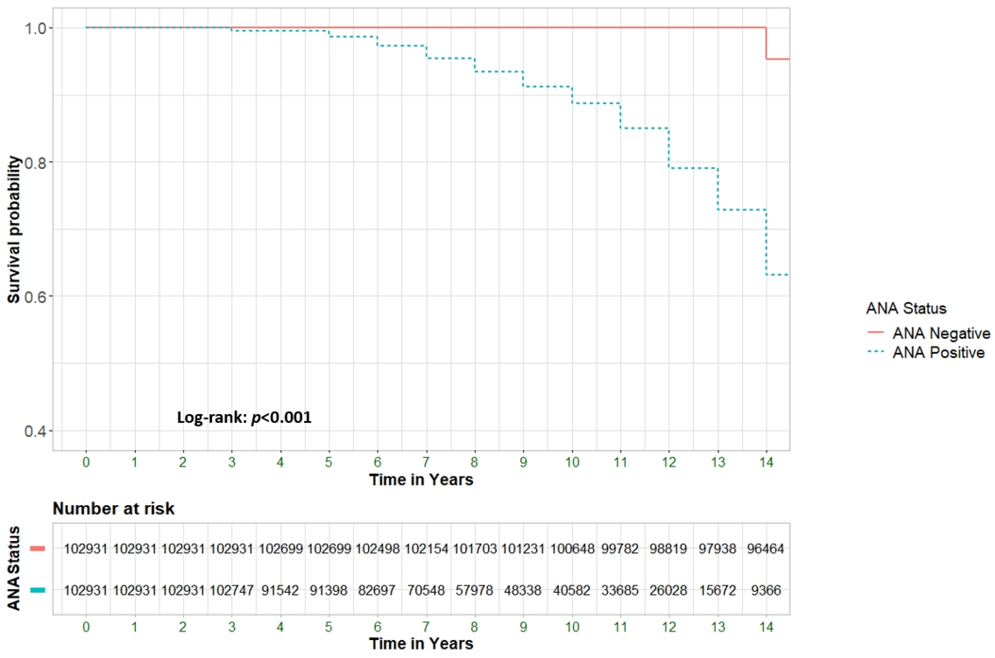
Figure 1 illustrates the progressively decreasing survival rates among ANA-positive patients compared to ANA-negative patients (p<0.001). To identify specific predictors of all-cause mortality, a multivariate analysis was performed to evaluate potential associations between clinical and epidemiological factors and mortality risk (supplementary table 1). The hazard ratio (HR) for the association between positive ANA status and all-cause mortality was 6.18 (95% CI 6.0–6.3, p<0.001). After adjusting for significant baseline variables, the adjusted HR (aHR) was 4.62 (95% CI 4.5–4.7, p<0.001).
Figure 1. Survival analysis of patients with positive ANA compared to patients with negative ANA.
Preprints 141530 g001
The presence of RIRDs were associated with increased all-cause mortality in ANA-positive patients, with a preliminary hazard ratio (HR) of 1.27 (95% CI, 1.22–1.32, p < 0.001). However, after adjusting for significant variables, there was a notable shift; the presence of RIRDs exhibited a relative protective effect, resulting in an adjusted hazard ratio (aHR) of 0.83 (95% CI, 0.79–0.86, p < 0.001). Other examined factors, such as age at ANA testing, Jewish ethnicity, hypertension, and malignancy, were significantly associated with all-cause mortality both before and after adjustment (p < 0.001).
  • ANA characteristics
Table 2 illustrates the relationship between specific ANA characteristics and all-cause mortality.
Different ANA titers were found to correlate with all-cause mortality, with the strongest correlation observed at the lowest titer of 1:40, which had an aHR of 4.70 (95% CI 4.53-4.88, p<0.001). Figure 2A visually represents the different survival curves for each ANA titer that was analyzed. Additionally, different ANA patterns were analyzed, and similar to the ANA titre analysis, all patterns were found to be significantly correlated with all-cause mortality. The highest associations were observed for the mitochondrial, dense fine speckled (DFS), and cytoplasmic patterns, with adjusted hazard ratios (aHRs) of 36.14 (95% CI 29.78–43.85, p < 0.001), 29.77 (95% CI 26.58–33.34, p < 0.001), and 20.97 (95% CI 18.77–23.43, p < 0.001), respectively. Figure 2B illustrates the survival curves for the different ANA patterns.
Figure 2. Survival rates among different ANA patterns [A] and different ANA titers [B].
Preprints 141530 g002
  • The role of concurrent RIRD
Given the established link between positive ANA serology and various rheumatological conditions, we evaluated the association of ANA positivity in specific subgroups of patients with confirmed rheumatological diagnoses, comparing them to those without these conditions. Table 3 outlines the baseline characteristics of the ANA-positive subgroup, while Figure 3 visually depicts the survival curve comparing patients with RIRD-associated ANA to those with asymptomatic ANA (aHR 0.9, 95% CI 0.86–0.95, p<0.001).
Table 3. Clinical and sociodemographic characteristics Associated with All-Cause Mortality Among ANA Positive Patients.
Variable All Patients Died Alive P-value
(n=102,931) (N=14,083) (N=88,848)
Patient Sex Male 27203(26.4) 5514(39.2) 21689(24.4) <0.001
Female 75728(73.6) 8569(60.8) 67159(75.6)
Age at ANA Test, years 53.2±18.5 72.4±13.0 50.1±17.3 <0.001
Ethnicity Jewish 86448(84.1) 12609(89.6) 73839(83.2) <0.001
Arab 16334(15.9) 1466(10.4) 14868(16.8)
Smoking 36305(35.6) 5920(42.0) 30385(34.2) <0.001
Comorbidities Diabetes Mellitus 24858(24.2) 6450(45.8) 18408(20.7) <0.001
Hyperlipidemia 60028(58.3) 11490(81.6) 48538(54.6) <0.001
Hypertension 41138(40.0) 11167(79.3) 29971(33.7) <0.001
Obesity 33540(32.6) 5458(38.8) 28082(31.6) <0.001
Rheumatological immune-related disorders (RIRD). Any 11982(11.6) 1684(12.0) 10298(11.6) 0.207
SLE 4814(4.6) 769(5.5) 4045(4.5) <0.001
Psoriatic arthritis 839(0.8) 37(0.3) 802(0.9) <0.001
Systemic sclerosis (diffuse) 1305(1.3) 370(2.6) 935(1.1) <0.001
Sjogren 2015(2.0) 273(1.9) 1742(2.0) 0.86
Myositis 122(0.1) 34(0.2) 88(0.1) <0.001
Reactive arthritis 6(0.0) 1(0.0) 5(0.0) 0.832
Rheumatoid arthritis 3994(3.9) 585(4.2) 3409(3.8) 0.07
Ankylosing Spondylitis 132(0.1) 54(0.1) 115(0.1) 0.424
Malignancy Any 17864(17.4) 6630(47.1) 11234(12.6) <0.001
Figure 3. Survival rates among patients with positive RIRD (Rheumatological immune-related disorder) status compared to asymptomatic patients. HR = Hazard Ratio, CI = Confidence Interval. Cox Regression adjusted to age, ethnicity, sex and comorbidities.
Preprints 141530 g003
In a sub analysis of specific RIRDs, classic ANA-positive diseases, including SLE, systemic sclerosis (SS) and myositis, were statistically associated with increased mortality. SLE was found to present in 769/14,083 patients (5.5%) who died until the end of study period, compared to 4045/88,848 patients (4.5%) who were alive (p<0.001). SS was found in 370/14,083 patients (2.6%) who died until the end of study period, compared to 935/88,848 patients (1.1%) who were alive (p < 0.001). Myositis was found in 34/14,083 patients (0.2%) who died, compared to 88/88,848 patients (0.1%) who were alive (p < 0.001). In contrast, Sjögren's syndrome and rheumatoid arthritis (RA) were not significantly correlated with mortality. RA was found in 585/14,083 patients (4.2%) who died, compared to 3,409/88,848 patients (3.8%) who were alive (p = 0.07) and Sjogren’s was present in 273/14,083 patients (1.9%) who died, compared to 1742/88,848 patients (2%) who were alive (p=0.86).
In this study, we also included rheumatological conditions driven by innate immunity or autoinflammatory processes, where ANA is not significant (e.g., PsA, ReA, AS). Most ANA-positive PsA patients had low titers of 1:40 (791/839, 94.2%), as did ANA-positive AS patients (107/132, 81.1%). PsA was negatively correlated with overall mortality, present in 802/88,848 patients (0.9%) who were alive by the end of the study, compared to 37/14,083 patients (0.3%) who died (p<0.001). AS and ReA were not significantly correlated with mortality in ANA-positive patients.

Discussion

Antinuclear antibodies (ANAs) target nuclear proteins and are a key screening tool for diagnosing chronic inflammatory conditions, especially autoimmune diseases like SLE, Sjögren's syndrome and MCTD. While ANA is sensitive for these diseases, it lacks specificity, as positive results also occur in healthy individuals, those with chronic infections, cancers, or medication-related effects [10,16].
To our knowledge, this is the largest cohort study to examine the relationship between ANA positivity and overall mortality. The main finding of our study is the association between positive ANA serology and all-cause mortality in randomly tested large cohort of 205,862 patients, with an aHR of 6.18 when comparing patients with positive ANA to those with negative ANA. The association between ANA and all-cause mortality in the general population has been explored in the past, but the evidence remains limited.
A study using data from the National Health and Nutrition Examination Survey (NHANES) from 1999-2004, which included 3,357 adults with ANA measurements and mortality data through 2011, found that ANA were not strongly associated with all-cause mortality (HR of 1.13, p=1.60) [4]. However, in subgroups with a history of cancer, positive ANA was associated with elevated all-cause mortality, particularly in men and older participants (≥75 years) [4]. In a study by Hurme et al., ANA positivity did not affect survival rates or inflammation levels in nonagenarians, suggesting that ANA positivity alone, without an accompanying autoimmune condition, does not significantly impact mortality [17]. Conversely, in a retrospective study by Selmi et al., which evaluated data from a random cohort of 2,822 patients in Italy over a 15-year period, there was initially a reduced unadjusted survival in subjects with positive ANA serology. However, this significance was lost after adjusting for age and sex [1]. Our study, which used a comparable follow-up period in a randomly assigned patient cohort, identified a clear and significant correlation between ANA positivity and mortality. This finding may be attributed to the larger cohort size in our study.
In the specific context of malignancy in patients with positive ANA, some studies suggest that ANA positivity may be linked to an increased risk of cancer and higher mortality, leading to a poorer prognosis. However, the evidence remains mixed. For example, a 15-year study found no significant association between ANA levels and elevated cancer risk or mortality [1]. Conversely, another study reported a significant correlation between the presence of ANA and survival rates, though no connection was found between ANA positivity and tumor stage, histological type, or the intensity and pattern of ANA staining [13]. In our study, since malignancy was statistically significantly more prevalent in the ANA-positive group compared to the ANA-negative group, we attempted to adjust for the impact of malignancy. However, the correlation between ANA positivity and mortality persisted, with an adjusted hazard ratio (aHR) of 4.62. This finding suggests that the association between ANA and overall mortality is independent of coexisting malignancy.
Aiming to identify specific predictors of all-cause mortality, we performed a multivariate regression analysis, adjusting for significant baseline variables. This adjustment resulted in a shift in the HR for rheumatological conditions, transforming them from a risk factor to a protective factor after accounting for ANA status and several other parameters. Given the prevalence of ANA positivity in both rheumatological conditions and the healthy population, we conducted a sub-analysis to assess the impact of rheumatological conditions on mortality in ANA-positive patients.
Over a 14-year follow-up period, we found that patients with rheumatological-associated ANA had a higher survival rate compared to those with asymptomatic ANA. Previous studies, particularly those focused on SLE, suggest that patients with known chronic inflammatory condition have more rigorous monitoring, regular check-ups, and supplementary examinations [18,19]. This heightened vigilance can facilitate the early detection of other potential causes of mortality. For instance, Yazdany et al. found that frequent physician visits and generalist involvement improved preventive care in SLE including cancer screening and immunizations, highlighting the importance of regular monitoring and comprehensive management [19]. Additionally, a study by Castro et al. demonstrated that SLE patients had higher screening rates for several metabolic and non-metabolic conditions, including hypertension, diabetes, and osteoporosis, compared to non-SLE counterparts [18]. The authors concluded that SLE patients are more likely to receive certain preventive services, potentially leading to earlier detection and management of comorbid conditions [18]. Furthermore, a study by Bruera et al. found that while cervical cancer screening rates were higher among women with SLE compared to the general population, a significant portion of SLE patients still did not undergo screening [20]. The study concluded that although SLE patients are more likely to receive certain preventive services, there remains a need for improvement in providing comprehensive care [20].
Our study found lower survival rates in asymptomatic ANA patients compared to those with rheumatological-associated ANA. We hypothesize that one potential explanation is less frequent monitoring of asymptomatic patients, which may delay the diagnosis of non-inflammatory but life-threatening conditions, such as malignancies or severe cardiovascular diseases. However, another possible explanation is that some patients may have atypical presentations of autoimmune or autoinflammatory conditions that went unrecognized, leading to underdiagnosis or delayed diagnosis. Studies show that delays in diagnosing chronic inflammatory conditions, like RA-ILD [21] and SLE [22], increase mortality and disease-related damage [21,22]. While one axSpA study found no significant impact of diagnostic delays on mortality [23], others linked delays to worse outcomes, including functional impairment and reduced quality of life [24,25]. In conclusion, our findings raise concerns about potential underdiagnosis, which may contribute to increased morbidity and mortality rates in this population.
In addition, our sub-analysis revealed that different ANA patterns variably to the increased mortality risk. Among the ANA patterns, the mitochondrial pattern exhibited the highest HR: (36.14, 95% CI: 29.78-43.85, p < 0.001), followed by the DFS pattern (HR: 29.77, 95% CI: 26.58-33.34, p < 0.001), and the cytoplasmic pattern (HR: 20.97, 95% CI: 18.77-23.43, p < 0.001). These patterns, which indicate specific types of autoantibodies, may reflect more severe systemic involvement or pathogenic processes, thus contributing to higher mortality. For instance, the mitochondrial pattern is mainly associated with primary biliary cirrhosis and the presence of anti-mitochondrial antibodies [26], with patients at a higher risk of malignancies, particularly hepatocarcinoma, but also lymphomas and other gastrointestinal cancers, which may explain the elevated mortality rate in these patients [27]. Surprisingly, the DFS pattern had the second-highest mortality rate. Although DFS is less commonly linked with traditional autoimmune diseases [28] and can often be found in healthy individuals [29], its strong association with increased mortality in our study may be explained by the role of its antigen (DFS70/LEDGFp75) as a stress protein, which potentially indicates cellular stress and inflammation and could contribute to a higher mortality risk [30]. This pattern has also been observed in non-autoimmune conditions, including malignancies, and is the most common among breast and prostate cancer patients with positive ANA .
The analysis of specific ANA titers also provided valuable insights. While all titer levels were associated with increased mortality risk, the highest titers did not consistently correspond to the highest hazard ratios. For example, a titer of 1:160 had a higher hazard ratio (HR: 4.17, 95% CI: 3.94-4.41, p < 0.001) compared to 1:320 (HR: 3.72, 95% CI: 3.19-4.33, p < 0.001) and 1:640 (HR: 3.64, 95% CI: 2.93-4.52, p < 0.001). This suggests that while higher ANA titers indicate significant autoimmune activity, lower titers might be associated with higher mortality due to potential underdiagnosis and subsequent undertreatment or other factors including generation non-specifically due to tissue damage or inflammation or even cancers. Patients with lower ANA titers may not be recognized as having a significant autoimmune condition, leading to delays in diagnosis and initiation of appropriate treatment. Additionally, lower titers might not prompt the same level of clinical vigilance, resulting in missed opportunities for early intervention and management. These factors could contribute to poorer outcomes and higher mortality in patients with lower ANA titers. Overall, these findings underscore the importance of detailed ANA characterization in assessing mortality risk, emphasizing that both the pattern and titer of ANA are crucial considerations.
Our study offers several strengths, including the potential for wide applicability due to the large patient cohort derived from a validated database. However, there are also limitations to consider. These include reliance on registry-based diagnoses and a study design that does not permit the establishment of causal relationships between the variables examined. Additionally, we did not have access to detailed information regarding patients' comorbidities or the specific therapies administered to these conditions, specifically anti-TNF biological therapies and hydralazine for example for hypertension, and autoimmune diseases such as thyroiditis, which could be related to the ANA positivity. Nonetheless, the diagnostic accuracy within the CHS registry is well-regarded, thanks to ongoing systematic validation and cross-verification from multiple data sources.

Conclusions

This is the first large-scale analysis of mortality in ANA-positive versus ANA-negative patients. ANA positivity, especially in asymptomatic individuals, is linked to increased mortality, even after adjusting for age, highlighting the need for improved screening. Further research is needed to confirm these findings.

Supplementary Materials

The following supporting information can be downloaded at the website of this paper posted on Preprints.org.

Author Contributions

Conceptualization, U.S.; methodology, O.R.; validation, P.D, N.B.S, D.M.; formal analysis, O.R; investigation, U.S; resources, A.W, O.W. ; data curation, U.S. M.Y; writing—original draft preparation, U.S; writing—review and editing, P.D. , I.B.S, ; supervision, A.W.; project administration, O.W, A.W; All authors have read and agreed to the published version of the manuscript.

Funding

This research received no external funding.

Institutional Review Board Statement

The Ethical Committee of Clalit Health Services (CHS) granted approval for the study, which was exempted from obtaining informed consent.

Data Availability Statement

The datasets generated and/or analyzed during the current research are available from the corresponding author on a reasonable request.

Conflicts of Interest

The authors declare no conflicts of interest.

References

  1. Selmi, C.; Ceribelli, A.; Generali, E.; Scirè, C.A.; Alborghetti, F.; Colloredo, G.; Porrati, L.; Achenza, M.I.S.; De Santis, M.; Cavaciocchi, F.; et al. Serum Antinuclear and Extractable Nuclear Antigen Antibody Prevalence and Associated Morbidity and Mortality in the General Population over 15 Years. Autoimmun. Rev. 2016, 15, 162–166. [CrossRef]
  2. Stochmal, A.; Czuwara, J.; Trojanowska, M.; Rudnicka, L. Antinuclear Antibodies in Systemic Sclerosis: An Update. Clin. Rev. Allergy Immunol. 2020, 58, 40–51. [CrossRef]
  3. Bonroy, C.; Vercammen, M.; Fierz, W.; Andrade, L.E.C.; Hoovels, L.V.; Infantino, M.; Fritzler, M.J.; Bogdanos, D.; Kozmar, A.; Nespola, B.; et al. Detection of Antinuclear Antibodies: Recommendations from EFLM, EASI and ICAP. Clin. Chem. Lab. Med. CCLM 2023, 61, 1167–1198. [CrossRef]
  4. Dinse, G.E.; Parks, C.G.; Weinberg, C.R.; Meier, H.C.S.; Co, C.A.; Chan, E.K.L.; Miller, F.W. Antinuclear Antibodies and Mortality in the National Health and Nutrition Examination Survey (1999-2004). PLoS ONE 2017, 12, e0185977. [CrossRef]
  5. Dinse, G.E.; Parks, C.G.; Weinberg, C.R.; Co, C.A.; Wilkerson, J.; Zeldin, D.C.; Chan, E.K.L.; Miller, F.W. Increasing Prevalence of Antinuclear Antibodies in the United States. Arthritis Rheumatol. Hoboken NJ 2020, 72, 1026–1035. [CrossRef]
  6. Guo, Y.-P.; Wang, C.-G.; Liu, X.; Huang, Y.-Q.; Guo, D.-L.; Jing, X.-Z.; Yuan, C.-G.; Yang, S.; Liu, J.-M.; Han, M.-S.; et al. The Prevalence of Antinuclear Antibodies in the General Population of China: A Cross-Sectional Study. Curr. Ther. Res. Clin. Exp. 2014, 76, 116–119. [CrossRef]
  7. Grainger, D.J.; Bethell, H.W.L. High Titres of Serum Antinuclear Antibodies, Mostly Directed against Nucleolar Antigens, Are Associated with the Presence of Coronary Atherosclerosis. Ann. Rheum. Dis. 2002, 61, 110–114. [CrossRef]
  8. Gauderon, A.; Roux-Lombard, P.; Spoerl, D. Antinuclear Antibodies With a Homogeneous and Speckled Immunofluorescence Pattern Are Associated With Lack of Cancer While Those With a Nucleolar Pattern With the Presence of Cancer. Front. Med. 2020, 7, 165. [CrossRef]
  9. Frost, E.; Hofmann, J.N.; Huang, W.-Y.; Parks, C.G.; Frazer-Abel, A.A.; Deane, K.D.; Berndt, S.I. Antinuclear Antibodies Are Associated with an Increased Risk of Diffuse Large B-Cell Lymphoma. Cancers 2023, 15, 5231. [CrossRef]
  10. Im, J.H.; Chung, M.-H.; Park, Y.K.; Kwon, H.Y.; Baek, J.H.; Lee, S.Y.; Lee, J.-S. Antinuclear Antibodies in Infectious Diseases. Infect. Dis. 2020, 52, 177–185. [CrossRef]
  11. Xiao, X.; Chang, C. Diagnosis and Classification of Drug-Induced Autoimmunity (DIA). J. Autoimmun. 2014, 48–49, 66–72. [CrossRef]
  12. Imai, H.; Ochs, R.L.; Kiyosawa, K.; Furuta, S.; Nakamura, R.M.; Tan, E.M. Nucleolar Antigens and Autoantibodies in Hepatocellular Carcinoma and Other Malignancies. Am. J. Pathol. 1992, 140, 859–870.
  13. Heegaard, N.H.H.; West-Nørager, M.; Tanassi, J.T.; Houen, G.; Nedergaard, L.; Høgdall, C.; Høgdall, E. Circulating Antinuclear Antibodies in Patients with Pelvic Masses Are Associated with Malignancy and Decreased Survival. PLOS ONE 2012, 7, e30997. [CrossRef]
  14. Gendelman, O.; Shapira, R.; Tiosano, S.; Kuntzman, Y.; Tsur, A.M.; Hakimian, A.; Comaneshter, D.; Cohen, A.D.; Buskila, D.; Amital, H. Utilisation of Healthcare Services and Drug Consumption in Fibromyalgia: A Cross-Sectional Analysis of the Clalit Health Service Database. Int. J. Clin. Pract. 2021, 75, e14729. [CrossRef]
  15. ICD - ICD-9-CM - International Classification of Diseases, Ninth Revision, Clinical Modification Available online: https://archive.cdc.gov/www_cdc_gov/nchs/icd/icd9cm.htm (accessed on 22 August 2024).
  16. Sur, L.M.; Floca, E.; Sur, D.G.; Colceriu, M.C.; Samasca, G.; Sur, G. Antinuclear Antibodies: Marker of Diagnosis and Evolution in Autoimmune Diseases. Lab. Med. 2018, 49, e62–e73. [CrossRef]
  17. Hurme, M.; Korkki, S.; Lehtimäki, T.; Karhunen, P.J.; Jylhä, M.; Hervonen, A.; Pertovaara, M. Autoimmunity and Longevity: Presence of Antinuclear Antibodies Is Not Associated with the Rate of Inflammation or Mortality in Nonagenarians. Mech. Ageing Dev. 2007, 128, 407–408. [CrossRef]
  18. Castro, L.L.; Lanna, C.C.D.; Ribeiro, A.L.P.; Telles, R.W. Recognition and Control of Hypertension, Diabetes, and Dyslipidemia in Patients with Systemic Lupus Erythematosus. Clin. Rheumatol. 2018, 37, 2693–2698. [CrossRef]
  19. Yazdany, J.; Tonner, C.; Trupin, L.; Panopalis, P.; Gillis, J.Z.; Hersh, A.O.; Julian, L.J.; Katz, P.P.; Criswell, L.A.; Yelin, E.H. Provision of Preventive Health Care in Systemic Lupus Erythematosus: Data from a Large Observational Cohort Study. Arthritis Res. Ther. 2010, 12, R84. [CrossRef]
  20. Bruera, S.; Lei, X.; Zogala, R.; Pundole, X.; Zhao, H.; Giordano, S.H.; Hwang, J.P.; Rauh-Hain, J.A.; Suarez-Almazor, M.E. Cervical Cancer Screening in Women With Systemic Lupus Erythematosus. Arthritis Care Res. 2021, 73, 1796–1803. [CrossRef]
  21. Cano-Jiménez, E.; Vázquez Rodríguez, T.; Martín-Robles, I.; Castillo Villegas, D.; Juan García, J.; Bollo de Miguel, E.; Robles-Pérez, A.; Ferrer Galván, M.; Mouronte Roibas, C.; Herrera Lara, S.; et al. Diagnostic Delay of Associated Interstitial Lung Disease Increases Mortality in Rheumatoid Arthritis. Sci. Rep. 2021, 11, 9184. [CrossRef]
  22. Kernder, A.; Richter, J.G.; Fischer-Betz, R.; Winkler-Rohlfing, B.; Brinks, R.; Aringer, M.; Schneider, M.; Chehab, G. Delayed Diagnosis Adversely Affects Outcome in Systemic Lupus Erythematosus: Cross Sectional Analysis of the LuLa Cohort. Lupus 2021, 30, 431–438. [CrossRef]
  23. Nieto, R.; Quintana, R.; Zavala-Flores, E.; Serrano, R.; Roberts, K.; Catoggio, L.J.; García, M.A.; Berbotto, G.A.; Saurit, V.; Bonfa, E.; et al. Time to Diagnosis in Systemic Lupus Erythematosus: Associated Factors and Its Impact on Damage Accrual and Mortality. Data from a Multi-Ethnic, Multinational Latin American Lupus Cohort. Lupus 2024, 33, 340–346. [CrossRef]
  24. Seo, M.R.; Baek, H.L.; Yoon, H.H.; Ryu, H.J.; Choi, H.-J.; Baek, H.J.; Ko, K.-P. Delayed Diagnosis Is Linked to Worse Outcomes and Unfavourable Treatment Responses in Patients with Axial Spondyloarthritis. Clin. Rheumatol. 2015, 34, 1397–1405. [CrossRef]
  25. Danve, A.; Deodhar, A. Axial Spondyloarthritis in the USA: Diagnostic Challenges and Missed Opportunities. Clin. Rheumatol. 2019, 38, 625–634. [CrossRef]
  26. Andrade, L.E.C.; Damoiseaux, J.; Vergani, D.; Fritzler, M.J. Antinuclear Antibodies (ANA) as a Criterion for Classification and Diagnosis of Systemic Autoimmune Diseases. J. Transl. Autoimmun. 2022, 5, 100145. [CrossRef]
  27. Schönau, J.; Wester, A.; Schattenberg, J.M.; Hagström, H. Risk of Cancer and Subsequent Mortality in Primary Biliary Cholangitis: A Population-Based Cohort Study of 3052 Patients. Gastro Hep Adv. 2023, 2, 879–888. [CrossRef]
  28. Dellavance, A.; Viana, V.S.T.; Leon, E.P.; Bonfa, E.S.D.O.; Andrade, L.E.C.; Leser, P.G. The Clinical Spectrum of Antinuclear Antibodies Associated with the Nuclear Dense Fine Speckled Immunofluorescence Pattern. J. Rheumatol. 2005, 32, 2144–2149.
  29. Mariz, H.A.; Sato, E.I.; Barbosa, S.H.; Rodrigues, S.H.; Dellavance, A.; Andrade, L.E.C. Pattern on the Antinuclear Antibody-HEp-2 Test Is a Critical Parameter for Discriminating Antinuclear Antibody-Positive Healthy Individuals and Patients with Autoimmune Rheumatic Diseases. Arthritis Rheum. 2011, 63, 191–200. [CrossRef]
  30. Ochs, R.L.; Mahler, M.; Basu, A.; Rios-Colon, L.; Sanchez, T.W.; Andrade, L.E.; Fritzler, M.J.; Casiano, C.A. The Significance of Autoantibodies to DFS70/LEDGFp75 in Health and Disease: Integrating Basic Science with Clinical Understanding. Clin. Exp. Med. 2016, 16, 273–293. [CrossRef]
  31. Nisihara, R.; Machoski, M.C.C.; Neppel, A.; Maestri, C.A.; Messias-Reason, I.; Skare, T.L. Anti-Nuclear Antibodies in Patients with Breast Cancer. Clin. Exp. Immunol. 2018, 193, 178–182. [CrossRef]
  32. Basu, A.; Rojas, H.; Banerjee, H.; Cabrera, I.B.; Perez, K.Y.; León, M.D.; Casiano, C.A. Expression of the Stress Response Oncoprotein LEDGF/P75 in Human Cancer: A Study of 21 Tumor Types. PLOS ONE 2012, 7, e30132. [CrossRef]
  33. Vlagea, A.; Falagan, S.; Gutiérrez-Gutiérrez, G.; Moreno-Rubio, J.; Merino, M.; Zambrana, F.; Casado, E.; Sereno, M. Antinuclear Antibodies and Cancer: A Literature Review. Crit. Rev. Oncol. Hematol. 2018, 127, 42–49. [CrossRef]
Table 2. Association Between ANA Characteristics to All-Cause Mortality.
Variable Adjusted HR b 95% CI Pv
Positive ANA Antibody 4.79 4.66-4.92 <0.001
ANA Pattern No ANA Antibody Reference
Centromere 17.54 15.00-20.51 <0.001
Cytoplasmatic 20.97 18.77-23.43 <0.001
DFS 29.77 26.58-33.34 <0.001
Homogenous 12.95 12.03-13.94 <0.001
Mitochondrial 36.14 29.78-43.85 <0.001
Mixed 9.71 9.08-10.37 <0.001
Nuclear 8.06 7.54-8.61 <0.001
Speckled 12.54 11.95-13.15 <0.001
ANA Titer No ANA Antibody
1:40 4.7 4.53-4.88 <0.001
1:80 2.54 2.40-2.69 <0.001
1:160 4.17 3.94-4.41 <0.001
1:320 3.72 3.19-4.33 <0.001
1:640 3.64 2.93-4.52 <0.001
Note: Boldface type indicates p<0.05. HR = Hazard Ratio; CI = Confidence Interval; Pv = P-value. b Multivariable Cox Regression, adjusted to patient age, sex, ethnicity, smoking history, autoimmune disease, diabetes mellites, hyperlipidemia, hypertension, obesity, and Malignancy.
Disclaimer/Publisher’s Note: The statements, opinions and data contained in all publications are solely those of the individual author(s) and contributor(s) and not of MDPI and/or the editor(s). MDPI and/or the editor(s) disclaim responsibility for any injury to people or property resulting from any ideas, methods, instructions or products referred to in the content.
Copyright: This open access article is published under a Creative Commons CC BY 4.0 license, which permit the free download, distribution, and reuse, provided that the author and preprint are cited in any reuse.
Prerpints.org logo

Preprints.org is a free preprint server supported by MDPI in Basel, Switzerland.

Subscribe

Disclaimer

Terms of Use

Privacy Policy

Privacy Settings

© 2026 MDPI (Basel, Switzerland) unless otherwise stated