Preprint
Article

This version is not peer-reviewed.

CNSL Based Green Catalyst Schiff Base Ligand and Its Metal(II) Complexes Synthesis, Characterization, Antibacterial, Anticancer and Molecular Docking Studies

Submitted:

27 November 2024

Posted:

27 November 2024

You are already at the latest version

Abstract

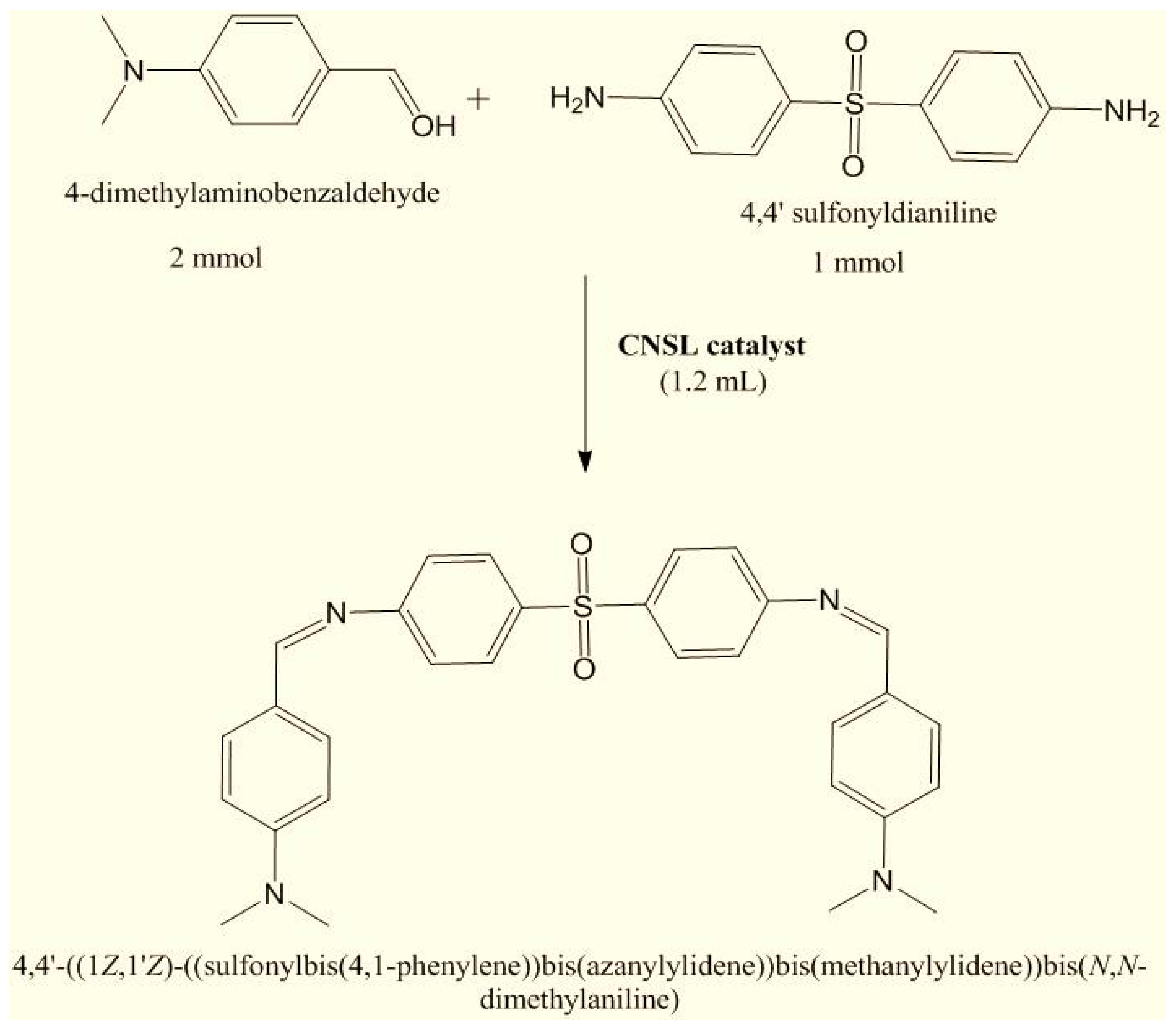
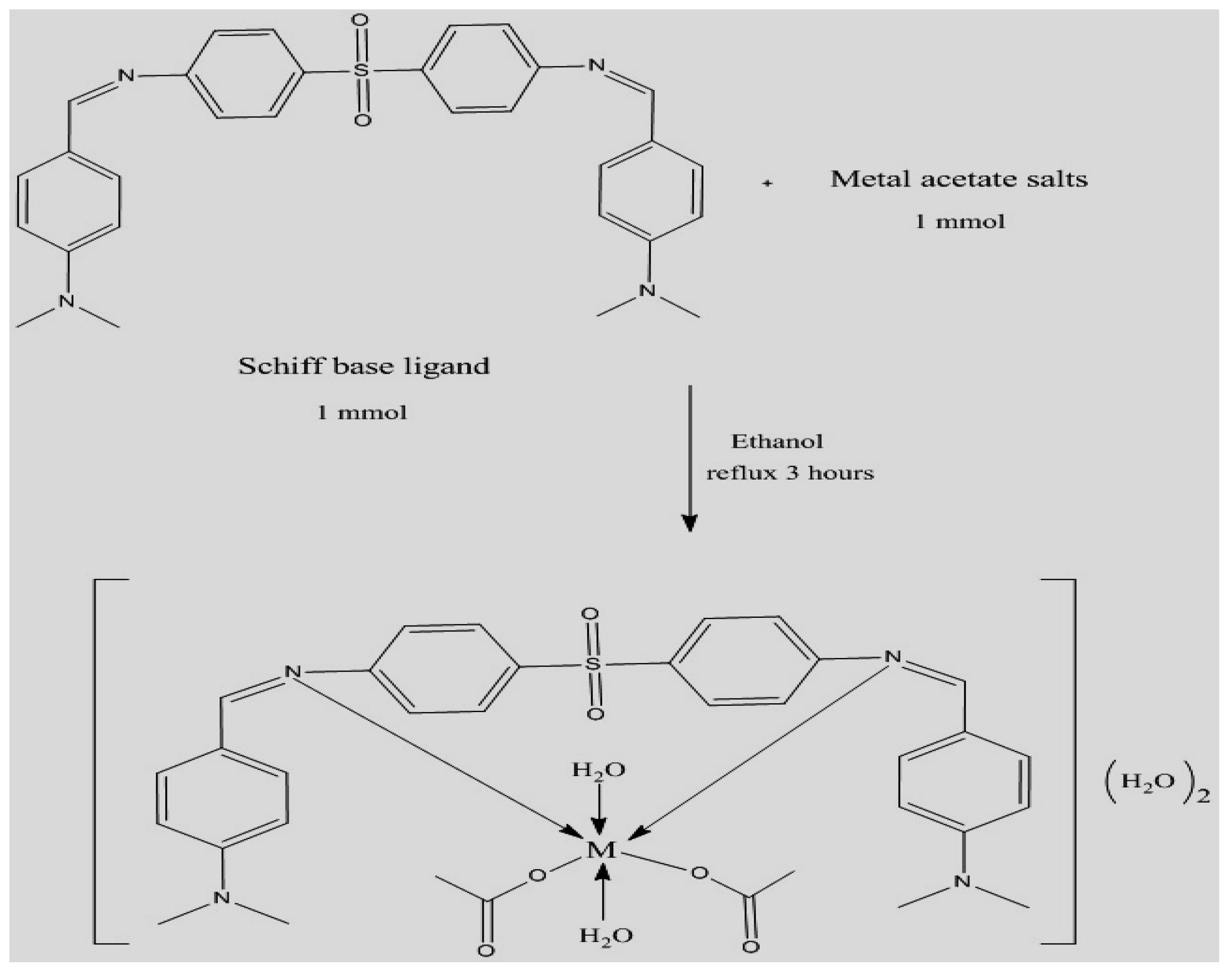
A Schiff base ligand is synthesized from the condensation of dapsone and 4-dimethylaminobenzaldehyde using cashew nut shell liquid (CNSL) anacardic acid as a green and natural effective catalyst via solvent-free simple physical grinding technique. Furthermore, metal(II) complexes Co(II), Cu(II) and Zn(II) coordinated by a new Schiff base ligand (L) were prepared. The composition of Schiff base ligand and its metal(II) complexes were analyzed by various analytical techniques. The Schiff base ligand and its complexes have been tested in vitro to evaluate their antimicrobial activity against Escherichia coli, Staphylococcus aureus and Candida albicans using well-diffusion method. It has been found that the Schiff base ligand and its complexes show significant antimicrobial activity against all tested bacterial species. Molecular docking study of Cu(II) complex with target protein HER2 has revealed good binding energy.

Keywords: 
;  ;  ;  

Introduction

Recently significant attention has been paid to environmental chemistry aimed at developing new mechanisms that can provide benefits for the synthesis of organic compounds in the absence of solvent [1]. The toxic effects and fragility of many organic solvents have presented a serious potential hazard to the atmosphere. Therefore, in recent years, the development of solvent-free catalytic reaction has gained significant recognition in the field of chemistry. Eco-friendly condensation reaction methods for the synthesis of Schiff bases have particular advantages, such as longer reaction times, higher reaction temperatures, less expensive dehydration reagents/catalysts, moisture-sensitive catalysts, specialized equipment, and so on [2]. The discovery of new and efficient catalysts with high catalytic activity, short reaction times, recyclability and simple reaction work-up to prepare Schiff bases is of great interest. Nowadays, dapsone is a drug commonly used as multidrug therapy (MDT) with rifampicin and clofazimine to treat leprosy infections. Dapsone exhibits efficient pharmacological activity against mycobacterium leprosy. Its effective binding to metal ion may have important biological properties making this antileprosy drug a very potent chelating agent [3].
The current study focused on the synthesis Schiff base ligand with CNSL and its Co(II), Cu(II) and Zn(II) complexes and spectroscopic characterization. Cashew Nut Shell Liquid (CNSL) as an effective, minimal-cost and natural catalyst for the efficient solvent-free mechanochemical assisted synthesis of a sulfone based Schiff base by the condensation of 4, 4’ sulfonyldianiline (dapsone) and 4-dimethylaminobenzaldehyde.

Materials Details

Metal(II) acetate salts were acquired from Sigma-Aldrich Ltd. and utilized without any additional purification. The compounds 4,4’-sulfonyldianiline (dapsone) and 4-dimethylaminobenzaldehyde, both of analytical reagent grade, were sourced from TCI Chemicals. The solvents employed for synthesis and characterization were of high-performance liquid chromatography grade.

Extraction of CNLS and Its Catalytic Activity

The shells of cashew nuts are soaked in petroleum ether for approximately three days, resulting in the production of dark brownish oil. This oil is then concentrated using a rotary evaporator system with a water bath set at 40o C.
The catalytic efficiency of CNSL was tested in the synthesis of Schiff base ligand. The reaction parameters were standardized in terms of catalyst quantity, response time and yield. The impact of the catalyst quantity on the prototype reaction has tabulated in Table 1. It is evident from this figure that in the absence of a CNSL catalyst, no product was formed. Furthermore, the percentage yield of the product increased with the addition of catalyst quantities ranging from 0.2 to 1.0 ml. However, when the catalyst quantity was further increased to 1.5 ml, no additional increase in output was observed. Additionally, there was no enhancement in product yield when using solvents such as DMF, methanol, ethanol and water, thereby confirming the crucial role of the catalyst in ensuring a highly effective reaction rate with excellent yields [4].

Experimental Procedure

Synthesis of Schiff Base Ligand Derived from 4, 4’ sulfonyldianiline

A Schiff base ligand was synthesized using 4, 4’ sulfonyldianiline (1 mmol) and 4-dimethylaminobenzaldehyde (2 mmol), to which 1.2 ml of CNSL catalyst was subsequently introduced. The components were mechanically mixed at ambient temperature for duration of 15 minutes, yielding a jelly-like texture. Subsequently, 4–5 ml of 95% ethanol was incorporated into the mixture to eliminate any waxy or gelatinous residues. The catalyst was then extracted from the mixture, thoroughly washed with petroleum ether (4-5 ml) and allowed to air dry. The crude product underwent purification through recrystallization using basic ethanol (Figure 1).

Synthesis of Schiff Base Metal(II) Complexes

An ethanolic solution of 1 mmol of Schiff base ligand was mixed to the solution of metal acetate salts of Co(II), Cu(II), and Zn(II) maintaining a molar ratio of 1:1. The mixture was stirred for duration of 3 hours. Subsequently, the final mixture underwent reflux for an additional 3 hours, leading to the isolation of the product (Figure 2). The resultant product was then filtered, washed multiple times with methanol, and finally dried under vacuum over anhydrous calcium chloride. The analytical and physical characteristics of the Schiff base ligand and its mononuclear complexes are compiled in Table 2.

M= Co(II), Cu(II), and Zn(II)

[Cu(L1) (OAc)(H2O)] (OAc) Preparation and Molecular Docking:

[Cu(L1)(OAc)(H2O)](OAc) molecule was prepared using Marvin 17.21.0 from Chemaxon (https://www.chemaxon.com), and its 2D structure was converted and optimized with Avogadro 1.2.0 software [5]. The inhibitory potential of [Cu(L1) (OAc)(H2O)] (OAc) against the HER2 protein, associated with breast cancer (PDB ID: 3MZW) [6], was evaluated through molecular docking. The protein and ligand were prepared by adding polar hydrogen’s and assigning AD4 atom types using AutoDock Tools 1.5.6, with rotatable bonds defined for selected atoms. Grid maps were generated based on a known affibody molecule, an established HER2 inhibitor, with the grid spacing set at 0.67 Å to facilitate ligand binding. The grid size was set to 50 × 50 × 50 points[7,8,9]. Molecular docking was performed using AutoDock 4.0.1, and the ligand-protein interactions were visualized with AutoDock Tools, while Chimera [10] was used to create images of the complexes.

Results and Discussion

Elemental Analyses and Molar Conductivity Measurements

The elemental analysis validated the stoichiometry of Schiff base ligand and its metal(II) complexes, as detailed in Table 2. It was observed that the Schiff base ligand and its complexes exhibit limited solubility in methanol, ethanol, acetone, and chloroform, while they are fully soluble in DMSO and DMF. The molar conductivity of measurements of Schiff base metal(II) complexes were recorded in DMSO and observed all complexes are in an ionic nature [11] as shown in Table 2.

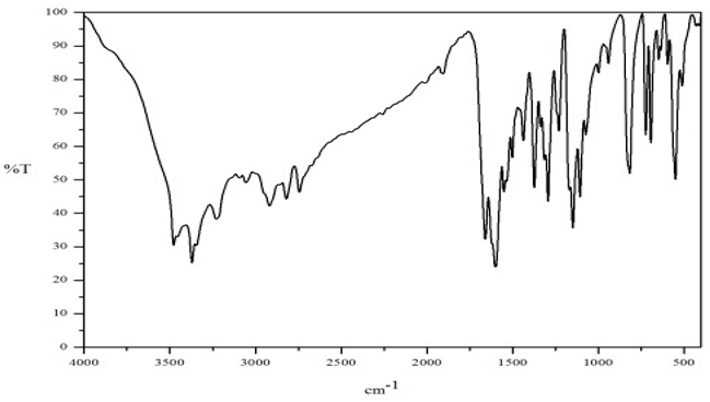
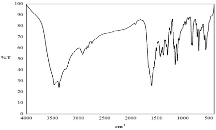
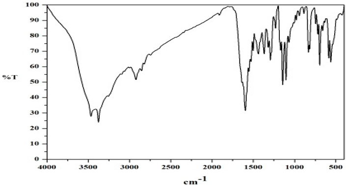
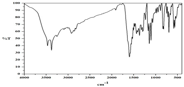
FT-IR Spectral Studies

The notable spectral features of the Schiff base ligand and its corresponding complexes are tabulated in Table 3 and depicted in Figures 3.1–3.4.
FT-IR spectra of metal(II) complexes indicate that the ligand coordinates with the metal ions in a bidentate manner, coordinating through the (-HC=N-) bond, as well as through OAc and H2O to the metal ions [12]. Schiff base ligand displays a significant absorption band at 1659 cm-1 attributed to the azomethine (-HC=N-) functional group. In the metal complexes, this band appears at a lower frequency range of 1596-1637 cm-1, indicating the coordination of the (-HC=N-) group with the metal ion [13]. All metal(II) complexes exhibit new bands in the ranges of 1720-1700 cm-1 and 1584-1581 cm-1 , which are associated with the υas(OAc) and υsy(OAc) vibrations, respectively [14]. Additionally, new bands are observed in all complexes around 3467-3378 cm-1 and 860-876 cm-1 attributed to the coordination of water molecules [15]. The spectra of the ligand and its metal(II) complexes reveal bands around 1301-1304 cm-1 and 1131-1140 cm-1 , corresponding to asymmetric and symmetric SO2 stretching vibrations [16]. The same positioning of these bands in both the ligand and its complexes suggests that the oxygen atom of - SO2 group is not coordinated with the metal ion. The coordination of azomethine nitrogen in metal(II) complexes are further supported by the new bands exhibits around 560-550 cm-1 which is due to formation of υ(M-N) respectively [17].
Preprints 141029 i001
Figure 3.1 IR spectra of the Ligand(L1).
Preprints 141029 i002
Figure 3.2 IR spectra of [CoL1(OAc)2(H2O)2](H2O)2.
Preprints 141029 i003
Figure 3.3 IR spectra of [[Cu(L1) (OAc)(H2O)] (OAc).
Preprints 141029 i004
Figure 3.4 IR spectra of [Zn(L1)(OAc)2(H2O)2].

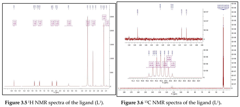
NMR Spectral Studies

1H NMR and 13C NMR spectrum of Schiff base ligand provided information about the formation of azomethine group. 1H NMR spectra of Schiff base ligand was resolved in the solution of DMSO with TMS as an internal standard as shown in Figure 3.5 and 3.6. 1H NMR spectrum of the ligand shows multiplet signal at δ (6.30 – 7.56) ppm are assigned to the aromatic protons respectively [18]. The azomethine group of ligand shows at 9.12 ppm. Signal shows at 3.06 ppm attributed to –NMe2 group in Schiff base ligand [19] 13C NMR spectrum of the ligand show signal at 158.26 ppm which may be assigned to azomethine carbon [20]. Signals due to phenyl carbons appeared in the range of 109.02 – 128.91 ppm [21].
Preprints 141029 i005

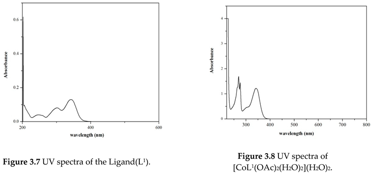
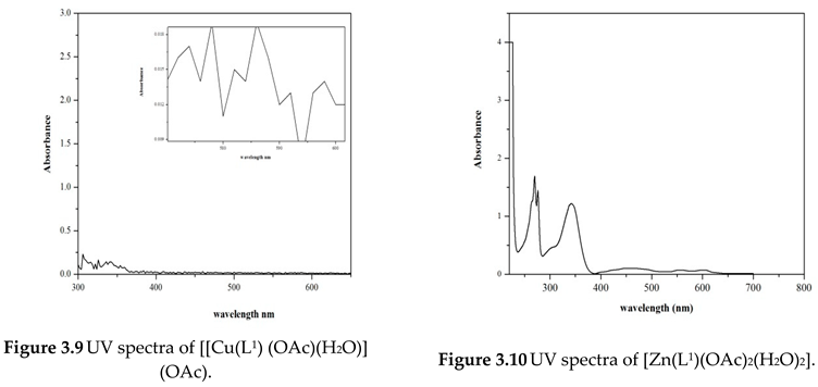
Electronic Spectral and Magnetic Moment Studies

The electronic absorption spectra of ligand and its metal(II) complexes were recorded in DMSO and the values are tabulated in Table 4 as shown in Figures 3.7–3.10.
An aromatic benzene ring and azomethine group of Schiff base ligand shows two high intensity absorption bands exhibited at 270 nm and 341 nm may be assigned for intraligand π-π* and n-π* transitions respectively [22]. But in spectra of metal(II) complexes, the positions of these bands are shifted to lower wavelengths suggesting the coordination of ligand to the metal ions.
The electronic spectrum of [Co(L1) (OAc)2(H2O)2] (H2O)2 , two new bands exhibit at 530 nm and 604 nm consequence to 4 T1g (F)→4T1g (P) and 4 T1g (F) → 4A2g (F) transitions respectively and its magnetic moment value is 4.12 B.M. which is supporting the suggestion of an octahedral geometry for Co(II) complex [23]. [Cu(L1) (OAc)(H2O)] (OAc) complex shows an intense broad band at 512 nm, which can be attributed to the 2B1g2A1g transition, indicative of a square planar geometry for the Cu(II) ion [24]. Additionally, the measured magnetic moment was found to be 1.70 B.M. [Zn(L1) (OAc)2 (H2O)2] shows a medium intensity band at 525 nm may be due to LMCT . The magnetic moment indicates diamagnetic nature which is further supported by an octahedral geometry for Zn(II) complex [25].
Preprints 141029 i006Preprints 141029 i007

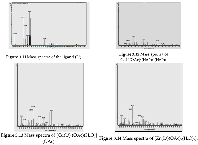
Mass Spectral Studies
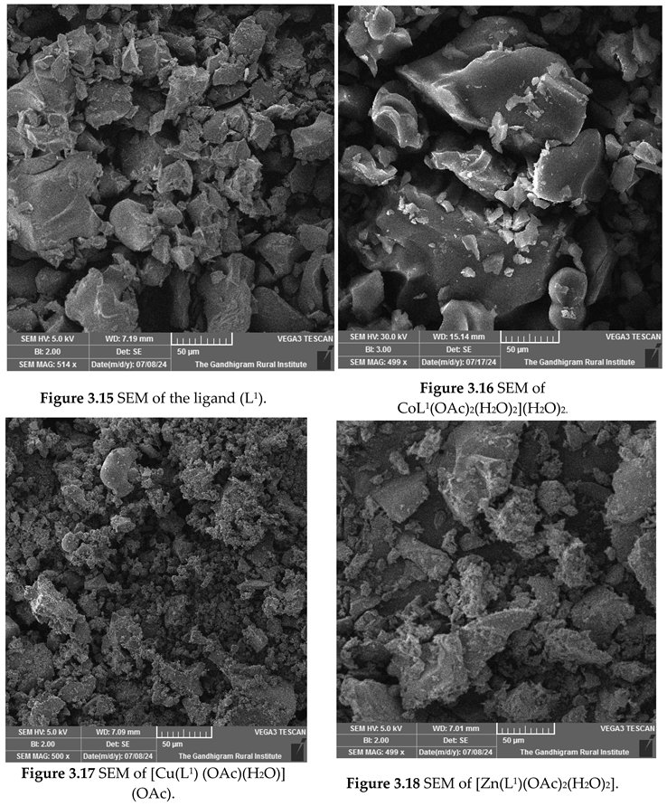
Mass spectrometry is extensively employed as an effective tool for structural characterization. The fragmentation patterns observed in the mass spectra of the free Schiff base ligand [C30H30N4O2S] closely align with the structure depicted in Scheme 1. The mass spectrum reveals a prominent peak at 512.49 m/z, which corresponds well to the calculated molecular weight of 510.12 m/z. The complexes observed m/z values at 743.43, 708.12 and 727.45 corresponds to the molecular ion peaks of the complexes C34H42N4O9S Co, C34H38N4O7SCu, C34H40N4O8SZn respectively, thereby Corresponding to their respective molecular weight of these complexes [26]. This information is further corroborated by the elemental analysis data provided in Table 1 and shown in Figures 3.11–3.14.
Preprints 141029 i008

SEM Analysis

The scanning electron microscopy (SEM) analysis conducted to evaluate the surface morphology of the ligand and its metal complexes is illustrated in Figure 3.15–3.18. SEM micrographs demonstrate significant variations in the surface morphology of the ligand and its metal complexes, attributed to the coordination of metal ions with the donor sites present in the ligand [27]. Furthermore, SEM images of the metal complexes indicate that the surface morphology is influenced by the type of metal ions used. The Schiff base ligand exhibits a non-uniform, broken rock - like structure. Co(II) and Zn(II) complexes present irregular, small-shaped grains resembling a cloud formation. In contrast, the Cu(II) complex displays microcrystalline structures formations.
Preprints 141029 i009

Biological Studies

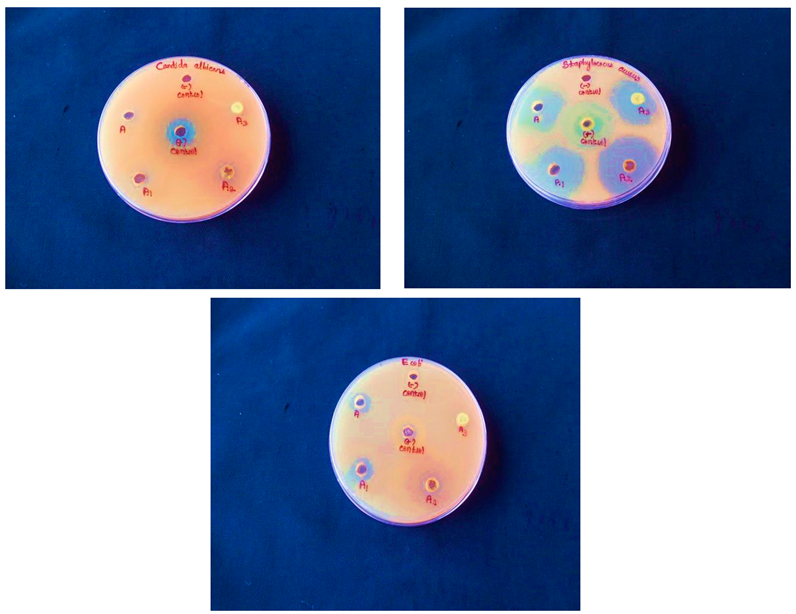
Antimicrobial Activity

The in vitro biological effects of the ligand and its mononuclear complexes were evaluated against one gram-positive bacteria (Staphylococcus aureus), one gram-negative bacteria (Escherichia coli), and one fungal species (Candida albicans) using the disc diffusion method. The findings are summarized in Table 5. The results indicate a moderate level of activity when compared to the reference standards, Tetracycline and Fluconazole, with DMSO solvent serving as a positive control. The data demonstrate that the complexes exhibit greater activity than the free ligand.
The copper(II) complex exhibits superior antibacterial and antifungal properties compared to Co(II) and Zn(II) complexes. This variation in antimicrobial efficacy can be attributed to the characteristics of the metal ions as well as the composition of the microorganisms cell membranes [28]. The enhanced activity of the Cu(II) complex can be elucidated by the fact that upon chelation, the polarity of the Cu(II) ion significantly diminishes due to the interaction between the ligand orbitals and the partial sharing of the copper ion's positive charge with the donor groups [29]. Consequently, Cu(II) ions adhere to the surface of microbial cell walls. The presence of these adsorbed Cu(II) ions disrupts the respiratory functions of the cells, thereby inhibiting protein synthesis and ultimately curtailing the growth of the organisms [30].
Preprints 141029 i010
Figure 3.19 Antimicrobial activity of Schiff base ligand and its metal(II) complexes.

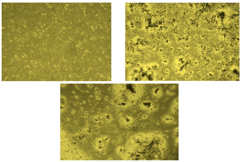
In Vitro Anticancer Activity

The anticancer properties of the Schiff base ligand and its copper(II) complex were evaluated using the MTT assay on the human breast cancer cell line MCF-7. The absorbance readings at 570 nm across a range of concentrations (3.125-50 μg/ml), along with the percentage of cell inhibition and IC50 values, are presented in Table 6. The results for cell viability and IC50 values suggest that the copper(II) complex exhibits greater efficacy against breast cancer cells compared to the Schiff base ligand. The IC50 value for the copper(II) complex is promising for further exploration of potential therapeutic agents. [31].
Preprints 141029 i011
Figure 3.20 Anticancer activity of standard, Schiff base ligand and its Copper(II) complex.

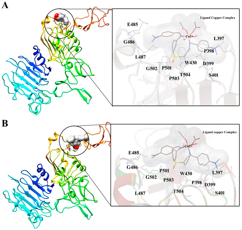
Molecular Docking and Interaction Analysis

The atomic interaction between a ligand molecule and a known target protein is commonly predicted using molecular docking analysis, which offers information on small molecule interaction behavior at the target protein binding site and clarifies important biochemical processes. H2L-based complexes have been shown to have strong anticancer efficacy against a variety of malignancies in recent years [32,33,34].Therefore, we employed the CuL molecule docked with the target protein HER2, which is related with breast cancer (PDB ID: 3MZW) [35].The binding energy, inhibition constant, and intermolecular energy were recorded in the Table 7, and the top two posture interactions were shown in the Figure 3.21. Based on the strong polar and non-polar interactions formed by L397, P398, D399, S401, W430, E485, G486, L487, P501, G502, P503, and T504 in comparison to pose 2, these results show that CuL-pose 1 has lower binding energy (-2.42 Kcal/mol) and good inhibition constant Ki (16.77 mM) against HER2 protein. All these amino acids are demonstrated to generate a strong contact with affibody molecule which is a best peptide inhibitor of HER2 protein. These results unequivocally demonstrate that the CuL molecule may be used as a novel HER2 protein inhibitor; hence, these discoveries will be helpful in the creation of potent medications for the treatment of breast cancer.
Preprints 141029 i012
Figure 3.21 The molecular interaction of CuL with Domain III of HER2 protein and the zoomed image shows the polar and non-polar residues actively contributing for the interaction. A) Top 1 pose of the ligand interaction with HER2 protein, B) Top 2 pose of the ligand interaction with HER2 protein.

Conclusion

A novel Schiff base ligand was synthesized from dapsone and 4-dimethylaminobenzaldehyde using a mechanochemical approach in the presence of a CNSL catalyst at room temperature and under solvent-free conditions. This study primarily highlights the advanced application and significance of environmental chemistry. Mononuclear complexes of Co(II), Cu(II), and Zn(II) with the Schiff base ligand were prepared in a 1:1 ratio and characterized through elemental and spectroscopic analyses. The physicochemical methods indicate an octahedral geometry for the Co(II) and Zn(II) complexes, while the Cu(II) complex exhibits a square planar geometry. The morphology of the complexes was examined using scanning electron microscopy (SEM). The Schiff base ligand and its metal(II) complexes were evaluated for antibacterial activity against gram-positive (Staphylococcus aureus), gram-negative (Escherichia coli) bacteria, and one fungal strain (Candida albicans). The findings revealed that the metal(II) complexes demonstrated greater activity than the Schiff base ligand, with the Cu(II) complex showing superior inhibition compared to the Co(II) and Zn(II) complexes. Docking studies further illustrated the inhibitory potential against the HER2 protein at the molecular level, with Cu(II) complex exhibiting excellent binding energies of -2.21 and -2.42, respectively. Consequently, this research underscores the potential of the newly synthesized Schiff base-derived complexes as prodrugs for cancer treatment, with future considerations aimed at enhancing their efficacy.

References

  1. Nagar, S.; Raizada, S.; Tripathee, N. A review on various green methods for synthesis of Schiff base ligands and their metal complexes. Results Chem. 2023, 6, 101153. [Google Scholar] [CrossRef]
  2. Msigala, S.C.; JEG Mdoe. Synthesis of organoamine-silica hybrids using cashew nut shell liquid components as templates for the catalysis of a model henry reaction. Tanz. J. Sci. 2012, 38, 24–34. [Google Scholar]
  3. Ahmed, A.A.; Turki, A.A.; Hanoosh, W.S. Synthesis, characterization of new di Schiff base derivatives from dapsone, their polymerization and thermal study. Bas. J. Sci. 2021, 39, 463–483. [Google Scholar] [CrossRef]
  4. Vidyavathi, G.T.; Kumar, B.V.; Aravinda, T.; Hani, U. Cashew nutshell liquid catalyzed green chemistry approach for synthesis of a Schiff base and its divalent metal complexes: molecular docking and dna reactivity. Nucleosides, Nucleotides and Nucleic Acids. 2021, 40, 264–287. [Google Scholar] [CrossRef]
  5. Hanwell, M.D.; Curtis, D.E.; Lonie, D.C.; Vandermeersch, T.; Zurek, E.; Hutchison, G.R. Avogadro: An advanced semantic chemical editor, visualization, and analysis platform. J. Cheminform. 2012, 4, 1–17. [Google Scholar] [CrossRef]
  6. Eigenbrot, C.; Ultsch, M.; Dubnovitsky, A.; Abrahmsén, L.; Härd, T. Structural basis for high-affinity HER2 receptor, binding by an engineered protein. Proc. Natl. Acad. Sci. 2010, 107, 15039–15044. [Google Scholar] [CrossRef]
  7. Morris, G.M.; Goodsell, D.S.; Halliday, R.S.; Huey, R.; Hart, W.E.; Belew, R.K.; Olson, A.J. Automated Docking Using a Lamarckian Genetic Algorithm and an Empirical Binding Free Energy Function. J. Comput. Chem. 1998, 19, 1639–1662. [Google Scholar] [CrossRef]
  8. Goodsell, D.S.; Morris, G.M.; Olson, A.J. Automated Docking of Flexible Ligands: Applications of AutoDock. J. Mol. Recognit. 1996, 9, 1–5. [Google Scholar] [CrossRef]
  9. Meng, X.Y.; Zhang, H.X.; Mezei, M.; Cui, M. Molecular Docking: A Powerful Approach for Structure-Based Drug Discovery. Curr. Comput. Aided-Drug Des. 2011, 7, 146–157. [Google Scholar] [CrossRef]
  10. Pettersen, E.F.; Goddard, T.D.; Huang, C.C.; Couch, G.S.; Greenblatt, D.M.; Meng, E.C.; Ferrin, T.E. UCSF Chimera—A Visualization System for Exploratory Research and Analysis. J. Comput. Chem. 2004, 25, 1605–1612. [Google Scholar] [CrossRef]
  11. Kavitha, A.; Anandhavelu, S.; Easwaramoorthy, D.; Karuppasamy, K.; Kim, H.-S.; Dhanasekaran, V. In vitro cytotoxicity activity of novel Schiff base ligand–lanthanide complexes. Sci Rep. 2018, 8, 1–12. [Google Scholar] [CrossRef]
  12. Bader, A.T.; Al-Abdaly, B.I.; Jassim, I.K. Synthesis and Characterization new metal complexes of heterocyclic units and study antibacterial and antifungal. J. Pharm. Sci. Res. 2019, 5, 2062–2073. [Google Scholar]
  13. Nair, M.S.; Arish, D.; Joseyphus, R.S. Synthesis, characterization, antifungal, antibacterial and DNA cleavage studies of some heterocyclic Schiff base metal complexes. J. Saudi Chem. Soc. 2012, 16, 83–88. [Google Scholar] [CrossRef]
  14. Sadafalea, V.A.; Thakareb, A.P.; Mandlik, P.R. Synthesis, spectral characterization and biological activity of Schiff base metal complexes derived from dapsone and salicylaldehyde. Int. J. Curr. Eng. Sci. Res. 2019, 6, 2394–0697. [Google Scholar]
  15. Al Zoubi, W.; Al-Hamdani, A.A.; Ahmed, S.D.; Ko, Y.G. Synthesis, characterization, and biological activity of Schiff bases metal complexes. J Phys Org Chem. 2017, 1–13. [Google Scholar] [CrossRef]
  16. Iqbal, N.; Iqbal, J.; Imran, M. Synthesis, characterization and anti-bacterial studies of some metal complexes of a Schiff base derived from benzaldehyde and sulphonamide. J. Sci. Res. 2009, XXXIX, 15–19. [Google Scholar] [CrossRef]
  17. Bhoopathy, P.; Jayalakshmi, R.; Rajavel, R. An Insight into Antibacterial and Anticancer Activity of Homo and Hetero Binuclear Schiff Base Complexes. Orient. J. Chem. 2017, 33, 1223–1234. [Google Scholar] [CrossRef]
  18. Ashokan, R.; Sathishkumar, S.; Akila, E.; Rajavel, R. Synthesis, characterization and biological activity of Schiff base metal complexes derived from 2, 4 dihydroxyactophenone. Chem Sci Trans. 2017, 6, 277–287. [Google Scholar] [CrossRef]
  19. Sura, H. Kathim. Synthesis and Characterization of the Inhibitory Activity of 4- (Dimethyl amino) benzaldehyde Schiff Bases. Wasit Journal for Pure Science 2023, 4, 254–268. [Google Scholar] [CrossRef]
  20. Burak, O.; Erdal, C. ; Hakan,Ş.; Mehmet, K. The synthesis, characterization of a novel Schiff base ligand and investigation of its transition metal complexes. Adıyaman university journal of science 2017, 7, 47–59. [Google Scholar]
  21. Shukla, S.N.; Gaur, P.R.; Jhariya, S.A.; Chaurasia, B.H.; Vaidya, P.R.; Dehariya, D.I.; Azam, M. Synthesis, Characterization, In Vitro Anti-diabetic, Antibacterial and Anticorrosive Activity of Some Cr(III) Complexes of Schiff Bases Derived from Isoniazid. Chem Sci Trans. 2018, 7, 424–444. [Google Scholar] [CrossRef]
  22. Moustafa, S.A.; Ali, M.M.; El-rashedy, A.A. Synthesis, anticancer activity and molecular docking study of Schiff base complexes containing thiazole moiety. Beni - Suef university journal of basic and applied sciences. 2016, 5, 85–96. [Google Scholar] [CrossRef]
  23. Deswal, Y.; Asija, S.; Kumar, D.; Jindal, D.K.; Chandan, G.; Panwar, V.; Saroya, S.; Kumar, N. Transition metal complexes of triazole-based bioactive ligands: synthesis, spectral characterization, antimicrobial, anticancer and molecular docking studies. Res. Chem. Intermed. 2022, 48, 703–729. [Google Scholar] [CrossRef]
  24. Krishnan, M.A.; Saranyaparvathi, S.; Raksha, C.; Vrinda, B. Transition Metal Complexes of 4-Aminoantipyrine Derivatives and Their Antimicrobial Applications. Russ. J. Coord. Chem. 2022, 48, 696–724. [Google Scholar] [CrossRef]
  25. Juyal, V.K.; Thakuri, S.C.V.K.; Panwar, M. Manganese (II) and Zinc(II) metal complexes of novel bidentate formamide-based Schiff base ligand: synthesis, structural characterization, antioxidant, antibacterial, and in-silico molecular docking study. Front. Chem. 2024, 7, 1–14. [Google Scholar] [CrossRef]
  26. Manjunatha, M.; Naik, V.H.; Kulkarni, A.D. DNA cleavage, antimicrobial, anti-inflammatory anthelmintic activities, and spectroscopic studies of Co(II), Ni(II), and Cu(II) complexes of biologically potential coumarin Schiff bases. J. Coord. Chem. 2011, 24, 4264–4275. [Google Scholar] [CrossRef]
  27. Leelavathy, C.; Antony, A. Structural elucidation and thermal studies of some novel mixed ligand Schiff base metal (II) complexes. Int. J. Basic Appl. Chem. Sci. 2013, 3, 88–95. [Google Scholar]
  28. Vamsikrishna, N.; Kumar, M.P.; Ramesh, G.; Ganji, N.; Daravath, S. DNA interactions and biocidal activity of metal complexes of benzothiazole Schiff bases: synthesis, characterization and validation. J. Chem. Sci. 2017, 129, 609–622. [Google Scholar] [CrossRef]
  29. Das, P.K.; Panda, N.; Behera, N.K. Synthesis, Characterization and Antimicrobial Activities of Schiff Base Complexes Derived from Isoniazid and Diacetylmonoxime. Int. J. Innov. Sci. Eng. Technol. 2016, 3, 42–54. [Google Scholar]
  30. Mahapatra, B.B.; Mishra, R.R.; Sarangi, A.K. Synthesis, characterisation, XRD, molecular modelling and potential antibacterial studies of Co(II), Ni(II), Cu(II), Zn(II), Cd(II) and Hg(II) complexes with bidentate azodye ligand. J. Saudi Chem. Soc. 2013, 1–9. [Google Scholar] [CrossRef]
  31. Gomathi, T.; Vedanayaki, S. Synthesis, Spectral, Electrochemical and Biological Studies on Co(II), Ni(II), Cu(II) and Zn(II) Complexes Derived from 4-(2-Aminoethyl)benzene-1,2-diol and Terephthalaldehyde. Asian J. Chem. 2022, 6, 1373–138234. [Google Scholar] [CrossRef]
  32. Kanagavalli, A.; Jayachitra, R.; Thilagavathi, G.; Padmavathy, M.; Elangovan, N.; Sowrirajan, S.; Thomas, R. Synthesis, structural, spectral, computational, docking and biological activities of Schiff base (E)-4-bromo-2-hydroxybenzylidene) amino)-N-(pyrimidin-2-yl) benzenesulfonamide from 5-bromosalicylaldehyde and sulfadiazine. J. Indian Chem. Soc. 2023, 100, 1–19. [Google Scholar] [CrossRef]
  33. Irfandi, R.; Raya, I. Potential anticancer activity of Mn (II) complexes containing arginine dithiocarbamate ligand on MCF-7 breast cancer cell lines. Ann. med. surg. 2020, 60, 396–402. [Google Scholar] [CrossRef]
  34. Sakthivel, R.V.; Sankudevan, P.; Vennila, P.; Venkatesh, G.; Kaya, S. Experimental and theoretical analysis of molecular structure, vibrational spectra and biological properties of the new Co(II), Ni(II) and Cu(II) Schiff base metal complexes. J. Mol. Struct. 2021, 1233, 1–13. [Google Scholar] [CrossRef]
  35. Pieper, U.; Webb, B.M.; Barkan, D.T.; Schneidman-Duhovny, D. ModBase, a database of annotated comparative protein structure models, and associated resources. Nucleic Acids Res. 2011, 39, 465–474. [Google Scholar] [CrossRef]
Figure 1. Synthesis of Schiff base ligand.
Preprints 141029 g001
Figure 2. Synthesis of Schiff base Metal(II) complexes.
Preprints 141029 g002
Table 1. Yield of synthesis of Schiff base ligand with CNSL.
S.NO Amount of catalyst (ml) Time (minutes) Yield
1 0.2 30 nil
2 0.4 25 30
3 0.6 15 55
4 0.8 5 85
5 1.0 5 85
Table 2. Analytical data for Schiff base and its metal(II) complexes.
Compound M.Wt. Color M.Pt.
°C
Molar conductance Ω- 1 cm2 mol-1 Elemental analysis
Calculated (Found) %
C H N M
Ligand(L1)
C30H30N4O2S
510.21 Brown 120 -- 70.16
(70.56)
5.96
(5.92)
10.13
(10.97)
-----
[Co(L1) (OAc)2
(H2O)2] (H2O)2
C34H42N4O9S Co
741.20 Reddish brown 220 18.16 55.12
(55.06)
5.45
(5.71)
7.23
(7.55)
7.81
(7.95)
[Cu(L1) (OAc)
(H2O)] (OAc)
C34H38N4O7SCu
709 brown 245 20.21 57.42
(57.49)
5.24
(5.39)
7.96
(7.89)
8.90
(8.95)
[Zn(L1) (OAc)2
(H2O)2]
C34H40N4O8SZn
728 brown 232 32.65 55.48
(55.93)
5.42
(5.52)
7.76
(7.67)
8.87
(8.95)
Table 3. IR spectral data analysis of the Schiff base ligand and its metal(II) complexes.
Compound ʋ(H2O) cm-1 υ(C=N) cm-1 ʋ(SO2)
cm-1
ʋ(M-N) cm-1
Ligand (L1) ------ 1659 1314 ---
[Co(L1) (OAc)2 (H2O)2] (H2O)2 3450 1637 1319 561
[Cu(L1) (OAc)
(H2O)] (OAc)
3467 1622 1317 560
[Zn(L1) (OAc)2
(H2O)2]
3378 1596 1317 534
Table 4. Electronic spectral data and magnetic moments of the Schiff base ligand and its metal(II) complexes.
Compound Absorption (nm) Band assignment Geometry Magnetic moment (BM)
Ligand (L1) 270
341
INCT
INCT
------- ------
[Co(L1) (OAc)2 (H2O)2] (H2O)2 275
348
429
534
INCT
INCT
4 T1g (F)—4 T1g (P)
4 T1g (F)—4 A2g (F)
Octahedral 4.12
[Cu(L1) (OAc)
(H2O)] (OAc)
272
355
512
INCT
INCT
2B1g2A1g
Square- planar 1.80
[Zn(L1) (OAc)2
(H2O)2]
275
345
495
INCT
INCT
LMCT
Octahedral Diamagnetic
Table 5. Antibacterial and antifungal activity of Schiff base ligand and its metal(II) complexes.
Sample Zone of inhibition (mm)
Gram positive Gram negative Fungi
Staphylococcus aureus Escherichia coli Candida albicans
Ligand (L1) 31 9 13
[Co(L1) (OAc)2 (H2O)2] (H2O)2 35 10 21
[Cu(L1) (OAc)
(H2O)] (OAc)
36 11 18
[Zn(L1) (OAc)2
(H2O)2]
33 9 12
Standard 30 20 17
Table 6. Anticancer effects of Schiff base and its Copper(II) complex in term of % cell inhibition at various concentrations.
Ligand (L1)
Concentration (μg/ml)
Cell Viability % IC50
(μM)
[Cu(L1) (OAc)(H2O)] (OAc) Concentration (μg/ml) Cell Viability % IC50
(μM)
3.125 96.79 35.78 3.125 94.16 24.62
6.25 91.42 6.25 90.67
12.5 83.66 12.5 72.78
25 67.78 25 55.81
50 35.54 50 30.16
Table 7. Molecular interaction of CuL with HER2 protein. The tables consist of binding energy, inhibition constant, intermolecular energy and interacting residues.
S. No Ligand copper complex Binding energy (Kcal/mol) Inhibition constant Ki (mM) Intermolecular Energy (Kcal/mol) Interacting Residues
1. Top 1 pose -2.42 16.77 -5.70 L397, P398, D399, S401, W430, E485, G486, L487, P501, G502, P503, T504,
2. Top 2 pose -2.21 24.14 -5.49 L397, P398, D399, S401, W430, E485, G486, L487, P501, G502, P503, T504,
Disclaimer/Publisher’s Note: The statements, opinions and data contained in all publications are solely those of the individual author(s) and contributor(s) and not of MDPI and/or the editor(s). MDPI and/or the editor(s) disclaim responsibility for any injury to people or property resulting from any ideas, methods, instructions or products referred to in the content.
Copyright: This open access article is published under a Creative Commons CC BY 4.0 license, which permit the free download, distribution, and reuse, provided that the author and preprint are cited in any reuse.
Prerpints.org logo

Preprints.org is a free preprint server supported by MDPI in Basel, Switzerland.

Subscribe

Disclaimer

Terms of Use

Privacy Policy

Privacy Settings

© 2025 MDPI (Basel, Switzerland) unless otherwise stated