

Submitted:
26 November 2024
Posted:
27 November 2024
You are already at the latest version
Abstract
Chronic enteropathy (CE) in dogs presents with persistent symptoms, such as vomiting, diarrhea, weight loss, and changes in appetite. Traditional diagnostic methods, including blood tests, ultrasonography, and endoscopy, often fail to assess the entire small intestine accurately. Capsule endoscopy offers a noninvasive approach that enables comprehensive visualization of the gastrointestinal tract and enhances diagnostic accuracy. In Case 1, a 10-year-old Maltese patient with chronic diarrhea and significant weight loss underwent capsule endoscopy, which revealed severe lacteal dilation and moderate gastric erythema, leading to a diagnosis of severe intestinal lymphangiectasia and moderate enteritis. Despite initial corticosteroid therapy and dietary changes, the clinical improvement was limited. Case 2 involved an 11-year-old mixed-breed dog with chronic vomiting, anorexia, and weight loss. Capsule endoscopy revealed moderate gastric erythema, severe small intestinal irregularities, and jejunal bleeding, leading to a diagnosis of mild esophagitis, moderate gastritis, and severe inflammatory bowel disease (IBD) with hemorrhage. Treatment with a hypoallergenic diet, corticosteroids, and acid suppressants gradually relieved symptoms. Capsule endoscopy provided crucial diagnostic insights and guided the effective treatment in both cases, mainly when conventional methods were inconclusive. This report highlights the utility of capsule endoscopy as a valuable tool for managing chronic enteropathy in dogs.
Keywords:
capsule endoscopy
; chronic enteropathy
; dog
; inflammatory bowel disease
; lymphangiectasia
1. Introduction
Chronic enteropathy (CE) is a common and challenging condition in veterinary medicine. It is characterized by persistent gastrointestinal symptoms, such as vomiting, diarrhea, weight loss, and altered appetite [1,2]. Diagnosing and managing chronic enteropathy in dogs is often complicated by overlapping clinical presentations and the limitations of traditional diagnostic methods [3,4].
Conventional diagnostic tools for CE, such as blood tests, abdominal ultrasound, and endoscopy, provide valuable information but are limited in visualizing the entire small intestine. Incomplete visualization can hinder accurate diagnosis and assessment of the disease’s extent [5]. Recent advances in diagnostic imaging, particularly capsule endoscopy, have opened new avenues for noninvasive visualization of the entire gastrointestinal tract, offering enhanced diagnostic capabilities for complex gastrointestinal disorders in veterinary patients [6,7].
Capsule endoscopy is a minimally invasive technique involving the ingestion of a small wireless video capsule that captures real-time images as it traverses the gastrointestinal tract. This technology allows detailed examination of the small intestine and detection of lesions or abnormalities that may be missed by conventional methods [7,8]. While capsule endoscopy has been widely used in human medicine to diagnose obscure gastrointestinal bleeding and inflammatory bowel conditions, its application in veterinary medicine, particularly in the diagnosis of chronic CE, is still in its nascent stages [9,10].
This case report presents a clinical approach using capsule endoscopy to diagnose and manage chronic enteropathy in two dogs. This study aimed to highlight the diagnostic value of capsule endoscopy in identifying mucosal abnormalities and guiding therapeutic decisions in dogs with this debilitating condition.
2. Case Description
Case 1
A 10-year-old castrated male Maltese dog weighing 3.8 kg presented with chronic diarrhea, weight loss, and abdominal distension at the Gyeongsang National University Animal Medical Center (GAMC).
The patient presented to a local animal hospital with a 2-month history of chronic diarrhea, abdominal distension, and mild dyspnea. Clinical evaluation performed at a local animal hospital revealed pleural effusion and ascites. Subsequent blood tests showed significant hypoproteinemia and hypoalbuminemia. Repeated pleural effusion and ascites drainage was performed at the local animal hospital, and corticosteroid therapy (prednisolone 1 mg/kg PO BID) was initiated; however, the patient showed minimal clinical improvement. The patient was referred to the GAMC for a thorough diagnostic evaluation to identify the underlying cause and guide the treatment plan.
The patient exhibited significant weight loss during the past 2 months, decreasing body weight from 4.8 kg to 3.8 kg. The dog’s body temperature was within normal limits at 37.8°C on physical examination. However, the patient’s respiratory and heart rates were slightly elevated. Auscultation revealed a grade 4/6 systolic murmur in the left apical region. Blood tests indicated elevated levels of symmetric dimethylarginine (43 µg/dL; reference range: 0–14 µg/dL), along with hypoproteinemia (4.7 g/dL; reference range: 5.2–8.2 g/dL) and hypoalbuminemia (1.8 g/dL; reference range: 2.2–3.9 g/dL).
Radiographic findings of the thorax and abdomen were consistent with suspected pleural effusion and ascites, respectively. Abdominal ultrasonography revealed hyperechoic changes with striations in the mucosal layers of the jejunum extending from the duodenum, with the most severe focal involvement observed in the mucosal layers of the descending duodenum. Mild thickening of the gastric wall at the pyloric level was noted, measuring up to 9.3 mm, and the colonic wall showed thickening of up to 2.5 mm. In addition, free anechoic fluid was detected throughout the abdominal cavity.
The Canine Chronic Enteropathy Clinical Activity Index (CCECAI) score was 14, indicating severe disease status. Following a discussion with the client, we proceeded with capsule endoscopy to further evaluate and differentiate the underlying gastrointestinal diseases. Then we proceeded with the capsule endoscopy examination (MiroVET®, VC1000, Intromedic, Republic of Korea; length: 23.0 mm, diameter: 9.5 mm) (Figure 1C). Before capsule endoscopic examination, the patient underwent a 12-h fasting period with access to water permitted. Simethicone (20 mg/dog) was administered 20 min before the procedure to reduce gastric gas and enhance image quality. The examination suite was placed on the patient (Figure 1A), and a capsule endoscope was administered orally.
The viewer on the receiver monitored the capsule in real-time during the capsule endoscopy. Once the procedure was completed, the data were transferred to a computer for detailed analysis. The total time for the capsule to reach the colon from the start of the procedure was 3 h 50 min.
Examination revealed mild erythematous changes in the gastric mucosa (Figure 2A) and severe lacteal dilation throughout the small intestine, including the duodenum, jejunum, and ileum, with the most severe changes noted in the jejunum (Figure 2B, 2C, and 2D). Due to the severity of lacteal dilation, it is challenging to assess changes in the small intestinal mucosa accurately. However, when the mucosa was intermittently visible, moderate-to-severe erythema and irregularities were observed (Figure 2E and 2F).
Based on capsule endoscopy findings, the patient was tentatively diagnosed with mild gastritis, severe intestinal lymphangiectasia, and moderate enteritis.
As a result, we prescribed prednisolone (Solondo®, Yunhanmedica, Republic of Korea; 1 mg/kg PO q12h) with a diet change to a low-fat diet (Low fat gastrointestinal®, Royal Canin, France). Although there was some initial improvement following treatment, the condition deteriorated again, and even with the administration of cyclosporine (Atopica®, Elanco, USA; 6 mg/kg PO q 24h), the therapeutic response was insufficient.
Case 2
An 11-year-old neutered female mixed-breed dog weighing 5.6 kg presented with chronic vomiting, anorexia, weight loss, and intermittent abdominal pain at the GAMC.
This patient had experienced intermittent vomiting and loss of appetite over the past 3–4 years. Recently, however, the frequency and severity of vomiting and abdominal pain have increased, with a progressively worsening loss of appetite over 3 months. Over the last 2 months, the patient’s weight has declined from 6.1 kg to 5.6 kg. Despite the administration of antiemetics and gastric acid suppressants at a local hospital, the patient’s symptoms did not improve. The patient was then referred to the GAMC for a comprehensive diagnostic evaluation to determine the underlying cause and inform the treatment plan.
The dog’s body temperature was slightly higher than the normal limits of 39.3°C on physical examination. Both the respiratory and heart rates were mildly elevated. Auscultation revealed a grade 3/6 systolic murmur in the left apical region. Blood tests indicated elevated levels of symmetric dimethylarginine (21 µg/dL; reference range: 0–14 µg/dL), along with elevated Blood urea nitrogen (BUN: 48 mg/dL; reference range: 7–27 mg/dL). All other blood parameters were within normal limits.
Thoracic and abdominal radiography revealed no abnormalities. Abdominal ultrasonography demonstrated thickening of the entire small intestinal muscular layer, with corrugation observed in duodenal and jejunal segments. No other abnormalities were identified in the lymph nodes of the abdominal cavity. Multiple hyperechoic lesions with associated reverberation artifacts were noted, which were attributed to the thickening of the gastric mucosal and muscular layers. The descending colonic wall exhibited localized thickening, measuring up to 3.9 mm.
The patient’s Canine Chronic Enteropathy Clinical Activity Index (CCECAI) score was 12, indicating severe disease. After consulting with the client, we proceeded with a capsule endoscopy for a more comprehensive evaluation to help differentiate underlying gastrointestinal conditions.
Capsule endoscopy was performed using the MiroVET® (VC1000, Intromedic, Republic of Korea; length: 23.0 mm, diameter: 9.5 mm) (Figure 1C). Before the procedure, the patients fasted for 12 h without access to water. Simethicone (20 mg/dog) was administered 20 min before the examination to minimize gastric gas and improve image quality. The patient was then fitted with an examination suit (Figure 1B), and the capsule endoscope was administered orally.
The viewer on the receiver monitored the capsule in real-time during the capsule endoscopy. Once the procedure was completed, the data were transferred to a computer for detailed analysis. The total time for the capsule to reach the colon from the start of the procedure was 2 h 30 min.
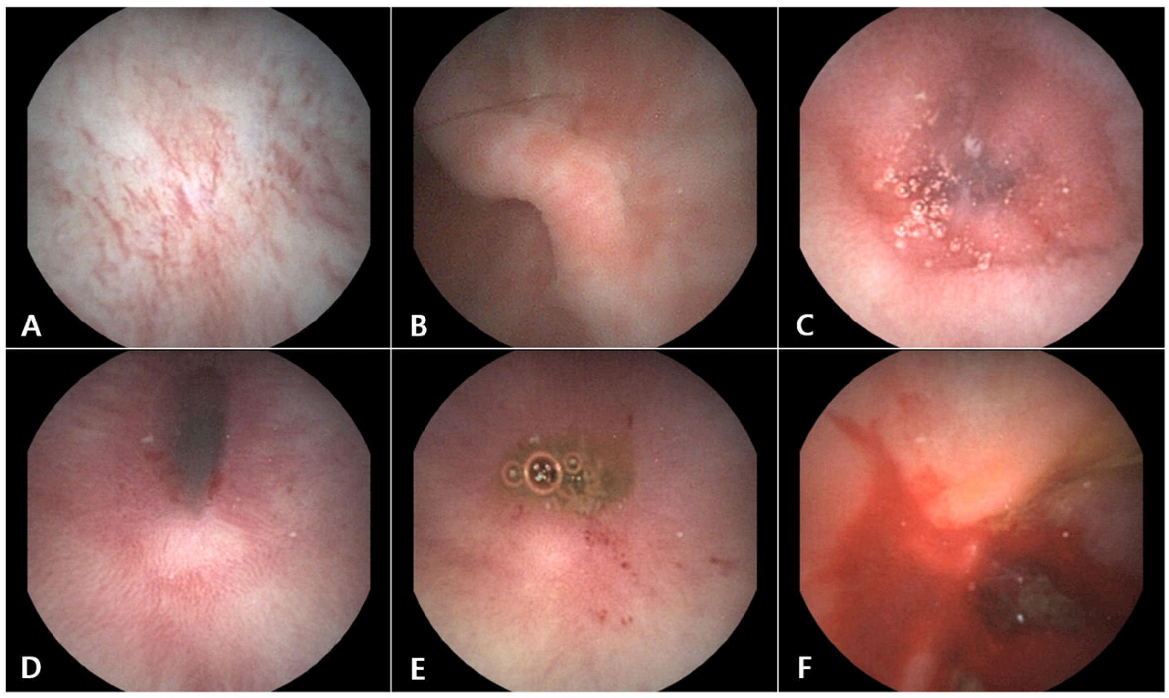
Capsule endoscopy revealed mild erythematous changes in the distal esophagus (Figure 3A) along with diffuse erythema with erosive lesions of the gastric mucosa (Figure 3B). Moderate-to-severe erythema and mucosal irregularities with edema were noted in the duodenum, jejunum, and ileum (Figure 3C). Persistent minor bleeding was observed in the erythematous areas of the duodenal and jejunal mucosa (Figure 3D and 3E). Active and substantial bleeding was observed in the colon (Figure 3F).
Based on capsule endoscopy findings, the patient was tentatively diagnosed with mild esophagitis, moderate gastritis, and severe inflammatory bowel disease (IBD) with evidence of intestinal hemorrhage.
After the diagnosis, a hypoallergenic diet (Hypoallergenic®, Royal Canin, France) was recommended, but it was substituted with Hill’s Z/D due to palatability issues. However, the soft stools persisted even after the introduction of Hill’s Z/D. After discussing with the owner, the diet was changed to low-fat (Low-fat gastrointestinal®, Royal Canin, France). We prescribed prednisolone (Solondo®, YunhanmedicaRepublic of Korea; 1 mg/kg PO q12h), omeprazole (Omed®, SK Chemical, Republic of Korea; 1 mg/kg PO q24h), and maropitant (Cerenia®, Zoetis, USA; 1 mg/kg SC q24h). The patient’s clinical symptoms gradually improved, and the steroid dosage was tapered accordingly.
3. Discussion
In this case report, we demonstrate the use of capsule endoscopy as a valuable diagnostic tool for evaluating chronic enteropathy in dogs. Both cases highlight the advantages of using capsule endoscopy for direct visualization of the small intestine. This allows for a more comprehensive assessment of mucosal abnormalities than traditional methods, such as ultrasound or standard endoscopy. Severe lacteal dilation, extensive mucosal erythema, and irregular mucosal patterns provide crucial diagnostic insights that can influence subsequent treatment decisions.
Our results are consistent with previous studies that reported the effectiveness of capsule endoscopy in detecting mucosal lesions associated with gastrointestinal diseases in dogs [7,8]. As observed in both cases, visualizing severe lacteal dilation is a hallmark of advanced intestinal lymphangiectasia, a condition that often leads to protein-losing enteropathy (PLE). This finding is consistent with the literature, suggesting that lymphangiectasia is a common underlying pathology of severe chronic enteropathies [1,2]. In a comparative review of canine intestinal lymphangiectasia, Jablonski (2022) emphasized the diagnostic challenge of lymphangiectasia due to its diffuse nature, which often prevents detection through conventional imaging techniques [1].
Integrating capsule endoscopy findings into managing chronic enteropathy aligns with Dandrieux [11], who discussed the variability in treatment outcomes based on clinical response types. Food-responsive enteropathy (FRE) is the most common condition, and dietary trials often result in rapid and sustained improvement. In contrast, immunosuppressant-responsive enteropathy (IRE) and non-responsive enteropathy (NRE) require more complex interventions [11]. This distinction was evident in our cases, particularly in Case 1, where corticosteroid monotherapy was inadequate, necessitating cyclosporine as an adjunctive treatment, consistent with findings highlighting the importance of tailored immunosuppressive therapies [11].
Capsule endoscopy facilitated a more targeted therapeutic approach in both cases. In Case 1, despite initial improvement with corticosteroid therapy, the patient’s condition deteriorated, necessitating the addition of cyclosporine. This reflects the complexity of managing severe cases of CE, where immunosuppressive therapies are often required; however, their effectiveness varies [4,11]. Similarly, in Case 2, the identification of extensive mucosal inflammation and bleeding led to the escalation of treatment, including dietary modifications and adjunctive pharmacotherapy, consistent with current recommendations for managing chronic inflammatory bowel conditions in veterinary medicine [3,4].
As highlighted in the literature, diet modification remains the cornerstone of CE management. Rodolphe and Mansfield (2019) emphasized that dietary interventions often yield the most favorable outcomes in FRE cases, mainly when hydrolyzed or novel antigen diets are employed [12]. This approach was supported by our findings, in which dietary changes contributed significantly to stabilizing symptoms, especially in Case 2. However, the poor response to diet alone in Case 1 underscores the need for multimodal therapeutic strategies for advanced cases of PLE [1,12].
One of the notable advantages of capsule endoscopy in this context is its ability to visualize the entire length of the small intestine, including deeper segments such as the jejunum and ileum, which are often inaccessible using conventional endoscopy [7,8]. This extended visualization capability allows the detection subtle lesions, such as diffuse erythema or irregular mucosal patterns, which other diagnostic methods may overlook. Our findings support the growing evidence that capsule endoscopy can enhance diagnostic accuracy in chronic gastrointestinal conditions by providing high-resolution, real-time images of the intestinal mucosa [9,10].
However, capsule endoscopy is not without limitations. One major drawback is the inability to obtain histopathological samples, which remain the gold standard for confirming specific diagnoses, such as inflammatory bowel disease (IBD) or neoplastic conditions. In our case, the absence of histopathological data limited the ability to differentiate between severe inflammatory enteropathy and potential neoplastic processes. Despite this limitation, capsule endoscopy provides critical visual information that guides clinical decision-making and helps formulate targeted treatment plans [7].
The clinical utility of capsule endoscopy in monitoring therapeutic response has been well-documented in human gastroenterology, particularly in the context of a “treat-to-target” strategy, which aims for mucosal healing as an endpoint [9]. Similar approaches can be adapted in veterinary practice, especially for chronic cases, where the ongoing assessment of mucosal changes may help tailor treatment regimens and improve long-term outcomes. Our cases underscore the potential role of capsule endoscopy as a noninvasive tool for initial diagnosis and follow-up monitoring in dogs with chronic enteropathy.
The Canine Chronic Enteropathy Clinical Activity Index (CCECAI) was used to assess disease severity and monitor the treatment response in both cases. In Case 1, the CCECAI score was 14, indicating severe disease, whereas in Case 2, it was 12. High CCECAI scores are associated with poor prognosis and a higher risk of mortality, particularly in dogs with protein-losing enteropathy (PLE) [3,4]. The elevated scores in our cases reflected an advanced stage of the disease and justified the use of capsule endoscopy as a more sensitive diagnostic tool. The ability of capsule endoscopy to detect extensive mucosal abnormalities correlated well with high CCECAI scores, supporting its utility for evaluating disease severity.
4. Conclusions
In conclusion, this case report illustrates capsule endoscopy’s diagnostic value in treating chronic enteropathy in dogs. Capsule endoscopy can complement traditional diagnostic tools and enhance diagnostic accuracy by enabling the detailed visualization of mucosal abnormalities throughout the gastrointestinal tract. Future studies with larger cohorts and more extended follow-up periods are warranted further to evaluate the prognostic implications of capsule endoscopic findings and refine their role in managing chronic enteropathies in veterinary patients.
Author Contributions
Conceptualization, H.J., and D.-I.J.; methodology, H.J., and D.-I.J.; software, H.J.; validation, H.J., and D.-I.J.; formal analysis, H.J., and D.-I.J.; investigation, H.J. and D.-I.J.; resources, H.J. and D.-I.J.; data curation, H.J. and D.-I.J.; writing—original draft preparation, H.J.; writing—review and editing, D.-I. J.; visualization, H.J. and D.-I.J.; supervision, D.-I.J.; project administration, D.-I.J.; funding acquisition, D.-I.J. All authors have read and agreed to the published version of this manuscript.
Funding
This work was supported by the Gyeongsang National University (Jinju, Republic of Korea) and Intromedic Co., Ltd. (Seoul, Republic of Korea).
Institutional Review Board Statement
Ethical review and approval were not required for the animal study, as we reported a case study from a veterinary medical teaching hospital. We obtained the consent of both the owners and veterinarians that dogs (which underwent the listed examinations, including capsule endoscopy) were treated for medical reasons rather than experimental purposes.
Informed Consent Statement
Informed consent was obtained from the owners of the animals used in this study.
Data Availability Statement
The original contributions of this study are presented in this article. Further inquiries can be directed to the corresponding authors.
Acknowledgments
We thank all Gyeongsang National University Animal Medical Center (GAMC) veterinary medical staff for their cooperation throughout the study.
Conflicts of Interest
The authors declare no conflict of interest.
References
- Jablonski, S.A. Pathophysiology, diagnosis, and management of canine intestinal lymphangiectasia: A comparative review. Animals 2022, 12, 2791. [Google Scholar] [CrossRef] [PubMed]
- Hodel, S.; Brugger, D.; Kook, P.H. Long-term evaluation of the initial response to therapy in 60 dogs with chronic inflammatory enteropathy. J. Vet. Intern. Med. 2024, 38, 2444–2453. [Google Scholar] [CrossRef] [PubMed]
- Kathrani, A.; Sánchez-Vizcaíno, F.; Hall, E.J. Association of chronic enteropathy activity index, blood urea concentration, and risk of death in dogs with protein-losing enteropathy. J. Vet. Intern. Med. 2019, 33, 536–543. [Google Scholar] [CrossRef] [PubMed]
- Wennogle, S.A.; Priestnall, S.L.; Webb, C.B. Histopathologic characteristics of intestinal biopsy samples from dogs with chronic inflammatory enteropathy with and without hypoalbuminemia. J. Vet. Intern. Med. 2017, 31, 371–376. [Google Scholar] [CrossRef]
- Mabry, K.; Davis, M.S.; Gould, E.; Gogal, R.M.; Steiner, J.M.; Tolbert, M.K.; Hill, T.L. Assessment of gastrointestinal health in racing Alaskan sled dogs using capsule endoscopy and inflammatory cytokines. J. Small Anim. Pract. 2023, 64, 574–580. [Google Scholar] [CrossRef] [PubMed]
- Davignon, D.L.; Lee, A.C.Y.; Johnston, A.N.; Bowman, D.D.; Simpson, K.W. Evaluation of capsule endoscopy to detect mucosal lesions associated with gastrointestinal bleeding in dogs. J. Small Anim. Pract. 2016, 57, 148–158. [Google Scholar] [CrossRef] [PubMed]
- Holmberg, J.; Ljungvall, I.; Pelander, L.; Defarges, A.; Stiller, J.; Ingman, J.; Harlos, C.; Spillmann, T.; Häggström, J. Video capsule endoscopy findings in dogs with chronic enteropathy and in healthy dogs. J. Vet. Intern. Med. 2024, 38, 2454–2463. [Google Scholar] [CrossRef] [PubMed]
- Le Berre, C.; Trang-Poisson, C.; Bourreille, A. Small bowel capsule endoscopy and treat-to-target in Crohn’s disease: A systematic review. World J. Gastroenterol. 2019, 25, 4534–4554. [Google Scholar] [CrossRef]
- Nakamura, M.; Yamamura, T.; Maeda, K.; Sawada, T.; Ishikawa, E.; Murate, K.; Furukawa, K.; Hirose, T.; Uetsuki, K.; Iida, T.; et al. Long-term monitoring and clinical implications of small bowel capsule endoscopy in patients with Crohn’s disease with small bowel lesions: A retrospective analysis. Digestion 2024, 105, 380–388. [Google Scholar] [CrossRef] [PubMed]
- Salavati Schmitz, S.S.; Gow, A.; Bommer, N.; Morrison, L.; Mellanby, R. Diagnostic features, treatment, and outcome of dogs with inflammatory protein-losing enteropathy. J. Vet. Intern. Med. 2019, 33, 2005–2013. [Google Scholar] [CrossRef]
- Dandrieux, J.R.S. Inflammatory Bowel Disease versus chronic enteropathy in dogs: Are they one and the same? J. Small Anim. Pract. 2016, 57, 589–599. [Google Scholar] [CrossRef] [PubMed]
- Dandrieux, J.R.S.; Mansfield, C.S. Chronic enteropathy in canines: Prevalence, impact and management strategies. Vet. Med. 2019, 10, 203–214. [Google Scholar] [CrossRef] [PubMed]
Figure 1.
The patients are dressed in examination suits for capsule endoscopy (A: Case 1, B: Case 2). The procedure was conducted without the attachment of electrodes, with the patients wearing examination suits. A photograph of the capsule endoscope (MiroVET®) was utilized in this case study (C).
Figure 1.
The patients are dressed in examination suits for capsule endoscopy (A: Case 1, B: Case 2). The procedure was conducted without the attachment of electrodes, with the patients wearing examination suits. A photograph of the capsule endoscope (MiroVET®) was utilized in this case study (C).
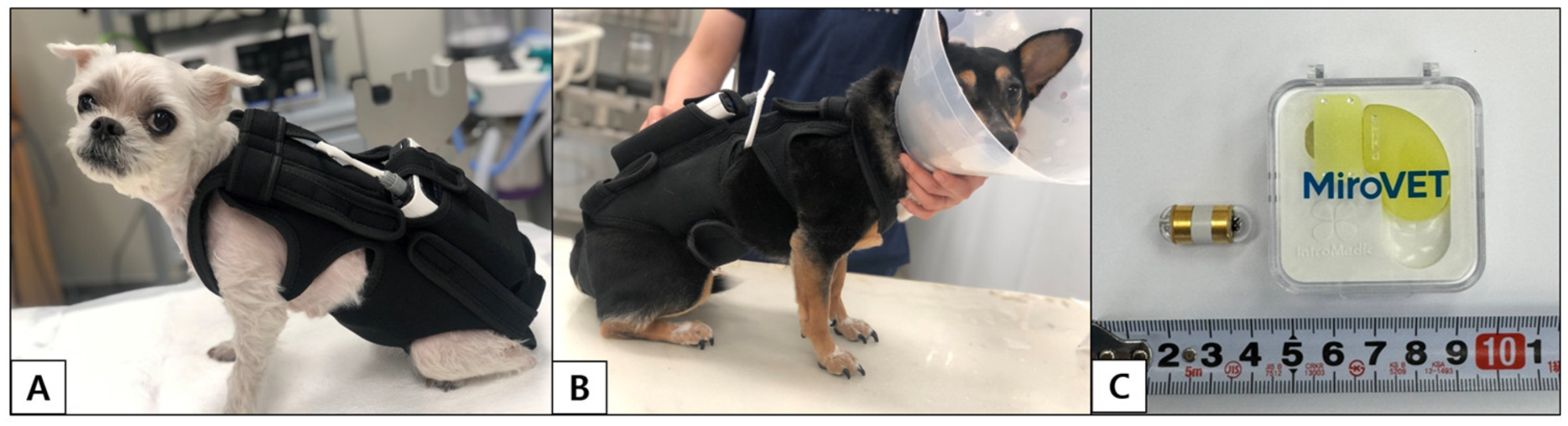
Figure 2.
Capsule endoscopic images of the stomach (A) and small intestine (B to F) in Case 1. Mild erythema is observed in the gastric mucosa (A). Dilated lacteals are evident in the duodenum (B), jejunum (C), and ileum (D). Lymphatic leakage is visible on the mucosal surface of the jejunum (C). The small intestinal mucosa with less severe lacteal dilation appears erythematous and demonstrates increased irregularity (E and F).
Figure 2.
Capsule endoscopic images of the stomach (A) and small intestine (B to F) in Case 1. Mild erythema is observed in the gastric mucosa (A). Dilated lacteals are evident in the duodenum (B), jejunum (C), and ileum (D). Lymphatic leakage is visible on the mucosal surface of the jejunum (C). The small intestinal mucosa with less severe lacteal dilation appears erythematous and demonstrates increased irregularity (E and F).
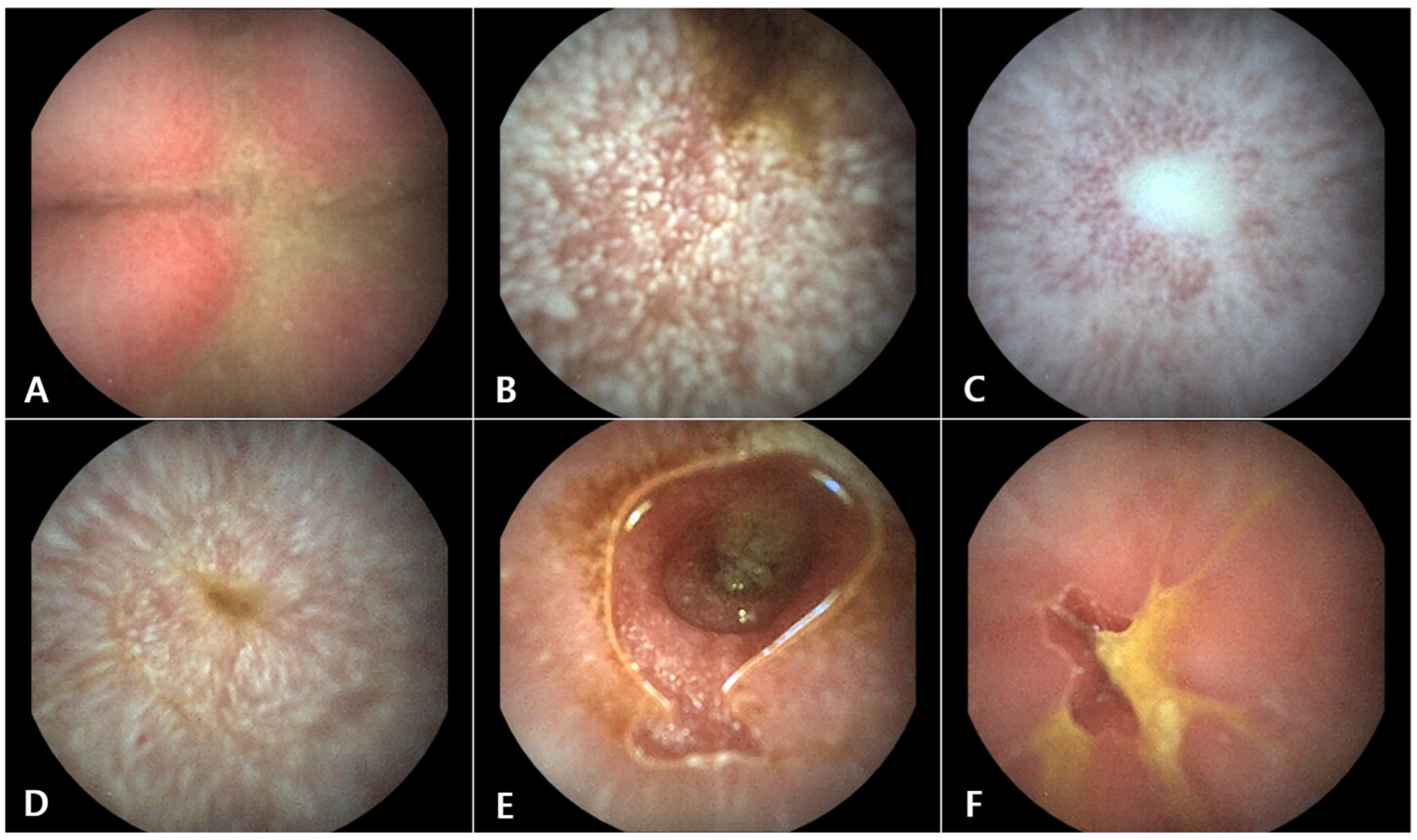
Figure 3.
Capsule endoscopic images of the esophagus (A), stomach (B), small intestine (C to E), and colon (F) in Case 2. Mild erythematous changes are observed in the distal esophagus (A). The gastric mucosa is diffusely erythematous with erosive lesions (B). The small intestinal mucosa is diffusely erythematous, with edema evident in specific segments (C). Persistent minor bleeding is noted in the erythematous areas of the duodenal and jejunal mucosa (D and E). Active and substantial bleeding is identified in the colon (F).
Figure 3.
Capsule endoscopic images of the esophagus (A), stomach (B), small intestine (C to E), and colon (F) in Case 2. Mild erythematous changes are observed in the distal esophagus (A). The gastric mucosa is diffusely erythematous with erosive lesions (B). The small intestinal mucosa is diffusely erythematous, with edema evident in specific segments (C). Persistent minor bleeding is noted in the erythematous areas of the duodenal and jejunal mucosa (D and E). Active and substantial bleeding is identified in the colon (F).
Disclaimer/Publisher’s Note: The statements, opinions and data contained in all publications are solely those of the individual author(s) and contributor(s) and not of MDPI and/or the editor(s). MDPI and/or the editor(s) disclaim responsibility for any injury to people or property resulting from any ideas, methods, instructions or products referred to in the content. |
© 2024 by the authors. Licensee MDPI, Basel, Switzerland. This article is an open access article distributed under the terms and conditions of the Creative Commons Attribution (CC BY) license (http://creativecommons.org/licenses/by/4.0/).
Copyright: This open access article is published under a Creative Commons CC BY 4.0 license, which permit the free download, distribution, and reuse, provided that the author and preprint are cited in any reuse.


